Embed Size (px)
Citation preview
Lebanese Medical Journal 2002 • Volume 50 (3) 75
EDITORIAL
In the spirit of fulfilling one of its majorgoals, namely continuing medical education,the Lebanese Medical Journal in this specialsupplement, the 5th in this series of specialissues, contains proceedings of the RegionalDensitometry Workshop that took place inBeirut on April 20th 2002 and the resultingNational Practice Guidelines regarding theuse of bone mineral density testing.
These practice guidelines were reviewed andunanimously approved by the five nationalmajor scientific societies of specialties thatcommonly deal with densitometry use.
The organizers of this landmark meeting,Drs Ghada El Hajj Fuleihan, Rafic Baddoura,Hassane Awada, Jad Okais and Paul Rizk,the guest expert Dr Michael McClung, thePresidents of the Lebanese Societies of Endo-crinology, Rheumatology, Orthopedics, Radi-ology and Obstetrics and Gynecology, andtheir scientific committees are all to be com -manded on a splendid job, the fruits of whichare presented herein. Also I would like toexpress my gratitude to Dr. Ghada El HajjFuleihan for having kindly accepted to be theguest editor of this special issue and for theexcellent work she did for it.
We trust that this first set of NationalPractice Guidelines published in the LMJ willundoubtedly reinforce quality care and opti -mize management of patients seeking attentionfor skeletal health matters.
Adel E. BIRBARIEditor in Chief
76 Lebanese Medical Journal 2002 • Volume 50 (3)
FOREWORD
Osteoporosis is a major public health problem projected to have an increasingly heavier social and economic toll inview of the demographic explosion of the aging population worldwide in general, and in developing countries includingin the Middle East in particular. The World Health Organization has listed osteoporosis as a major item on the list of non-communicable diseases, that account for 60% of mortality worldwide.
Because bone mass or bone mineral density is the strongest single predictor of osteoporotic fracture, both theInternational Consensus Panel on Osteoporosis (1993) as well as the National Institute of Health Consensus Panel onOsteoporosis (2001) have used the term bone mass or bone density in their definition of osteoporosis. The World HealthOrganization working group has proposed several years ago an operational definition of osteoporosis based on bonemineral density derived cut-offs.
Because of the increasing recognition of osteoporosis as a disease and demand for identification of a larger numberof subjects at risk, densitometry technology has undergone substantial growth as well as changes over the years. Aplethora of instruments are therefore available on the market to-date, with a myriad of validated and not validated tech-nologies, as well as a diversity of databases from which fracture risk assessment could be derived, justifiably and not so-justifiably depending on the specific instance. The Middle East in general and Lebanon in particular has not escaped thisglobal phenomenon, that added to uncertainties regarding the status of densitometry use amongst practicing physicians.Thus guidelines regarding densitometry testing have been put forth and further refined over the years in the light of thesubstantial body of evidence that has accumulated from prospective studies evaluating risk factors and fracture risk, andfrom large randomized controlled trials evaluating the safety and efficacy of various osteoporosis treatment strategies.
On April 20, 2002, in an effort to further optimize the quality of care in osteoporosis nationally, a group of expertsconvened and presented regional data on osteoporosis, and provided guidelines based on the review of the evidencebehind currently published guidelines on densitometry use as put forth by several organizations including the WorldHealth Organization, The National Osteoporosis Foundation, the International Osteoporosis Foundation, the AmericanAssociation of Clinical Endocrinologists, the Canadian Medical Association and the North American MenopauseSociety. This local meeting was a collaborative effort between several Lebanese Scientific Societies and the EasternMediterranean Regional Office of the World Health Organization. The resultant guidelines, the document justifying suchguidelines and the proceedings of this meeting are detailed in this special supplement of the Lebanese Medical Journal.These guidelines were unanimously endorsed after careful evaluation by the Lebanese Society of Endocrinology, theLebanese Society of Obstetrics and Gynecology, the Lebanese Society of Orthopedics, the Lebanese Society ofRadiology and the Lebanese Society of Rheumatology, and the Eastern Mediterranean Regional Office of the WorldHealth Organization.
We would like to thank the Presidents and constituents of the societies for the time and input in reviewing and endors-ing the current guidelines : Ibrahim Salti, MD, PhD, Paula Atallah, MD, Georges Halaby, MD, Pierre Najm, MD andCharles Saab, MD (Lebanese Society of Endocrinology) ; Georges Kaadeh, MD, Muhieddine Seoud, MD and JihadEzzedine, MD (Lebanese Society of Obstetrics and Gynecology) ; Raja Shaftari, MD, and Assaad Taha, MD (LebaneseSociety of Orthopedics) ; Georges Rouhana, MD and Naji Atallah, MD (Lebanese Society of Radiology) ; Abdel FattahMasri, MD and Said Atweh, MD (Lebanese Society of Rheumatology) ; Ussama El-Khatib, MD, PhD (Eastern Medi-terranean Regional Office of the World Health Organization). Special thanks to Professor Eric Orwoll, Oregon HealthSciences University, for his thoughts on guidelines for men.
Such guidelines have been put forth to provide a framework around which sound clinical decision-making can bebuilt. They are not meant to supercede the prerogative of the practicing physician making decisions and treating an indi-vidual patient, nor are they meant as rigid yardsticks to measure standard of care. Rather, they are meant to provide aplatform for an evidence-based approach to osteoporosis management based on data available to-date. Such guidelineswill undoubtedly continue to be refined as our knowledge base on this challenging silent disease keeps evolving glob-ally, regionally and last but not least nationally.
Ghada EL-HAJJ FULEIHAN, MD, MPH Guest Editor, on the behalf of
Guest International Expert Organizing & Scientific CommitteeMichael R. McClung, MD Ghada El-Hajj Fuleihan, MD, MPH
Rafic Baddoura, MD, MPHHassane Awada, MD
Jad Okais, MD Paul Rizk, MD
GUIDELINES GUIDELINES GUIDELINES GUIDELINES GUIDELINES GUIDELINES
Lebanese Guidelines for Osteoporosis Assessment and Treatment Lebanese Medical Journal 2002 • Volume 50 (3) 77
Bone Mineral Density (BMD) Measurement Guidelines 2002
Endorsed by
Lebanese Society of EndocrinologyLebanese Society of OBGYN
Lebanese Society of OrthopedicsLebanese Society of Radiology
Lebanese Society of Rheumatology
WHO Eastern Mediterranean Region
Recommandations sur la mesure dela densité minérale osseuse (DMO) 2002
Agréées par les Sociétés libanaises
d’Endocrinologied’Obstétrique et de Gynécologie
d’Orthopédiede Radiologie
de Rhumatologieet
le Bureau de l’OMS de la Méditerranée orientale
Utilité de la mesure de la DMO
• La DMO prédit les fractures (risque relatif)
• La DMO est mesurable avec précision et sans risque
• Les données cliniques ne permettent pas de prédire la DMO
• La DMO influence la décision thérapeutique.
• Des traitements peuvent réduire l’incidence des fractures
• La DMO indirectement diminue l’incidence des fractures
NOF JBMR 1989; 4:Supplement 2
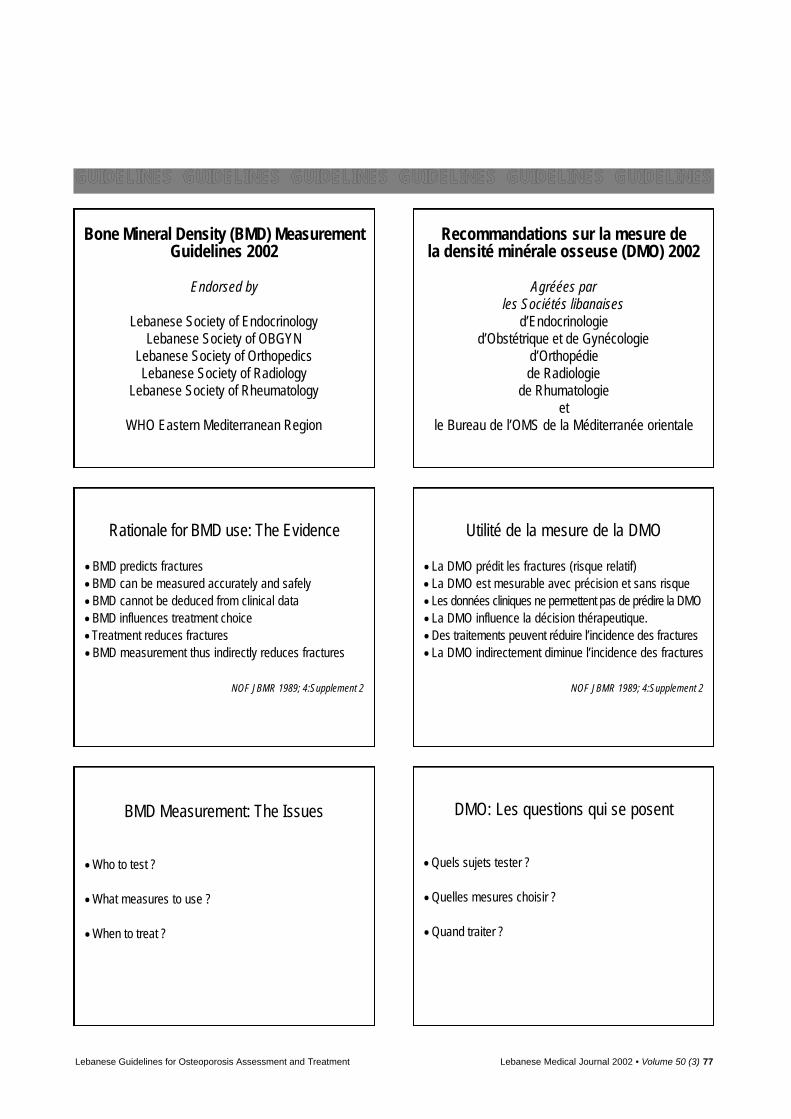
Rationale for BMD use: The Evidence
• BMD predicts fractures
• BMD can be measured accurately and safely
• BMD cannot be deduced from clinical data
• BMD influences treatment choice
• Treatment reduces fractures
• BMD measurement thus indirectly reduces fractures
NOF JBMR 1989; 4:Supplement 2
DMO: Les questions qui se posent
• Quels sujets tester ?
• Quelles mesures choisir ?
• Quand traiter ?
BMD Measurement: The Issues
• Who to test ?
• What measures to use ?
• When to treat ?
78 Lebanese Medical Journal 2002 • Volume 50 (3) Lebanese Guidelines for Osteoporosis Assessment and Treatment
GUIDELINES GUIDELINES GUIDELINES GUIDELINES GUIDELINES GUIDELINES
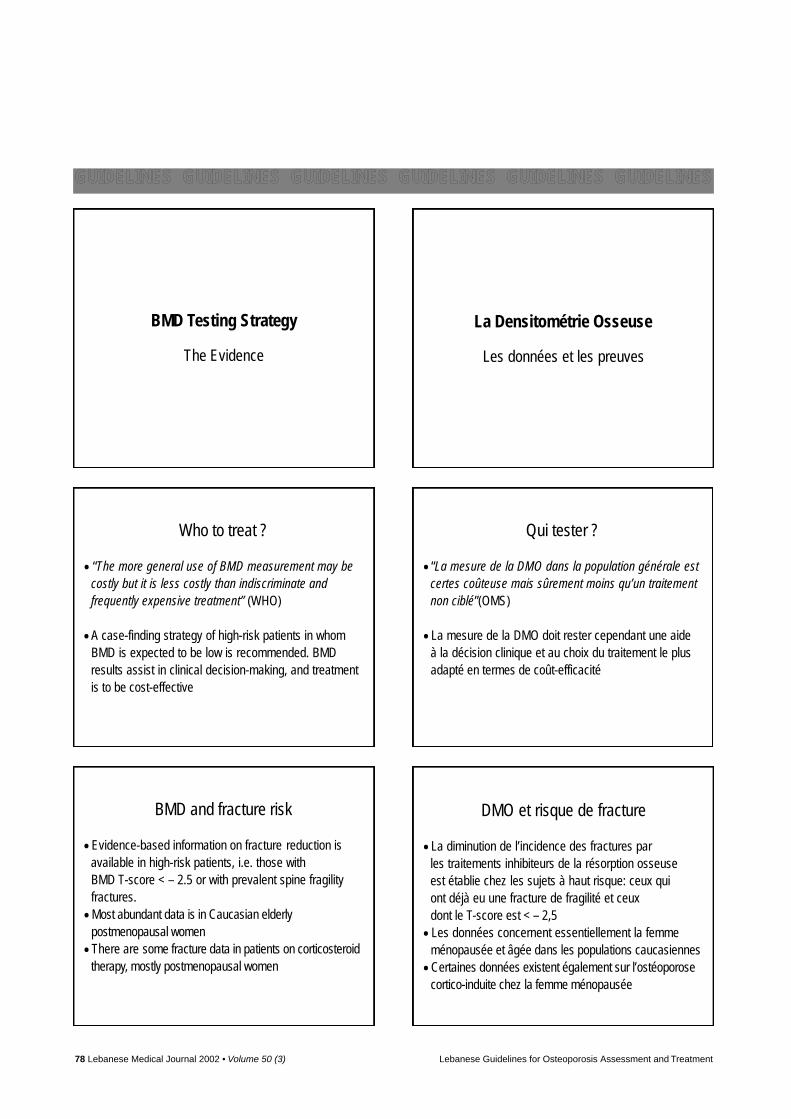
BMD Testing Strategy
The Evidence
La Densitométrie Osseuse
Les données et les preuves
Qui tester ?
• “La mesure de la DMO dans la population générale estcertes coûteuse mais sûrement moins qu’un traitementnon ciblé”(OMS)
• La mesure de la DMO doit rester cependant une aideà la décision clinique et au choix du traitement le plusadapté en termes de coût-efficacité
DMO et risque de fracture
• La diminution de l’incidence des fractures par les traitements inhibiteurs de la résorption osseuse est établie chez les sujets à haut risque: ceux qui ont déjà eu une fracture de fragilité et ceux dont le T-score est < – 2,5
• Les données concernent essentiellement la femmeménopausée et âgée dans les populations caucasiennes
• Certaines données existent également sur l’ostéoporosecortico-induite chez la femme ménopausée
Who to treat ?
• “The more general use of BMD measurement may becostly but it is less costly than indiscriminate and frequently expensive treatment” (WHO)
• A case-finding strategy of high-risk patients in whomBMD is expected to be low is recommended. BMDresults assist in clinical decision-making, and treatmentis to be cost-effective
BMD and fracture risk
• Evidence-based information on fracture reduction isavailable in high-risk patients, i.e. those with BMD T-score < – 2.5 or with prevalent spine fragilityfractures.
• Most abundant data is in Caucasian elderly postmenopausal women
• There are some fracture data in patients on corticosteroidt h e r a p y, mostly postmenopausal women
Lebanese Guidelines for Osteoporosis Assessment and Treatment Lebanese Medical Journal 2002 • Volume 50 (3) 79
GUIDELINES GUIDELINES GUIDELINES GUIDELINES GUIDELINES GUIDELINES
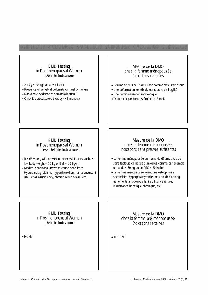
BMD Testingin Postmenopausal Women
Definite Indications
• > 65 years: age as a risk factor
• Presence of vertebral deformity or fragility fracture
• Radiologic evidence of demineralization
• Chronic corticosteroid therapy (> 3 months)
Mesure de la DMOchez la femme ménopausée
Indications certaines
• Femme de plus de 65 ans: l’âge comme facteur de risque
• Une déformation vertébrale ou fracture de fragilité
• Une déminéralisation radiologique
• Traitement par corticostéroïdes > 3 mois
Mesure de la DMO chez la femme ménopausée
Indications sans preuves suffisantes
• La femme ménopausée de moins de 65 ans avec ousans facteurs de risque surajoutés comme par exempleun poids < 50 kg ou un IMC < 20 kg/m2
• La femme ménopausée ayant une ostéoporose secondaire: hyperparathyroïdie, maladie de Cushing,traitements anti-convulsifs, insuffisance rénale,insuffisance hépatique chronique, etc
Mesure de la DMO chez la femme pré-ménopausée
Indications certaines
• AUCUNE
BMD Testingin Postmenopausal Women
Less Definite Indications
• If < 65 years, with or without other risk factors such aslow body weight < 50 kg or BMI < 20 kg/m2
• Medical conditions known to cause bone loss: Hyperparathyroidism, hyperthyroidism, anticonvulsantuse, renal insufficiency, chronic liver disease, etc.
BMD Testingin Pre-menopausal Women
Definite Indications
• NONE
80 Lebanese Medical Journal 2002 • Volume 50 (3) Lebanese Guidelines for Osteoporosis Assessment and Treatment
GUIDELINES GUIDELINES GUIDELINES GUIDELINES GUIDELINES GUIDELINES
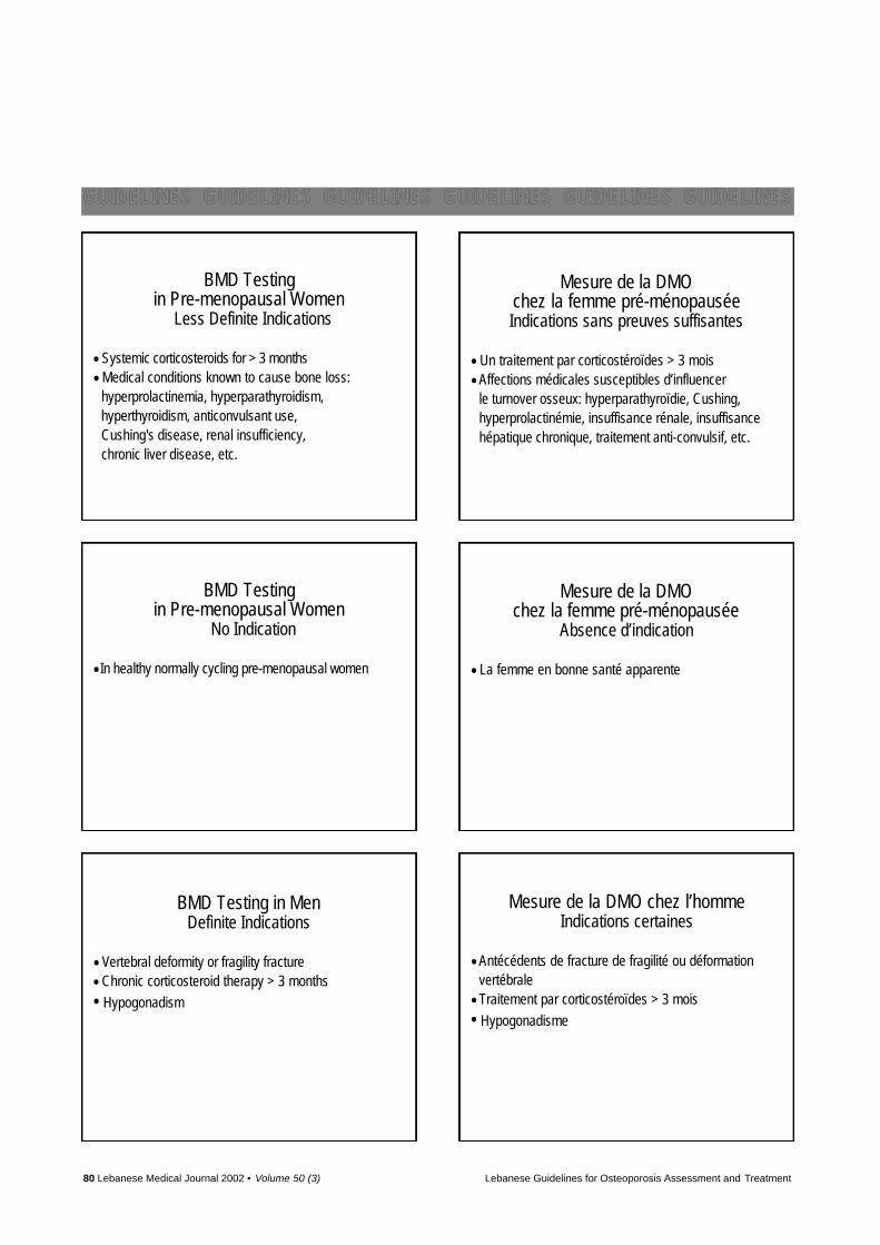
BMD Testingin Pre-menopausal Women
Less Definite Indications
• Systemic corticosteroids for > 3 months
• Medical conditions known to cause bone loss: hyperprolactinemia, hyperparathyroidism,hyperthyroidism, anticonvulsant use,Cushing's disease, renal insufficiency,chronic liver disease, etc.
Mesure de la DMOchez la femme pré-ménopausée Indications sans preuves suffisantes
• Un traitement par corticostéroïdes > 3 mois
• Affections médicales susceptibles d’influencer le turnover osseux: hyperparathyroïdie, Cushing,hyperprolactinémie, insuffisance rénale, insuffisancehépatique chronique, traitement anti-convulsif, etc.
Mesure de la DMOchez la femme pré-ménopausée
Absence d’indication
• La femme en bonne santé apparente
Mesure de la DMO chez l’hommeIndications certaines
• Antécédents de fracture de fragilité ou déformationvertébrale
• Traitement par corticostéroïdes > 3 mois
• Hypogonadisme
BMD Testingin Pre-menopausal Women
No Indication
• In healthy normally cycling pre-menopausal women
BMD Testing in MenDefinite Indications
• Vertebral deformity or fragility fracture
• Chronic corticosteroid therapy > 3 months
• Hypogonadism
Lebanese Guidelines for Osteoporosis Assessment and Treatment Lebanese Medical Journal 2002 • Volume 50 (3) 81
GUIDELINES GUIDELINES GUIDELINES GUIDELINES GUIDELINES GUIDELINES
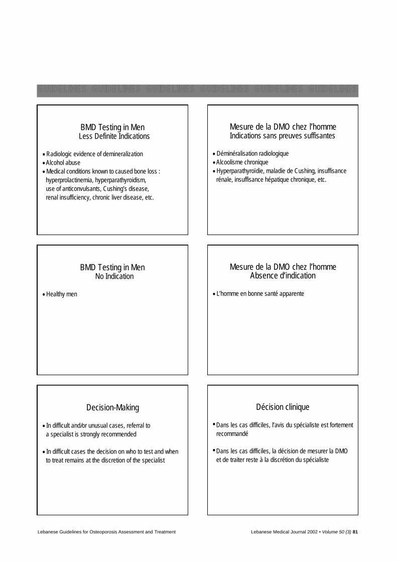
BMD Testing in MenLess Definite Indications
• Radiologic evidence of demineralization
• Alcohol abuse
• Medical conditions known to caused bone loss :hyperprolactinemia, hyperparathyroidism,use of anticonvulsants, Cushing’s disease,renal insufficiency, chronic liver disease, etc.
Mesure de la DMO chez l’hommeIndications sans preuves suffisantes
• Déminéralisation radiologique
• Alcoolisme chronique
• Hyperparathyroïdie, maladie de Cushing, insuffisancerénale, insuffisance hépatique chronique, etc.
Mesure de la DMO chez l’hommeAbsence d’indication
• L’homme en bonne santé apparente
Décision clinique
• Dans les cas difficiles, l’avis du spécialiste est fortementrecommandé
• Dans les cas difficiles, la décision de mesurer la DMOet de traiter reste à la discrétion du spécialiste
BMD Testing in MenNo Indication
• Healthy men
Decision-Making
• In difficult and/or unusual cases, referral toa specialist is strongly recommended
• In difficult cases the decision on who to test and whento treat remains at the discretion of the specialist
82 Lebanese Medical Journal 2002 • Volume 50 (3) Lebanese Guidelines for Osteoporosis Assessment and Treatment
GUIDELINES GUIDELINES GUIDELINES GUIDELINES GUIDELINES GUIDELINES
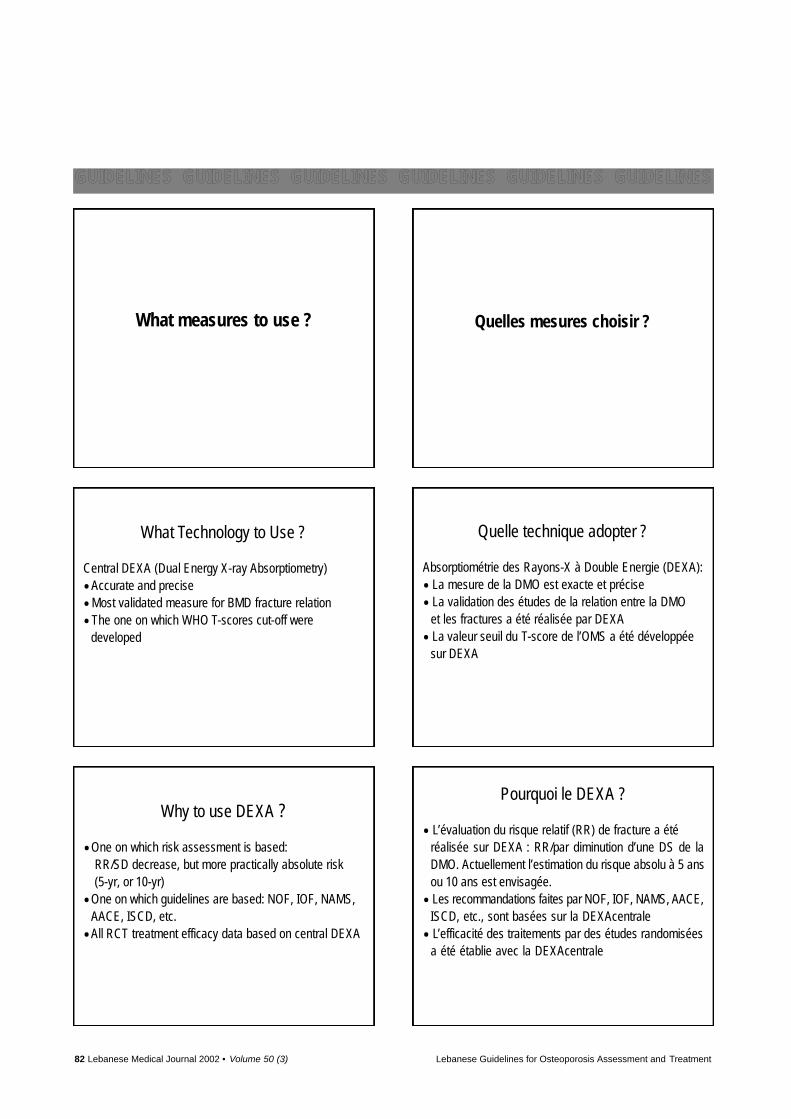
What measures to use ? Quelles mesures choisir ?
Quelle technique adopter ?
Absorptiométrie des Rayons-X à Double Energie (DEXA):
• La mesure de la DMO est exacte et précise
• La validation des études de la relation entre la DMOet les fractures a été réalisée par DEXA
• La valeur seuil du T-score de l’OMS a été développéesur DEXA
What Technology to Use ?
Central DEXA (Dual Energy X-ray Absorptiometry)
• Accurate and precise
• Most validated measure for BMD fracture relation
• The one on which WHO T-scores cut-off were developed
Why to use DEXA ?
• One on which risk assessment is based: RR/SD decrease, but more practically absolute risk(5-yr, or 10-yr)
• One on which guidelines are based: NOF, IOF, NAMS,AACE, ISCD, etc.
• All RCT treatment efficacy data based on central DEXA
Pourquoi le DEXA ?
• L’évaluation du risque relatif (RR) de fracture a été réalisée sur DEXA : RR/par diminution d’une DS de laDMO. Actuellement l’estimation du risque absolu à 5 ansou 10 ans est envisagée.
• Les recommandations faites par NOF, IOF, NAMS, A A C E ,ISCD, etc., sont basées sur la DEXAcentrale
• L’efficacité des traitements par des études randomiséesa été établie avec la DEXAcentrale
Lebanese Guidelines for Osteoporosis Assessment and Treatment Lebanese Medical Journal 2002 • Volume 50 (3) 83
GUIDELINES GUIDELINES GUIDELINES GUIDELINES GUIDELINES GUIDELINES
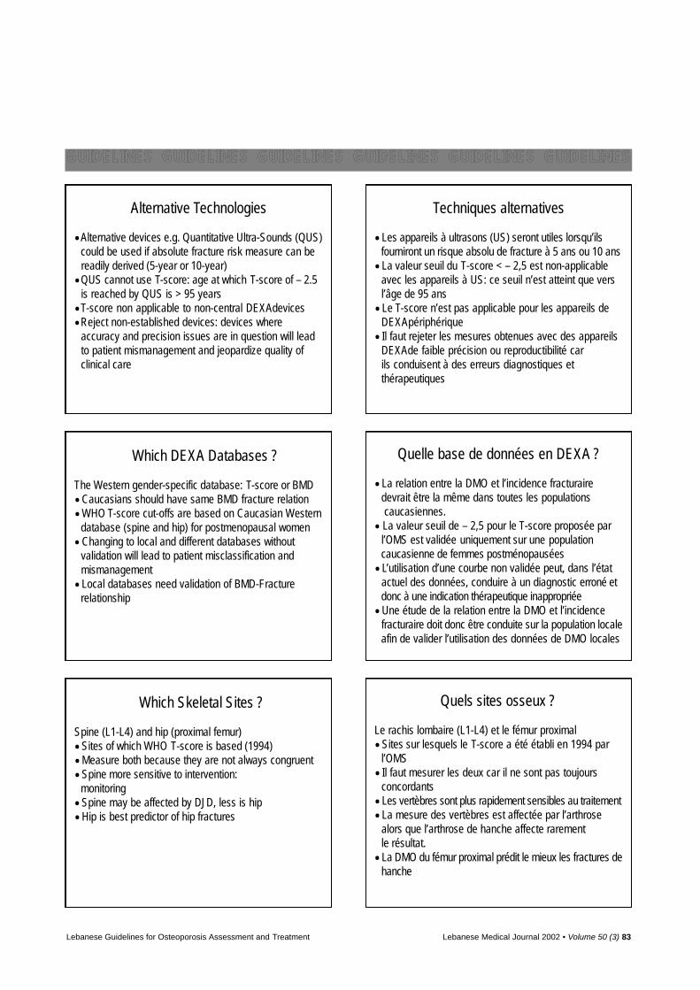
Alternative Technologies
• Alternative devices e.g. Quantitative Ultra-Sounds (QUS)could be used if absolute fracture risk measure can bereadily derived (5-year or 10-year)
• QUS cannot use T-score: age at which T-score of – 2.5is reached by QUS is > 95 years
• T-score non applicable to non-central DEXAdevices• Reject non-established devices: devices where
accuracy and precision issues are in question will leadto patient mismanagement and jeopardize quality ofclinical care
Which DEXA Databases ?
The Western gender-specific database: T-score or BMD• Caucasians should have same BMD fracture relation• WHO T-score cut-offs are based on Caucasian Western
database (spine and hip) for postmenopausal women• Changing to local and different databases without
validation will lead to patient misclassification andmismanagement
• Local databases need validation of BMD-Fracturerelationship
Quelle base de données en DEXA ?
• La relation entre la DMO et l’incidence fracturairedevrait être la même dans toutes les populationscaucasiennes.
• La valeur seuil de – 2,5 pour le T-score proposée parl’OMS est validée uniquement sur une population caucasienne de femmes postménopausées
• L’utilisation d’une courbe non validée peut, dans l’étatactuel des données, conduire à un diagnostic erroné etdonc à une indication thérapeutique inappropriée
• Une étude de la relation entre la DMO et l’incidencefracturaire doit donc être conduite sur la population localeafin de valider l’utilisation des données de DMO locales
Which Skeletal Sites ?
Spine (L1-L4) and hip (proximal femur)• Sites of which WHO T-score is based (1994)• Measure both because they are not always congruent• Spine more sensitive to intervention:
monitoring• Spine may be affected by DJD, less is hip• Hip is best predictor of hip fractures
Quels sites osseux ?
Le rachis lombaire (L1-L4) et le fémur proximal• Sites sur lesquels le T-score a été établi en 1994 par
l’OMS• Il faut mesurer les deux car il ne sont pas toujours
concordants• Les vertèbres sont plus rapidement sensibles au traitement • La mesure des vertèbres est affectée par l’arthrose
alors que l’arthrose de hanche affecte rarement le résultat.
• La DMO du fémur proximal prédit le mieux les fractures deh a n c h e
Techniques alternatives
• Les appareils à ultrasons (US) seront utiles lorsqu’ilsfourniront un risque absolu de fracture à 5 ans ou 10 ans
• La valeur seuil du T-score < – 2,5 est non-applicableavec les appareils à US: ce seuil n’est atteint que versl’âge de 95 ans
• Le T-score n’est pas applicable pour les appareils deDEXApériphérique
• Il faut rejeter les mesures obtenues avec des appareilsDEXAde faible précision ou reproductibilité car ils conduisent à des erreurs diagnostiques etthérapeutiques
84 Lebanese Medical Journal 2002 • Volume 50 (3) Lebanese Guidelines for Osteoporosis Assessment and Treatment
GUIDELINES GUIDELINES GUIDELINES GUIDELINES GUIDELINES GUIDELINES
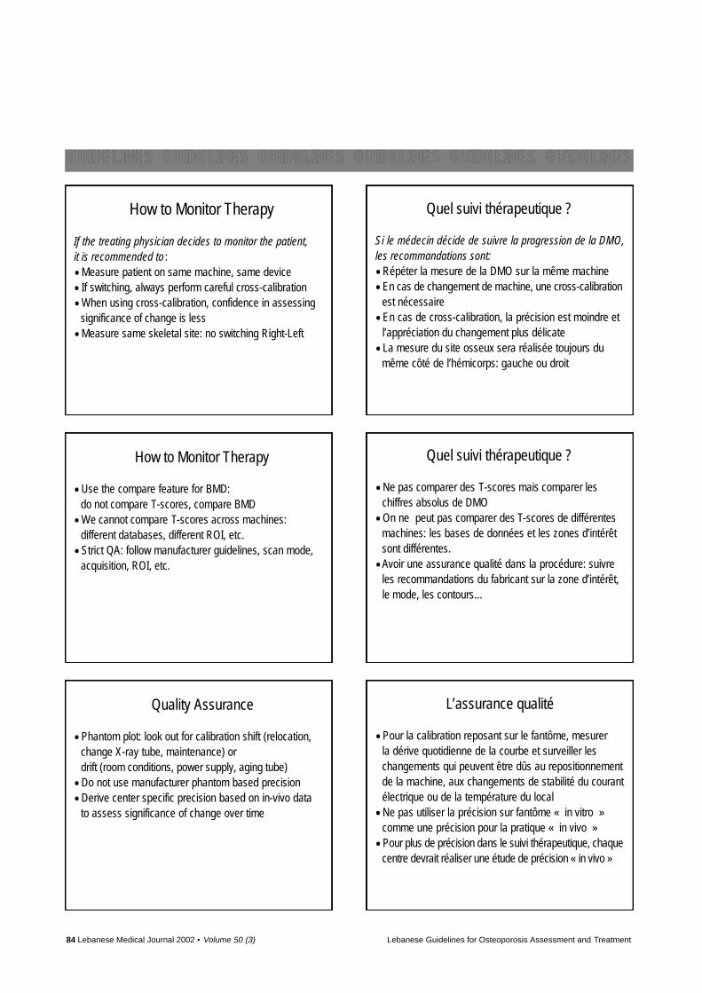
How to Monitor Therapy
If the treating physician decides to monitor the patient,it is recommended to :
• Measure patient on same machine, same device
• If switching, always perform careful cross-calibration
• When using cross-calibration, confidence in assessingsignificance of change is less
• Measure same skeletal site: no switching Right-Left
How to Monitor Therapy
• Use the compare feature for BMD: do not compare T-scores, compare BMD
• We cannot compare T-scores across machines: different databases, different ROI, etc.
• Strict QA: follow manufacturer guidelines, scan mode,acquisition, ROI, etc.
Quality Assurance
• Phantom plot: look out for calibration shift (relocation,change X-ray tube, maintenance) or drift (room conditions, power supply, aging tube)
• Do not use manufacturer phantom based precision
• Derive center specific precision based on in-vivo datato assess significance of change over time
Quel suivi thérapeutique ?
Si le médecin décide de suivre la progression de la DMO,les recommandations sont:
• Répéter la mesure de la DMO sur la même machine
• En cas de changement de machine, une cross-calibrationest nécessaire
• En cas de cross-calibration, la précision est moindre etl’appréciation du changement plus délicate
• La mesure du site osseux sera réalisée toujours dumême côté de l’hémicorps: gauche ou droit
Quel suivi thérapeutique ?
• Ne pas comparer des T-scores mais comparer leschiffres absolus de DMO
• On ne peut pas comparer des T-scores de différentesmachines: les bases de données et les zones d’intérêtsont différentes.
• Avoir une assurance qualité dans la procédure: suivreles recommandations du fabricant sur la zone d’intérêt,le mode, les contours…
L’assurance qualité
• Pour la calibration reposant sur le fantôme, mesurer la dérive quotidienne de la courbe et surveiller leschangements qui peuvent être dûs au repositionnementde la machine, aux changements de stabilité du courantélectrique ou de la température du local
• Ne pas utiliser la précision sur fantôme « in vitro »comme une précision pour la pratique « in vivo »
• Pour plus de précision dans le suivi thérapeutique, chaquecentre devrait réaliser une étude de précision « in vivo »
Lebanese Guidelines for Osteoporosis Assessment and Treatment Lebanese Medical Journal 2002 • Volume 50 (3) 85
GUIDELINES GUIDELINES GUIDELINES GUIDELINES GUIDELINES GUIDELINES
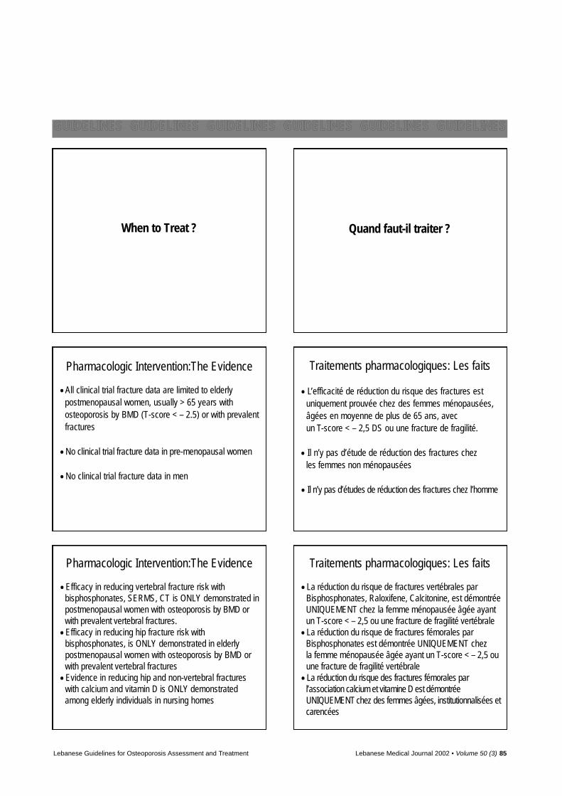
When to Treat ? Quand faut-il traiter ?
Traitements pharmacologiques: Les faits
• L’efficacité de réduction du risque des fractures estuniquement prouvée chez des femmes ménopausées,âgées en moyenne de plus de 65 ans, avec un T-score < – 2,5 DS ou une fracture de fragilité.
• Il n’y pas d’étude de réduction des fractures chez les femmes non ménopausées
• Il n’y pas d’études de réduction des fractures chez l’homme
Traitements pharmacologiques: Les faits
• La réduction du risque de fractures vertébrales par Bisphosphonates, Raloxifene, Calcitonine, est démontréeU N I Q U E M E N T chez la femme ménopausée âgée ayant un T-score < – 2,5 ou une fracture de fragilité vertébrale
• La réduction du risque de fractures fémorales par Bisphosphonates est démontrée U N I Q U E M E N T chez la femme ménopausée âgée ayant un T-score < – 2,5 ou une fracture de fragilité vertébrale
• La réduction du risque des fractures fémorales par l’association calcium et vitamine D est démontrée U N I Q U E M E N T chez des femmes âgées, institutionnalisées etc a r e n c é e s
Pharmacologic Intervention:The Evidence
• All clinical trial fracture data are limited to elderlypostmenopausal women, usually > 65 years with osteoporosis by BMD (T-score < – 2.5) or with prevalentfractures
• No clinical trial fracture data in pre-menopausal women
• No clinical trial fracture data in men
Pharmacologic Intervention:The Evidence
• Efficacy in reducing vertebral fracture risk with bisphosphonates, SERMS, CT is ONLY demonstrated inpostmenopausal women with osteoporosis by BMD orwith prevalent vertebral fractures.
• Efficacy in reducing hip fracture risk with bisphosphonates, is ONLY demonstrated in elderly postmenopausal women with osteoporosis by BMD orwith prevalent vertebral fractures
• Evidence in reducing hip and non-vertebral fractureswith calcium and vitamin D is ONLY demonstratedamong elderly individuals in nursing homes
86 Lebanese Medical Journal 2002 • Volume 50 (3) Lebanese Guidelines for Osteoporosis Assessment and Treatment
GUIDELINES GUIDELINES GUIDELINES GUIDELINES GUIDELINES GUIDELINES
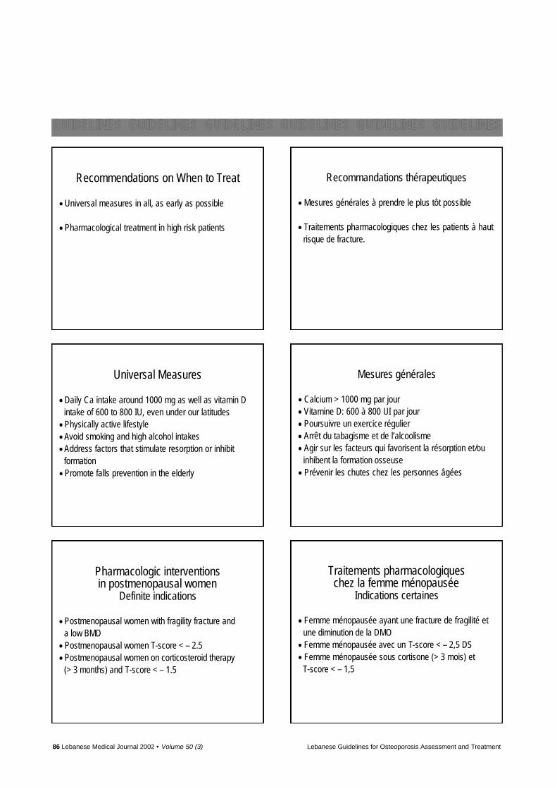
Recommendations on When to Treat
• Universal measures in all, as early as possible
• Pharmacological treatment in high risk patients
Recommandations thérapeutiques
• Mesures générales à prendre le plus tôt possible
• Traitements pharmacologiques chez les patients à hautrisque de fracture.
Mesures générales
• Calcium > 1000 mg par jour
• Vitamine D: 600 à 800 UI par jour
• Poursuivre un exercice régulier
• Arrêt du tabagisme et de l’alcoolisme
• Agir sur les facteurs qui favorisent la résorption et/ouinhibent la formation osseuse
• Prévenir les chutes chez les personnes âgées
Traitements pharmacologiques chez la femme ménopausée
Indications certaines
• Femme ménopausée ayant une fracture de fragilité etune diminution de la DMO
• Femme ménopausée avec un T-score < – 2,5 DS
• Femme ménopausée sous cortisone (> 3 mois) et T-score < – 1,5
Universal Measures
• Daily Ca intake around 1000 mg as well as vitamin Dintake of 600 to 800 IU, even under our latitudes
• Physically active lifestyle
• Avoid smoking and high alcohol intakes
• Address factors that stimulate resorption or inhibit formation
• Promote falls prevention in the elderly
Pharmacologic interventionsin postmenopausal women
Definite indications
• Postmenopausal women with fragility fracture and a low BMD
• Postmenopausal women T-score < – 2.5
• Postmenopausal women on corticosteroid therapy(> 3 months) and T-score < – 1.5
Lebanese Guidelines for Osteoporosis Assessment and Treatment Lebanese Medical Journal 2002 • Volume 50 (3) 87
GUIDELINES GUIDELINES GUIDELINES GUIDELINES GUIDELINES GUIDELINES
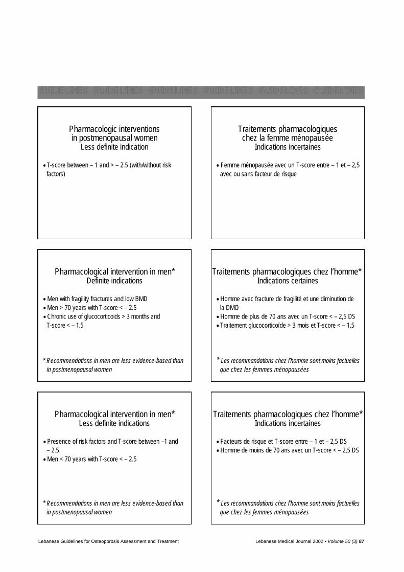
Pharmacologic interventions in postmenopausal women
Less definite indication
• T-score between – 1 and > – 2.5 (with/without riskfactors)
Traitements pharmacologiques chez la femme ménopausée
Indications incertaines
• Femme ménopausée avec un T-score entre – 1 et – 2,5avec ou sans facteur de risque
Traitements pharmacologiques chez l’homme*Indications certaines
• Homme avec fracture de fragilité et une diminution dela DMO
• Homme de plus de 70 ans avec un T-score < – 2,5 DS
• Traitement glucocorticoïde > 3 mois et T-score < – 1,5
* Les recommandations chez l’homme sont moins factuellesque chez les femmes ménopausées
Traitements pharmacologiques chez l’homme*Indications incertaines
• Facteurs de risque et T-score entre – 1 et – 2,5 DS
• Homme de moins de 70 ans avec un T-score < – 2,5 DS
* Les recommandations chez l’homme sont moins factuellesque chez les femmes ménopausées
Pharmacological intervention in men*Definite indications
• Men with fragility fractures and low BMD
• Men > 70 years with T-score < – 2.5
• Chronic use of glucocorticoids > 3 months and T-score < – 1.5
* Recommendations in men are less evidence-based thanin postmenopausal women
Pharmacological intervention in men*Less definite indications
• Presence of risk factors and T-score between –1 and – 2.5
• Men < 70 years with T-score < – 2.5
* Recommendations in men are less evidence-based thanin postmenopausal women
88 Lebanese Medical Journal 2002 • Volume 50 (3) Lebanese Guidelines for Osteoporosis Assessment and Treatment
GUIDELINES GUIDELINES GUIDELINES GUIDELINES GUIDELINES GUIDELINES
Anti-resorptive therapy is definitely not indicated in
• Normal healthy pre-menopausal women with low bonemass (– 2.5 < T-score < – 1)
• Normal healthy men with low bone mass (– 2.5 < T-score < – 1)
Middle East Densitometry WorkshopApril 20, 2002
Who to test?What measures to use?
When to treat?
Ghada El-Hajj Fuleihan, MD, MPH.Rafic Baddoura, MD, MPH.
Hassane Awada, MD. Jad Okais, MD. Paul Rizk, MD.
Michael McClung, MD.
Traitements pharmacologiques Absences certaines d’indication
• Une femme en pré-ménopause normalement régléeavec une diminution de la DMO (– 2,5 < T-score < – 1)ne doit pas recevoir de traitement anti-résorption osseuse
• Un homme avec une diminution de la DMO (– 2,5 < T-score< – 1) ne doit pas recevoir de traitementanti-résorption osseuse
Atelier densitométrie osseuse pour la Méditerranée orientale
20 avril 2002
Quels sujets faut-il tester?Quelles mesures choisir?
Quand faut-il traiter?
Ghada El-Hajj Fuleihan, MD, MPH.Rafic Baddoura, MD, MPH.
Hassane Awada, MD. Jad Okais, MD. Paul Rizk, MD.
Michael McClung, MD.
Lebanese Medical Journal 2002 • Volume 50 (3) 89
El-Hajj Fuleihan G, Baddoura R, Awada H, Okais J, Rizk P,McClung M. Practice guidelines on the use of bone mineraldensity measurements. Who to test ? What measures to use ?When to treat ? A Consensus report from the Middle East Den-sitometry Workshop Leb Med J 2002 ; 50 (3) : 89-104.
WHO TO TEST ?
When the question is “to test or not test” using bonemineral density (BMD) one could anticipate the answeris not straightforward. As with any diagnostic procedure,indications should be linked to clinical decision-makingand this has to do with issues of sensitivity, specificity,predictive value, and balance between health and eco-nomic consequences of false positive and false negativeresults. Moreover targeting osteoporosis adds some pe-culiarities to the analytical process. The outcome is aprobability, i.e. the risk of fracture and the test is a quan-titative measure with an arbitrary cut-off threshold value[1-3].
Clinical decision-making [4] can occur in the settingof either initiating or monitoring therapy. In the first sit-uation, two approaches are identified, mass screening ortargeting high-risk population [5-6]. The latter is cur-rently the main policy by cost-effectiveness considera-tions using evidence-based knowledge about osteoporo-sis. Therefore the question of who to test for BMD canbe first approached as of who is at high risk of fracture.The difficulty comes however from the very objective ofBMD testing that is to estimate the risk of fracture [7].
Regarding the risk of fracture, epidemiological evi-dence supports the role of multiple risk factors for frac-ture, commonly classified into bone and none bone relat-ed determinants (see Table I). The latter are related to therisk of falls namely locomotor problems and environ-ment characteristics and the former to bone strengthdeterminants including bone density and bone quality[8-14].
So far bone density remains the most important deter-minant of fracture in terms of relative risk, that we can
estimate with enough confidence using DEXAtechnolo-gy and that is amenable to modification through pharma-cological interventions [15-19].
However, in practice we are dealing with two differ-ent estimations of the risk of fracture, the absolute risk orremaining lifetime risk [20] and the relative risk [21].The lifetime risk is the probability of sustaining a frac-ture over life expectancy which is higher for early post-menopausal than for late postmenopausal women. Therelative risk is the ratio of the probability of sustaining afracture when the risk factor is present compared toprobability of sustaining a fracture when the risk factoris absent. Relative risk is higher for late postmeno-pausal than early postmenopausal women [22-23].
Besides BMD simple clinical variables could be iden-tified as determinants of fracture risk based on epidemi-ological data [24]. However these variables poorly pre-dict BMD [25]. Therefore BMD testing remains the cor-ner stone in the evaluation of the risk of fracture. Guide-lines have been developed to select people at high risk offracture based on those simple clinical variables [26-29].
Since guidelines necessarily reflect health system pat-terns one might anticipate several guidelines to be pub-lished. Literature review provides guidelines reportsfrom the American National Osteoporosis Foundation[27], the American Association of Clinical Endocrinol-ogists [30], the American College of Rheumatology[31], the North American Menopause Society [32], theUS Preventive Services Task Force [33-34], the Inter-national Society of Clinical Densitometry [28], the Os-teoporosis Society of Canada [29], the European Found-ation for Osteoporosis now known as IOF [35-36], theAustralian National Consensus Conference [37] andWHO [1].
Despite the apparent diversity of recommendations,common and simple clinical variables associated withincreased fracture risk constitute the core set of the clin-ical decision making rule of proposed guidelines. How-ever their diagnostic value might be different. This issuehas been recently addressed in an original contribution [38], where the diagnostic value of the NOF guidelines
PRACTICE GUIDELINES ON THE USE OFBONE MINERAL D E N S I T Y M E A S U R E M E N T SWHO TO T E S T ? W H AT MEASURES TO USE ? WHEN TO T R E AT ?A Consensus report from the Middle East Densitometry Workshop
Ghada EL-HAJJ FULEIHAN, Rafic BADDOURA, Hassane AWADA, Jad OKAIS, Paul RIZK, Michael McCLUNG*
* Faculty members of The Middle East Densitometry Workshop. See Proceedings p. 109. Address correspondence to : Ghada El-Hajj Fuleihan, MD, MPH. Calcium Metabolism and Osteoporosis Program. American
University of Beirut Medical Center. Bliss Street. POBox 11-0236 Riad El-Solh. 4407 2020 Beirut. Lebanon Tel. : 961 1 374374 Ext. 5360/5362 Fax : 961 1 744464 E-mail : [email protected]
was compared to four other clinical decision rules [39-42] based on simple clinical criteria identified throughMEDLINE search excluding decision aids based onregression models or involving detailed questionnaires.The study concluded to the superiority of Simple Calcu-lated Osteoporosis Risk Estimation (SCORE) and Osteo-porosis Risk Assessment Instrument (ORAI) methods,compared to the NOF recommendations in terms of sen-sitivity and specificity. The strength of the study risesfrom the database on which the comparison was made,that is a population based community sample from theCanadian Multi-center Osteoporosis Study [42]. How-ever this might not apply to other populations and yetremains the issue of cost effectiveness that reflectshealth system priorities and practices. As an indicator,BMD testing cost and reimbursement differ widelyacross national health systems and as a consequenceaccess to BMD testing may be dependent on patterns ofhealth care provision [1].
Until further progress can be made in the validation ofwidely applicable rules [42-43], we recommend the useof a core set of clinical variables that are accepted uni-versally to select individuals at high risk of fracture towhom BMD testing will help clinical decision makingand therefore add value to health outcomes.
RECOMMENDATIONSFOR WHO TO TEST
FOR WOMEN, the core set of clinical risk factors in-
cludes menopausal status, age, weight, past history
of fragility fracture and steroid therapy. Therefore we
recommend the following :
In postmenopausal women, regardless of age,BMD testing is definitely indicated if :a. There is evidence of radiological deminerali-
zation.b. Vertebral deformity or fragility fracture is present.c. Corticosteroid therapy for > 3 months is con-
templated.
In postmenopausal women, in the absence of the
conditions above, age is an important issue and we
recommend the following :
In late postmenopausal women (aged 65 yearsand above) BMD testing is definitely indicatedregardless of clinical risk factors to make a deci-sion about pharmacological intervention. In early postmenopausal women (age less than 65years), BMD testing is less definitely indicated,and can be considered in conditions associated
with increased fracture risk such as maternal his-tory of fragility fractures, low body weight (Wt < 50 kgor BMI < 20 kg/m2 or conditions associated with
secondary causes of bone loss, since the like-
lihood of identifying subjects with osteoporosis
is higher. These medical conditions associated
with bone loss, include premature menopause
< 45 years, asymptomatic primary hyperpara-
thyroidism, hyperthyroidism, chronic renal failure,
chronic liver disease, malabsorption, and use of
anticonvulsants, etc.
In premenopausal women with medical conditionsknown to be associated with bone loss BMD is lessdefinitely indicated in clinical decision makingregarding these conditions. These conditions in-
clude : anorexia nervosa, asymptomatic primary
hyperparathyroidism, hyperthyroidism, chronic
renal failure, chronic liver disease, malabsorption,
use of anticonvulsants, etc.
Î In apparently healthy premenopausal women,BMD testing is definitely not indicated since theprevalence of low BMD is rare and the safety ande fficacy of pharmacological intervention is notestablished.
FOR MEN although epidemiological data is less
abundant, however recent evidence suggests a simi -
lar BMD fracture relationship and BMD response to
anti-resorptive agents in men as in women [44-49].
However, the evidence is less definite.
We suggest that the following set of clinical risk fac -
tors for fracture in men is to be considered. These
include past history of fragility fracture, chronic
steroid therapy, hypogonadism, alcohol abuse, de-
mineralization, low weight and medical conditions
associated with bone loss. The main difference would
be that the efficacy of osteoporosis therapies is less
established in men and the incidence rate of fracture
is lower in men compared to women. Therefore, test -
ing in men would be recommended on less defin -
itive grounds. Until further information on the epi -
demiology of osteoporosis in men becomes available,
we recommend the following :
BMD is definitely indicated in the presence of ver-tebral deformity or fragility fracture, hypogonadismor chronic steroid therapy.BMD is less definitely indicated in the case of alco-hol abuse, low weight, radiologic evidence of de-mineralization, medical conditions that are asso-ciated with bone loss such as hyperparathyroid-ism, renal insufficiency, chronic liver disease, andanticonvulsant use.
90 Lebanese Medical Journal 2002 • Volume 50 (3) G. EL-HAJJ F U L E I H A N et al – Practice guidelines on the use of BMD measurements
BMD testing is definitely not indicated in healthy menin the absence of clinical risk factors.
However, these recommendations represent generalguidelines. For difficult and unusual cases referral toa specialist is strongly recommended. Deciding whoto test and how to treat is then left to the discretion ofthe expert.
WHAT MEASURES TO USE ?
What measures to use to assess BMD ? 1. To evaluate the risk of fracture.2. To monitor response to therapy.In order to adequately address that question, the fol-
lowing four issues need to be covered :a. BMD and fracture risk b. Which technique and device ?c. Which parameter (BMD/T-score) and which data-
base ?d. Which skeletal site to measure ?
1. What measures to use to assess the risk of fracture
a. Bone Mineral Density as a strong predictor of fracture :the evidence Before we discuss which measures to use to assess the
risk of fracture, let us review two widely recognized def-initions of osteoporosis.
1. “A systemic skeletal disease characterized by lowbone mass and microarchitectural deterioration of bonetissue, with a consequent increase in bone fragility andsusceptibility to fractures.” International ConsensusDefinition 1993 [50].
2. “Askeletal disorder characterized by compromisedbone strength predisposing to an increased risk of frac-ture. Bone strength reflects the integration of two mainfeatures : bone density and bone quality.” NIH Consen-sus Development Panel 2001 [51].
ä WHO WORKING GROUP OPERATIONAL DEFINITION OFOSTEO -
POROSIS IS BASED ON BMD
Bone mass or BMD are recurrent terms in the abovetwo definitions, that were coined almost a decade apart.This is due to the fact that over the last twenty yearsabundant data has accumulated, establishing BMD asone of the strongest, if not the strongest predictor of frac-tures. As a matter of fact, it is a stronger predictor offracture, than cholesterol is a predictor of coronaryartery disease (CAD), and is at least as good as hyper-tension is a predictor of stroke. This was the main reasonwhy the World Health Organization working groupdeveloped an operational definition for osteoporosis
based on BMD. Although the BMD fracture relationshipis an exponential one, a specific BMD-based cutoff waschosen for osteoporosis diagnosis : a BMD T-score(number of SD below peak bone mineral density) below– 2.5 [1]. “ Such a cutoff value identifies approximately30% of postmenopausal women as having osteoporosisusing measurements made at the spine, hip, or forearm.This percentage is approximately equivalent to the life-time risk of fracture at these sites” [1]. Indeed, Melton etal recently demonstrated that the proportion of post-menopausal women who have a BMD T-score < – 2.5 atthe femoral neck, spine, forearm, or at any of these threesites corresponds to the same proportion of women witha lifetime risk of hip, spine, wrist, or any of these threefractures, respectively [52-53]. This – 2.5 T-score cutoffonly applies to postmenopausal Caucasian women andonly to DEXA densitometry measurements [1, 54].
ä ME A S U R E S VA L I D AT I N GT H EU S EO F BMD TO P R E D I C TF R A C T U R E S
The rationale for the use of BMD in fracture predic-tion is validated by the following observations :
1. Biomechanical testing in the laboratory supports astrong relationship between BMD and bone strength asassessed by failure load, etc. [55].
2. Ample epidemiologic data from longitudinal stud-ies such as the Study of Osteoporotic fractures, the EPI-DOS study, the Rochester study, the Rotterdam study,and the Hawaii Osteoporosis study documenting BMDto be a strong predictor of fractures. Indeed, for each SDdecreases in BMD the RR of fracture is 1.7-3, dependingon the fracture type, the skeletal site, and the devicebeing used [56-65].
3. An evaluation of the large randomized controlledtrials using pharmacologic treatment reveals that BMDincrements account for a significant proportion of thevariance in vertebral fracture risk reduction [66-71].However, the proportion of variance in fracture reduc-tion that is explained by BMD changes has varied wide-ly depending on the study [66-67, 69-71]. Althoughmeasurement of bone mineral density only captures oneaspect of bone strength, since there is no additional read-ily measure of bone quality todate, bone mineral densityremains a pivotal tool in the diagnosis of patients at riskfor fracture.
b. Which device and technique to use ?There are multiple devices on the market to measure
bone mineral density in the central or the peripheralskeleton. The techniques used in these devices are alsodifferent. The main techniques available today are dualenergy x-ray absorptiometry (DEXA), single energy x-ray absorptiometry (SXA), quantitative computerizedtomo-graphy (QCT) and ultrasonometry (QUS).
G. EL-HAJJ F U L E I H A N et al – Practice guidelines on the use of BMD measurements Lebanese Medical Journal 2002 • Volume 50 (3) 91
ä TECHNICAL ISSUES
X-ray technology as DEXA or SXA : uses ionizingradiation and measures areal density (bone mineral con-tent/ area), this can be affected by size, growth etc.DEXA can be used to measure BMD at the central aswell as peripheral skeleton. Central DEXA is the yard-stick to which all measures are compared (see section onvalidation of techniques used below). Central devicesmeasure BMD at the spine, hip, forearm and total body.Peripheral devices are based on single energy (SXA) ordual energy (pDEXA) technology and measure BMD atthe forearm or calcaneus’s (SXA) or at the finger, toe,heel, forearm (pDEXA) [72].
Quantitative computerized tomography (QCT) : usesionizing radiation and measures true volumetric as op-posed to areal density such as measured by SXA andDEXA. QCT can be used for central measurements atthe spine, and a special QCTis available to measure vol-umetric density at the forearm (pQCT). Recent advancesin spiral CT and recent automated software make hipmeasurement also feasible. A major drawback of centralQCT is high radiation exposure, doses reaching 40 timesthat of a DEXA. Although QCTdoes offer high sensitiv-ity in detecting osteoporosis and excellent fracture dis-crimination in cross-sectional studies there are no longi-tudinal studies relating QCT BMD measures to fracturerisk. Furthermore, the standard T-scores do not applywell to QCT, therefore caution must be exercised ininterpretation of results.
Quantitative ultrasound (QUS) : this technology usessound waves, measures speed of sound and broadbandultrasound attenuation that can then yield calculatedparameters (e.g. stiffness, etc. [73]). Both the directlymeasured and the derived parameters are lower in thepatient with osteoporosis. Similarly to QCT, T-scoresalso do not apply to QUS.
ä VALIDATION OF THE TECHNIQUE USED
DEXA : is by far the most widely accepted technolo-gy and one that is most well validated by all three crite-ria listed above. It is the technique with which we havethe most information and is the gold standard today. Ithas very good accuracy and excellent precision in experthands (see below), and incurs low radiation exposure.DEXA is FDA approved for the diagnosis of osteoporo-sis. Indeed, the biomechanical data was mostly obtainedusing DEXA, most epidemiologic studies establishingthe close relationship between fracture risk and BMDused DEXA [59-60, 63, 74]. Similarly, the data from therandomized controlled trials linking treatment efficacyto fracture outcome, exclusively used DEXA (as op-posed to pDEXA or SXA) as an intermediary measurefor efficacy, as required by the FDA.
pDEXA, SXA : Data from the NORA study of over200,000 women screened across the United States with awide variety of devices demonstrates a significant rela-tionship between BMD as assessed by any of these tech-niques/devices (SXA, pDEXA, DXA) and fracture risk,with variations in the risk measure depending on thedevice [64, 75].
QUS : Several prospective studies such as the SOFstudy, EPIDOS and the NORA studies, have demon-strated a direct correlation between QUS measured andderived parameters and fracture risk [61-62, 64].However, it is the calculated QUS parameters and notthe directly measured ones that are used to calculateT-scores, and therefore by inference fracture risk.
QCT : Two studies have recently demonstrated theability of QCT and pQCT to predict risk of vertebralfracture for the former, and spine, hip and global fracturefor the latter [76-77]. However, QCT is to be consideredof experimental value compared to DEXA. QCT also in-curs the highest radiation exposure, around 40X that ofa DEXA.
ä ACCURACY AND PRECISION
Key characteristics to be considered in the choice of adevice to be used to diagnose and monitor a clinical con-dition.
The main critical characteristic to be considered in thechoice of a technique/device to diagnose a condition isits accuracy : how close the measure is to what it is sup-posed to measure. In this instance, this can be evaluatedby measuring BMD/BMC of a bone specimen using thedifferent techniques and devices and comparing that tothe actually measured bone mineral content by ashingthat specimen afterwards. This was carefully studied andthe accuracy for various devices/techniques listed abovevaries between 3-6%, it may be slightly higher for bothQ C T and pQCT, with a range of 8-15% [78-79].Precision (reproducibility) on the other hand is the mostimportant variable to consider when using a technique tomonitor therapy [79-82], see section below “What mea-sures to use to monitor the patient”.
Central DEXA technology is the most establishedtechnology in which the BMD-fracture relationship hasbeen validated in longitudinal studies, and the one withwhich WHO T-score based cutoffs have been estab -lished. FDA approved central DEXAbased densitometrydevices are therefore the preferred method of choice forevaluating fracture risk, when available. Alternativemeasures that could be used are QUS, pDEXA, QCT, orother validated devices. However, non-validated DEXAlike devices are to be avoided in view of their poor accu -racy and their probable poor precision.
92 Lebanese Medical Journal 2002 • Volume 50 (3) G. EL-HAJJ F U L E I H A N et al – Practice guidelines on the use of BMD measurements
c. Which parameter, which device and which databasesIt is generally agreed that the relationship between
BMD and fracture risk is an inversely exponential one,as BMD decreases fracture risk increases ; expressed dif-ferently, for each SD decrease in BMD fracture riskalmost doubles. This assessment was derived from sev-eral large epidemiologic studies conducted mostly inCaucasian populations : SOF in the United States [83],Rotterdam study in the Netherlands [74] ; EPIDOS inFrance [63] ; The Hawaii Osteoporosis study in Hawaii[56], although some scarcer data is available with otherraces.
To-date, fracture risk can either be expressed in one oftwo ways :
1. An absolute risk, lifetime or 5-year risk, for a spe-cific BMD at a certain age (since age is anotherindependent predictor of fractures) such as provid-ed in the Rotterdam study [74].
2. More commonly but in less practical terms as RR/SD decrease. Therefore an individual with a T-score of – 3 has a fracture risk that is twice that ofan individual with a T-score of – 2. The latterassessment is less useful in the clinical setting as itexpresses risk in relative rather than absoluteterms, the latter being a much more clinicallyapplicable and relevant risk assessment tool [59-60, 63, 83-84].
Very few studies have expressed absolute fracture riskas a function of BMD such as the Rotterdam study [74].However, since absolute BMD in gm/cm2 may varydepending on central DEXA manufacturer, appropriateconversions are to be implemented prior to the ability touse such data [85]. In view of the paucity of absolutefracture risk data published, the practice has been to tryto use the more abundant data using RR/SD decrease inBMD, and hence the practice to use T-scores to assessfracture risk, and to establish T-score based thresholdsfor intervention. Two important points are to be made atthis juncture : the WHO T-score cut-offs for the diagno-sis of osteoporosis are applicable only to central DEXAgenerated data in postmenopausal Caucasian femalesubjects only. Conversely, T-score derived from othertechnologies such as pDEXA, QUS and QCT are notcomparable to DEXA derived T-scores for multiple rea-sons including differences in what is measured withthese technologies, differences in normative databasesand the lack of agreed upon diagnostic criteria [86-87].Work in progress between committees from the NOF, theISCD and ASBMR with the goal of deriving T-scoreequivalents that vary depending on the device, to esti-mate fracture risk may help partially resolve this issue.Alternatively, other algorithms are currently being eval-uated to estimate absolute 5 (or 10)-yr fracture risk using
absolute BMD adjusting for variation in densitometertypes (DEXA, U/S, pDEXA, etc.).
The second issue of relevance in our part of the worldis how to use the BMD-fracture data expressed in theEuropean and American Caucasians to our part of theworld, the Middle East. That really gets to the questionof how do absolute BMD/fracture curves compareacross populations within the same racial category, forour purposes, Caucasians. A comparison of absoluteBMD vs fracture risk across populations of the samerace would be needed to evaluate that question. Suchdata is just not available to-date for populations from theMiddle East. Therefore, resorting to T-score was the nextavailable strategy, to assess fracture risk in individuals inthe Middle East. This would be sound if the followingtwo conditions were met :
1. We assume that the absolute BMD/fracture rela-tionship is overall the same in all Caucasians re-gardless of the population. There is no reason to-date to think otherwise.
2. We use the appropriate device and database inwhich the BMD-fracture relationship and thereforeT-score cut-off was derived. These would be a cen-tral DEXAdevice and a Caucasian postmenopausalfemale normative database.
Let us turn to the data available to us to-date from theregion trying to address that issue. Peak BMD has beenstudied mostly in non-population based [88-90] and inpopulation-based samples [91-92]. The studies availablefrom our region reveal peak BMD in these subjects maybe slightly lower than (in 4 studies) or equal to (in onestudy) that of European and American Caucasians, pos-sibly due to differences in body size, chronic vitamin Ddeficiency or less physical activity [89, 93-94]. The pre-valence of vertebral fractures in postmenopausal womenand hip fracture rates are comparable to data for Westerncounterparts [95-98]. Finally, mean BMD in hip fracturein Lebanese subjects is comparable to mean BMD in hipfracture subjects from the West [99-100]. The latterinformation suggests that the absolute BMD-fracturerelationship may be the same in our region as it is in theWest. The situation may very well be different in otherraces i.e. Asians, African-Americans, etc.
In view of the above observations, the application ofWestern standards for the diagnosis and assessment offracture risk in Caucasian subjects from the Middle Eastare prudent, until additional forthcoming data from theregion becomes available. This is consistent with therecommendations from the International OsteoporosisFoundation [35]. Therefore, we recommend the use ofcentral DEXA devices and Western databases (for e.g.NHANES, etc.) for the derivation of T-scores to assess
G. EL-HAJJ F U L E I H A N et al – Practice guidelines on the use of BMD measurements Lebanese Medical Journal 2002 • Volume 50 (3) 93

f r a c t u re risk, or absolute BMD/fx risk data suchas provided in the Rotterdam study after appropriatetransformation of the data to obtain comparable densit -ometry units (see above ). Any other practice will resultin a tendency to erroneously diagnose osteoporosis andwrongly estimate fracture risk. WHO T-score based cri -teria are not applicable to non-Caucasian postmeno-pausal women, to premenopausal women, to men, tochildren and non-Caucasians. They are also not applic -able to other technologies such as QCT, pQCT, QUS,pDEXA and SXA.
Algorithms are currently being evaluated to use in-formation gathered from non-central DEXA devices toestimate absolute 5-year or 10-year fracture risk, how -ever such data is not readily available yet.
d. Which skeletal sites to measure ?It is generally agreed that the relationship between
BMD and fracture risk is an inversely exponential one,as BMD decreases fracture risk increases ; expressed dif-ferently for each SD decrease in BMD fracture risk in-creases by 1.5-2.8 folds. This range is due to variationsin the skeletal site used to estimate fracture risk (L2-L4,hip, forearm, et.) and the specific fracture for which therisk is predicted (wrist, hip, or vertebral fracture).
Global risk of fracture : Several studies have estab-lished that the global relative risk of fracture, relativerisk of developing an osteoporotic fracture anywhere inthe skeleton is the same 1.4-1.6/SD decrease in BMD asmeasured at any site in the skeleton [84].
Site-specific fracture risk : although site-specific frac-ture risk assessment can be estimated by measuringBMD at any skeletal site, the predictive value is higherif a site-specific assessment is conducted : e.g. whereasspine, hip and forearm all predict fracture risk at the hipand spine, hip BMD is the best predictor of hip fractureand spine BMD is the best predictor of vertebral fracture[59-60, 83-84].
Central DEXA : RR/SD decrease in BMD from a meta-analysis of 11 prospective cohort studies, 1985-1994,90,000 person-years, > 2000 fractures [84].
RR/SD decrease in BMDSpine BMD for vertebral fractures : 2.3 [1.9-2.8]Femoral neck BMD for hip fractures : 2.6 [2.0-3.5]Distal radius for wrist fractures : 1.7 [1.4-2.0]
ä HOW MANY SKELETAL SITES TO MEASURE ?
1. Although there is correlation in BMD between onesite and the other (r = 0.4-0.6), it is not perfect.Therefore measuring only one site may underesti-mate a subject’s osteoporosis risk [87, 101-103].
2. Hip BMD is the best predictor of hip fracture, spine
BMD is the best predictor of vertebral fractures, asoutlined in the previous section.
3. At the menopause, accelerated bone loss takes placemore so at the spine than at the hip. So measuringonly a hip BMD may miss the lower bone mass atthe spine [102].
4. Aging results in degenerative changes at the spinethat may falsely increase BMD by 0.5-1SD [104-105]. Measuring the hip in the elderly is of particu-lar importance.
5. The spine site is the most responsive skeletal site topharmacologic intervention and may be importantin monitoring a patient [69].
6. Forearm : Some clinical conditions such as prima-ry or secondary hyperparathyroidism may lowerforearm BMD the most [106]. In such instances aforearm measurement is indicated. A forearm isalso indicated in the very obese patient in whom aspine or hip cannot be performed due to large size.
ä EFFO POSITION (NOW IOF) : In a position paper theEuropean Foundation for Osteoporosis has suggestedmeasuring only one skeletal site for the young patient(spine, hip, or forearm) and the hip only in the elderly asit best predicts the occurrence of the most importantfracture, and avoids running into the problems of DJD ofthe spine [35].
ä ISCD POSITION : ISCD forum July 2001. Measurespine and hip for all patients, non-dominant forearm is tobe added if one of the above two skeletal sites cannot beused, if the patient has suspected hyperparathyroidism,or if the patient is obese. Total body BMC measurementis recommended in children.
ä NOF : NOF analyses for cost-effectiveness were allbased on BMD measurement at the hip.
Although it is controversial whether measurement ofmore than one skeletal site improves our discriminativeability in predicting the patient at risk for fractures, atwo-site central DEXA measurement is preferred for theabove-mentioned reasons.
We therefore recommend following the guidelines ofthe ISCD (ISCD Syllabus, Version 4, Jan 2002) for skele -tal site selection :
* Spine and hip for all patients.
* Non-dominant forearm is added in the following situations : One skeletal site cannot be used : arthritis, prosthesis, etc.Hyperparathyroidism is suspectedThe patient is obese
* For spine the use of L1-L4 is recommended, and for the
94 Lebanese Medical Journal 2002 • Volume 50 (3) G. EL-HAJJ F U L E I H A N et al – Practice guidelines on the use of BMD measurements
hip ISCD suggests the use of the lowest T-score of all3 hip sites total, femoral neck, trochanter.
2. What measures to use to monitor the patient ?
A complete and detailed overview of that topic is pro-vided in the ISCD Clinical track syllabus, Version 4,January 2002.
a. Skeletal sites to monitor BMD change over timeand what is a significant change
b. Interval time for repeating BMD to assess responseto therapy.
The purpose of this discussion is not to advisewhether a patient (whether on therapy or not) shouldhave serial BMD measured, but rather once the decisionis made to repeat BMD measurements which skeletalsites should be used, and when should the follow-upscan take place.
a. Skeletal sites to monitor BMD change over time andwhat is a significant changeIn order to assess change over time the following con-
ditions should be met : 1. The same skeletal site as measured on the same de-
vice, not just same model, should be used. In theevent of a change in the machine, careful cross-cali-bration is mandatory.
2. Absolute BMD, rather than T-scores should be used.3. Cannot compare T-score on scans performed on dif-
ferent manufacturers due to differences in norma-tive databases [92, 107] and differences in identify-ing ROI (e.g. L1-L4 vs. L2-L4 ; differences in algo-rithm used to define ROI for femoral neck).
4. The ROI of BMD sites being compared should beidentical and the area should be within 2% betweento the two duplicates, otherwise the comparison ofareal BMD is not valid.
5. Strict adherence to manufacturer guidelines for po-sition and analysis are of utmost importance.
6. The skeletal site to be chosen for monitoring BMDchange over time is one that has the highest preci-sion (≤ 1%), is the most responsive to change withtreatment, and is the least affected by potential arti-facts. The spine definitely fulfills the first two crite-ria [81-82]. In case of DJD of the spine, the total hip(rather than femoral neck, better precision lessaffected by rotation) is the next preferred site. Theforearm is unlikely to show changes over time dueto its lower bone turnover.
7. Center specific precision data should be available.Ideally such precision (duplicate BMD scans on thesame patient, performed on over 30 individuals fewdays apart) should be calculated in each center, ontheir own machines, in the population being evalu-ated, namely postmenopausal women. Indeed, wehave demonstrated that same day precision is betterthan different day precision, and precision derivedfrom osteoporotic patients is worse than in normalsubjects [82]. Use of in-vitro precision based onphantom duplicate measurements, provided by thedensitometer manufacturers and used by the densi-tometry software to assess significance of changesin an individual over time should be discouraged.Indeed, these estimates are not applicable to the realclinical situations but are unfortunately used bymany centers.
8. The mean SD (rather than CV) derived from allduplicate scans is calculated, and the root meansquare average for the entire group is then calculat-ed by summing the square of the SD, then dividingby the number of patients (e.g. N = 30), and thentaking the mean square root MSR [79].
G. EL-HAJJ F U L E I H A N et al – Practice guidelines on the use of BMD measurements Lebanese Medical Journal 2002 • Volume 50 (3) 95
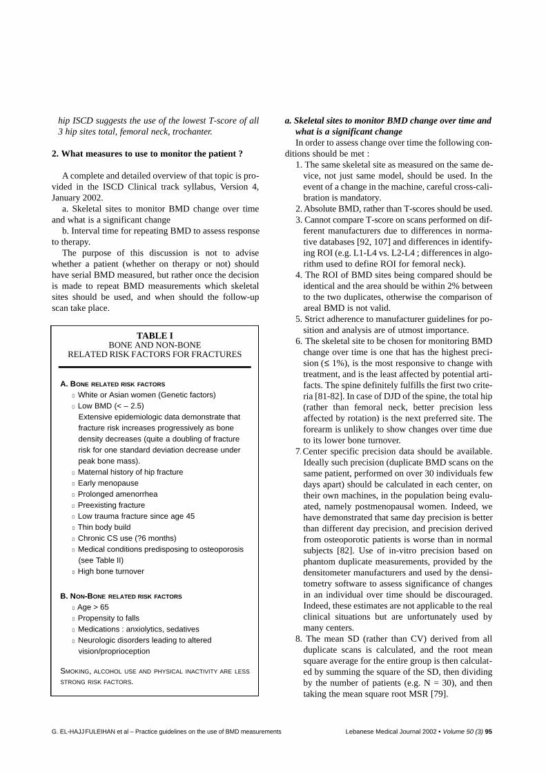
TABLE IBONE AND NON-BONE
RELATED RISK FACTORS FOR FRACTURES
A. BONE RELATED RISK FACTORS
n White or Asian women (Genetic factors)n Low BMD (< – 2.5)
Extensive epidemiologic data demonstrate thatfracture risk increases progressively as bonedensity decreases (quite a doubling of fracturerisk for one standard deviation decrease underpeak bone mass).
n Maternal history of hip fracturen Early menopausen Prolonged amenorrhean Preexisting fracturen Low trauma fracture since age 45n Thin body buildn Chronic CS use (?6 months)n Medical conditions predisposing to osteoporosis
(see Table II) n High bone turnover
B. NON-BONE RELATED RISK FACTORS
n Age > 65n Propensity to fallsn Medications : anxiolytics, sedativesn Neurologic disorders leading to altered
vision/proprioception
SMOKING, ALCOHOL USE AND PHYSICAL INACTIVITY ARE LESS
STRONG RISK FACTORS.
9. A change over time is real if it exceeds the LeastSignificant Change (LSC), a number derived fromthe precision, preferably calculated from SD fromd u p l icates rather than CV%. LSC is calculated as2.77 x MSR of the data, with a 95% confidenceinterval [79]. Even in the centers with the best pre-cision, one should not repeat BMD before 1.5-2y e a r s , unless one expects accelerated bone loss such(see below).
b. Interval time for repeating BMD to assess responseto therapyThe interval of time is determined by the expected
change in BMD over time (the latter depends on type oftherapy and specific skeletal site), the center derivedLSC. Mean Treatment Interval = LSC/expectedchange/year [79]. This implies that even in expert cen-ters with an MSR of 1, LSC of 2.77, and an expectedmean change in BMD of 0.03 gm/cm2, repeating a BMDbefore 1.5-2 years is not indicated. The interval time isobviously shorter in cases of anticipated fast incrementsand/or decrements in BMD (as seen post-oophorectomy,post GnRH therapy, with high doses chronic corticos-teroid therapy, or with bone anabolic therapies) in whichinstances the interval may be as short as six months to ayear.
To conclude, if the decision is to monitor a patient, itis strongly recommended to evaluate the patient ideallyon the same device, not just same model, with strict QAmeasures for scan acquisition and analysis. This in-cludes choice of scanning mode, choice of site, ROI, andthe derivation of center specific patient based precisiondata for the skeletal site of interest, to determine a cen -ter specific LSC measure and therefore significance of achange in an individual patient. The skeletal site we rec -ommended for monitoring is the spine, the hip can beused instead in select situations or in addition. Monitor-ing interval depends on the center specific LSC data andexpected changes in BMD in each patient/year. Usually,follow-up scans should not be done before 1.5-2 yrs.
WHEN TO TREAT ?
Over the last decade there has been an effort to expandguidelines from “who to test” to “who and when to treat”using the body of evidence provided by the large random-ized osteoporosis trials with the solid endpoints of osteo-porotic fractures. With the increasing evidence for a rela-tively rapid rate of treatment onset and offset for theseinterventions, there has been a move away from long-termpreventive strategies towards the use of shorter-term ther-apy in high risk individuals as outlined below and in pub-
96 Lebanese Medical Journal 2002 • Volume 50 (3) G. EL-HAJJ F U L E I H A N et al – Practice guidelines on the use of BMD measurements
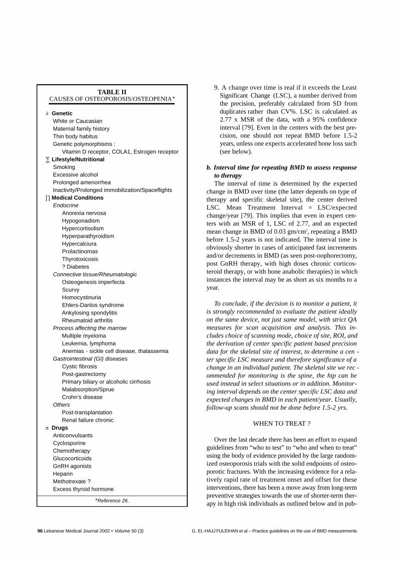
TABLE IICAUSES OF OSTEOPOROSIS/OSTEOPENIA*
GeneticWhite or CaucasianMaternal family historyThin body habitusGenetic polymorphisms :
Vitamin D receptor, COLA1, Estrogen receptorLifestyle/NutritionalSmokingExcessive alcoholProlonged amenorrheaInactivity/Prolonged immobilization/SpaceflightsMedical ConditionsEndocrine
Anorexia nervosaHypogonadismHypercortisolismHyperparathyroidismHypercalciuraProlactinomasThyrotoxicosis? Diabetes
Connective tissue/Rheumatologic
Osteogenesis imperfectaScurvyHomocystinuriaEhlers-Danlos syndromeAnkylosing spondylitisRheumatoid arthritis
Process affecting the marrow
Multiple myelomaLeukemia, lymphomaAnemias - sickle cell disease, thalassemia
Gastrointestinal (GI) diseases
Cystic fibrosisPost-gastrectomyPrimary biliary or alcoholic cirrhosisMalabsorption/SprueCrohn’s disease
Others
Post-transplantationRenal failure chronic
DrugsAnticonvulsantsCyclosporineChemotherapyGlucocorticoidsGnRH agonistsHeparinMethotrexate ?Excess thyroid hormone
*Reference 26.
lished guidelines or reviews on that topic [27, 35, 108-111]. The pivotal randomized controlled trials that areresponsible for the switch in the treatment strategies willalso be highlighted in this overview.
Definition of prevention and treatment strategies andthe evidence for intervention
– Prevention is defined by primary prevention, i.e. pre-vention of bone loss in early postmenopausal womenwithout established osteoporosis i.e. with BMD T-score between – 1 and – 2.5.
– Prevention studies are conducted with a primary end-point of BMD, not fracture. Indeed, in these womenthe absolute risk of fracture is very low and thus,within the relatively short time frame of the majorityof these studies, anti-fracture efficacy cannot be test-ed. As with any preventive treatment, prevention ofosteoporosis should be cost-effective and easy to usein large populations.
– Treatment is defined as reduction in fracture risk inpostmenopausal women with established osteoporo-sis (BMD T-score below – 2.5, with or without a pre-vious prevalent fracture). Usually, the much higherrisk of fragility fracture in the treatment populations,in late (older) postmenopausal women, enables as-sessment of anti-fracture efficacy.
To help evaluating known evidence related to theseinterventions, the Royal College of Medicine establishedthe following grading of evidence. (Grading of evidencelevels as well as tables I and II are taken from the RoyalCollege of Medicine Guidelines, updated in July 2000
[112]. The United States Preventive Services Task Forceas well as the Osteoporosis Society of Canada have alsorecently reviewed osteoporosis treatment efficacy aswell as issued clinical guidelines for osteoporosisscreening [34, 113].
Grade A : Meta-analysis of randomized clinical trials(RCT) or at least one RCT.At least one well designed controlled studywithout randomization.Valid cohort study for prognosis or riskassessment purpose.
Grade B : At least one other type of well designedquasi-experimental study.Well-designed non-experimental descrip-tive studies (comparative, correlation or case-control studies).
Grade C : Expert committee reports/opinions and/orclinical experience of authorities.
Among risk factors for osteoporosis, some may bemodified through behavioral or environmental interven-tions (see Tables I and II) whereas others may be targetsfor pharmacological intervention. It has been suggested that an adequate work-up to rule out secondary causes ofosteoporosis could include a 24-hour urinary calcium,serum calcium and serum PTH to all postmenopausalwomen with osteoporosis, and a TSH in those on chron-ic supplementation [114].
G. EL-HAJJ F U L E I H A N et al – Practice guidelines on the use of BMD measurements Lebanese Medical Journal 2002 • Volume 50 (3) 97
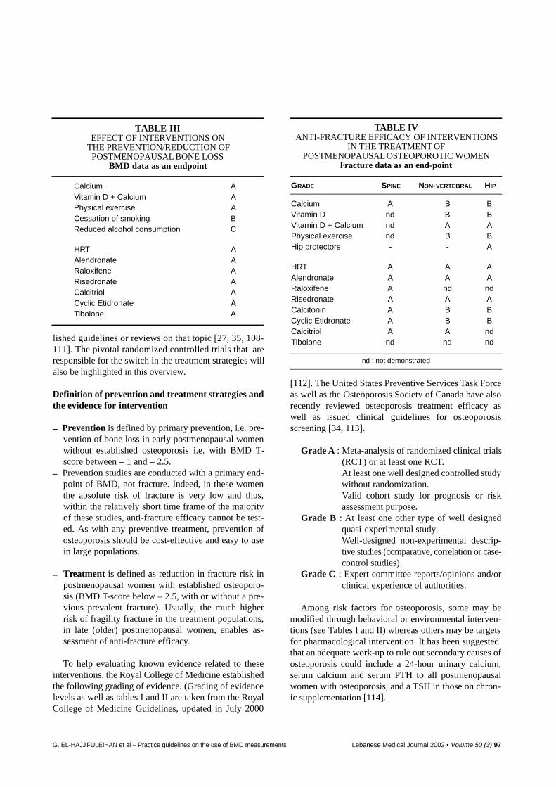
TABLE IIIEFFECT OF INTERVENTIONS ON
THE PREVENTION/REDUCTION OFPOSTMENOPAUSAL BONE LOSS
BMD data as an endpoint
Calcium AVitamin D + Calcium APhysical exercise ACessation of smoking BReduced alcohol consumption C
HRT AAlendronate ARaloxifene ARisedronate ACalcitriol ACyclic Etidronate ATibolone A
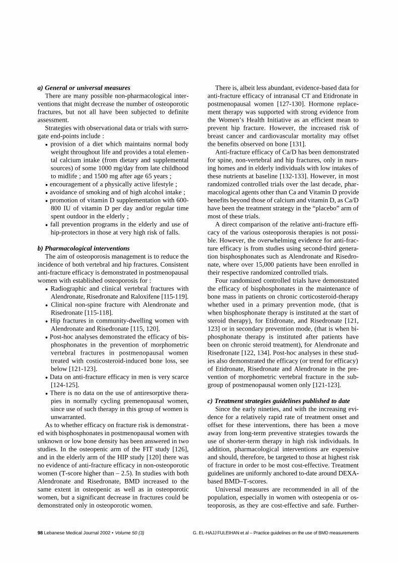
TABLE IVANTI-FRACTURE EFFICACY OF INTERVENTIONS
IN THE TREATMENT OF POSTMENOPAUSAL OSTEOPOROTIC WOMEN
Fracture data as an end-point
GRADE SPINE NON-VERTEBRAL HIP
Calcium A B BVitamin D nd B BVitamin D + Calcium nd A APhysical exercise nd B BHip protectors - - A
HRT A A AAlendronate A A ARaloxifene A nd ndRisedronate A A ACalcitonin A B BCyclic Etidronate A B BCalcitriol A A ndTibolone nd nd nd
nd : not demonstrated
a) General or universal measuresThere are many possible non-pharmacological inter-
ventions that might decrease the number of osteoporoticfractures, but not all have been subjected to definiteassessment.
Strategies with observational data or trials with surro-gate end-points include :
• provision of a diet which maintains normal bodyweight throughout life and provides a total elemen-tal calcium intake (from dietary and supplementalsources) of some 1000 mg/day from late childhoodto midlife ; and 1500 mg after age 65 years ;
• encouragement of a physically active lifestyle ;
• avoidance of smoking and of high alcohol intake ;
• promotion of vitamin D supplementation with 600-800 IU of vitamin D per day and/or regular timespent outdoor in the elderly ;
• fall prevention programs in the elderly and use ofhip-protectors in those at very high risk of falls.
b) Pharmacological interventionsThe aim of osteoporosis management is to reduce the
incidence of both vertebral and hip fractures. Consistentanti-fracture efficacy is demonstrated in postmenopausalwomen with established osteoporosis for :
• Radiographic and clinical vertebral fractures withAlendronate, Risedronate and Raloxifene [11 5 - 11 9 ] .
• Clinical non-spine fracture with Alendronate andRisedronate [115-118].
• Hip fractures in community-dwelling women withAlendronate and Risedronate [115, 120].
• Post-hoc analyses demonstrated the efficacy of bis-phosphonates in the prevention of morphometricvertebral fractures in postmenopausal womentreated with costicosteroid-induced bone loss, seebelow [121-123].
• Data on anti-fracture efficacy in men is very scarce[124-125].
• There is no data on the use of antiresorptive thera-pies in normally cycling premenopausal women,since use of such therapy in this group of women isunwarranted.
As to whether efficacy on fracture risk is demonstrat-ed with bisphosphonates in postmenopausal women withunknown or low bone density has been answered in twostudies. In the osteopenic arm of the FIT study [126],and in the elderly arm of the HIP study [120] there wasno evidence of anti-fracture efficacy in non-osteoporoticwomen (T-score higher than – 2.5). In studies with bothAlendronate and Risedronate, BMD increased to thesame extent in osteopenic as well as in osteoporoticwomen, but a significant decrease in fractures could bedemonstrated only in osteoporotic women.
There is, albeit less abundant, evidence-based data foranti-fracture efficacy of intranasal CT and Etidronate inpostmenopausal women [127-130]. Hormone replace-ment therapy was supported with strong evidence fromthe Women’s Health Initiative as an efficient mean toprevent hip fracture. However, the increased risk ofbreast cancer and cardiovascular mortality may offsetthe benefits observed on bone [131].
Anti-fracture efficacy of Ca/D has been demonstratedfor spine, non-vertebral and hip fractures, only in nurs-ing homes and in elderly individuals with low intakes ofthese nutrients at baseline [132-133]. However, in mostrandomized controlled trials over the last decade, phar-macological agents other than Ca and Vitamin D providebenefits beyond those of calcium and vitamin D, as Ca/Dhave been the treatment strategy in the “placebo” arm ofmost of these trials.
A direct comparison of the relative anti-fracture effi-cacy of the various osteoporosis therapies is not possi-ble. However, the overwhelming evidence for anti-frac-ture efficacy is from studies using second-third genera-tion bisphosphonates such as Alendronate and Risedro-nate, where over 15,000 patients have been enrolled intheir respective randomized controlled trials.
Four randomized controlled trials have demonstratedthe efficacy of bisphosphonates in the maintenance ofbone mass in patients on chronic corticosteroid-therapywhether used in a primary prevention mode, (that iswhen bisphosphonate therapy is instituted at the start ofsteroid therapy), for Etidronate, and Risedronate [121,123] or in secondary prevention mode, (that is when bi-phosphonate therapy is instituted after patients havebeen on chronic steroid treatment), for Alendronate andRisedronate [122, 134]. Post-hoc analyses in these stud-ies also demonstrated the efficacy (or trend for efficacy)of Etidronate, Risedronate and Alendronate in the pre-vention of morphometric vertebral fracture in the sub-group of postmenopausal women only [121-123].
c) Treatment strategies guidelines published to dateSince the early nineties, and with the increasing evi-
dence for a relatively rapid rate of treatment onset andoffset for these interventions, there has been a moveaway from long-term preventive strategies towards theuse of shorter-term therapy in high risk individuals. Inaddition, pharmacological interventions are expensiveand should, therefore, be targeted to those at highest riskof fracture in order to be most cost-effective. Treatmentguidelines are uniformly anchored to-date around DEXA-based BMD–T-scores.
Universal measures are recommended in all of thepopulation, especially in women with osteopenia or os-teoporosis, as they are cost-effective and safe. Further-
98 Lebanese Medical Journal 2002 • Volume 50 (3) G. EL-HAJJ F U L E I H A N et al – Practice guidelines on the use of BMD measurements
more, most of the guidelines published to-date favorpharmacologic intervention in high risk individuals asdefined with T-score < – 2.5 or a T-score < – 2 in thepresence of additional independent risk factors for frac-ture [27, 32, 35]. These would include high on the listprevalent fracture at entry and glucocorticoid use. In-deed, the number needed to treat to prevent a vertebralfracture in older postmenopausal women with a low T-score at entry and prevalent fractures varies between9 and 20 depending on the study [115, 117-119, 131,135]. Similar analyses conducted in older postmeno-pausal women with a BMD T-score at entry of less than2.5 but without fractures, is calculated at 35 for Alen-dronate and 45 for Raloxifene [115, 119]. In contrast,subgroup analyses of the FITtrial revealed that the num-ber needed to treat to prevent a vertebral fracture, evenin older postmenopausal women, increases from 35 ifthe T-score at study entry is < – 2.5, to 59 if the entryT-score is between – 2 and – 2.5, and is as high as 363for older postmenopausal women with entry T-score be-tween – 1.6 and – 2 [115].
Despite increasing awareness of people as well asphysicians about osteoporosis and its related complica-tions, a high percentage of people with osteoporotic frac-tures remain untreated [98, 136]. This paradox betweenscientific data and current clinical practice is commonworldwide [136], in particular in our country where, asshown in a recent study, no more than 5% of people over50 with a fracture were receiving anti-resorptive treat-ments [98][.
d) In conclusionThe only patients in whom fracture prevention with
pharmacological intervention has been proven are thoseat high risk of fracture : elderly postmenopausal womenwith preexisting fracture, or with BMD T-score lowerthan – 2.5, or postmenopausal women on chronic gluco-corticoid therapy, albeit with more limited evidence.Treating young postmenopausal women who do nothave osteoporosis for several years with anti-resorptivetherapy preserves bone density but does not seem tobe associated with reduction in spine or hip fractures.Therefore, the timing of the institution of pharmacolog-ical intervention in that subgroup after menopauseremains to be determined.
Treatment of acute vertebral fracture
Treatment of acute and chronic pain related to verte-bral fractures depends on specific measures and not onanti-osteoporotic drugs. These measures include pain-killers, NSAIDs, bed rest, back support and soft mas-sages as well as mild exercises for subacute and chronic
pain. Calcitonin also has additional analgesic effects.Vertebroplasty, that is injection of intravertebral meta-crylate, can be helpful to alleviate morbidity from verte-bral fractures in case of prolonged pain or refractoryconditions. It should not be routinely used as its safetyand long-term effects are unknown [137].
RECOMMENDATIONS FOR WHO AND WHEN TO TREAT
UNIVERSAL MEASURES are recommended inde -
pendently from BMD measurement
• Maintain a physically active lifestyle with ade-quate exposure to sunlight.
• Avoid smoking and high alcohol intakes.
• Maintain a total dietary calcium intake around1.5 gm of elemental calcium in postmenopausalestrogen deficient women or men > 65 years, aswell as vitamin D intake of 600 to 800 IU/day,even under our latitudes. Provide calcium andvitamin D supplementation in the elderly.
• Avoid a low weight < 60 kg in men or 50 kg inwomen or a low Body Mass Index BMI < 20kg/m2.
• The prevention of osteoporosis begins with opti-mal bone mass acquisition during growth.Factors hindering bone mass acquisition, suchas malnutrition, should be considered, identi-fied, and addressed during childhood.
• Address known factors that stimulate boneresorption or inhibit bone formation, includingh y p o g o n adism, primary hyperparathyroidism,h y p e r t h y r o i d i s m and hypercortisolism.
• Develop fall prevention awareness and pro-grams in the elderly. Hip protection and/or softfloor covering in elderly environment.
PHARMACOLOGICALINTERVENTION is warranted
in high-risk individuals. BMD assessment is pivotal in
clinical decision-making regarding pharmacological
interventions.
Specifically it is definitely indicated in postmeno-pausal women with :
• BMD T-score < – 2.5.
• Prevalent fragility fractures when further docu-mented with low BMD.
• On chronic corticosteroid therapy with BMD T-score < – 1.5.
No clear evidence is available to demonstrate the effi-cacy of pharmacological intervention in postmeno-pausal women with – 2.5 < T-score < – 1. No treat-ment (in addition to universal measures) is indicatedif BMD T-score > – 1.
G. EL-HAJJ F U L E I H A N et al – Practice guidelines on the use of BMD measurements Lebanese Medical Journal 2002 • Volume 50 (3) 99
In premenopausal women
All known treatments were studied in postmenopausalwomen. Their efficacy in premenopausal women isunknown. Thus, in the absence of any establishedtreatment for normally cycling premenopausal womenwith low bone density, such patients should be re-ferred to specialized centers for investigation of possi-ble underlying causes and advice on further manage-ment. Treatment should not be started in such patientsbefore appropriate investigations and diagnoses areachieved.
In men
Given the few number of studies done in men osteo-porosis, no definite recommendation other than uni-versal measures can be given for men. These univer-sal measures as outlined above include reversal ofconditions associated with bone loss. Preliminary datafrom one trial only suggest treatment efficacy of highrisk individuals with Alendronate. Treatment is probably indicated in men :
• with prevalent fragility fractures when furtherdocumented with low BMD
• > 70 years and BMD T-score < – 2.5
• on chronic (> 3 months) corticosteroid therapyand a BMD T-score < – 1.5.
It is less clearly indicated in men :
• with - 1< T-score < - 2.5 in the presence of riskfactors
• < 70 years and with a T-score < – 2.5
The above guidelines, were endorsed by the fivemajor Lebanese scientific societies, the LebaneseSociety of Endocrinology, the Lebanese Society ofObstetrics and Gynecology, the Lebanese Society ofOrthopedics, the Lebanese Society of Radiology, theLebanese Society of Rheumatology, and by the EasternMediterranean Regional Office of the World HealthO rganization. They are meant to provide a structuralframe-work to be used by the physician treating thepatient at risk for or with osteoporosis. They are cer-tainly not meant to supercede the ultimate decision ofthe practicing physician. In the case of rare and/or diff i-cult cases referral to an osteoporosis specialist is highlyr e c o m m e n d e d .
ACKNOWLEDGEMENTS
The authors would like to thank Mrs MicheleValligny for her tireless efforts in coordinating the edit-ing, formatting and printing of this document for theLebanese Guidelines on Osteoporosis Assessment andTr e a t m e n t .
REFERENCES
1. Assessment of fracture risk and its application toscreening for postmenopausal osteoporosis. Report of aWHO study group. WHO technical report series 1994 ;843 : 1-129.
2. Wasnich R. Bone mass measurement : prediction ofrisk. Am J Med 1993 ; 95: Suppl A : 6S-10S.
3. Delmas PD. Do we need to change the WHO definition of osteoporosis ? Osteoporos Int 2000 ; 11 : 189-91.
4. Laupacis A, Sekar N, Steill IG. Clinical predictionrules. A review and suggested modifications of method-ological standards. JAMA1997 ; 277 : 488-94.
5. Law MR, Wald NJ, Meade TW. Strategies for preven-tion of osteoporosis and hip fracture. BMJ 1991 ; 303 :453-9.
6. Johnston CC Jr, Slemenda CW. Risk assessment : theo-retical considerations. Am J Med 1993 ; 95 : Suppl A:2S-5S.
7. Cummings SR, Black D. Bone mass measurement andrisk of fracture in Caucasian women : a review of find-ings from prospective studies. Am J Med 1995 ; 98(Suppl A) : 24S-28S.
8. Ross PD, Davis JW, Epstein RS, Wasnich RD. Pre-existing fractures and bone mass predict vertebral frac-ture incidence in women. Ann Intern Med 1991 ; 114 :919-23.
9. Cummings SR. Treatable and untreatable risk factorsfor hip fracture. Bone 1996 ; 18 : 165S-167S.
10. Kanis JA, McCloskey EV. Evaluation of the risk of hipfracture. Bone 1996 ; 18 : 127S-132S.
11. Silman AJ. The patient with fracture : the risk of subse-quent fracture. Am J Med 1995 ; 98 : Suppl A : 12S-16S.
12. Carroll J, Testa MA, Erat K, LeBoff MS, El-HajjFuleihan G. Modeling fracture risk using bone density,age and years since menopause. Am J Prev Med 1997 ;13 : 447-52.
13. Blake GM, Gluer CC, Fogelman I. Bone densitometrycurrent status and future prospects. Br J Radiol 1997 ;70 : 177S-186S.
14. Levis S, Altman R. Bone densitometry. Clinical Con-siderations. Arthritis Rheum 1998 ; 41 ; 577-87.
15. Rizzoli R, Solsman D, Bonjour JP. The role of DEXAof lumbar spine and proximal femur in the diagnosisand follow-up of osteoporosis. Am J Med 1995 ; 98(2A) : 33S-36S.
16. Sturtridge W, Lentle B, Hanley DA. Prevention andmanagement of osteoporosis : consensus statementsfrom the Scientific Advisory Board of the OsteoporosisSociety of Canada. The use of bone densitometry mea-surement in the diagnosis and management of osteo-porosis. CMAJ 1996 ; 155 : 924-9.
17. Bracker MD, Watts NS. How to get most out of bonedensitometry ? Results can help assess fracture risk andguide therapy. Postgrad Med 1998 ; 104 : 77-9, 83-6.
18. Melton LJ III, Kan SH, Wahner HW, Riggs BL. Life-time fracture risk : an approach to hip fracture risk
100 Lebanese Medical Journal 2002 • Volume 50 (3) G. EL-HAJJ F U L E I H A N et al – Practice guidelines on the use of BMD measurements
assessment based on bone mineral density and age. JClin Epidemiol 1988 ; 41 : 985-94.
19. Christiansen C. Practical application of risk assessment.Osteoporos Int 1998 ; Suppl 1 : 43S-46S.
20. Christiansen C. What should be done at the time ofmenopause ? Am J Med 1995 ; 98 (Suppl A) : 56-8.
21. Black DM. Why elderly women should be screened andtreated to prevent osteoporosis ? Am J Med 1995 ; 98(Suppl A) : 66S-75S.
22. Cummings SR, Nevitt MC, Browner WS, et al. Risk fac-tors for hip fracture in white women. Study of Osteo-porotic Fractures Research Group. N Engl J Med 1995 ;332 : 767-73.
23. Slemenda CW, Hui SL, Longcope C, Wellman H,Johnston CC Jr. Predictors of bone mass in perimeno-pausal women. A prospective study of clinical datausing photon absorptiometry. Ann Intern Med 1990 ;112 : 96-101.
24. Johnston CC. Development of clinical practice guide-lines for prevention and treatment of osteoporosis.Calcif Tissue Int 1996 ; 59 (Suppl 1) ; 30S-33S.
25. Baran DT, Faulkner KG, Genant HK, Miller PD, P a c i -fici R. Diagnosis and management of osteoporosis :guidelines for the utilization of bone densitometry.Calcif Tissue Int 1997 ; 61: 433-40.
26. El-Hajj Fuleihan G. Osteoporosis : an overview of prac-tice guidelines for bone density measurements and os-teoporosis treatment strategies. Leb Med J 1999 ; 47 :221-8.
27. Eddy DM, Delmas P, Buckhardt P, et al. Osteoporosis :Review of the evidence for prevention, diagnosis, treat-ment and cost-effectiveness analysis. Osteoporos Int1998 ; * (Suppl 4) : 7S-80S.
28. Miller PD, Bonnick SL, Rosen CJ et al. Clinical utilityof bone mass measurement in adults : consensus of aninternational panel. Semin Arthritis Rheum 1996 ; 25 :361-72.
29. Clinical practice guidelines for diagnosis and manage-ment of osteoporosis. Scientific advisory board, osteo-porosis society of Canada. CMAJ 1996 ; 155 : 1113-33.
30. Hodgson SF,Watts NB, Bilezikian JPet al. AACE 2001medical guidelines for clinical practice for the preven-tion and treatment of osteoporosis. Endocr Prac 2001 ;7 : 293-312.
31. Recommendations for the prevention and treatment ofgluco-corticoid induced osteoporosis : 2001 update.ACR Ad Hoc Committee on GIO. Arthritis Rheum2001 ; 44 (7) : 1496-503.
32. Position Statement. Management of postmenopausalosteoporosis : position statement of the North AmericanMenopause Society. Menopause 2002 ; 9 : 84-101.
33. Kanis JA, Black D, Cooper C et al, on behalf of theInternational Osteoporosis Foundation and the NationalOsteoporosis Foundation, USA. A new approach to thedevelopment of assessment guidelines for osteoporosis.Osteoporos Int 2002 ; 13 : 527-36.
34. Nelson HD, Hefland M, Woof SH, Allan JD. Screeningfor postmenopausal osteoporosis : a review of the evi-
dence for the US Preventive Task Force. Ann Int Med2002 ; 137 : 529-41.
35. Kanis JA, Delmas P, Buckhardt P, Cooper C, TogersonD. Guidelines for diagnosis and management of osteo-porosis on behalf of the European Foundation of Osteo-porosis. Osteoporos Int 1997 ; 7 : 390-406.
36. Kanis JA, Gluer CC. An update on the diagnosis andassessment of osteoporosis with densitometry. Com-mittee of scientific advisors of the International Osteo-porosis Foundation. Osteoporos Int 2000 ; 11 : 192-202.
37. Sambrook P, O’Neill S, Diamond T, Flicker L, Mac-Lennan A. Postmenopausal osteoporosis treatmentguidelines. Aust Fam Physician 2000 ; 29 (8) : 756-8.
38. Cadatrette SM. Evaluation of decision rules for referringfor bone densitometry by DEXA. JAMA 2001; 286 :5 7 - 6 3 .
39. Weinstein L, Ullery B. Identification of at-risk womenfor osteoporosis screening. Am J Obstet Gynecol 2000 ;183 : 547-9.
40. Michaelsson K, Bergestrom R, Mallim H, Holmberg L,Wolk A. Screening for osteopenia and osteoporosis :selection by body composition. Osteoporos Int 1996 ; 6 :1 2 0 - 6 .
41. Lydick E, Cook K, Turpin J, Melton M, Stine R, ByrnesC. Development and validation of a simple question-naire to facilitate identification of women likely to havelow bone density. Am J Manag Care 1998 ; 4 : 37-48.
42. Cadarette SM, Jaglal SB, Kreiger N, McIsaac WJ,Darlington GA, Tu JV. Development and validation ofthe Osteoporosis Risk Assessment Instrument to facili-tate selection of women for bone densitometry. CMAJ2000 ; 162 : 1289-94.
43. Siris E, Miller P, Barrett-Connor E, Abbott T, SherwoodL, Berger M. Design of NORA, the National Osteo-porosis Risk Assessment Program. A longitudinal USregistry of postmenopausal women. Osteoporos Int1998 ; Suppl 1 : 62S-69S.
44. De Laet CEDH, Van Hout BA, Burger H, et al. Bonedensity and risk of hip-fracture in men and women :cross-sectional analysis. J Bone Miner Res 1998 ; 13 :1587-93.
45. Selby PL, Davies M, Adams JE. Do men and womenfracture bones at similar bone densities ? OsteoporosInt 2000 ; 11 (2) : 153-7.
46. Melton LJ III, Atkinson EJ, O’Connor MK, et al. Bonedensity and fracture risk in men. J Bone Miner Res1998 ; 13 : 1915-23.
47. Orwoll E. Perspective. Assessing bone density in men.J Bone Miner Res 2000 ; 15 : 1867-70.
48. Orwoll ES, Belknap JK, Klein RF. Gender specificity inthe genetic determinants of peak bone mass. J BoneMiner Res 2001 ; 16 : 1962-71.
49. Melton LJ III, Atkinson EJ, O’Connor MK, O’FallonWM, Riggs BL. Bone density and fracture risk in men.J Bone Miner Res 1998 ; 13 : 1915-23.
50. Consensus Development Conference : Diagnosis, pro-phylaxis, and treatment of osteoporosis. Am J Med1993 ; 94 : 646-50.
G. EL-HAJJ F U L E I H A N et al – Practice guidelines on the use of BMD measurements Lebanese Medical Journal 2002 • Volume 50 (3) 101
51. Osteoporosis Prevention Diagnosis and Therapy : NIHConsensus Development on Osteoporosis PreventionDiagnosis and Therapy. JAMA2001 ; 285 : 785.
52. Melton LJ III, Chrischilles EA, Cooper C, Lane AW,Riggs BL. Perspective. How many women have osteo-porosis ? J Bone Miner Res 1992 ; 7 : 1005-10.
53. Melton LJ III. Who has osteoprosis ? A conflict betweenclinical and public health perspectives. J Bone MinerRes 2000 ; 15 (12) : 2309-14.
54. Kanis JA, Devogelaer J-P, Gennari C. Practical guidefor the use of bone mineral measurements in the assess-ment of treatment of osteoporosis : a position paper ofthe European Foundation for Osteoporosis and BoneDisease. Osteoporos Int 1996 ; 6 : 256-61.
55. Bouxsein ML, Augat P. Biomechanics of Bone. In :Njeh CF ed. Quantitative US. London : Martin Dunitz,1999 : 21-46.
56. Wasnich RD, Ross PD, Heilbrun LK, Vogel JM. Pre-diction of postmenopausal fracture risk with use of bonemineral measurements. Am J Obstet Gynecol 1985 ;153 : 745-51.
57. Hui SL, Slemenda CS, Johnston CC Jr. Age and bonemass as predictors of fracture in a prospective study.JCI 1988 ; 81 : 1804-9.
58. Hui SL, Slemenda CW, Johnston CC. Baseline mea-surement of bone mass predicts fracture in whitewomen. Ann Int Med 1989 ; 111 : 355-61.
59. Black DM, Cummings SR, Genant HK, Nevitt MC,Palermo L, Browner W. Axial and appendicular bonedensity predict fractures in older women. J Bone MinerRes 1992 ; 7 : 633-8.
60. Melton LJ III, Atkinson EJ, O’Fallon WM, WahnerHW, Riggs BL. Long-term fracture prediction by bonemineral assessed at different skeletal sites. J BoneMiner Res 1993 ; 8 : 1227-33.
61. Hans D, Dargent-Molina, Schott AM, et al. Ultrasono-graphic heel measurements to predict hip fractures inelderly women : the EPIDOS prospective study. Lancet1996 ; 348 : 511-14.
62. Bauer DC, Gluer CC, Cauley JA, et al. Broadband ultra-sound attenuation predicts fractures strongly and inde-pendently of densitometry in older women. A prospec-tive study. Study of Osteoporotic Fractures ResearchGroup. Arch Int Med 1997 ; 157 : 629-34.
63. Schott AM, Cormier C, Hans D, et al. How hip andwhole-body bone mineral density predict hip fracture inelderly women : the EPIDOS prospective study. Osteo-porosis Int 1998 ; 8 : 247-54.
64. Faulkner K, Abbott III TA, Furman WD, et al. Fracturerisk assessment in NORA is comparable across periph-eral sites. J Bone Miner Res 2000 ; 15S : Abstract 1021.
65. Blake GM, Fogelman I. Peripheral or central densito-metry : does it matter which technique we use? J ClinDensitometry 2001 ; 4 : 83-96.
66. Hochberg MC, Ross PD, Black D, et al. Larger increas-es in bone mineral density during alendronate therapyare associated with lower risk of new vertebral fracturesin women with postmenopausal osteoporosis. Arthritis
Rheum 1999 ; 42 : 1246-54.67. Wasnich RD, Miller PD. Antifracture efficacy of anti-
resorptive agents are related to changes in bone density.J Clin Endocrinol Metab 2000 ; 85 : 231-6.
68. Faulkner K. Bone matters : are density increases neces-sary to reduce fractures risk ? J Bone Miner Res 2000 ;15 : 183-7.
69. Hochberg MC, Greenspan S, Wasnich RD, Miller P,Thompson DE, Ross PD. Changes in bone density andturnover explain the reduction in incidence of nonverte-bral fractures that occur during treatment with anti-resorptive agents. J Clin Endocrinol Metab 2002 ; 87 :1586-92.
70. Cummings SR, Karpf DB, Harris F, et al. Improvementin spine bone density and reduction in risk of vertebralfractures during treatment with antiresorptive drugs.JAMA2002 ; 112 : 281-9.
71. Marcus R, Wong M, Heath H III, Stock J. Antiresorp-tive treatment of postmenopausal osteoporosis : com-parison of study designs and outcomes in large clinicaltrials with fractures as an end point. Endocrin Rev2002 ; 23 : 16-37.
72. Miller PD, Bonnick SL, Johnston CC, et al. The chal-lenges of peripheral bone density testing. Which pa-tients need additional central density skeletal measure-ments. J Clin Densitometry 1998 ; 1 : 211-17.
73. Gluer CC. Quantitative ultrasound techniques for theassessment of osteoporosis : expert agreement on currentstatus. The International Quantitative Ultrasound Con-sensus Group. J Bone Miner Res 1997 ; 12 : 1280-8.
74. De Laet CE, Van Hout BA, Burger H, Weel AE,Hofman A, Pols HA. Hip fracture prediction in elderlymen and women : validation of the Rotterdam study. JBone Miner Res 1998 ; 13 : 1587-93.
75. Siris ES, Miller PD, Barrett-Connor E, et al. Identifi-cation and fracture outcomes of undiagnosed low bonemineral density in postmenopausal women. Resultsfrom the National Osteoporosis Risk A s s e s s m e n t .JAMA2001 ; 286 : 2815-22.
76. Formica CA, Nieves JW, Cosman F, Garrett P, LinsdayR. Comparative assessment of bone mineral measure-ments using dual X-ray absorptiometry and peripheralquantitative tomography. Osteoporos Int 1998 ; 8 :460-7.
77. Jergas M, Breitenseher M, Gluer CC, Yu W, GenantHK. Estimates of volumetric bone density from projec-tional measurements improve the discriminative capa-bility of dual X-ray absorptiometry. J Bone Miner Res1995 ; 10 : 1101-10.
78. Genant HK, Engelke K, Fuerst T, et al. Noninvasiveassessment of bone mineral and structure : state of theart. J Bone Miner Res 1996 ; 11 : 707-30.
79. Bonnick SL, Johnston CC, Kleerekoper M, et al. Im-portance of precision in bone density measurements. JClin Densitometry 2001 ; 4 : 105-10.
80. LeBoff MS, El-Hajj Fuleihan G, Angell JE, Chung S,Curtis K. Dual-Energy X-Ray Absorptiometry of theforearm : reproducibility and correlation with Single
102 Lebanese Medical Journal 2002 • Volume 50 (3) G. EL-HAJJ F U L E I H A N et al – Practice guidelines on the use of BMD measurements
Photon Absorptiometry. J Bone Miner Res 1992 ; 7 :841-6.
81. Mazess R, Chesnut CH III, McClung M, Genant H.Enhanced precision with dual energy X-ray Absorptio-metry. Calcif Tissue Int 1992 ; 51 : 14-17.
82. El Hajj Fuleihan GE, Testa M, Angell J, Porrino N,L e B o ff MS. Reproducibility of DEXAdensitometry : amodel for bone loss estimates. J Bone Miner Res 1995 ;10 : 1004-14.
83. Cummings SR, Black DM, Nevitt MC, et al. Bone den-sity at various sites for prediction of hip fracture. TheStudy of Osteoporotic Fractures Research group.Lancet 1993 ; 341 : 72-5.
84. Marshall D, Johnell O, Wedel H. Meta-analysis of howwell measures of bone mineral density predict oc-currence of osteoporotic fractures. BMJ 1996 ; 312 :1254-9.
85. Genant H, Grampp S, Gluer C, et al. Universal stan-dardization for dual x-ray absorptiometry : Patient andphantom cross-calibration results. J Bone Miner Res1994 ; 10 : 1503-14.
86. Faulkner KG, Von Stetten E, Miller P. Discordance inpatient classification using T-scores. J Clin Densitom1999 ; 2 : 343-50.
87. Lofman O, Larsson L, Toss G. Bone mineral density indiagnosis of osteoporosis. Reference population, defin-ition of peak bone mass, and measured site determineprevalence. J Clin Densit 2000 ; 3 : 177-86.
88. El-Desouki M. Bone mineral density of the spine andfemur in the normal Saudi population. Saudi Med J1995 ; 16 : 30-5.
89. Ghannam NN, Hammani MM, Bakheet SM, Khan BA.Bone mineral density of the spine and femur in healthySaudi females : relation to vitamin D status, pregnancy,and lactation. Calcif Tissue Int 1999 ; 65 : 23-8.
90. Maalouf G, Salem S, Sandid M, et al. Bone mineraldensity of the Lebanese reference population. Osteo-poros Int 2000 ; 11 : 756-64.
91. Dougherty G, Al-Marzouk N. Bone density measuredby dual-energy X-ray absorptiometry in healthy Ku-waiti women. Calcif Tissue Int 2001 ; 68 : 225-9.
92. El-Hajj Fuleihan G, Baddoura R, Awada H, Salam N,Salamoun M, Rizk P. Low peak bone mineral density inhealthy young Lebanese subjects. Bone 2002 ; 31 : 520-8.
93. El Hajj-Fuleihan G, Deeb M. Hypovitaminosis D in asunny country. N Eng J Med 1999 ; 340 : 1840-1.
94. El-Hajj Fuleihan G, Nabulsi M, Choucair M, et al.Hypovitaminosis D in healthy school children. Pediat-rics 2001 ; 4 : 1-7.
95. Al-Nuaim AR, Kremli M, Al-Nuaim M, Sandgki S.Incidence of proximal femur fracture in an urbanizedcommunity in Saudi Arabia. Calcif Tissue Int 1995 ; 56 : 536-8.
96. Memom A, Popsula WM, Tantawy AY,Abdul-Ghafar S,Suresh A, Al-Rowaih A. Incidence of hip fracture in Ku-wait. International Epidemiological Association 1998 ;27 : 860-5.
97. Saliba N, Maalouf G, Johnell O. The prevalence of os-
teoporosis (OP) and related vertebral fractures in Leb-anon. First Pan Arab Congress of Endocrinology andMetabolic Diseases 1999 ; Abtract : OPF3.
98. Baddoura R, Okais J, Awada H. Incidence fracturaireaprès 50 ans et implications d’ostéoporose dans la po-pulation libanaise. Revue Epidemiol Santé Publique2001 ; 49 : 27-32.
99. El-Hajj Fuleihan G, Badra M, Tayim A, et al. Lebanesepatients with hip fractures are relatively young, buthave osteoporosis. J Bone Miner Res 2001 ; Suppl 1 :Abstract M 337.
100. Baddoura R, Salam N, Okais J, Dagher F, Awada H.Does the risk of hip fracture using hip BMD measure-ments varies across Caucasian populations ? A n nRheum Dis 2002 ; 61 (Suppl 1) : Abstract FR10304.
101. Greenspan SL, Maitland-Ramsey L, Myers E. Classifi-cation of osteoporosis in the elderly is dependant on site-specific analysis. Calcif Tissue Int 1996 ; 58 : 409-14.
102. Arlot ME, Sornay-Rendu E, Garnero P, Vey-Marty B, Del-mas P. Apparent pre- and postmenopausal bone loss eval-uated by DEXAat different skeletal sites in women : theO F E LYcohort. J Bone Miner Res 1997 ; 12 : 683-90.
103. Deng HW, Li J-L, Li J, Davies KM, Recker RR. Hetero-geneity of bone mineral density across skeletal sites andits clinical implications. J Clin Densitometry 1998 ; 1 :339-53.
104. Cann CE, Rutt BK, Genant HK. Effect of extraosseouscalcification on vertebral mineral measurement. CalcifTissue Int 1983 ; 35 : 667-
105. Rand T, Seidl G, Kianberger F, et al. Impact of spinaldegenerative changes on the evaluation of bone mineraldensity with dual-energy X-ray absortiometry (DEXA).Calcif Tissue Int 1997 ; 60 : 430-3.
106. Silverberg SJ, Shane E, De La Cruz L, et al. Skeletaldisease in primary hyperparathyroidism. J Bone MinerRes 1989 ; 4 : 283-90.
107. Faulkner KG, Roberts LA, McClung MR. Discrepan-cies in normative data between Lunar and HologicDEXA. Osteoporosis Int 1996 ; 7 : 432-8.
108. Meunier PJ, Delmas PD, Eastell R, et al. Diagnosis and management of Osteoporosis in postmenopausalwomen : Clinical guidelines. Clinical T h e r a p e u t i c s1999 ; 21 : 1025-44.
109. Genant HK, Cooper C, Poor G, et al. Interim report andrecommendations of the World Health OrganizationTask-force for osteoporosis. Osteoporos Int 1999 ; 10 :259-64.
110. Hochberg M. Preventing fractures in postmenopausalwomen with Osteoporosis. A review of recent con-trolled trials of antiresorptive agents. Drugs and Aging2000 ; 4 : 317-30.
111. Royal College of Physicians : Osteoporosis : Clinicalguidelines for prevention and treatment, 1999.
112. Royal College of Physicians : Osteoporosis : Clinicalguidelines for prevention and treatment. Update onpharmacological interventions and an algorithm formanagement, July 2000. http://www.rcplondon.ac.uk/pubs/wp_osteo_update.htm
G. EL-HAJJ F U L E I H A N et al – Practice guidelines on the use of BMD measurements Lebanese Medical Journal 2002 • Volume 50 (3) 103
113. Brown JP, Josse RG and the Scientific A d v i s o r yCouncil of the Osteoporosis Society of Canada. 2002clinical practice guidelines for the diagnosis and man-agement of osteoporosis in Canada. CMAJ 2002 ; 167(Suppl 10) : 1S.
114. Tannenbaum C, Clark J, Schwarzamn K, et al. Yield oflaboratory testing to identify secondary contributors toosteoporosis in otherwise healthy women. J Clin Endo-crinol Metab 2002 ; 87 : 4431-7.
115. Black DM, Cummings SR, Karpf DB, et al. Random-ised trial of effect of alendronate on risk of fracture inwomen with existing vertebral fractures. Lancet 1996 ;348 : 1535-41.
116. Black DM, Desmond DE, Bauer DC, et al. Fracture riskreduction with alendronate in women with osteoporosis :the Fracture Intervention Trial. J Clin Endocrin Metab2000 ; 85 : 4188-4124.
117. Harris ST, Watts NB, Genant HK, et al. Effect of rise-dronate treatment on vertebral and non-vertebral frac-tures in women with postmenopausal osteoporosis. Arandomized controlled trial. JAMA1999 ; 282 : 1344-52.
118. Reginster JY, Minne HW, Sorensen OH, et al. Ran-domized trial of the effects of risedronate on vertebralfractures in women with established postmenopausalosteoporosis. Osteoporos Int 2000 ; 11 : 83-91.
119. Ettinger B, Black DM, Mitlak BH, et al. Reduction of ver-tebral fracture risk in postmenopausal women with osteo-porosis treated with Raloxifene. Results from a 3-yearrandomized clinical trial. JAMA1999 ; 192 : 637-45.
120. McClung MR, Geusens P, Miller PD, et al. Effect ofResidronate on the risk of hip fracture in elderlywomen. N Eng J Med 2001 ; 344 : 333-40.
121. Adachi JD, Bensen WG, Brown J, et al. Intermittentetidronate therapy to prevent corticosteroid inducedosteoporosis. N Engl J Med 1997 ; 337 : 382-7.
122. Saag KG, Emkey R, Schnitzer TJ, et al. Alendronate forthe prevention and treatment of glucocorticoid-inducedosteoporosis. N Engl J Med 1998 ; 339 : 292-9.
123. Cohen S, Levy RM, Keller M, et al. Risedronate thera-py prevents corticosteroid-induced bone loss. A twelve-month, multicenter, randomized, placebo-controlledparallel group study. Arthritis Rheumat 1999 ; 42 :2309-18.
124. Orwoll E, Ettinger M, Weiss S, et al. Alendronate forthe treatment of osteoporosis in men. N Engl J Med2000 ; 343 : 604-10.
125. Ringe JD, Faber H, Dorst A. Alendronate treatment ofestablished primary osteoporosis in men : results of a 2-year prospective study. J Clin Endocrinol Metab 2001 ;
86 : 5252-5. 126. Cummings SR, Black DM, Thomson DE, et al. Effect
of Alendronate on risk of fracture in women with lowbone density but without vertebral fracture : results fromthe Fracture Intervention Trial. JAMA 1998 ; 280 :2 0 7 7 - 8 2 .
127. Storm T, T h a m s b o rg G, Steiniche T, Genant H, Soren-sen OH. Effect of intermittent cyclical etidronate thera-py on bone mass and fracture rate in women with post-menopausal osteoporosis. N Engl J Med 1990 ; 322 :1265-71.
128. Watts NB, Harris ST, Genant H, et al. Intermittent cycli-cal etidronate treatment of postmenopausal osteoporo-sis. N Engl J Med 1990 ; 323 : 73-9.
129. Lufkin EG, Wahner HW, O’Fallon WM, et al. Treat-ment of postmenopausal osteoporosis with transdermalestrogen. Ann Int Med 1992 ; 117 : 1-9.
130. Chesnut CH III, Silverman S, Andriano K, et al. A ran-domized trial of nasal spray salmon calcitonin in post-menopausal women with established osteoporosis : thePrevent Recurrence Of Osteoporosis Fractures study.PROOF Study Group. Am J Med 2000 ; 109 : 267-76.
131. Writing group for the Women’s Health Initiative. Riskand benefits of estrogen plus progestin in healthy post-menopausal women. Principal results from theWo m e n ’s Health Initiative Randomized ControlledTrial. JAMA2002 ; 288 : 321-33.
132. Chapuy MC, Arlot ME, DuBouef F, et al. Vitamin D3and calcium to prevent hip fractures in elderly women.N Engl J Med 1992 ; 327 : 1837-42.
133. Dawson-Hughes B, Harris SS, Krall EA, et al. A con-trolled calcium and vitamin D supplementation trial inmen and women age 65 years and older. N Engl J Med1997 ; 337 : 670-6.
134. Reid DM, Hughes RA, Laan R FJM, et al. Efficacy andsafety of daily risedronate in the treatment of corticos-teroid-induced osteoporosis in men and women : a ran-domized trial. J Bone Miner Res 2000 ; 15 : 1006-13.
135. Watts NB, Josse RG, Hamdy RC, et al. Risedronateprevents new vertebral fractures in postmenopausalwomen at high risk. J Clin Endocrinol Metab 2003 ; 88 :5 4 2 - 9 .
136. Juby AG, De Geus-Wenceslau CM. Evaluation of os-teoporosis treatment in seniors after hip fractures.Osteoporos Int 2002 ; 13 : 205-10.
137. Watts NB, Harris ST, Genant HK. Treatment of painfulosteoporotic vertebral fractures with percutaneous ver-tebroplasty or kyphoplasty. Osteoporos Int 2001 ; 12 :429-37.
104 Lebanese Medical Journal 2002 • Volume 50 (3) G. EL-HAJJ F U L E I H A N et al – Practice guidelines on the use of BMD measurements

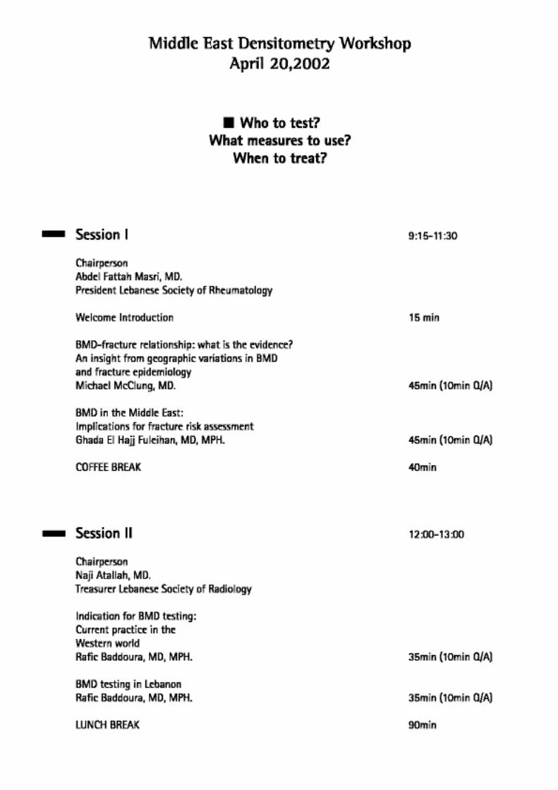
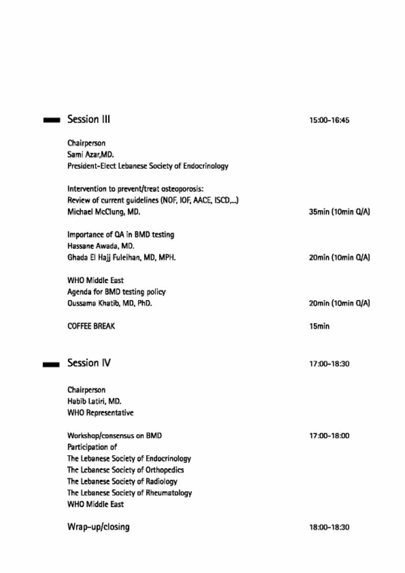
6
BMD-Fracture Relationship: What is the Evidence?An Insight from Geographic Variations in BMD and Fracture Epidemiology
Michael McClung, MD.
Low bone mineral density (BMD) is a risk factor for fractures of the spine and hip. However,
other major determinants of fracture risk include age, previous fractures, skeletal geometry, body
size and falls. The interplay of these various risk factors differs markedly between spine and hip
fractures and among different populations of people.
Spine fractures are the quintessential osteoporotic (low BMD) fractures. Across different
populations of older women, the relationship between absolute BMD values and vertebral fracture
risk appears to be very similar. (Data regarding these relationships in men are inadequate for
comparisons to be made.) This observation suggests that a common database, as opposed to
racial, regional or national databases, should be used to calculate T-scores, to estimate fracture
risk and to determine thresholds for treatment.
In contrast, substantial differences are observed between BMD and hip fracture risk among
different populations of women. Asian women have lower BMD than do Caucasian women but have
variably lower hip fracture rates. The hip fracture incidence among African American women is
lower than predicted by racial differences in BMD. This indicates that BMD alone is inadequate to
predict the risk of hip fracture across populations, and that other risk factors (especially hip
geometry, body size and physical fitness) must be included, along with BMD, in the clinical
estimate of hip fracture risk. It is probable that the specific contribution of low BMD to hip fracture
risk is similar among various populations, but that this risk is modified to very different extents by
the other determinants of risk. This means that BMD - no matter how it is expressed or how T-
scores are calculated - will never adequately account for population differences in hip fracture risk.
Rather than develop different racial or national BMD databases upon which T-scores are based,
population-specific information and data about the non-BMD risk factors must be gathered.
Models combining BMD with these other risk factors need to be developed to estimate hip fracture
risk. We will then move from the use of T-scores alone toward more appropriate estimates of
absolute fracture risk as thresholds for patient evaluation and especially for pharmacological
intervention.
8
Osteoporosis in the Middle East
Ghada El-Hajj Fuleihan, MD, MPH.
Osteoporosis is a major public health problem worldwide to-date. Although it is projected that
the magnitude of the problem may be even larger in the developing countries including those in the
Middle East, such projection is however based on several assumptions. This paper will evaluate the
above conclusion. In order to achieve that the following will be reviewed:
1- BMD in various groups/populations from the Middle East: Cross-sectional data.2- Prevalence of osteopenia and osteoporosis in our populations using the
WHO operational definition for osteoporosis3- Fracture prevalence data 4- BMD-fracture relationship: BMD in hip fracture patients
Peak BMD is a major determinant of BMD and therefore fractures in later life. Several studies
have demonstrated that young healthy subjects from the region have a BMD that is 3-10% lower than
the published Western norms, depending on the study, gender, skeletal site and type of densitometer.
Body size differences may account for some of these differences. These decrements in BMD seem to
be sustained at older ages, potentially inferring a higher fracture risk in the elderly. Using the WHO
operational definition of osteopenia/osteoporosis, the prevalence of osteopenia ranges between 37-
66% at the lumbar spine and 47-54% at the femoral neck in female subjects after age 50 years
depending on the study; whereas for osteoporosis these numbers are 28-33% for the spine and 6-18%
for the hip, respectively. The overall prevalence of spontaneous clinical fractures in women after age
50 years, is estimated to vary between 11-17%; a study of morphometric vertebral fractures estimates
a prevalence of 20-22% after age 60 years. Finally, mean BMD in hip fractures subjects is comparable
if not identical to mean BMD in hip fracture patients from the US/Europe, however they seem to occur
at a slightly younger age. Limited data on hip fracture incidence also suggests that it is comparable
to that from the Western world.
In conclusion, BMD in subjects from the region is slightly lower than that of US/European
counterparts when matched for age and gender. Although fracture estimates are comparable, this
seems to be possibly explained by their occurrence at a younger age. Therefore, the burden of
osteoporosis in the Middle East is comparable to the Western world, and can only be expected to
increase based on the steady increase in our aging population.
10
Bone densitometry is one of the most powerful clinical tools in the diagnostic evaluation of thepatient with or at high risk for osteoporosis, thus allowing the appropriate choice of therapeuticstrategies. As early as 1989, the National Osteoporosis Foundation Scientific Advisory Board has putforth its first set of Clinical Indications for BMD measurements. This was the result of earlyobservations that demonstrated the following: BMD predicts fractures, BMD can be measured safelyand accurately, clinical risk factors alone do not accurately predict BMD, information about BMDinfluences choice of therapy, treatment of osteoporosis affects fracture outcomes, and therefore BMDmeasurements should lead to fracture reduction. Since then, abundant information has come throughconfirming these points as summarized throughout this meeting. Furthermore, meta-analyses ofseveral large randomized controlled trials have shown since then that the further the BMDincrements, the lower is the risk of fractures. In this paper the currently published western guidelinesfor densitometry testing are reviewed.
The WHO1 : recommends BMD had in patients with radiographic evidence of osteopenia orvertebral deformity; loss of height or thoracic kyphosis; previous low trauma fracture; and thepresence of risk factors such as prolonged steroid therapy, hypogonadism in either sex,hyperthryoidism or hyperparathyroidism, low BMI < 19 kg/m2, low calcium intake.
The NOF2: recommends BMD to be measured in the following groups of women: women< age65 with one or more risk factors (family history, personal history of fractures, smoking, weight<57kgs); all women > 65 years; postmenopausal women with fractures; women considering therapy ifBMD influences the decision; women on HRT for prolonged periods.
The EFFO 3 now IOF: recommends BMD measurement in patients with radiographic evidence ofosteopenia or deformity, previous fragility fracture, significant height loss or kyphosis, presence ofstrong risk factors, and monitoring therapy.
The ISCD4 : recently came out with its recommendations as follow: all women >65 years, all men>70 years, anyone with a fragility fracture, anyone with a disease or condition associated withosteoporosis, anyone considering therapy for osteoporosis based on BMD, anyone on prolonged HRTtherapy.
The AACE5: in 1996 adopted the older NOF guidelines from 1989 and gave its revisedrecommendentions in 2001. BMD is recommended in peri- or postmenopausal women, in women withx-ray findings of osteoporosis, in women on long term glucocorticoid therapy, in peri- orpostmenopausal women with hyperparathyroidism. In women on osteoporosis treatment formonitoring response to therapy.
The principle behind all of the above commonly shared recommendations is that of a cost-effective case-finding strategy using risk factors that predict low BMD in order to identify the highrisk individuals in whom identification and treatment will result in a significant reduction in futurefractures, whereas the role of prevention in the younger perimenopausal women is not established.
1. Genant et al. Osteoporos Int 1999; 10:259-2. Anonymous. Osteoporos Int 1998;8(S4):1-883. Kanis et al. Osteoporos Int 1996; 6:256- 4. ISCD Bone Densitometry Forum 2001; JCD in press5. Hogson et al. Endocrinology Practice 2001;7(4): 293-312
Indication for BMD testing: Rationale from the Western World
Rafic Baddoura, MD, MPH.
12
INTRODUCTION: Bone mineral density (BMD) measurement using Dual Energy X-ray Absorptiometry(DEXA) is the core of osteoporosis diagnosis and management (1). Disease definition is based on DEXA-based BMD assessment as well as decision to treat (2). However technology costs and access are significantbarriers to screening policies for osteoporosis and justify the efforts to identify at high risk populations toachieve cost effective case identification (3). Published clinical decision rules addressing this issue have tobe challenged methodologically before gaining large adherence from health professionals and authorities(4).
OBJECTIVES : The aims are to characterize demographically and clinically referred population for BMDtesting and to evaluate BMD diagnostic performance by international standards.
MATERIAL AND METHOD: Our sample is driven from referred population for BMD testing in threehospital-based osteoporosis units during fall 2001. Subjects were submitted a questionnaire collectinginformation about age, gender and known risk factors for osteoporosis. Using W.H.O. definition ofosteoporosis we evaluated the diagnostic performance of clinical variables for case identification andcompared the actual performance with propose theoretical models.
RESULTS: Sample size is 722 with 95.0% females. Mean (SD) age is 58.5 (11.0) for females and 56.5(16.9) for males. The overall proportion of individuals >=65 years is 31.2% in females and 38.9% in males.Proportion of post-menopausal women is 83.4% (573/687 women) and mean (SD) age at menopause is46.6 (5.6). Prevalence of common osteoporosis risk factors is as follows:
Low weight defined as less than 70kg 57.3%Current smoking 34.6% Early menopause defined as less that 45 years of age 24.7% Personal history of fracture after a fall 21.3% Family history of osteoporotic fracture in first-degree relatives 18.9% Oral steroid therapy 10.9%
384 women are tested for diagnosis as compared to treatment evaluation among others and 306 (79.7%)were post-menopausal. Based on W.H.O definition, prevalence of osteoporosis in post-menopausal womenis 37.6%, 25.5% and 17.7% using spine, radius and hip BMD values respectively. Using hip BMD for casedefinition, osteoporosis is significantly associated with all common risk factors. Corresponding sensitivity(Se) and specificity (Sp) are the following:
Se. Sp.Age >=65 years
62.3 73.4Low weight defined as less than 70kg 77.1 91.5Current smoking Early menopause defined as less that 45 years of age 29.5 85.1 No prior estrogen use 98.4 98.0Personal/family (first-degree) history of osteoporotic fracture 50.8 89.1 Oral steroid therapy 18.0 85.4
Using NOF guidelines, the Osteoporosis Risk Assessment Instrument and the weight criterion, percentof post-menopausal women at high risk of osteoporosis in our target population would be 47.7%, 60.5%and 56.5% respectively.
CONCLUSION: Subject selection for osteoporosis case identification using DEXA may be improvedprovided common risk factors are taken along with consensus guidelines.
Main issues to be discussed.Who are those who benefit from DEXA bone density measurement?What is our diagnostic performance using DEXA? Where do we stand compared to NOF, IOF and literature recommendations?
BMD Testing In Lebanon: Sould We Move Our Target?
Rafic Baddoura, Ghada El Hajj Fuleihan, Nadia Salam, Hassane Awada, Marianna Salamoun, Jad Okais, Paul Rizk.
The objective of managing patients with or at risk for osteoporosis is to prevent orminimize the occurrence of new fractures. We can identify patients at high fracture risk onthe basis of their BMD, their history of previous fractures, their age and other importantdeterminants of fracture risk. Therapies exist to preserve or even increase bone density andto reduce the incidence of both vertebral and hip fractures. The main challenge today forclinicians is to determine in which patients our new therapies are most appropriate.
Although our current anti-remodeling drugs prevent bone loss in men andpostmenopausal women regardless of age or BMD, the only patients in whom fractureprotection has been documented are those at high current risk of fracture - those withprevious spine fractures or BMD values consistent with osteoporosis. The clinical utility andcost effectiveness of treating high-risk patients is clear. However, the rationale and need fortreating women at lower risk is much less certain. Treating young postmenopausal womenwho do not have osteoporosis for up to 6 years with estrogen or bisphosphonates preservesbone density but is not associated with a reduction in spine or hip fractures. These datademonstrate that pharmalogical therapy to prevent fractures in low-risk populations willrequire many years before fracture protection is afforded, and that the use of bone-specificagents to prevent osteoporosis is neither cost effective nor necessary.
Clinical guidelines for osteoporosis management in the United States have evolved overthe past 4 years in recognition of the evidence from clinical trials that our therapeuticresources should be aimed at individuals with recognized risk factors for fracture rather thanat preventing those risk factors. The various guidelines are consistent in recommending thatwomen with fragility vertebral fractures be treated. The NOF guidelines of 1998 suggestedtreating all women with BMD T-scores <-2 and other women with T-scores of less than -1.5and another risk factor. The AACE guidelines, published in 2000, proposed treating womenwith T-scores -2.5 or lower and those with values < -1.5 and other risk factors. Both the NOFand ACCE guidelines suggest treating all women with a fragility fracture. Most recently, theguidelines of the North American Menopause Society (Menopause, March 2002), recommendtreating women with T-scores of -2.5 or lower and those with other risk factors and low BMD(T-score lower than -2). The NAMS guidelines do not suggest treating all women with non-spine fragility fractures but rather that these women undergo evaluation including BMDtesting, and that those non-spine fractures be treated as a risk factor to modify the BMDthreshold at which treatment be considered. These most recent US guidelines have movedtoward the recommendations of the European osteoporosis community that has always hada more conservative approach toward treatment, restricting pharmacological therapy topatients known to have osteoporosis.
We now define osteoporosis as a risk factor for fracture - not as a patient who has had afracture. Consequently, treating patients to prevent osteoporosis would be analogous totreating patients with lipid-lowering drugs to prevent hyperlipidemia - a strategy that is notaccepted except in very high-risk patients.
Guidelines for osteoporosis management are moving toward risk-based interventionthresholds. This is similar to the recent guidelines for lipid-lowering therapy that recommendtherapy at different levels of serum cholesterol depending on the individual’s other riskfactors. Only is this way will our treatments be used most effectively and efficiently to reducethe burden of osteoporotic fractures.
Intervention to prevent/treat osteoporosis: Review of current guidelines
Michael McClung, MD.
Importance of Quality Assurance (QA) in BMD Testing
Hassane Awada, MD.Ghada El-Hajj Fuleihan, MD, MPH.
Bone densitometry technology has undergone fast changes over the last decade, withdual energy X-ray absorptiometry as the gold standard measure. In the clinical setting,densitometry is used in following situations:
1- The diagnostic evaluation of the patient with or at risk for osteoporosis2- In the monitoring of such patients
Densitometry in the diagnostic evaluation of a patient at risk or with ostoporosis.A key characteristic to be considered in the choice of a study in the management of
osteoporosis is its accuracy: how close is the measurement to what it is supposed tomeasure, (ie how close is measured BMC to actual BMC determined by ashing the bone)and how well does BMD predict fractures? Accuracy error has been measured on all devicesavailable and is better for DXA than it is for QCT, ranging between 4-10% depending onthe skeletal site. Because QUS does not really measure BMC, it is not possible to define theaccuracy of that technique. QA data from phantom measurements allows detection ofmachine drifts or shifts that affect accuracy. As to fracture prediction, DXA is undoubtedlythe most validated measure to-date. Indeed, BMD as measured by DXA predicts failureload, BMD is one of the strongest predictors of fractures, and as BMD increases fracturerisk decreases. For those reasons the WHO working group on osteoporosis proposed cut-offs based on DXA derived BMD T-scores to diagnose osteoporosis. These T-score cut-offsdo not apply to other densitometry technologies. However in order for DXA to fulfill itsfunction, strict adherence to manufacturerís guidelines in scan acquisition and analysis areof utmost importance. Furthermore, adherence to FDA approved densitometers will in greatpart insure excellent accuracy.
Densitometry in the monitoring of the patient at risk for or with osteoporosis Precision (reproducibility) is the most important characteristic to consider in the choice
of a technique to monitor a patientís progress. Precision of a measure reflects its ability toreproduce the same result on repeat (duplicate) measurements, in the event of absence ofa real biological change, provided the test is conducted in an identical situation. Indeed,any changes exceeding the precision error by a certain magnitude, otherwise called leastsignificant change (LSC, see below) are likely to reflect true biological changes. Sources ofprecision error include the machine itself, but more importantly changes in technicians,inconsistencies in patient positioning, choice of ROI, scan mode, and scan analysis, as wellas artifacts and some biological variations. The precision of the instrument should beassessed continuously in each center, and derived from situations identical to the clinicalsituation in which the test is performed: on osteopenic/or osteoporotic patients ideallymeasured few days apart. It should not be based on phantom measurements. This allowsthe assessment of significance of changes in an individual over time.
The 95% CI for the LSC=2.77X precision error =2.77X RMS SD. Time interval at which a LSC can be detected = LSC/expected change/year.
Strict adherence to quality assurance measures is critical to the sound use ofdensitometry. Failure to do so will undermine the validity of the densitometry study andtherefore the quality of clinical care delivered.
Agenda for BMD Testing Policy
Dr Oussama Khatib, MD-PhD.
With the advent of quantitative techniques to measure bone mineral content a little more
than decade ago, our ability to quantify changes in bone mass and assess osteoporosis has
markedly improved. The relation between bone density and fracture is important, and an
estimate of bone mineral density can provide an effective assessment of fracture risk. It is
thus possible to choose a value for bone mineral density that defines the presence of
osteoporosis. According to World Health Organization (WHO) criteria, two diagnostic
threshold of bone den sity for Caucasian women, which are based on distribution of skeletal
mass in young healthy individuals.
• Normal: A value of bone mineral within 1 SD of young adult reference mean (t-score>-
1.0) is considered normal.
• Low bone mass (Osteopenia): A value of bone mineral more than 1SD below the young
adult mean but less than 2.5 SD below this value (-1.0>t-score(-2.5). Low bone mass is
visualized as a risk factor for fracture.
• The term osteoporosis is used to designate bone mass value more than 2.5 standard
deviations (SD) below the young adult mean (t-score(-2.5).
• Severe (established) osteoporosis: A value for bone mineral 2.5 SD or more below the
young adult in the presence of one or more fragility fractures.
Suitable diagnostic cut-off values for non-Caucasian women and for men are less secure.
It has been suggested that a similar absolute value for bone mineral density to that used in
women can be taken as a cut-off point for the diagnosis of osteoporosis in men, that is, a
value 2.5 SD below the average for adult pre-menopausal women. It is necessary to be
recognized that cut-off values are arbitrary and may differ according to sites measured, race,
and type of equipment . Because of this difficulty in accurate measurement and
standardization between instruments and sites, controversy still exists among experts
regarding the use of the above-mentioned criterion in non-DXA devices. Similarly, it is to be
stressed that the use of locally generated databases is not-validated and should be avoided.