Embed Size (px)
Citation preview

Edward H. ANGLE SOCIETY OFORTHODONTISTS MIDWEST COMPONENT
ADMISSIONS PROCEDURE MANUALRevised January 2016
2
Dear Doctor,
We are pleased to learn that you have been invited to attend the next annual meeting of the Midwest Component of the Edward H. Angle Society of Orthodontists. Your attendance at this meeting offers you the opportunity to embark upon admission into the Society.
The path to membership is rigorous but rewarding. The purpose of the Society is to provide a setting for leaders in the field to engage in dialogue and to challenge its members to actively participate and grow intellectually as their orthodontic careers progress.
The Angle Society encourages and maintains a balance between clinical orthodontics and research. To this end, clinical skills are measured during the admission process, but a research project of your choosing is also required. While these requirements may appear daunting on their surface, Society membership is such that you will be aided in your endeavors throughout the entire process. Each and every Angle Society member you will encounter has gone through the same protocol and you may be sure that they will be willing to help at stations along the way.
The orthodontic cases you offer for evaluation will be examined by the Society’s Admissions Committee in an effort not only to gain an understanding of your clinical background, but to initiate a dialogue between you and the Society. You should find the exchange of treatment ideas a valuable learning experience. As you progress through the admission process, you will be encouraged to interact with members of the Society regarding the progress of your cases.
The research project in which you will engage is intended to add to the scientific base of knowledge available to orthodontic clinicians and academicians. The Angle Society’s Study Committee is charged with the responsibility of assisting you in bringing worthy ideas before the membership. Your sponsor will guide you through the process as the Study Committee offers advice regarding topic selection, research design, data collection and analysis. This culminates in the presentation of your research project before members of the Society at one of its annual meetings. Every effort will be made to ensure that your admission process into the Edward H. Angle Society of Orthodontists is a rewarding experience and an enjoyable challenge.
Chip Rigsbee , D.D.S., M.S. Chair, Admissions Committee
Introduction
3
The Guest / The Candidate / Initial Meeting 4
The Second Meeting 5
The Third Meeting / The Fourth Meeting 6
The Fifth Meeting 7
Special At-Large Membership 8
Summary of Requirements for Admissions 9
Appendix 1: Synopsis of Case Reports 10
Appendix 2: Case Report: Initial Records 11
Appendix 3: Case Report: Progress Records 15
Appendix 4: Case Report: Final Evaluation 18
Diagnostic Study Records / Pre-treatment and Progress 21
Diagnostic Study Records / Post-treatment 22
Dental Casts 23
Dental Casts 24
Photographs 26
Appendix 5: Radiographic Records 28
Cephalometric Summary 30
Composite Tracings 31
Cephalometric Tracing 32
Cranofacial Composite / Maxilla Composite / Mandibular Composite 33
Appendix 6: Case Selection 34
Appendix 7: Admissions Commitee 35
Study Commitee 36
Appendix 8: Example of Permission Form for Patients 37
Summary
4
Edward H. Angle Society of Orthodontists Midwest Component
Your invitation to attend the next annual session of the Angle Society affords you the choice of attending either as a guest or a candidate.
Should you decide to attend as a guest, you have the privilege of participating in all scientific sessions and social events. Guests are free to do nothing more than observe and contemplate the advantages of Society membership. The Midwest Component of the Angle Society is reputed to be an active, participating group. You will have ample opportunity to decide whether this is for you. We hope it is. You may attend only one annual session as a guest.
Should you decide to attend as a candidate for membership, you share the same privileges as a guest, but become directly involved in the Admissions process at the onset. In either case, whether a guest or a candidate, you must accept your sponsor’s invitation at least sixty days prior to the start of the annual session. Please notify your sponsor of your intent, otherwise you will not be properly listed in the annual meeting directory.
You may attend only one meeting as a guest. A guest has no requirements and is invited to attend all scientific sessions and social functions. The next meeting that you attend would be as a candidate.
Initial Meeting
As a candidate, you are required to display pre-treatment and post-treatment diagnostic records for five cases you have treated. These cases must have been completed, i.e., placed in retention, no more than five (5) years prior to the opening date of the annual session you are attending. It is assumed that you have been the sole provider of orthodontic treatment for these patients, but if you share treatment supervision with another operator, it is your duty to inform the Chairperson of the Admissions Committee of this fact.
As a candidate, a full-time faculty member can present cases that had treatment provided by residents under his/her supervision. However, during the Affiliate process, only full-time faculty with no intra or extramural practice opportunity and who are Diplomates of the American Board of Orthodontics may present patient records of supervised cases.
The Admissions Committee requires that your study records and diagnostic/treatment reports adhere to the guidelines layout in Appendix 1 and 2. All paper, photographic, and radiographic records must be placed in page protectors and submitted in one three-ring binder per case. Pre and post-treatment cephalometric tracings, including superimpositions, are required. Digital models will be accepted for candidate cases only. They are not acceptable for cases utilized in the admissions process.
The Candidate
The Guest
Admissions Protocol
You may present cases previously submitted to the ABO, but the five year time limit mentioned above applies here as well.
The quality of the cases provided for review is intended to be indicative of your acumens and treatment skills. Bring only your very best work! Treatment results compromised by the difficulty of a case, while understandable, are not acceptable for admission into the Society. Likewise, cases which would reasonably be judged by your peers as relatively unchallenging will not be accepted. The Admissions Committee is looking for cases of reasonable difficulty that have been finished to the best of your ability. See Appendix 6 for case selection. You must display at least two cases requiring extraction of permanent teeth. It is expected that occlusion on all cases has been idealized from second molar to second molar. This is an opportunity to demonstrate to the membership your level of competence.
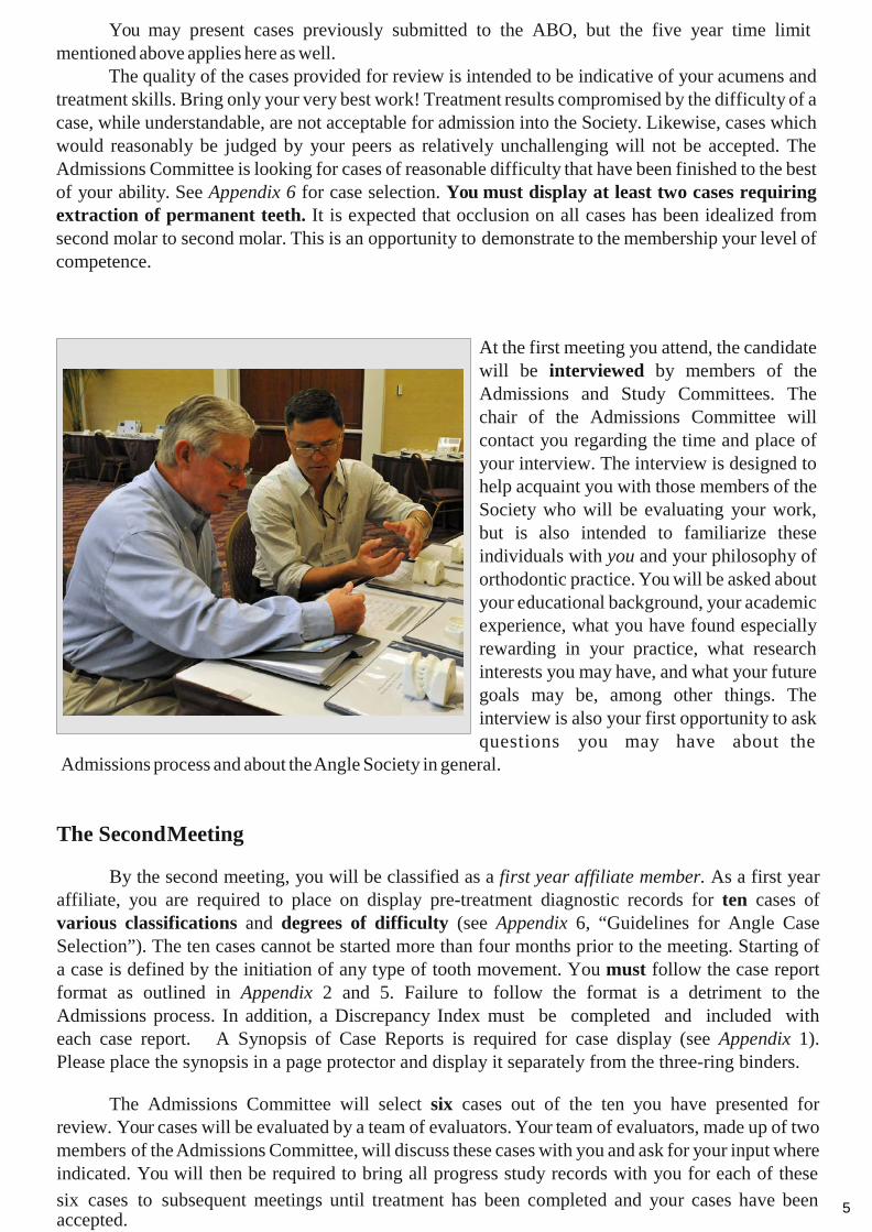
At the first meeting you attend, the candidate will be interviewed by members of the Admissions and Study Committees. The chair of the Admissions Committee will contact you regarding the time and place of your interview. The interview is designed to help acquaint you with those members of the Society who will be evaluating your work, but is also intended to familiarize these individuals with you and your philosophy of orthodontic practice. You will be asked about your educational background, your academic experience, what you have found especially rewarding in your practice, what research interests you may have, and what your future goals may be, among other things. The interview is also your first opportunity to ask questions you may have about the
Admissions process and about the Angle Society in general.
The Second Meeting
By the second meeting, you will be classified as a first year affiliate member. As a first year affiliate, you are required to place on display pre-treatment diagnostic records for ten cases of various classifications and degrees of difficulty (see Appendix 6, “Guidelines for Angle Case Selection”). The ten cases cannot be started more than four months prior to the meeting. Starting of a case is defined by the initiation of any type of tooth movement. You must follow the case report format as outlined in Appendix 2 and 5. Failure to follow the format is a detriment to the Admissions process. In addition, a Discrepancy Index must be completed and included with each case report. A Synopsis of Case Reports is required for case display (see Appendix 1). Please place the synopsis in a page protector and display it separately from the three-ring binders.
The Admissions Committee will select six cases out of the ten you have presented for review. Your cases will be evaluated by a team of evaluators. Your team of evaluators, made up of two members of the Admissions Committee, will discuss these cases with you and ask for your input where indicated. You will then be required to bring all progress study records with you for each of these six cases to subsequent meetings until treatment has been completed and your cases have been 5accepted.
6
The ten cases that you display must follow the Guidelines for Case Selection found in Appendix 6. Approval from the Admissions Committee Chairperson is required for any substitution.
At the second meeting, you must submit evidence of completion of (or application for) Part II of the American Board of Orthodontics. Finally, you will meet with the Study Committee regarding ideas you may have been contemplating for your research project. The Study Committee will help you evaluate your proposal to make sure it will meet the criteria for a paper which is acceptable for membership. The committee will also help with ideas for a project, should this need arise. It is strongly encouraged that you establish a timeline for your research presentation. Without a personal timeline, you will be unable to present your paper as a third year prospective affiliate which is expected.
The Third Meeting
By the time of the third meeting, you will be a second year affiliate member. At this time, you are required to present progress study records on each of your six cases. Please refer to Appendix 3 and 5 for a sample of the required progress report since that format differs from the initial report format. These records may not be recorded more than two months prior to the starting date of the third meeting. Again, you are required to obtain records on all ten cases, but submit for review only the six cases selected at the previous meeting.
You are further required to submit to the Study Committee a brief written summary of an anticipated protocol for your research project. Ameeting with the Study Committee in this regard will be arranged for you.
The Fourth Meeting
At this meeting, you have become classified a third year affiliate member. You are required to present post-treatment study records for those cases which have been placed into retention and all progress study records for those cases not yet completed. Please refer to Appendix 4 and 5 for samples of required final reports since the format differs from the initial and progress reports. A Cast- Radiograph Evaluation should also be included in the final report. Should it become necessary to substitute one of your alternate cases for an originally selected case, prior approval must be obtained from the Admissions Committee Chairperson.
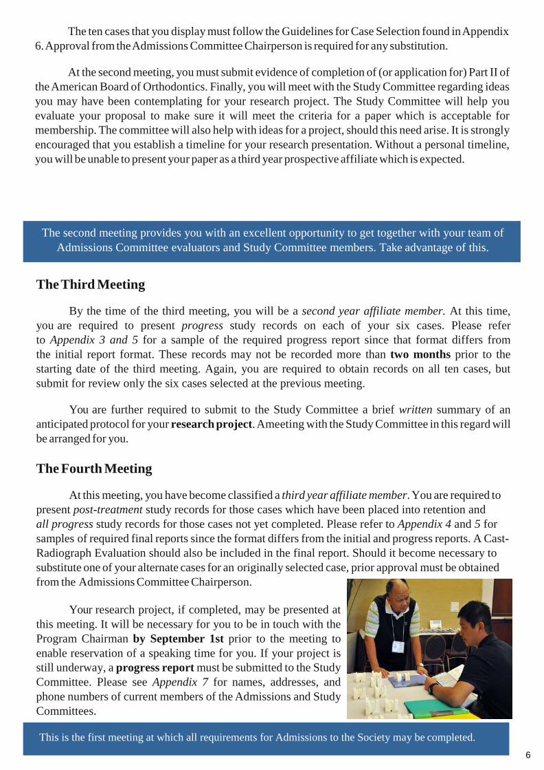
Your research project, if completed, may be presented at this meeting. It will be necessary for you to be in touch with the Program Chairman by September 1st prior to the meeting to enable reservation of a speaking time for you. If your project is still underway, a progress report must be submitted to the Study Committee. Please see Appendix 7 for names, addresses, and phone numbers of current members of the Admissions and Study Committees.
The second meeting provides you with an excellent opportunity to get together with your team of Admissions Committee evaluators and Study Committee members. Take advantage of this.
This is the first meeting at which all requirements for Admissions to the Society may be completed.

7
The Fifth Meeting
At this meeting, you become a fourth year affiliate member. You are required to present for review all six cases (including those already accepted) until all cases are completed.
Your research project should be finished by now and a paper prepared for presentation before the general membership. It will be necessary for you to be in touch with the Program Chairman by September 1st prior to the meeting to enable reservation of a speaking time for you. Your research project must be completed no later than one year after all cases have been completed.
8
Edward H. Angle Society of Orthodontists Midwest Component
Purpose: To create a membership classification that will enable the members of the Midwest Component of the Edward H. Angle Society of Orthodontists to invite select individuals to membership who previously have made outstanding contributions to our speciality and who have received their orthodontic education outside the United States and Canada. They should possess outstanding credentials, including excellence in clinical orthodontics, and should be able to contribute substantially to the intellectual environment of our Society.
Criteria: Guidelines for selection may include, but are not limited to the following:
1. The candidate must have completed an accredited full-time graduate program or residencyprogram in the speciality of orthodontics of at least two years in duration. The suitability of thecandidate’s education will be determined by the Board of Directors of the Midwest Component.2. The candidate should be affiliated with a teaching or research institituion or have equivalentexperience (e.g., lecturing at national or international meetings).3. The candidate should have shown evidence of a history of scholarly activity that may includepublication in a respected journal (e.g., Angle Orthodontist, American Journal of Orthodonticsand Dentofacial Orthopedics, European Journal of Orthodontics) or equivalent.
Sponsorship: The candidate’s sponsor should have known the candidate for a period of time, be fairly certain of his or her character, and be able to vouch for the candidate’s ability to communicate in English. The candidate should assure the sponsor of his or her ability and willingness to attend all annual meetings of the Angle Society during the admissions process as well as during regular meetings.
Procedure: The sponsor will provide by June 1st prior to the next annual meeting, the following documents:
1. Aletter of sponsorship.2. The curriculum vitae of the candidate.3. An additional letter or recommendation from a well-known orthodontist from the candidate’scountry or region.4. Verification of the candidate’s education (including a copy of an official certificate ofcompletion of an accredited orthodontic graduate program).
The preceeding material should be sent to the chairpersons of the Study and Admissions Committees. They will sit on a Special At-Large Membership Committee with two other members selected by the Board of Directors. If this committee feels the candidate merits consideration, they will send their recommendation to the Board. The Board then will make the final decision as to the candidacy of the individual.
The prospective Special At-Large candidate must attend one annual meeting as a guest. UNDER NO CIRCUMSTANCES should the sponsor bring the individual to our meeting as a candidate until his or her special status has been approved by the Board of Directors.
Special At-Large Membership
9
A summary of requirements for Admissions into the Midwest Component of the Edward H. Angle Society of Orthodontists is as follows:
First Meeting: Prepare and display five treated cases taken from your private practice which have been completed in the last five years prior to the meeting date. Two of these cases must have had extraction therapy requiring space closure mechanics. This is required of candidates not guests.
Second Meeting: Meet with the Admissions and Study Committees. Submit evidence of completion of or application for Part II of the ABO. Bring ten pre-treatment cases (that meet the Case Selection Criteria found in Appendix 2) of which six will be selected for treatment.
Third Meeting: Bring initial records, progress records and Progress Case Reports for the six selected cases. Submit a written description of your anticipated research project to the Study Committee.
Fourth Meeting: Bring all initial, progress and post-treatment study records for all of your six selected cases. Present your research paper or provide a written summary of progress to the Study Committee.
Fifth Meeting: Bring all initial, progress and post-treatment study records for all of your six selected cases. Present your research paper before the general membership if not done so at the previous meeting.
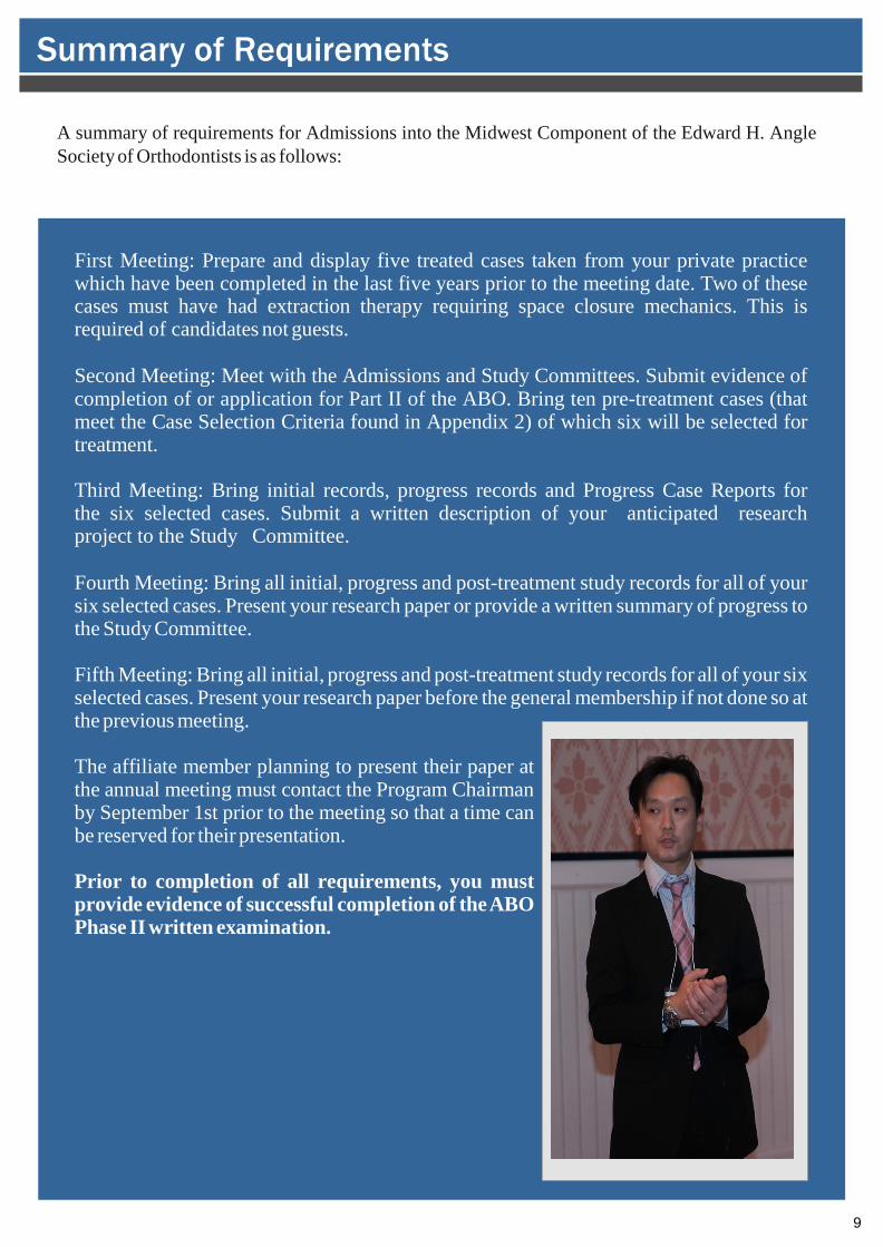
The affiliate member planning to present their paper at the annual meeting must contact the Program Chairman by September 1st prior to the meeting so that a time can be reserved for their presentation.
Prior to completion of all requirements, you must provide evidence of successful completion of the ABO Phase II written examination.
Summary of Requirements
10
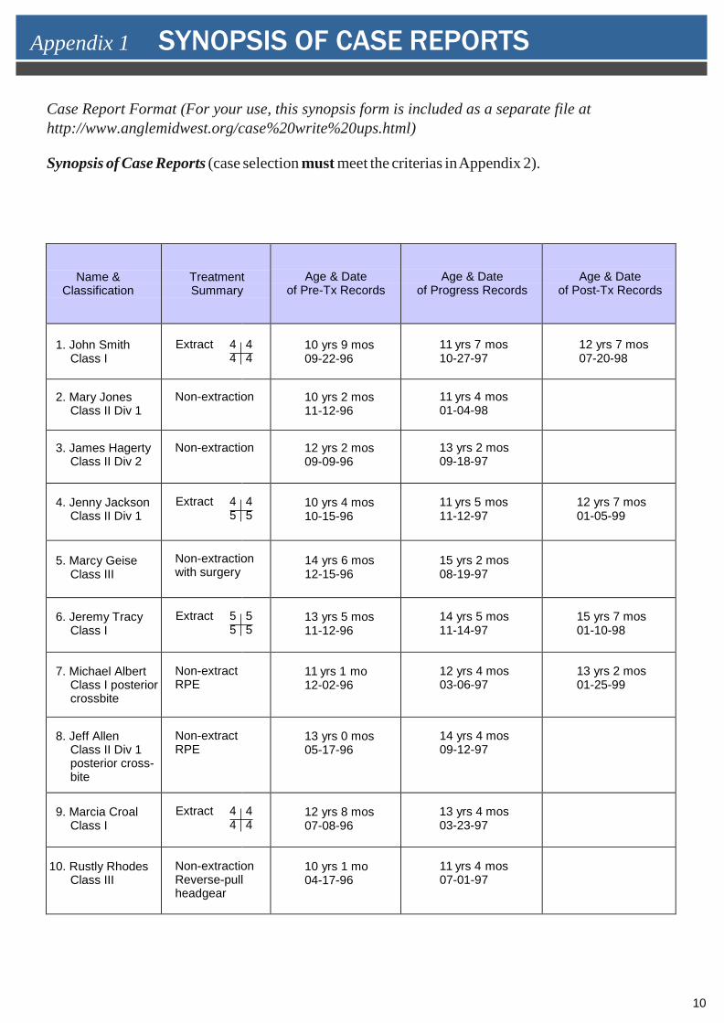
Case Report Format (For your use, this synopsis form is included as a separate file at http://www.anglemidwest.org/case%20write%20ups.html)
Synopsis of Case Reports (case selection must meet the criterias in Appendix 2).
Name & Classification
Treatment Summary
Age & Date of Pre-Tx Records
Age & Date of Progress Records
Age & Date of Post-Tx Records
1. John SmithClass I
Extract 44
44
10 yrs 9 mos 09-22-96
11 yrs 7 mos 10-27-97
12 yrs 7 mos 07-20-98
2. Mary JonesClass II Div 1
Non-extraction 10 yrs 2 mos 11-12-96
11 yrs 4 mos 01-04-98
3. James HagertyClass II Div 2
Non-extraction 12 yrs 2 mos 09-09-96
13 yrs 2 mos 09-18-97
4. Jenny JacksonClass II Div 1
Extract 45
45
10 yrs 4 mos 10-15-96
11 yrs 5 mos 11-12-97
12 yrs 7 mos 01-05-99
5. Marcy GeiseClass III
Non-extraction with surgery
14 yrs 6 mos 12-15-96
15 yrs 2 mos 08-19-97
6. Jeremy TracyClass I
Extract 55
55
13 yrs 5 mos 11-12-96
14 yrs 5 mos 11-14-97
15 yrs 7 mos 01-10-98
7. Michael AlbertClass I posteriorcrossbite
Non-extract RPE
11 yrs 1 mo 12-02-96
12 yrs 4 mos 03-06-97
13 yrs 2 mos 01-25-99
8. Jeff AllenClass II Div 1posterior cross-bite
Non-extract RPE
13 yrs 0 mos 05-17-96
14 yrs 4 mos 09-12-97
9. Marcia CroalClass I
Extract 44
44
12 yrs 8 mos 07-08-96
13 yrs 4 mos 03-23-97
10. Rustly RhodesClass III
Non-extraction Reverse-pull headgear
10 yrs 1 mo 04-17-96
11 yrs 4 mos 07-01-97
Appendix 1 SYNOPSIS OF CASE REPORTS
11
Edward H. Angle Society
Midwest Component
Case Report
Presented by:
Appendix 2 THE CASE REPORT
12
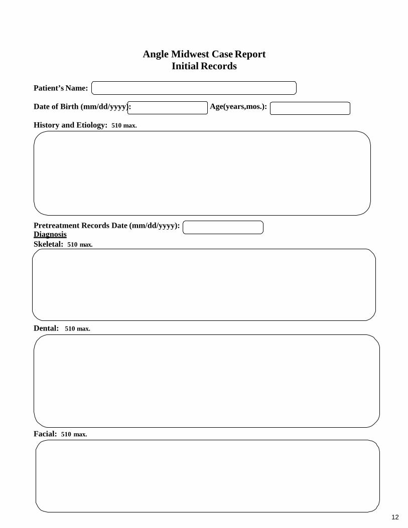
Angle Midwest Case Report Initial Records
Patient’s Name:
Date of Birth (mm/dd/yyyy):
History and Etiology: 510 max.
Pretreatment Records Date (mm/dd/yyyy): Diagnosis Skeletal: 510 max.
Age(years,mos.):
Dental: 510 max.
Facial: 510 max.
13
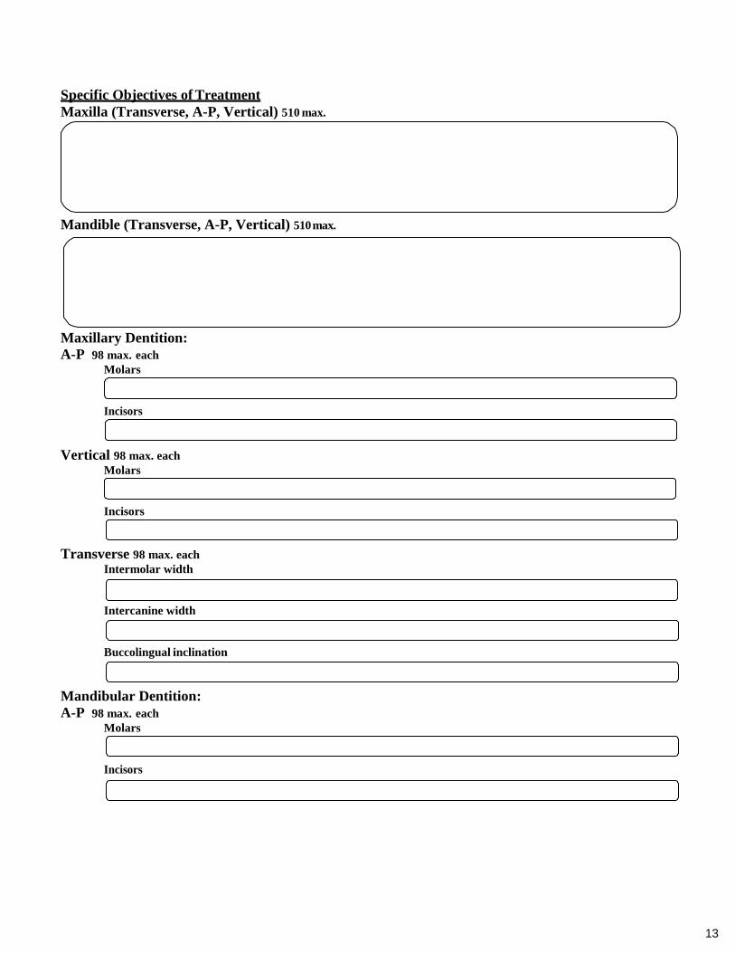
Specific Objectives of Treatment Maxilla (Transverse, A-P, Vertical) 510 max.
Mandible (Transverse, A-P, Vertical) 510 max.
Maxillary Dentition: A-P 98 max. each
Molars
Incisors
Vertical 98 max. each Molars
Incisors
Transverse 98 max. each Intermolar width
Intercanine width
Buccolingual inclination
Mandibular Dentition: A-P 98 max. each
Molars
Incisors
14
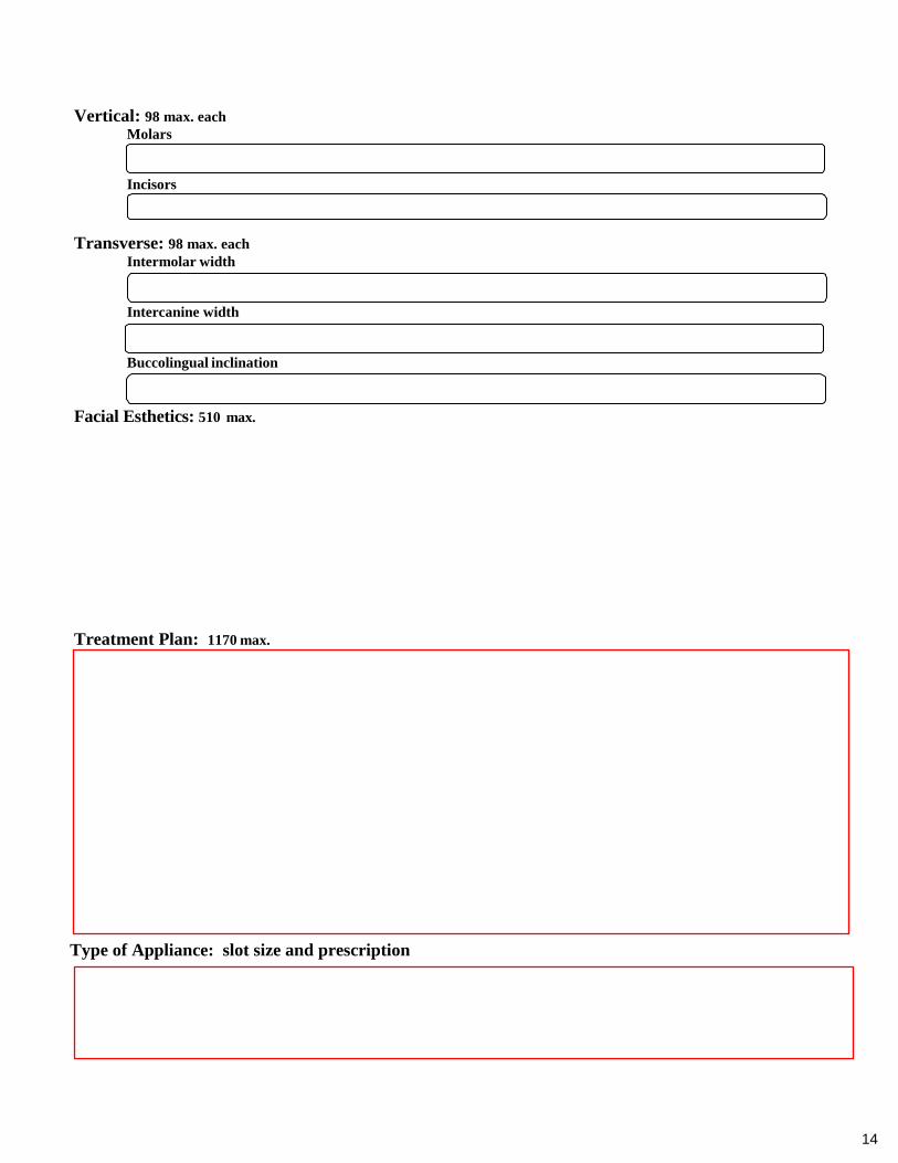
Vertical: 98 max. each Molars
Incisors
Transverse: 98 max. each Intermolar width
Intercanine width
Buccolingual inclination
Facial Esthetics: 510 max.
Treatment Plan: 1170 max.
Type of Appliance: slot size and prescription
15
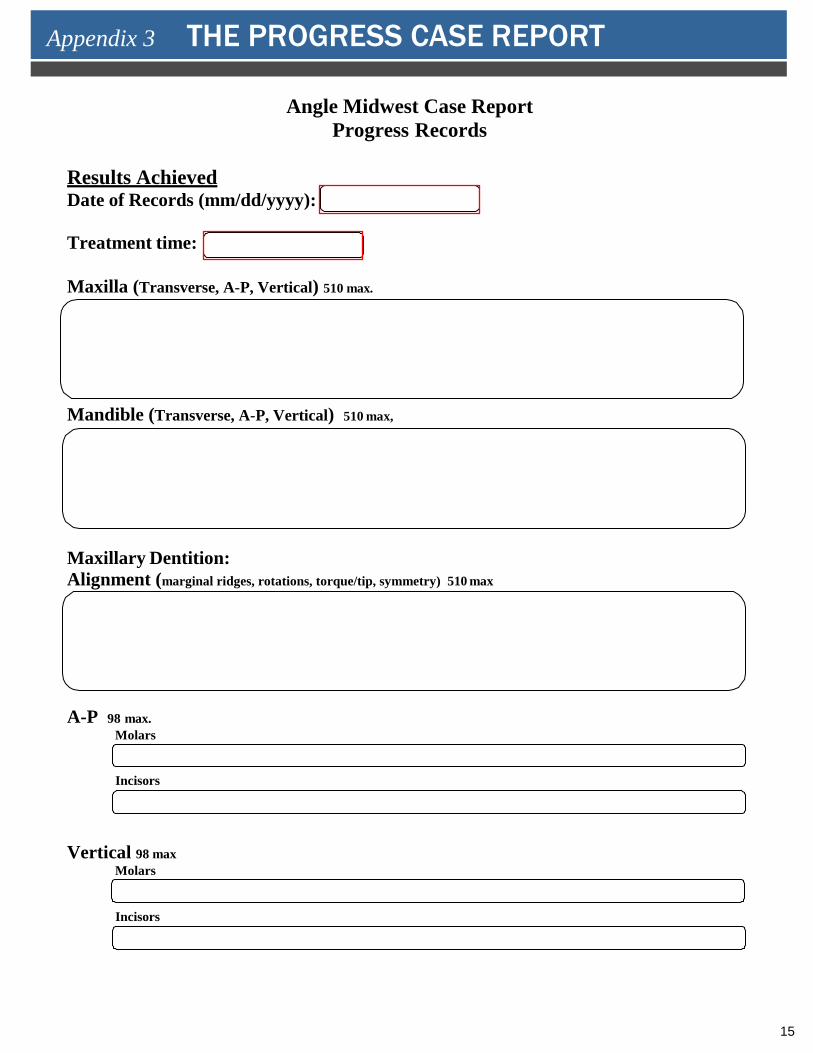
Angle Midwest Case Report Progress Records
Results Achieved Date of Records (mm/dd/yyyy):
Treatment time:
Maxilla (Transverse, A-P, Vertical) 510 max.
Mandible (Transverse, A-P, Vertical) 510 max,
Maxillary Dentition: Alignment (marginal ridges, rotations, torque/tip, symmetry) 510 max
A-P 98 max.Molars
Incisors
Vertical 98 maxMolars
Incisors
Appendix 3 THE PROGRESS CASE REPORT
16
Transverse 98 max.Intermolar width
Intercanine width
Buccolingual inclination
Mandibular Dentition: Alignment (marginal ridges, rotations, torque/tip, symmetry) 510 max
A-P 98 max.Molars
Incisors
Vertical 98 maxMolars
Incisors
Transverse 98 max.Intermolar width
Intercanine width
Buccolingual inclination
Facial Esthetics 510 max.
17
Future Treatment Concerns and Summary 1170 max.
18
max
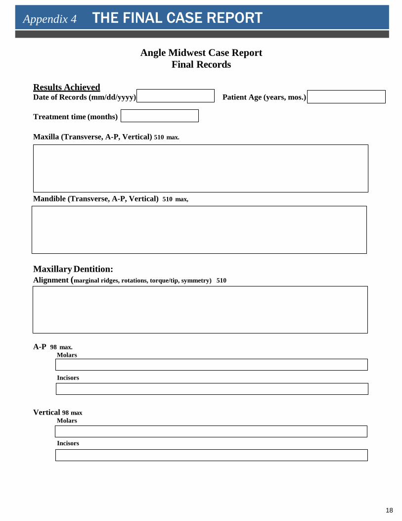
Angle Midwest Case Report Final Records
Results Achieved Date of Records (mm/dd/yyyy) Patient Age (years, mos.)
Treatment time (months)
Maxilla (Transverse, A-P, Vertical) 510 max.
Mandible (Transverse, A-P, Vertical) 510 max,
Maxillary Dentition: Alignment (marginal ridges, rotations, torque/tip, symmetry) 510
A-P 98 max.Molars
Incisors
Vertical 98 max Molars
Incisors
Appendix 4 THE FINAL CASE REPORT
19
max
Transverse 98 max. Intermolar width
Intercanine width
Buccolingual inclination
Mandibular Dentition: Alignment (marginal ridges, rotations, torque/tip, symmetry) 510 max.
A-P 98 max.Molars
Incisors
Vertical 98 max Molars
Incisors
Transverse 98 max. Intermolar width
Intercanine width
Buccolingual inclination
Facial Esthetics 510 max.
20
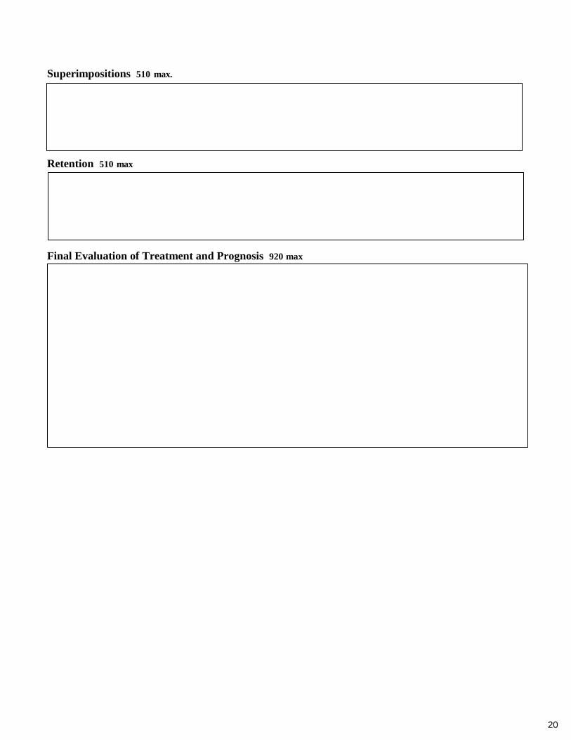
Superimpositions 510 max.
Retention 510 max
Final Evaluation of Treatment and Prognosis 920 max
21
The importance of quality study records cannot be overestimated. By implication, they set the standard for the level of treatment to follow. The following records are required for your cases:
PRE-TREATMENT RECORDS
• Dental Casts
• FMX or panoramic radiographs• Cephalometric radiograph
• Cephalometric tracing
• Facial photographs: profile, frontal with lips in repose, and frontal smiling
• Intra-oral photographs: frontal, right lateral, left lateral, maxillary occlusal, mandibularocclusal
• Summary of cephalometric measurements
• Discrepancy Index
PROGRESS RECORDS
• Dental Casts
• FMX or panoramic radiographs
• Cephalometric radiograph
• Cephalometric tracing along with serial composite superimpositions
• Facial photographs: profile, frontal with lips in repose, and frontal smiling
• Intra-oral photographs: frontal, right lateral, left lateral, maxillary occlusal, mandibularocclusal
• Summary of cephalometric measurements
DIAGNOSTIC STUDY RECORDS
22
POST-TREATMENT RECORDS
• Dental Casts• FMX or panoramic adiographs
• Cephalometric radiograph
• Cephalometric tracing
• Facial photographs: profile, frontal with lips in repose, and frontal smiling
• Intra-oral photographs: frontal, right lateral, left lateral, maxillary occlusal,mandibular occlusal
• Summary of cephalometric measurements
• Cast Radiograph Evaluation
DIAGNOSTIC STUDY RECORDS
23
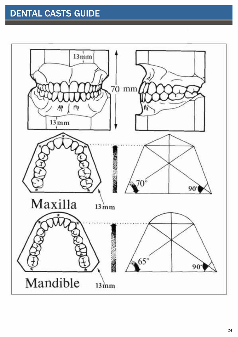
Cast Trimming Requirements
Impressions should extend far enough into the sulcus to allow accurate reproduction of all soft tissue anatomy in the dental casts. Impressions are to include the most distal tooth in each quadrant with an adequate replication of the tuberosity. The casts must be trimmed so that when placed in occlu- sion and resting on their backs on the table top, they replicate the patient’s occlusion with condyles seated in the fossae. Page 19 illustrates trimming requirement. If your dental casts are mounted on adjustable articulators, they must be converted to hand-held models. In addition, to the hand- held models, you may also display your original mounted casts if so desired. It is strongly encouraged that magnetic mounting plates be utilized.
When trimming the posterior portion of the maxillary cast, be careful to preserve the tuberosity when possible, and certainly the second molars. Likewise, preserve a reasonable portion of the retromolar area when trimming the mandibular cast. Partially trimmed off second molars is not acceptable. Trimming or carving on the anatomical portion of the dental casts should be limited to the removal of bubbles and other defects. During cast finishing, take care to avoid obliteration of soft and hard tissue anatomy.
All diagnostic records are to be of standards that would be satisfactory for presentation to the American Board of Orthodontics. 1. All models, photos, x-rays and cephalometric tracings are to be labeled with the followinginformation:
a. Affilate nameb. Case numberc. Date Records were takend. Age of Patiente. Colored dot for quick reference: initial (black), progress (blue) or final (red) recordsf. To be in compliance with HIPPA standards, all displayed case reports must have a signed copy
of a permission form in the pocket at the back of the patient binder. An example of a permission form is available in Appendix 8.
SUPLEMENTAL INFORMATION FOR CASE REPORTS
DENTAL CASTS
24
DENTAL CASTS GUIDE
25
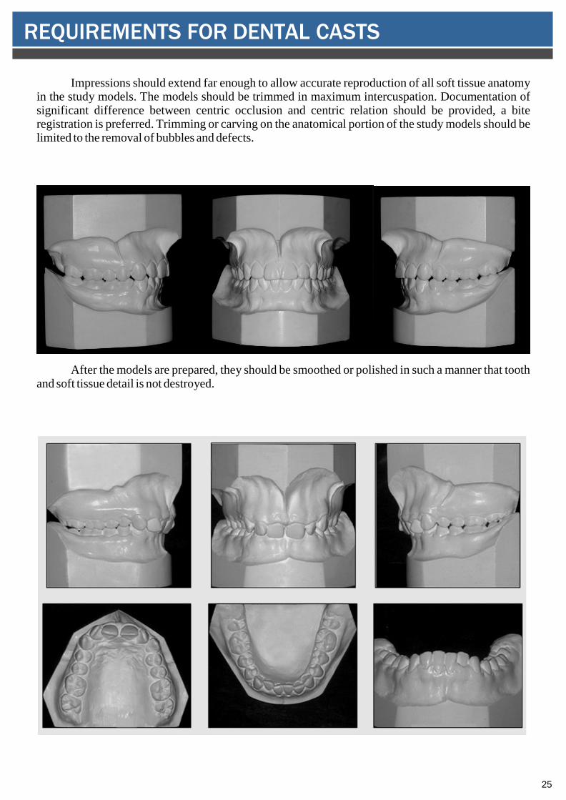
Impressions should extend far enough to allow accurate reproduction of all soft tissue anatomy in the study models. The models should be trimmed in maximum intercuspation. Documentation of significant difference between centric occlusion and centric relation should be provided, a bite registration is preferred. Trimming or carving on the anatomical portion of the study models should be limited to the removal of bubbles and defects.
After the models are prepared, they should be smoothed or polished in such a manner that tooth and soft tissue detail is not destroyed.
REQUIREMENTS FOR DENTAL CASTS
26
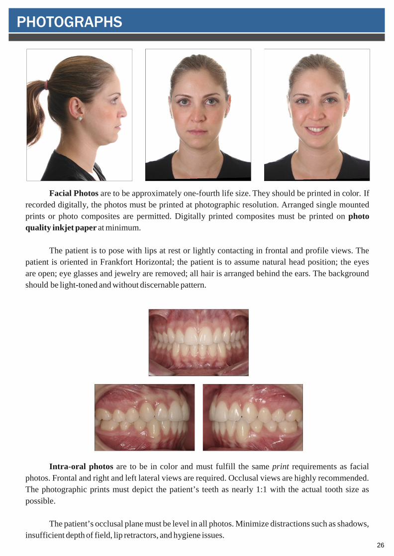
Facial Photos are to be approximately one-fourth life size. They should be printed in color. If recorded digitally, the photos must be printed at photographic resolution. Arranged single mounted prints or photo composites are permitted. Digitally printed composites must be printed on photo quality inkjet paper at minimum.
The patient is to pose with lips at rest or lightly contacting in frontal and profile views. The patient is oriented in Frankfort Horizontal; the patient is to assume natural head position; the eyes are open; eye glasses and jewelry are removed; all hair is arranged behind the ears. The background should be light-toned and without discernable pattern.
Intra-oral photos are to be in color and must fulfill the same print requirements as facial photos. Frontal and right and left lateral views are required. Occlusal views are highly recommended. The photographic prints must depict the patient’s teeth as nearly 1:1 with the actual tooth size as possible.
The patient’s occlusal plane must be level in all photos. Minimize distractions such as shadows, insufficient depth of field, lip retractors, and hygiene issues.
PHOTOGRAPHS
27
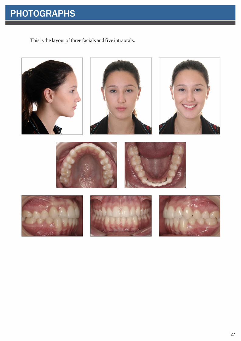
This is the layout of three facials and five intraorals.
PHOTOGRAPHS
28
Full Mouth or Panoramic Radiographs
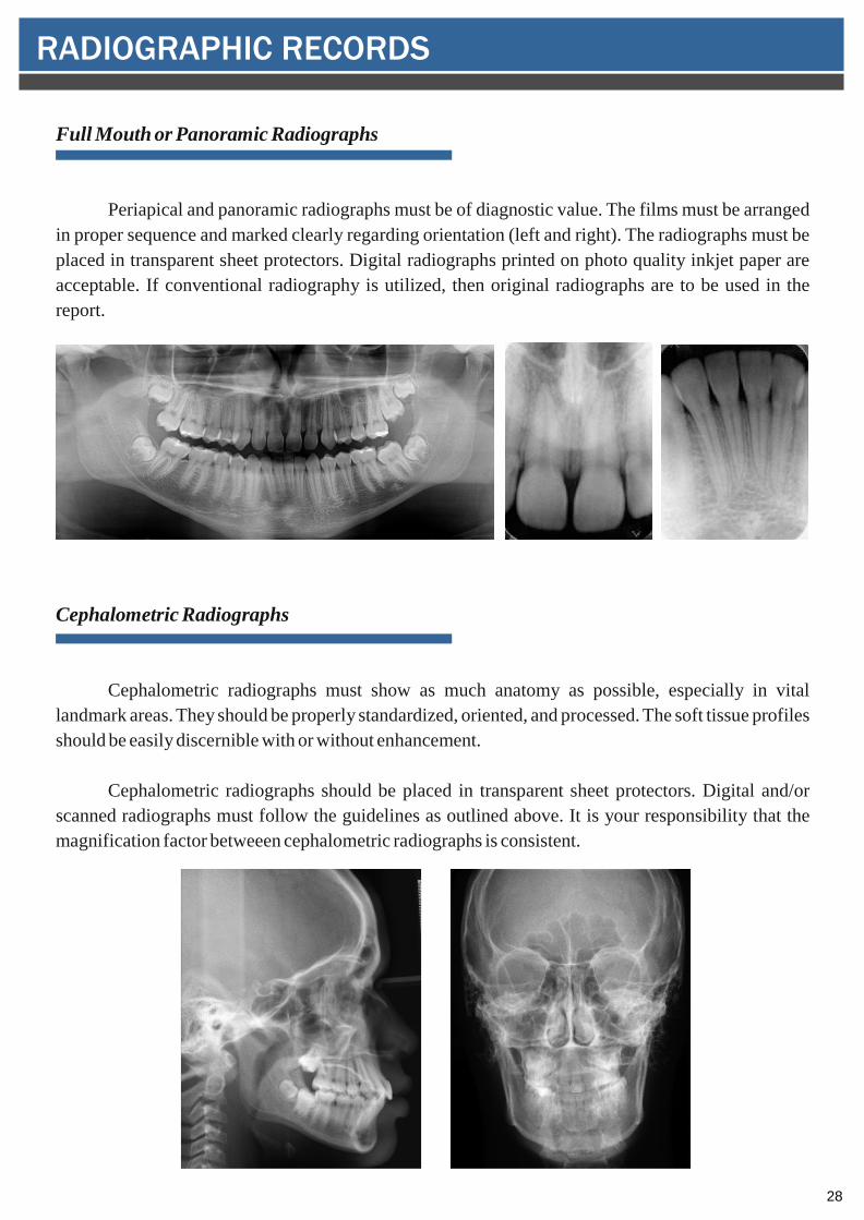
Periapical and panoramic radiographs must be of diagnostic value. The films must be arranged in proper sequence and marked clearly regarding orientation (left and right). The radiographs must be placed in transparent sheet protectors. Digital radiographs printed on photo quality inkjet paper are acceptable. If conventional radiography is utilized, then original radiographs are to be used in the report.
Cephalometric Radiographs
Cephalometric radiographs must show as much anatomy as possible, especially in vital landmark areas. They should be properly standardized, oriented, and processed. The soft tissue profiles should be easily discernible with or without enhancement.
Cephalometric radiographs should be placed in transparent sheet protectors. Digital and/or scanned radiographs must follow the guidelines as outlined above. It is your responsibility that the magnification factor betweeen cephalometric radiographs is consistent.
RADIOGRAPHIC RECORDS
29
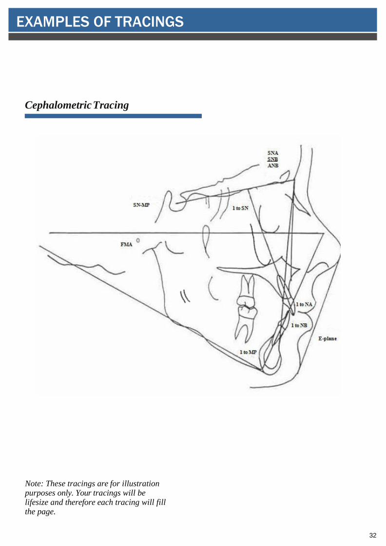
Cephalometric Tracings
Cephalometric radiographs may be manually and accurately traced on acetate film by the candidate/affiliate. Computer generated tracings are also acceptable. Templates may be used for tracing of teeth. Do not record measurements on the cephalometric acetate tracing. All measurements used must be recorded on the Cephalometric Summary sheet (Appendix 5). Record Frankfort Horizontal from anatomic porion. Soft tissue outlines must be included on the tracings. The tracings should be placed in transparent sheet protectors and displayed in the pocket at the back of the patient binder. Pre-treatment tracings (black), progress tracings (blue), and post- treatment tracings (red).
Besides all the mesurements that are on the cephalometric summary sheet (Appendix 5), you may also use your customary landmarks, lines, and measurements provided valid standards are available. Any additional cephalometric measurements that are in addition to those on the Cephalometric Summary Sheet must be recorded on a supplemental sheet.
RADIOGRAPHIC RECORDS
30
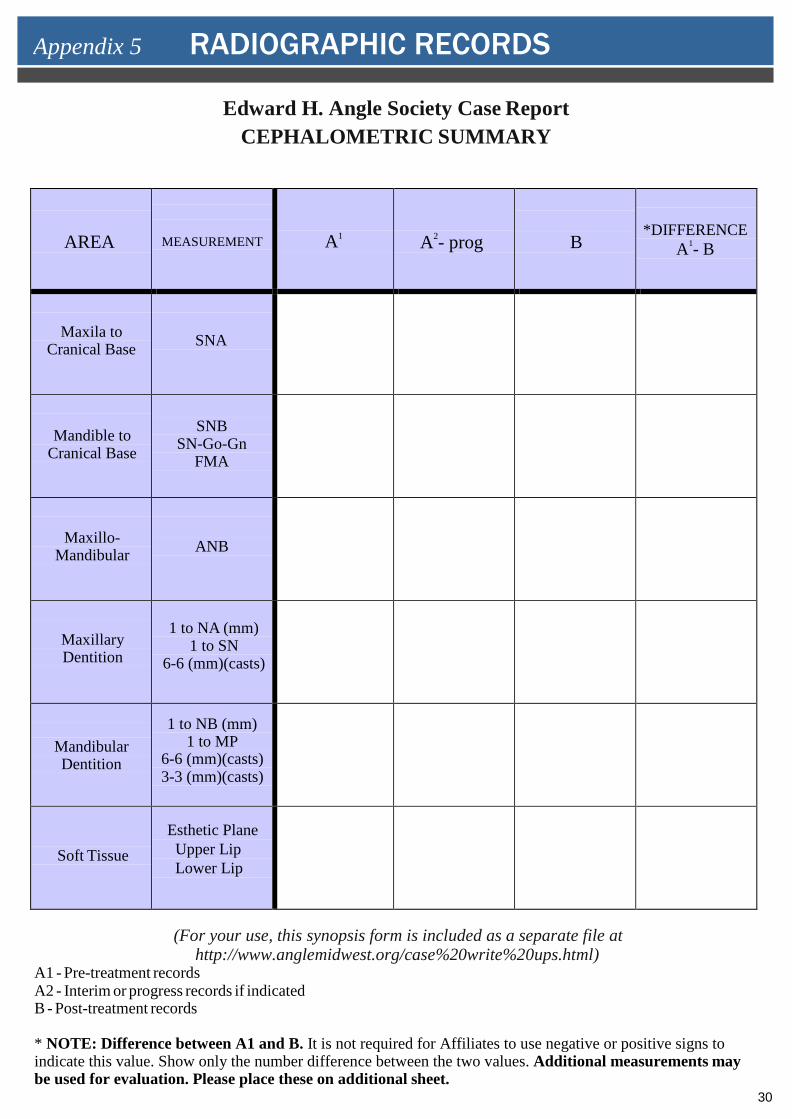
Edward H. Angle Society Case Report CEPHALOMETRIC SUMMARY
AREA MEASUREMENT A1 A2- prog B *DIFFERENCE
A1- B
Maxila to Cranical Base SNA
Mandible to Cranical Base
SNB SN-Go-Gn
FMA
Maxillo- Mandibular ANB
Maxillary Dentition
1 to NA (mm) 1 to SN
6-6 (mm)(casts)
Mandibular Dentition
1 to NB (mm) 1 to MP
6-6 (mm)(casts)3-3 (mm)(casts)
Soft Tissue Esthetic Plane Upper Lip Lower Lip
(For your use, this synopsis form is included as a separate file at http://www.anglemidwest.org/case%20write%20ups.html)
A1 - Pre-treatment records A2 - Interim or progress records if indicated B - Post-treatment records
* NOTE: Difference between A1 and B. It is not required for Affiliates to use negative or positive signs toindicate this value. Show only the number difference between the two values. Additional measurements maybe used for evaluation. Please place these on additional sheet.
Appendix 5 RADIOGRAPHIC RECORDS
31
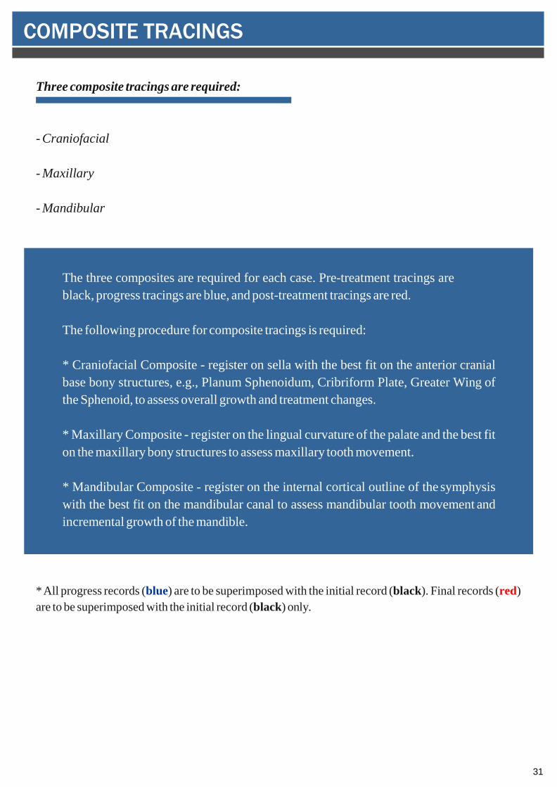
Three composite tracings are required:
- Craniofacial
- Maxillary
- Mandibular
The three composites are required for each case. Pre-treatment tracings are black, progress tracings are blue, and post-treatment tracings are red.
The following procedure for composite tracings is required:
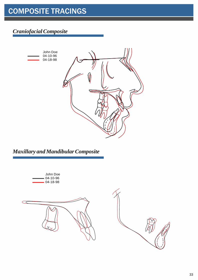
* Craniofacial Composite - register on sella with the best fit on the anterior cranialbase bony structures, e.g., Planum Sphenoidum, Cribriform Plate, Greater Wing ofthe Sphenoid, to assess overall growth and treatment changes.
* Maxillary Composite - register on the lingual curvature of the palate and the best fiton the maxillary bony structures to assess maxillary tooth movement.
* Mandibular Composite - register on the internal cortical outline of the symphysiswith the best fit on the mandibular canal to assess mandibular tooth movement andincremental growth of the mandible.
* All progress records (blue) are to be superimposed with the initial record (black). Final records (red)are to be superimposed with the initial record (black) only.
COMPOSITE TRACINGS
32
Cephalometric Tracing
Note: These tracings are for illustration purposes only. Your tracings will be lifesize and therefore each tracing will fill the page.
EXAMPLES OF TRACINGS
33
Craniofacial Composite
Maxillary and Mandibular Composite
John Doe 04-10-9604-18-98
John Doe 04-10-9604-18-98
COMPOSITE TRACINGS
34
The clinical requirement of the Admissions process requires the prospective affiliate to bring a total of 10 cases with complete clinical diagnostic records. These cases permit the Admissions Committee to evaluate clinical proficiency in a variety of areas. These cases should demonstrate differing malocclusion types. These must include cases from the following areas:
1. Two cases that require the removal of maxillary and mandibular permanent teeth for resolution of the problem that require space closure biomechanics.
2. Two cases that present with a significant antero/posterior discrepancy: an ANB a) 6 degrees or greater, or b) –1 degrees or more negative (more Class III).
3. Two cases that require clinical skills to manage vertical changes: SN-MP of 36 degrees or greater.
4. Of the 10 cases, the following limitations apply:
a. No more than two cases being treated in a two-phase treatment modality. b. No more than one case requiring orthognathic surgery. c. No more than one case being treated with TMJ splint therapy.
If there are any questions regarding these requirements, please contact the Chairperson of the
Admissions Committee. To identify the Chairperson refer to Appendix 7 in the back of your Admissions Procedure Manual or contact your sponsor. Good luck and thank you!
Appendix 6 GUIDELINES FOR ANGLE CASE SELECTION
35
Edward H. Angle Society of Orthodontists Midwest Component
Chip Rigsbee, Chair (2017) 13590B N. Meridian, Suite 205 Carmel, IN 46032 317-574-0612317-574-0614 [email protected]
Ken Eberle (2016) 3909 Arctic Boulevard #202 Anchorage, AK 99503 907-344-8383907-345-4971 [email protected]
Steve Sherman (2018) 8311 Bluebonnet Boulevard Baton Rouge, LA 70810 225-769-1276225-769-3751 [email protected]
Michael Frazier (2019) 1225 Park Way Drive Zionsville, IN 46077 317 873-3399 317 873-3497 fax [email protected] Roberto Hernandez-Orsini (2020) Camino Alejandro #4, Vila Clementina Guaynabo, Puerto Rico 00969 787-731-8424787-790-1859 [email protected]
Anthony Puntillo (2021) 1549 S. Court Street #A Crown Point, IN 46307 [email protected]
Appendix 7 ADMISSION COMMITEE MEMBERSHIP 2013
36
Bradley Pearson (2022) 7450 France Avenue S #270 Edino, MN 55435 952-926-2551952-926-6516 [email protected]
Jay Whitley (2023) 541 Shadows Lane #A Baton Rouge, LA 70806-6555 225-924-4383225-924-4364 [email protected]
STUDY COMMITTEE MEMBERSHIP 2012-2017
Jim Hartsfield (2016) 800 Rose Street, Room D416 Lexington, KY 40536-0297 859-323-0296859-257-8878 [email protected]
Eustaquio Araujo (2017) 3320 Rutger Street St. Louis, MO 63104 [email protected]
Sarandeep Huja (2018) 800 Rose Street, Room D-406 Lexington, KY 40536-0297 859-323-5371859-257-8878 [email protected]
Pamela Hanson (2019) 15855 West National Avenue New Berlin, WI 53151 [email protected]
37
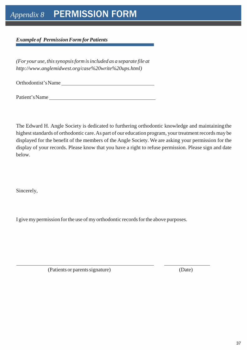
Example of Permission Form for Patients
(For your use, this synopsis form is included as a separate file at http://www.anglemidwest.org/case%20write%20ups.html)
Orthodontist’s Name
Patient’s Name
The Edward H. Angle Society is dedicated to furthering orthodontic knowledge and maintaining the highest standards of orthodontic care. As part of our education program, your treatment records may be displayed for the benefit of the members of the Angle Society. We are asking your permission for the display of your records. Please know that you have a right to refuse permission. Please sign and date below.
Sincerely,
I give my permission for the use of my orthodontic records for the above purposes.
(Patients or parents signature) (Date)
Appendix 8 PERMISSION FORM