Embed Size (px)
Citation preview

河川のアユ病魚から分離されたEdwardsiella ictaluriの性状
誌名誌名 魚病研究
ISSNISSN 0388788X
著者著者
永井, 崇裕岩本, 恵美坂井, 貴光有馬, 多恵子天社, こずえ飯田, 悦左飯田, 貴次中井, 敏博
巻/号巻/号 43巻4号
掲載ページ掲載ページ p. 158-163
発行年月発行年月 2008年12月
農林水産省 農林水産技術会議事務局筑波産学連携支援センターTsukuba Business-Academia Cooperation Support Center, Agriculture, Forestry and Fisheries Research CouncilSecretariat

魚病研究 Fish Pathology, 43 (4),158-163,2008.12 。2008The Japanese Society of Fish Pathology
Characterization of Edwardsiella ictaluri Isolated from
Wild Ayu Plecoglossus altivelis in Japan
Takahiro Nagai1, 2*, Emi Iwamoto2, Takamitsu Sakai3, TaelくoArima4,
Kozue Tensha5, Yoshisuke lida¥ Takaji lida3 and Toshihiro Nakai2
1円sheriesand Marine Technology Center, Hiroshima Prefectural Technology Rθsearch Institute, Kure, Hiroshima 737-1207, Japan
2 GraduatθSchool of Biosphere Sciθnce, Hiroshima University, Higashi“Hiroshima, 739・8528,Japan
3National Research Institute of Aquaculture, Fisheries Research Agθncy, Minami-Ise, Mie 516・0193,Japan
4γ"okyo Metropolitan Islands Area Research and Development Center of Agriculture,
Forestry and Fisheries, Minato, Tokyo 105・・0022,Japan51nland Sea Division, Yamaguchi Prefectural Fisheries Research Center,
Aio欄 Fu臼shima,Yamaguchi 754開 0893,Japan
(Received August 30, 2008)
ABSTRACT -A Gram-negative bacterium was isolated from diseased wild ayu Plecoglossus
altivelis, which were caught in rivers in Japan from August to October in 2007. AII four isolates
examined showed the same morphological, physiological and biochemical characteristics and were
c1assified into the genus Edwardsiella. The isolates were differentiated from E. tarda in respect of
its negative production of indole and no growth at 370C, and different from a reference strain of E.
ictaluri (JCM1680) in its positive production of hydrogen sulfide. AIi isolates were completely iderト
tical in the pa吋ialnucleotide sequences of 16S rDNA, a type 1 fimbrial gene (etfA) and a heat shock
protein gene (dnaみ andthese sequences showed high similarity (100%,99.7% and 100%, respec-
tively) with E. ictaluri but low similarity (99.7%, 92.5% and 87.3%, respectively) with E.
tarda. 8ased on these phenotypic and genetic characteristics, the present isolates from ayu were
identified as E. ictaluri.
Key words: Edwardsiella ictaluri, characterization, Plecoglossus altivelis, ayu
The genus Edwardsiella includes two fish patho幽
gens, E. tarda and E. ictaluri, both of which have caused
serious disease problems in fish culture industries. E.
tarda (εwing et al., 1965), which was earlier called
Paracolobactrum anguillimortiferum (Hoshina, 1962;
Wakabayashi and Egusa, 1973; Sakazaki and Tamura,
1975), is the causative agent of edwardsiellosis in many
cultured freshwater and marine fish species worldwide
(Plumb, 1999; Muroga, 2001). In Japan, edwardsieliosis was first reported in cultured Japanese eel Anguilla
japonica (Wakabayashi and Egusa, 1973), and then the
disease has caused a serious disease problem not only
in cultured eel but also in cultured marine fishes, particLト
larly Japanese flounder Paralichthys olivaceus and red
認 Correspondingauthor らmail:[email protected]
sea bream pagrus major (Kusudaθt al., 1977;
Yasunaga et al., 1982; Nakatsugawa, 1983). E. ictaluri
(Hawke et al., 1981) is the causative agent of enteric
septicemia of catfish (ESC), the most important disease
for economic loss in cultured channel catfish Ictalurus
punctatus in the United States (Plumb, 1999). E.
ictaluri was also isolated from some cultured freshwater
catfish species in Thailand (Kasornchandra et al., 1987),
Vietnam (Crumlish et al., 2002) and Indonesia (Yuasa et
al., 2003). However, there have been no reports on
isolation of the bacterium in Japan
In 2007, a Granトnegativebacterium was isolated
from diseased wild ayu Plecoglossus altivelis, which
were caught in rivers of three locations, Tokyo Metropo圃
lis, Yamaguchi Prefecture and Hiroshima Prefecture,
Japan (Sakai et al., 2008), and the disease was diag-
nosed as E. icおluriinfection. This paper describes the
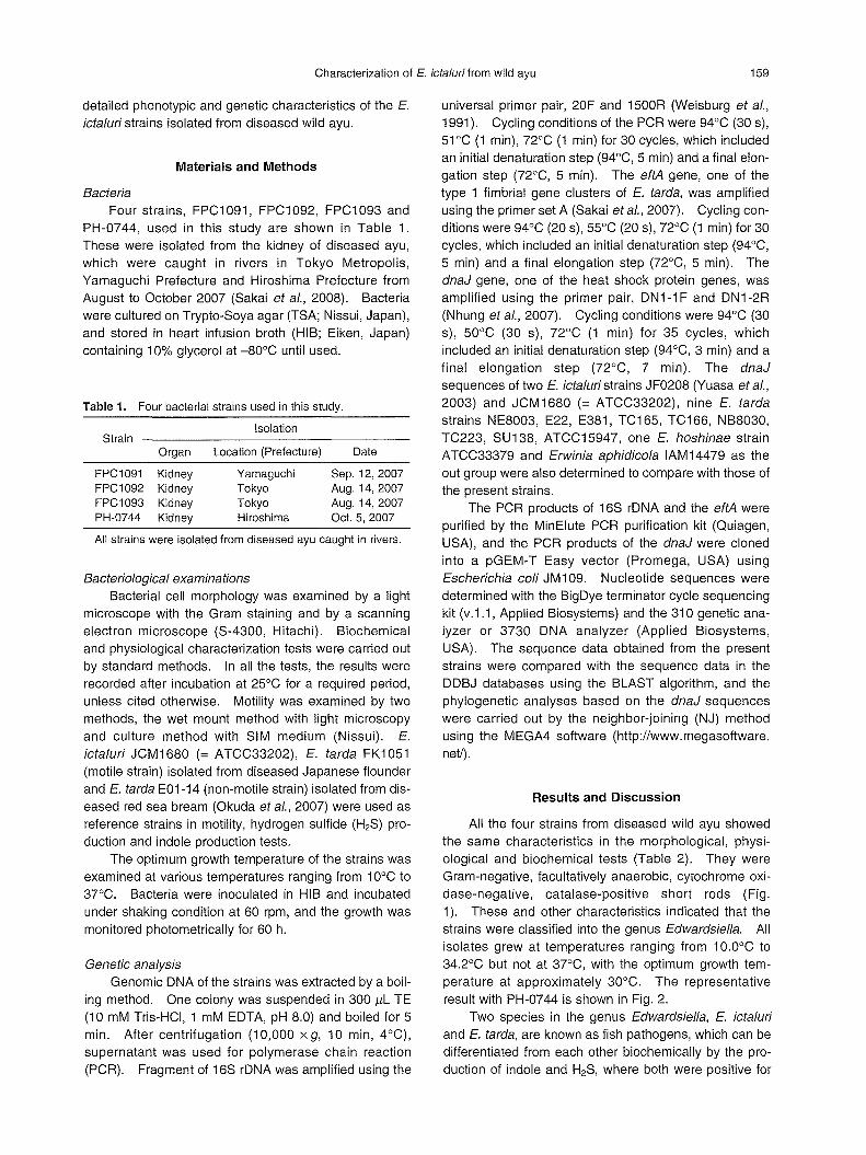
Characterization 01 E. iclaluri Irom wild ayu 159
detailed phenotypic and genetic characteristics of the E.
ictaluri strains isolated from diseased wild ayu.
Materials and Methods
Bacteria
Four strains, FPC1 091, FPC1092, FPC1093 and
PH-0744, used in this study are shown in Table 1.
These were isolated from the kidney of diseased ayu,
which were caught in rivers in Tokyo Metropolis,
Yamaguchi Prefecture and Hiroshima Prefecture from
August to October 2007 (8akai et al., 2008). Bacteria
were cultured on Trypto-80ya agar (T8A; Nissui, Japan),
and stored in heart infusion broth (HIB; Eiken, Japan)
containing 10% glycerol at -80oC until used.
Table 1. Four bacterial strains used in this study.
Isolation Strain
Organ Location (Prelecture) Date
FPC1091 Kidney FPC1092 Kidney FPC1093 Kidney PH-0744 Kidney
Yamaguchi Tokyo Tokyo Hiroshima
Sep.12,2007 Aug.14,2007 Aug.14,2007 Oct. 5, 2007
AII strains were isolated Irom diseased ayu caught in rivers.
Bacteriological examinations
Bacterial cell morphology was examined by a light
microscope with the Gram staining and by a scanning
electron microscope (8-4300, Hitachi). Biochemical
and physiological characterization tests were carried out
by standard methods. In all the tests, the results were
recorded after incubation at 250C for a required period,
unless cited otherwise. Motility was examined by two
methods, the wet mount method with light microscopy
and culture method with 81M medium (Nissui). E.
ictaluri JCM 1680 (= ATCC33202),εtarda FK1051
(motile strain) isolated from diseased Japanese flounder
and E. tarda E01イ4(norトmotilestrain) isolated from dis幽
eased red sea bream (Okuda et al., 2007) were used as
reference strains in motility, hydrogen sulfide (H28) pro-
duction and indole production tests.
The optimum growth temperature of the strains was
examined at various temperatures ranging from 100C to
370C. Bacteria were inoculated in HIB and incubated
under shaking condition at 60 rpm, and the growth was
monitored photometrically for 60 h.
Genetic anaケ"Sis
Genomic DNA of the strains was extracted by a boil-
ing method. One colony was suspended in 300μL TE
(10 mM Tris-HCI, 1 mM EDTA, pH 8.0) and boiled for 5
min. After centrifugation (10,000 xg, 10 min, 40C),
supernatant was used for polymerase chain reaction
(PCR). Fragment of 168 rDNA was amplified using the
universal primer pair, 20F and 1500R (Weisburg et al.,
1991). Cycling conditions of the PCR were 940C (30 s),
510C (1 min), 720C (1 min) for 30 cycles, which included
an initial denaturation step (940C, 5 min) and a final elon-
gation step (720C, 5 min). The eftA gene, one of the
type 1 fimbrial gene clusters of E. tarda, was amplified
using the primer set A (8akai et al., 2007). Cycling con-
ditions were 940C (20 s), 550C (20 s), 720C (1 min) for 30
cycles, which included an initial denaturation step (940C,
5 min) and a final elongation step (720C, 5 min). The
dnaJ gene, one of the heat shock protein genes, was
amplified using the primer pair, DN1-1 F and DN1闘 2R
(Nhung et al., 2007). Cycling conditions were 940C (30
s), 500C (30 s), 720C (1 min) for 35 cycles, which
included an initial denaturation step (940C, 3 min) and a
final elongation step (720C, 7 min). The dnaJ
sequences of two E. ictaluri strains JF0208 (Yuasa et al.,
2003) and JCM1680 (= ATCC33202), nine E. tarda
strains NE8003, E22, E381, TC165, TC166, NB8030,
TC223, 8U138, ATCC15947, one E. hoshinae strain
ATCC33379 and Erwinia aphidicola IAM14479 as the
out group were also determined to compare with those of
the present strains.
The PCR products of 168 rDNA and the eftA were
purified by the MinElute PCR purification kit (Quiagen,
U8A), and the PCR products of the dnaJ were cloned
into a pG巨M-TEasy vector (Promega, U8A) using
Escherichia coli JM1 09. Nucleotide sequences were
determined with the BigDye terminator cycle sequencing
kit (v.1.1, Applied Biosystems) and the 310 genetic ana-
Iyzer or 3730 DNA analyzer (Applied Biosystems,
U8A). The sequence data obtained from the present
strains were compared with the sequence data in the
DDBJ databases using the BLA8T algorithm, and the
phylogenetic analyses based on the dnaJ sequences
were carried out by the neighbor-joining (NJ) method
using the MEGA4 software (http://www.megasoftware.
ne的
Results and Discussion
AII the four strains from diseased wild ayu showed
the same characteristics in the morphological, physi-
ological and biochemical tests (Table 2). They were
Gram-negative, facultatively anaerobic, cytochrome oxi-
daseィlegative,catalase-positive short rods (Fig・
1). These and other characteristics indicated that the
strains were classified into the genus Edwardsiella. AII
isolates grew at temperatures ranging from 10.0oC to
34.20C but not at 370C, with the optimum growth tem胴
perature at approximately 30oC. The representative
result with PH闘 0744is shown in Fig. 2.
Two species in the genus Edwardsiella, E. ictaluri
and εtarda, are known as fish pathogens, which can be
differentiated from each other biochemically by the pro帽
duction of indole and H28, where both were positive for

160 T. Nagai, E. Iwamoto, T. Sakai, T. Arima, K. Tensha, Y. lida, T. lida and T. Nakai
Table 2. Morphological, physiological and biochemical characteristics of the present strains,
Edwardsiella ictaluri and εtarda.
Characteristic Present strains E. ic伺lun*l E. tarda判
Gram stain Form Short rod Short rod Short rod Motility at 250C +場2 + + Growth at 3rC + + NaCI tolerance
1.0% + + + 1.5% + + + 4.0% +
Cytochrome oxidase Catalase + + + OFtest F F F
H2S + *3 + Indole + Methyl red test + + Voges-Proskauer test Citrate *4 第5 *5
Arginine dihydrolase Lysine decarboxylase + + + Ornithine decarboxylase + + + Gas from glucose + + + Acid production from
D-mannos巴 + + + Maltose + + + D-mannitol Sucrose
Trehalose L-arabinos巴
判 Plumb(1999) partly modified. 説 2Weak motility was observed in SIM medium at 250C, but not by the wet mount method
under light microscopy 持3Weak H2S production was observed in SIM medium at 250C 判 Simmon's
*5 Christensen's
、元一"
v
-V -Fig. 1. Gram-negative short rod (PH-0744 strain) isolated from diseased wild ayu. (A) Light microscopy (Gram staining), (8) Scan-
ning electron microscopy
E. tarda and both negative for E. ictaluri (Plumb,
1999). E. tarda grows well at 370C, whereas E. ictaluri
shows poor or no growth at 3JOC (Plumb, 1999). E.
ictaluri is biochemically less active than other
Edwardsiella species, and it appears to be homoge-
neous (Waltman et al., 1986). The characteristics of
the present strains were clearly different from those of E.
tarda in points of negative production of indole and no
growth at 370C, and were different from εictaluri in its
positive but weak production of H2S and positive reaction
in methyl red (MR) test (Table 2). Neither motility by
the wet mount method under light microscopy nor flagel-

Characterization of E. ictalurifrom wild ayu
lation by electron microscopy was observed (Fig.
1 B). However, when the cultures of 81M at 250C were
compared with the non-motile E. tarda strain, slightly
spreading growth from inoculated area was observed in
the present strains and E. ictaluri JCM 1680 strain (Fig.
n
u
n
u
n
u
n
u
n
u
n
U
0
8
6
4
2
0
4
l
n
u
n
u
n
u
n
u
n
u
E
C
O∞∞百.口
.0
10 13 16 19 22 25 28 31 34 37
Temperature CC)
Fig. 2. Effect of temperature on the growth of the strain PH-0744. Bacteria were cultured in heart infusion broth by shaking (60 rpm) at 10-37"C, and the growth after 14 h-(・)and 60 h-incubation (0) was shown as optトcal density (00) at 660 nm
A
B
C
1 234 567 Fig.3. Motility, H28 and indole tests in 81M medium of E. tarda
and E. ictaluri strains. Bacteria were cultured at 250C for 48 h. (A) for motility, (B) for H28 and indole production (Ehrlich-Bohne reagent-I), (C) for indole production 10 min after addition of Ehrlich-Bohne re-agent-II. 1: E. tarda FK1051, 2: E. tarda E01-14, 3 E. ictaluri JCM1680, 4-7: the present strains, PH-0744. FPC1091. FPC1092. FPC1093.
161
3A), suggesting weak motility. A slight black coloration
indicating production of H28 was found in the center of
the inoculated 81M medium after 48 h or longer inocula-
tion at 250C, but not in E. ictaluri JCM1680 strain (Fig.
3A and B). The coloration around the limited area was
quite similar to that of the non-motile E. tarda strain.
Waltman et al. (1986) repoパedthat E. ictaluri exhibited
no H28 production in 81M medium at any incubation tem-
peratures of 20oC, 300C and 37"C, but H28 production
was observed in some isolates using lead acetate
paper. These results suggest that H28 productivity is
not a key character to differentiate between E. tarda and
E. ictaluri. Both the present strains and E. ictaluri
JCM 1680 strain gave negative result in the indole pro-
duction test with 81M medium (Fig. 3B and C). The
present strains were positive in the MR test, but there is
also discrepancy in the MR test between Waltman et al.
(1986) and Plumb (1999). Although the present strains
were closely similar to the repo吋edE. ictaluri strains on
the biochemical characteristics, further comparative
examinations with reference strains of E. ictaluri are
required on motility ,ト~28 production, MR test and other
characteristics in order to understand diversity among E
ictaluri strains
The similarities of nucleotide sequences between
the present strains and Edwardsiella species are shown
in Table 3. AII the strains were completely identical in
the partial nucleotide sequences of 168 rDNA, the 倒的
and dnaJ genes. The 168 rDNA (1426 bp) of the iso-
lates showed 100% similarity to that of εictaluri (DDBJ
accession no. AB050826) and 99.7% similarity to that
of E. tarda (DDBJ accession no. AB050832). Because
the sequences of 168 rDNA were very close between E.
ictaluri and E. tarda, the etfA, one of type 1 fimbrial gene
Table 3. 8imilarities in the partial sequences of 168 rONA, etfA and dnaJ among the present strains, E. ictaluri and E. tarda
Percent similarity to the Gene Bacteria present strains
(target size) (FPC1 091, FPC1092,
FPC1093, PH-0744)
E. ictaluri 100
168 rONA (AB050826判)
(1426 bp) E. tarda 99.7
(AB050832判)
E. ictaluri etfA (AY626368*') 99.7
(372 bp) E. tarda 92.5
(AF491964判)
E. ictaluri 100
dnaJ JF0208円 2
(719 bp) E. tarda 87.3
ATCC15947
*, Accession no. in OOBJ お2Catfish strain (Yuasa et al., 2003).

162 T. Nagai, E. Iwamoto, T. Sakai, T. Arima, K. Tensha, Y. lida, T. lida and T. Nakai
FPC1091 (Ayu Plecoglossus altivelis)
FPC1092 (Ayu)
951 FPC1093 (Ayu)
PH・0744(Ayu)
E. ictaluri JF0208 (Striped catfish Pangasius hypophthalmus)
E. ictaluri JCM1680 (Channel catfish Ictalurus punctaus)
8司E.tarda NE8003 (Japanese flounder Paralichthys olivaceus)
E. tarda E22 (Japanese eel Anguilla japonica)
E. tarda E381 (Japanese eel)
E. tarda TC165 (Ayu)
E. tarda TC166 (Ayu)
E. tarda NB8030 (Red sea bream Pagrus major)
E. tarda TC223 (Gizzard shad Konosirus punctatus)
E. tarda SU 138 (Eel intestinal content)
E. tarda ATCC15947
E. hoshinae ATCC33379
Erwinia aphidicola IAM14479
トTア吋
Fig. 4. Phylogenetic relationships 01 the genus Edwardsiella and present strains. The phylogenetic tree was inlerred Irom the dnaJ sequences by the neighbor-joining (NJ) method. Bootstrap probabilities (%) are shown on the internal branches. The origins of the strains are shown in parentheses following strain name.
clusters of E. tarda, which were involved in the patho-
genesis (Sakai et al., 2007), and the dnaJ, heat shock
protein gene (Nhung et al., 2007), were compared for
further genetic analysis. The nucleotide sequences of
the etfA (372 bp) of the strains showed 99.7% similarity
to that of E. ictaluri (DDBJ accession no. AY626368),
while the similarity was lower (92.5%) to that of E. tarda
(DDBJ accession no. AF491964). The nucleotide se-
quences of the dnaJ (719 bp) of the strains showed
100% similarity to that of E. ictaluri JF0208, while the
similarity was lower (87.3%) to that of E. tarda
ATCC15947. Phylogenetic tree inferred from the dnaJ
sequences is shown in Fig. 4. The present strains were
included in the cluster of E. ictaluri, which was clearly
separated from the clusters of E. tarda. Therefore, the
strains are genetically closely related to E. ic匂luri. The
nucleotide sequence data obtained in this study appear
in the DDBJ database under accession numbers
AB453281, AB453282 and AB454422-AB454435.
From these phenotypic and genetic characterization
tests, it was concluded that the present ayu isolates can
be identified as E. ictaluri.
As cited before, ESC has a fairly limited geographi-
cal distribution, the United States and Southeast Asian
countries, and furthermore εictaluri has a considerably
narrow host range than other wamトwaterfish patho-
genic bacteria (Plumb, 1999). E. ictaluri has been
mostly isolated from affected catfish species; channel
catfish Ictalurus punctatus, blue catfish 1. furcatus, white
catfish Ameiurus catus, brown bullhead A. nebulosus,
walking catfish Clarias batrachus and striped catfish
Pangasius hypophthalmus (Plumb, 1999; Crumlish et
al., 2002), except for a few ornamental fishes (Humphrey
et al., 1986; Plumb, 1999) and one salmonid fish, rain-
bow trout Oncorhynchus mykiss (Keskin et al.,
2004). Therefore, ayu in the present study is a new
host species of E. ictaluri.
E. ictaluri infection of fish in Japan was first found in
wild populations of ayu in 2007, as described in Sakai et
al. (2008). No mass mortalities by 丘 ictaluげinfection
have been recorded in cultured populations of ayu
before and after the above occurrence. Considering
that new diseases are usually noticed first in cultured
fishes and E. ictaluri has a narrow host range, the
present E. ictaluri infection in wild ayu seems to be
strange. The routes, from where E. ictaluri was intro-
duced into Japanese rivers, are remained unlくnown,
though there are some speculative ideas. ドurther
detailed epidemiological and pathological studies on the
infection and the agent are required to know the trans-
mission route and a possibility that E. ictaluri infection
spreads to other domestic freshwater fishes living in
Japanese rivers, particularly catfish species.
Acknowledgements
We thank Dr. S. Tanaka and Dr. K. Yuasa for pro醐
viding E. tarda strain (E01-14) and E. ictaluri strain
(JF0208), respectively, used in this study. We also
thank Dr.トLTakayama for his help on the observation of

Characterization of E. ictaluri from wild ayu 163
bacterial strain by SEM. This study was supported in
part by a special grant from the Japan Fisheries
Resource Conservation Association.
References
Crumlish, M., T. T. Dung, J. F. Turnbull, N. T. N. Ngoc and H
W. Ferguson (2002): Identification of Edwardsiella ictaluri
from diseased freshwater catfish, Pangasius hypophthalmus (Sauvage), cultured in the Mekong Delta, Vietnam. J.
Fish Dis., 25, 733-736 Ewing, W. H., A. C. McWhorter, M. R. Escobar and A. H. Lubin
(1965): Edwardsiella, a new genus of Enterobacteriaceae
based on a new species, Edwardsiella tarda. Int. Bull.
Bact. Nom. Taxon., 15, 33-38 Hawke, J. P., A. C. McWhorter, A. C. Steigerwalt and D. J.
Brenner (1981): Edwardsiella ictaluri sp. nov., the caus-ative agent of enteric septicemia of catfish. Int. J. Syst.
Bacteriol., 31, 396-400. トloshina,T. (1962)・Ona new bacterium, Paracolobactrum
anguillimortiferum n. sp. Bull. Japan. Soc. Sci円'sh., 28, 162-164
Humphrey, J. D., C. Lancaster, N. Gudkovs and W. McDonald
(1986): Exotic bacterial pathogens Edwardsiella tarda and
Edwardsiella ictaluri from imported ornamental fish Beta
splendens and Puntius conchonius, respectively: isolation and quarantine significance. Aust. Vet. J., 63, 369-371
Kasornchandra, J., W. A. Rogers and J. A. Plumb (1987): Edwardsiella ictaluri from walking catfish, Clarias batrachus L., in Thailand. J. Fish Dis., 10, 137-138
Keskm, 0., S. Secer, M. Izgur, S. Tur主yilmazand R.S.
Mkakosya (2004): Edwardsiella ictaluri infection in rainbow
trout (Oncorhynchus mykiss). Turk. J. Vet. Anim. Sci., 28,
649-653.
Kusuda, R., T. Itami, M. Munekiyo and H. Nakajima (1977) Characteristics of a Edwardsiella sp. from an epizootic of
cultured crimson sea breams. Bull. Japan. Soc. Sci. Fish.,
43, 129-134. (In Japanese with English abstract) Muroga, K. (2001): Viral and bacterial diseases of marine fish
and shellfish in Japanese hatcheries. Aquaculture, 202, 23-44.
Nakatsugawa, T. (1983): Edwardsiella tarda isolated from cul-tured flounder. Fish Pathol., 18, 99-101. (In Japanese
with巨nglishabstract)
Nhung, P. H., K. Ohkusu, N. Mishima, M. Noda, M. M. Shah, X. Sun, M. Hayashi and T. Ezaki (2007): Phylogeny and spe-
cies identification of the family Enterobacteriaceae based
on dnaJ sequences. Diagn. Microbiol. Infect. Dis., 58, 153-161.
Okuda, J., F. I¥Aurayama, E. Yamanoi, E. Iwamoto, S
Matsuoka, M. Nishibuchi and T. Nakai (2007): Base changes in the fliC gene of Edwardsiella tarda, possible ef-
fects on flagellation and motility. Dis. Aquat. Org., 76,
113-121.
Plumb, J. A. (1999)・Catfishbacterial diseases. In “Health main-
tenance and principal microbial diseases of cultured fish"
lowa State University Press, lowa, pp. 181-209 Sakai, T., T. lida, K. Osatomi and K. Kanai (2007): Detection of
type 1 fimbrial genes in fish pathogenic and non-pathoω
genic Edwardsiella tarda strains by PCR. Fish Pathol., 42,115-117
Sakai, T., T. Kamaishi, M. Sano, K. Tensha, T. Arima, Y. lida, T. Nagai, T. Nakai and T. lida (2008): Outbreaks of Edwardsiella ictaluri Infection in Ayu Plecoglossus altivelis
in Japanese Rivers. Fish Pathol., 43, 152-157 Sakazaki, R. and K.τamura (1975): Priority of the specific epト
thet anguillimorti俗rumover the specific epithet tarda in the
name of the organism presently known as Edwardsiella
tarda. Int. J. Syst. Bacteriol., 25, 219-220. Wakabayashi, H. and S. Egusa (1973): Edwardsiella tarda
(Paracolobactrum anguil/imortiferum) associated with
pond-cultured eel diseases. Bull. Japan. Soc. Sci丹'sh.,39, 931-939.
Waltman, W. D., E. B. Shotts and T. C. Hsu (1986): Biochemical
characteristics of Edwardsiella ictaluri. Appl. Environ.
Microbiol., 51, 101-104 Weisburg, W. G., S. M. Barns, D. A. Pelletier and D. J. Lane
(1991): 16S ribosomal DNA amplification for phylogenetic
study. J. Bacteriol., 173, 697-703.
Yasunaga, N., S. Ogawa and K. Hatai (1982): Characteristics of the fish pathogen Edwardsiella isolated from several spe-
cies of cultured marine fishes.βull. Nagasaki Pref. Inst.
Fish., 8, 57-65. (In Japanese with English abstract) Yuasa, K., E. B. Kholidin, N. Panigoro and K. Hatai (2003): First
isolation of Edwardsiella ictaluri from cultured striped cat-
fish Pangasius hypophthalmus in Indonesia. Fish Pathol., 38,181-183.