Embed Size (px)
Citation preview
Ž .International Journal of Psychophysiology 29 1998 255]275
EEG activity and heart rate during recall of emotionalevents in hypnosis: relationships with hypnotizability and
suggestibility
V. De Pascalisa,U, W.J. Ray b, I. Tranquilloa, D. D’Amicoa
aDepartment of Psychology, Via dei Marsi 78, Uni ersity of Rome, Rome 00185, ItalybDepartment of Psychology, Penn State Uni ersity, Uni ersity Park, PA 16802, USA
Received 25 March 1997; received in revised form 10 November 1997; accepted 22 December 1997
Abstract
The purpose of the present research was to find physiological and cognitive correlates of hypnosis, imaginativesuggestibility and emotional experiences. After the administration of a standard hypnotic induction, the EEG and
Ž .heart rate HR were recorded during self-generated happy and sad emotions using a relaxation condition as aŽ .control. Physiological recordings were also obtained during three eyes-open and eyes-closed baseline periods: 1
Ž . Ž . Ž . Žwaking rest; 2 early-rest in hypnosis just after the hypnotic induction ; 3 late-rest hypnosis at the end of hypnotic. Ž . Ž . Žcondition . EEG was recorded at frontal F3, F4 , central C3, C4 , and posterior sites middle of O1-P3-T5 and
. Ž .O2-P4-T6 triangles . Using log transform of mean spectral amplitude, eight EEG frequency bands 4]44 Hz wereŽ .evaluated. High hypnotizable subjects, as compared to the lows, produced a higher theta1 amplitude 4]6 Hz across
both left- and right-frontal and right-posterior areas. These subjects also produced smaller alpha1 amplitudeŽ .8.25]10 Hz over both left and right frontal recording sites. High suggestible subjects, during resting conditions,
Ž .disclosed higher theta2 6.25]8 Hz and alpha1 amplitudes in eyes-closed as compared to an eyes-open conditionthan did low suggestible subjects. High suggestible subjects also showed, in hypnosis-rest condition, higher 40-Hz
Ž .amplitudes 36]44 Hz and HR activity than did low suggestible subjects. Hypnotizability and not suggestibility wasfound to moderate emotional processing: high hypnotizable individuals self-reported greater levels of emotionalexperiences than did low hypnotizables especially in terms of negative emotion. High hypnotizables, duringprocessing of emotional material, also disclosed opposite 40-Hz hemispheric asymmetries over anterior and posteriorregions of the scalp. These subjects during happiness showed an increased production of 40-Hz activity in the leftfrontal and central regions of the scalp, while during sadness they showed an increased activity in the right centraland posterior regions. The hemispheric asymmetries for relaxation condition were similar, but less marked, to those
U Corresponding author. E-mail: [email protected].@eit
0167-8760r98r$19.00 Q 1998 Published by Elsevier Science B.V. All rights reserved.Ž .P I I S 0 1 6 7 - 8 7 6 0 9 8 0 0 0 0 9 - 9
( )V. De Pascalis et al. r International Journal of Psychophysiology 29 1998 255]275256
obtained for happiness. No significant interactions involving both hypnotizability and imaginative suggestibility werefound for physiological variables considered in this study. This demonstrates that hypnotizability and suggestibilityreflect different underlying psychophysiological activities. Q 1998 Published by Elsevier Science B.V. All rightsreserved.
Keywords: Emotion; EEG; 40-Hz EEG; Heart rate; Laterality; Hypnosis; Hypnotizability; Sensory suggestibility
1. Introduction
Two dominant themes in contemporary hypno-Ž .sis over the last 30 years can be shown: 1 theŽquestion of the existence of a hypnotic state state
. Ž .vs. non-state and 2 hypnotic susceptibility as aŽstable individual difference i.e. trait vs. situatio-
. Žnal manipulation see Fellows, 1990; Hilgard,.1973; Kirsch and Lynn, 1995; Kihlstrom, 1997 .
However, upon closer examination, hypnosis re-search has addressed a variety of dimensions thatdo not fall easily into these two camps. Recently
Ž .Kirsch and Lynn 1995 suggested a convergenceof position with many of the positions best viewedas lying on a continuum. However, KihlstromŽ .1997 points to a number of important distinc-tions which separate the more cognitive theoreti-cal explanations including the neodissociative onefrom the more sociocognitive ones. According to
Ž .Kihlstrom 1997 , hypnosis is a complex pheno-menon, simultaneously consisting of a social in-teraction between subject and hypnotist and of astate of altered consciousness involving basic cog-nitive mechanisms underlying perception, me-mory and thought. In the present study, we ad-dress the question of the relationship of hypnosisand suggestion and the manner in which theseheighten emotional experiences.
1.1. State ¨s. non-state positions
One of the best articulated state positions isŽ .the neodissociative approach Hilgard, 1977, 1992
which suggests that hypnosis reflects the activa-tion of subordinate cognitive control systemsoperation in a hierarchical order. According toHilgard’s neodissociation theory, when a hypnoticsuggestion becomes operant, the suggestive be-havior is characterized by a reduced influence ofexecutive control and volition and by an increasedactivation of subordinate systems of control whose
automatic activity produces the suggested re-sponse. The result is a division in consciousness,i.e. the control executive system becoming ig-norant of the intentional activities performed bythe subordinate system and, therefore, experi-ences these activities as involuntary. This modelassumes that hypnosis alters normal cognitivefunctioning and that such alteration is reflected in
Žboth behavior and experience e.g. amnesia, anal-.gesia as well as psychophysiological functions. In
Ž .line with this view Kihlstrom 1987 , in his excel-lent review on ‘cognitive unconscious’, defineshypnosis phenomena as a category of ‘subconsci-ous declarative knowledge’ and invokes Janet’s
Ž .concept of dissociative phenomena Hilgard, 1986to describe hypnosis. In certain phenomena ofhypnosis in which dissociation is operating, themental representations fully activated by percep-tual inputs, are resident in working memory andtherefore available to introspection, they appearto be temporarily set aside and seem neverthelessinaccessible to conscious awareness. The alterna-tive position, commonly named the social-psycho-
Ž .logical approach Spanos and Coe, 1992 , viewshypnosis as a social situation in which hypnoticbehavior may be described in terms of suggestibil-ity, expectations, and demands present in thehypnotic situation. In particular, a number of
Žresearchers e.g. Barber, 1969; Kirsch, 1997;.Weitzenhoffer, 1963, 1980 propose that sugges-
tion, rather than hypnosis per-se, is the basicphenomenon that may describe hypnotic behav-
Ž .ior. Recently Kirsch 1997 indicated that hyp-notic susceptibility scales are better measures ofwaking suggestibility than they are of hypnotiz-ability. He proposed a definition of hypnosis aschanges in experience following suggestions byreducing the hypnotic induction rituals to beingnothing more than a particular set of suggestions,rather than the means to establish a hypnotic
( )V. De Pascalis et al. r International Journal of Psychophysiology 29 1998 255]275 257
state. He proposed to reinterpret hypnotizabilityscores as indexes of non-hypnotic imaginativesuggestibility.
Assuming that hypnotic phenomena are accu-rately described by individual differences in imag-inative suggestibility than by individual differ-ences in hypnotizability, then we would expect tofind, after the administration of an induction,more suggestion-induced differences in physiolog-ical responding between high and low suggestiblesubjects than between high and low hypnotizableones. Anyway, if suggestibility and hypnotizabilityrefer to similar behavior then they should sharesimilar patterns of physiological activity.
1.2. Hypnotic susceptibility
The second major question directing hypnoticŽresearch is that of hypnotic susceptibility for an
overview of hypnotic susceptibility research see.Fromm and Nash, 1992 . In this line of research
various measures of hypnotic susceptibility haveevidenced reliable correlation coefficients above
Ž .0.60 Bowers, 1983 . Even more impressive is thefinding that hypnotic susceptibility has been shownto be as stable a measure of individual differencesas measures of I.Q. or various personality inven-tories with a test-retest correlation of 0.71 whenconsidered over 10-, 15-, and 25-year periodsŽ .Morgan et al., 1974; Piccione et al., 1989 . Giventhis stability, it is surprising that there exist fewindividual difference variables that can reliably
Žpredict hypnotizability Kirsch and Council, 1992;.Silva and Kirsch, 1992 . Even conceptually similar
measures such as dissociative experiences showan orthogonal relationship with hypnotic suscepti-
Žbility Faith and Ray, 1994; Ray and Faith, 1995;.Ray, 1996 . One possibility in predicting hypnotiz-
ability may lie in psychophysiological measures asŽhave been shown in other contexts e.g. To-
.marken et al., 1990 .Initial studies on psychophysiological indicators
Žof hypnotic susceptibility cf. London et al., 1968;.Morgan et al., 1974; Nowlis and Rhead, 1968
have reported a positive relationship between thetime-percentage occurrence of EEG alpha waves
Ž .in resting eyes closed conditions and hypnotiz-Ž .ability. However, Evans 1972 did not find this
difference and suggested that previous resultswere affected by demand characteristics. In his
Ž .review, Dumas 1977 suggested that the alphahypnotizability relationship resulted from biasedsubject selection, a conclusion not supported by
Ž .Barabasz 1983 . In their critical review of alphaŽ .and hypnotizability, Perlini and Spanos 1991
concluded that there is little support for analpha-hypnotizability relationship.
Ž .According to Crawford and Gruzelier 1992Žthe choice of alpha band ranges e.g. 7]12 Hz or
.8]13 Hz may have influenced findings since re-Žcent studies have revealed two alpha bands slow
parietal alpha: 7]10 Hz and fast occipital alpha:. Ž10]13 Hz that are functionally different Cop-
.pola and Chassy, 1986; Coppola, 1986 . The slowalpha activity has been found to be related to a
Žrelative cognitive inactivity or low alertness e.g.Bosel, 1992; Crawford et al., 1995; Gale and Ed-
.wards, 1983 and the fast alpha activity to mentalŽworkload e.g. Pfurtscheller and Klimesch, 1991;
.Sterman et al., 1994 and memory performanceŽ .e.g. Klimesch et al., 1990 .
The most consistent relationship between EEGactivity and hypnotizability exists in the 4]8 Hz
Ž .theta band see Crawford and Gruzelier, 1992 .Ž .Crawford 1990, 1991 found that high hypnotiz-
ables generated greater power than low hypnotiz-Ž .ables in the high theta 5.5]7.5 Hz , but not the
Ž .low theta 3.5]5.5 Hz range from frontal to pos-terior regions of the scalp. Recently, Crawford et
Ž .al. 1996 reported that high hypnotizables, incomparison to the lows, showed significantlygreater right hemisphere activity compared to theleft hemisphere in parietal region in high thetaŽ . Ž .5.5]7.5 Hz , high alpha 11.5]13.45 Hz , and
Ž . Ž .beta activity 16.5]25 Hz . Vogel et al. 1968describe two inhibitory processes reflected in theslow EEG activity: ‘class I inhibition’ and ‘class IIinhibition’. Class I inhibition is characterized bygross inactivation of a global excitatory processsuch as that which would result in relaxation ordrowsiness. Class II inhibition is associated toselective inactivation as present during the per-formance of automatized behaviors. According to
Ž .Vogel et al. 1968 class II inhibition is reflectedin the EEG frequency which differentiates thetafrom alpha, designated for normal adults at about
( )V. De Pascalis et al. r International Journal of Psychophysiology 29 1998 255]275258
6 Hz. In other studies theta has been associatedwith continuous concentration of attention and
Žselective attention e.g. Basar-Eroglu et al., 1992;.Bruneau et al., 1993; Schacter, 1977 . In the field
Ž .of hypnosis research, Galbraith et al. 1970 re-ported the highest significant positive correlationbetween 5 Hz theta and hypnotizability. The au-thors interpreted their findings by suggesting thatan increase of theta activity in high hypnotizableswas reflecting the ability of these subjects to focusattention on relevant stimuli and to shut off theirrelevant ones. In general, a number of subse-quent studies have reported a strong relationship
Ž .between theta 4]8 Hz band and hypnotizabilityŽ .e.g. Graffin et al., 1995; Sabourin et al., 1990 .
Ž .Barabasz 1990 reported that high but not lowhypnotizable subjects generated more theta powerjust after a condition of restricted environmentalstimulation.
1.3. Hypnosis and suggestion
It is generally assumed that suggestibility is notŽ .a unitary phenomenon Eysenck, 1947, 1991 and
the variety of procedures used to measure sug-gestibility is somewhat diverse. Consequently, the
Žresults among studies are often contradictory seeŽ . .Gheorghiu 1989 for an overview . Recently a
test measuring sensory suggestibility was con-Ž .structed Gheorghiu et al., 1993 . Sensory sug-
gestibility refers to a variety of imaginal situationscharacterized by the individual tendency to judgesimulated stimuli as real. With the constructionof this reliable test, it is now possible to measuresensory suggestibility.
Ž .Evans 1989 , after a historical and empiricalreview, concluded that suggestibility and hypnoti-zability can be considered as the product of dif-ferent processes and therefore one is not an
Žexhaustive expression of the other. Hilgard 1973,.1991 also concluded that hypnotic behavior can-
not be defined simply as a response to suggestionsince there are several forms of suggestion includ-ing impersonal suggestions, conformity andplacebo responses which do not belong to thehypnosis condition, and vice versa, hypnoticphenomena which cover more than specific re-sponses to suggestion.
Further, although there exists a large literatureexamining psychophysiological concomitants of
Žhypnosis and hypnotic susceptibility see Craw-ford et al., 1996; Graffin et al., 1995; Sabourin etal., 1990; for a review see Crawford and Gruze-
.lier, 1992 , there are few, if any, studies examin-ing psychophysiological contributions in the areaof suggestibility. This may result from the lack ofintegrative theoretical approaches to direct re-search in relation to the variety of suggestive
Ž .phenomena Gheorghiu, 1989 . However, one hy-pothesis to be tested is that individual differencesin sensory suggestibility and hypnotizability areexpressions of the same psychophysiologicalprocess.
1.4. Attention and emotion
A variety of studies have focused on site speci-fic differences during emotional processing. For
Ž .example, Davidson et al. 1979, 1990 and David-Ž .son 1992 , using alpha activity, found that corti-
Ž .cal activation less alpha activity in the rightfrontal site and anterior temporal area correlatedwith disliked segments of film clips and a relativefrontal left hemisphere activation with liked ones.In experiments in which subjects used a moodinduction procedure to recall past emotional ex-
Žperiences sad, happy, depression or sexual emo-.tions there was an activation of parietal region of
Žthe cortex Tucker et al., 1981; Tucker and Daw-. Ž .son, 1984 . Stenberg 1992 found an increased
theta activity in the right lateral frontal regionduring recall of pleasant and unpleasant events.
Ž .Ray and Cole 1985 engaged subjects in positiveand negative emotional tasks which required in-ternal vs. external attentional focus. They found
Ž .more abundant right temporal beta 16]24 Hzactivity was associated with positive as comparedto negative emotional tasks. The role of hemi-spheric asymmetry in emotional processing is gen-erally accepted but it is still not clear how eachhemisphere contributes in the generation of emo-tional state. This is since a number of othervariables such as personality traits or emotion
Žintensity may affect hemispheric asymmetry seee.g. Davidson et al., 1990; Kline et al., 1994;Tomarken et al., 1990; Tucker et al., 1981; Wexler
( )V. De Pascalis et al. r International Journal of Psychophysiology 29 1998 255]275 259
. Ž .et al., 1992 . Recently, Heller 1993 carried outan interesting model of emotion in which theright parietotemporal regions of the cortex notonly are specialized for the processing of au-tonomic and behavioural arousal in emotionalstates. In the context of this model it is suggestedthat the right parietotemporal system operates inconjunction with a system localized to the frontallobes that is involved in the modulation of theemotional valence of experience.
Hypnosis is often viewed as a condition ofŽheightened experience and attention e.g. Craw-
ford, 1982; Crawford et al., 1996; De Pascalis,.1993; Gruzelier and Warren, 1993 in that hyp-
notically responsive individuals in the waking aswell as in hypnosis conditions, both in cognitivetask performances and self-report measures, de-monstrate more controlled attention, and greater
Žabsorption in everyday events Crawford, 1982;Crawford et al., 1993; Karlin et al., 1979; Tellegen
.and Atkinson, 1974 . The assertion that high hyp-notizable persons have a greater ability to controlthe activity of the attentional system is supportedby a growing number of neurophysiological find-ings. One of the most reliable findings is that
Žthere are changes in ERP components N1, P2,.N2 or P3 during hypnosis when high hypnotiz-
ables are able to reduce or eliminate the con-Žscious awareness of incoming auditory Crawford
. Žet al., 1996; Jutai et al., 1993 , visual De Pascalis,.1994; Jasiukaitis et al., 1996; Spiegel et al., 1985 ,
Žolfactory Barabasz and Lonsdale, 1983; Spiegel. Žand Barabasz, 1988 or somatosensory Kropotov
et al., 1997; De Pascalis and Carboni, 1997; Spiegel.et al., 1989; Zachariae and Bjierring, 1994 sti-
muli.A robust finding in the hypnosis literature is
that high hypnotizables in waking, as well as inŽhypnosis conditions e.g. Bower, 1981; Bryant and
McConkey, 1989; Crowson et al., 1991; Crawford.et al., 1995; De Pascalis et al., 1987, 1989 , usually
report more intense affect when experiencingpositive and negative emotions. Using 40-Hz EEGas a physiological indicator of focused arousal as
Ž .suggested by Sheer 1975 , some evidence hasbeen provided that such greater ability in access-ing emotions is due to the greater ability of thesesubjects to focus their attention to relevant emo-
tional events and to shut off the irrelevant ones.In a previous study carried out in our laboratoryhigh hypnotizable subjects, while they were inhypnosis, experienced greater feeling ratings ofself-generated negative events than they did in a
Ž .waking condition De Pascalis et al., 1989 . Theseemotional patterns were paralleled by an in-creased level of 40-Hz activity in hypnosis ascompared to waking state.
The purpose of the present research is to bringtogether the areas of hypnosis, suggestibility, andemotional experiences. Given the current state ofresearch in hypnosis and suggestibility, we suggestthree major hypotheses and a number of pilotquestions. Firstly, we expect to find greater thetaactivity in highly hypnotizable persons comparedto the low hypnotizable ones. Secondly, emotio-nality should be experienced more strongly byhigh as compared with low hypnotically suscepti-ble individuals. And, thirdly, based upon priorhypnosis research on electrocortical processing of
Žemotional material Crawford et al., 1996; DePascalis et al., 1987; Heller et al., 1995; Ray and
.Cole, 1985 , we expected greater hemisphericasymmetries during induced emotions in highhypnotizables compared to low hypnotizables.High subjects were expected to show greater EEGasymmetries associated with focused attention i.e.high frequency theta, high alpha, beta and 40-HzEEG activity. Further, using a more advancedmeasure of 40-Hz EEG spectral amplitude, wesought to examine EEG differences in high andlow sensory suggestible subjects in an attempt toreplicate and extend our previous 40-Hz EEGcorrelates of emotionality. In particular, we ex-pected to find that such asymmetries should bemanifested in a reciprocal relationship betweenfrontal and posterior sites with left frontal andright temporo-parieto-occipital sites associated topositive affect and with posterior right site associ-ated with negative one. Assuming that suggestionand hypnosis are part of the same phenomenon,then we expected to find a common physiologicalsubstrate between individuals characterized by ex-treme levels of sensory suggestibility and hypnoti-zability dimensions.
ŽPast research Kahneman et al., 1969; Lacey,.1967; Lacey and Lacey, 1970 had demonstrated
( )V. De Pascalis et al. r International Journal of Psychophysiology 29 1998 255]275260
that cognitive effort in generating imagery andother internal activity is accompanied by heart
Ž .rate HR increase. However, HR increases havealso been found to reflect emotionality. Manystudies have found HR modifications during al-
Žmost every emotional state for a review see Ca-cioppo et al., 1993; Levenson, 1992; Palomba et
.al., 1997 . Thus, HR has some claim to be con-sidered as a reasonable index of cognitive andemotional work. Whether HR increases reflectcognitive effort or emotional arousal may be de-pendent on individual differences and specificexperimental factors. Assuming that highly hyp-notizable subjects become more emotionally in-
Žvolved in their imagining than lows Hilgard, 1979;.Tellegen and Atkinson, 1974 , the last but not
least aim of the present study was to evaluate thevalidity of the suggestion that self-generated emo-tional events produce a greater HR increase inhigh than low hypnotizable subjects. A similarHR trend was expected for high and low sensorysuggestible subjects.
2. Methods
2.1. Subjects
Twenty right-handed females between the ageof 20 and 25 years were selected from a sample of85 psychology students. The subjects were allvolunteers who were tested in small groups of 3]6persons. They were informed that the experimentinvolved evaluating their sensibility to perceivesensory stimuli, but were unaware that a standardsuggestibility scale was being administered, the
Ž .SSKG Gheorghiu et al., 1993 . This is a groupscale consisting of 10 items designed to measurean individual’s level of suggestibility based onsensory judgement with scores ranging from 0 to40. A second was held in which subjects wereadministrated individually the Stanford Hypnotic
ŽSusceptibility Scale, Form C SHSS: C; Weitzen-.hoffer and Hilgard, 1962 . The SHSS: C scale was
administered by tape-recorder after the experi-menter had established rapport with the subject.The experimenter was a woman. The experi-menter stopped the tape if extra time was neces-sary to the subject to comply with task sugges-
tions. The hypnotic induction was administeredby tape for standardization purposes and becausethere is evidence of little or no difference betweenthe effects of live versus taped inductions in
Žvoluntary students e.g. Shor and Orne, 1963;.Ulett et al., 1971 . Following the administration
of the SHSS, the subjects were asked to writedown two emotionally-meaningful situations intheir lives, one of happiness and another of sad-ness, and one neutral-relaxation control situationŽi.e. with no markedly positive or negative emotio-
.nal valence . Handedness was evaluated with theItalian version of the Edinburgh Inventory Ques-
Ž .tionnaire Salmaso and Longoni, 1985 . Subjectswere also administered the Tellegen Absorption
Ž .Scale TAS: Tellegen and Atkinson, 1974 and theŽBetts’ questionnaire of mental imagery Sheehan,
.1967 . The subjects were all women since it isreported that women are significantly more sus-
Ž .ceptible to hypnosis than men Bowers, 1971 andthat sex is a moderating variable in the relation-ship between hypnotic susceptibility and hemi-
Ž .spheric asymmetry Gur and Gur, 1974 . More-over, there is experimental evidence for a greatertask-induced hemispheric asymmetry in males
Žcompared to females see e.g. Glass et al., 1984;.Ray et al., 1976 .
Ten high and 10 low hypnotically susceptiblefemales were selected such that each high andlow hypnotizable group included half of the sub-jects with high and half with low level of sensorysuggestibility. Subjects were designed as being
Žhigh hypnotizables Ns10, means10.1, S.D.s.0.69 when their scores on SHSS: C were 0.9 S.D.
Žabove the group mean Ns60, mean s6.5,.S.D.s2.33 ; an equivalent but opposite deviation
Ždesignated the low hypnotizable subjects Ns10,.means2.9, S.D.s1.1 . In the same way subjects
Žwere designed as being high suggestibles Ns10,.means23.0, S.D.s3.2 when their scores on
ŽSSKG were 0.9 S.D. above the group mean Ns.60, means13.8, S.D.s7.1 ; an equivalent but
opposite deviation designated the low suggestibleŽ .subjects Ns10, means5.3, S.D.s2.2 . At the
beginning of the experimental psychophysiologysession, subjects were administered the StateAnxiety Version of the State-Trait Anxiety Inven-
Ž .tory STAI, Spielberger et al., 1970 . Subjects who
( )V. De Pascalis et al. r International Journal of Psychophysiology 29 1998 255]275 261
had reported psychiatric or neurological disorderswere excluded from EEG recordings. Since men-
Ž .strual cycle may affect EEG activity Glass, 1968 ,no subjects were experiencing a menstrual periodduring the time of the EEG recording.
2.2. Procedure
The subjects were shown the experimental set-ting including the psychophysiological monitoringequipment. Neither the specific experimental hy-potheses nor their level of hypnotizability wasdiscussed with the subjects. The experimental ses-sion was carried out between 15:00 and 18:30 h.After that electrodes were attached, each subjectsat in a comfortable armchair in a sound-at-tenuated dimly-lit room. During the experimentalsession the hypnotist and the subject were intouch with another experimenter by means of anintercom system. The hypnotist of this session wasa woman, but not the same person who previouslyassessed the hypnotic susceptibility and sug-gestibility of all the subjects. After a 2-min relax-ation period, the physiological recordings startedwith two 1.5-min waking resting periods one witheyes closed and with eyes open. After the admin-istration of a tape-recorded hypnotic inductionŽthat included the SHCS; Morgan and Hilgard,
.1978 , two additional relaxation 1.5-min baselinesŽ .eyes open and eyes closed were taken. After thisperiod the subject was told to keep her eyesclosed and to recall from memory one of three
Žemotional events experienced happiness, sadness.or neutral relaxation events described previously.
The hypnotist emphasized that the emotional me-mory should be experienced with strong visualimagery and emotional feeling as in the reality.Each emotional task lasted for 2-min. Each pe-riod of emotionality was preceded by a 1-min restperiod. After these three tasks were completed, a
Žfinal two 1.5-min baselines eyes open and eyes.closed were completed. The order of baselines
and emotional tasks were counterbalanced acrosssubjects.
Subjects were told if they were unable to per-form any of the tasks to inform the experimenter.In those cases in which the subject had difficulties
performing the task, she was given extra time.The subject signalled the start of the task byraising her right index finger. At the end of eachemotional task the subject rated along a 10-point
Žscale the vividness of visual imagery i.e. 1 ‘noimage at all’, to 10 ‘perfectly clear and vivid as in
. Žthe reality’ and level of emotional feeling i.e. 1‘no emotion at all’, to 10 ‘perfectly identical feel-
. Žing as in reality’ and relaxation i.e. 1 ‘not re-.laxed at all’, to 10 ‘relaxed as in reality’ .
2.3. EEG recordings
EEG recordings were made using silver-silverŽ .chloride cup electrodes Ag-AgCl attached byŽ .collodion, on F3 and F4 anterior sites , on C3
Ž .and C4 central sites , and in the middle of O1-Ž .P3-T5 and of O2-P4-T6 triangles posterior sites .
These anterior and posterior recording sites wereŽ .chosen according to Sheer 1989 in terms of
regulation of arousal. Raw EEG signals wereobtained by using an eight-channel EEG machineŽ .‘ERA-9’-OTE Biomedica Italiana . Time con-stants were set at 0.3 s and filters at 75 Hz. EEGsignals were recorded on tape by an 8-channel
Ždigital recorder OTE-Biomedica Italiana, 0]80.Hz bandwidth .
The EEG was acquired in digital form, using anIBM PC-AT computer, by sampling at 128 Hz per
Žchannel with a 12-bit interface Metrabyte Dash-. Ž16 . For each condition, 20 epochs of 4 s 512
.points per epoch per channel were recorded on3.50 microdisks. Forty-Hz EEG, for each elec-trode, was obtained off-line from the digitizedraw EEG by filtering the EEG in the 36]44 Hzband and then correcting the filtered signal for 60
Ž .Hz EMG 56]64 Hz bandwidth contamination.The correction consisted in subtracting from thefiltered 40-Hz EEG scores the predicted EEGvalues, as obtained by linear regression of filtered
Ž40-Hz EEG on filtered 60-Hz EMG for moreŽ .details see Raghavan et al. 1986 and Sheer
Ž ..1975 .Ž .Eye movements EOG were recorded with
Ag-AgCl electrodes in a bipolar arrangement,with the superior orbit referenced to the outercanthus of the right eye. All electrode impedances
( )V. De Pascalis et al. r International Journal of Psychophysiology 29 1998 255]275262
were less than 7 kV. EEG epochs were excludedfrom analysis when the eye movements produceda slow potential variation greater than 30]40 mV.The EEG was processed by Fast Fourier Trans-
Ž .form FFT over 4 s epoch. FFT was implementedby using a Keithley Asyst-4.0 software system.Thus, 512 point transforms were accomplishedcreating a resolution of 0.25 Hz in the resultingspectra. To reduce discontinuities at the windowedges a Blackman window was applied to theacquired EEG data before computing the spec-trum.
Mean EEG spectral amplitudes were obtainedŽin the following frequency bands: theta1 4]6
. Ž . Ž .Hz , theta2 6.25]8 Hz , alpha1 8.25]10 Hz ,Ž . Ž .alpha2 10.25]13 Hz , beta1 13.25]16 Hz , beta2
Ž . Ž .16.25]20 Hz , beta3 20.25]35.75 Hz , correctedŽ .40-Hz EEG 36]44 Hz . A log transform of spec-
tral amplitude data was used as database forstatistical analysis.
2.4. Cardiac recording
Ž .An electrocardiogram EKG was obtainedfrom two Ag-AgCl electrodes attached to themanubrium sterni and to the left rib cage. TheEKG signal was detected by a computerized sys-
Ž .tem SATEM, Biolab PT104SC that measuredR-R intervals by supplying one data point forevery 100 ms. For each resting and task conditiona recording period of 80 s was obtained. The R-Rinterval time series was off-line processed forartifact correction. First, values lower than 400ms and greater than 1200 ms were excluded forthe computation of the cardiac series. Second,each R-R value that was less than 2.5 standarddeviations below the R-R mean and that wasgreater than 3 standard deviations above the R-Rmean were considered to be errors. Errors werethen corrected only in the cases in which one ortwo consecutive data points were out of the range,by using a smoothing procedure. In the cases inwhich there were from 3]6 consecutive artefacteddata points they were excluded from data analy-sis. R-R intervals were measured in ms and ex-
Ž .pressed as beatsrmin bpm .
2.5. Statistical data analyses
2.5.1. EEG bandsFor each EEG band a repeated measures anal-
Ž .ysis of variance ANOVA was performed acrossresting and emotion conditions. The GLM proce-
Ž .dure of the Statistical Analysis System SAS wasused with the following design: 2 HypnotizabilityŽ . Ž .high, low = 2 Suggestibility high, low = 2
Ž .Hemisphere left, right =3 Intrahemispheric Lo-Ž . Žcation frontal, central, posterior =3 Trial posi-
.tive, negative, neutral . A similar analysis was alsoperformed across eyes openrclosed resting condi-tions.
2.5.2. Heart rateA repeated ANOVA was performed for R-R
intervals across emotion conditions according tothe following design: 2 Hypnotizability=2 Sug-gestibility =3 Trial. A similar analysis was per-formed across three resting baselines by consider-ing the eye-condition factor.
An ANOVA was also performed for self-ratingscores of visual imagery and emotional feeling.
For EEG, R-R time, and self-rating scores sig-nificant effects were assessed using the Green-
Ž .house-Geisser epsilon Vasey and Thayer, 1987 .A rejection region with at least a value of P-0.05was selected and used throughout. Multiple com-parisons of the means were carried out by Tukey’s
Ž .HSD test Kirk, 1968, p. 88 . Comparisons ad-dressing hypnotizability and suggestibility withhemisphere functioning are emphasized.
3. Results
3.1. Self-report measures
No significant differences for state anxiety werefound between hypnotizability groups or between
Žhigh and low suggestible subjects ts0.65 and.ts0.78, P)0.05, respectively .
The ANOVA for visual-imagery ratings demon-Žstrated a main effect for Hypnotizability F1,16s
.10.5, MSes157.7, P-0.005 that indicated ahigher level of visual imagery for the high hypno-
( )V. De Pascalis et al. r International Journal of Psychophysiology 29 1998 255]275 263
tizables compared to the low hypnotizables acrossŽall emotional task conditions highs: 7.3, 7.8, and
8.1; lows: 4.2, 5.0, and 6.1; respectively for neu-.tral, positive and negative emotion conditions .
ANOVAs involving emotional self-report rat-Ž .ings showed the following effects: 1 Hypnotiz-
Ž . Ž .ability F1,16s6.63, MSes170.0, P-0.02 ; 2Ž . Ž .Trial F2,32s9.5, MSes1.8, P-0.001 ; 3
ŽHypnotizability=Trial F2,32s3.44, MSes1.8,.P-0.05 . The first effect indicated that high hyp-
notizables experienced higher levels of emotional-Žity compared to the low hypnotizables 7.4 vs. 5.3,
.respectively for high vs. low hypnotizables . Thesecond effect indicated that high hypnotizablesexperienced more pronounced negative emotio-nal feelings compared to positive emotionalityand relaxation. The low hypnotizables, in con-trast, did not evidence differences between posi-tive and negative emotions, but both emotional
Žratings were higher than relaxation ratings highs:8.2 vs. 7.1 and 7.0; lows: 6.1 vs. 6.0 and 3.7;respectively for negative vs. positive and relax-
.ation feelings .In order to evaluate the relationships between
the personality dimensions, a correlation coeffi-cient was calculated for the entire subject pool
Ž .with complete data Ns60 . Hypnotizability wasŽsignificantly related with sensory suggestibility r
.s0.447, Ps0.0003 , with vividness of mentalŽ .imagery rsy0.294, Ps0.0225 , and with ab-Ž .sorption rs0.270, Ps0.0369 . Absorption was
also found related to Bett’s vividness of visualŽ .imagery rsy0.323, Ps0.0118 . The other cor-
relation coefficients between personality variableswere not significant. The negative sign in this caseindicates that absorption increases with vividnessof visual imagery, since the level of imagery wasscored in the way that at higher scores werecorresponding lower levels of vividness.
3.2. EEG bands
3.2.1. Resting conditionsThere were significant site effects for alpha2,
Žbeta1 and beta3 amplitudes alpha2: F2,32s15.01, MSes0.786, P-0.0001; beta1: F2,32s11.01, MSes0.197, P-0.0002; beta3: F2,32s
.6.10, MSes0.312, P-0.01 that indicated greater
amplitudes over posterior sites compared tofrontal and central sites. Common to alpha2,beta1, beta2 and beta3 activity was a significant
Žeffect for the eyes openrclosed-condition alpha2:F1,16 s 49.37, MSe s 2.12, P - 0.0001; beta1:F1,16s5.58, MSes0.309, Ps0.03; beta2: F1,16s9.13, MSes0.341, Ps0.0081; beta3: F1,16s
.8.19, MSes0.407, P-0.01 . This effect indicatedthat alpha2, beta1 and beta2 amplitudes werehigher in eyes-closed condition compared toeyes-open condition, while an opposite trend wasfound for beta3 amplitude.
3.2.1.1. Theta1. The following effects wereŽ .found for theta1 amplitude: 1 Eye-condition
Ž . Ž .F1,16s18.7, MSes0.286, P-0.0005 ; 2 Lo-Ž .cation F2,32s77.9, MSes0.139, P-0.0001 ;
Ž . Ž3 Eye-condition=Location F2,32s37.6, MSe. Ž .s0.07, P-0.0001 ; 4 Location=Hemisphere
Ž . Ž .F2,32s68.6, MSes0.02, P-0.0001 ; 5 Hyp-notizability = Hemisphere = Location = TrialŽ .F4,64s3.7, MSes0.004, Ps0.009 . The firsteffect indicated that there was a greater thetaamplitude in eyes-open compared to an eyes-
Žclosed condition 1.56 vs. 0.983, for eyes-open vs..eyes-closed condition . The second effect indi-
cated that there was a monotone decrease ofamplitude from frontal to central and posteriorleads. The third effect showed that this decreasewas more pronounced in eyes-open compared to
Žthe eyes-closed condition eyes-open: 1.484, 1.085and 0.879; eyes-closed: 1.012, 0.925 and 0.914; for
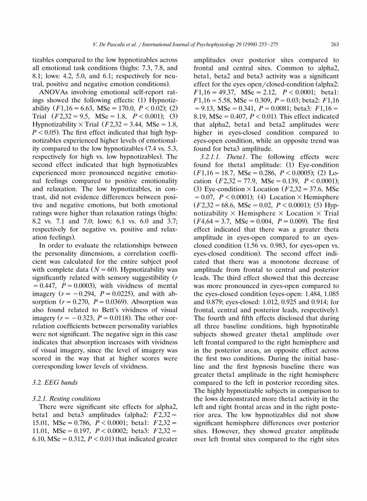
.frontal, central and posterior leads, respectively .The fourth and fifth effects disclosed that duringall three baseline conditions, high hypnotizablesubjects showed greater theta1 amplitude overleft frontal compared to the right hemisphere andin the posterior areas, an opposite effect acrossthe first two conditions. During the initial base-line and the first hypnosis baseline there wasgreater theta1 amplitude in the right hemispherecompared to the left in posterior recording sites.The highly hypnotizable subjects in comparison tothe lows demonstrated more theta1 activity in theleft and right frontal areas and in the right poste-rior area. The low hypnotizables did not showsignificant hemisphere differences over posteriorsites. However, they showed greater amplitudeover left frontal sites compared to the right sites
( )V. De Pascalis et al. r International Journal of Psychophysiology 29 1998 255]275264
Ž . Ž .Fig. 1. Theta1 4]6 Hz spectral amplitude in left and right hemispheres at frontal, central and parieto-temporo-occipital PTOŽ . Ž . Ž .scalp leads for high and low hypnotizable subjects during waking-rest W , hypnosis-rest1 H-1 , and hypnosis-rest2 H-2 conditions.
Data natural log transformed.
Ž .across all conditions see Fig. 1 . No effects in-volving sensory suggestibility were found.
3.2.1.2. Theta2. Effects for Location, Eye-con-dition, and Eye-condition=Location were sig-
Žnificant F2,32s27.47, MSes0.205, P-0.0001;F1,16s4.97, MSes0.823, Ps0.04; and F2,32
.s23.73, MSes0.065, P-0.0001 . The Locationeffect disclosed that there was a greater theta2amplitude over frontal scalp area as compared to
Žcentral and posterior sites 1.35, 1.12 and 1.06,.respectively . The other effects indicated that in
eyes-closed compared to eyes-open conditionthere was a significantly greater production of
Žtheta2 activity over posterior scalp sites frontal:1.34 vs. 1.36; central: 1.06 vs. 1.17; parietal: 0.9 vs.1.23, respectively for eyes-open vs. eyes-closed
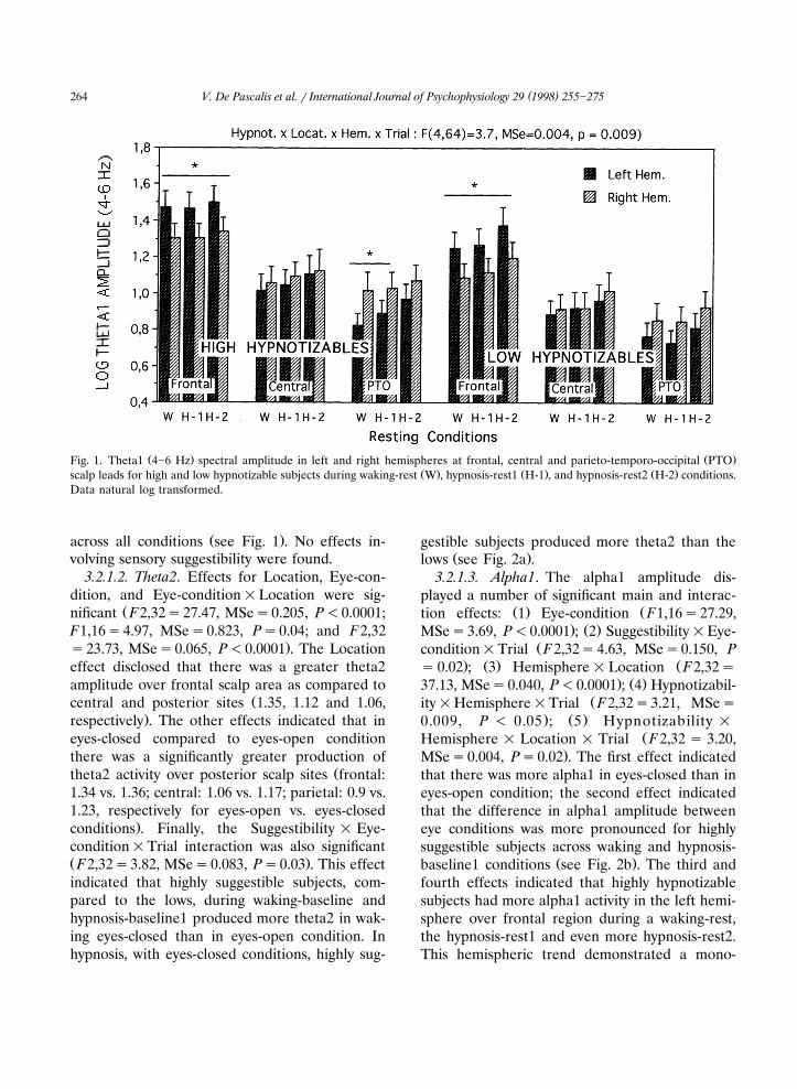
.conditions . Finally, the Suggestibility = Eye-condition=Trial interaction was also significantŽ .F2,32s3.82, MSes0.083, Ps0.03 . This effectindicated that highly suggestible subjects, com-pared to the lows, during waking-baseline andhypnosis-baseline1 produced more theta2 in wak-ing eyes-closed than in eyes-open condition. Inhypnosis, with eyes-closed conditions, highly sug-
gestible subjects produced more theta2 than theŽ .lows see Fig. 2a .
3.2.1.3. Alpha1. The alpha1 amplitude dis-played a number of significant main and interac-
Ž . Žtion effects: 1 Eye-condition F1,16s27.29,. Ž .MSes3.69, P-0.0001 ; 2 Suggestibility=Eye-
Žcondition=Trial F2,32s4.63, MSes0.150, P. Ž . Žs 0.02 ; 3 Hemisphere = Location F2,32 s
. Ž .37.13, MSes0.040, P-0.0001 ; 4 Hypnotizabil-Žity=Hemisphere=Trial F2,32s3.21, MSes
. Ž .0.009, P - 0.05 ; 5 Hypnotizability =ŽHemisphere = Location = Trial F2,32 s 3.20,
.MSes0.004, Ps0.02 . The first effect indicatedthat there was more alpha1 in eyes-closed than ineyes-open condition; the second effect indicatedthat the difference in alpha1 amplitude betweeneye conditions was more pronounced for highlysuggestible subjects across waking and hypnosis-
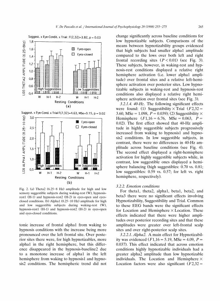
Ž .baseline1 conditions see Fig. 2b . The third andfourth effects indicated that highly hypnotizablesubjects had more alpha1 activity in the left hemi-sphere over frontal region during a waking-rest,the hypnosis-rest1 and even more hypnosis-rest2.This hemispheric trend demonstrated a mono-
( )V. De Pascalis et al. r International Journal of Psychophysiology 29 1998 255]275 265
Ž . Ž .Fig. 2. a Theta2 6.25]8 Hz amplitude for high and lowŽ .sensory suggestible subjects during waking-rest W , hypnosis-
Ž . Ž .rest1 H-1 and hypnosis-rest2 H-2 in eyes-open and eyes-Ž . Ž .closed conditions. b Alpha1 8.25]10 Hz amplitude for high
Ž .and low suggestible subjects during waking-rest W ,Ž . Ž .hypnosis-rest1 H-1 and hypnosis-rest2 H-2 in eyes-open
and eyes-closed conditions.
tonic increase of frontal alpha1 from waking tohypnosis conditions with the increase being morepronounced over the left frontal site. Over poste-rior sites there were, for high hypnotizables, morealpha1 in the right hemisphere, but this differ-ence disappeared in the hypnosis-baseline2 dueto a monotone increase of alpha1 in the lefthemisphere from waking to hypnosis1 and hypno-sis2 conditions. The hemispheric trend did not
change significantly across baseline conditions forlow hypnotizable subjects. Comparisons of themeans between hypnotizability groups evidencedthat high subjects had smaller alpha1 amplitudecompared to the lows over both left and right
Ž . Ž .frontal recording sites P-0.01 see Fig. 3 .These subjects, however, in waking-rest and hyp-nosis-rest conditions displayed a relative right
Žhemisphere activation i.e. lower alpha1 ampli-.tude over frontal sites and a relative left-hemi-
sphere activation over posterior sites. Low hypno-tizable subjects in waking-rest and hypnosis-restconditions also displayed a relative right hemi-
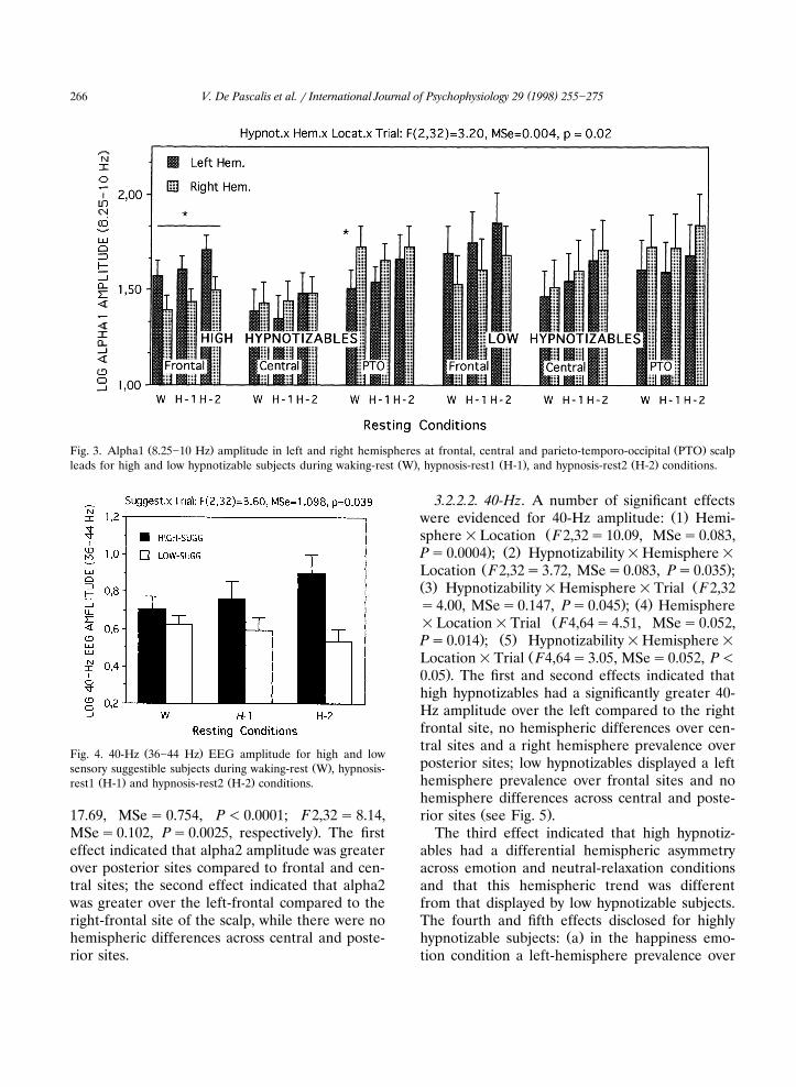
Ž .sphere activation over frontal sites see Fig. 3 .3.2.1.4. 40-Hz. The following significant effects
Ž . Žwere found: 1 Suggestibility=Trial F2,32s. Ž .3.60, MSes1.098, Ps0.039 ; 2 Suggestibility=
ŽHemisphere F1,16 s 6.76, MSe s 0.063, P s.0.02 . The first effect showed that 40-Hz ampli-
tude in highly suggestible subjects progressivelyincreased from waking to hypnosis1 and hypno-sis2 conditions. In low suggestible subjects, incontrast, there were no differences in 40-Hz am-
Ž .plitude across baseline conditions see Fig. 4 .The second effect displayed a right-hemisphereactivation for highly suggestible subjects while, incontrast, low suggestible ones displayed a hemi-
Žsphere balancing high suggestibles: 0.70 vs. 0.81;low suggestibles: 0.59 vs. 0.57; for left vs. right
.hemisphere, respectively .
3.2.2. Emotion conditionsFor theta1, theta2, alpha1, beta1, beta2, and
beta3 there were no significant effects involvingHypnotizability, Suggestibility and Trial. Commonto these EEG bands were the significant effectsfor Location and Hemisphere=Location. Theseeffects indicated that there were higher ampli-tudes over posterior recording sites and that theseamplitudes were greater over left-frontal scalpsites and over right-posterior scalp sites.
3.2.2.1. Alpha2. A main effect for Hypnotizabil-Žity was evidenced F1,16s5.19, MSes4.09, Ps
.0.037 . This effect indicated that across emotionconditions highly hypnotizable individuals had agreater alpha2 amplitude than low hypnotizableindividuals. The Location and Hemisphere =
ŽLocation factors were also significant F2,32s
( )V. De Pascalis et al. r International Journal of Psychophysiology 29 1998 255]275266
Ž . Ž .Fig. 3. Alpha1 8.25]10 Hz amplitude in left and right hemispheres at frontal, central and parieto-temporo-occipital PTO scalpŽ . Ž . Ž .leads for high and low hypnotizable subjects during waking-rest W , hypnosis-rest1 H-1 , and hypnosis-rest2 H-2 conditions.
Ž .Fig. 4. 40-Hz 36]44 Hz EEG amplitude for high and lowŽ .sensory suggestible subjects during waking-rest W , hypnosis-
Ž . Ž .rest1 H-1 and hypnosis-rest2 H-2 conditions.
17.69, MSe s 0.754, P - 0.0001; F2,32 s 8.14,.MSes0.102, Ps0.0025, respectively . The first
effect indicated that alpha2 amplitude was greaterover posterior sites compared to frontal and cen-tral sites; the second effect indicated that alpha2was greater over the left-frontal compared to theright-frontal site of the scalp, while there were nohemispheric differences across central and poste-rior sites.
3.2.2.2. 40-Hz. A number of significant effectsŽ .were evidenced for 40-Hz amplitude: 1 Hemi-
Žsphere=Location F2,32s10.09, MSes0.083,. Ž .Ps0.0004 ; 2 Hypnotizability=Hemisphere=Ž .Location F2,32s3.72, MSes0.083, Ps0.035 ;
Ž . Ž3 Hypnotizability=Hemisphere=Trial F2,32. Ž .s4.00, MSes0.147, Ps0.045 ; 4 Hemisphere
Ž=Location=Trial F4,64s4.51, MSes0.052,. Ž .Ps0.014 ; 5 Hypnotizability=Hemisphere=
ŽLocation=Trial F4,64s3.05, MSes0.052, P-.0.05 . The first and second effects indicated that
high hypnotizables had a significantly greater 40-Hz amplitude over the left compared to the rightfrontal site, no hemispheric differences over cen-tral sites and a right hemisphere prevalence overposterior sites; low hypnotizables displayed a lefthemisphere prevalence over frontal sites and nohemisphere differences across central and poste-
Ž .rior sites see Fig. 5 .The third effect indicated that high hypnotiz-
ables had a differential hemispheric asymmetryacross emotion and neutral-relaxation conditionsand that this hemispheric trend was differentfrom that displayed by low hypnotizable subjects.The fourth and fifth effects disclosed for highly
Ž .hypnotizable subjects: a in the happiness emo-tion condition a left-hemisphere prevalence over
( )V. De Pascalis et al. r International Journal of Psychophysiology 29 1998 255]275 267
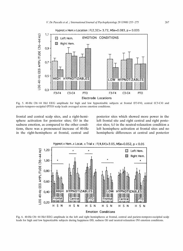
Ž . Ž . Ž .Fig. 5. 40-Hz 36]44 Hz EEG amplitude for high and low hypnotizable subjects at frontal F3-F4 , central C3-C4 andŽ .parieto-temporo-occipital PTO scalp leads averaged across emotion conditions.
frontal and central scalp sites, and a right-hemi-Ž .sphere activation for posterior sites; b in the
sadness emotion, as compared to the other condi-tions, there was a pronounced increase of 40-Hzin the right-hemisphere at frontal, central and
posterior sites which showed more power in theleft frontal site and right central and right poste-
Ž .rior sites; c in the neutral-relaxation condition aleft hemisphere activation at frontal sites and nohemispheric differences at central and posterior
Ž .Fig. 6. 40-Hz 36]44 Hz EEG amplitude in the left and right hemispheres at frontal, central and parieto-temporo-occipital scalpŽ . Ž . Ž .leads for high and low hypnotizable subjects during happiness H , sadness S and neutral-relaxation N emotion conditions.
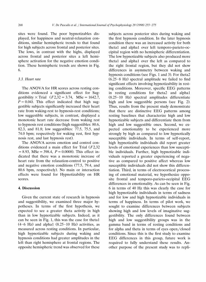
( )V. De Pascalis et al. r International Journal of Psychophysiology 29 1998 255]275268
sites were found. The poor hypnotizables dis-played, for happiness and neutral-relaxation con-ditions, similar hemispheric trends to that foundfor high subjects across frontal and posterior sites.The lows, in contrast with the highs, displayedacross frontal and posterior sites a left hemi-sphere activation for the negative emotion condi-tion. These hemispheric trends are shown in Fig.6.
3.3. Heart rate
The ANOVA for HR scores across resting con-ditions evidenced a significant effect for Sug-
Žgestibility = Trial F2,32 s 3.74, MSe s 1642.7,.Ps0.04 . This effect indicated that high sug-
gestible subjects significantly increased their heartrate from waking-rest to hypnosis-rest conditions;low suggestible subjects, in contrast, displayed amonotonic heart rate decrease from waking rest
Žto hypnosis rest conditions high suggestibles: 80.6,82.3, and 81.8; low suggestibles: 77.5, 75.5, and74.0 bpm; respectively for waking rest, first hyp-
.nosis rest, and last hypnosis rest .The ANOVA across emotion and control con-
Žditions evidenced a main effect for Trial F2,32.s8.93, MSes398.4, Ps0.0008 . This effect in-
dicated that there was a monotonic increase ofheart rate from the relaxation-control to positive
Žand negative emotion conditions 77.5, 79.4, and.80.6 bpm, respectively . No main or interaction
effects were found for Hypnotizability on HRscores.
4. Discussion
Given the current state of research in hypnosisand suggestibility, we examined three major hy-potheses. In terms of the first hypothesis, weexpected to see a greater theta activity in highthan in low hypnotizable subjects. Indeed, as itcan be seen in Fig. 1, this was the case for theta1Ž . Ž .4]6 Hz and alpha1 8.25]10 Hz activities, asmeasured across resting conditions. In particular,high hypnotizable subjects during waking andhypnosis conditions had greater amplitudes in theleft than right hemisphere at frontal regions. Theopposite hemispheric trend was observed for these
subjects across posterior sites during waking andthe first hypnosis condition. In the later hypnosiscondition there was an increased activity for boththeta1 and alpha1 over left temporo-parieto-oc-cipital region with no hemispheric differentiation.The low hypnotizable subjects also produced moretheta1 and alpha1 over the left as compared tothe right frontal region, but they did not showdifferences in asymmetry between waking and
Ž .hypnosis conditions see Figs. 1 and 3 . For theta2Ž .6.25]8 Hz spectral amplitude we failed to findsignificant effects involving hypnotizability in rest-ing conditions. Moreover, specific EEG patternsin resting conditions for theta2 and alpha1Ž .8.25]10 Hz spectral amplitudes differentiate
Ž .high and low suggestible persons see Fig. 2 .Thus, results from the present study demonstratethat there are distinctive EEG patterns amongresting baselines that characterize high and lowhypnotizable subjects and differentiate them fromhigh and low suggestible ones. Second, we ex-pected emotionality to be experienced morestrongly by high as compared to low hypnoticallysusceptible individuals. In terms of self-report,high hypnotizable individuals did report greaterlevels of emotional experiences than low suscepti-ble individuals. Further, high hypnotizable indi-viduals reported a greater experiencing of nega-tive as compared to positive affect whereas lowsusceptible individuals did not show this differen-tiation. Third, in terms of electrocortical process-ing of emotional material, we hypothesize oppo-site frontal and temporo-parieto-occipital EEGdifferences in emotionality. As can be seen in Fig.6 in terms of 40 Hz this was clearly the case forhigh hypnotizable individuals in terms of sadnessand for low and high hypnotizable individuals interms of happiness. In terms of pilot work, wesought to examine differences between subjectsshowing high and low levels of imaginative sug-gestibility. The only differences found betweenhigh and low suggestibility groups was in thegamma band in terms of resting conditions andfor alpha and theta in terms of eyes openrclosedconditions. Since this is the first study to examineEEG differences in this group, future work isrequired to fully understand these results. An-other purpose of the present study was to repli-
( )V. De Pascalis et al. r International Journal of Psychophysiology 29 1998 255]275 269
cate and extend our previous 40-Hz EEG corre-lates of emotionality using a more advanced mea-sure of 40-Hz EEG spectral amplitude.
4.1. Psychophysiological differences during rest inwaking and hypnosis
The theta1 finding is consistent with previousŽhypnosis research e.g. Galbraith et al., 1970;
Graffin et al., 1995; Sabourin and Cutcomb, 1980;.Sabourin et al., 1990; Tebecis et al., 1975 . In-
deed, as with the present study, Graffin et al.Ž . Ž .1995 found the strongest theta 4]8 Hz differ-ences between susceptibility groups in the morefrontal areas of the cortex. Previous research hasevidenced that increased frontal midline theta,especially at 6 Hz, has been associated with atten-
Žtional readiness or attentive performance in rest-. Žing condition of cognitive tasks Bruneau et al.,
1993; Ishihara and Yoshii, 1972; Lang et al., 1988;.Mizuki et al., 1980 . Moreover, looking at the
Ž .Galbraith et al. 1970 findings, the highest corre-lated theta frequency with hypnotizability was at5 Hz which is corresponding to the center fre-quency of our theta1 band. As in the Galbraith etal. study, our theta1 findings may be seen asreflecting a class II inhibition process i.e. thathigh hypnotizable individuals might manifest aheightened state of attentional readiness and fo-calization of attention. In the present study wefailed to find theta2 differences between high andlow hypnotizable subjects as, indeed, it was found
Ž .in the recent study of Crawford et al. 1996 . Wethink that the apparent conflicting findings maylie in the sometimes incorrect assumption thatlow theta is associated with class I inhibition andhigh theta with class II inhibition, since we can-not exactly know the band edges which define thehigh and low theta activities. It may be reason-able to think that these edges are dependent froma number of factors such as personality, sampling,and situation-specific variables.
The alpha1 activity has been found to be asso-ciated with decreased cortical activity or with
Žpoor sustained attentional abilities e.g. Bosel,1992; Gale and Edwards, 1983; Klimesch et al.,
.1990 . If we assume that an increase in theta1activity reflects an increase in focused attention
and that an increase in alpha1 reflects an aspeci-fic effect of relaxation, then the increased theta1and alpha1 amplitudes found over left temporo-parieto-occipital site in highly hypnotizable sub-jects, indicate that these subjects, in resting hyp-notic condition, were not only more relaxed, butalso more facilitated in processing relevant sti-muli than they did in a waking condition. More-over, the finding that high hypnotizables, in com-parison with lows, displayed higher theta1 andlower alpha1 amplitudes over left and right frontalregions, is indicating that highs were actively moreinvolved in the hypnosis condition than did thelows. Finally, the alpha1 trend observed acrosswaking and hypnosis conditions over frontal andtemporo-parieto-occipital regions of the lefthemisphere is consistent with the Gruzelier neu-rophysiological model of hypnosis wherein thecentral inhibitory processes, particularly of theleft hemisphere, are considered as the main indi-
Žcators characterizing hypnosis Gruzelier et al.,1984; Gruzelier, 1988; Gruzelier and Warren,
.1993 .The HR data failed to evidence significant dif-
ferences among resting conditions between highand low hypnotizable subjects. This result doesnot confirm the expected finding, i.e. that hypno-sis induction, being composed of relaxation sug-
Žgestions, produces a decrease in HR see e.g.. Ž .Sarbin and Slagle, 1979 . Gray et al. 1970 and
Ž .Bauer and McCanne 1980 obtained findings inline with this prediction: they observed a decreasein HR during a rest period after the hypnoticinduction as compared to a rest period before theinduction. However, the present result appears inagreement with the Lamas and del Valle-Inclan`Ž .1994 findings supporting that the inductionprocedure does not produce HR deceleration inhigh hypnotizable subjects.
4.2. EEG differences during emotions in hypnosis
During the emotional conditions, high hypnoti-zable subjects, compared to the lows, reportedgreater ability in experiencing positive and nega-tive emotions, more alpha2 activity and morepronounced emotion-related 40-Hz EEG hemi-spheric shifts. Specifically, in terms of 40-Hz am-
( )V. De Pascalis et al. r International Journal of Psychophysiology 29 1998 255]275270
plitude high hypnotizables showed three mainŽ .findings see Fig. 6 . Firstly, in the happiness
condition an increased activity in the frontal lefthemisphere that produced a marked hemisphericasymmetry in favor of the left compared to theright hemisphere; a similar but less pronouncedasymmetry in favor of the left hemisphere wasalso found across central sites, while an oppositehemispheric asymmetry was evidenced in the pos-terior region. This pattern of findings is consistentwith previous research showing more left frontalŽ . Žcf. Davidson, 1992 and right posterior cf.
.Tucker, 1981 cortical involvement in the process-ing of positive emotionality. Secondly, in the sad-ness condition, compared to the other conditions,there was an increased activity over the righthemisphere that produced a hemispheric balanc-ing in the frontal region and a right hemisphereactivation in the central and even more in theposterior regions. These results are consistent
Ž .with the predictions of the Pribram 1981 ante-rio-posterior neurophysiological model of emo-tions. Thirdly, the relaxation condition producedmore activity in the left than in the right frontalregion, along with more equal hemispheric activ-ity in the central and posterior regions.
Overall findings in terms of 40-Hz activity par-allels those of previous research from our lab.One major EEG difference between positive andnegative emotional experience was an increasedproduction of 40-Hz activity in the left frontalregion for happiness and a significant increase inthe right posterior regions for sadness. Theseresults support prior research of self-generatedemotional states that demonstrate, using alpha,activation of parietal region for positive and nega-
Žtive emotions Collet and Duclaux, 1987; Harmanand Ray, 1977; Karlin et al., 1979; Meyers andSmith, 1987; Smith et al., 1987, 1989; Tucker et
.al., 1981; Tucker and Dawson, 1984 . These re-Ž .sults also support the validity of the Heller 1993
model indicating that parietotemporal regionsplay a critical role in the experience of emotion.Our observations are parallel to those of Sten-
Ž .berg 1992 who found the frontal lateral regionof the brain mainly engaged in emotional process-ing. Similar 40-Hz hemispheric trends were re-ported in our previous studies in which 40-Hz
Ždensity was the dependent variable De Pascalis.et al., 1987, 1989 . In our previous studies, 40-Hz
density increased in the right hemisphere duringnegative emotions. The current study found anincreased activity in the left and even more in theright posterior sites by displaying a hemisphericasymmetry in favor of the right hemisphere. Start-ing from the assumption that 40-Hz EEG rhythmis the physiological expression of ‘focused arousal’Ž .e.g. Sheer, 1989; Tiitinen et al., 1993 , our 40-HzEEG findings during emotional states are consis-tent with the dual-attentional model of Kins-
Ž . Ž .bourne 1982 and of Pribram 1981 wherein theleft hemisphere is more involved to maintain se-lective or focal attention and the right to main-tain ‘sustained’ attention in order to form a cont-inuous synthesis of the incoming information.During positive emotions, as compared to a relax-ation condition, the frontal and central regions inthe left hemisphere are more engaged to extractinformation from the recollecting material andthe posterior-right hemisphere to form a connota-tional synthesis of the ongoing processed stimuli.That is, it is the posterior area that gives color tothe emotional experience. The recollection of anegative emotion, with respect to a relaxationcondition, requires an engagement of both rightand left frontal areas and mainly of the rightcentral and posterior areas of the cortex.
In terms of HR activity it was found that HRincreased during positive and even more duringnegative emotional events as compared to a relax-ation condition. The HR activity failed to evi-dence, among emotion conditions, different trendsbetween high and low hypnotizable subjects. Thisresult confirms findings from other studies usingfacial configuration or relived emotion tasks. The
ŽHR increases for sadness, fear and anger Ekman.et al., 1983; Levenson et al., 1991 and happiness
Ž .Levenson et al., 1990 . The HR activity has beenused in hypnosis research as a physiological mea-sure of attentional demands. It has been found toincrease with stressful active suggestions and to
Ždecrease with relaxation ones see e.g. Barber,.1965; Sarbin and Slagle, 1979 . The HR changes
should be more pronounced in highly hypnotiz-able subjects since they are more engaged thanthe lows in the suggested tasks, as indeed it was
( )V. De Pascalis et al. r International Journal of Psychophysiology 29 1998 255]275 271
Ž .the case in the study of Sabourin et al. 1990 .Our HR changes were sensitive to the emotionalvalence of the recollected material but failed toevidence differences in trends between high andlow hypnotizable subjects. This lacking result maylay in the fact that attentional demands and emo-tional valence have an additive effect on HR
Žincrease e.g. Cook et al., 1991; Hawk et al.,.1992 . This effect may have been sufficient to
produce an HR increase also in the low hypnotiz-able subjects.
4.3. Suggestibility
The HR increase during hypnosis resting condi-tions as compared to a waking resting conditiondifferentiate suggestible from non-suggestiblesubjects. In terms of EEG, theta2, alpha1, and40-Hz EEG activities were sensitive to individualdifferences in suggestibility, but only between eyesopen and closed conditions. Common to theta2and alpha1 was the tendency for highly sug-gestible persons to display a significantly greateramount of these activities in resting eyes-closedas compared to resting eyes-open conditions. Ingeneral, high suggestible individuals compared tolow suggestible ones showed a differential in-crease of theta2 in the last eye-closed baselinecondition. In terms of alpha, high suggestibleindividuals showed more alpha1 in the eyes closed
Ž .condition see Fig. 2 . The 40-Hz EEG in highlysuggestible subjects also displayed a monotonicincrease from waking to the first and last hypno-
Ž .sis-rest conditions see Fig. 4 . These results to donot form a consistent picture but can be under-stood as related to the greater reactivity of highlysuggestible subjects to the hypnosis task per-se.The validity of this interpretation is also sup-ported by the more pronounced heart rate in-crease found for these subjects in the hypnosis-rest conditions as compared to the initial wakingcondition. The lack of relationship between sen-sory suggestibility and absorption scores as well asa lack of EEG theta differentiation would suggestthat individual differences in the capacity to beabsorbed in imaginative activity is separate fromthat of suggestibility. Since no significant interac-tional effects involving both hypnotizability and
suggestibility were found, for each of the vari-ables considered in this study we maintained thatthese two dimensions are expressions of differentunderlying psychophysiological activities.
Overall, the present study replicates a varietyof EEG research, especially theta and 40 Hzactivity, in relation to hypnosis. As in previousstudies, high and low hypnotically susceptible in-dividuals differed in EEG theta and in emotionalprocessing as reflected in both self-report and 40Hz activity. The present study also supportedprevious theoretical speculations suggestingfrontalrposterior differences in emotional pro-cessing. Finally, it gives psychophysiological sup-port to the theoretical position that suggestibilityand hypnosis reflect different physiological mech-anisms, but it also indicates that it might beworthwhile to take into account suggestibility infuture research.
Acknowledgements
We greatly acknowledge the assistance of thefollowing students: Giovanna Carbone, SimonaTomassetti. This research was assisted in part bya collaborative research grant from NATO.
References
Barabasz, A.F., 1983. EEG alpha-hypnotizability correlationsare not simple covariates of subject self-selection. Biol.Psychol. 17, 169]172.
Barabasz, A.F., 1990. Effects of sensory deprivation on EEGtheta and skin conductance. Paper presented at the FifthInternational Congress of Psychophysiology, Budapest,Hungary.
Barabasz, A.F., Lonsdale, C., 1983. Effects of hypnosis onP300 olfactory-evoked potential amplitudes. J. Abnorm.Psychol. 4, 520]523.
Barber, T.X., 1969. Hypnosis: A Scientific Approach. VanNostrand Reinhold, New York.
Barber, T.X., 1965. Physiological effects of ‘hypnotic sugges-tions’: a critical review of recent research. Psychol. Bull. 63,201]222.
Basar-Eroglu, C., Basar, E., Demiralp, T., Schurmann, M.,1992. P300-response: possible psychophysiological corre-lates in delta and theta frequency channels. A review. Int.J. Psychophysiol. 13, 161]179.
Bauer, K.E., McCanne, T.R., 1980. Autonomic and centralnervous system responding: during hypnosis and simulationof hypnosis. Int. J. Clin. Exp. Hypn. 28, 148]163.
( )V. De Pascalis et al. r International Journal of Psychophysiology 29 1998 255]275272
Bosel, R., 1992. Slow alpha in the EEG power spectrum as anindicator for conceptual arousal. Zur. Exp. Angew. Psychol.39, 372]395.
Bower, G.H., 1981. Mood and memory. Am. Psychol. 36,129]148.
Bowers, K.S., 1971. Sex and susceptibility as moderator vari-ables in the relationship of creativity and hypnotic suscepti-bility. J. Abnorm. Psychol. 78, 93]100.
Bowers, K.S., 1983. Hypnosis for the Seriously Curious. Nor-ton, New York.
Bruneau, N., Sylvie, R., Gurin, P., Garreau, B., Lelord, G.,1993. Auditory stimulus intensity responses and frontalmidline theta rhythm. Electroencephalogr. Clin. Neuro-physiol. 86, 213]216.
Bryant, R.A., McConkey, K.M., 1989. Hypnotic emotions andphysical sensations: a real-simulating analysis. Int. J. Clin.Exp. Hypn. 37, 305]319.
Cacioppo, J.T., Klein, D.J., Berntson, G.G., Hatfield, E., 1993.The psychophysiology of emotion. In: Lewis, M., Haviland,
Ž .J.M. Eds. , Handbook of Emotions. The Guilford Press,New York, pp. 119]142.
Collet, L., Duclaux, R., 1987. Hemispheric lateralization ofemotions: absence of electrophysiological arguments. Phys-iol. Behav. 40, 215]220.
Cook, E.W., III, Hawk, L.W., Davis, T.L., Stevenson, V.E.,1991. Affective individual differences and startle reflexmodulation. J. Abnorm. Psychol. 100, 5]13.
Coppola, R., 1986. Issues in topographic analysis of EEGŽ .activity. In: Duffy, F.H. Ed. , Topographic Mapping of
Brain Electrical Activity. Butterworths, Boston, pp.339]346.
Coppola, R., Chassy, J., 1986. Subjects with low versus highfrequency alpha rhythm reveal different topographic struc-ture. Electroencephalogr. Clin. Neurophysiol. 63, 41.
Crawford, H.J., 1982. Hypnotizability, daydreaming styles,imagery vividness, and absorption: a multidimensionalstudy. J. Pers. Soc. Psychol. 42, 915]926.
Crawford, H.J., 1990. Cognitive and psychophysiological corre-lates of hypnotic responsiveness and hypnosis. In: Fass,
Ž .M.L., Brown, D.P. Eds. , Creative Mastery in Hypnosisand Hypnoanalysis: a Festschrift for Erika Fromm. PlenumPress, New York, pp. 47]54.
Crawford, H.J., 1991. The hypnotizable brain: attentional anddisattentional processes. Presidential address delivered atthe 42nd Annual Scientific Meeting of The Society forClinical and Experimental Hypnosis, New Orleans, LA.
Crawford, H.J., Brown, A.M., Moon, C.E., 1993. Sustainedattentional and disattentional abilities: differences betweenlow and high hypnotizable persons. J. Abnorm. Psychol.102, 534]543.
Crawford, H., Clarke, S., Kitner-Triolo, M., 1996. Self-gener-ated happy and sad emotions in low and highly hypnotiz-able persons during waking and hypnosis: laterality andregional EEG activity differences. Int. J. Psychophysiol. 24,239]266.
Crawford, H.J., Gruzelier, J.H., 1992. A midstream view of theneuropsychophysiology of hypnosis: recent research and
Ž .future directions. In: Fromm, E., Nash, M.R. Eds. , Con-temporary Hypnosis Research. Guilford Press, New York,pp. 227]266.
Crawford, H.J., Kapelis, L., Harrison, D.W., 1995. Visual fieldasymmetry in facial affect perception: moderating effects ofhypnosis, hypnotic susceptibility level, absorption, and sus-tained attentional abilities. Int. J. Neurosci. 82, 11]23.
Crowson, J.J., Jr., Conroy, A.M., Chester, T.D., 1991. Hypnoti-zability as related to visually induced affective reactivity.Int. J. Clin. Exp. Hypn. 39, 140]144.
Davidson, R.J., 1992. Anterior cerebral asymmetry and thenature of emotion. Brain Cogn. 20, 125]151.
Davidson, R.J., Ekman, P., Saron, C., Senulis, R., Friesen,W.V., 1990. Approach-withdrawal and cerebral asymmetry:I. Emotional expression and brain physiology. J. Pers. Soc.Psychol. 58, 333]341.
Davidson, R.J., Schwartz, G.E., Saron, C., Bennett, J., Gole-man, D.J., 1979. Frontal versus parietal EEG asymmetryduring positive and negative affect. Psychophysiology 16,202]203.
De Pascalis, V., 1993. EEG spectral analysis during hypnoticinduction, hypnotic dream and age regression. Int. J. Psy-chophysiol. 15, 153]166.
De Pascalis, V., 1994. Event-related potentials during hypnotichallucination. Int. J. Clin. Exp. Hypn. 1, 39]55.
De Pascalis, V., Carboni, G., 1997. P300 event-related-poten-tial amplitudes and evoked cardiac responses during hyp-notic alteration of somatosensory perception. Int. J. Neu-rosci., in press.
De Pascalis, V., Marucci, F.S., Penna, M.P., 1989. 40-Hz EEGasymmetry during recall of emotional events in waking andhypnosis: differences between low and high hypnotizables.Int. J. Psychophysiol. 7, 85]96.
De Pascalis, V., Marucci, F.S., Penna, M.P., Pessa, E., 1987.Hemispheric activity of 40 Hz EEG during recall of emo-tional events: differences between low and high hypnotiz-ables. Int. J. Psychophysiol. 5, 167]180.
Dumas, R.A., 1977. EEG alpha-hypnotizability correlations: areview. Psychophysiology 14, 431]438.
Ekman, P., Levenson, R.W., Friesen, W.V., 1983. Autonomicnervous system activity distinguishes among emotions. Sci-ence 221, 1208]1210.
Evans, F.J., 1972. Hypnosis and sleep: techniques for exploringcognitive activity during sleep. In: Fromm, E., Shor, R.E.Ž .Eds. , Hypnosis: Research Developments and Perspec-tives. Aldine-Atherton, Chicago, pp. 43]83.
Evans, F.J., 1989. The hypnotizable patient. In: Waxman, D.,Ž .Pedersen, D., Wilkie, I., Mellett, P. Eds. , Hypnosis: The
Fourth European Congress at Oxford. Whurr, London, pp.18]28.
Eysenck, H.J., 1947. Dimensions of Personality. Routledgeand Kegan Paul, London.
Ž .Eysenck, H.J., 1991. Is suggestibility? In: Schumaker, J. Ed. ,Human Suggestibility. Routledge, New York, pp. 76]90.
Faith, M., Ray, W.J., 1994. Hypnotizability and dissociation ina college age population: orthogonal individual differences.Pers. Individ. Differ. 17, 211]216.
( )V. De Pascalis et al. r International Journal of Psychophysiology 29 1998 255]275 273
Fellows, B.J., 1990. Current theories of hypnosis: a criticaloverview. Br. J. Exp. Clin. Hypn. 7, 81]92.
Fromm, E., Nash, M., 1992. Contemporary Hypnosis Re-search. Guilford Press, New York.
Galbraith, G.C., London, P., Leibovitz, M.P., Cooper, L.M.,Hart, J.T., 1970. EEG and hypnotic susceptibility. J. Comp.Physiol. Psychol. 72, 125]131.
Gale, A., Edwards, J.A., 1983. Psychophysiology and individualdifferences: theory, research procedures, and the interpre-tation of data. Aust. J. Psychol. 35, 361]378.
Gheorghiu, V., 1989. The development of research on sug-gestibility: critical considerations. In: Gheorghiu, V.A.,
Ž .Netter, P., Eysenck, H.J., Rosenthal, R. Eds. , Suggestionand Suggestibility } Theory and Research. Springer, Hei-delberg, pp. 3]55.
Gheorghiu, V., Kock, E., Hubner, M., 1993. A group scale forthe influence of suggestion on sensory judgments. Paperpresented at the 6th European Congress of Hypnosis, Vi-enna, August 14]20, 1993.
Glass, A., 1968. Intensity of attenuation of alpha activity bymental arithmetic in females and males. Physiol. Behav. 3,217]220.
Glass, A., Butler, S.R., Carter, J.C., 1984. Hemisphericasymmetry of EEG alpha activation: effects of gender andfamiliar handedness. Biol. Psychol. 19, 169]187.
Graffin, N.F., Ray, W.J., Lundy, R., 1995. EEG concomitantsof hypnosis and hypnotic susceptibility. J. Abnorm. Psychol.1, 123]131.
Gray, A.L., Bowers, K.S., Fenz, W.D., 1970. Heart rate inanticipation of and during a negative hallucination. Int. J.Clin. Exp. Hypn. 18, 41]81.
Gruzelier, J.H., 1988. The neuropsychology of hypnosis. In:Ž .Heap, M. Ed. , Hypnosis: Current Clinical, Experimental
and Forensic Practices. Croom Helm, London, pp. 68]76.Gruzelier, J.H., Brow, T.D., Perry, A., Rhonder, J., Thomas,
R., 1984. Hypnotic susceptibility: a lateral predispositionand altered cerebral asymmetry under hypnosis. Int. J.Psychophysiol. 2, 131]139.
Gruzelier, J.W., Warren, K., 1993. Neuropsychological evi-dence of reductions on left frontal tests hypnosis. Psychol.Med. 23, 93]101.
Gur, R.C., Gur, R.E., 1974. Handedness, sex and eyedness asmoderating variables in the relation between hypnotic sus-ceptibility and functional brain asymmetry. J. Abnorm.Psychol. 83, 635]643.
Harman, D.W., Ray, W.J., 1977. Hemispheric activity duringaffective verbal stimuli: an EEG study. Neuropsychologia15, 457]460.
Hawk, L.W., Stevenson, V.E., Cook, E.W., III, 1992. Theeffect of eyelid closure on affective imagery and eyeblinkstartle. J. Psychophysiol. 6, 299]310.
Heller, W., 1993. Neuropsychological mechanisms of individ-ual differences in emotion, personality and arousal. Neu-ropsychologia 7, 476]489.
Heller, W., Etienne, M.A., Miller, G.A., 1995. Patterns ofperceptual asymmetry in depression and anxiety: Implica-
tions for neuropsychological models of emotion and psy-chopathology. J. Abnorm. Psychol. 104, 327]333.
Hilgard, E.R., 1973. The domain of hypnosis: with some com-ments on alternative paradigms. Am. Psychol. 23, 972]982.
Hilgard, E.R., 1977. Divided Consciousness: Multiple Controlsin Human Thought and Action. Wiley, New York.
Hilgard, E.R., 1979. Divided consciousness in hypnosis: theimplications of the hidden observer. In: Fromm, E., Shor,
Ž .R.E. Eds. , Hypnosis: Developments in Research and NewPerspectives, 2nd ed. Aldine, New York, pp. 45]79.
Hilgard, E.R., 1986. Divided Consciousness: Multiple Controlsin Human Thought and Action, revised ed. Wiley, NewYork.
Hilgard, E.R., 1991. A neodissociation interpretation of hyp-Ž .nosis. In: Lynn, S.J., Rhue, J.W. Eds. , Theories of Hypno-
sis: Current Models and Perspectives. Guilford Press, NewYork, pp. 83]104.
Hilgard, E.R., 1992. Dissociation and theories of hypnosis. In:Ž .Fromm, E., Nash, M.R. Eds. , Contemporary Hypnosis
Research. Guilford Press, New York, pp. 69]101.Jasiukaitis, P., Nouriani, B., Spiegel, D., 1996. Left hemisphere
superiority for event-related potential effects of hypnoticobstruction. Neuropsychologia 7, 661]668.
Jutai, J., Gruzelier, J., Golds, J., Thomas, M., 1993. Bilateralauditory-evoked potentials in conditions of hypnosis andfocused attention. Int. J. Psychophysiol. 15, 167]176.
Ishihara, T., Yoshii, N., 1972. Multivariate analytic study ofEEG and mental activity in juvenile delinquents. Elec-troencephalogr. Clin. Neurophysiol. 33, 71]80.
Kahneman, D., Tursky, B., Shapiro, D., Crider, A., 1969.Pupillary, heart rate, and skin resistance changes during amental task. J. Exp. Psychol. 79, 164]167.
Karlin, R., Weinapple, M., Rochford, J., Goldstein, L., 1979.Quantitated EEG features of negative affective states: re-port of some hypnotic studies. Res. Commun. Psychol.Psychiatr. Behav. 4, 397]413.
Kihlstrom, J.F., 1987. The cognitive unconscious. Science 237,1445]1452.
Kihlstrom, J., 1997. Convergence in understanding hypnosis?Perhaps, but perhaps not so fast. Int. J. Clin. Exp. Hypn.45, 324]332.
Kinsbourne, M., 1982. Hemispheric specialization and thegrowth of human understanding. Am. Psychol. 37, 411]420.
Kirk, R.E., 1968. Experimental Design: Procedures for theBehavioral Sciences. BrooksrCole, Belmont.
Kirsch, I., 1997. Hypnotic suggestion: a musical metaphor.Am. J. Clin. Hypn. 39, 271]282.
Kirsch, I., Council, J., 1992. Situational and personality corre-lates of hypnotic responsiveness. In: Fromm, E., Nash, M.Ž .Eds. , Contemporary Hypnosis Research. Guilford Press,New York, pp. 267]291.
Kirsch, I., Lynn, S., 1995. The altered state of hypnosis. Am.Psychol. 50, 846]858.
Klimesch, W., Schimke, H., Ladurner, G., Pfurtscheller, G.,1990. Alpha frequency and memory performance. J. Psy-chophysiol. 4, 381]390.
Kline, J.P., Schwartz, G.E., Allen, J.J., Dikman, Z.V., Fernan-
( )V. De Pascalis et al. r International Journal of Psychophysiology 29 1998 255]275274
dez, M., 1994. EEG correlates of unconscious registrationof emotional words in repressive and defensive copingstyles. Psychophysiology 31, S62.
Kropotov, D.J., Crawford, H.J., Polyakov, Y.I., 1997. So-matosensory event-related potential changes to painful sti-muli during hypnotic analgesia: anterior cingulate cortexand anterior temporal cortex intracranial recordings inobsessive-compulsive. Int. J. Psychophysiol. 27, 1]8.
Lacey, J., 1967. Somatic response patterning and stress: somerevisions of activation theory. In: Appley, M.H., Trumbell,
Ž .R. Eds. , Psychological Stress. Appleton-Century-Crofts,New York, pp. 14]37.
Lacey, J.I., Lacey, B.C., 1970. Some autonomic-central ner-Ž .vous system interrelationships. In: Black, P. Ed. , Physio-
logical Correlates of Emotion. Academic Press, New York.Lamas, J.R., del Valle-Inclan, F., 1994. Attentional demands`
during hypnotic responding: an investigation using heartrate variability. Paper presented at the 7th InternationalCongress of Psychophysiology, Thessaloniki, 27 Sept.]2 Oct.
Lang, W., Lang, M., Kornhuber, A., Diekmann, V., Kornhu-ber, H.H., 1988. Event related EEG spectra in a conceptformation task. Hum. Neurobiol. 6, 295]301.
Levenson, R.W., Carstensen, L.L., Friesen, W.V., Ekman, P.,1991. Emotion, physiology, and expression in old age. Psy-chol. Ag. 6, 28]35.
Levenson, R.W., 1992. Autonomic nervous system patterningin emotion. Psychol. Sci. 3, 23]27.
Levenson, R.W., Ekman, P., Friesen, W.V., 1990. Voluntaryfacial action generates emotion-specific autonomic nervoussystem activity. Psychophysiology 27, 363]384.
London, P., Hart, J.T., Leibovitz, M.P., 1968. EEG alpharhythms and susceptibility to hypnosis. Nature 219, 71]72.
Meyers, M.B., Smith, B.D., 1987. Cerebral processing of non-verbal affective stimuli: differential effects of cognitive andaffective sets on hemispheric asymmetry. Biol. Psychol. 24,67]84.
Mizuki, Y., Tanaka, M., Isozaki, H., Nishijima, H., Inanaga,K., 1980. Periodic appearance of theta rhythm in the frontalmidline area during performance of a mental task. Elec-troencephalogr. Clin. Neurophysiol. 49, 345]351.
Morgan, A.H., Hilgard, J.R., 1978. The Stanford HypnoticClinical Scale for adults. Am. J. Clin. Hypn. 21, 134]147.
Morgan, A.H., MacDonald, H., Hilgard, E.R., 1974. EEGalpha: lateral asymmetry related to task and hypnotizabil-ity. Psychophysiology 11, 275]282.
Nowlis, D.P., Rhead, J.C., 1968. Relation of eyes-closed rest-ing EEG alpha activity to hypnotic susceptibility. Percep.Mot. Skills 27, 1047]1050.
Palomba, D., Angrilli, A., Mini, A., 1997. Visual evoked poten-tials, heart rate responses and memory to emotional pic-torial stimuli. Int. J. Psychophysiol. 27, 55]67.
Perlini, A.H., Spanos, N.P., 1991. EEG alpha methodologiesand hypnotizability: a critical review. Psychophysiology 28,511]530.
Pfurtscheller, G., Klimesch, W., 1991. Event-related desyn-chronization during motor behavior and visual informationprocessing. In: Brunia, C.H.M., Mulder, G., Verbaten, M.N.
Ž . Ž .Eds. , Event-related Brain Research EEG Suppl. 42 pp.58]65.
Piccione, C., Hilgard, E.R., Zimbardo, P.G., 1989. On thedegree of stability of measured hypnotizability over a 25-year period. J. Pers. Soc. Psychol. 56, 289]295.
Pribram, K.H., 1981. Emotions. In: Filskov, S.B., Boll, T.J.Ž .Eds. , Handbook of Clinical Neuropsychology. Wiley, NewYork, pp. 102]134.
Raghavan, N., Glover, J.R., Sheer, D.E., 1986. A microproces-sor-based system for diagnosis of cognitive dysfunction.IEEE Trans. Biomed. Eng. 33, 942]948.
Ray, W.J., 1996. Dissociation in normal populations. In:Ž .Michelson, L., Ray, W. Eds. , Handbook of Dissociation.
Plenum, New York.Ray, W.J., Cole, H.W., 1985. EEG alpha activity reflects
attentional demands, and beta activity reflects emotionaland cognitive processes. Science 228, 750]752.
Ray, W., Faith, M., 1995. Dissociative experiences in a collegeage population: follow-up with 1190 subjects. Pers. Individ.Differ. 18, 223]230.
Ray, W.J., Morrel, M., Frediani, A., Tucker, D.M., 1976. Sexdifferences and lateral specialization of hemispheric func-tioning. Neuropsychologia 14, 391]394.
Sabourin, M.E., Cutcomb, S.D., 1980. EEG spectral analysisand hypnotic susceptibility. Paper presented at the AnnualMeeting of the Society for Clinical and Experimental Hyp-nosis, Chicago.
Sabourin, M.E., Cutcomb, S.D., Crawford, H.J., Pribram, K.,1990. EEG correlates of hypnotic susceptibility and hyp-notic trance: spectral analysis and coherence. Int. J. Psy-chophysiol. 10, 125]142.
Salmaso, D., Longoni, A.M., 1985. Problems in the assessmentof hand preference. Cortex 21, 533]549.
Sarbin, T.R., Slagle, R.W., 1979. Hypnosis and psychophysio-Ž .logical outcomes. In: Fromm, E., Shor, R.E. Eds. , Hypno-
sis: Research Developments and Perspectives, 2nd ed. Al-dine, New York, pp. 273]303.
Schacter, D.L., 1977. EEG theta waves and psychologicalphenomena: a review and analysis. Biol. Psychol. 5, 47]82.
Sheehan, P.W., 1967. A shortened form of Betts’ question-naire upon mental imagery. J. Clin. Psychol. 3, 386]389.
Sheer, D.E., 1975. Biofeedback training of 40 Hz EEG andŽ .behavior. In: Burch, N., Altshuler, H.L. Eds. , Behavior
and Brain Electrical Activity. Plenum, New York, pp.325]362.
Sheer, D.E., 1989. Focused arousal and the cognitive 40-Hzevent-related potentials: differential diagnosis ofAlzheimer’s disease. In: Igbal, K., Wisniewski, H.M., Win-
Ž .blad, B. Eds. , Alzheimer’s Disease and Related Disorders.Alan R. Liss, Inc., pp. 79]94.
Shor, R.E., Orne, E.C., 1963. Norms on the Harvard GroupScale of Hypnotic Susceptibility: Form A. Int. J. Clin. Exp.Hypn. 11, 39]47.
Silva, C., Kirsch, I., 1992. Interpretative sets, expectancy, fan-tasy proneness, and dissociation as predictors of hypnoticresponse. J. Pers. Soc. Psychol. 63, 847]856.
Smith, B.D., Meyers, M., Kline, R., Bozman, A., 1987. Hemi-
( )V. De Pascalis et al. r International Journal of Psychophysiology 29 1998 255]275 275
spheric asymmetry and emotion: lateralized parietal pro-cessing of affect and cognition. Biol. Psychol. 25, 247]260.
Smith, B.D., Womack, W.M., Chen, A.C.N., 1989. Hypnotiz-ability does not predict outcome of behavioral treatment ofpediatric headache. Am. J. Clin. Hypn. 31, 237]241.
Spanos, N., Coe, W., 1992. A social psychological approach toŽ .hypnosis. In: Fromm, E., Nash, M. Eds. , Contemporary
Hypnosis Research. Guilford Press, New York, pp. 102]130.Spiegel, D., Barabasz, A.F., 1988. Effects of hypnotic instruc-
tions on P300 event-related-potential amplitudes: researchand clinical implications. Am. J. Clin. Hypn. 31, 11]17.
Spiegel, D., Bierre, P., Rootenberg, J., 1989. Hypnotic alter-ation of somatosensory perception. Am. J. Psychiatry 146,749]754.
Spiegel, D., Cutcomb, S., Ren, C., Pribram, K., 1985. Hypnotichallucination alters evoked potentials. J. Abnorm. Psychol.94, 249]255.
Spielberger, C.D., Gorsuch, R., Lushene, R., 1970. The State-Ž .Trait Anxiety Inventory STAI Test Manual. Consulting
Psychologists Press, Palo Alto.Stenberg, G., 1992. Personality and the EEG: arousal and
emotional arousability. Pers. Individ. Diff. 13, 1097]1113.Sterman, M.B., Mann, C.A., Kaiser, D.A., Suyenobu, B.Y.,
1994. Multiband topographic EEG analysis of a simulatedvisuomotor aviation task. Int. J. Psychophysiol. 16, 49]56.
Tebecis, A.K., Provins, K.A., Farnbach, R.W., Pentony, P.,1975. Hypnosis and the EEG: a quantitative investigation.J. Nerv. Ment. Dis. 161, 1]17.
Tellegen, A., Atkinson, C., 1974. Openness to absorbing andŽ .self-altering experiences ‘absorption’ , a trait related to
hypnotic susceptibility. J. Abnorm. Psychol. 83, 268]277.Tiitinen, H., Sinkkonen, J., Reinikainen, K., Alho, K.,
Lavikainen, J., Naatanen, R., 1993. Selective attention en-hances the auditory 40-Hz transient response in humans.Nature 364, 59]60.
Tomarken, A.J., Davidson, R.J., Henriques, J.B., 1990. Restingfrontal brain asymmetry predicts affective response to films.J. Pers. Soc. Psychol. 59, 791]801.
Tucker, D.M., Dawson, S.L., 1984. Asymmetric EEG changesas method actors generated emotions. Biol. Psychol. 19,63]75.
Tucker, D.M., 1981. Lateral brain function, emotion and con-ceptualization. Psychol. Bull. 89, 19]46.
Tucker, D.M., Stenslie, C.E., Roth, R.S., Shearer, S.L., 1981.Right frontal lobe activation and right hemisphere perfor-mance decrement during a depressed mood. Arch. Gen.Psychiatry 38, 169]174.
Ulett, G.A., Akpinar, S., Itil, T.M., 1971. Investigations ofhypnosis utilizing induction by video tape. Psychosomatics12, 250]255.
Vasey, M.W., Thayer, J.F., 1987. The continuing problem offalse positives in repeated measures ANOVA in psy-chophysiology: a multivariate solution. Psychophysiology24, 166]175.
Vogel, G.W., Broverman, D.M., Klaiber, E.L., 1968. EEG andmental abilities. Electroencephalogr. Clin. Neurophysiol.24, 166]175.
Weitzenhoffer, A.M., 1963. Hypnotism: An Objective Study inSuggestibility, 2nd ed. Wiley, New York.
Weitzenhoffer, A.M., 1980. Hypnotic susceptibility revisited.Am. J. Clin. Hypn. 22, 130]146.
Weitzenhoffer, A.M., Hilgard, E.R., 1962. Stanford HypnoticSusceptibility Scale, Form C. Consulting Psychologists Press,Palo Alto, CA.
Wexler, B.E., Warrenburg, S., Schwartz, G.E., Janer, L.D.,1992. EEG and EMG responses to emotion-evoking stimuliprocesses without conscious awareness. Neuropsychology30, 1065]1079.
Zachariae, R., Bjierring, P., 1994. Laser-induced pain-relatedbrain potentials and sensory pain ratings in high and lowhypnotizable subjects during hypnotic suggestions of relax-ation, dissociated imagery, focused analgesia, and placebo.Int. J. Clin. Exp. Hypn. XLII, 56]80.