Embed Size (px)
Citation preview
133
Journal of Epilepsy and Clinical Neurophysiology
J Epilepsy Clin Neurophysiol 2011;17(4):133-139
a Laboratory of Electroencefalography.b Laboratory of Neuroimaging.Received Nov. 05, 2011; accepted Dec. 10, 2011.
EEG and Magnetic Resonance Imaging Abnormalities in Patients with Acute Limbic Encephalitis
Angélica Lizcanoa, Luciana Carriçoa, Patrícia Barbosaa, Maria Imaculada Carvalhoa, Clarissa Yasudab, Maria Augusta Montenegroa, Marilisa Guerreiroa, Carlos Guerreiroa, Fernando Cendesa,b
University of Campinas – Campinas, SP, Brazil
ABSTRACT
Objective: To describe the characteristics of electroencephalography (EEG) and MRI in patients with acute limbic encephalitis (LE). Patients and Methods: We retrospectively reviewed medical records of 57 patients with diagnosis of LE from May 1994 to April 2010 and selected those with available EEG and MRI records. We analyzed EEG characteristics (type of abnormality, location and periodicity), reviewed MRIs and compared both. Results: We included 22 patients, age ranging from 3 months to 71 years. EEG was abnormal in 21 patients (95.5%): Periodic lateralized epileptiform discharges (PLEDs) in 9 patients (40.9%), epileptiform discharges and/or temporal slow waves in 7 (31.8%) and 5 (22.7 %) with only background slowing. MRI showed abnormalities in temporal lobes of 19 patients (86.4%). The presence of PLEDs was strongly associated with hyperintense MRI-FLAIR signal involving hippocampus and temporal lobe cortex. Bilateral periodic lateralized epileptiform discharges (Bi-PLEDs) seen in two patients were related to extensive symmetrical bitemporal hyperintense MRI-FLAIR signal. Three patients with PLEDs had only mild ipsilateral FLAIR-hyperintense abnormalities, while contralateral temporal areas were moderate to severely affected on MRI. In four patients with smaller asymmetric bilateral lesions we observed PLEDs in the most affected side. Diffuse slow waves were observed in three patients with discrete hyperintense signal on MRI. Conclusion: PLEDs are a typical EEG finding in LE, but not present in all cases. However EEG may predict the extension of MRI abnormalities: slow, mild and nonspecific EEG abnormalities may be related to discrete MRI lesions, while PLEDs to extensive lesions. Moreover, in bilateral, asymmetrical and widespread lesions PLEDS may be contralateral to the most affected (“burned-out”) area on MRI.
Keywords: acute limbic encephalitis; PLEDs; magnetic resonance imaging.
RESUMO
Anormalidades de EEG e ressonância magnética em pacientes com encefalite límbica agudaObjetivo: descrever as características de EEG e ressonância magnética em pacientes com encefalite límbica aguda (EL). Pacientes e Métodos: Foram analisados retrospectivamente os prontuários de 57 pacientes com diagnóstico de EL de maio de 1994 a abril de 2010 e selecionados aqueles com registros disponíveis de eletroencefalograma e ressonância magnética. Analisamos as características do EEG (tipo de anormali- dade, a localização e periodicidade) e ressonância magnética e comparamos os resultados dos dois exames. Resultados: Foram incluídos 22 pacientes, com idade variando de 3 meses a 71 anos. O EEG foi anormal em 21 pacientes (95,5%): Descargas periódicas epileptiformes lateralizadas (PLEDs) em 9 pacientes (40,9%), descargas epileptiformes e /ou ondas lentas temporais em 7 (31,8%) e 5 (22,7%) com apenas lentificação da atividade de base. A ressonância magnética mostrou anormalidades nos lobos temporais de 19 pacientes (86,4%). A presença de PLEDs foi fortemente associada com o sinal FLAIR-RM hiperintenso envolvendo hipocampo e córtex do lobo temporal. Bi-PLEDs (descargas periódicas epileptiformes bilaterais) visto em dois pacientes estavam relacionados com extensa alteração de sinal FLAIR-RM bitemporal simétrica. Três pacientes com PLEDs apresentaram apenas anormalidades leves ipsilaterais nas imagens FLAIR, enquanto que a ressonância

134
Lizcano A, Carriço L, Barbosa P et al.
magnética mostrou áreas em lobo temporal contralateral com alterações de sinal moderadas a intensa. Em quatro pacientes com pequenas lesões bilaterais assimétricas na RM observamos PLEDs no lado mais afetado. Ondas lentas difusas foram observadas em três pacientes com hipersinal discreto na ressonância magnética. Conclusão: PLEDs são achados típicos de EEG na LE, mas não estão presentes em todos os casos. No entanto o EEG pode prever a extensão de alterações à RM: Anormalidades EEG leves do tipo ondas lentas podem estar relacionada a lesões discretas de ressonância magnética, enquanto PLEDs estão relacionados à lesões extensas. Além disso, em lesões bilaterais, assimétricas e difusas nas imagens FLAIR os PLEDs podem estar localizados no lobo temporal contralateral à zona mais afetada na ressonância magnética.
Unitermos: encefalite límbica aguda; PLEDs; ressonância magnética; EEG.
INTRODUCTION
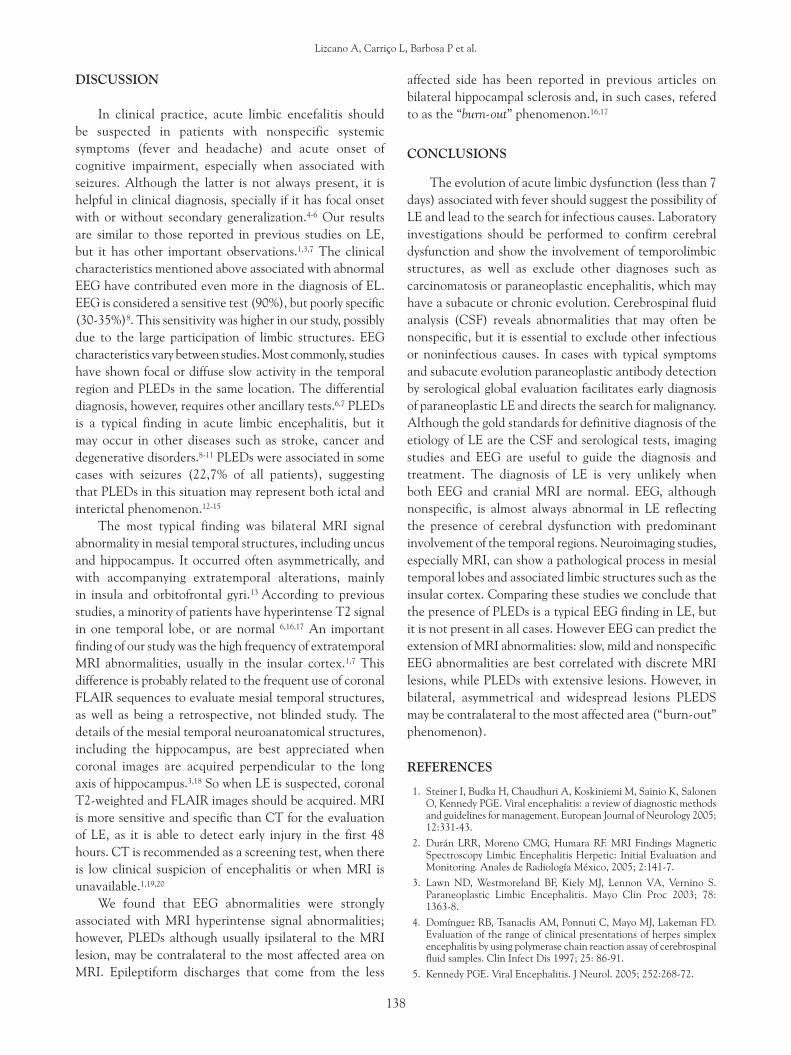
Encephalitis is the presence of inflammation in the brain parenchyma associated with clinical evidence of brain dysfunction. It can be due to a noninfective condition such as in acute disseminated encephalomyelitis (ADEM) or to an infection, which is diffuse and usually of viral origin. Herpes simplex virus type 1 (HSV-1), varicella-zoster virus (VZV), Epstein-Barr virus (EBV), mumps, measles and entero-viruses are responsible for most cases of viral encephalitis in immune competent individuals.1 Herpes simplex is the most common cause of viral encephalitis. The adult form of herpes simplex encephalitis is known as herpetic limbic encephalitis due to the virus predilection for the limbic system.2 Acute limbic encephalitis (LE) typically affects the mesial temporal lobe and limbic mesial cortical structures (cingulate gyrus, orbito-frontal cortex, and mamillary bodies).3 It usually involves one or both temporal lobes, in particular the hippocampus, amygdala, insular and parahipocampal gyrus. Spread may occur to the frontal and parietal lobes and it may lead to prominent memory and personality disturbances and seizures as sequelae (Fig. 1).2,3
A thorough diagnostic evaluation typically includes cranial magnetic resonance imaging (MRI), electro- encephalography (EEG), and cerebrospinal fluid (CSF) examination, but abnormalities may be nonspecific. This study was undertaken to define the spectrum of abnormalities found in the investigation of LE and to identify and compare any findings that may be specific to LE.3
PATIENTS AND METHODS
Clinical Material
Cases in which another neurological disorder was identified were excluded. Data from CT, MRI and CSF tests were considered to exclude other non-infective causes.
We retrospectively reviewed medical records of 57 patients with diagnosis of acute limbic encephalitis in the context of a febrile disease accompanied by headache, altered level of consciousness, and symptoms and signs of cerebral dysfunction (cognitive dysfunction, behavioural changes and seizures) with less than 7-day duration, from May 1994 to April 2010 and selected those with available EEG and MRI. We analyzed EEG characteristics (type of abnormality, location and periodicity), reviewed MRIs and compared both.
Laboratory Data
Electroencephalography was performed using analog and digital 16-channel and 21-channel instruments; scalp electrodes were placed according to the standard 10-20 International System. All EEGs were reviewed independently. Magnetic resonance imaging was performed according to standard clinical protocols. Sagittal and axial T1-weighted and T2-weighted images and thin-section coronal fluid-attenuated inversion recovery (FLAIR) sequences were obtained for all patients. Additionally CT was performed on the first day of admission. MRl abnormalities were evaluated qualitatively for each patient and compared with EEG results.
Figure 1. T1 weighted – IR (inversion recovery) coronal MRI. The structures most commonly involved of the limbic system in herpes encephalitis are identified (Yellow: cingulate gyrus; red: hippocampus; green: temporal lobe neocortex; blue: insula, pink: uncus; P: parahippocampal gyrus).
135
EEG and magnetic resonance imaging abnormalities in patients ...
RESULTS
Clinical Features
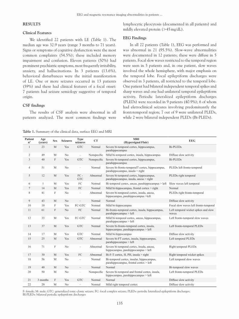
We identified 22 patients with LE (Table 1). The median age was 32.9 years (range 3 months to 71 years). Signs or symptoms of cognitive dysfunction were the most common complaints (54.5%); these included memory impairment and confusion. Eleven patients (50%) had prominent psychiatric symptoms, most frequently irritability, anxiety, and hallucinations. In 3 patients (13.6%), behavioral disturbances were the initial manifestation of LE. One or more seizures occurred in 13 patients (59%) and these had clinical features of a focal onset: 7 patients had seizure semiology suggestive of temporal origin.
CSF findings
The results of CSF analysis were abnormal in all patients analyzed. The most common findings were
lymphocytic pleocytosis (documented in all patients) and mildly elevated protein (>45 mg/dL).
EEG Findings
In all 22 patients (Table 1), EEG was performed and was abnormal in 21 (95.5%). Slow-wave abnormalities were documented in 12 patients; these were diffuse in 5 patients. Focal slow waves restricted to the temporal region were seen in 5 patients and, in one patient, slow waves involved the whole hemisphere, with major emphasis on the temporal lobe. Focal epileptiform discharges were observed in 3 patients, all restricted to the temporal lobe. One patient had bilateral independent temporal spikes and sharp waves and one had unilateral temporal epileptiform activity. Periodic lateralized epileptiform discharges (PLEDs) were recorded in 9 patients (40.9%); 6 of whom had eletroclinical seizures involving predominantly the frontotemporal region; 7 out of 9 were unilateral PLEDs, while 2 were bilateral independent PLEDs (Bi-PLEDs).
Table 1. Summary of the clinical data, surface EEG and MRI
Patient nº
Age (years) Sex Seizures Type
seizures CT MRI(Hypersignal Flair) EEG
1 23 M Yes GTC Normal Severe bi-temporal cortex, hippocampus, parahippocampus
Bi-PLEDs
2 49 M No – Nonspecific Mild bi-temporal cortex, insula, hippocampus Diffuse slow activity 3 40 F Yes GTC Nonspecific Severe bi-temporal cortex, hippocampus,
parahippocampusBi-PLEDs
4 51 M No – Normal Severe bi-fronto-temporalT cortex, hippocampus, parahippocampus, insula > right
PLEDs left fronto-temporal
5 12 M Yes FC -GTC
Abnormal Severe bi-temporal cortex, hippocampus, parahippocampus, insula, uncus > right
PLEDs right temporal
6 1 M Yes FC Normal Bi-temporal cortex, uncus, parahippocampus > left Slow waves left temporal 7 14 M Yes FC Normal Mild bi-hippocampus, frontal cortex > right Normal 8 41 F No – Abnormal Severe bi-temporal cortex, insula, uncus,
hippocampus, parahippocampus >leftPLEDs right fronto-temporal
9 43 M No – Normal Normal Diffuse slow activity 10 10 F Yes FC-GTC Normal Mild bi-hippocampus Focal slow waves left fronto-temporal11 61 F No FC Normal Bi-fronto-temporal cortex, insula, hippocampus,
parahippocampus > leftLeft temporal wicket spikes and slow waves
12 33 M Yes FC-GTC Normal Mild bi-temporal cortex, uncus, hippocampus, parahippocampus > left
Left fronto-temporal slow waves
13 37 M Yes GTC Normal Severe bi-fronto-temporal cortex, insula, hippocampus, parahippocampus > left
Left fronto-temporal PLEDs
14 17 M Yes GTC Normal Mild bi-hippocampus Diffuse slow activity 15 25 M Yes GTC Abnormal Severe bi-FT cortex, insula, hippocampus,
parahippocampus > leftLeft temporal PLEDs
16 71 F No – Abnormal Severe bi-temporal cortex, insula, uncus, hippocampus, parahippocampus > left
Right temporal PLEDs
17 30 M Yes FC Abnormal Bi-F-T cortex, H, PH, insula > right Right temporal wicket spikes18 56 M No – Normal Bi-temporal cortex, insular, hippocampus,
parahippocampus, frontal cortex > left Left temporal slow waves
19 40 M No – Normal Normal Bi-temporal slow waves20 50 M No – Nonspecific Severe bi-temporal and frontal cortex, insula,
hippocampus, parahippocampus > leftLeft fronto-temporal PLEDs
21 3 months F Yes GTC Normal Normal Diffuse slow activity 22 20 M No – Normal Mild right temporal cortex Diffuse slow activity
F: female; M: male; GTC: generalized tonic-clonic seizure; FC: focal complex seizure; PLEDs: periodic lateralized epileptiform discharges; BI-PLEDs: bilateral periodic epileptiform discharges

136
Lizcano A, Carriço L, Barbosa P et al.
Imaging Findings
CT was performed in all patients, with abnormal results related to research in only 5 and 3 patients with nonspecific findings.
Brain MRI was performed in all patients (Table 1) and images from 22 patients were available for retrospective review. Of these, 19 (86.4%) showed either symmetrical or asymmetrical temporal lobe abnormalities. Fifteen scans (68.2%) showed increased T2 and FLAIR signals in the mesial temporal region, 11 (50%) of which bilateral. Extratemporal increased signal was observed in 13 individuals (59.1%), mainly in the insular region and orbitofrontal cortex and subcortex.
Relation of MRI and EEG abnormalities
EEGs from the 2 patients with normal MRI showed focal or diffuse, nonspecific slow waves. This was also
present in EEGs from patients that had asymmetrical FLAIR hyperintensities over the temporal region, hippocampus and insula (Patient 1). The presence of PLEDs was strongly associated with hyperintense FLAIR signal involving hippocampus and temporal lobe neocortex. BI-PLEDs (2 patients) were related to extensive symmetrical bitemporal hyperintense FLAIR signal (Patient 2). Three patients with PLEDs on EEG had discrete ipsilateral and severe contra- lateral FLAIR-hyperintense abnormalities (Patient 3). In 4 patients with smaller bilateral asymmetrical lesions, we observed PLEDs in the most affected side (Patient 4). Diffuse slow waves were observed in 3 patients with discrete hyperintense signal on MRI (Patient 5).
Patient 1. Male, 56 years. A) EEG, longitudinal bipolar mountage: continuous slow activity in the left temporal region. B) MRI-FLAIR, coronal image: bilateral increased signal over the insula, hippocampus and temporal basal cortex.
Patient 2. Female, 40 years. A) EEG, referential (average) mountage: interval of 4sec between Bi-PLEDs in the temporal regions. B) MRI-FLAIR, coronal view: bilateral symmetrical hyperintense signal in the hippocampi, uncus and para- hippocampal gyri.
A B
A B

137
EEG and magnetic resonance imaging abnormalities in patients ...
Patient 3. Male, 51 years. A) EEG, longitudinal mountage: PLEDs occuring every 3 sec in the left temporal region. B) MRI-FLAIR, coronal image: hyperintense signal over medial temporal regions, parahippocampal cortex, hippocampus and insula, with right sided predominance.
Patient 4. Male, 37 years. A) EEG, longitudinal mountage: PLEDs occuring every 1.5 sec in the left temporal region. B) MRI-FLAIR, coronal image: hyperintense signal in mesial temporal structures, insular region, as well as in the supraorbital, inferior frontal gyrus and cingulate gyrus (not shown here).
Patient 5. Male, 20 years. A) EEG, longitudinal mountage: Diffuse slow activity. B) MRI-FLAIR, coronal image: hyperintense signal in the mesial temporal regions.
A B
A B
A B
138
Lizcano A, Carriço L, Barbosa P et al.
DISCUSSION
In clinical practice, acute limbic encefalitis should be suspected in patients with nonspecific systemic symptoms (fever and headache) and acute onset of cognitive impairment, especially when associated with seizures. Although the latter is not always present, it is helpful in clinical diagnosis, specially if it has focal onset with or without secondary generalization.4-6 Our results are similar to those reported in previous studies on LE, but it has other important observations.1,3,7 The clinical characteristics mentioned above associated with abnormal EEG have contributed even more in the diagnosis of EL. EEG is considered a sensitive test (90%), but poorly specific (30-35%)8. This sensitivity was higher in our study, possibly due to the large participation of limbic structures. EEG characteristics vary between studies. Most commonly, studies have shown focal or diffuse slow activity in the temporal region and PLEDs in the same location. The differential diagnosis, however, requires other ancillary tests.6,7 PLEDs is a typical finding in acute limbic encephalitis, but it may occur in other diseases such as stroke, cancer and degenerative disorders.8-11 PLEDs were associated in some cases with seizures (22,7% of all patients), suggesting that PLEDs in this situation may represent both ictal and interictal phenomenon.12-15
The most typical finding was bilateral MRI signal abnormality in mesial temporal structures, including uncus and hippocampus. It occurred often asymmetrically, and with accompanying extratemporal alterations, mainly in insula and orbitofrontal gyri.13 According to previous studies, a minority of patients have hyperintense T2 signal in one temporal lobe, or are normal 6,16,17 An important finding of our study was the high frequency of extratemporal MRI abnormalities, usually in the insular cortex.1,7 This difference is probably related to the frequent use of coronal FLAIR sequences to evaluate mesial temporal structures, as well as being a retrospective, not blinded study. The details of the mesial temporal neuroanatomical structures, including the hippocampus, are best appreciated when coronal images are acquired perpendicular to the long axis of hippocampus.3,18 So when LE is suspected, coronal T2-weighted and FLAIR images should be acquired. MRI is more sensitive and specific than CT for the evaluation of LE, as it is able to detect early injury in the first 48 hours. CT is recommended as a screening test, when there is low clinical suspicion of encephalitis or when MRI is unavailable.1,19,20
We found that EEG abnormalities were strongly associated with MRI hyperintense signal abnormalities; however, PLEDs although usually ipsilateral to the MRI lesion, may be contralateral to the most affected area on MRI. Epileptiform discharges that come from the less
affected side has been reported in previous articles on bilateral hippocampal sclerosis and, in such cases, refered to as the “burn-out” phenomenon.16,17
CONCLUSIONS
The evolution of acute limbic dysfunction (less than 7 days) associated with fever should suggest the possibility of LE and lead to the search for infectious causes. Laboratory investigations should be performed to confirm cerebral dysfunction and show the involvement of temporolimbic structures, as well as exclude other diagnoses such as carcinomatosis or paraneoplastic encephalitis, which may have a subacute or chronic evolution. Cerebrospinal fluid analysis (CSF) reveals abnormalities that may often be nonspecific, but it is essential to exclude other infectious or noninfectious causes. In cases with typical symptoms and subacute evolution paraneoplastic antibody detection by serological global evaluation facilitates early diagnosis of paraneoplastic LE and directs the search for malignancy. Although the gold standards for definitive diagnosis of the etiology of LE are the CSF and serological tests, imaging studies and EEG are useful to guide the diagnosis and treatment. The diagnosis of LE is very unlikely when both EEG and cranial MRI are normal. EEG, although nonspecific, is almost always abnormal in LE reflecting the presence of cerebral dysfunction with predominant involvement of the temporal regions. Neuroimaging studies, especially MRI, can show a pathological process in mesial temporal lobes and associated limbic structures such as the insular cortex. Comparing these studies we conclude that the presence of PLEDs is a typical EEG finding in LE, but it is not present in all cases. However EEG can predict the extension of MRI abnormalities: slow, mild and nonspecific EEG abnormalities are best correlated with discrete MRI lesions, while PLEDs with extensive lesions. However, in bilateral, asymmetrical and widespread lesions PLEDS may be contralateral to the most affected area (“burn-out” phenomenon).
REFERENCES
Steiner I, Budka H, Chaudhuri A, Koskiniemi M, Sainio K, Salonen 1. O, Kennedy PGE. Viral encephalitis: a review of diagnostic methods and guidelines for management. European Journal of Neurology 2005; 12:331-43.
Durán LRR, Moreno CMG, Humara RF. MRI Findings Magnetic 2. Spectroscopy Limbic Encephalitis Herpetic: Initial Evaluation and Monitoring. Anales de Radiología México, 2005; 2:141-7.
Lawn ND, Westmoreland BF, Kiely MJ, Lennon VA, Vernino S. 3. Paraneoplastic Limbic Encephalitis. Mayo Clin Proc 2003; 78: 1363-8.
Domínguez RB, Tsanaclis AM, Ponnuti C, Mayo MJ, Lakeman FD. 4. Evaluation of the range of clinical presentations of herpes simplex encephalitis by using polymerase chain reaction assay of cerebrospinal fluid samples. Clin Infect Dis 1997; 25: 86-91.
Kennedy PGE. Viral Encephalitis. J Neurol. 2005; 252:268-72.5.
139
EEG and magnetic resonance imaging abnormalities in patients ...
Tu¨zu¨n E, Dalmau J. Limbic Encephalitis and Variants: Classification, 6. Diagnosis and Treatment. The Neurologist 2007; 13:261-71.
Al-Shekhlee A, Kocharian N, Suarez JJ. Re-evaluating the Diagnostic 7. Methods in Herpes Simplex Encephalitis. HERPES 2006; 13:1.
Lai CW, Gragasin ME. Electroencepha-lography in herpes simplex 8. encephalitis. J Clin Neurophysiol. 1988;5: 87–103.
Roches JC, Probst A, Scollo-Lavizzari G. How specific are periodic 9. complexes in the diagnosis of herpes simplex encephalitis? Eur Neurol 1984; 23:466-71.
Verma NP, Kooi KA. Contralateral epilep-tiform transients in stroke 10. (CETS). Epilepsia 1986; 27:437-40.
Sainio K, Granstro¨m ML, Pettay O et al. EEG in neonatal herpes 11. simplex encepha-litis. Electroenceph Clin Neurophysiol 1983; 56: 556-61.
Daly DD, Markand ON. Focal brain lesions. In: Daly DD, Pedley 12. TA, eds. Current Practice of Clinical Electroencephalography. 2nd ed. New York, NY: Raven Press; 1990. 335-70.
Westmoreland BF. The EEG in cerebral inflammatory processes. 13. In: Niedermeyer E, Lopes Da Silva F, eds. Electroence-phalography. 4th ed. Baltimore: Williams & Wilkins; 1999. p. 302-16.
W. Fitzpatrick, Lowry N. PLEDs: Clinical Correlates. Can. J. Neurol. 14. Sci. 2007; 34:443-50
Giridhar P. Kalamangalam, Beate Diehl, Richard C. Burgess. 15. Neuroimaging and Neurophysiology of Periodic Lateralized Epileptiform Discharges: Observations and Hypotheses. Epilepsia 2007; 48(7):1396-405.
Dirr LY, Elster AD, Donofrio PD, Smith M. Evolution of brain MRI 16. abnormalities in limbic encephalitis. Neurology 1990; 40:1304-6.
Kodama T, Numaguchi Y, Gellad FE, Dwyer BA, Kristt DA. Magnetic 17. resonance imaging of limbic encephalitis. Neuroradiology 1991; 33: 520-3.
Jack Jr CR, Theodore WH, Cook M, McCarthy G. MRI-based 18. hippocampal volumetrics: data acquisition, normal ranges, and optimal protocol. Magn Reson Imaging 1995; 13:1057-64.
Whitley RJ, Kimberlin DW. Herpes simplex: encephalitis in children 19. and adolescents. Semin Pediatr Infect Dis 2005; 16:17-23.
Maschke M, Kastrup O, Forsting M, Diener HC. Update on 20. neuroimaging in infectious central nervous system disease. Curr Opin Neurol 2004; 17:475-80.
Cendes F, Dubeau F, Andermann F, Quesney LF, Gambardella A, 21. Jones-Gotman M, Bizi J, Oliver A, Gotman J, Arnold DL. Significance of mesial temporal atrophy in relation to intracranial ictal and interictal stereo EEG abnormalities. Brain 1996; 119(Pt 4):1317-26.
Mintzer S, Cendes F, Soss J, Andermann F, Engel Jr J, Dubeau F, 22. Olivier A, Fried I. Unilateral hippocampal sclerosis with contralateral temporal scalp ictal onset. Epilepsia. 2004;45(7): 792-802.
Endereço para correspondência:Fernando CendesDepto de Genética Clínica – UNICAMPCaixa Postal 6111CEP 13083-971, Campinas, São Paulo, BrasilE-mail: [email protected]


![individual’s - Home | Aetna Medicaid imaging], magnetic resonance spectroscopy (MRS), PET and SPECT) Neuropsychiatric EEG‐based assessment aid (NEBA) System Otoacoustic emissions](https://img.pdfslide.net/doc/110x75/5ad2c1137f8b9aff738d1421/individuals-home-aetna-medicaid-imaging-magnetic-resonance-spectroscopy.jpg)