Embed Size (px)
Citation preview
52International Journal of Scientifi c Study | February 2015 | Vol 2 | Issue 11
Effi cacy of Modifi ed Vacuum Assisted Closure in Wound HealingLoka Vijayan Siddha1, Sunil Kumar Shetty2, Thangam Varghese3
1Post-graduate, Department of General Surgery, Kasturba Medical College, Manipal University, Mangalore, Karnataka, India, 2Associate Professor, Department of General Surgery, Kasturba Medical College, Manipal University, Mangalore, Karnataka, India, 3Professor, Department of General Surgery, Kasturba Medical College, Manipal University, Mangalore, Karnataka, India
method is removal of blood and serous collection from the wound site by the application of negative pressure. This will be done by applying a piece of foam and a drain over the wound surface after debridement and is covered over by a semi permeable plastic adherent membrane securing it to skin margin and the drain is given connection to a vacuum creating unit. The plastic membrane forms like a barrier preventing the contamination from outside environment and the foam will help to distribute the negative pressure uniformly over the entire wound surface area preventing the chance of necrosis at a single place due to high pressure at a single place. The standardized average negative pressure applied is around −125 mm Hg. The interface material used in the VAC therapy stretches the cells at the base of the wound bed, promoting the response for divide and
INTRODUCTION
Vacuum assisted closure (VAC), may also be known as negative pressure wound therapy or Microdeformational wound therapy, which has brought a revolution in wound care since past 15 years. This method was fi rst described by Fleischmann et al. in 1993.1 The basic concept of this
Original Article
Abstract
Background: Vacuum assisted wound healing is a recent trend and proven method of fast and better healing of wounds. The basic concept is the removal of blood and serous collection from the wound site with negative pressure and promoting the healing process rapidly by altering the local microcellular environment.
Aim: The aim of this study is to evaluate effi cacy of the modifi ed method of vacuum dressing in wound healing in low resource settings.
Objectives: To fi nd out the rate of wound contraction, infection clearance, duration of hospital stay in comparison to betadine dressings.
Materials and Methods: In our prospective non-randomized comparative study, a total of 100 patients were taken and divided into two groups with 50 each for conventional betadine dressing and modifi ed vacuum assisted dressing. Vacuum dressing done with autoclaved sponge, opsite, glove, sterile plastic cover and creating a vacuum with 50 cc syringe, romovac, pedal suction, portable motorized suction apparatus. Comparison between the groups made in categories of wound cultures, wound area, wound scoring, duration of hospital stay, cost-effectiveness.
Results: Among 50 patients of vacuum dressing, 9 patients are excluded due to various reasons. There is 29.72% decrease in wound area in the experimental group than compared to 19.97% decrease in conventional dressing with P = 0.000. In wound scoring, 68.16% improvement is seen in the experimental group as compared with 57.10% in the control group with P = 0.002. There is 19.41% decreased duration of hospital stay in the experimental group. There is a signifi cant decrease in wound infection clearance of 63.4% in the experimental group as compared to 34% in the control group with P = 0.005. The median cost for modifi ed vacuum dressing was Rs. 311.1111 and compared to Rs. 610.5477 in the control group.
Conclusion: Modifi ed vacuum assisted dressing in low resource settings proven effective than conventional betadine dressing.
Key words: Negative pressure wound therapy, Opsite, Surgical glove
DOI: 10.17354/ijss/2015/52
Access this article online
www.ijss-sn.com
Month of Submission : 12-2014Month of Peer Review : 01-2015Month of Acceptance : 01-2015Month of Publishing : 02-2015
Corresponding Author: Dr. Loka Vijayan Siddha, Falnir Mens Hostel, Falnir, Mangalore - 575 001, Karnataka, India. Phone: +91-9901832864. E-mail: [email protected]
Siddha, et al.: Efficacy of Modified Vacuum Assisted Closure in Wound Healing
53 International Journal of Scientifi c Study | February 2015 | Vol 2 | Issue 11
proliferates. It also creates an environment of hypoxia over the surface leading to promotion of angiogenesis in addition to it keeps the wound warm, moist and prevents desiccation.
In the era of modern wound care negative pressure therapy for treatment of wounds has been routinely used and become integral part of the treatment plan. Its usage in acute, chronic, and complex wounds has been proven more effective and promotes for faster healing, early discharge with good quality of life with cost-effectiveness
The trademarked VAC therapy belonging to KCI VAC2 needed a sophisticated equipment with specialized foam and drain and trained personnel for application and maintenance, which is possible at high cost settings.
There are many studies done to make negative pressure dressing more cost effective and can also be practiced in low resource settings, like Danu and Rosadi,3 Singh et al.4 who have proven their effi cacy in using negative pressure therapy in low resource settings.
Main objectives of negative pressure therapy are:1. To promote rapid healing2. To decrease the frequency of change of dressings3. To prepare faster granulation bed for the wound for
change to other surgical intervention procedure4. To promote contraction of the wound edges5. To minimize the contamination of the wound6. To decrease the hospital stay.
In this study, we practiced to do the negative pressure dressing by using easily available materials to a surgeon and making in cost effective manner than compared to standard method and achieving similar results in a low resource setting.
MATERIALS AND METHODS
This prospective comparative study was undertaken at Kasturba Medical College Hospitals and Government Wenlock Hospital attached to Kasturba Medical College, Mangalore, India from October 2012 to September 2014. Ethical approval was obtained for this study from local ethical committee. A total of 100 patients in the study divided into experimental and control groups each of 50 patients in each group and all patients informed consent was taken. All patients are of above 18 years of age of both sexes. Modifi ed method of vacuum dressing applied for the experimental group, and conventional betadine dressing applied for the control group. For vacuum dressing, the inclusion and exclusion criteria are as follows:
Inclusion Criteria1. Chronic pressure ulcers2. Neuropathic ulcers3. Dehisced wounds or wounds with exposed bone/
tendons4. Partial thickness burns.
Exclusion Criteria1. Wounds of very large surface area (area more than 30%
body surface area, areas like groin, perineum, axilla)2. Malignancy in wound3. Cavity or sinus of unknown depth or origin4. Untreated osteomyelitis within vicinity of the wound5. Wound with unstable fractures or loose fragments of
bone6. Ulcers over the extremities with peripheral vascular
disease7. Wound with exposed blood vessels or organs8. Acute burns.
Materials Needed1. Autoclaved sponge foam (double autoclaved at
pressure of 20 PSI, 250°F for 30 min)4
2. Disposable syringes (10 cc, 20 cc, 50 cc), romovac, mucus suckers, pedal suction apparatus, portable electrical suction machine
3. Tegaderm/opsite/plastic cover/surgical glove of appropriate size
4. Suction catheter/Ryle’s tube/infant feeding tube5. Transparent adhesive tape/micropore6. Cling drape7. Graph paper8. Plastic sheet9. Marker pen.
Method of ApplicationAfter thoroughly debriding the wound from necrotic slough after hemostasis wound surface area is measured by imprint of plastic sheet over graph paper and recorded in cm2. Sponge foam which is normally available at hardware stores of 8 mm thickness is taken and is autoclaved and is cut in to shape of the wound with slightly larger size than the wound. Wound swab is taken for culture sensitivity.
Over the wound surface if there is clean granulation tissue present then bactigrass or Vaseline gauge can be applied so that while removing of dressing the sponge surface will not be adherent to the wound surface and during its removal bleeding can be reduced. A suction catheter/Ryle’s tube with adequate number of fenestrations made depending upon the wound size is placed in between the two sponge layers and the whole wound area is sealed with tegaderm/opsite/sterilized polyethylene cover5/sterile surgical glove.6 The exit site of the suction catheter to the opsite T-tailing
Siddha, et al.: Efficacy of Modified Vacuum Assisted Closure in Wound Healing
54International Journal of Scientifi c Study | February 2015 | Vol 2 | Issue 11
should be done to prevent the tubing exit site leakage.7 The suction catheter on the other end is connected to vacuum creating device and is charged.
The syringe/romovac/mucus sucker/pedal suction machine is cleared of drainage and recharged with vacuum after each clearance at timely intervals.
The method of application of dressings and the negative pressures that can be created with portable suction, romovac, syringe and mucus sucker are shown in Figures 1-6.
The negative pressure applied will be from −75 mm Hg to −200 mm Hg depending upon the type of modality used to create the vacuum. The characteristic of exuded fl uid and quantity is noted down. The wounds surroundings were inspected at time of change of dressing for any spreading cellulitis or maceration. Dressings are changed at intervals of 48-72 h depending upon the amount of exudates
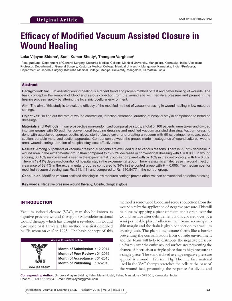
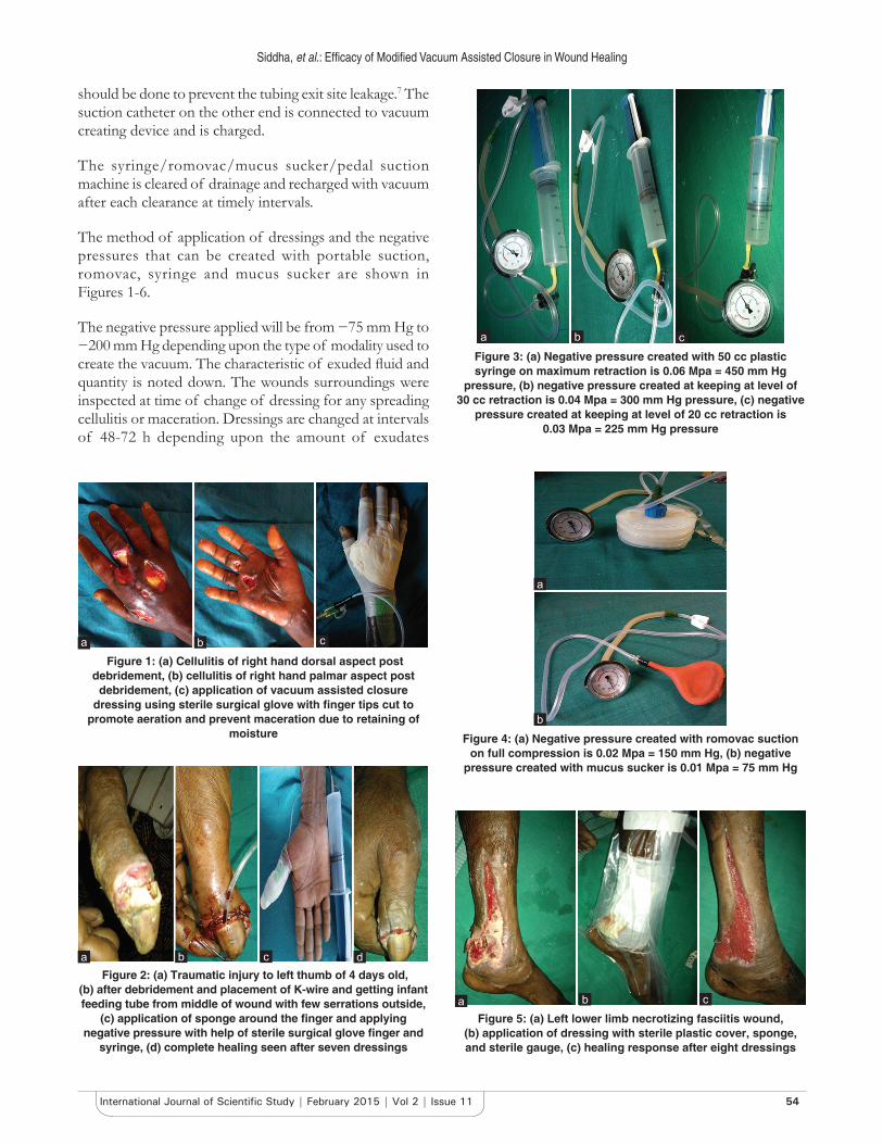
Figure 1: (a) Cellulitis of right hand dorsal aspect post debridement, (b) cellulitis of right hand palmar aspect post
debridement, (c) application of vacuum assisted closure dressing using sterile surgical glove with fi nger tips cut to
promote aeration and prevent maceration due to retaining of moisture
Figure 2: (a) Traumatic injury to left thumb of 4 days old, (b) after debridement and placement of K-wire and getting infant feeding tube from middle of wound with few serrations outside,
(c) application of sponge around the fi nger and applying negative pressure with help of sterile surgical glove fi nger and
syringe, (d) complete healing seen after seven dressings
Figure 3: (a) Negative pressure created with 50 cc plastic syringe on maximum retraction is 0.06 Mpa = 450 mm Hg
pressure, (b) negative pressure created at keeping at level of 30 cc retraction is 0.04 Mpa = 300 mm Hg pressure, (c) negative
pressure created at keeping at level of 20 cc retraction is 0.03 Mpa = 225 mm Hg pressure
Figure 4: (a) Negative pressure created with romovac suction on full compression is 0.02 Mpa = 150 mm Hg, (b) negative
pressure created with mucus sucker is 0.01 Mpa = 75 mm Hg
Figure 5: (a) Left lower limb necrotizing fasciitis wound, (b) application of dressing with sterile plastic cover, sponge, and sterile gauge, (c) healing response after eight dressings
cba
ba
dcba
c
a
b
a b c
Siddha, et al.: Efficacy of Modified Vacuum Assisted Closure in Wound Healing
55 International Journal of Scientifi c Study | February 2015 | Vol 2 | Issue 11
drained, leakage from the sealed area due to fl uid logging in and also upon the state of the wound and pictures of the wound and measurements are taken.
If maceration present then the next dressing will be applied after 12-24 h interval period to allow the skin to get back to the normal state. When surgical glove is used for dressing then, by creating small fenestrations in the distal end of the glove will allow the minimal air leak and decrease the maceration.
Wound swab and also the exudates obtained in the vacuum device are sent for culture at weekly intervals. The size of the wound is measured every time on change of dressing over the graph paper, and successive measurements will be recorded. The improvement in the wound is assessed by revised photographic wound assessment tool8 and the scores are noted. The total cost analysis of the dressing for each patient is analyzed separately and recorded.
The vacuum dressing are done till the granulation tissue of the wound fi lls till to the skin surface and left to healing by secondary intention or as secondary method of wound closure as secondary suturing, split skin grafting, fl ap repair.
RESULTS
In our study, among the experimental group nine patients were considered as failure (two patients underwent amputation, two patients developed leak in vacuum, one patient was not willing to continue, four patients not improved and changed to other modality of dressing) and these patients were excluded from study population of vacuum dressing, thereby experimental group n = 41, control group n = 50. The mean age distribution in the experimental group is 45.39 ± 9.95 and in control group 46.72 ± 7.63 and sex distribution in experimental
group 34 patients (82.9%) are males and 7 patients (17%) females, in control group 41 patients (82%) males and 9 patients (18%) females. The ulcers are located predominantly over lower limbs and other sites also like upper limb, clavicular region, abdomen, amputation stump, back, neck and scrotum.
The modality to create vacuum are by portable suction machine in 28 cases (68%), by using syringe in 10 cases (24%), pedal suction 2 cases (5%) and using romovac in 1 case (3%), by comparing with Fischer’s exact test P = 0.0001 proving highly signifi cant. The materials used for vacuum dressing are with opsite in 21 cases (51%), surgical glove in 12 cases (29%) and plastic cover in 8 cases (20%). On comparison by mode of healing in both groups split skin grafting was done in 41 (82%) cases and 22 (53.7%) cases in control and experimental groups, respectively and healing by secondary intention in 9 (18%) and 17 (41.5%) cases in control and experimental groups respectively, scrotum reconstruction done in 1 (2.4%) case in experimental group, and secondary suturing in 1 (2.4%) case in experimental group with P = 0.008 proving highly signifi cant.
The effi cacy of wound healing indicated by clearing the infection is measured by sequential wound swab cultures in both experimental and control group and the results are shown in Table 1.
In Culture 1 the predominant organisms being pseudomonas in 16 (40%) and 19 (38%) cases in experimental and control group and Staphylococcus aureus in 16 (40%) and 24 (48%) cases in experimental and control group and Klebisiella in 4 (10%) and 2 (4%) cases in experimental and control group. In Culture 3 S. aureus in 12 (80.0%) and 27 (81.8%) cases in experimental and control group and Klebisiella in 3 (20%) cases in experimental group and proteus in 3 (9%) cases and pseudomonas in 3 (9%) cases of control group.
The wound healing is also compared between the experimental and control groups in the parameters such as wound areas initial and fi nal, number of debridements, number of dressings, number of days of hospital stay,
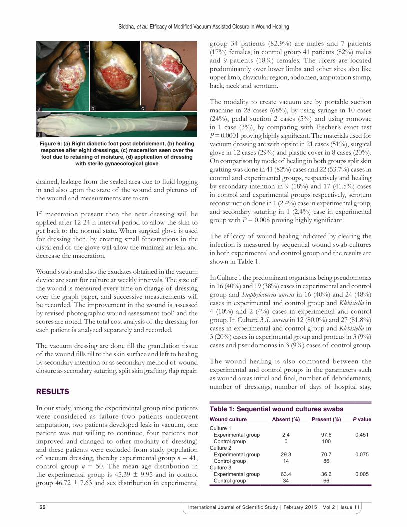
Figure 6: (a) Right diabetic foot post debridement, (b) healing response after eight dressings, (c) maceration seen over the foot due to retaining of moisture, (d) application of dressing
with sterile gynaecological glove
Table 1: Sequential wound cultures swabsWound culture Absent (%) Present (%) P valueCulture 1
Experimental group 2.4 97.6 0.451Control group 0 100
Culture 2Experimental group 29.3 70.7 0.075Control group 14 86
Culture 3Experimental group 63.4 36.6 0.005Control group 34 66
d
cba
Siddha, et al.: Efficacy of Modified Vacuum Assisted Closure in Wound Healing
56International Journal of Scientifi c Study | February 2015 | Vol 2 | Issue 11
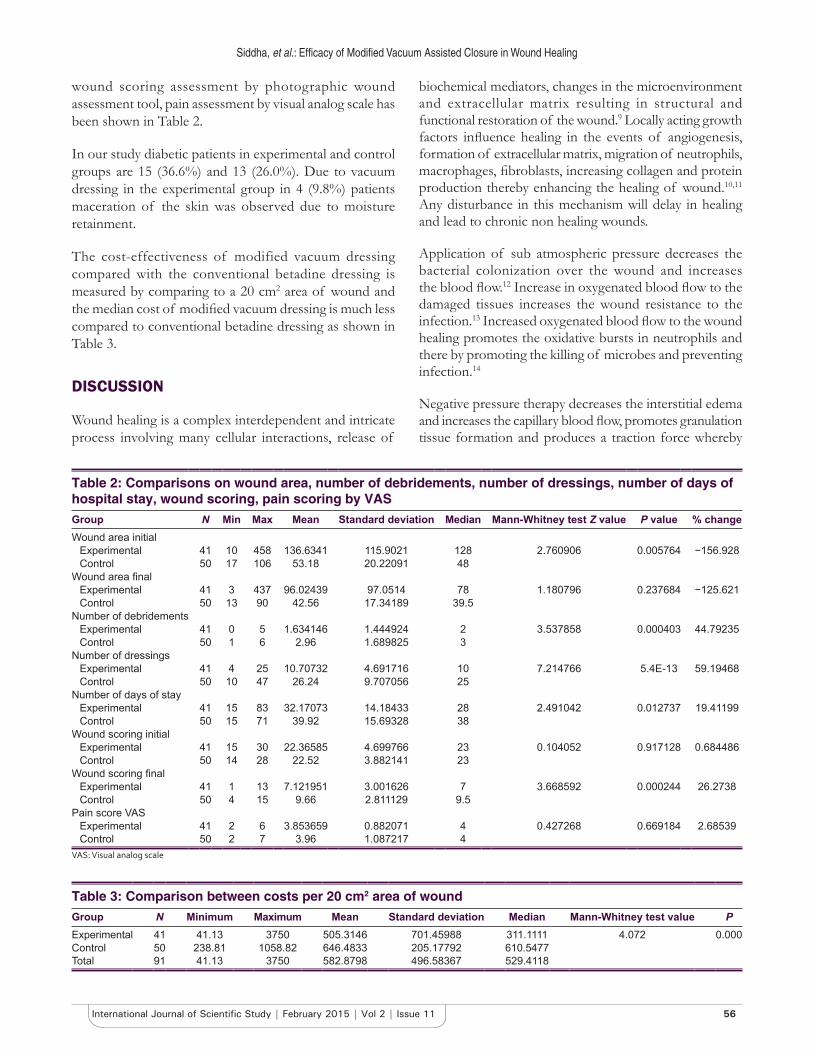
wound scoring assessment by photographic wound assessment tool, pain assessment by visual analog scale has been shown in Table 2.
In our study diabetic patients in experimental and control groups are 15 (36.6%) and 13 (26.0%). Due to vacuum dressing in the experimental group in 4 (9.8%) patients maceration of the skin was observed due to moisture retainment.
The cost-effectiveness of modified vacuum dressing compared with the conventional betadine dressing is measured by comparing to a 20 cm2 area of wound and the median cost of modifi ed vacuum dressing is much less compared to conventional betadine dressing as shown in Table 3.
DISCUSSION
Wound healing is a complex interdependent and intricate process involving many cellular interactions, release of
biochemical mediators, changes in the microenvironment and extracellular matrix resulting in structural and functional restoration of the wound.9 Locally acting growth factors infl uence healing in the events of angiogenesis, formation of extracellular matrix, migration of neutrophils, macrophages, fi broblasts, increasing collagen and protein production thereby enhancing the healing of wound.10,11 Any disturbance in this mechanism will delay in healing and lead to chronic non healing wounds.
Application of sub atmospheric pressure decreases the bacterial colonization over the wound and increases the blood fl ow.12 Increase in oxygenated blood fl ow to the damaged tissues increases the wound resistance to the infection.13 Increased oxygenated blood fl ow to the wound healing promotes the oxidative bursts in neutrophils and there by promoting the killing of microbes and preventing infection.14
Negative pressure therapy decreases the interstitial edema and increases the capillary blood fl ow, promotes granulation tissue formation and produces a traction force whereby
Table 2: Comparisons on wound area, number of debridements, number of dressings, number of days of hospital stay, wound scoring, pain scoring by VASGroup N Min Max Mean Standard deviation Median Mann-Whitney test Z value P value % changeWound area initial
Experimental 41 10 458 136.6341 115.9021 128 2.760906 0.005764 −156.928Control 50 17 106 53.18 20.22091 48
Wound area fi nalExperimental 41 3 437 96.02439 97.0514 78 1.180796 0.237684 −125.621Control 50 13 90 42.56 17.34189 39.5
Number of debridementsExperimental 41 0 5 1.634146 1.444924 2 3.537858 0.000403 44.79235Control 50 1 6 2.96 1.689825 3
Number of dressingsExperimental 41 4 25 10.70732 4.691716 10 7.214766 5.4E-13 59.19468Control 50 10 47 26.24 9.707056 25
Number of days of stayExperimental 41 15 83 32.17073 14.18433 28 2.491042 0.012737 19.41199Control 50 15 71 39.92 15.69328 38
Wound scoring initialExperimental 41 15 30 22.36585 4.699766 23 0.104052 0.917128 0.684486Control 50 14 28 22.52 3.882141 23
Wound scoring fi nalExperimental 41 1 13 7.121951 3.001626 7 3.668592 0.000244 26.2738Control 50 4 15 9.66 2.811129 9.5
Pain score VASExperimental 41 2 6 3.853659 0.882071 4 0.427268 0.669184 2.68539Control 50 2 7 3.96 1.087217 4
VAS: Visual analog scale
Table 3: Comparison between costs per 20 cm2 area of woundGroup N Minimum Maximum Mean Standard deviation Median Mann-Whitney test value PExperimental 41 41.13 3750 505.3146 701.45988 311.1111 4.072 0.000Control 50 238.81 1058.82 646.4833 205.17792 610.5477Total 91 41.13 3750 582.8798 496.58367 529.4118
Siddha, et al.: Efficacy of Modified Vacuum Assisted Closure in Wound Healing
57 International Journal of Scientifi c Study | February 2015 | Vol 2 | Issue 11
decreases the wound surface area and increases the mitoticity in cells around the area.15
It has been proposed four primary mechanisms for healing by negative pressure therapy1. Macrodeformation or wound shrinkage at the base2. Microdeformation near the interface sponge3. Removal of excess fl uid4. Optimizing the wound environment.16
MacrodeformationIt refers to decrease in the wound surface due to shrinkage of sponge and action of centripetal forces over the wound surface. In studies made by Borgquist et al. in porcine models, exposing the sponge to negative pressure of 125 mm Hg will decrease the foam volume by 80% leading to decrease in wound surface area.17 Due to the inherent tension which is present in the dermis near the wound and underlying attachments of different wounds over different sites contract to a different extent. The macrodeformation effects depend upon the variety of tissues, amount of negative pressure, volume of the interface material, and the surrounding tissues deformability.16
MicrodeformationThe negative pressure transmitted through interface material acting over the undulated surface of the wound produces changes occur in μ to mm range scale. Depending on the common diameters of pores of interface material in range of 400-600 μm, on application of the negative pressure there will be 5-20% tissue strain over the wound surface.18 These mechanical forces are transmitted to every cell through the extracellular matrix and lead to cell deformation causing modifi cations in cell function for adaptation of stress.19,20
Removal of Excess FluidThe total body fl uid is distributed in three compartments. They are: (1) Intracellular, (2) extracellular (3) intravascular. Translocation of fl uid in between these compartments across the semi permeable membrane is governed by the differential between osmotic and hydrostatic pressures derived by Starling’s equation. Extracellular compartment is the most variable compartment among the three. Excess fl uid in this compartment leads to edema and deprivement leads to signs of dehydration. This compartment is drained by lymphatics; abnormality in this may lead to lymphedema.
Chronic wounds and edema are often concomitant more commonly in lower limbs. Excess of fl uid will lead to delay in healing due to the compressive effect exerted over the tissues. While healing intrinsic tension will be developed within the individual cells through their cytoskeleton and extracellular matrix interactions, increased fl uid pressure
will dampen the building up intrinsic tension and prevent proliferative response.16
Removal of this excess fl uid will decrease the compression of microvasculature there by promoting the perfusion to the local area.21 The semi permeable nature of the occlusive drape will allow a little leakage of air into the system, which helps in preventing the fluid lock and thereby allowing the evacuation of fl uid continuously. Along with excess extracellular fl uid toxins formed over the wound and microbes were also cleared by the negative pressure therapy.22 Negative pressure therapy also allows for developing of lymphatics at the wound edges thereby improving the fl uid drainage.23 The semi occlusive drape is not permeable to microorganisms thereby signifi cantly reducing the contamination and also helps in maintaining moist and warmth environment, which promotes the healing response.24,25
Conditions where negative pressure therapy is contraindicated are:1. Untreated osteomyelitis2. Unexplored and nonenteric fi stulas3. Necrotic tissue along with eschar4. Exposed blood vessels5. Wounds with malignancy6. Exposed nerves7. Exposed anastamotic sites8. Exposed internal organs.26
FDA has proposed risk factors and other warrant conditions before consideration of a patient of negative pressure therapy. They are:1. Treatment with platelet aggregation inhibitors or
anticoagulants2. High risk for bleeding3. Infected blood vessels, wounds, osteomyelitis, exposed
blood vessels, nerves, tendons, ligaments, anastamosis, spinal cord injuries, enteric fi stulas, sharp edges at wound edges
4. Patient requirement for hyperbaric oxygen therapy, magnetic resonance imaging, defi brillation
5. Patient size and weight6. Circumferential dressing application7. Proximity of foam to the vagus nerve8. Continuous or intermittent suction application.26,27
The negative pressure therapy will cause deformations in the cell cytoskeleton architecture leading to cellular proliferation, differentiation, and migration. This has been supported by studies in diabetic mouse model by application of short term intermittent negative pressure there is increased expression of Ki67 which is a marker for proliferation.28
Siddha, et al.: Efficacy of Modified Vacuum Assisted Closure in Wound Healing
58International Journal of Scientifi c Study | February 2015 | Vol 2 | Issue 11
Negative pressure therapy treated wounds in the proliferation phase there will be robust granulation tissue, proliferation of cells, angiogenesis and the maturation of collagen exhibit mast cell dependence in proliferation and remodeling phases.
Morykwas et al. studies showed a decrease in the bacterial load in wounds treated with negative pressure therapy,29 Mouës et al. studies showed there is a decrease in non fermentive Gram-negative bacilli and S. aureus increased.30 The effect of negative pressure therapy on bacterial culture from the wounds should be more studied particularly in responses of different strains that are elicited. Traditional VAC dressing’s uses polyurethane ether foam, reduction of bacterial load can be achieved in the wound by silver coating added to the foam. Stinner et al. study in the goat model with silver dressings placed beneath the foam in complex wounds with high bacterial load demonstrated reduction in bacterial growth particularly S. aureus when compared to standard VAC dressings.31 Instillation therapy adding of fl uid to the wound through a tubing in form of normal saline or other antimicrobials like sodium hypochlorite solution, dilute betadine, doxycycline, phenytoin, lactoferrin are done but trials are needed to prove its effi cacy.
Treatment by negative pressure therapy provides cosmetic as well as functional outcomes by promoting the local vascularity and decrease in scar height. Negative pressure therapy can be used for preparation of recipient sites for dermal scaffolds and skin grafts over exposed bones or tendons which provide complete vascularized wound bed before skin grafting.
For the treatment with negative pressure therapy, many factors to be considered in view of goal of therapy, type of dressing, suction pressure application. For different types of wounds, there is different amount pressure protocols and the duration of treatment changes. In acute wounds, it is benefi cial to start within 48 h initially with continuous suction followed by intermittent suction therapy. For chronic wounds they benefi t more by continuous negative pressure therapy. Short and intermittent negative pressure therapy32 shows improved tissue response than compared to the continuous effect, but it may not be applicable for all types of cases. Intermittent negative pressure therapy may not be tolerated by some patients due to discomfort. The optimal pressure to be applied for improvement of the wound is not yet currently known, there are different studies with application from −75 mm Hg to −150 mm Hg pressure and achieved good healing responses. Frequent change of vacuum dressings may be required for wounds with increased risk of infection.
All wounds are not amenable to negative pressure therapy. Due to hypersensitivity for the adhesive drape and pain
caused due to the suction effect some patients may not tolerate the therapy. Pain can be reduced by modalities like decreasing the negative pressure, and if pain is from the surrounding skin then framing the wound with hydrocolloid dressing at the borders and adhesive drape can be placed over hydrocolloid so that the friction force is relieved over the skin. Tissue integrity must be checked during every change of dressing. If hematoma or bruises appears over the wound negative pressure should be decreased, if still persists then negative pressure therapy should be discontinued substituted by alternative type of dressing.
CONCLUSION
Through our study it has been proven that modifi ed vacuum assisted therapy is more benefi cial when compared to the conventional moist betadine dressings, compared in parameters of granulation tissue formation, clearance of the infection over the wound, decreasing the duration of hospital stay, and cost effectiveness than compared to moist dressings.
REFERENCES
1. Fleischmann W, Strecker W, Bombelli M, Kinzl L. Vacuum sealing as treatment of soft tissue damage in open fractures. Unfallchirurg 1993;96:488-92.
2. KCI VAC, 2014. Available from: http://www.kci1.com/KCI1/educationtraining. [Last accessed on 2014 Nov 11].
3. Danu M, Rosadi S. The simplest modifi ed vacuum assisted closure to treat chronic wound: Serial case report. J Plastik Rekonstruksi 2012;1:117-22.
4. Singh M, Singh R, Singh S, Pandey V, Singh D. Vacuum assisted closure in wound management – Poor man’s VAC. Int J Plast Surg 2008;6.
5. Kumar P. Exploiting potency of negative pressure in wound dressing using limited access dressing and suction-assisted dressing. Indian J Plast Surg 2012;45:302-15.
6. Hemmanur SR, Siddha LV. Role of the surgical glove in modifi ed vacuum-assisted wound healing. Arch Plast Surg 2013;40:630-2.
7. Ram S, Vijayan SK, Kini A. T tail confi guration of opsite on suction tubing outlet for modifi ed vacuum assisted closure. Int J Plast Surg 2012;8.
8. Thompson N, Gordey L, Bowles H, Parslow N, Houghton P. Reliability and validity of the revised photographic wound assessment tool on digital images taken of various types of chronic wounds. Adv Skin Wound Care 2013;26:360-73.
9. Clarkeand RA, Henson PM, editors. The Molecular and Cellular Biology of Wound Repair. NewYork, NY, USA: Plenum Press; 1988.
10. Laiho M, Keski-Oja J. Growth factors in the regulation of pericellular proteolysis: A review. Cancer Res 1989 15;49:2533-53.
11. Whitby DJ, Ferguson MW. Immunohistological studies of the extracellular matrix and soluble growth factors in fetal and adult wound healing. In: Adzick NS, Longaker MT, editors. Fetal Wound Healing. New York, NY, USA: Elsevier Science; 1992. p. 161-77.
12. Argenta LC, Morykwas MJ. Vacuum-assisted closure: A new method for wound control and treatment: Clinical experience. Ann Plast Surg 1997;38:563-76.
13. Hunt TK. The physiology of wound healing. Ann Emerg Med;17:1265-73.14. Ryan TJ. Microcirculation in psoriasis: Blood vessels, lymphatics and
tissue fl uid. Pharmacol Ther 1980;10:27-64.15. Leininger BE, Rasmussen TE, Smith DL, Jenkins DH, Coppola C.
Experience with wound VAC and delayed primary closure of contaminated soft tissue injuries in Iraq. J Trauma 2006;61:1207-11.
Siddha, et al.: Efficacy of Modified Vacuum Assisted Closure in Wound Healing
59 International Journal of Scientifi c Study | February 2015 | Vol 2 | Issue 11
16. Orgill DP, Manders EK, Sumpio BE, Lee RC, Attinger CE, Gurtner GC, et al. The mechanisms of action of vacuum assisted closure: More to learn. Surgery 2009;146:40-51.
17. Borgquist O, Ingemansson R, Malmsjö M. The infl uence of low and high pressure levels during negative-pressure wound therapy on wound contraction and fl uid evacuation. Plast Reconstr Surg 2011;127:551-9.
18. Saxena V, Hwang CW, Huang S, Eichbaum Q, Ingber D, Orgill DP. Vacuum-assisted closure: Microdeformations of wounds and cell proliferation. Plast Reconstr Surg 2004;114:1086-96.
19. Ingber DE, Levin M. What lies at the interface of regenerative medicine and developmental biology? Development 2007;134:2541-7.
20. McLeod KJ, Lee RC, Ehrlich HP. Frequency dependence of electric fi eld modulation of fi broblast protein synthesis. Science 1987;236:1465-9.
21. Adámková M, Tymonová J, Zámecníková I, Kadlcík M, Klosová H. First experience with the use of vacuum assisted closure in the treatment of skin defects at the burn center. Acta Chir Plast 2005;47:24-7.
22. Lancerotto L, Bayer LR, Orgill DP. Mechanisms of action of microdeformational wound therapy. Semin Cell Dev Biol 2012;23:987-92.
23. Labanaris AP, Polykandriotis E, Horch RE. The effect of vacuum-assisted closure on lymph vessels in chronic wounds. J Plast Reconstr Aesthet Surg 2009;62:1068-75.
24. Winter GD, Scales JT. Effect of air drying and dressings on the surface of a wound. Nature 1963;197:91-2.
25. Hinman CD, Maibach H. Effect of air exposure and occlusion on experimental human skin wounds. Nature 1963;200:377-8.
26. V.A.C. Therapy Indications and Contraindications. Available from: http://www.kci1.com/KCI1/indicationsandcontraindications. [Last accessed on 2014 Nov 12].
27. FDA Safety Communication: UPDATE on Serious Complications Associated with Negative Pressure Wound Therapy Systems, 2011. Available from: http://www.fda.gov/MedicalDevices/Safety/AlertsandNotices/ucm244211.htm. [Last accessed on 2014 Nov 12].
28. Scherer SS, Pietramaggiori G, Mathews JC, Orgill DP. Short periodic applications of the vacuum-assisted closure device cause an extended tissue response in the diabetic mouse model. Plast Reconstr Surg 2009;124:1458-65.
29. Morykwas MJ, Simpson J, Punger K, Argenta A, Kremers L, Argenta J. Vacuum-assisted closure: State of basic research and physiologic foundation. Plast Reconstr Surg 2006;117:121S-6.
30. Mouës CM, Vos MC, van den Bemd GJ, Stijnen T, Hovius SE. Bacterial load in relation to vacuum-assisted closure wound therapy: A prospective randomized trial. Wound Repair Regen 2004;12:11-7.
31. Stinner DJ, Waterman SM, Masini BD, Wenke JC. Silver dressings augment the ability of negative pressure wound therapy to reduce bacteria in a contaminated open fracture model. J Trauma 2011;71:S147-50.
32. Mendez-Eastman S. Guidelines for using negative pressure wound therapy. Adv Skin Wound Care 2001;14:314-22.
How to cite this article: Siddha LV, Shetty SK, Varghese T. Effi cacy of Modifi ed Vacuum Assisted Closure in Wound Healing. Int J Sci Stud 2015;2(11):52-59.
Source of Support: Nil, Confl ict of Interest: None declared.