Embed Size (px)
Citation preview
UCTEA Chamber of Metallurgical & Materials Engineers Proceedings Book
328 IMMC 2016 | 18th International Metallurgy & Materials Congress
Eff ect of Time on the Direct Formation of Hydroxyapatite Coatings on Commercially Pure Zirconium by Plasma Electrolytic Oxidation
Salim Levent Aktuğ¹, Metin Usta¹,²
¹Gebze Technical University, ²TÜBİTAK Marmara Research Center - Türkiye
Abstract In this study, hydroxyapatite (HAp) and calcium-based ceramic composite coatings were produced directly on commercially pure zirconium by plasma electrolytic oxidation (PEO) in an electrolyte at 0.260 A/cm2 current densities for 5, 10 and 15 minutes. The phase structure, Cross-sectional morphology of the coatings, surface roughness and coating thickness were characterized by X-ray diffraction (XRD), scanning electron microscope (SEM), electron probe micro-analysis via wavelength-dispersive spectroscopy (EPMA-WDS), surface profilometer and eddy current method respectively. The XRD results showed that Zr, c-ZrO2, Ca0.15Zr0.85O1.85, CaZrO3 and Ca10(PO4)6(OH)2 (HAp) phases were formed on the surface of the commercially pure zirconium. Surface roughness of coatings were measured as of 2.04, 3.44 and 4.33 m for 5, 10 and 15 min, respectively. The average coating thicknesses were measured as 17.7, 25.8 and 34.4
m for 5, 10 and 15 min Hydroxyapatite and calcium-based bioceramic coatings on commercially pure zirconium produced by PEO method have potential in terms of biomaterial application areas such as dentistry and surgery. 1. Introduction Zirconium and its alloys that have good biocompatibility, excellent corrosion resistance and higher mechanical properties are widely used in dental and orthopedic implant applications [1-3]. However, zirconium does not interact with the adjacent tissues after being inserted surgically into the body [4]. There are some limitations on using zirconium due to the chemical binding and fixing problems to the bone tissues [5]. Hydroxyapatite (HA) which exhibits bioactive and biocompatible characteristics is used in biomedical applications because synthetic hydroxyapatite is similar to bone and tooth mineral [6]. The use of hydroxyapatite itself is limited in biomedical applications due to its brittleness, low fatigue resistance and poor mechanical properties [7, 8]. Various coating methods have been developed for
zirconium and its alloys to deposit the apatite coating on the surface in recent years in order to both use the capabilities of the HA and improve poor properties of the HA at the same time. These methods are SBF immersion [9], sol-gel technique [10], anodization [11] and chemical treatment [12]. There is another method in use which is called plasma electrolytic oxidation (PEO) to enhance bioactivity and biocompatibility of the zirconium and its alloys surface. However, these studies compared to PEO method are limited and rare. PEO technique is well-developed coating method for biometals such as titanium, magnesium and zirconium in order to produce ceramic oxides on the surface. The composite coatings that have hard, wear-resistant, better adhesion to the base material, porous, high corrosion and heat-resistance properties are produced by PEO [13]. Although hydroxyapatite coated phase are generally formed on zirconium by PEO at two stages in the literature [14-16], this is a time consuming process which takes too much operation time for days even weeks to achieve HA phase in the coating structure. However, with a single process it is possible for this to produce directly HA coating on the zirconium substrate by PEO [17]. In this work, HA and calcium-based coatings were directly coated on the commercially pure zirconium surface by PEO method at different treatment times. The aim of this study was to investigate effect on time for the production of hydroxyapatite and calcium apatite-based coatings that are bioactive and biocompatible to be used as a biomaterial in biomedical applications by PEO method. The surface properties of the coated zirconium were characterized to investigate the phase structures, cross-sectional morphologies and chemical compositions of the surfaces in the coating by X-ray diffraction (XRD), scanning electron microscopy (SEM), electron probe micro-analysis via wavelength-dispersive spectroscopy (EPMA-WDS), surface profilometer and eddy current method respectively.
TMMOB Metalurj i ve Malzeme Mühendisleri Odas ıBildir i ler Kitab ı
32918. Uluslararas ı Metalurj i ve Malzeme Kongresi | IMMC 2016
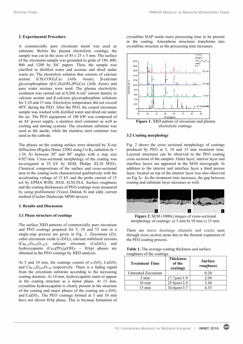
2. Experimental Procedure A commercially pure zirconium metal was used as substrate. Before the plasma electrolytic coatings, the sample was cut in the sizes of 45 x 25 x 5 mm. The surface of the zirconium sample was grounded to grids of 180, 400, 800 and 1200 by SiC papers. Then, the sample was clarified in distilled water and acetone and dried under warm air. The electrolyte solution that consists of calcium acetate (CH3COO)2Ca) (Alfa Aesar), -calcium glycerophosphate ( -C3H5(OH2)PO4Ca) (Alfa Aesar) and pure water mixture were used. The plasma electrolytic oxidation was carried out at 0,260 A/cm2 current density in calcium acetate and -calcium glycerophosphate solutions for 5,10 and 15 min. Electrolyte temperature did not exceed 40oC during the PEO. After the PEO, the coated zirconium sample was washed with distilled water and dried out under the air. The PEO equipment of 100 kW was composed of an AC power supply, a stainless steel container as well as cooling and stirring systems. The zirconium substrate was used as the anode, while the stainless steel container was used as the cathode. The phases on the coating surface were detected by X-ray diffraction (Rigaku Dmax 2200) using Cu-K radiation (k = 1.54 Å) between 20o and 80o angles with a step size 0.02o/min. Cross-sectional morphology of the coating was investigated at 25 kV by SEM; Philips XL30 SFEG. Chemical compositions of the surface and cross-sectional area in the coating were characterized qualitatively with the accelerating voltage of 15 kV and the probe current of 15 nA by EPMA-WDS; JEOL 8230-JXA. Surface roughness and the coating thicknesses of PEO coatings were measured by using profilometer (Veeco Dektak 8) and eddy current method (Fischer Dualscope MP40 device). 3. Results and Discussion 3.1 Phase structure of coatings The surface XRD patterns of commercially pure zirconium and PEO coatings prepared for 5, 10 and 15 min in a single-step process are given in Fig. 1. Zirconium (Zr), cubic-zirconium oxide (c-ZrO2), calcium stabilized zirconia (Ca0.15Zr0.85O1.85), calcium zirconate (CaZrO3) and hydroxyapatite (Ca10(PO4)6(OH)2 ~ HAp) phases are obtained in the PEO coatings by XRD analysis. At 5 and 10 min, the coatings consist of c-ZrO2, CaZrO3 and Ca0.15Zr0.85O1.85 respectively. There is a fading signal from the zirconium substrate according to the increasing coating duration. At 10 min, hydroxyapatite starts to appear in the coating structure as a minor phase. At 15 min, crystalline hydroxyapatite is clearly present in the structure of the coating and major phases of the coating are c-ZrO2 and CaZrO3. The PEO coatings formed at 5 and 10 min have not shown HAp phase. This is because formation of
crystalline HAP needs more processing time to be present in the coating. Amorphous structures transforms into crystalline structure as the processing time increases.
Figure 1. XRD pattern of zirconium and plasma
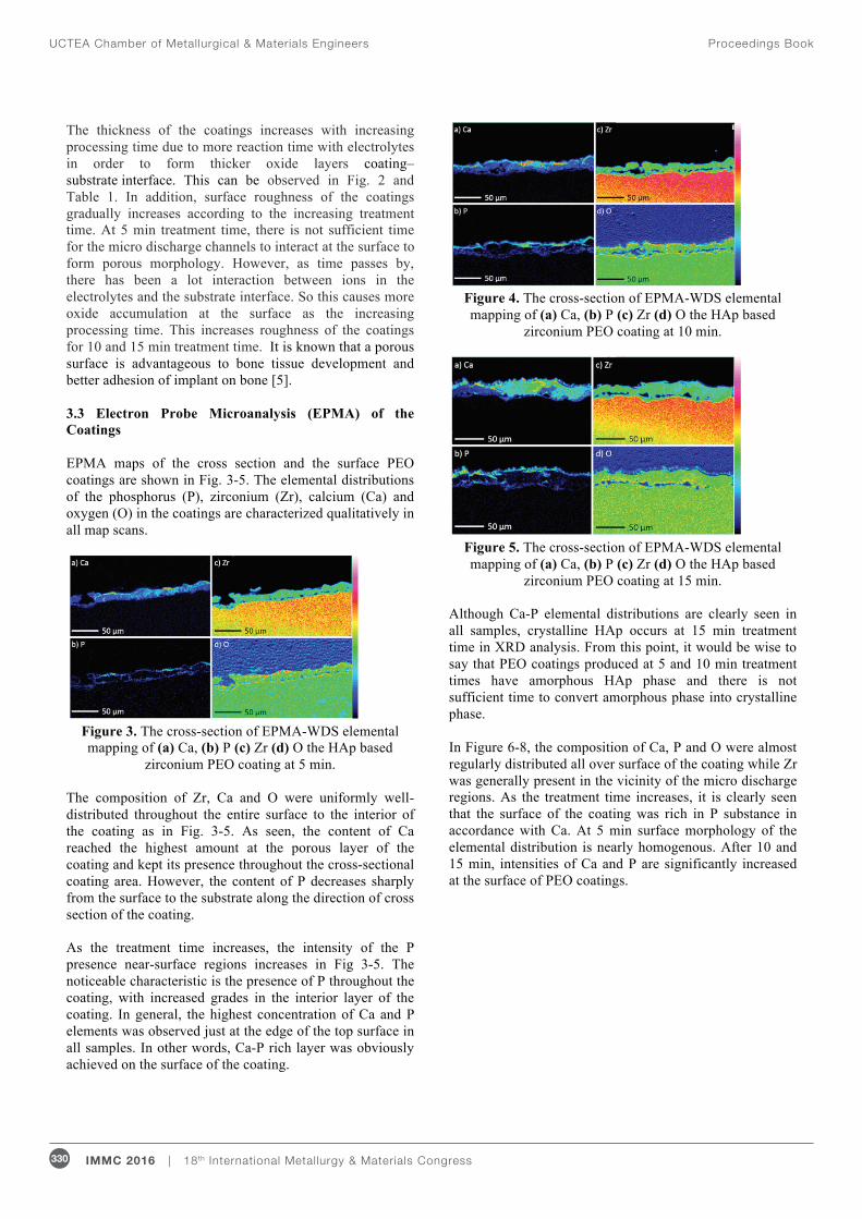
electrolytic coatings. 3.2 Coating morphology Fig. 2 shows the cross sectional morphology of coatings produced by PEO at 5, 10 and 15 min treatment time. Layered structures can be observed in the PEO coating cross sections of the samples. Outer layer, interior layer and interface layers are appeared in the SEM micrograph. In addition to the interior and interface layer a third porous layer, located on top of the interior layer was also observed on Fig 2c. As the treatment time increases, the gap between coating and substrate layer increases as well.
Figure 2. SEM (1000x) images of cross-sectional
morphology of coatings: a) 5 min b) 10 min c) 15 min. There are micro discharge channels and cracks seen through cross section areas due to the thermal expansion of the PEO coating process. Table 1. The average coating thickness and surface roughness of the coatings.
Treatment Time Thickness
of the coatings
Surface roughness
Untreated Zirconium - 0.20 5 min 17.7μm±1.9 2.04
10 min 25.8μm±2.4 3.44 15 min 34.4μm±3.7 4.33
UCTEA Chamber of Metallurgical & Materials Engineers Proceedings Book
330 IMMC 2016 | 18th International Metallurgy & Materials Congress
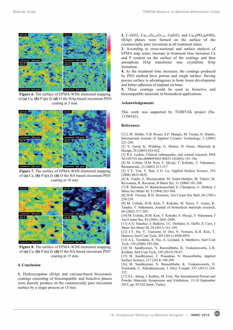
The thickness of the coatings increases with increasing processing time due to more reaction time with electrolytes in order to form thicker oxide layers coating–substrate interface. This can be observed in Fig. 2 and Table 1. In addition, surface roughness of the coatings gradually increases according to the increasing treatment time. At 5 min treatment time, there is not sufficient time for the micro discharge channels to interact at the surface to form porous morphology. However, as time passes by, there has been a lot interaction between ions in the electrolytes and the substrate interface. So this causes more oxide accumulation at the surface as the increasing processing time. This increases roughness of the coatings for 10 and 15 min treatment time. It is known that a porous surface is advantageous to bone tissue development and better adhesion of implant on bone [5]. 3.3 Electron Probe Microanalysis (EPMA) of the Coatings EPMA maps of the cross section and the surface PEO coatings are shown in Fig. 3-5. The elemental distributions of the phosphorus (P), zirconium (Zr), calcium (Ca) and oxygen (O) in the coatings are characterized qualitatively in all map scans.
Figure 3. The cross-section of EPMA-WDS elemental mapping of (a) Ca, (b) P (c) Zr (d) O the HAp based
zirconium PEO coating at 5 min.
The composition of Zr, Ca and O were uniformly well-distributed throughout the entire surface to the interior of the coating as in Fig. 3-5. As seen, the content of Ca reached the highest amount at the porous layer of the coating and kept its presence throughout the cross-sectional coating area. However, the content of P decreases sharply from the surface to the substrate along the direction of cross section of the coating. As the treatment time increases, the intensity of the P presence near-surface regions increases in Fig 3-5. The noticeable characteristic is the presence of P throughout the coating, with increased grades in the interior layer of the coating. In general, the highest concentration of Ca and P elements was observed just at the edge of the top surface in all samples. In other words, Ca-P rich layer was obviously achieved on the surface of the coating.
Figure 4. The cross-section of EPMA-WDS elemental mapping of (a) Ca, (b) P (c) Zr (d) O the HAp based
zirconium PEO coating at 10 min.
Figure 5. The cross-section of EPMA-WDS elemental mapping of (a) Ca, (b) P (c) Zr (d) O the HAp based
zirconium PEO coating at 15 min. Although Ca-P elemental distributions are clearly seen in all samples, crystalline HAp occurs at 15 min treatment time in XRD analysis. From this point, it would be wise to say that PEO coatings produced at 5 and 10 min treatment times have amorphous HAp phase and there is not sufficient time to convert amorphous phase into crystalline phase. In Figure 6-8, the composition of Ca, P and O were almost regularly distributed all over surface of the coating while Zr was generally present in the vicinity of the micro discharge regions. As the treatment time increases, it is clearly seen that the surface of the coating was rich in P substance in accordance with Ca. At 5 min surface morphology of the elemental distribution is nearly homogenous. After 10 and 15 min, intensities of Ca and P are significantly increased at the surface of PEO coatings.
TMMOB Metalurj i ve Malzeme Mühendisleri Odas ıBildir i ler Kitab ı
33118. Uluslararas ı Metalurj i ve Malzeme Kongresi | IMMC 2016
Figure 6. The surface of EPMA-WDS elemental mapping of (a) Ca, (b) P (c) Zr (d) O the HAp based zirconium PEO
coating at 5 min.
Figure 7. The surface of EPMA-WDS elemental mapping of (a) Ca, (b) P (c) Zr (d) O the HA based zirconium PEO
coating at 10 min.
Figure 8. The surface of EPMA-WDS elemental mapping of (a) Ca, (b) P (c) Zr (d) O the HA based zirconium PEO
coating at 15 min.
4. Conclusion 1. Hydroxyapatite (HAp) and calcium-based bioceramic coatings consisting of biocompatible and bioactive phases were directly produce on the commercially pure zirconium surface by a single process at 15 min.
2. C-ZrO2, Ca0.15Zr0.85O1.85, CaZrO3 and Ca10(PO4)6(OH)2 (HAp) phases were formed on the surface of the commercially pure zirconium at all treatment times. 3. According to cross-sectional and surface analysis of EPMA map scans; increase in treatment time increases Ca and P content on the surface of the coatings and then amorphous HAp transforms into crystalline HAp formation. 4. As the treatment time increases, the coatings produced by PEO method have porous and rough surface. Having porous surface is advantageous to bone tissue development and better adhesion of implant on bone. 5. These coatings could be used as bioactive and biocompatible materials in biomedical applications. Acknowledgements
This work was supported by TUBITAK project (No. 113M162). References [1] L.W. Hobbs, V.B. Rosen, S.P. Mangin, M. Treska, G. Hunter, International Journal of Applied Ceramic Technology, 2 (2005) 221-246. [2] V. Good, K. Widding, G. Hunter, D. Heuer, Materials & Design, 26 (2005) 618-622. [3] R.S. Laskin, Clinical orthopaedics and related research, DOI 10.1097/01.blo.0000093003.90435.1f(2003) 191-196. [4] M. Uchida, H.M. Kim, F. Miyaji, T. Kokubo, T. Nakamura, Biomaterials, 23 (2002) 313-317. [5] Y.Y. Yan, Y. Han, C.G. Lu, Applied Surface Science, 254 (2008) 4833-4839. [6] K. Fatehi, F. Moztarzadeh, M. Solati-Hashjin, M. Tahriri, M. Rezvannia, R. Ravarian, B Mater Sci, 31 (2008) 101-108. [7] R. Halouani, D. Bernacheassolant, E. Champion, A. Ababou, J Mater Sci-Mater M, 5 (1994) 563-568. [8] M.B. Thomas, R.H. Doremus, Am Ceram Soc Bull, 60 (1981) 258-259. [9] M. Uchida, H.M. Kim, T. Kokubo, M. Nawa, T. Asano, K. Tanaka, T. Nakamura, Journal of biomedical materials research, 60 (2002) 277-282. [10] M. Uchida, H.M. Kim, T. Kokubo, F. Miyaji, T. Nakamura, J Am Ceram Soc, 84 (2001) 2041-2044. [11] A.G. Sanchez, J. Ballarre, J.C. Orellano, G. Duffo, S. Cere, J Mater Sci-Mater M, 24 (2013) 161-169. [12] J.Y. Ha, Y. Tsutsumi, H. Doi, N. Nomura, K.H. Kim, T. Hanawa, Surf Coat Tech, 205 (2011) 4948-4955. [13] A.L. Yerokhin, X. Nie, A. Leyland, A. Matthews, Surf Coat Tech, 130 (2000) 195-206. [14] M. Sandhyarani, N. Rameshbabu, K. Venkateswarlu, L.R. Krishna, Surf Coat Tech, 238 (2014) 58-67. [15] M. Sandhyarani, T. Prasadrao, N. Rameshbabu, Applied Surface Science, 317 (2014) 198-209. [16] M. Sandhyarani, N. Rameshbabu, K. Venkateswarlu, D. Sreekanth, C. Subrahmanyam, J Alloy Compd, 553 (2013) 324-332. [17] S.L. Aktug, I. Kutbay, M. Usta, The International Porous and Powder Materials Symposium and Exhibition, 15-18 September 2015, pp. 95/242 Izmir, Turkey.