Embed Size (px)
Citation preview
Journal of Clinical InvestigationVol. 46, No. 2, 1967
Effect of Diet upon Intestinal Disaccharidases andDisaccharide Absorption *
J. J. DEREN, S. A. BROITMAN, ANDN. ZAMCHECKt(From the Gastroenterology Laboratory, Mallory Institute of Pathology; the Second and
Fourth [Harvard] Medical Services, Boston City Hospital; the Department ofMedicine, Harvard Medical School; and the Departments of Microbiology
and Pathology, Boston University School of Medicine, Boston, Mass.)
Summary. The administration of a carbohydrate-containing diet for 24hours to rats previously fasted for 3 days led to a twofold increase in totalintestinal sucrase and sucrase specific activity. The specific activity of mal-tase was similarly increased, but lactase activity was unaffected. The su-crose-containing diet led to a greater increase in sucrase than maltase ac-tivity, whereas the converse was true of the maltose-containing diet. Acarbohydrate-free isocaloric diet led to a slight increase in the total intestinalsucrase, but sucrase specific activity was unchanged. Assay of sucrase ac-tivity of mixed homogenates from casein-fed and sucrose-fed rats or fastedand sucrose-fed animals yielded activities that were additive. The Michaelisconstant (Km) of the enzyme hydrolyzing sucrose was similar in the fasted,casein-fed, and sucrose-fed rats. The maximal velocity (Vmax) was twicegreater in sucrose-fed as compared to casein-fed or fasted rats, suggestingan increased quantity of enzyme subsequent to sucrose feeding.
Adrenalectomized rats maintained on 1.0% salt intake had sucrase andmaltase levels comparable to those of controls. Steroid administration didnot significantly increase their activities. The response to sucrose feedingwas similar in both control and adrenalectomized rats, indicative of the ab-sence of steroidal control on sucrase and maltase activity in the adult animal.
Studies using intestinal ring preparations indicated that sucrose hydrolysisby the intact cells proceeded more rapidly when animals were fed sucrose.Additional corroboration of the physiologic significance of the increased en-zyme levels in homogenates was afforded by intestinal perfusion studies.Sucrose hydrolysis increased twofold and fructose absorption fourfold inanimals fed sucrose when compared to either fasted or casein-fed rats.
Introduction
The demonstration of isolated deficiencies ofthe intestinal disaccharidases as well as the non-specific depression of these enzymes in diversesmall bowel diseases has renewed interest in disac-charide absorption (1-5). To date, little is known
* Submitted for publication June 6, 1966; acceptedOctober 20, 1966.
This investigation was supported by research grantsT1 AM 5320, National Institute of Arthritis and Meta-bolic Diseases; CA 02090 and CA 04486, National Can-cer Institute; and GMO9628, National Institute of Gen-
about the factors that regulate the tissue levels ofthese enzymes and their physiologic significance.Marked variability of disaccharidase levels char-acterizes the peroral biopsy specimen in normalhumans and is due in part to variable numbers of
eral Medical Sciences, National Institutes of Health,Bethesda, Md. A preliminary report appeared in Fed.Proc. 1965, 24, 403.
t Present address: Dept. of Medicine, State Universityof New York (Downstate) and Dept. of Medicine,Maimonides Hospital, Brooklyn, N. Y.
t Address requests for reprints to Dr. N. Zamcheck,Boston City Hospital, Boston, Mass. 02118.
186
DISACCHARIDASESAND DISACCHARIDE ABSORPTION
epithelial cells and inflammation within the laminapropria. Furthermore, enzyme activity may be in-fluenced by previous dietary experience. In therat at least, intestinal sucrase (6) and alkalinephosphatase (7, 8) can be modified by diet. Al-though evidence on this point is not available inhumans, there is little doubt that enzymatic adap-tation occurs in mammalian tissue and that thismay be induced by appropriate substrates. How-ever, the relationship of adaptation to physiologicfunction is not well defined. Thus, depressed lev-els of disaccharidases can be implicated as func-tionally significant in disaccharide malabsorptiononly if the enzyme level is rate-limiting and varia-tions in disaccharidase levels are reflected bychanges in disaccharide absorption.
Weinitiated the present studies 1) to determinethe influence of diet and adrenocorticoid hormonesupon intestinal disaccharidase levels, 2) to definethe mechanism, and 3) to study the physiologicalsignificance of these changes measured in vitro andin Z'IVO.
Methods
Male albino rats 1 (175 to 350 g) were kept at constant
temperature (70 10 F) for at least 1 week before ini-tiation of the experiment, during which time they were
fed Purina laboratory chow ad libitum. The rats were
fasted but allowed water ad libitum for 3 days, and thenthey were placed for 24 hours on one of the syntheticdiets (Table I). Adrenalectomized rats (maintainedwith lo% sodium chloride in their drinking water) were
fasted but allowed free access to 1%o sodium chloridesolution for 24 hours; then they were placed on one ofthe synthetic diets for 24 hours.
Homogenate assays. Rats were decapitated and thesmall bowel rinsed in situ with ice-cold 0.9%o saline. Theentire small bowel or the third 10-cm segment distal tothe pylorus was removed and homogenized in seven timesits volume of cold saline in a glass homogenizer. Weassayed samples as described below at various dilutionsand different time intervals to ensure linearity of theassays. Although maltose and sucrose are hydrolyzedby several apparently different enzymes with overlappingsubstrate affinities, for convenience the enzymes that hy-drolyze each substrate are referred to collectively as
sucrase or maltase.Sucrose hydrolysis by intestinal rings. The entire
small bowel was rinsed in, situ with ice-cold saline, re-
moved, everted with a metal probe, and cut into ringsweighing 20 to 30 mg each according to the generalmethod of Agar, Hird, and Sidhu (9). After randomi-zation, ten to fifteen rings were placed in an Erlenmeyer
1 Charles River Laboratories, Wilmington, Mass.
TABLE I
Synthetic diets
DietarySubstance content
g/100 g dietSucrose, maltose, glucose, fructose,
or vitamin-free casein 68Casein (vitamin-free) 17Beef fat 8Salt mixture* 4Cellulose flour 2Vitamin powdert 1
* Salt mixture contained Ca(C3H502)2-5 H20, 35%;CaCO3, 5.06%; Ca(H2PO4) -H20, 14.6%; K2HPO4, 6.46%;NaH2PO4-H2O, 18.76%; NaCl, 9.34%; MgSO4, 7.18%;ZnSO4, 0.035%; CuSO4, 0.039%70; KI, 0.00039%; Fecitrate, 3.2%; and MnSO4 H2O, 0.33%.
t Vitamin powder contained thiamine, 0.05%; riboflavin,0.025%; pyridoxine, 0.02%; calcium pantothenate, 0.1%;and nicotinic acid, 0.1%.
flask containing 20 ml of 28 mMd(+)-sucrose 2 and 1%polyethylene glycol 3 (4,000) (PEG) in a phosphate-saline buffer (pH 7.4) and incubated at 370 C in a shak-ing water bath. Samples of the incubation medium wereremoved at 30, 45, and 60 minutes and analyzed forPEG, fructose, glucose, and sucrose. An estimate of su-crase activity was obtained by homogenizing a sampleof the rings in cold saline and assaying in a phosphate-saline buffer that contained 28 mMsucrose (pH 7.4).
Perfusion studies. Rats were fasted and refed as de-scribed above. A general method described by Schanker,Tocco, Brodie, and Hogben was used in the perfusionstudies (10). The rat was anesthetized with Pentothal(5 mg per 100 g body weight) and placed in an incuba-tor maintained at 370 C. A ligature was placed aroundthe pylorus, a small incision was made in the first por-tion of the duodenum, and a cannula was inserted. Afterthe ileocecal area had been ligated, a polyethylene cathe-ter was inserted into the distal ileum. A solution con-taining 18 to 20 mMsucrose in 140 mMNaCl with 1%polyethylene glycol maintained at 37° C was perfused bya constant perfusion pump 4 for 30 minutes at 1.5 ml perminute, and the effluent was discarded. Six consecutive10-minute collection periods were obtained; the effluentswere assayed for sucrose, glucose, fructose, and PEG asdescribed below. The per cent water absorption was de-termined from the change in PEG concentration (11).
Analytical methods. Sucrase, maltase, and lactase ac-tivities were determined as described by Dahlqvist (12)with minor modifications. Sucrase and maltase activitieswere measured in a 25 mMsodium maleate buffer (pH6.2) with the substrate at a 28 mMconcentration exceptwhere otherwise specified; lactase activity was measuredin a phosphate-citrate buffer (pH 4.8) with lactose pres-ent at 14 mMconcentration. Alkaline phosphatase wasmeasured as described by Moog (13) using 100 mMso-
2 Mallinckrodt Chemical Works, St. Louis, Mo.3 Union Carbide Corp., New York, N. Y.4 Harvard Apparatus Co., Dover, Mass.
187
J. J. DEREN, S. A. BROITMAN, AND N. ZAMCHECK
(6)
80-
.~C SE
60-440
20 -
SUCRASE LACTASE MALTASEACTIVITY ACTIVITY ACTIVITY
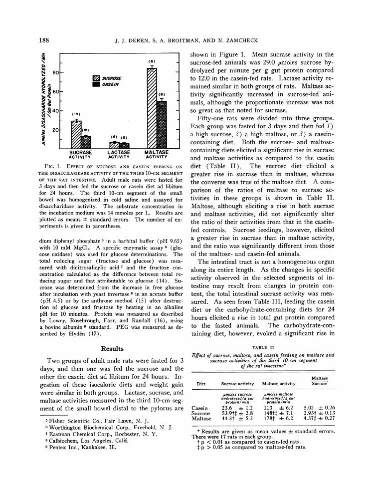
FIG. 1. EFFECT OF SUCROSEAND CASEIN FEEDING ON
THE DISACCHARIDASEACTIVITY OFTHETHIRD 10-CM SEGMENT
OF THE RAT INTESTINE. Adult male rats were fasted for3 days and then fed the sucrose or casein diet ad libitumfor 24 hours. The third 10-cm segment of the smallbowel was homogenized in cold saline and assayed fordisaccharidase activity. The substrate concentration inthe incubation medium was 14 mmoles per L. Results are
plotted as means standard errors. The number of ex-
periments is given in parentheses.
dium diphenyl phosphate 5 in a barbital buffer (pH 9.65)with 10 mMMgCl5. A specific enzymatic assay 6 (glu-cose oxidase) was used for glucose determinations. Thetotal reducing sugar (fructose and glucose) was mea-
sured with dinitrosalicylic acid 7 and the fructose con-
centration calculated as the difference between total re-
ducing sugar and that attributable to glucose (14). Su-crose was determined from the increase in free glucoseafter incubation with yeast invertase 8 in an acetate buffer(pH 4.5) or by the anthrone method (15) after destruc-tion of glucose and fructose by heating in an alkalinepH for 10 minutes. Protein was measured as describedby Lowry, Rosebrough, Farr, and Randall (16), usinga bovine albumin 9 standard. PEGwas measured as de-scribed by Hyden (17).
Results
Two groups of adult male rats were fasted for 3days, and then one was fed the sucrose and theother the casein diet ad libitum for 24 hours. In-gestion of these isocaloric diets and weight gainwere similar in both groups. Lactase, sucrase, andmaltase activities measured in the third 10-cm seg-
ment of the small bowel distal to the pylorus are
5 Fisher Scientific Co., Fair Lawn, N. J.6Worthington Biochemical Corp., Freehold, N. J.7 Eastman Chemical Corp., Rochester, N. Y.8 Calbiochem, Los Angeles, Calif.9 Pentex Inc., Kankakee, Ill.
shown in Figure 1. Mean sucrase activity in thesucrose-fed animals was 29.0 jumoles sucrose hy-drolyzed per minute per g gut protein comparedto 12.0 in the casein-fed rats. Lactase activity re-mained similar in both groups of rats. Maltase ac-tivity significantly increased in sucrose-fed ani-mals, although the proportionate increase was notso great as that noted for sucrase.
Fifty-one rats were divided into three groups.Each group was fasted for 3 days and then fed 1 )a high sucrose, 2) a high maltose, or 3) a casein-containing diet. Both the sucrose- and maltose-containing diets elicited a significant rise in sucraseand maltase activities as compared to the caseindiet (Table II). The sucrose diet elicited agreater rise in sucrase than in maltase, whereasthe converse was true of the maltose diet. A com-parison of the ratios of maltase to sucrase ac-tivities in these groups is shown in Table II.Maltose, although eliciting a rise in both sucraseand maltase activities, did not significantly alterthe ratio of their activities from that in the casein-fed controls. Sucrose feedings, however, eliciteda greater rise in sucrase than in maltase activity,and the ratio was significantly different from thoseof the maltose- and casein-fed animals.
The intestinal tract is not a homogeneous organalong its entire length. As the changes in specificactivity observed in the selected segments of in-testine may result from changes in protein con-tent, the total intestinal sucrase activity was mea-sured. As seen from Table III, feeding the caseindiet or the carbohydrate-containing diets for 24hours elicited a rise in total gut protein comparedto the fasted animals. The carbohydrate-con-taining diet, however, evoked a significant rise in
TABLE II
Effect of sucrose, maltose, and casein feeding on maltase andsucrase activities of the third 10-cm segment
of the rat intestine*
MaltaseDiet Sucrase activity Maltase activity Sucrase
jAmoles sucrose jimoles maltosehydrolyzed/g gut hydrolyzed/g gut
protein/min protein/minCasein 23.6 i 1.2 113 ± 6.2 5.02 i 0.26Sucrose 53.9tt ± 2.8 148tt i 7.1 2.93t i 0.13Maltose 44.3t i 5.2 178t i 6.2 4.37t i 0.27
* Results are given as mean values ± standard errors.There were 17 rats in each group.
t p < 0.01 as compared to casein-fed rats.t p > 0.05 as compared to maltose-fed rats.
188
DISACCHARIDASESAND DISACCHARIDE ABSORPTION
TABLE III
Total intestinal sucrase activity of rats fed various dietary regimens*
MeanNo. of body Total intestinal
Diet animals wt Total gut protein Specific activity sucrase activity
g g 5moles hydrolywed/g pmoles hydrolyzed/minprotein/min
Fasted 15 165 0.564 4- 0.031 20.0 4 1.79 10.8 i 1.27Casein 16 164 0.723 i 0.039 25.8t ± 1.30 18.7$ i 1.44Sucrose 16 165 0.688 + 0.031 46.1$ i 2.74 31.5T i 2.41Fructose 6 135 0.766 i 0.116 45.51 i 3.88 33.7$ ± 4.54Glucose 7 136 0.760 i 0.071 43.4t ± 2.06 27.3t 4t 3.10Maltose 16 165 0.670 :1: 0.040 36.4$ i 2.89 23.6t ± 1.30
* Results are expressed as means ± standard errors.t p < 0.05 > 0.01 when compared to fasted animals.$ p < 0.01 when compared to fasted animals.
the specific activity of sucrase. The casein-con-taining diet yielded a minimal increase in sucrasespecific activity owing to the increased gut pro-tein in this group. Total intestinal sucrase activitywas, however, significantly increased in the casein-fed animals as compared to fasted controls.
Changes in sucrase activity observed with thedifferent dietary regimens may represent changesin the quantity of enzyme present or alterations inthe kinetic characteristics of a similar quantity ofenzyme. Accordingly, the following studies wereperformed. Jejunal homogenates from sucrose-and casein-fed rats were prepared, and sucrase ac-tivity was determined. Portions of each fractionwere mixed together; the sucrase activity of themixed homogenates was measured (Table IV).The sucrase activity of the mixed homogenate wasnot significantly different from that predicted onthe basis of an additive effect. Similar resultswvere obtained from mixed homogenates from su-crose-fed and fasted animals, indicating that thedifferences observed were not attributable to vari-ations in easily dissociable inhibitors or activators.
TABLE IV
Mean sucrase activity of mixed homogenates from fasted,sucrose-fed, and casein-fed rats*
Mixed homogenates
Mean differ-Theoret- ence between
Obtained ical pairs ±SE
Sucrose Casein26.7 (6) 15.7 21.9 21.5 0.33±40.22Sucrose Fasted32.6 (5) 11.3 23.8 22.6 1.2 ±0.75
* The results are expressed as micromoles sucrose hydrolyzed pergram gut protein per minute and are given as the mean value. Thenumber of experiments is given in parentheses.
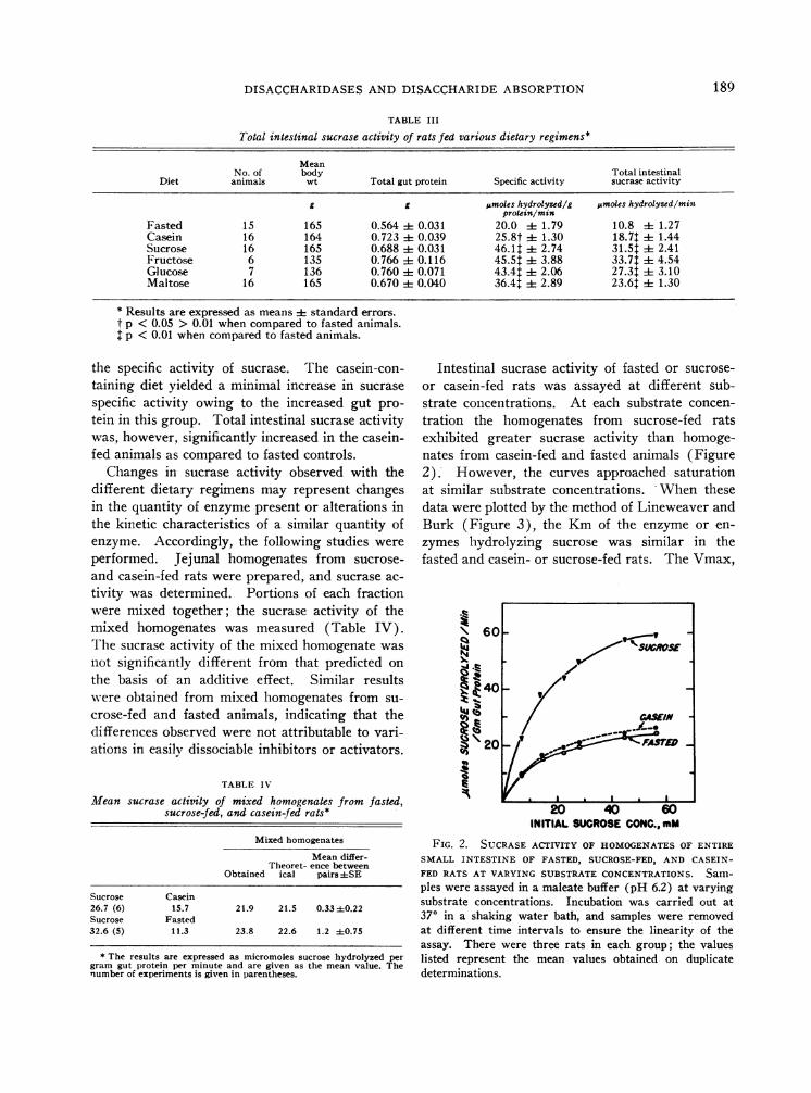
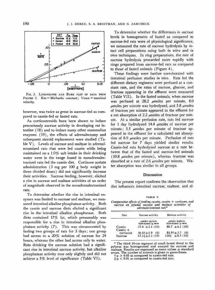
Intestinal sucrase activity of fasted or sucrose-or casein-fed rats was assayed at different sub-strate concentrations. At each substrate concen-tration the homogenates from sucrose-fed ratsexhibited greater sucrase activity than homoge-nates from casein-fed and fasted animals (Figure2). However, the curves approached saturationat similar substrate concentrations. When thesedata were plotted by the method of Lineweaver andBurk (Figure 3), the Kmof the enzyme or en-zymes hydrolyzing sucrose was similar in thefasted and casein- or sucrose-fed rats. The Vmax,
\60-
20 ~AS~M
I * I . * I
INITIAL SUCROSECONG.. mM
FIG. 2. SUCRASEACTIVITY OF HOMOGENATESOF ENTIRESMALL INTESTINE OF FASTED, SUCROSE-FED, AND CASEIN-FED RATS AT VARYING SUBSTRATECONCENTRATIONS. Sam-ples were assayed in a maleate buffer (pH 6.2) at varyingsubstrate concentrations. Incubation was carried out at370 in a shaking water bath, and samples were removedat different time intervals to ensure the linearity of theassay. There were three rats in each group; the valueslisted represent the mean values obtained on duplicatedeterminations.
189
J. J. DEREN, S. A. BROITMAN, AND N. ZAMCHECK
-.05 0 .05 0.1 Q.15
[SUBSTRATE]FIG. 3. LINEWEAVER AND BURK PLOT OF DATA FROM
FIGURE 2. Km= Michaelis constant; Vmax= maximalvelocity.
however, was twice as great in sucrose-fed as com-
pared to casein-fed or fasted rats.As corticosteroids have been shown to induce
precociously sucrase activity in developing rat in-testine (18) and to induce many other mammalianenzymes (19), the effects of adrenalectomy andsubsequent steroid replacement were studied (Ta-ble V). Levels of sucrase and maltase in adrenal-ectomized rats that were fed casein while beingmaintained on a 1.0%c salt intake in their drinkingwater were in the range found in nonadrenalec-tomized rats fed the casein diet. Cortisone acetateadministration (3 mg per 100 g body weight inthree divided doses) did not significantly increasetheir activities. Sucrose feeding, however, eliciteda rise in sucrase and maltase activities of an orderof magnitude observed in the nonadrenalectomizedrats.
To determine whether the rise in intestinal en-
zymes was limited to sucrase and maltase, we mea-
sured intestinal alkaline phosphatase activity. Boththe casein and sucrose diets elicited a significantrise in the intestinal alkaline phosphatase. Bothdiets contained 17% fat, which presumably was
responsible for a rise in intestinal alkaline phos-phatase activity (7). This was circumvented byfasting two groups of rats for 3 days; one group
had access to a 20%o solution of sucrose for 24hours, whereas the other had access only to water.Rats drinking the sucrose solution had a signifi-cant rise in intestinal sucrase activity, but alkalinephosphatase activity rose only slightly and did notachieve a 5% level of significance (Table VI).
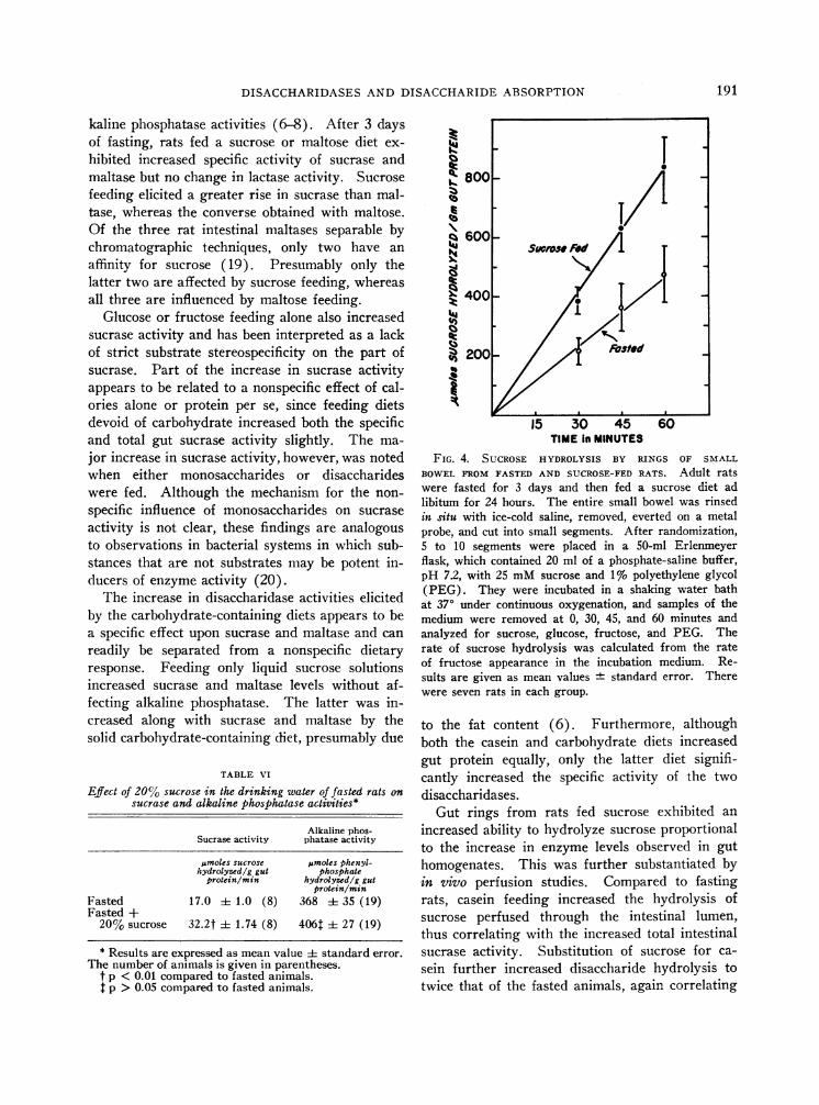
To determine whether the differences in sucraselevels in homogenates of fasted as compared tosucrose-fed rats were of physiological significance,we measured the rate of sucrose hydrolysis by in-tact cell preparations using both in 7itro and invivo techniques. In ring preparations, the rate ofsucrose hydrolysis proceeded more rapidly withrings prepared from sucrose-fed rats as comparedto those of fasted animals (Figure 4).
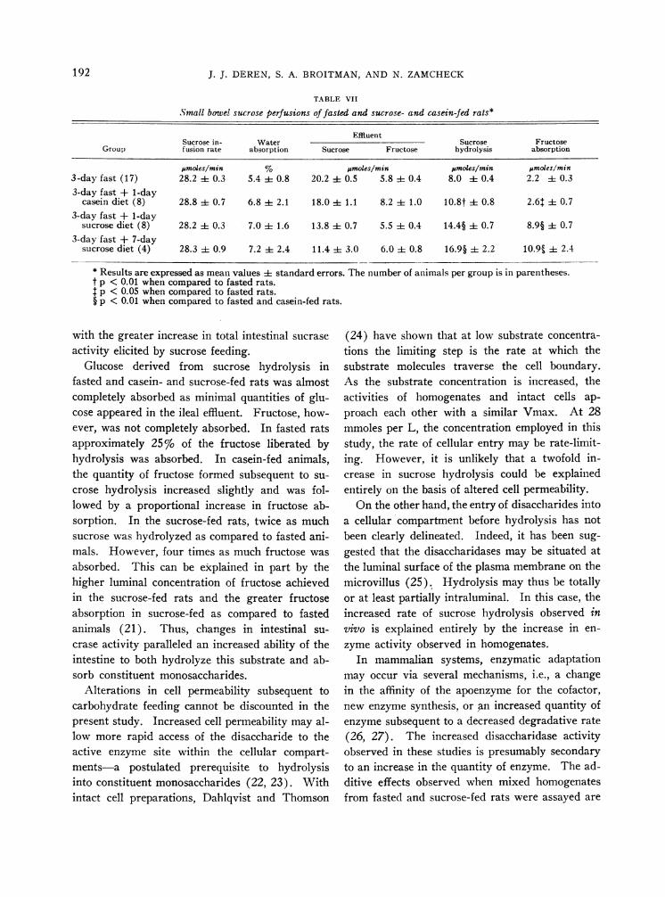
These findings were further corroborated withintestinal perfusion studies in vivo. Rats fed thedifferent dietary regimens were perfused at a con-stant rate, and the rates of sucrose, glucose, andfructose appearing in the effluent were measured(Table VII). In the fasted animals, when sucrosewas perfused at 28.2 pLmoles per minute, 8.0,umoles per minute was hydrolyzed, and 5.8 ,fmolesof fructose per minute appeared in the effluent fora net absorption of 2.2 pumoles of fructose per min-ute. At a similar perfusion rate, rats fed sucrosefor 1 day hydrolyzed 14.4 Ftmoles of sucrose perminute; 5.5 Mmoles per minute of fructose ap-peared in the effluent for a calculated net absorp-tion of 8.9 /.Lmoles per minute. Perfusion of ratsfed sucrose for 7 days yielded similar results.Casein-fed rats hydrolyzed sucrose at a rate be-tween that of the fasted and sucrose-fed animals(10.8 jumoles per minute), whereas fructose wasabsorbed at a rate of 2.6,umoles per minute. Wa-ter absorption was similar in all groups.
Discussion
The present report confirms the observation thatdiet influences intestinal sucrase, maltase, and al-
TABLE V
Comparative effects of feeding casein, casein + cortisone, andsucrose on jejunal sucrase and maltase activities of
adrenalectomized rats*
Diet Sucrase activity Maltase activity
5moles sucrose pmoles maltosehydrolyzed/g/min hydrolyzed/g/min
Casein 27.4 ±2.1 (10) 80.7 ±4.1 (10)Casein +
cortisone 36.4t± 1.9 (6) 84.9t-3.2 (6)Sucrose 67.1+±2.2 (10) 1301 ±8.5 (10)
* The third 10-cm segment of small bowel distal to thepylorus was homogenized and assayed for sucrase andmaltase. Results are expressed as mean values ± standarderrors. The number of animals is given in parentheses.
t p > 0.05 as compared to casein-fed rats.t p < 0.01 as compared to casein-fed rats.
190
DISACCHARIDASESAND DISACCHARIDE ABSORPTION
kaline phosphatase activities (6-8). After 3 daysof fasting, rats fed a sucrose or maltose diet ex-hibited increased specific activity of sucrase andmaltase but no change in lactase activity. Sucrosefeeding elicited a greater rise in sucrase than mal-tase, whereas the converse obtained with maltose.Of the three rat intestinal maltases separable bychromatographic techniques, only two have anaffinity for sucrose (19). Presumably only thelatter two are affected by sucrose feeding, whereasall three are influenced by maltose feeding.
Glucose or fructose feeding alone also increasedsucrase activity and has been interpreted as a lackof strict substrate stereospecificity on the part ofsucrase. Part of the increase in sucrase activityappears to be related to a nonspecific effect of cal-ories alone or protein per se, since feeding dietsdevoid of carbohydrate increased both the specificand total gut sucrase activity slightly. The ma-jor increase in sucrase activity, however, was notedwhen either monosaccharides or disaccharideswere fed. Although the mechanism for the non-specific influence of monosaccharides on sucraseactivity is not clear, these findings are analogousto observations in bacterial systems in which sub-stances that are not substrates may be potent in-ducers of enzyme activity (20).
The increase in disaccharidase activities elicitedby the carbohydrate-containing diets appears to bea specific effect upon sucrase and maltase and canreadily be separated from a nonspecific dietaryresponse. Feeding only liquid sucrose solutionsincreased sucrase and maltase levels without af-fecting alkaline phosphatase. The latter was in-creased along with sucrase and maltase by thesolid carbohydrate-containing diet, presumably due
TABLE VI
Effect of 20% sucrose in the drinking water of fasted rats onsucrase and alkaline phosphatase activities*
Alkaline phos-Sucrase activity phatase activity
osmoles sucrose pumoles phenyl-hydrolyzed/g gut phosphate
protein/mm hydrolyzed/g gutprotein/mon
Fasted 17.0 i 1.0 (8) 368 ± 35 (19)Fasted +
20% sucrose 32.2t + 1.74 (8) 406t ± 27 (19)
800 -
_q 6003
400 -
200- Fasted
i5 30- 45 60TIME In MINUTES
FIG. 4. SUCROSE HYDROLYSIS BY RINGS OF SMALLBOWELFROMFASTED AND SUCROSE-FEDRATS. Adult ratswere fasted for 3 days and then fed a sucrose diet adlibitum for 24 hours. The entire small bowel was rinsedin situ with ice-cold saline, removed, everted on a metalprobe, and cut into small segments. After randomization,5 to 10 segments were placed in a 50-ml Erlenmeyerflask, which contained 20 ml of a phosphate-saline buffer,pH 7.2, with 25 mMsucrose and 1% polyethylene glycol(PEG). They were incubated in a shaking water bathat 370 under continuous oxygenation, and samples of themedium were removed at 0, 30, 45, and 60 minutes andanalyzed for sucrose, glucose, fructose, and PEG. Therate of sucrose hydrolysis was calculated from the rateof fructose appearance in the incubation medium. Re-sults are given as mean values standard error. Therewere seven rats in each group.
to the fat content (6). Furthermore, althoughboth the casein and carbohydrate diets increasedgut protein equally, only the latter diet signifi-cantly increased the specific activity of the twodisaccharidases.
Gut rings from rats fed sucrose exhibited anincreased ability to hydrolyze sucrose proportionalto the increase in enzyme levels observed in guthomogenates. This was further substantiated byin vivo perfusion studies. Compared to fastingrats, casein feeding increased the hydrolysis ofsucrose perfused through the intestinal lumen,thus correlating with the increased total intestinalsucrase activity. Substitution of sucrose for ca-sein further increased disaccharide hydrolysis totwice that of the fasted animals, again correlating
* Results are expressed as mean value + standard error.The number of animals is given in parentheses.
t p < 0.01 compared to fasted animals.t p > 0.05 compared to fasted animals.
191
J. J. DEREN, S. A. BROITMAN, AND N. ZAMCHECK
TABLE VII
Small bowel sucrose perfusions of fasted and sucrose- and casein-fed rats*
EffluentSucrose in- Water Sucrose Fructose
Group fusion rate absorption Sucrose Fructose hydrolysis absorption
jumoles/min % tmoles/min jumoles/min pmoles/min3-day fast (17) 28.2 i 0.3 5.4 i 0.8 20.2 i 0.5 5.8 i 0.4 8.0 i 0.4 2.2 ± 0.33-day fast + 1-day
casein diet (8) 28.8 i 0.7 6.8 i 2.1 18.0 i 1.1 8.2 ± 1.0 10.8t i 0.8 2.6 ± 0.73-day fast + 1-day
sucrose diet (8) 28.2 i 0.3 7.0 ± 1.6 13.8 i 0.7 5.5 i 0.4 14.4§ ± 0.7 8.9§ i 0.73-day fast + 7-day
sucrose diet (4) 28.3 i 0. 9 7.2 i 2.4 11.4 i 3.0 6.0 ± 0.8 16.9§ i 2.2 10.95 i 2.4
* Results are expressed as mean values i standard errors. The number of animals per group is in parentheses.t p < 0.01 when compared to fasted rats.t p < 0.05 when compared to fasted rats.§ p < 0.01 when compared to fasted and casein-fed rats.
with the greater increase in total intestinal sucraseactivity elicited by sucrose feeding.
Glucose derived from sucrose hydrolysis infasted and casein- and sucrose-fed rats was almostcompletely absorbed as minimal quantities of glu-cose appeared in the ileal effluent. Fructose, how-ever, was not completely absorbed. In fasted ratsapproximately 25%o of the fructose liberated byhydrolysis was absorbed. In casein-fed animals,the quantity of fructose formed subsequent to su-crose hydrolysis increased slightly and was fol-lowed by a proportional increase in fructose ab-sorption. In the sucrose-fed rats, twice as muchsucrose was hydrolyzed as compared to fasted ani-mals. However, four times as much fructose wasabsorbed. This can be explained in part by thehigher luminal concentration of fructose achievedin the sucrose-fed rats and the greater fructoseabsorption in sucrose-fed as compared to fastedanimals (21). Thus, changes in intestinal su-crase activity paralleled an increased ability of theintestine to both hydrolyze this substrate and ab-sorb constituent monosaccharides.
Alterations in cell permeability subsequent tocarbohydrate feeding cannot be discounted in thepresent study. Increased cell permeability may al-low more rapid access of the disaccharide to theactive enzyme site within the cellular compart-ments-a postulated prerequisite to hydrolysisinto constituent monosaccharides (22, 23). Withintact cell preparations, Dahlqvist and Thomson
(24) have shown that at low substrate concentra-tions the limiting step is the rate at which thesubstrate molecules traverse the cell boundary.As the substrate concentration is increased, theactivities of homogenates and intact cells ap-proach each other with a similar Vmax. At 28mmoles per L, the concentration employed in thisstudy, the rate of cellular entry may be rate-limit-ing. However, it is unlikely that a twofold in-crease in sucrose hydrolysis could be explainedentirely on the basis of altered cell permeability.
On the other hand, the entry of disaccharides intoa cellular compartment before hydrolysis has notbeen clearly delineated. Indeed, it has been sug-gested that the disaccharidases may be situated atthe luminal surface of the plasma membrane on themicrovillus (25). Hydrolysis may thus be totallyor at least partially intraluminal. In this case, theincreased rate of sucrose hydrolysis observed invivo is explained entirely by the increase in en-zyme activity observed in homogenates.
In mammalian systems, enzymatic adaptationmay occur via several mechanisms, i.e., a changein the affinity of the apoenzyme for the cofactor,new enzyme synthesis, or an increased quantity ofenzyme subsequent to a decreased degradative rate(26, 27). The increased disaccharidase activityobserved in these studies is presumably secondaryto an increase in the quantity of enzyme. The ad-ditive effects observed when mixed homogenatesfrom fasted and sucrose-fed rats were assayed are
192
DISACCHARIDASESAND DISACCHARIDE ABSORPTION
indirect evidence for an increased quantity of en-zyme. The similar Kmof the enzymes hydrolyz-ing sucrose in both fasted and sucrose-fed ratswith a twofold greater Vmax in the latter animalsis best explained by the presence of twice the quan-tity of enzyme in the sucrose-fed rats. Direct con-firmation of this hypothesis would require assayof the quantity of enzymes by means independentof activity measurements, as has been shown im-munologically with other enzymes (28, 29).
It has been suggested that the mechanism regu-lating enzyme levels in the adult may be similarto those regulating the accumulation of the en-zyme in the developing organism (29). Althoughrat intestinal sucrase may be precociously inducedby steroids (19) and its appearance delayed in anadrenalectomized rat (30), sucrase and maltaselevels in the adult were not affected by adrenalec-tomy or steroid administration. Rat intestinalmaltase has been previously observed to be unal-tered after adrenalectomy (31). However, as thehalf-lives of the intestinal sucrases are unknown,caution must be exercised in the interpretation ofthe lack of steroidal stimulation. Schimke, Swee-ney, and Berlin (32) have shown that the rise inenzyme activity after steroid or substrate ad-ministration is related to the half-life of the en-zyme. The lack of steroid stimulation may merelyindicate a prolonged half-life, so that during theperiod of observation after the stimulus has beenapplied only a small portion of the enzyme mole-cules have had a chance to turn over. However,as a 24-hour fast leads to a 50% reduction in in-testinal sucrase, it would appear that these en-zymes are turning over at a rapid rate.
The histology of the small intestine from fastedand casein- and disaccharide-fed animals did notsignificantly differ. However, it is possible thateven during short periods of dietary manipula-tion, changes in intestinal epithelial cell popula-tion may occur as the epithelium is turning overat a rapid rate (33, 34). Consequently, mea-surements of the total epithelial cell populationwould be required to demonstrate that the en-
zyme changes observed represented a change inthe number of enzyme molecules per cell ratherthan an increase in cell population.
In the absence of such measurements, enzymeactivity is expressed in terms of wet or dry weightor of nitrogen content. Clinical interpretation ofthese results is further limited when they are ob-tained on fragments of human intestinal mucosa.In addition to the epithelial cells, many other cel-lular constituents of these biopsy specimens in-cluding inflammatory infiltrates of the lamina pro-pria are included in homogenates prepared for en-zyme analysis. Since it is accepted that disacchari-dase activity resides in the brush border of theepithelial cells, the enzymatic activity may be arti-factually lowered by the protein contribution of in-flammatory cells. An additional factor contributingto the variability noted in the peroral biopsy speci-mens of normal individuals may relate to the die-tary intake preceding the biopsy. Moreover, pa-tients who experience symptoms after the intakeof certain food products are likely to refrain fromthem, so that any depression of measured physio-logical function may result in part at least fromlack of suitable "substrates" induction. It wouldappear to be important to determine whether die-tary factors similarly influence disaccharidases anddisaccharide absorption in human subjects. In-deed, changes in intestinal disaccharidases havebeen reported in human subjects after periods ofprolonged fasting (35). Previous attempts to in-duce lactase activity in deficient adults by feedingmilk have not been successful (36). However,lactase may be less responsive to dietary manipula-tion, as evidenced by the failure to acutely inducelactase activity in experimental animals. Underconditions of prolonged ingestion of large quan-tities of lactose, total lactase activity in rats hasbeen reported to increase, secondary to an increasein total gut weight and length, but without an in-crease in lactase specific activity (37, 38). Con-versely, others have reported a statistically sig-nificant increase in lactase activity after prolonged(6 to 9 weeks) ingestion of lactose (39).
193
J. J. DEREN, S. A. BROITMAN, AND N. ZAMCHECK
Acknowledgments
Wewould like to acknowledge the invaluable assistanceof Mrs. Joan Kenney, Miss Joan Zecker, and Mr. PaulColon.
References
1. Durand, P. Lactosuria idiopathica in una pazientecon diarrea cronica ed acidosi. Minerva pediat.1958, 10, 706.
2. Holzel, A., V. Schwarz, and K. W. Sutcliffe. De-fective lactose absorption causing malnutrition ininfancy. Lancet 1959, 1, 1126.
3. Weijers, H. A., J. H. Van de Kamer, W. K. Dicke,and J. Ij sseling. Diarrhea caused by deficiencyof sugar splitting enzymes. Acta paediat. (Upp-sala) 1961, 50, 55.
4. Dahlqvist, A. Specificity of the human intestinaldisaccharidases and implications for hereditarydisaccharide intolerance. J. clin. Invest. 1962, 41,463.
5. Plotkin, G. R., and K. J. Isselbacher. Secondarydisaccharidase deficiency in adult celiac disease(nontropical sprue) and other malabsorptionstates. New Engl. J. Med. 1964, 271, 1033.
6. Blair, D. G. R., W. Yakimets, and J. Tuba. Ratintestinal sucrase. II. The effects of rat age andsex and of diet on sucrase activity. Canad. J. Bio-chem. 1963, 41, 917.
7. Tuba, J., and M. I. Robinson. The response of in-testinal alkaline phosphatase of fasted rats toforced feeding of fat. J. biol. Chem. 1953, 203,947.
8. Tuba, J., and N. Dickie. The role of alkaline phos-phatase in intestinal absorption. II. The effects ofvarious carbohydrates on levels of the enzyme inintestinal mucosa. Canad. J. Biochem. 1954, 32,621.
9. Agar, W. T., F. J. R. Hird, and G. S. Sidhu. Theuptake of amino acids by the intestine. Biochim.biophys. Acta (Amst.) 1954, 14, 80.
10. Schanker, L. S., D. J. Tocco, B. B. Brodie, andC. A. M. Hogben. Absorption of drugs from therat small intestine. J. Pharmacol. exp. Ther. 1958,123, 81.
11. Jacobson, E. D., D. C. Bondy, S. A. Broitman, andJ. S. Fordtran. Validity of polyethylene glycol inestimating intestinal water volume. Gastroenter-ology 1963, 44, 761.
12. Dahlqvist, A. Method for assay of intestinal disac-charidases. Analyt. Biochem. 1964, 7, 18.
13. Moog, F. Alkaline and acid phosphomonoesteraseactivity in chick embryos. J. cell. comp. Physiol.1946, 28, 197.
14. Sumner, J. B. The estimation of sugar in diabeticurine, using dinitrosalicylic acid. J. biol. Chem.1924, 62, 287.
15. Scott, T. A., Jr., and E. H. Melvin. Determination ofdextran with anthrone. Analyt. Chem. 1953, 25,1656.
16. Lowry, 0. H., N. J. Rosebrough, A. L. Farr, andR. J. Randall. Protein measurement with theFolin phenol reagent. J. biol. Chem. 1951, 193,265.
17. Hyden, S. A. A turbidometric method for the de-termination of higher polyethylene glycols in bio-logical materials. Ann. roy. agricul. Coll. Sweden1955, 22, 139.
18. Doell, R. G., and N. Kretchmer. Intestinal inver-tase: precocious development of activity after in-jection of hydrocortisone. Science 1964, 143, 42.
19. Dahlqvist, A. Rat-intestinal dextranase. Localiza-tion and relation to the other carbohydrases ofthe digestive tract. Biochem. J. 1963, 86, 72.
20. Monod, J. Remarks on the mechanism of enzyme in-duction in Enzymes: Units of Biological Structureand Function. Henry Ford International Sym-posium. New York, Academic Press, 1956, p. 7.
21. Broitman, S. A., J. J. Deren, and N. Zamcheck.Dietary influence on intestinal sucrase activity, su-
crose hydrolysis and absorption in the rat. Fed.Proc. 1966, 25, 321.
22. Miller, D., and R. K. Crane. The digestive func-tion of the epithelium of the small intestine. I.An intra cellular locus of disaccharide and sugar
phosphate ester hydrolysis. Biochim. biophys.Acta (Amst.) 1961, 52, 281.
23. Miller, D., and R. K. Crane. The digestive func-tion of the epithelium of the small intestine. II.Localization of disaccharide hydrolysis in the iso-lated brush border portion of intestinal epithelialcells. Biochim. biophys. Acta (Amst.) 1961, 52,293.
24. Dahlqvist, A., and D. L. Thomson. The hydrolysisof sucrose by intact and homogenized cells of ratsmall intestine. Influence of pH and substrateconcentration. Biochim. biophys. Acta (Amst.)1964, 92, 99.
25. Crane, R. K. Enzymes and malabsorption: a con-
cept of brush border membrane disease. Gastro-enterology 1966, 50, 254.
26. Greengard, O., and P. Feigelson. The activation andinduction of rat liver tryptophan pyrrolase in vivoby its substrate. J. biol. Chem. 1961, 236, 158.
27. Feigelson, P., and 0. Greengard. Immunochemicalevidence for increased titers of liver tryptophanpyrrolase during substrate and hormonal en-
zyme induction. J. biol. Chem. 1962, 237, 3814.28. Kenney, F. T. Induction of tyrosine-a-ketoglutarate
transaminase in rat liver. III. Immunochemicalanalysis. J. biol. Chem. 1962, 237, 1610.
29. Greengard, O., M. A. Smith, and G. Acs. Relationof cortisone and synthesis of ribonucleic acid toinduced and developmental enzyme formation. J.biol. Chem. 1963, 238, 1548.
30. Jirsovaf, V., and A. Heringova. Effect of aldosteroneand corticosterone on 6-galactosidase and invertaseactivity in the small intestine of rats. Nature(Lond.) 1965, 206, 300.
194
DISACCHARIDASESAND DISACCHARIDE ABSORPTION
31. Levin, R. J., H. Newey, and D. H. Smyth. Theeffects of adrenalectomy and fasting on intestinalfunction in the rat. J. Physiol. (Lond.) 1965,177, 58.
32. Schimke, R. T., E. W. Sweeney, and C. M. Berlin.An analysis of the kinetics of rat liver tryptophanpyrrolase induction: the significance of both en-
zyme synthesis and degradation. Biochem. bio-phys. Res. Commun. 1964, 15, 214.
33. Thaysen, E. H., and J. H. Thaysen. Morphologicalchanges in the gastrointestinal tract of the whiterat following inanition. Acta path. microbiol.scand. 1949, 26, 370.
34. Hooper, C. S., and M. Blair. The effect of starva-tion on epithelium renewal in the rat duodenum.Exp. Cell Res. 1958, 14, 175.
35. Knudsen, K. B., H. M. Bellamy, R. R. Lecocq, E. M.Bradley, and J. D. Welsh. The influence of fast-
ing and refeeding on jejunal disaccharidases. Clin.Res. 1966, 14, 300.
36. Cuatrecasas, P., D. H. Lockwood, and J. R. Cald-well. Lactase deficiency in the adult: a common
occurrence. Lancet 1965, 1, 14.37. Fischer, J. E. Effects of feeding a diet containing
lactose upon P-D-galactosidase activity and organ
development in the rat digestive tract. Amer. J.
Physiol. 1957, 188, 49.38. Huber, J. T., R. J. Rifkin, and J. M. Keith. Effect
of level of lactose upon lactase concentrations inthe small intestines of young calves. J. DairySci. 1964, 47, 789.
39. Giradet, P., R. Richterich, and I. Antener. Adapta-tion de la lactase intestinale a l'administration delactose chez le rat adulte. Helv. physiol. Pharma-col. Acta 1964, 22, 7.
195