Embed Size (px)
Citation preview

I. Feghiu. Moldovan Medical Journal. June 2019;62(3):7-12ORIGINAL ReseARch
DOI: 10.5281/zenodo.3404088UDC: 617-089.5:612.13
Effect of induction of general anesthesia with propofol and fentanyl on hemodynamic response
Iuliana Feghiu, MD, Assistant ProfessorValeriu Ghereg Department of Anesthesiology and Intensive Care No 1
Department of Pathophysiology and Clinical PathophysiologyNicolae Testemitsanu State University of Medicine and Pharmacy, Chisinau, the Republic of Moldova
Corresponding author: [email protected] received July 01, 2019; revised manuscript August 23, 2019
AbstractBackground: Induction of general anesthesia with propofol and fentanyl is frequently associated with changes in arterial blood pressure and heart rate. At present, there are no clinical studies investigating the relation between baseline cardiac autonomic tonus and cardiovascular instability after induction of general anesthesia with propofol and fentanyl.Material and methods: A randomized prospective study was performed with approval of Ethic Committee. Written informed consent was obtained from all patients. We enrolled in the study 47 ASA physical status I–II patients scheduled for elective surgical procedures. Heart rate variability by Holter ECG, arterial blood pressure (systolic, diastolic, mean), and heart rate were measured at baseline, after premedication, as well as after induction of general anesthesia with propofol 2.5mg/kg and fentanyl 1.0 mkg/kg.Results: our research revealed that increased baseline cardiac parasympathetic tonus was a risk factor for development of sinus bradycardia (OR = 21.0 (95%CI 3.9-112.8, p<0.0002) and sinus bradycardia associated with arterial hypotension (OR = 19.2 (95%CI 4.1-88.6, p<0.0001).Conclusions: Induction of general anesthesia with propofol and fentanyl was associated frequently with arterial hypotension and sinus bradycardia. Increased cardiac parasympathetic tonus at rest represents a risk factor for development of arterial hypotension and sinus bradycardia after administration of propofol and fentanyl for induction of general anesthesia.Key words: arterial hypotension, sinus bradycardia, cardiac autonomic tonus.
Introduction
Propofol is a frequently used hypnotic for sedation as well as for induction of general anesthesia. But, when this drug is injected rapidly it can lead to hemodynamic insta-bility, mainly to arterial hypotension and changes in heart rhythm. Both, sinus tachycardia and sinus bradycardia were reported after administration of propofol for sedation or for induction of general anesthesia [1-3]. Many mechanisms have been involved for explanation of propofol induced arterial hypotension, mainly direct depression of myocar-dium, reduced peripheral vascular resistance caused by di-rect vasodilatory effect of the drug, reduction of preload and afterload. The studies anyway, showed controversial results, and any of these factors could be imputed for hemodynamic instability after administration of propofol for sedation or for induction of general anesthesia [2-7]. Administration of propofol for moderate or deep sedation is frequently associ-ated with a significant decrease in mean blood pressure. This hypotensive effect of the drug can be caused by reduction of sympathetic cardiac tonus or disturbances in baroreceptor-mediated cardiac activity [4-6]. Similar to other intravenous anesthetic agents like benzodiazepines and barbiturates, propofol exerts its hypnotic actions by activation of the cen-tral inhibitory neurotransmitter – gamma-aminobutyric acid (GABA) [7].
Most often propofol administration is combined with opioid (fentanyl or sufentanyl). This combination has a beneficial effect as can reduce the requirement in myorelax-ants in the course of general anesthesia. But, on the other hand the combination between propofol and opioid can en-hance the risk for arterial hypotension and bradycardia in patients [8,9]
Heart rate variability (HRV) is a noninvasive electro-cardiographic marker which reflects the sympathetic and parasympathetic influences on sinus node of the heart. In other words, HRV analysis shows the baseline autonomic function of the heart. Measurements of HRV are noninva-sive, and highly reproducible. They may be performed on the basis of 24 hour Holter recordings or on shorter periods ranging from 0.5 to 5 minutes particularly in the field of dy-namic electrocardiography. Most studies in anesthesia and intensive care which used the HRV for analysis of changes in sympathetic-parasympathetic balance of the heart per-formed the 5 minutes analysis of HRV [10,11].
The purpose of this clinical research was to find a re-lationship between autonomic heart tonus at rest and the risk for development of arterial hypotension and changes in heart rate after induction of general anesthesia with propo-fol and fentanyl.
7

8
I. Feghiu. Moldovan Medical Journal. June 2019;62(3):7-12 ORIGINAL ReseARch
Material and methods
We performed a prospective randomized study to evalu-ate the relationship between baseline cardiac autonomic to-nus of the heart and the risk for development of cardiovas-cular instability after induction of general anesthesia with fentanyl and propofol. The study protocol was approved by the Ethic Committee of the State University of Medicine and Pharmacy “Nicolae Testemiţanu”, Chișinău (No.20, 2.02.2016).
Between March 2017 and September 2017, ASA physi-cal status I-II patients aged less than 60 years (to exclude age-related changes of HRV) scheduled for elective surger-ies with normal sinus rhythm on ECG were enrolled in the study. We obtained an informed consent from all research participants. Patients with diseases that could affect auto-nomic cardiac regulation (endocrine, neurological, cardio-vascular diseases) were excluded from the study.
In the operating room, the patients were monitored (Holter ECG (Holter TLC 5000, USA)), non-invasive blood pressure, pulse oximetry and capnography). Baseline heart rate (HR), systolic blood pressure (SBP), diastolic blood pressure (DBP), and respiratory rate were recorded. During induction of general anesthesia, oxygen was delivered to maintain SpO2 above 95%. All patients received 10 ml/kg of crystalloid before induction of anaesthesia.
HRV parameters, HR, SBP, DBP and respiratory rate were recorded at baseline (T1), 5 minutes after premedica-tion with Fentanyl 1.0 mkg/kg (T2) and 5 minutes after in-duction of general anesthesia with propofol 2.5 mg/kg and fentanyl 1.0 mkg/kg (T3). If after receiving propofol and fentanyl, patients developed bradypnea or apnea, the mask ventilation was initiated at a rate of 14-16 breaths/min and tidal volume of 7-8 ml/kg, an important requirement for correct registration and analysis of HRV.
HRV parameters were analyzed by Holter computer-ized system. HRV was interpreted according to the rec-ommendations of the Task Force of the European Society of Cardiology and the North American Society of Pacing and Electrophysiology [12]. In this clinical study HRV was used for assessment of autonomic heart tonus at rest.
Sinus tachycardia was considered in any patient who had a heart rate more than 100 beats/min, and sinus bradycardia – a heart rate less than 60 beats/min.
We considered systolic arterial hypertension when SBP was more than 140 mmHg or an increase in SBP of more than 20% from baseline values, systolic arterial hypotension – when SBP was less than 90 mmHg or a decrease in SBP more than 20% below baseline, and diastolic hypotension – when DBP was less than 60 mmHg or a decrease in DBP more than 20% below baseline.
Statistical analysis of the results was performed using GraphPad Prism 6 (GraphPad Software, San Diego, California, SUA). Values with parametric distribution were analyzed by t-pair and repeated measures ANOVA tests. Values with non-parametric distribution were analyzed by Wilcoxon and Friedman tests. The Fisher’s exact test was used to compare categorical variables. Results are expressed as 95% confidence interval of odd ratio (parametric data) and median with interquartile range (IQR, non-parametric data). A p value of less than 0.05 was considered statistically significant.
Results and discussion
The study group consisted of 47 patients (26 females and 21 males), aged 37.5±11.9 years. The mean body mass index was 24.6±3.4 kg/m2 (it ranged between 16.1 and 30.0 kg/m2).
Holter heart rate variability analysis revealed that in baseline 38.3% of patients were with enhanced sympathetic tonus of the heart and 38.3% – with enhanced parasympa-thetic tonus of the heart. Another 23.4% of patients present-ed with heart eutonia (fig. 1).
Fig. 1. Structure of the study group in function of vegetative tonus of the heart at rest.
There were no significant changes in SBP, DBP, MAP and HR after premedication with fentanyl (1.0 mkg/kg), but after induction of general anesthesia with propofol 2.5 mkg/kg and fentanyl 1.0 mkg/kg, SBP decreased by
Table 1Changes in blood pressure and heart rate after premedication and induction of general anesthesia
Parameters T1 T2 T3 p
SBP 138.9(134.0-143.7)
133.9(129.6-138.1)
99.7(95.9-103.5)
0.001
DBP 85.4*(81.5-89.2)
79.0*(75.5-82.5)
51.1*(48.6-53.5)
0.001
MAP 103.1*(98.9-107.3)
97.7 *(94.1-101.3)
67.9*`(65.5-70.4)
0.001
HR 73.1(69.9-76.4)
72.8(69.4-76.1)
61.4(59.1-63.6)
0.001
*Blood pressure and HR values are represented as mean and 95%CI or as median and interquartile range for data with nonparametric distribution.
I. Feghiu. Moldovan Medical Journal. June 2019;62(3):7-12ORIGINAL ReseARch
25.5% (from 133.9 mmHg at T2 to 99.7 mmHg at T3; p=0.001), DBP – decreased by 35.5% (from 79.0 mmHg at T2 to 51.1 mmHg at T3; p<0.001), MAP – decreased by 30.5% (from 97.7 mmHg at T2 to 67.9 mmHg at T3; p<0.001), and HR – decreased by 15.7% (from 72.8 beats/min at T2 to 61.4 beats/min at T3; p<0.001), (tab. 1).
After induction of general anesthesia with propofol and fentanyl most patients developed systolic-diastolic or dia-stolic hypotension (41 patients – 87.2%) and sinus brady-cardia (24 patients – 51.1%) (fig. 2, fig. 4). More frequently there was attested diastolic hypotension (35 patients – 74.5%), only in 6 patients (12.8%) there was found systolic-diastolic hypotension. Minimal SBP was 69.0 mmHg, mini-mal DBP was 37.0 mmHg and minimal registered MAP was 49.0 mmHg. More frequently systolic-diastolic hypoten-sion or diastolic hypotension was registered at 3-5 minutes (4.6±0.3 min) after administration of propofol and fentanyl. Arterial hypotension was corrected with fluids, and none of the patients required vasopressor support. In the study group only in 3 patients (6.4%) was found arterial hyperten-sion. It is worth mentioning the fact that in all these patients arterial hypertension was present only the first 1-3 minutes (1.1±0.6 min) after administration of propofol and fentanyl. Maximal SBP was 169.0 mmHg, maximal DBP was 109.0 mmHg, and maximal registered MAP was 134.0 mmHg. All these 3 patients who developed arterial hypertension after induction of general anesthesia presented enhanced sympa-thetic tonus of the heart at rest.
Fig. 2. The number of patients with normal BP, arterial hypertension and arterial hypotension at rest, after premedication and induction of general anesthesia.
It is important to remark the fact that out of 41 patients who developed arterial hypotension after induction of gen-eral anesthesia, in 18 patients (43.9%) was attested enhanced parasympathetic tonus of the heart at rest. Holter ECG analysis revealed that all 18 patients with enhanced cardiac parasympathetic tonus in baseline developed arterial hypo-tension after administration of propofol and fentanyl. Other 15 patients (36.6%) who developed arterial hypotension were with enhanced sympathetic tonus of the heart at rest, and 8 patients (19.5%) – with heart eutonia at rest.
Fisher’s exact test of the relation between presence of en-hanced parasympathetic tonus of the heart at rest and the risk for development of arterial hypotension after admin-istration of propofol and fentanyl for induction of general anesthesia revealed: Odds ratio – 10.2 (95%CI 0.54-19.8)
(p=0.06), with sensitivity – 1.0 (95%CI 0.8-1.0), specificity – 0.2 (95%CI 0.08-0.39). Even if, OR and sensitivity are high, specificity is reduced, such enhanced parasympathetic to-nus of the heart in baseline doesn’t represent a risk factor for development of arterial hypotension after administration of propofol and fentanyl (fig. 3).
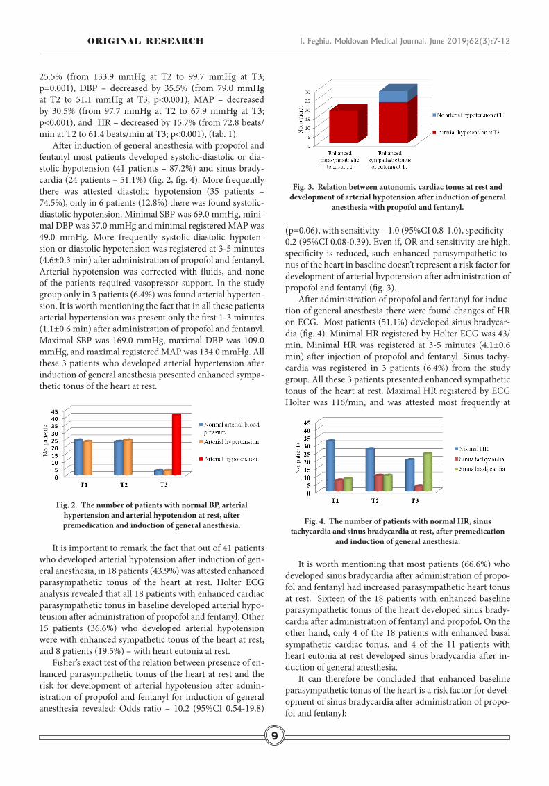
After administration of propofol and fentanyl for induc-tion of general anesthesia there were found changes of HR on ECG. Most patients (51.1%) developed sinus bradycar-dia (fig. 4). Minimal HR registered by Holter ECG was 43/min. Minimal HR was registered at 3-5 minutes (4.1±0.6 min) after injection of propofol and fentanyl. Sinus tachy-cardia was registered in 3 patients (6.4%) from the study group. All these 3 patients presented enhanced sympathetic tonus of the heart at rest. Maximal HR registered by ECG Holter was 116/min, and was attested most frequently at 2.1±0.6 min after administration of propofol and fentanyl.
Fig. 4. The number of patients with normal HR, sinus tachycardia and sinus bradycardia at rest, after premedication
and induction of general anesthesia.
It is worth mentioning that most patients (66.6%) who developed sinus bradycardia after administration of propo-fol and fentanyl had increased parasympathetic heart tonus at rest. Sixteen of the 18 patients with enhanced baseline parasympathetic tonus of the heart developed sinus brady-cardia after administration of fentanyl and propofol. On the other hand, only 4 of the 18 patients with enhanced basal sympathetic cardiac tonus, and 4 of the 11 patients with heart eutonia at rest developed sinus bradycardia after in-duction of general anesthesia.
It can therefore be concluded that enhanced baseline parasympathetic tonus of the heart is a risk factor for devel-opment of sinus bradycardia after administration of propo-fol and fentanyl:
Fig. 3. Relation between autonomic cardiac tonus at rest and development of arterial hypotension after induction of general
anesthesia with propofol and fentanyl.
9

10
I. Feghiu. Moldovan Medical Journal. June 2019;62(3):7-12 ORIGINAL ReseARch
Odds Ratio – 21.0 (95%CI 3.9-112.8; p<0.0002); sensibility 0.89 (95%CI 0.65-0.99) and specificity 0.72 (95%CI 0.53-0.87) (fig. 5).
In 21 patients from the study group (44.7%) after injec-tion of propofol and fentanyl was attested arterial hypoten-sion associated with sinus bradycardia. It is important to mention the fact that Holter ECG analysis revelead that out of these 21 patients in 15 (71.4%) was attested enhanced parasimpathetic cardiac tonus at rest. On the other hand, only 2 of the 18 patients with enhanced baseline sympathe-tic cardiac tonus, and 4 of the 11 patients with heart eutonia at rest developed sinus bradycardia associated with arterial hypotension after induction of general anesthesia.
Fisher’s exact test of the relation between cardiac auto-nomic tonus at rest and the risc for development of arte-rial hypotension associated with sinus bradycardia revealed: Odds Ratio – 19.2 (95%CI 4.1–88.6; p<0.0001); sensitivity – 0.83 (95%CI 0.58-0.96) and specificity – 0.79 (95%CI 0.60-0.92) (fig. 6 ). It can therefore be concluded that enhanced parasympathetic tonus of the heart at rest is a risk factor for development of sinus bradycardia associated with arterial hypotension after administration of propofol and fentanyl for induction of general anesthesia.
Fig. 6. Relation between autonomic cardiac tonus at rest and development of arterial hypotension associated with sinus
bradycardia after induction of general anesthesia
Discussion
Propofol is an intravenous hypnotic agent, which com-monly is used for anesthesia induction due to rapid onset, short duration of action, anti-nausea and vomiting effect and feeling comfortable after surgery. The most promi-nent effect of propofol is a decrease in arterial blood pres-
sure during induction of anesthesia and is associated with a decrease in cardiac output, stroke volume, and systemic vascular resistance. Furthermore, propofol induces severe vasodilation while the effects of myocardial depression are not exactly clear. Vasodilation occurs in both venous and arterial circulation, which leads to reduced preload and af-terload [1, 5, 7, 13, 14].
Although there are many studies that evaluated the effect of propofol and fentanyl on cardiac autonomic nervous sys-tem using heart rate variability [2-6], the relation between cardiac autonomic tonus at rest and the risk for cardiovas-cular instability after induction of anesthesia with propofol and fentanyl have not been investigated before.
In the literature there are published several studies which compare the hemodynamic effects of propofol with other hypnotic drugs, most often from the group of barbiturates or benzodiazepines.
In a study by Frolich M.A. et al. which involved 60 healthy volunteers ASA I physical status, subjects received 4 dose level ranges of propofol to provide anxyolitic level to moderate sedative effect. Predicted propofol concentrations in this study were 0.1, 0.2, 0.4 and 0.8 mkg/kg. A signifi-cant dose dependent blood pressure reduction was in the propofol group compared to midazolam group. Significant decreases in blood pressure were at propofol concentration 0.4 and 0.8 mkg/kg [13].
In another prospective, double-blinded, randomized clinical study by Kilic E. et al., proved that combination of alfentanil (10.0 mkg/kg) with propofol 0.7 mg/kg for seda-tion in upper gastrointestinal system endoscopy in morbid-ly obese patient is more frequently combined with bradycar-dia and arterial hypotension than combination of propofol with ketamine [8]. Chidambaran V. et al. conducted a study to evaluate total intravenous anesthesia with propofol and fentanyl in obese children and adolescents (aged 9-18 years) during laparoscopic surgery. Propofol was administered at a standardized infusion rate of 1000 μg/kg/min combined with fentanyl 50-100 μg. In all patients were attested hemo-dynamic changes and drop in SBP, DBP and MAP was more than 20% from the baseline value [9].
In a recent study, aiming to compare the effect of the ketamine-propofol mixture (ketofol) and propofol on the insertion conditions of laryngeal mask airway and hemo-dynamic stability in pediatrics patient (age ranging from 2 to 15 years) physical status ASA I and II, and undergo-ing elective surgical procedures, Aberra B. et al., proved a significant decrease in blood pressure and heart rate in the group of patients who received propofol (3.5 mg/kg) com-pared with the group of patients who received a mixture of propofol and ketamine [14]. Soleimani A. et al. proved that induction of general anesthesia in patients with left ven-tricular dysfunction with diazepam is safer than induction with propofol in term of hemodynamics. In this study in the group of patients who received propofol (1.5 mg/kg) after premedication with fentanyl (2.0 µg/kg) the decrease in SBP, DBP and MAP was significantly grater than in the group of
Fig. 5. Relation between autonomic cardiac tonus at rest and development of sinus bradycardia after induction of general
anesthesia.

I. Feghiu. Moldovan Medical Journal. June 2019;62(3):7-12ORIGINAL ReseARch
patients who received midazolam or etomidate for induc-tion of general anesthesia [15]. Another group of authors [16], compared the hemodynamic effects of propofol with the hemodynamic effects of etomidate when used for induc-tion of general anesthesia in patients undergoing coronary artery bypass grafting /mitral valve and aortic valve replace-ment surgery. In this study all patients received fentanyl 2.0 mkg/kg 3 minutes prior to induction. The dose of propofol for induction of general anesthesia was 2.0 mg/kg. There was significant decrease in SBP, DBP and MAP between the groups after induction, after intubation and 5 min postintu-bation. There was significant decrease in cardiac output and cardiac index in propofol group when compared to baseline values after induction, after intubation and 5 minutes after intubation, but not in etomidate group.
There are several studies which compare the hemody-namic effects of propofol used for sedation in patients un-dergoing gastrointestinal endoscopy [17-19]. In a prospec-tive, randomized, double-blind study by Usman S. et al. [17] was compared the hemodynamic effects of propofol (1.0 mg/kg propofol followed by repeated doses of 10 to 20 mg propofol intravenously) with that of midazolam associated with meperidine (0.4 mg/kg meperidine intravenously fol-lowed three minutes later by 0.05 mg/kg midazolam intra-venously) in 100 patients scheduled for diagnostic upper gastrointestinal endoscopy. The authors observed signifi-cantly more adverse cardiopulmonary events with propo-fol compared to meperidine/midazolam (20% vs. 4%, p = 0.025). Hypotension incidence was significantly higher in the propofol group compared to the meperidine/midazol-am group (12% vs. 0%, p=0.027). In this study, the authors found that midazolam/meperidine is superior to propofol with respect to the occurrence of adverse cardiopulmonary events, particularly hypotension. Tsai H. C. et al. performed a meta-analysis of randomized clinical trials aiming to com-pare the efficacy and safety of propofol and midazolam for sedation in cirrhotic patients undergoing endoscopy. Five studies between 2003 and 2012, including 433 patients, were included. In four of the selected randomized clinical trials arterial hypotension was more frequently in the patients who receive propofol than in patients who receive midazol-am. In three of the selected studies was evaluated the inci-dence of bradycardia based on a heart rate (HR) < 55 beats per minute. The incidence of bradycardia was 6% (9/150) in the propofol group and 2.86% (4/140) in the midazolam group [18]. In another recent meta-analysis conducted to compare the efficacy and safety of midazolam and propofol in gastrointestinal endoscopy five randomized controlled trials involving 552 patients were included [19]. The con-clusion of this meta-analysis was that propofol sedation for gastrointestinal endoscopy results in higher endoscopist satisfaction scores, but may increase the incidence of hypo-tension and bradycardia.
Another study examined the safety and effectiveness of the procedural sedation analgesia technique carried out in the emergency department. The research was done to com-
pare the effectiveness and efficacy of moderate sedation of fentanyl (0.1 mkg/kg) combined with propofol (1.0 mg/kg) or midazolam (1.0 mg/kg). None of the patients in ei-ther group developed any adverse events during and after the procedures. No significant drops in blood pressure and heart rate were observed during and after the procedures. Even though a few parameters, such as MAP, SBP and DBP, dropped intra-procedure, these values normalized post-procedure, and the changes were statistically insignificant within and between the groups [20].
Most studies which analyze the hemodynamic effects of propofol alone or propofol associated with opioid (as in this study) proved that a drop in SBP, DBP, MAP and HR can develop. These hemodynamic changes can be present even after administration of propofol and fentanyl in dos-es lower than in this study. However, there is not a single clinical research which studied the relation between base-line cardiac autonomic tonus of the heart and the risk for development of arterial hypotension and changes of heart rate after administration of propofol and fentanyl for in-duction of general anesthesia. This study revealed that en-hanced baseline cardiac parasympathetic tonus represents a risk factor for development of sinus bradycardia and ar-terial hypotension associated with sinus bradycardia after administration of propofol and fentanyl for induction of general anesthesia.
Conclusions
1. Induction of general anesthesia with propofol and fen-tanyl is frequently associated with arterial hypotension and sinus bradycardia.
2. Enhanced parasympathetic tonus of the heart at rest is a risk factor for development of sinus bradycardia and sinus bradycardia associated with arterial hypotension after injec-tion of propofol and fentanyl.
References1. Rawal P, Bajracharya U. Hemodynamic response to sevoflurane and
propofol induction: a comparative study. J Soc Anaesthesiol Nepal. 2015;2(1):2-7.
2. Mohit M, Radhakrishnan M, Umamaheswara R, Kavyashree KV. As-sessment of heart rate variability during different propofol effect site concentrations in patients with supratentorial tumours: a pilot study. J Neuroanaesthesiol Crit Care. 2017;4:108-113.
3. Tarvainen MP, Georgiadis S, Lipponen JA, Laitio T, Karjalainen PA, Scheinin H, Kaskinoro K. Analysis of heart rate variability dynamics during propofol and dexmedetomidine anesthesia. In: 32nd Annual International Conference of the IEEE EMBS; 2010 Aug 31-Sept 4; Buenos Aires, Argentina, 2010. P. 1634-7.
4. Tsugayasu R, Handa T, Kaneko Y, Ichinohe T. Midazolam more effec-tively suppresses sympathetic activations and reduces stress feelings during mental arithmetic task than propofol. J Oral Maxillofac Surg. 2010;68:590-6.
5. Win NN, Fukayama H, Kohase H, Umino M. The different effects of intravenous propofol and midazolam sedation on hemodynamic and heart rate variability. Anesth Analg. 2005;101:97-102.
6. Hidaka S, Kawamoto M, Kurita S, Yuge O. Comparison of the effects of propofol and midazolam on the cardiovascular autonomic nervous
11

12
I. Feghiu. Moldovan Medical Journal. June 2019;62(3):7-12 ORIGINAL ReseARch
system during combined spinal and epidural anesthesia. J Clin Anesth. 2005;17:36-43.
7. Sahinovic MM, Struys MM, Absalom AR. Clinical pharmacoki-netics and pharmacodynamics of propofol. Clin Pharmacokinet. 2018;57(12):1539-1558.
8. Kılıc E, Demiriz B, Isıkay N, Yıldırım AE, Can S, Basmacı C. Alfentanil versus ketamine combined with propofol for sedation during upper gastrointestinal system endoscopy in morbidly obese patient. Saudi Med J. 2016;37:1191-1195.
9. Chidambaran V, Sadhasivam S, Diepstraten J, Esslinger H, Cox S, Schnell BM, Samuels P, Inge T, Vinks AA, Knibbe CA. Evaluation of propofol anesthesia in morbidly obese children and adolescents. BMC Anesthesiol. 2013;13:8.
10. Anderson T. Heart rate variability: implications for perioperative anesthesia care. Curr Opin Anaesthesiol. 2017;30(6):691-697.
11. Pichot V, Roche F, Celle S, Barthélémy JC, Chouchou F. HRV analysis: a free software for analyzing cardiac autonomic activity. Front Physiol. 2016 Nov 22;7:557.
12. Heart rate variability: standards of measurement, physiological in-terpretation and clinical use. Task Force of the European Society of Cardiology and the North American Society of Pacing and Electro-physiology. Circulation. 1996;93(5):1043-1065.
13. Frölich MA, Arabshahib A, Katholi C, Prasain J, Barnes S. Hemody-namic characteristics of midazolam, propofol, and dexmedetomidine in healthy volunteers. J Clin Anesth. 2011;23(3):218-223.
14. Aberra B, Aregawi A, Teklay G, Tasew H. Effect of ketofol versus pro-pofol as an induction agent on ease of laryngeal mask airway insertion
conditions and hemodynamic stability in pediatrics: an observational prospective cohort study. BMC Anesthesiol. 2019;19(1):41.
15. Soleimani A, Heidari N, Habibi MR, Kiabi FH, Khademloo M, Emami Zeydi A, Sohrabi FB. Comparing hemodynamic responses to diazepam, propofol and etomidate during anesthesia induction in patients with left ventricular dysfunction undergoing coronary artery bypass graft surgery: a double-blind, randomized clinical trial. Med Arch. 2017 Jun;71(3):198-203.
16. Kaushal RP, Vatal A, Pathak R. Effect of etomidate and propofol induc-tion on hemodynamic and endocrine response in patients undergoing coronary artery bypass grafting/mitral valve and aortic valve replace-ment surgery on cardiopulmonary bypass. Ann Card Anaesth. 2015 Apr-Jun;18(2):172-178.
17. Uzman S, Gurbulak B, Gurbulak EK, Donmez T, Hut A, Yildirim D. A comparison of propofol and midazolam/meperidine sedation in up-per gastrointestinal endoscopy. Wideochir Inne Tech Maloinwazyjne. 2016;11(3):178-185.
18. Tsai HC, Lin YC, Ko CL, Lou HY, Chen TL, Tam KW, Chen CY. Propo-fol versus midazolam for upper gastrointestinal endoscopy in cirrhotic patients: a meta-analysis of randomized controlled trials. PLoS One. 2015;10(2):e0117585.
19. Zhang R, Lu Q, Wu Y. The comparison of midazolam and propofol in gastrointestinal endoscopy: a systematic review and meta-analysis. Surg Laparosc Endosc Percutan Tech. 2018;28(3):153-158.
20. Rahman NH, Hashim A. Is it safe to use propofol in the emergency department? A randomized controlled trial to compare propofol and midazolam. Int J Emerg Med. 2010;3(2):105-113.