Embed Size (px)
Citation preview

Basic Research—Technology
Effect of Instrument Design and Access Outlineson the Removal of Root Canal ObturationMaterials in Oval-shaped Canals
Tuomas K. Niemi, DMD,* Melissa A. Marchesan, DDS, MS, PhD,* Adam Lloyd, BDS, MS,*and Robert J. Seltzer, DMD†Abstract
SignificanceChoosing rotary instruments that work throughsmaller access cavity designs and efficiently re-moveall obturatingmaterials is of clinical relevanceto the practicing endodontist.
Introduction: The aim of this study was to comparethe effectiveness of TRUShape (TS) instruments withProFile Vortex Blue (VB) instruments for the removalof obturation materials during retreatment of single-canal mandibular premolars performed through 2 ac-cess outlines. Methods: Initial root canal treatmentwas completed through a contracted endodontic cav-ity (CEC) design. Canals were instrumented to an F2ProTaper instrument, obturated with warm lateralcondensation of gutta-percha with AH Plus sealer,and allowed to set for 30 days at 37�C and 100%humidity. For retreatment, specimens were dividedinto 2 groups (n = 24) on the basis of accessoutline, CEC or traditional endodontic cavity (TEC).Retreatment was initiated by using ProTaper Retreat-ment instruments (D1–D3). Specimens were thenstratified, further divided (n = 12), and reinstru-mented up to TS 40 .06v or 40 .06 VB. Irrigationwas performed by using 8.25% NaOCl and QMix2in1. Retreatment time was recorded. Teeth weresectioned and photographed, and the percentage ofremaining obturation materials was measured. Datawere analyzed with Kruskal-Wallis analysis of vari-ance for two-factor tests (a < 0.05). Results: Theinteraction between access design and instrumenttype showed that the combination of CEC-VB pre-sented significantly higher amounts of remainingobturation materials on the canal surface whencompared with TEC-VB, CEC-TS, and TEC-TS(P # .05). None of these other combinations weredifferent from each other (P > .05). Significantlymore time was required for retreatment with CEC-TS (27.68 � 1.4 minutes) than the other groups(P < .05). Conclusions: Neither retreatment protocolwas able to completely eliminate all obturation mate-rials from the root canal surface of mandibular premo-lars. However, in the presence of a CEC access design,
From the *University of Tennessee Health Science Center CollegeDavie, Florida.
Address requests for reprints to Dr Adam Lloyd, Department of EMemphis, TN 38163. E-mail address: [email protected]/$ - see front matter
Copyright ª 2016 American Association of Endodontists.http://dx.doi.org/10.1016/j.joen.2016.07.011
1550 Niemi et al.
using TS instruments removed more obturating material in single-rooted, oval-shapedcanals. (J Endod 2016;42:1550–1554)
Key WordsEndodontic cavity, instrumentation efficacy, nickel-titanium instrument, ProFile VortexBlue, retreatment, TRUShape
The goal of retreatmentof endodontically
treated teeth is to eradicatepersistent or emergedapical periodontitis andprovide a favorable envi-ronment for healing
(1, 2). Retreatment aims to remove all filling materials from the canal system,followed by chemomechanical disinfection and obturation (3). The most commonobturation material is gutta-percha in combination with a sealer or cement as a lutingagent (4). Mechanical removal of gutta-percha is routinely performed by using handfiles, rotary instruments, ultrasonic tips, or heating devices (5–8). The presence ofresidual obturation materials on root canal walls can prevent irrigating solutions andintracanal medicaments from contacting the surface of the underlying dentin,hindering disinfection (2).Amajor complicating factor in the elimination of materials from root canal systemsis the cross-sectional anatomy. Rotary instrumentation with nickel-titanium (NiTi) in-struments often machines a round area with limitations in treatment of oval-shaped ca-nals because instrumentation is directed largely by the shape-memory of the alloy andthe canal curvature (9–11). Although brushing or circumferential filing movements areoften used to overcome this challenge, a high percentage of walls still remain untouched(11). Although the overall design of NiTi engine-driven instruments is suitable for au-gering root-filling debris coronally, to date, no studies have shown complete removal ofthe obturation materials during the retreatment of root canals regardless of the tech-nique or instruments used (4, 7, 8, 12).
Instrument designs have been progressively altered to increase their cutting effi-ciency and resistance to fatigue. Newer instruments have focused on alterations to thetaper, cross-sectional shape, variable pitch, and helical angles (13–15). Advances havealso beenmade in the metallurgy, optimizing the nanocrystalline structure (16, 17) andwith post-grinding heat treatment (15, 16). Despite changes in metallurgy and design,
of Dentistry, Memphis, Tennessee; and †Nova Southeastern University College of Dental Medicine,
ndodontics, University of Tennessee Health Science Center College of Dentistry, 875 Union Avenue,
JOE — Volume 42, Number 10, October 2016

Basic Research—Technology
the long axis of endodontic instruments has remained linear to a largeextent. TRUShape (TS) instruments (Dentsply Tulsa Dental Specialties,Tulsa, OK) have a multi-planar S-shaped curve from the tip of the instru-ment to the beginning of the shank, thereby creating an envelope of mo-tion with the aim of increasing the percentage of walls touched duringinstrumentation of irregular cross sections. The ability of TS instru-ments to compress into smaller canal spaces and return to their originalshape as the canal widens could enable their use through a contractedendodontic cavity (CEC) design minimizing tooth structure removal.Few studies have examined instrumentation through CEC (18, 19),and no studies have investigated the impact such an access designwould have on retreatment effectiveness.Therefore, the aim of the present study was to compare the effec-tiveness of the TS and ProFile Vortex Blue (VB) rotary instruments onthe removal of obturation materials from single-rooted, oval-shapedmandibular premolars through CEC or traditional endodontic cavity(TEC) access designs.
Materials and MethodsAfter Institutional Review Board approval (14-03594-XM), human
first and second mandibular premolars were obtained from a bank ofteeth and screened in clinical and proximal radiographic views. Forty-eight teeth were selected with the following inclusion criteria: intactcrowns, fully formed apices, single roots and canals, 21–24 mm length,and canals wide in the buccolingual direction. All endodontic proced-ures were performed by a single operator under the clinical microscopeat �10.9 magnification (OPMI Pico; Carl Zeiss Meditec Inc, Jena,Germany).
Initial Root Canal TreatmentTeeth were accessed with a high-speed mosquito 392 bur (Spring
Health Diamonds, St Louis Park, MN) under water spray. The CEC ac-cess approach was used in all teeth (18). Briefly, premolars were ac-cessed 1 mm buccal to the central fossa, and cavities were extendedapically, maintaining part of the chamber roof and lingual shelf. Anew bur was used for each specimen.
For instrumentation and reinstrumentation procedures, canalswere approached by changing the entry angle of the instruments as ifthe teeth presented as 2 separate systems in a buccolingual directionbecause of the oval shape configuration. Working length was estab-lished by visualizing the tip of ISO #10 K-file at the canal foramen
Figure 1. Photographs of occlusal surface of mandibular premolars showing (A) C
JOE — Volume 42, Number 10, October 2016
and subtracting 0.5 mm. A confirmatory radiograph was exposed toensure accurate working length, and values were recorded for all spec-imens. Instrumentation was performed up to F2 ProTaper instrument(Dentsply Tulsa Dental Specialties). Canals were irrigated with 2 mL8.25% NaOCl (Clorox Professional Products Company, Oakland, CA)between instruments, followed by irrigation with 5 mL 17% EDTA(Roth International LTD, Chicago, IL) for 3 minutes and finally by5 mL 8.25% NaOCl. Canals were dried with paper points. AH Plusroot canal sealer (Dentsply De Trey GmbH, Konstanz, Germany) wasapplied to the canal walls by using a 30/.04 gutta-percha master cone(Brasseler USA, Savannah, GA) and obturated with modified lateralcompaction of warm gutta-percha. A proximal radiograph showingdense obturation material from orifice to apex with no voids wasconsidered adequate. If voids were observed in the obturation mass,the specimen was replaced. Access cavities were sealed with Cavit G(3M ESPE, Neuss, Germany), and the teeth were stored in 100% humid-ity at 37�C for 30 days to allow full setting of the sealer.
Retreatment TechniqueTeeth were divided into 2 groups by using a random number
generator according to the access design used for retreatment, CECor TEC. The CEC group was composed of 24 specimens from whichonly the temporary restorations were removed. The remaining 24 spec-imens were further enlarged to a TEC with an LA Axxess high-speeddiamond (SybronEndo, Glendora, CA) under water spray to allowstraight-line access to the obturation material and pulp chamber andeliminate remaining pulp horns. Final access outlines are demonstratedin Figure 1A and C. In all specimens, the bulk of the obturation materialwas removed with ProTaper Universal Retreatment instruments (Dents-ply Tulsa Dental) at pre-set lengths: D1 (30/.09, 16 mm, coronal onethird), D2 (25/.08, 18 mm, middle one third), and D3 (20/.07,21 mm, apical one third) at 600 rpm. The stratified specimens werefurther divided into 2 subgroups, TS and VB.
Reinstrumentation with TS was carried out as follows: 20./08v TSorificemodifier followed by 20/.06v, 25/.06v, 30/.06v, and 40/.06v. Theinstruments were used passively at 300 rpm in the presence of 8.25%sodium hypochlorite, with gentle 2–5 mm pecking motions up to themid-root and 2–3 mm pecking motions toward working length. The re-maining 24 teeth were instrumented with 16 mm 20./08 ProFile VortexOrifice Opener (Dentsply Tulsa Dental) and VB 20.06, 25.06, 30.06,and 40.06 at 500 rpm. Retreatment was considered complete when
EC before instrumentation, (B) CEC after instrumentation, and (C) TEC design.
Effect of Access Outline and Instrument Design on Retreatment 1551
Basic Research—Technology
instruments were removed with no obturation materials visible on theirsurface. A new set of instruments was used for each specimen.All specimens were irrigated with 8.25% NaOCl between instru-ments. Final irrigation was done for all specimens with 5 mL QMix2in1 (Dentsply Tulsa Dental Specialties), followed by 5 mL 8.25%NaOCl. Total time (minutes) for retreatment was recorded.
The pulp chambers were temporarily sealed (Blu-Tack, Bostik,Victoria, Australia) during post-processing to prevent debris fromentering the canal system. A 30-mm diameter � 0.030-mm widthdouble-sided diamond disk (AXIS; SybronEndo) was used at low speedto create a longitudinal groove along the buccal and lingual surfaces ofthe root at�10.9 magnification, using care not to reach the root canal.Teeth were fractured longitudinally with a bi-tapered chisel and a sur-gical hammer and photographed at 1:1 ratio (EOS Rebel T5i; Canon,Melville, NY). The photographs were evaluated by 2 independent ob-servers who were calibrated and standardized before examining thespecimens. The evaluators were blinded as to which retreatmentmethod was used. Adobe Photoshop CC 2014 (Adobe Systems Inc,San Jose, CA) was used to measure the total area of the root canal spaceand the areas of remaining obturation material in millimeters squaredfor separate root canal thirds (apical, middle, and coronal). The mea-surements were repeated to ensure reproducibility. Mean percentagevalues were determined and compared. The intraclass and interclasscorrelation coefficients were calculated by using a 95% confidenceinterval.
Data AnalysisThe Shapiro-Wilk test was used to assess normality of the data.
Data that followed a non-normal distribution were expressed as medianand interquartile range. The interaction between the access design andtype of instrumentation technique was analyzed with the Kruskal-Wallisanalysis of variance (ANOVA) for 2-factor tests (access � instrument)and Mann-Whitney rank sum for 1-factor test (access or instrument).The location (coronal, middle, and apical) of the remaining obturationmaterials was analyzed with the Friedman repeated-measures ANOVA,followed by the Tukey honestly significant difference pairwise testing.Time required for retreatment procedures was analyzed with one-wayANOVA and Tukey honestly significant difference tests. The level of sig-nificance was set at a < 0.05 (SigmaPlot 13.0; Systat, San Jose, CA).
ResultsData obtained for the percentage of remaining obturation mate-
rials on the root canal surface and total retreatment time are shownin Table 1. Significantly more time was required for retreatment withthe CEC-TS (27.68� 1.43 minutes) when compared with other groups(P < .05).
There was a highly significant difference between the instrumentsoverall (P < .001), with VB showing more remaining obturation mate-rials on the root canal surface. There was also a highly significant
TABLE 1. Percentage of Remaining Obturation Materials in Each Canal Third (Me(mean � standard deviation)
Instrument Access Coronal
TS CEC 0.02A (0.00–4.13) 0TEC 0.46A (0.00–1.93) 0
VB CEC* 11.85B (6.69–18.93) 13TEC 0.96A (0.33–2.63) 0
CEC, contracted endodontic cavity; TEC, traditional endodontic cavity; TS, TRUShape; VB, ProFile Vortex B
Pairwise comparison tests showed that the combination of CEC-VB resulted in significantly more debris than
each row. Different symbols indicate statistically significant differences between treatments (P < .05).
1552 Niemi et al.
difference between CEC and TEC access designs overall (P = .001),with CEC resulting in more obturation materials remaining on theroot canal surface.
The interaction between access design and instrument typeshowed that the combination of CEC-VB presented significantly higheramounts of remaining obturation materials on the canal surfacewhen compared with TEC-VB, CEC-TS, and TEC-TS (P # .05). Noneof these other combinations were different from each other (P > .05;Fig. 2).
When evaluating the canal thirds, the CEC-VB group showed higheramounts of obturation materials remaining in the coronal and middlethirds when compared with the apical third (P# .05). All other accessdesign–instrument combinations showed no differences in the locationof remaining obturation materials (P > .05).
The intraobserver and interobserver agreements were 1.000(ranging from 0.999 to 1.000) and 0.990 (ranging from 0.985 to0.994), respectively, which suggest an excellent level of agreement.
DiscussionFor CEC designs to be adopted, they must not be less effective than
their traditional counterparts, regardless of trends toward maintenanceof pericervical dentin. To our knowledge, there have been no previousreports in the literature that assessed the efficacy of different access cav-ity designs on the ability of NiTi rotary instruments to remove obturationmaterials.
The initial CEC access outline followed the design proposed byKrishan et al (18) and resulted in a tapered preparation in anocclusal-apical direction because of the inherent shape of the bur.Initial root canal instrumentation with F2 ProTaper instrument uninten-tionally enlarged the cavity because of the cutting blades of the rotaryinstrument. Similarly, after the retreatment phase, the CEC access un-derwent gradual expansion (Fig. 1B). The volume of remaining pericer-vical dentin was not evaluated in this study because of limitations in themethodology used. However, the clinician should take this into consid-eration when determining how small the access can be to eliminate un-necessary stress on the rotary instruments and avoid unnecessaryincreased treatment time for the patient. In our study, significantlymore time was required for retreatment with the CEC-TS combinationwhen compared with other groups.
The constricted nature of the CEC access design for the single-rooted teeth used in this study did not permit continuous wave obtura-tion because of the dimensions of the reduced access. Insufficient spaceprevented retrieval of gutta-percha with System B heat source because itremained in the pulp horns on withdrawing the carrier. Moreover,matched F2 gutta-percha was too large to be accommodated throughthe constricted access; hence there was the need to use .04 taperedgutta-percha cones. Therefore, a modified warm lateral compactiontechnique was used. A CEC access design in multirooted teeth has
dian and Interquartile Range) and Time Required (minutes) for Retreatment
Middle Apical Time (min)
.11A (0.00–3.28) 0.34A (0.00–1.63) 27.7 � 1.4*
.07A (0.00–0.79) 0.00A (0.00–0.33) 23.9 � 5.1†
.94B (9.29–28.9) 2.27A (0.84–4.94) 20.1 � 3.3†
.06A (0.00–2.74) 1.35A (0.02–2.32) 22.6 � 1.7†
lue.
all other combinations (P# .05). Different letters indicate statistically significant differences within
JOE — Volume 42, Number 10, October 2016

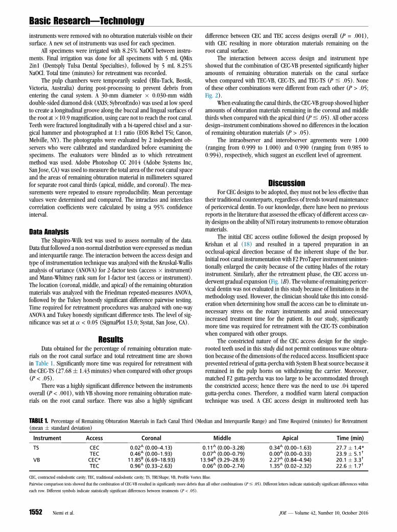
Figure 2. Photographs of mandibular premolars split in buccolingual direction showing remaining obturation materials on root canal surface for different in-strument and access design combinations: (A) CEC-TS, (B) CEC-VB, (C) TEC-TS, and (D) TEC-VB.
Basic Research—Technology
greater potential for sufficient space, allowing easier retrieval of gutta-percha because the access would be larger.
Several methods have been used to determine the amount of resid-ual obturation materials on the canal surface after retreatment (4, 7, 8,12, 20). Similar to previous studies that used single-rooted teeth, ourspecimens were split longitudinally for evaluation (7, 8). Moreaccurate measurements can be obtained with longitudinal sectioningwhen compared with conventional radiographic techniques becausethe latter may be subject to magnification or distortion (20). In ourstudy, high-resolution images were traced by using Adobe Photoshopto quantitatively assess the presence of obturation material remnantsand were reported in relation to the total canal surface area. No attemptwas made to distinguish between residual sealer or gutta-percha.
Neither evaluated NiTi instrumentation systems completelyremoved all obturation materials from the root canals of mandibular
JOE — Volume 42, Number 10, October 2016
premolars. These findings are in agreement with several studies thatevaluated different instruments and systems for this purpose (4, 7, 8,12). In our study, mandibular premolars with similar length andinternal and external anatomy were used to allow standardization ofthe sample. Canals were initially prepared to a 25/.08 instrument andfurther enlarged apically in the retreatment phase to minimize thepresence of residual filling materials (12, 21). No solvents were usedto allow for sole evaluation of the effect of instrumentation. Eachtooth was re-treated with new instruments to decrease fatigue and avoidinstrument separation. The active cutting tip of the D1 ProTaper retreat-ment instrument easily created a pilot space in the gutta-percha/sealermass (22). Retreatment was initiated with the entire ProTaper retreat-ment series in all groups and was followed with either TS or VB instru-ments. Patency was reestablished during retreatment in all specimensindependently of access design or instrument used.
Effect of Access Outline and Instrument Design on Retreatment 1553

Basic Research—Technology
Our results demonstrated that the use of VB instruments through aCEC access design showed higher amounts of obturation materials onthe root canal walls, particularly in the cervical and middle thirds. Apossible reason for this is that the constricted access and the lineardesign of the VB instruments prevented the file from engaging thegutta-percha, despite best efforts to introduce the instruments intothe canal in a buccolingual direction as if the teeth presented as 2 sepa-rate systems because of the oval canal shape. The material remnantswere burnished against the canal walls.
A recent study on the removal of obturation materials from rootcanals when using TS instruments showed no advantage of the latterwhen compared with Reciproc files (23). Our results showed thatthe use of TS instruments through a CEC access design provided thegreatest amount of obturation material removal from the root canal sur-face, similar to the use of VB and TS instruments in a TEC design. If acontracted design is desired by the clinician during access, then TS in-struments can be used with no deleterious effect on the overall amountof obturation material removal. Per this study, this is specific to root ca-nals of single-rooted, oval-shaped mandibular premolars. However,regardless of the retreatment protocol or access design used, residualobturation materials were evident on the root canal surface.
ConclusionsWithin the limitations of this ex vivo study, neither retreatment
protocol was able to render root canals free of obturation materials.However, in the presence of a CEC access design, TS instrumentsremoved more obturation material in single-rooted, oval-shapedcanals.
AcknowledgmentsThis study was supported by an AAE Foundation resident
research grant and is submitted in partial fulfillment of the re-quirements for the degree of Master of Dental Science.
The authors deny any conflicts of interest related to this study.
References1. Farzaneh M, Abitbol S, Friedman S. Treatment outcome in endodontics: the Toronto
Study—phases I and II: orthograde retreatment. J Endod 2004;30:627–33.2. Rocas IN, Siqueira JF Jr. Characterization of microbiota of root canal-treated teeth
with posttreatment disease. J Clin Microbiol 2012;50:1721–4.3. Moiseiwitsch JR, Trope M. Nonsurgical root canal therapy treatment with apparent
indications for root-end surgery. Oral Surg Oral Med Oral Pathol Oral Radiol Endod1998;86:335–40.
1554 Niemi et al.
4. Roggendorf MJ, Legner M, Ebert J, et al. Micro-ct evaluation of residual material incanals filled with activ gp or guttaflow following removal with niti instruments. IntEndod J 2010;43:200–9.
5. Wilcox LR, Krell KV, Madison S, et al. Endodontic retreatment: evaluation of gutta-percha and sealer removal and canal reinstrumentation. J Endod 1987;13:453–7.
6. Colaco AS, Pai VA. Evaluation of the efficiency of manual and rotary gutta-percharemoval techniques. J Endod 2015;41:1871–4.
7. Rios MD, Villela AM, Cunha RS, et al. Efficacy of 2 reciprocating systemscompared with a rotary retreatment system for gutta-percha removal. J Endod2014;40:543–6.
8. €Ozy€urek T, Demiry€urek EO. Efficacy of different nickel-titanium instruments inremoving gutta-percha during root canal retreatment. J Endod 2016;42:646–9.
9. Wu MK, R’Oris A, Barkis D, et al. Prevalence and extent of long oval canals inthe apical third. Oral Surg Oral Med Oral Pathol Oral Radiol Endod 2000;89:739–43.
10. Barbizam JVB, Fariniuk LF, Marchesan MA, et al. Effectiveness of manual and rotaryinstrumentation techniques for cleaning flattened root canals. J Endod 2002;28:365–6.
11. Peters OA. Current challenges and concepts in the preparation of root canal systems:a review. J Endod 2004;30:559–67.
12. Hassanloo A, Watson P, Finer Y, et al. Retreatment efficacy of the epiphany soft resinobturation system. Int Endod J 2007;40:633–43.
13. Versluis A, Kim HC, Lee W, et al. Flexural stiffness and stresses in nickel-titaniumrotary files for various pitch and cross-sectional geometries. J Endod 2012;38:1399–403.
14. Zhang EW, Cheung GS, Zheng YF. Influence of cross-sectional design and dimensionon mechanical behavior of nickel-titanium instruments under torsion and bending:a numerical analysis. J Endod 2010;36:1394–8.
15. Shen Y, Zhou HM, Zheng YF, et al. Current challenges and concepts of the thermo-mechanical treatment of nickel-titanium instruments. J Endod 2013;39:163–72.
16. Alapati SB, Brantley WA, Iijima M, et al. Metallurgical characterization of a newnickel-titanium wire for rotary endodontic instruments. J Endod 2009;35:1589–93.
17. Johnson E, Lloyd A, Kuttler S, et al. Comparison between a novel nickel-titaniumalloy and 508 nitinol on the cyclic fatigue life of profile 25/.04 rotary instruments.J Endod 2008;34:1406–9.
18. Krishan R, Paque F, Ossareh A, et al. Impacts of conservative endodontic cavity onroot canal instrumentation efficacy and resistance to fracture assessed in incisors,premolars, and molars. J Endod 2014;40:1160–6.
19. Eaton JA, Clement DJ, Lloyd A, et al. Micro-computed tomographic evaluation of theinfluence of root canal system landmarks on access outline forms and canal curva-tures in mandibular molars. J Endod 2015;41:1888–91.
20. Maciel ACD, Scelza MFZ. Efficacy of automated versus hand instrumentation duringroot canal retreatment: an ex vivo study. Int Endod J 2006;39:779–84.
21. Friedman S, Moshonov J, Trope M. Efficacy of removing glass ionomer cement, zincoxide eugenol, and epoxy resin sealers from retreated root canals. Oral Surg OralMed Oral Pathol 1992;73:609–12.
22. Takahashi CM, Cunha RS, De Martin AS, et al. In vitro evaluation of the effectivenessof protaper universal rotary retreatment system for gutta-percha removal with orwithout a solvent. J Endod 2009;35:1580–3.
23. Zuolo AD, Zuolo ML, Bueno CED, et al. Evaluation of the efficacy of trushape andreciproc file systems in the removal of root filling material: an ex vivo micro-computed tomographic study. J Endod 2016;42:315–9.
JOE — Volume 42, Number 10, October 2016