Embed Size (px)
Citation preview
RETINAL DISORDERS
Effect of intravitreal injection of aflibercept or ranibizumabon chorioretinal atrophy in myopic choroidal neovascularization
Kaori Sayanagi1 & Sato Uematsu1& Chikako Hara1 & Taku Wakabayashi1 & Yoko Fukushima1 & Shigeru Sato1
&
Yasushi Ikuno1& Kohji Nishida1
Received: 2 July 2018 /Revised: 2 December 2018 /Accepted: 6 December 2018 /Published online: 14 January 2019
AbstractPurpose To compare chorioretinal atrophy (CRA) progression in myopic choroidal neovascularization (mCNV) between intra-vitreal injections of ranibizumab (IVR) and aflibercept (IVA) in the eyes with mCNV.Methods Thirty eyes (28 patients) with treatment-naïve mCNVwere included in this study. IVR or IVAwas administered for upto 1 year. The best-corrected visual acuity (BCVA) was measured, and fundus photographs and fundus autofluorescence wereobtained before and 1, 3, 6, and 12 months after the initial treatment. The clinical characteristics including the macular choroidalthickness in various areas and CRA progression were compared between the drugs. The clinical characteristics and macularchoroidal thicknesses were compared between eyes with and without CRA progression.Results The BCVA improved significantly (p < 0.05 for all comparisons) from 0.44 to 0.26, 0.19, 0.20, and 0.17 after 1, 3, 6, and12months, respectively. CRA progressed in 12 (40%) eyes over 1 year. The CRA progression did not differ significantly betweenaflibercept and ranibizumab. The foveal choroid was significantly (p = 0.0043) thinner in aflibercept-treated eyes compared withranibizumab-treated eyes at 1 year. Subfoveal CNV tended to cause CRA progression more frequently at 1 year, although this didnot reach significance.Conclusions IVA to treat mCNV caused more severe thinning of the foveal choroid than ranibizumab; however, no significantdifference was seen in CRA progression between the drugs and the choroidal thickness should not be associated with CRAprogression. The CNV location may predict CRA progression after anti-vascular endothelial growth factor therapy for mCNV.
Keywords Myopic choroidal neovascularization . Anti-VEGF therapy . Ranibizumab . Aflibercept . Macular atrophy .
Chorioretinal atrophy . Choroidal thickness
Introduction
Myopic choroidal neovascularization (mCNV), a severe com-plication of pathologicmyopia, occurs in up to 10% ofmyopicpatients and up to 40% in highly myopic patients [1–3]. Thelong-term visual prognosis is poor without treatment [4].Although various treatments, including laser photocoagula-tion, macular translocation, surgical CNV removal, photody-namic therapy (PDT), and anti-vascular endothelial growth
factor (anti-VEGF) therapy, have been performed [5], thelong-term visual outcomes are extremely poor primarily dueto the development of chorioretinal atrophy (CRA) around theregressed CNV [6–12]. To improve the long-term visual out-comes of mCNV, treatments are needed that also prevent de-velopment of CRA. The mechanism of the development ofCRA around the CNV is unknown, although Ohno-Matsuiet al. observed holes in Bruch’s membrane at the myopic cho-roidal atrophy including CRA around the CNV using swept-source optical coherence tomography (OCT) [13]. Several riskfactors for CRA, including age, CNV size and location, andchoroidal thickness, have been reported [12, 14, 15], in con-trast, Oishi et al. reported that no patient characteristics signif-icantly affected the CRA [9].
In the eyes with age-related macular degeneration (AMD),macular atrophy develops despite anti-VEGF therapy and af-fects the long-term visual outcomes in some cases [16].
* Kaori [email protected]
1 Department of Ophthalmology, Osaka University Medical School,Osaka, Japan
Graefe's Archive for Clinical and Experimental Ophthalmology (2019) 257:749–757https://doi.org/10.1007/s00417-018-04214-w
# The Author(s) 2019
Macular atrophy develops in about 10% of patients withAMD and 37% of those with retinal angiomatous proliferation(RAP) [17, 18]. The mechanism of the development of mac-ular atrophy is unknown; however, several investigators havereported that macular atrophy was associated significantlywith the baseline macular choroidal thickness and the numberof anti-VEGF injections administered [17, 19].
The first-line treatment for mCNV recently has becomeanti-VEGF therapy. Two large, multi-center, double-masked,randomized, controlled clinical trials, i.e., the RADIANCE(Ranibizumab And PDT evAluation iN myopic ChoroidalnEovascularization) study and the MYRROR (IntravitrealAflibercept Injection in Patients with Myopic ChoroidalNeovascularization) study, have reported beneficial effects oftreatment [5, 20, 21]. However, it is unclear if anti-VEGFtherapy also prevents later development of CRA in high my-opia. In addition, although a recent study reported that theincidence of macular atrophy was higher in the eyes withRAP treated with intravitreal injections of aflibercept (IVA)(Eylea, Regeneron Pharmaceuticals, Tarrytown, NY, BayerAG, Leverkusen, Germany) compared with intravitreal injec-tions of ranibizumab (IVR) (Lucentis, Genentech, South SanFrancisco, CA) [18, 22], it is unclear if the prevalence rates ofCRA development after administration of anti-VEGF therapyfor mCNV differ between IVA and IVR. The current studycompared the progression of CRA between IVA and IVR andinvestigated the difference between those with and withoutCRA enlargement.
Methods
We retrospectively reviewed the records of patients whowere treated with IVA or IVR for mCNV. The diagnosis ofmCNV was based on the characteristic findings on fluores-cein angiography (FA), indocyanine green angiography(ICGA) images, and fundus photographs. The inclusioncriteria were a refractive error of − 6.0 diopters (D) or higheror axial length of 26.5 mm or higher, the presence ofsubfoveal or juxtafoveal CNV, and treatment with IVA orIVR at Osaka University Hospital. The exclusion criteriaincluded any treatment for mCNV other than anti-VEGFtherapy before or during the observation period, a follow-up period of less than 12 months, intraocular surgery otherthan cataract surgery, or development of other ocular dis-eases during the follow-up.
The best-corrected visual acuity (BCVA) was measuredand a dilated fundus examination that included indirect oph-thalmoscopy, color fundus photography, FA, OCT, and fundusautofluorescence (FAF) was performed. FA and OCT wereused to determine the location of the CNV, which was definedas subfoveal if the CNV was present in the foveal center,juxtafoveal if the CNVedge was within 200 μm of the foveal
center, and extrafoveal if the CNV edge was more than200 μm from the foveal center. For the purposes of analysis,the location of the CNV was classified as subfoveal or notsubfoveal. When it was difficult to determine the location ofthe CNV using only FA images, OCT images were used todetermine if the CNV was beneath the foveal center. OCTfindings including intraretinal and subretinal fluid and CNVwith a fuzzy border also were observed. The BCVAwas con-verted to the logarithm of the minimum angle of resolution foranalysis.
After topical anesthesia was applied, aflibercept (2.0 mg/0.05 ml) or ranibizumab (0.5 mg/0.05 ml) was injected 3.5 to4.0 mm posterior to the corneal limbus into the vitreous cavityusing a 30-gauge needle. Prophylactic topical antibiotics wereapplied for 3 days after the injection. Follow-up evaluationswere conducted at 1, 3, 6, and 12months. Additional follow-upvisits were planned for each patient at the clinician’s discretion.After the initial treatment, additional treatment was adminis-tered as needed. The criteria for retreatment were determinedbased on objective/subjective visual declines, exudativechanges seen on the OCT images, and/or dye leakage on FA.
The research adhered to the tenets of the Declaration ofHelsinki. The institutional review board of Osaka UniversityHospital approved this retrospective study.
FA, ICGA, and FAF examinations
All examinations were performed using the Spectralis instru-ment (Heidelberg Retina Angiograph+OCT, Heidelberg,Germany) and a fundus camera (TRC-50DX, TopconCorporation, Tokyo, Japan).
Choroidal and retinal thickness measurements
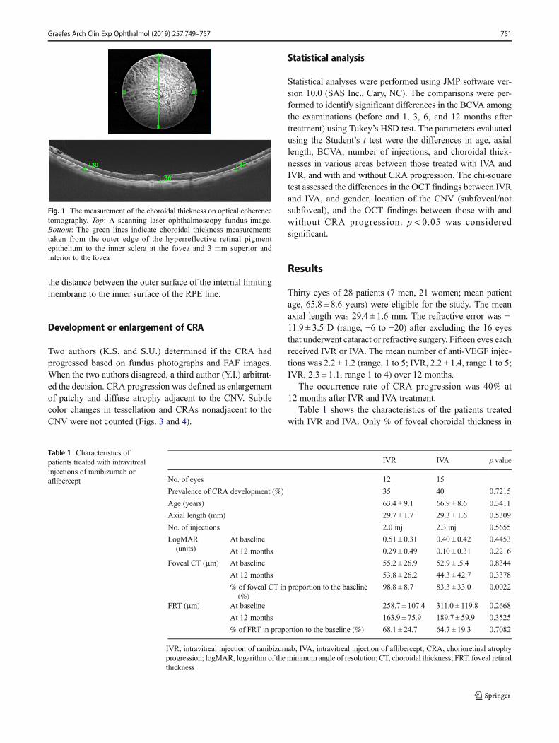
Swept-source OCTDRI OCT-1 Atlantis (Topcon Corporation)was used to obtain the measurements. The choroidal thick-nesses were manually segmented and defined as the distancefrom the retinal pigment epithelial (RPE) line to thehyperreflective line behind the large vessel layers of the cho-roid, presumed to be the choroidal-scleral interface.
A five-line cross scan centered on the fovea was performed.After the B-scan scale was adjusted to 1:1 and the size of theimage, the observer measured the choroidal thickness usingthe caliper function, which is a built-in linear measuring tool(Fig. 1). These measurements were performed manually andobtained at the fovea and 3.0 mm superior, inferior, nasal, andtemporal to the fovea. The choroidal thickness at 12 monthsand % of foveal choroidal thickness in proportion to the base-line choroidal thickness at 3.0 mm nasal to the fovea wereexcluded from the analysis because the choroidal thicknessat 3.0 mm nasally was too thin to be measured due toperipapillary atrophy. The retinal thickness was defined as
750 Graefes Arch Clin Exp Ophthalmol (2019) 257:749–757
the distance between the outer surface of the internal limitingmembrane to the inner surface of the RPE line.
Development or enlargement of CRA
Two authors (K.S. and S.U.) determined if the CRA hadprogressed based on fundus photographs and FAF images.When the two authors disagreed, a third author (Y.I.) arbitrat-ed the decision. CRA progression was defined as enlargementof patchy and diffuse atrophy adjacent to the CNV. Subtlecolor changes in tessellation and CRAs nonadjacent to theCNV were not counted (Figs. 3 and 4).
Statistical analysis
Statistical analyses were performed using JMP software ver-sion 10.0 (SAS Inc., Cary, NC). The comparisons were per-formed to identify significant differences in the BCVA amongthe examinations (before and 1, 3, 6, and 12 months aftertreatment) using Tukey’s HSD test. The parameters evaluatedusing the Student’s t test were the differences in age, axiallength, BCVA, number of injections, and choroidal thick-nesses in various areas between those treated with IVA andIVR, and with and without CRA progression. The chi-squaretest assessed the differences in the OCT findings between IVRand IVA, and gender, location of the CNV (subfoveal/notsubfoveal), and the OCT findings between those with andwithout CRA progression. p < 0.05 was consideredsignificant.
Results
Thirty eyes of 28 patients (7 men, 21 women; mean patientage, 65.8 ± 8.6 years) were eligible for the study. The meanaxial length was 29.4 ± 1.6 mm. The refractive error was −11.9 ± 3.5 D (range, −6 to −20) after excluding the 16 eyesthat underwent cataract or refractive surgery. Fifteen eyes eachreceived IVR or IVA. The mean number of anti-VEGF injec-tions was 2.2 ± 1.2 (range, 1 to 5; IVR, 2.2 ± 1.4, range 1 to 5;IVR, 2.3 ± 1.1, range 1 to 4) over 12 months.
The occurrence rate of CRA progression was 40% at12 months after IVR and IVA treatment.
Table 1 shows the characteristics of the patients treatedwith IVR and IVA. Only % of foveal choroidal thickness in
Fig. 1 The measurement of the choroidal thickness on optical coherencetomography. Top: A scanning laser ophthalmoscopy fundus image.Bottom: The green lines indicate choroidal thickness measurementstaken from the outer edge of the hyperreflective retinal pigmentepithelium to the inner sclera at the fovea and 3 mm superior andinferior to the fovea
Table 1 Characteristics ofpatients treated with intravitrealinjections of ranibizumab oraflibercept
IVR IVA p value
No. of eyes 12 15
Prevalence of CRA development (%) 35 40 0.7215
Age (years) 63.4 ± 9.1 66.9 ± 8.6 0.3411
Axial length (mm) 29.7 ± 1.7 29.3 ± 1.6 0.5309
No. of injections 2.0 inj 2.3 inj 0.5655
LogMAR(units)
At baseline 0.51 ± 0.31 0.40 ± 0.42 0.4453
At 12 months 0.29 ± 0.49 0.10 ± 0.31 0.2216
Foveal CT (μm) At baseline 55.2 ± 26.9 52.9 ± .5.4 0.8344
At 12 months 53.8 ± 26.2 44.3 ± 42.7 0.3378
% of foveal CT in proportion to the baseline(%)
98.8 ± 8.7 83.3 ± 33.0 0.0022
FRT (μm) At baseline 258.7 ± 107.4 311.0 ± 119.8 0.2668
At 12 months 163.9 ± 75.9 189.7 ± 59.9 0.3525
% of FRT in proportion to the baseline (%) 68.1 ± 24.7 64.7 ± 19.3 0.7082
IVR, intravitreal injection of ranibizumab; IVA, intravitreal injection of aflibercept; CRA, chorioretinal atrophyprogression; logMAR, logarithm of the minimum angle of resolution; CT, choroidal thickness; FRT, foveal retinalthickness
Graefes Arch Clin Exp Ophthalmol (2019) 257:749–757 751
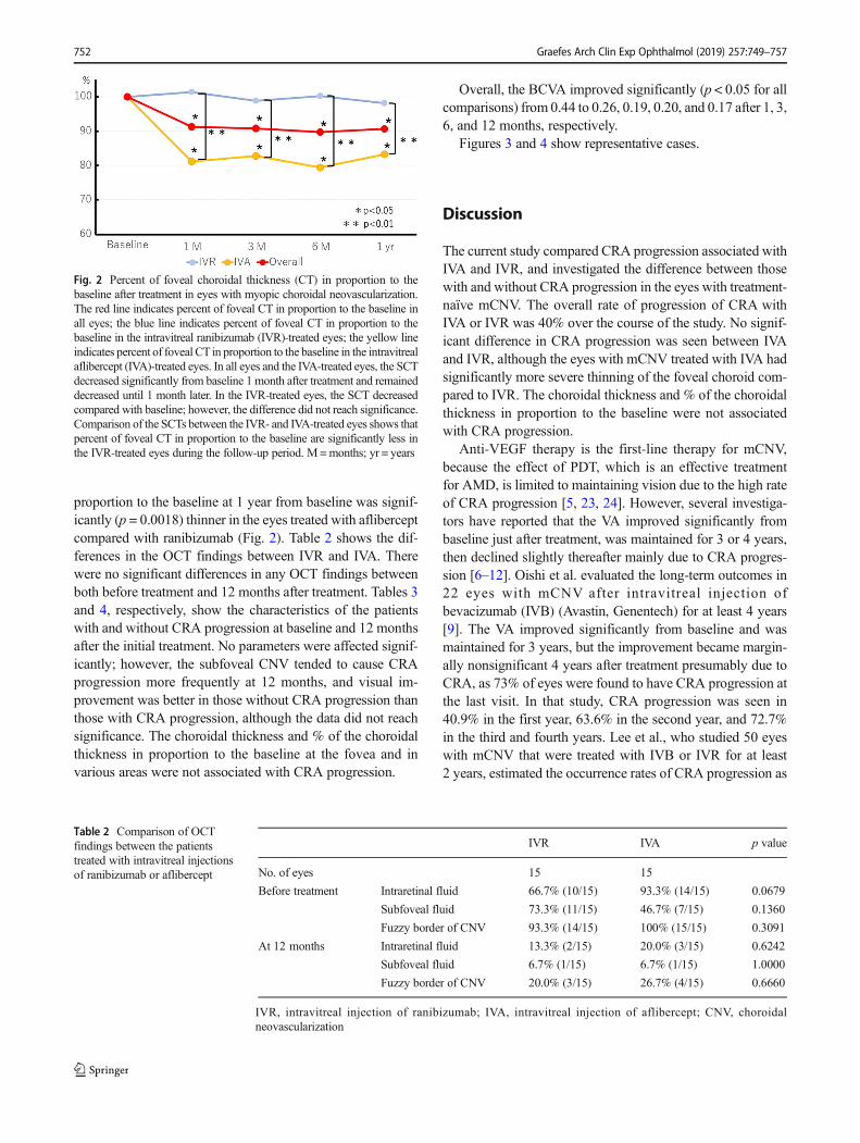
proportion to the baseline at 1 year from baseline was signif-icantly (p = 0.0018) thinner in the eyes treated with afliberceptcompared with ranibizumab (Fig. 2). Table 2 shows the dif-ferences in the OCT findings between IVR and IVA. Therewere no significant differences in any OCT findings betweenboth before treatment and 12 months after treatment. Tables 3and 4, respectively, show the characteristics of the patientswith and without CRA progression at baseline and 12 monthsafter the initial treatment. No parameters were affected signif-icantly; however, the subfoveal CNV tended to cause CRAprogression more frequently at 12 months, and visual im-provement was better in those without CRA progression thanthose with CRA progression, although the data did not reachsignificance. The choroidal thickness and % of the choroidalthickness in proportion to the baseline at the fovea and invarious areas were not associated with CRA progression.
Overall, the BCVA improved significantly (p < 0.05 for allcomparisons) from 0.44 to 0.26, 0.19, 0.20, and 0.17 after 1, 3,6, and 12 months, respectively.
Figures 3 and 4 show representative cases.
Discussion
The current study compared CRA progression associated withIVA and IVR, and investigated the difference between thosewith and without CRA progression in the eyes with treatment-naïve mCNV. The overall rate of progression of CRA withIVA or IVR was 40% over the course of the study. No signif-icant difference in CRA progression was seen between IVAand IVR, although the eyes with mCNV treated with IVA hadsignificantly more severe thinning of the foveal choroid com-pared to IVR. The choroidal thickness and % of the choroidalthickness in proportion to the baseline were not associatedwith CRA progression.
Anti-VEGF therapy is the first-line therapy for mCNV,because the effect of PDT, which is an effective treatmentfor AMD, is limited to maintaining vision due to the high rateof CRA progression [5, 23, 24]. However, several investiga-tors have reported that the VA improved significantly frombaseline just after treatment, was maintained for 3 or 4 years,then declined slightly thereafter mainly due to CRA progres-sion [6–12]. Oishi et al. evaluated the long-term outcomes in22 eyes with mCNV after intravitreal injection ofbevacizumab (IVB) (Avastin, Genentech) for at least 4 years[9]. The VA improved significantly from baseline and wasmaintained for 3 years, but the improvement became margin-ally nonsignificant 4 years after treatment presumably due toCRA, as 73% of eyes were found to have CRA progression atthe last visit. In that study, CRA progression was seen in40.9% in the first year, 63.6% in the second year, and 72.7%in the third and fourth years. Lee et al., who studied 50 eyeswith mCNV that were treated with IVB or IVR for at least2 years, estimated the occurrence rates of CRA progression as
Fig. 2 Percent of foveal choroidal thickness (CT) in proportion to thebaseline after treatment in eyes with myopic choroidal neovascularization.The red line indicates percent of foveal CT in proportion to the baseline inall eyes; the blue line indicates percent of foveal CT in proportion to thebaseline in the intravitreal ranibizumab (IVR)-treated eyes; the yellow lineindicates percent of foveal CT in proportion to the baseline in the intravitrealaflibercept (IVA)-treated eyes. In all eyes and the IVA-treated eyes, the SCTdecreased significantly from baseline 1 month after treatment and remaineddecreased until 1 month later. In the IVR-treated eyes, the SCT decreasedcompared with baseline; however, the difference did not reach significance.Comparison of the SCTs between the IVR- and IVA-treated eyes shows thatpercent of foveal CT in proportion to the baseline are significantly less inthe IVR-treated eyes during the follow-up period. M=months; yr = years
Table 2 Comparison of OCTfindings between the patientstreated with intravitreal injectionsof ranibizumab or aflibercept
IVR IVA p value
No. of eyes 15 15
Before treatment Intraretinal fluid 66.7% (10/15) 93.3% (14/15) 0.0679
Subfoveal fluid 73.3% (11/15) 46.7% (7/15) 0.1360
Fuzzy border of CNV 93.3% (14/15) 100% (15/15) 0.3091
At 12 months Intraretinal fluid 13.3% (2/15) 20.0% (3/15) 0.6242
Subfoveal fluid 6.7% (1/15) 6.7% (1/15) 1.0000
Fuzzy border of CNV 20.0% (3/15) 26.7% (4/15) 0.6660
IVR, intravitreal injection of ranibizumab; IVA, intravitreal injection of aflibercept; CNV, choroidalneovascularization
752 Graefes Arch Clin Exp Ophthalmol (2019) 257:749–757
10% in the first year, 19.1% in the second year, 23.6% in thethird and fourth years, and 35.4% in the fifth year [12]. In thecurrent study, the occurrence rate of CRA progression12 months after treatment corresponded to the results reportedby Oishi et al. [9], but was higher than that of Lee et al. [12].While the reason for this discrepancy is unknown, one hy-pothesis might be differences in the diagnosis and definitionof CRA progression.
In the eyes with AMD, macular atrophy after anti-VEGFtherapy is considered problematic. After anti-VEGF therapy isadministered to the eyes with AMD, the long-term progress ispoor due to macular atrophy, which is CRA at the maculararea [16]. The incidence of macular atrophy in AMD afteranti-VEGF therapy is about 10% in the first year. Based ondisease type, 12 months after treatment, the occurrence rateswere reported to be 36.6% in the eyes with RAP, 3.8% in the
Table 3 Baseline characteristicsof patients with and withoutprogression of choroidal atrophy
CRA No CRA p value
No. of eyes 12 18
Age (years) 63.9 ± 4.9 66.0 ± 10.4 0.4795
Axial length (mm) 29.5 ± 1.5 29.3 ± 1.7 0.6984
Baseline logMAR (units) 0.51 ± 0.40 0.40 ± 0.35 0.8844
Location of CNV (subfoveal/not subfoveal) 9/3 7/11 0.0717
CT (μm) Fovea 54.4 ± 28.9 53 ± 22.9 0.8864
3 mm superior 74.6 ± 45.3 74.9 ± 41.9 0.9855
3 mm inferior 55.1 ± 29.7 68.3 ± 43.0 0.3789
3 mm temporal 71.1 ± 43.0 82.4 ± 35.2 0.4530
3 mm nasal 25.4 ± 9.7 24.1 ± 12.8 0.7736
FRT (μm) 311.8 ± 129.6 267.7 ± 94.2 0.3074
Intraretinal fluid 91.7% (11/12) 72.2% (13/18) 0.1921
Subretinal fluid 50.0% (6/12) 66.7% (12/18) 0.3613
Fuzzy border of CNV 100% (12/12) 94.4% (17/18) 0.4063
CRA, chorioretinal atrophy progression; CNV, choroidal neovascularization; logMAR, logarithm of theminimumangle of resolution; CT, choroidal thickness; FRT, foveal retinal thickness; CNV, choroidal neovascularization
Table 4 Characteristics ofpatients with and withoutprogression of choroidal atrophyat 12 months after treatment
CRA No CRA pvalue
No. of eyes 12 18
LogMAR at 12 months (units) 0.29 ± 0.56 0.09 ± 0.22 0.1787
No. of injections 2.6 ± 1.1 2.0 ± 1.3 0.2060
CT (μm) Fovea 49.8 ± 26.8 47.6 ± 22.3 0.8134
3 mm superior 71.3 ± 42.4 71.9 ± 42.5 0.9705
3 mm inferior 53.8 ± 27.6 63.5 ± 35.8 0.4468
3 mm temporal 69.1 ± 39.5 77.9 ± 32.4 0.5217
% of CT in proportion to thebaseline (%)
Fovea 91.8 ± 14.9 89.9 ± 12.4 0.7096
3 mm superior 97.4 ± 14.8 94.1 ± 16.0 0.5878
3 mm inferior 101.9 ± 14.8 95.9 ± 12.9 0.2674
3 mm temporal 100.1 ± 19.7 95.7 ± 14.2 0.4969
FRT (μm) At 12 months 175.3 ± 73.2 183.2 ± 61.8 0.7627
% of FRT in proportion to thebaseline
61.1 ± 24.9 70.8 ± 17.1 0.2321
Intraretinal fluid 25.0%(3/12)
11.1%(2/18)
0.3173
Subretinal fluid 8.3% (1/12) 5.6% (1/18) 0.7651
Fuzzy border of CNV 8.3% (1/12) 33.3%(6/18)
0.1127
CRA, chorioretinal atrophy progression; logMAR, logarithm of the minimum angle of resolution; CT, choroidalthickness; FRT, foveal retinal thickness; CNV, choroidal neovascularization
Graefes Arch Clin Exp Ophthalmol (2019) 257:749–757 753
eyes with AMD other than RAP treated with IVR, and 10.6%treated with IVA [17, 18, 25]. In another report, the incidencerates of macular atrophy in the eyes with RAPwere 19% in theIVR-treated group and 42.9% in the IVA-treated group12 months after treatment, a difference that reached signifi-cance [22]. Several factors such as the frequency of injections,baseline choroidal thickness, and AMD subtype have beenreported as risk factors for macular atrophy, but no definitiveconclusions have been reached [17, 19]. In addition, it is un-known why the IVA treatment group has a greater occurrencerate of macular atrophy in the eyes with AMD compared to theIVR treatment group. In the current study, the overall occur-rence rate of CRA progression was 40% and there was nodifference between the two treatment groups. The current oc-currence rate of CRA progression is comparable to the inci-dence of macular atrophy in the eyes with RAP. Certainly, thechoroid is thin in the eyes with RAP and highmyopia [26, 27],and the choroid becomes thinner after treatment with IVA thanwith IVR [28]. Lee et al. reported that the choroidal thick-nesses at the fovea and in various areas were not associatedsignificantly with CRA progression, and only the ratio of the
subfoveal choroid to the inferior choroid at 3 mm was associ-ated strongly with CRA progression [12]. The current studydid not analyze the association between the ratio of thesubfoveal choroid to the choroid in various areas and CRAprogression; however, considering that the baseline and 12-month choroidal thicknesses and the choroid thickness chang-es were not associated with CRA in the current report, wespeculated that the choroidal circulation is related somehowto the atrophy of the macular region and IVAmight have moreof a negative impact on the choroidal circulation compared toIVR. However, the choroidal circulation might not necessarilybe evaluated by the choroidal thickness. Future studies thatinclude use of other methods to evaluate the choroidal circu-lation, such as laser speckle flowgraphy and OCT angiogra-phy, are needed to elucidate the pathology of CRA progres-sion [29, 30] as are longer studies with more eyes, whichmight help differentiate the effect on the choroidal morpholo-gy between treatment with IVA and IVR in the eyes withmCNV.
Another interesting result in the current study was thelocation of the CNV. The subfoveal CNV tended to cause
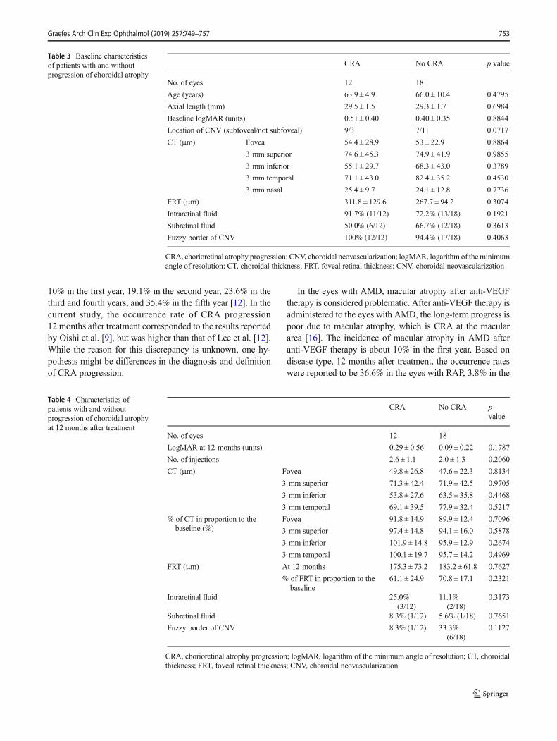
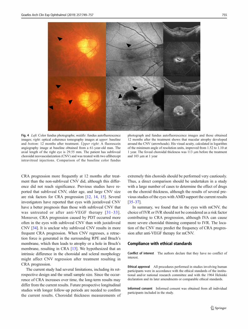
Fig. 3 Left: Color fundus photographs; middle: fundus autofluorescenceimages; right: optical coherence tomography images at upper: baselineand bottom: 12 months after treatment. Upper right: A fluoresceinangiography image at baseline obtained from a 60-year-old woman.The axial length of the right eye is 32.92 mm. The patient has subfovealchoroidal neovascularization (CNV) and was treated with one intravitrealranibizumab injection. Comparison of the baseline color fundus
photograph and fundus autofluorescence image and those obtained12 months after treatment shows the obvious macular atrophy aroundthe CNV (arrowheads) and the patchy atrophy close to the enlargedCNV (arrows). Her visual acuity, calculated in logarithm of the minimumangle of resolution units, improved from 0.82 to 0.40 at 1 year. The fovealchoroidal thickness was 26 μm before the treatment and 27 μm at 1 year
754 Graefes Arch Clin Exp Ophthalmol (2019) 257:749–757
CRA progression more frequently at 12 months after treat-ment than the non-subfoveal CNV did, although this differ-ence did not reach significance. Previous studies have re-ported that subfoveal CNV, older age, and large CNV sizeare risk factors for CRA progression [12, 14, 15]. Severalinvestigators have reported that eyes with juxtafoveal CNVhave a better prognosis than those with subfoveal CNV thatwas untreated or after anti-VEGF therapy [31–33].Moreover, CRA progression caused by PDT occurred moreoften in the eyes with subfoveal CNV than with juxtafovealCNV [34]. It is unclear why subfoveal CNV results in morefrequent CRA progression. When CNV regresses, a retrac-tion force is generated in the surrounding RPE and Bruch’smembrane, which then leads to atrophy or a hole in Bruch’smembrane, resulting in CRA [13]. We hypothesized that anintrinsic difference in the choroidal and scleral morphologymight affect CNV regression after treatment resulting inCRA progression.
The current study had several limitations, including its ret-rospective design and the small sample size. Since the occur-rence of CRA increases over time, the long-term results maydiffer from the current results. Future prospective longitudinalstudies with longer follow-up periods are needed to confirmthe current results. Choroidal thickness measurements of
extremely thin choroids should be performed very cautiously.Thus, a direct comparison should be undertaken in a studywith a large number of cases to determine the effect of drugson the choroid thickness, although the results of several pre-vious studies of the eyes with AMD support the current results[35–37].
In summary, we found that in the eyes with mCNV, thechoice of IVR or IVR should not be considered as a risk factorcontributing to CRA progression, although IVA can causemore severe choroidal thinning compared to IVR. The loca-tion of the CNV may predict the frequency of CRA progres-sion after anti-VEGF therapy for mCNV.
Compliance with ethical standards
Conflict of interest The authors declare that they have no conflict ofinterest.
Ethical approval All procedures performed in studies involving humanparticipants were in accordance with the ethical standards of the institu-tional and/or national research committee and with the 1964 Helsinkideclaration and its later amendments or comparable ethical standards.
Informed consent Informed consent was obtained from all individualparticipants included in the study.
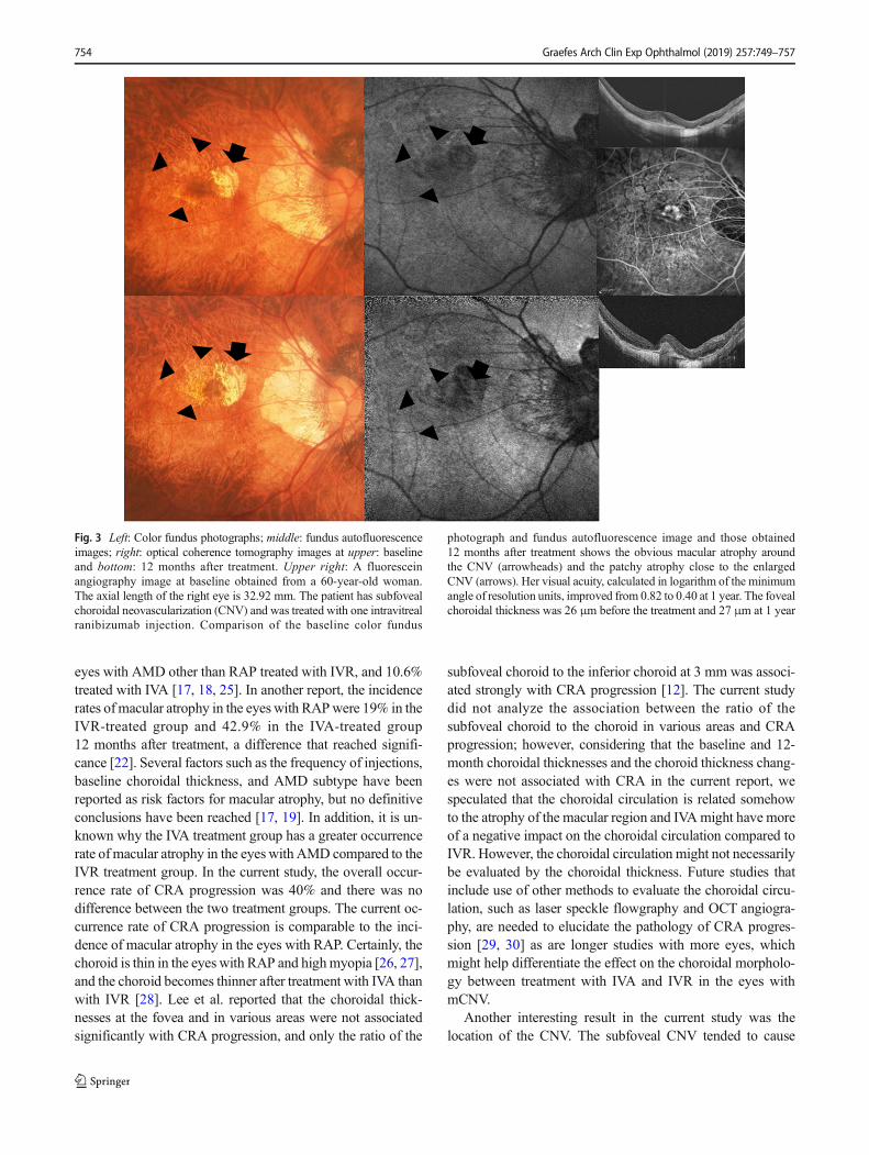
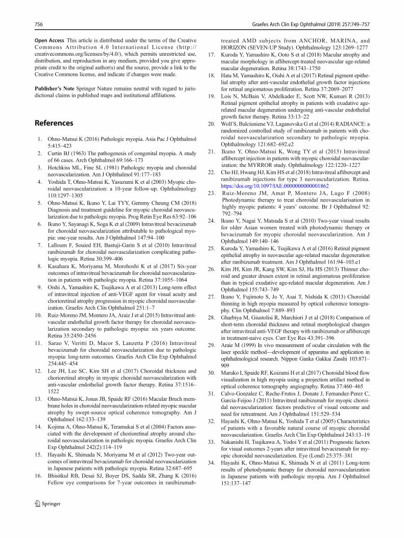
Fig. 4 Left: Color fundus photographs; middle: fundus autofluorescenceimages; right: optical coherence tomography images at upper: baselineand bottom: 12 months after treatment. Upper right: A fluoresceinangiography image at baseline obtained from a 61-year-old man. Theaxial length of the right eye is 29.55 mm. The patient has subfovealchoroidal neovascularization (CNV) and was treated with two afliberceptintravitreal injections. Comparison of the baseline color fundus
photograph and fundus autofluorescence images and those obtained12 months after the treatment shows that macular atrophy developedaround the CNV (arrowheads). His visual acuity, calculated in logarithmof the minimum angle of resolution units, improved from 1.52 to 1.10 at1 year. The foveal choroidal thickness was 113 μm before the treatmentand 103 μm at 1 year
Graefes Arch Clin Exp Ophthalmol (2019) 257:749–757 755
Open Access This article is distributed under the terms of the CreativeCommons At t r ibut ion 4 .0 In te rna t ional License (h t tp : / /creativecommons.org/licenses/by/4.0/), which permits unrestricted use,distribution, and reproduction in any medium, provided you give appro-priate credit to the original author(s) and the source, provide a link to theCreative Commons license, and indicate if changes were made.
References
1. Ohno-Matsui K (2016) Pathologic myopia. Asia Pac J Ophthalmol5:415–423
2. Curtin BJ (1963) The pathogenesis of congenital myopia. A studyof 66 cases. Arch Ophthalmol 69:166–173
3. Hotchkiss ML, Fine SL (1981) Pathologic myopia and choroidalneovascularization. Am J Ophthalmol 91:177–183
4. Yoshida T, Ohno-Matsui K, Yasuzumi K et al (2003) Myopic cho-roidal neovascularization: a 10-year follow-up. Ophthalmology110:1297–1305
5. Ohno-Matsui K, Ikuno Y, Lai TYY, Gemmy Cheung CM (2018)Diagnosis and treatment guideline for myopic choroidal neovascu-larization due to pathologic myopia. Prog Retin Eye Res 63:92–106
6. Ikuno Y, Sayanagi K, Soga K et al (2009) Intravitreal bevacizumabfor choroidal neovascularization attributable to pathological myo-pia: one-year results. Am J Ophthalmol 147:94–100
7. Lalloum F, Souied EH, Bastuji-Garin S et al (2010) Intravitrealranibizumab for choroidal neovascularization complicating patho-logic myopia. Retina 30:399–406
8. Kasahara K, Moriyama M, Morohoshi K et al (2017) Six-yearoutcomes of intravitreal bevacizumab for choroidal neovasculariza-tion in patients with pathologic myopia. Retina 37:1055–1064
9. Oishi A, Yamashiro K, Tsujikawa A et al (2013) Long-term effectof intravitreal injection of anti-VEGF agent for visual acuity andchorioretinal atrophy progression in myopic choroidal neovascular-ization. Graefes Arch Clin Ophthalmol 251:1–7
10. Ruiz-Moreno JM,Montero JA, Araiz J et al (2015) Intravitreal anti-vascular endothelial growth factor therapy for choroidal neovascu-larization secondary to pathologic myopia: six years outcome.Retina 35:2450–2456
11. Sarao V, Veritti D, Macor S, Lanzetta P (2016) Intravitrealbevacizumab for choroidal neovascularization due to pathologicmyopia: long-term outcomes. Graefes Arch Clin Exp Ophthalmol254:445–454
12. Lee JH, Lee SC, Kim SH et al (2017) Choroidal thickness andchorioretinal atrophy in myopic choroidal neovascularization withanti-vascular endothelial growth factor therapy. Retina 37:1516–1522
13. Ohno-Matsui K, Jonas JB, Spaide RF (2016) Macular Bruch mem-brane holes in choroidal neovascularization-related myopic macularatrophy by swept-source optical coherence tomography. Am JOphthalmol 162:133–139
14. Kojima A, Ohno-Matsui K, Teramukai S et al (2004) Factors asso-ciated with the development of chorioretinal atrophy around cho-roidal neovascularization in pathologic myopia. Graefes Arch ClinExp Ophthalmol 242(2):114–119
15. Hayashi K, Shimada N, Moriyama M et al (2012) Two-year out-comes of intravitreal bevacizumab for choroidal neovascularizationin Japanese patients with pathologic myopia. Retina 32:687–695
16. Bhisitkul RB, Desai SJ, Boyer DS, Sadda SR, Zhang K (2016)Fellow eye comparisons for 7-year outcomes in ranibizumab-
treated AMD subjects from ANCHOR, MARINA, andHORIZON (SEVEN-UP Study). Ophthalmology 123:1269–1277
17. Kuroda Y, Yamashiro K, Ooto S et al (2018) Macular atrophy andmacular morphology in aflibercept-treated neovascular age-relatedmacular degeneration. Retina 38:1743–1750
18. Hata M, Yamashiro K, Oishi A et al (2017) Retinal pigment epithe-lial atrophy after anti-vascular endothelial growth factor injectionsfor retinal angiomatous proliferation. Retina 37:2069–2077
19. Lois N, McBain V, Abdelkader E, Scott NW, Kumari R (2013)Retinal pigment epithelial atrophy in patients with exudative age-related macular degeneration undergoing anti-vascular endothelialgrowth factor therapy. Retina 33:13–22
20. Wolf S, Balciuniene VJ, Laganovska G et al (2014) RADIANCE: arandomized controlled study of ranibizumab in patients with cho-roidal neovascularization secondary to pathologic myopia.Ophthalmology 121:682–692.e2
21. Ikuno Y, Ohno-Matsui K, Wong TY et al (2015) Intravitrealaflibercept injection in patients with myopic choroidal neovascular-ization: the MYRROR study. Ophthalmology 122:1220–1227
22. ChoHJ, HwangHJ, KimHS et al (2018) Intravitreal aflibercept andranibizumab injections for type 3 neovascularization. Retina.https://doi.org/10.1097/IAE.0000000000001862
23. Ruiz-Moreno JM, Amat P, Montero JA, Lugo F (2008)Photodynamic therapy to treat choroidal neovascularisation inhighly myopic patients: 4 years’ outcome. Br J Ophthalmol 92:792–794
24. Ikuno Y, Nagai Y, Matsuda S et al (2010) Two-year visual resultsfor older Asian women treated with photodynamic therapy orbevacizumab for myopic choroidal neovascularization. Am JOphthalmol 149:140–146
25. Kuroda Y, Yamashiro K, Tsujikawa A et al (2016) Retinal pigmentepithelial atrophy in neovascular age-related macular degenerationafter ranibizumab treatment. Am J Ophthalmol 161:94–103.e1
26. Kim JH, Kim JR, Kang SW, Kim SJ, Ha HS (2013) Thinner cho-roid and greater drusen extent in retinal angiomatous proliferationthan in typical exudative age-related macular degeneration. Am JOphthalmol 155:743–749
27. Ikuno Y, Fujimoto S, Jo Y, Asai T, Nishida K (2013) Choroidalthinning in high myopia measured by optical coherence tomogra-phy. Clin Ophthalmol 7:889–893
28. Gharbiya M, Giustolisi R, Marchiori J et al (2018) Comparison ofshort-term choroidal thickness and retinal morphological changesafter intravitreal anti-VEGF therapywith ranibizumab or afliberceptin treatment-naive eyes. Curr Eye Res 43:391–396
29. Araie M (1999) In vivo measurement of ocular circulation with thelaser speckle method—development of apparatus and application inophthalmological research. Nippon Ganka Gakkai Zasshi 103:871–909
30. Maruko I, Spaide RF, Koizumi H et al (2017) Choroidal blood flowvisualization in high myopia using a projection artifact method inoptical coherence tomography angiography. Retina 37:460–465
31. Calvo-Gonzalez C, Reche-Frutos J, Donate J, Fernandez-Perez C,Garcia-Feijoo J (2011) Intravitreal ranibizumab for myopic choroi-dal neovascularization: factors predictive of visual outcome andneed for retreatment. Am J Ophthalmol 151:529–534
32. Hayashi K, Ohno-Matsui K, Yoshida T et al (2005) Characteristicsof patients with a favorable natural course of myopic choroidalneovascularization. Graefes Arch Clin Exp Ophthalmol 243:13–19
33. Nakanishi H, Tsujikawa A, Yodoi Yet al (2011) Prognostic factorsfor visual outcomes 2-years after intravitreal bevacizumab for my-opic choroidal neovascularization. Eye (Lond) 25:375–381
34. Hayashi K, Ohno-Matsui K, Shimada N et al (2011) Long-termresults of photodynamic therapy for choroidal neovascularizationin Japanese patients with pathologic myopia. Am J Ophthalmol151:137–147
Publisher’s Note Springer Nature remains neutral with regard to juris-dictional claims in published maps and institutional affiliations.
756 Graefes Arch Clin Exp Ophthalmol (2019) 257:749–757
35. Koizumi H, KanoM, Yamamoto A, SaitoM et al (2015) Short-termchanges in choroidal thickness after aflibercept therapy forneovascular age-related macular degeneration. Am J Ophthalmol159:627–633
36. Yamazaki T, Koizumi H, Yamagishi T, Kinoshita S (2012)Subfoveal choroidal thickness after ranibizumab therapy for
neovascular age-related macular degeneration: 12-month results.Ophthalmology 119:1621–1627
37. Ellabban AA, Tsujikawa A, Ogino K et al (2012) Choroidal thick-ness after intravitreal ranibizumab injections for choroidal neovas-cularization. Clin Ophthalmol 6:837–844
Graefes Arch Clin Exp Ophthalmol (2019) 257:749–757 757