Embed Size (px)
Citation preview

1
CLINICAL RELEVANCE
The ventilatory apparatus and the cardiovascular system have profound effects on each other. 1 , 2 Acute hypoxia impairs car-diac contractility and vascular smooth muscle tone, promot-ing cardiovascular collapse. Hypercarbia causes vasodilation and increases pulmonary vascular resistance. Hyperinflation increases pulmonary vascular resistance, which impedes right-ventricular (RV) ejection and also compresses the heart inside the cardiac fossa in a fashion analogous to tamponade. Lung collapse also increases pulmonary vascular resistance, impeding RV ejection. 3 Acute RV failure, or cor pulmonale, is not only difficult to treat, but it can induce immediate cardiovascular collapse and death.
Ventilator technologies and numerous vasoactive drugs have been developed as means to improve oxygenation of arterial blood. These advances are the subjects of other chapters in this volume. The complex interactions, however,
36 EFFECT OF MECHANICAL VENTILATION ON HEART–LUNG INTERACTIONS Hernando Gomez Michael R. Pinsky
CLINICAL RELEVANCE PHYSIOLOGY OF HEART–LUNG INTERACTIONS Effect of Lung Volume Effect of Intrathoracic Pressure SPONTANEOUS BREATHING VERSUS MECHANICAL POSITIVE-PRESSURE VENTILATION DETECTION AND MONITORING Weaning Failure Using Ventilation to Define Cardiovascular Performance CLINICAL SCENARIOS Initiating Mechanical Ventilation Comparing Different Ventilator Modes Upper Airway Obstruction Chronic Obstructive Pulmonary Disease Auto–Positive End-Expiratory Pressure
Acute Respiratory Distress Syndrome and Acute Lung Injury Congestive Heart Failure Intraoperative State STEPS TO LIMIT OR OVERCOME DETRIMENTAL HEART–LUNG INTERACTIONS Minimize Work of Breathing Minimize Negative Swings in Intrathoracic Pressure Prevent Hyperinflation Fluid Resuscitation during Initiation of Positive-Pressure
Ventilation Prevent Volume Overload during Weaning Augment Cardiac Contractility IMPORTANT UNKNOWNS THE FUTURE SUMMARY AND CONCLUSIONS
The heart and lungs are intimately coupled by their anatomi-cal proximity within the thorax and, more importantly, by their responsibility to deliver the O 2 requirements of indi-vidual cells and organs while excreting the CO 2 by-product of metabolism. During critical illness, if these two organ sys-tems fail, either alone or in combination, the end result is an inadequate O 2 delivery to the body with inevitable tissue ischemia, progressive organ dysfunction, and if untreated, death. Thus, restoration and maintenance of normalized car-diopulmonary function is an essential and primary goal in the management of critically ill patients. Heart failure can impair gas exchange by inducing pulmonary edema and lim-iting blood flow to the respiratory muscles. Ventilation can alter cardiovascular function by altering lung volume, and intrathoracic pressure (ITP), and by increasing metabolic demands. These processes are discussed from the perspec-tive of the impact that ventilation has on the cardiovascular system.
Tobin_Ch36_001-030.indd 1Tobin_Ch36_001-030.indd 1 2/9/12 4:42 PM2/9/12 4:42 PM

2 Part IX Physiologic Effect of Mechanical Ventilation
limiting absolute cardiac volumes analogous to cardiac tam-ponade, except that with hyperinflation both pericardial pressure and ITP increase by a similar amount.
AUTONOMIC TONE
Although neurohumoral processes define a few immedi-ate effects of ventilation on the heart, these neurohumoral processes probably play a primary role in all the long-term effects of ventilation on the cardiovascular system. Most of the immediate effects of ventilation of the heart are sec-ondary to changes in autonomic tone. The lungs are richly enervated with somatic and autonomic fibers that originate, traverse through, and end in the thorax. These networks mediate multiple homeostatic processes through the auto-nomic nervous system altering instantaneous cardiovascular function. The most commonly known of these are the vagally mediated heart rate changes during ventilation. 4 , 5 Inflation of the lung to normal tidal volumes (<10 mL/kg) induces vagal-tone withdrawal, accelerating heart rate. This phe-nomenon is known as respiratory sinus arrhythmia 6 and can be used to document normal autonomic control, 7 especially in patients with diabetes who are at risk for peripheral neu-ropathy. 8 Inflation to larger tidal volumes (>15 mL/kg), how-ever, decreases heart rate by a combination of both increased vagal tone 9 and sympathetic withdrawal. Sympathetic with-drawal also creates arterial vasodilation. 4 , 10 – 14 This inflation–vasodilation response can reduce LV contractility in healthy volunteers 15 and in ventilator-dependent patients with the initiation of high-frequency ventilation 4 or hyperinflation. 12 This inflation–vasodilation response is presumed to be the cause of the initial hypotension seen when infants are placed on mechanical ventilation. It appears to be mediated at least partially by afferent vagal fibers, because it is abolished by selective vagotomy. Hexamethonium, guanethidine, and bre-tylium, however, also block this reflex. 16 , 17 These data sug-gest that lung inflation mediates its reflex cardiovascular effects by modulating central autonomic tone. Interestingly, the almost total lack of measurable hemodynamic effects of unilateral hyperinflation in subjects with normal lungs receiving split-lung ventilation 18 suggests that these auto-nomic cardiovascular effects require a general increase in lung volume to be realized. This is not a minor point because selective hyperinflation within lung units commonly occurs in patients with acute lung injury (ALI) and chronic obstruc-tive pulmonary disease (COPD). If localized hyperinflation were able to induce cardiovascular impairment, these sub-jects would be profoundly compromised.
Humoral factors, including compounds blocked by cyclooxygenase inhibition, 19 released from pulmonary endothelial cells during lung inflation may also induce this depressor response 20 – 22 within a short (15 seconds) time frame. These interactions, however, do not appear to grossly alter cardiovascular status. 23 Ventilation also alters the more chronic control of intravascular fluid balance via hormonal release. The right atrium func-tions as the body’s effective circulating blood-volume
between the heart, circulation, and lungs often leads to a par-adoxical worsening of one organ system function while the function of the other is either maintained or even improved by the use of these technologies and drugs. To minimize these deleterious events, and in the hope of more efficiently and effectively treating critical ill patients with cardiorespi-ratory failure, a better knowledge and understanding of the integrated behavior of the cardiopulmonary system, dur-ing both health and critical illness is essential. Based on this perspective, the health care provider can more appropriately manage this complex and challenging group of patients.
Respiratory function alters cardiovascular function and cardiovascular function alters respiratory function. A useful way to consider the cardiovascular effects of ventilation is to group them by their impact on the determinants of car-diac performance. The determinants of cardiac function can be grouped into four interrelated processes: heart rate, preload, contractility, and afterload. Phasic changes in lung volume and ITP can simultaneously change all four of these hemodynamic determinants for both ventricles. Our current understanding of cardiovascular function also emphasizes both the independence and interdependence of RV and left-ventricular (LV) performance on each other and to external stresses. Complicating these matters further, the direction of interdependence, from right to left or left to right, can be similar or opposite in direction, depending on the baseline cardiovascular state. It is clear, therefore, that a comprehen-sive understanding of the specific cardiopulmonary inter-actions and their relative importance in defining a specific cardiovascular state is a nearly impossible goal to achieve in most patients. By understanding the components of this process, however, one can come to a better realization of its determinants, and, to a greater or lesser degree for any indi-vidual patient, predict the limits of these interactions and how the patient may respond to stresses imposed by either adding or removing artificial ventilatory support.
PHYSIOLOGY OF HEART–LUNG INTERACTIONS
Both spontaneous and positive-pressure ventilation increase lung volume above an end-expiratory baseline. Many of the hemodynamic effects of all forms of ventilation are similar despite differences in the mode of ventilation. ITP, however, decreases during spontaneous inspiration and increases dur-ing positive-pressure ventilation. Thus, the primary reasons for different hemodynamic responses seen during spontane-ous and positive-pressure breathing are related to the changes in ITP and the energy necessary to produce those changes.
Effect of Lung Volume
Changing lung volume phasically alters autonomic tone and pulmonary vascular resistance. At very high lung volumes, the expanding lungs compress the heart in the cardiac fossa,
Tobin_Ch36_001-030.indd 2Tobin_Ch36_001-030.indd 2 2/9/12 4:42 PM2/9/12 4:42 PM
Chapter 36 Effect of Mechanical Ventilation on Heart–Lung Interactions 3
cor pulmonale can precipitate profound cardiovascular col-lapse secondary to excessive RV dilation, RV ischemia, and compromised LV filling. Ventilation can alter pulmonary vascular resistance by either altering pulmonary vasomotor tone, via a process known as hypoxic pulmonary vasoconstric-tion , or mechanically altering vessel cross-sectional area, by changing transpulmonary pressure.
Hypoxic Pulmonary Vasoconstriction. Unlike systemic ves sels that dilate under hypoxic conditions, the pulmonary vasculature constricts. Once alveolar partial pressure of oxygen decreases below 60 mm Hg, or acidemia develops, pulmonary vasomotor tone increases. 38 Hypoxic pulmonary vasoconstriction is mediated, in part, by variations in the synthesis and release of nitric oxide by endothelial nitric oxide synthase localized on pulmonary vascular endothelial cells, and in part by changes in intracellular calcium fluxes in the pulmonary vascular smooth muscle cells. The pulmonary endothelium normally synthesizes a low basal amount of nitric oxide, keeping the pulmonary vasculature actively vasodilated. Loss of nitric oxide allows the smooth muscle to return to its normal resting vasomotor tone. Nitric oxide synthesis is dependent on adequate amounts of O 2 and is inhibited by both hypoxia and acidosis. Presumably, hypoxic pulmonary vasoconstriction developed to minimize ventilation–perfusion mismatches caused by local alveolar hypoventilation. Generalized alveolar hypoxia, however, increases global pulmonary vasomotor tone, impeding RV ejection. 32 At low lung volumes, terminal bronchioles collapse, trapping gas in the terminal alveoli. With continued blood flow, these alveoli lose their O 2 and also may collapse. Patients with acute hypoxemic respiratory failure have small lung volumes and are prone to both alveolar hypoxia and spontaneous alveolar collapse. 39 , 40 This is one of the main reasons why pulmonary vascular resistance is increased in patients with acute hypoxemic respiratory failure.
Based on the above considerations, mechanical ventila-tion may reduce pulmonary vasomotor tone by a variety of mechanisms. First, hypoxic pulmonary vasoconstriction can be inhibited if the patient is ventilated with gas enriched with O 2 increasing alveolar partial pressure of oxygen. 41 – 44 Second, mechanical breaths and positive end-expiratory pressure (PEEP) can refresh hypoventilated lung units and recruit col-lapsed alveolar units, causing local increases in alveolar par-tial pressure of oxygen, 3 , 45 – 47 especially if small lung volumes are returned to resting functional residual capacity (FRC) from an initial smaller lung volume. 48 Third, mechanical ventilation often reverses respiratory acidosis by increasing alveolar ventilation. 44 Fourth, decreasing central sympathetic output, by sedation or decreased stress of breathing against high-input impedance during mechanical ventilation, also reduces vasomotor tone. 49 , 50 Importantly, these effects do not require endotracheal intubation to occur; they occur with mere reexpansion of collapsed alveoli. 51 , 52 Thus, PEEP, CPAP, recruitment maneuvers, and noninvasive ventilation may all reverse hypoxic pulmonary vasoconstriction and may all improve cardiovascular function.
sensor. Circulating levels of a family of natriuretic pep-tides increase in heart failure states secondary to right-atrial stretch. 24 These hormones promote sodium and water diuresis. The levels of these hormones vary directly with the degree of heart failure. Both positive-pressure ventilation and sustained hyperinflation decrease right-atrial stretch mimicking hypovolemia. During positive-pressure ventilation, plasma norepinephrine and renin increase, 25 , 26 whereas atrial natriuretic peptide decreases. 27 This humoral response is the primary reason why venti-lator-dependent patients gain weight early in the course of respiratory failure, because protein catabolism is also usually seen. Interestingly, when patients with congestive heart failure (CHF) are given nasal continuous positive airway pressure (CPAP), plasma atrial natriuretic peptide activity decreases in parallel with improvements in blood flow. 28 , 29 This finding suggests that some of the observed benefit of CPAP therapy in heart failure is mediated in part through humoral mechanisms, owing to the mechan-ical effects of CPAP on cardiac function.
PULMONARY VASCULAR RESISTANCE
Changing lung volume alters pulmonary vascular resistance. 3 Marked increases in pulmonary vascular resistance, as may occur with hyperinflation, can induce acute cor pulmonale and cardiovascular collapse. The reasons for these changes are multifactorial. They can reflect conflicting cardiovascu-lar processes and almost always reflect both humoral and mechanical interactions.
Lung volume can only increase if its distending pressure increases. Lung-distending pressure, called the transpulmo-nary pressure , equals the pressure difference between alveo-lar pressure (Palv) and ITP. If lung volume does not change, then transpulmonary pressure does not change. Thus, occluded inspiratory efforts (Mueller maneuver) and expi-ratory efforts (Valsalva maneuver) cause ITP to vary by an amount equal to Palv, but do not change pulmonary vascular resistance. Although obstructive inspiratory efforts, as occur during obstructive sleep apnea, are usually associated with increased RV afterload, the increased afterload is caused pri-marily by either increased vasomotor tone (hypoxic pulmo-nary vasoconstriction) or backward LV failure. 30 , 31
RV afterload is maximal RV systolic wall stress. 32 , 33 By law of Laplace, wall stress equals the product of the radius of curvature of a structure and its transmural pressure. Systolic RV pressure equals transmural pulmonary artery pressure. Increases in transmural pulmonary artery pressure increases RV afterload, impeding RV ejection, 34 decreasing RV stroke volume, 35 inducing RV dilation, and passively causing venous return to decrease. 19 , 21 If such acute increases in transmural pulmonary artery pressure are not reduced, or if RV con-tractility is not increased by artificial means, then acute cor pulmonale rapidly develops. 36 If RV dilation and RV pres-sure overload persist, RV free-wall ischemia and infarction can develop. 37 These concepts are of profound clinical rel-evance because rapid fluid challenges in the setting of acute
Tobin_Ch36_001-030.indd 3Tobin_Ch36_001-030.indd 3 2/9/12 4:42 PM2/9/12 4:42 PM

4 Part IX Physiologic Effect of Mechanical Ventilation
vessels more distended as lung volume increases, 45 , 56 , 57 just as increasing lung volume increases airway diameter. These radial forces also act upon the extraalveolar vessels, causing them to remain dilated, increasing their capacitance. 58 This tethering is reversed with lung deflation, thereby increasing extraalveolar vascular resistance. 42 , 45 Thus, pulmonary vas-cular resistance is increased at small lung volumes owing to the combined effect of hypoxic pulmonary vasoconstriction and extraalveolar vessel collapse, and at high lung volumes by alveolar compression.
Right-Ventricular Afterload. The right ventricle, as opposed to the left ventricle, ejects blood into a low-pressure, high-compliance system: the pulmonary cir cu la tion. The pulmonary circulation is capable of accom modating high volumes of blood without generating high pressure, which is beneficial for the right ventricle. Despite being compliant, this circuit does pose resistance to the ejecting right ventricle as quantified by pulmonary artery pressure, which is the pressure limit the right ventricle has to overcome to open the pulmonary valve. RV afterload is conceptually similar to LV afterload and is determined by the wall tension of the right ventricle. RV afterload is highly dependent on the distribution of blood flow in the lung, namely, the proportion of West zones 1 and 2, as compared to zone 3, as originally described by Permutt et al. 59 Zones 1 and 2 exist whenever the intraluminal pressure of juxtaalveolar capillaries is lower than the Palv during the respiratory cycle, thus collapsing vessels and increasing pulmonary vascular resistance. In contrast, zone 3 occurs when intraluminal capillary pressure is higher than Palv, decreasing pulmonary resistance. Importantly, intraluminal pressure of alveolar capillaries tracks changes in ITP, 60 and thus decreases less than Palv during spontaneous inspiration, and increases less than Palv during positive-pressure inspiration. Consequently, both spontaneous and positive-pressure inspiration above FRC increase the afterload to the right ventricle as opposed to the LV afterload, which is reduced by increased ITP.
VENTRICULAR INTERDEPENDENCE
Because right ventricle output is linked to left ventricle out-put serially, if right ventricle output decreases, left ventricle output must eventually decrease. The two ventricles, how-ever, are also linked in parallel through their common sep-tum, circumferential fibers, and pericardium, which limits total cardiac volume. For this reason, the diastolic filling of the RV has a direct influence on the shape and compli-ance of the LV, and vice versa. This phenomenon is known as ventricular diastolic interdependence . 61 The most common manifestation of ventricular interdependence is pulsus para-doxus . Changes in RV end-diastolic volume inversely alter LV diastolic compliance. 62 Because venous return can and often does vary by as much as 200% between inspiration and expiration, owing to associated changes in the pressure gradient for venous return (infra vide), right ventricle filling also changes in parallel. Increasing RV end-diastolic volume,
AQ2
Volume-Dependent Changes in Pulmonary Vascular Re sist ance. Changes in lung volume directly alter pul-mo nary vasomotor tone by compressing the alveolar vessels. 39 , 46 , 47 The actual mechanisms by which this occurs have not been completely resolved, but appear to reflect vascular compression induced by a differential extraluminal pressure gradient. The pulmonary circulation lives in two environments, separated from each other by the pressure that surrounds them. 46 The small pulmonary arterioles, venules, and alveolar capillaries sense Palv as their surrounding pressure, and are called alveolar vessels . The large pulmonary arteries and veins, as well as the heart and intrathoracic great vessels of the systemic circulation, sense interstitial pressure or ITP as their surrounding pressure, and are called extraalveolar vessels . Because the pressure difference between Palv and ITP is transpulmonary pressure, increasing lung volume increases this extraluminal pressure gradient. Increases in lung volume progressively increase alveolar vessel resistance by increasing this pressure difference once lung volumes increase much above FRC ( Fig. 36-1 ). 42 , 53 Similarly, increasing lung volume, by stretching and distending the alveolar septa, may also compress alveolar capillaries, although this mechanism is less well substantiated. Hyperinflation can create significant pulmonary hypertension and may precipitate acute RV failure (acute cor pulmonale) 54 and RV ischemia. 37 Thus, PEEP may increase pulmonary vascular resistance if it induces overdistension of the lung above its normal FRC. 55
Extraalveolar vessels are also influenced by changes in transpulmonary pressure. Normally, radial interstitial forces of the lung, which keep the airways patent, only make the large
Hypoxicpulmonary
vasoconstriction
Alveolarvessels
Residualvolume
Functionalresidualcapacity
Totallung
capacity
Lung volume
Total PVR
Alveolarcompression
Extraalveolarvessels
Pu
lmo
nar
y va
scu
lar
resi
stan
ce
FIGURE 36-1 Schematic of the relationship between changes in lung volume and pulmonary vascular resistance ( PVR ), where the extraal-veolar and alveolar vascular components are separated. Pulmonary vascular resistance is minimal at resting lung volume or functional residual capacity. As lung volume increases toward total lung capac-ity or decreases toward residual volume, pulmonary vascular resistance also increases. The increase in resistance with hyperinflation is caused by increased alveolar vascular resistance, whereas the increase in resis-tance with lung collapse is caused by increased extraalveolar vessel tone.
AQ1
Tobin_Ch36_001-030.indd 4Tobin_Ch36_001-030.indd 4 2/9/12 4:42 PM2/9/12 4:42 PM

Chapter 36 Effect of Mechanical Ventilation on Heart–Lung Interactions 5
right ventricle dilation and left ventricle collapse can occur during recruitment maneuvers. 66 This is an important con-cept when treating patients with borderline RV failure. Thus, recruitment maneuvers should be used with caution and be restricted to 10 seconds or less of an end-inspiratory hold to avoid significant hemodynamic derangements.
The presence of ventricular interdependence can be assessed in mechanically ventilated patients based on heart–lung interactions. Using echocardiographic techniques, Mitchell et al 67 and Jardin et al 68 showed that positive- pressure breaths decrease RV dimensions, whereas both LV dimensions and LV flows increase. Still, the changes in RV output generated by positive-pressure inspiration are much less than the changes in LV output. 69 If ventricular interde-pendence was the primary process driving hemodynamic interactions during a positive-pressure breath, then a phasic increase in LV stroke volume would occur during inspira-tion. If the primary process was a phasic decrease in venous return, however, a phasic decrease in LV stroke volume would be observed two to three beats later, usually during the expiratory phase, suggesting the right ventricle is preload responsive. These points underscore the use of LV stroke vol-ume variation during positive-pressure ventilation to iden-tify volume responsiveness.
MECHANICAL HEART–LUNG INTERACTIONS BECAUSE OF LUNG VOLUME
With inspiration, the expanding lungs compress the heart in the cardiac fossa, 70 increasing juxtacardiac ITP. Because the chest wall and diaphragm can move away from the expand-ing lungs, whereas the heart is trapped within this cardiac fossa, juxtacardiac ITP usually increases more than these external ITPs. 71 , 72 This effect is a result of increasing lung volume. It is not affected by the means whereby lung volume is increased. Both spontaneous 73 and positive- pressure-induced hyperinflation 56 , 57 induce similar compressive effects on cardiac filling. If one measured only intraluminal LV pressure, then it would appear as if LV diastolic compli-ance was reduced, because the associated increase in peri-cardial pressure and ITP would not be seen. 74 – 76 When LV function, however, is assessed as the relationship between end-diastolic volume and output, no evidence for impaired LV contractile function is seen 77 , 74 despite the continued application of PEEP. 78 These compressive effects can be considered as analogous to cardiac tamponade 79 – 81 and are discussed further in the next section.
Effect of Intrathoracic Pressure
The heart lives within the thorax, a pressure chamber inside a pressure chamber. Thus, changes in ITP affect the pressure gradients for both systemic venous return to the right ventri-cle and systemic outflow from the left ventricle, independent of the heart itself ( Fig. 36-3 ). Increases in ITP, by increas-ing right-atrial pressure (Pra) and decreasing transmural
AQ3
as occurs during spontaneous inspiration and spontane-ous inspiratory efforts, will reduce LV diastolic compliance, immediately decreasing LV end-diastolic volume. Positive-pressure ventilation may decrease venous return causing RV volumes to decrease, increasing LV diastolic compliance. Except in acute cor pulmonale or biventricular overloaded states, however, the impact of positive-pressure ventilation on LV end-diastolic volume is minimal.
Ventricular interdependence functions through two sep-arate processes. First, increasing RV end-diastolic volume induces an intraventricular septal shift into the LV, thereby decreasing LV diastolic compliance ( Fig. 36-2 ). 63 Because left ventricle wall stress is unaltered, any change in LV output does not reflect a change in LV preload. Because spontane-ous inspiration increases venous return, causing right ven-tricle dilation, LV end-diastolic compliance decreases during spontaneous inspiration. Whereas right ventricle volumes usually do not increase during positive-pressure inspira-tion, ventricular interdependence usually has less impact over the patient’s hemodynamic status. Second, if pericardial restraint or absolute cardiac fossal volume restraint limits absolute biventricular filling, then right ventricle dilation will increase pericardial pressure, with minimal to no sep-tal shift because the pressure outside of both ventricles will increase similarly. 64 , 65
Positive-pressure ventilation, however, can still display right ventricle dilation-associated ventricular interdepen-dence. If positive-pressure inspiration overdistends alveoli, as for example during lung recruitment maneuvers, pulmo-nary vascular resistance will increase. Despite the fact that hemodynamic changes elicited by recruitment maneuvers do not cause persistent cardiovascular insufficiency, transient
0
0
10
20
10 20 30 40
Left ventricle volume (mL)
LV P
ress
ure
(mm
Hg)
Right ventricle volume (mL)
50 35 20 0
Changing RV end-diastolic volume changes LVdiastolic compliance
FIGURE 36-2 Schematic of the effect of increasing right-ventricular (RV) volumes on the relationship between left-ventricular (LV) dia-stolic pressure and left ventricle volume (filling). Increases in right ventricle volumes decrease LV diastolic compliance, such that a higher filling pressure is required to generate a constant end-diastolic volume. (Adapted, with permission, from Taylor RR, Covell JW, Sonnenblick EH, Ross J Jr. Dependence of ventricular distensibility on filling the opposite ventricle. Am J Physiol . 1967;213:711–718.)
Tobin_Ch36_001-030.indd 5Tobin_Ch36_001-030.indd 5 2/9/12 4:42 PM2/9/12 4:42 PM
6 Part IX Physiologic Effect of Mechanical Ventilation
If changes in Pra were the only process that altered venous return, then positive-pressure ventilation would induce pro-found hemodynamic insufficiency in most patients. The decrease in venous return during positive-pressure ventila-tion, however, is often lower than one might expect based on the increase in Pra.
The reasons for this preload-sparing effect seen during positive-pressure ventilation are twofold. First, when cardiac output does decrease, increased sympathetic tone decreases venous capacitance, increasing mean systemic pressure, which tends to restore the pressure gradient for venous return, even in the face of an elevated Pra. Increases in sympathetic tone, however, would increase steady-state cardiac output and would not alter the phasic changes in venous return seen during positive-pressure ventilation. The decreased phasic reductions in venous return are caused by associ-ated increases in mean systemic pressure during inspiration. Diaphragmatic descent and abdominal-muscle contraction increase intraabdominal pressure, decreasing intraabdomi-nal vascular capacitance. 103 , 104 Because a large proportion of venous blood is in the abdomen, the net effect of both inspiration and PEEP is to increase mean systemic pressure and Pra in a parallel but unequal fashion. 105 – 107 Accordingly, the pressure gradient for venous return may not be reduced as much as predicted as predicted from a pure increase in Pra. This is an important adaptive response by the body to positive-pressure ventilation and PEEP, both of which produce this effect secondary to the associated increase in lung volume, which promotes diaphragmatic descent. This preload-sparing effect is especially well demonstrated in
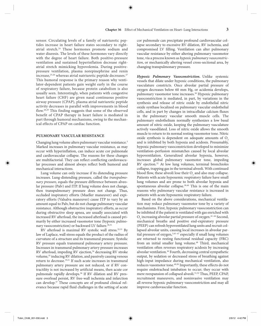
LV systolic pressure, will reduce the pressure gradients for venous return and LV ejection decreasing intrathoracic blood volume. Using the same argument, decreases in ITP will augment venous return and impede LV ejection, increas-ing intrathoracic blood volume. The increases in ITP during positive-pressure ventilation show marked regional differ-ences; juxtacardiac ITP increases more than lateral chest wall ITP as inspiratory flow rate and tidal volume increase. 71 Interestingly, lung compliance plays a minimal role in defin-ing the positive-pressure-induced increase in ITP. For the same increase in tidal volume, ITP usually increases simi-larly if tidal volume is kept constant. 82 , 83 If, however, chest wall compliance decreases, then ITP will increase for a fixed tidal volume. 84 , 85
SYSTEMIC VENOUS RETURN
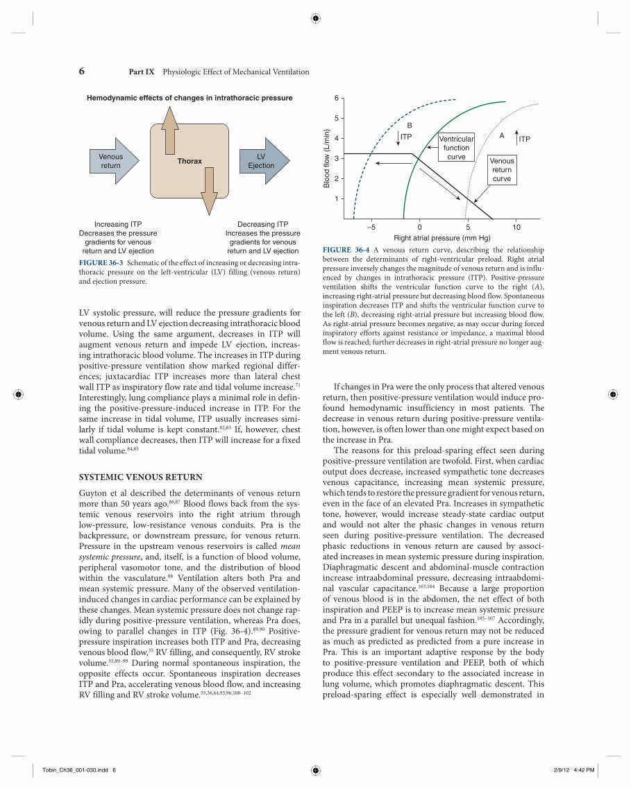
Guyton et al described the determinants of venous return more than 50 years ago. 86 , 87 Blood flows back from the sys-temic venous reservoirs into the right atrium through low-pressure, low-resistance venous conduits. Pra is the backpressure, or downstream pressure, for venous return. Pressure in the upstream venous reservoirs is called mean systemic pressure , and, itself, is a function of blood volume, peripheral vasomotor tone, and the distribution of blood within the vasculature. 88 Ventilation alters both Pra and mean systemic pressure. Many of the observed ventilation-induced changes in cardiac performance can be explained by these changes. Mean systemic pressure does not change rap-idly during positive-pressure ventilation, whereas Pra does, owing to parallel changes in ITP ( Fig. 36-4 ). 89 , 90 Positive-pressure inspiration increases both ITP and Pra, decreasing venous blood flow, 35 RV filling, and consequently, RV stroke volume. 35 , 89 – 99 During normal spontaneous inspiration, the opposite effects occur. Spontaneous inspiration decreases ITP and Pra, accelerating venous blood flow, and increasing RV filling and RV stroke volume. 35 , 36 , 64 , 93 , 96 , 100 – 102
Hemodynamic effects of changes in intrathoracic pressure
Increasing ITPDecreases the pressure
gradients for venousreturn and LV ejection
Decreasing ITPIncreases the pressure
gradients for venousreturn and LV ejection
LVEjection
Venousreturn
Thorax
FIGURE 36-3 Schematic of the effect of increasing or decreasing intra-thoracic pressure on the left-ventricular (LV) filling (venous return) and ejection pressure.
6
5B
ITP Ventricularfunctioncurve Venous
returncurve
4
Right atrial pressure (mm Hg)
3
Blo
od fl
ow (
L/m
in)
2
1
–5 0 5 10
ITPA
FIGURE 36-4 A venous return curve, describing the relationship between the determinants of right-ventricular preload. Right atrial pressure inversely changes the magnitude of venous return and is influ-enced by changes in intrathoracic pressure (ITP). Positive-pressure ventilation shifts the ventricular function curve to the right ( A ), increasing right-atrial pressure but decreasing blood flow. Spontaneous inspiration decreases ITP and shifts the ventricular function curve to the left ( B ), decreasing right-atrial pressure but increasing blood flow. As right-atrial pressure becomes negative, as may occur during forced inspiratory efforts against resistance or impedance, a maximal blood flow is reached; further decreases in right-atrial pressure no longer aug-ment venous return.
Tobin_Ch36_001-030.indd 6Tobin_Ch36_001-030.indd 6 2/9/12 4:42 PM2/9/12 4:42 PM

Chapter 36 Effect of Mechanical Ventilation on Heart–Lung Interactions 7
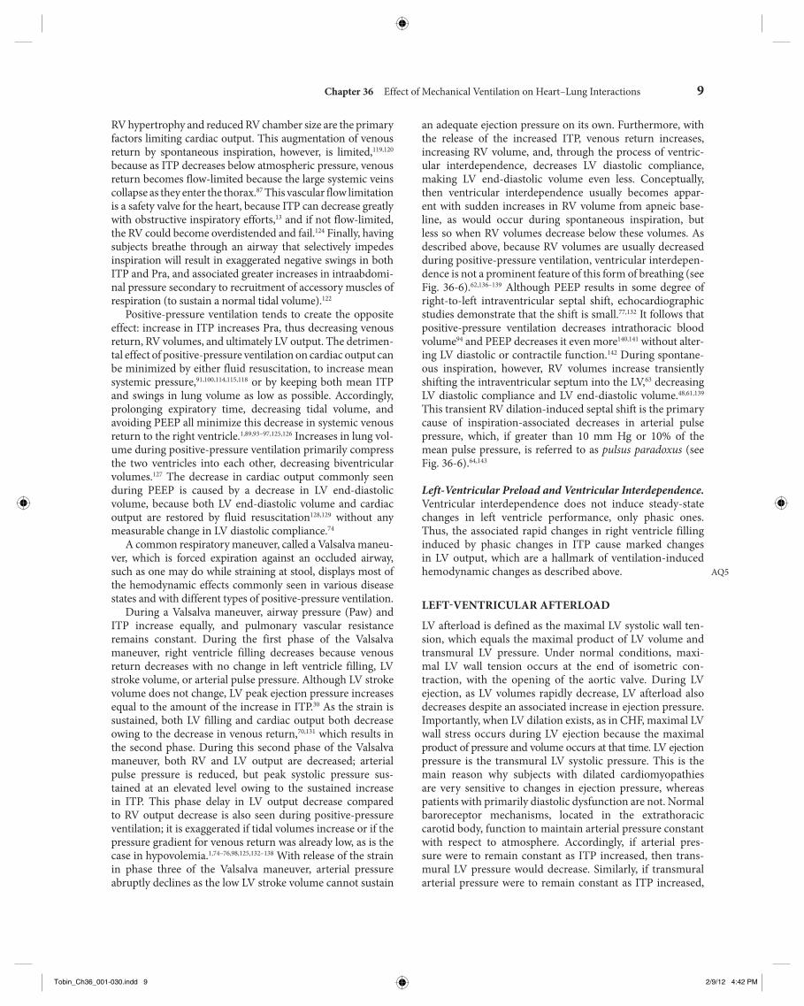
filling occurs with minimal changes in filling pressure. 81 Spontaneous inspiratory efforts usually increase venous return because of the combined decrease in Pra 64 , 94 – 96 , 116 and increase in intraabdominal pressure. 103 , 104 For Pra to remain very low, however, RV diastolic compliance must be high and RV output must equal venous return. Otherwise, sustained increases in venous blood flow would distend the RV and increase Pra. During normal spontaneous inspiration, although venous return increases, ITP decreases at the same time, minimizing any potential increase in Pra, which might otherwise occur if ITP were not to decrease. 89 Aiding in this process of minimizing RV workload, the pulmonary arterial inflow circuit is highly compliant and can accept large increases in RV stroke volume without changing pressure. 35 , 117 Thus, increases in venous return proportionally increase pulmonary arterial inflow without significant changes in RV filling or ejection pressures. Accordingly, this compensatory system fails if RV diastolic compliance decreases or if Pra increases independent of changes in RV end-diastolic volume. Figure 36-6 illustrates these differential effects of negative (spontaneous inspiration) and positive (positive-pressure inspiration) swings in ITP on dynamic RV and LV performance. In RV failure states, spontaneous inspiration does not decrease Pra and Pra actually increases. This results in the physical sign of increased jugular venous distension during spontaneous inspiration.
Note further in Figure 36-6 that not only does RV stroke volume increase with spontaneous inspiration and decrease with positive-pressure inspiration, but also that LV stroke volume decreases only during spontaneous inspiration (ven-tricular interdependence); during positive-pressure inspira-tion, however, any change in LV stroke volume occurs late, as the decrease in RV output finally reaches the left ventricle. RV diastolic compliance can acutely decrease in the setting of
patients with hypervolemia. In fact, both the translocation of blood from the pulmonary to the systemic capacitance vessels, 108 as well as abdominal pressurization secondary to diaphragmatic descent, may be the major mechanisms by which the decrease in venous return is minimized during positive-pressure ventilation. 109 – 113 In fact, van den Berg et al 114 documented that up to 20 cm H 2 O CPAP did not signifi-cantly decrease cardiac output, as measured 30 seconds into an inspiratory-hold maneuver, in fluid-resuscitated, post-operative cardiac surgery patients. Although CPAP induced an increase in Pra, intraabdominal pressure also increased, preventing a significant change in RV volumes ( Fig. 36-5 ). Interest in inverse-ratio ventilation has raised questions as to its hemodynamic effect, because its application includes a large component of hyperinflation.
Current data clearly show that detrimental effects of increased ITP and PEEP on venous return are far more com-plex than an effect on the pressure gradient between mean systemic pressure and Pra, and that geometric deformation of the venous vasculature and its flow distribution, which alter the resistance to flow, may be a better explanation. 115 Animal data suggest that compression and deformation of capacitance vessels at the entrance of the thorax 103 and com-pression of the portal circulation by diaphragmatic descent 115 may account for these increments in venous resistance and thus decreased venous return.
Relevance of Intrathoracic Pressure on Venous Return. It is axiomatic that the heart can only pump out that amount of blood that it receives and no more. Thus, venous return is the primary determinant of cardiac output and the two must be the same. 88 Because Pra is the backpressure to venous return and because Pra is normally close to zero relative to atmospheric pressure, venous return is maintained near maximal levels at rest, 12 , 87 , 94 , 98 , 99 because right ventricle
1.0
0.9
0.7
0.5
0.3
0.1
0
ΔPraΔPaw
0.9
0.8
0.6
0.4
0.2
0
ΔPabdΔPaw
6
4
2
0
–4
–2
–6
ΔRVEDV
ΔPawRVEDV
(%/cm H2O)
FIGURE 36-5 Effect of increasing levels of continuous positive airway pressure (CPAP) on the relations between increasing airway pressure ( Paw ) and right-atrial pressure ( Pra ) ( left graph ), Paw and intraabdominal pressure ( Pabd ) ( center graph ), and Paw and changes in right-ventricular end-diastolic volume ( RVEDV ) ( right graph ) in forty-three postoperative fluid-resuscitated cardiac surgery patients. (Data derived, with permission, from data in Van den Berg et al. J Appl Physiol . 2002;92:1223–1231.) AQ4
Tobin_Ch36_001-030.indd 7Tobin_Ch36_001-030.indd 7 2/9/12 4:42 PM2/9/12 4:42 PM

8 Part IX Physiologic Effect of Mechanical Ventilation
Finally, with exaggerated negative swings in ITP, as occur with obstructed inspiratory efforts, venous return behaves as if abdominal pressure is additive to mean systemic pres-sure in augmenting venous blood flow. 118 – 121 These findings have some investigators to suggest that obstructive breathing may be a therapeutic strategy in sustaining cardiac output in patients in hemorrhagic shock. 122 Interestingly, negative pres-sure ventilation, by augmenting venous return, increases car-diac output by 39% in children following repair of tetralogy of Fallot. 123 In this condition, impaired RV filling secondary to
acute RV dilation or cor pulmonale (pulmonary embolism, hyperinflation, and RV infarction). Importantly, acute RV dilation and acute cor pulmonale can not only induce rapid cardiovascular collapse, but they are singularly not respon-sive to fluid resuscitation. Because spontaneous inspiration and inspiratory efforts cause both ITP and Pra to decrease, RV dilation may occur in patients with occult heart failure. Accordingly, some patients who were previously stable and ventilator-dependent can develop acute RV failure during weaning trials.
17
SV
rvm
L
01 s
PA
Om
m H
g
1050
250
17
SV
lvm
L
0
150
Spontaneousventilation
Positive pressureventilation
Pla
tmm
m H
g
10
5Ppa
tmm
m H
g
8
0Pra
tmm
m H
g
0
5
Pra
mm
Hg
–8
Ppl
mm
Hg
0
Paw
mm
Hg
FIGURE 36-6 Strip chart recording of right and left-ventricular stroke volumes ( SVrv and SVlv , respectively), aortic pressure (Pa O ), left-atrial, pulmo-nary arterial, and right-atrial transmural pressures ( Pla tm , Ppa tm , and Pra tm , respectively), airway pressure ( Paw ), pleural pressure ( Ppl ), and right-atrial pressure ( Pra ) during spontaneous ventilation ( left ) and similar tidal volume positive-pressure ventilation ( right ) in an anesthetized, intact canine model. (Used, with permission, from Pinsky MR, Matuschak GM, Klain M. Determinants of cardiac augmentation by elevations in intrathoracic pres-sure. J Appl Physiol . 1985;58:1189–1198.)
Tobin_Ch36_001-030.indd 8Tobin_Ch36_001-030.indd 8 2/9/12 4:42 PM2/9/12 4:42 PM
Chapter 36 Effect of Mechanical Ventilation on Heart–Lung Interactions 9
an adequate ejection pressure on its own. Furthermore, with the release of the increased ITP, venous return increases, increasing RV volume, and, through the process of ventric-ular interdependence, decreases LV diastolic compliance, making LV end-diastolic volume even less. Conceptually, then ventricular interdependence usually becomes appar-ent with sudden increases in RV volume from apneic base-line, as would occur during spontaneous inspiration, but less so when RV volumes decrease below these volumes. As described above, because RV volumes are usually decreased during positive-pressure ventilation, ventricular interdepen-dence is not a prominent feature of this form of breathing (see Fig. 36-6 ). 62 , 136 – 139 Although PEEP results in some degree of right-to-left intraventricular septal shift, echocardiographic studies demonstrate that the shift is small. 77 , 132 It follows that positive-pressure ventilation decreases intrathoracic blood volume 94 and PEEP decreases it even more 140 , 141 without alter-ing LV diastolic or contractile function. 142 During spontane-ous inspiration, however, RV volumes increase transiently shifting the intraventricular septum into the LV, 63 decreasing LV diastolic compliance and LV end-diastolic volume. 48 , 61 , 139 This transient RV dilation-induced septal shift is the primary cause of inspiration-associated decreases in arterial pulse pressure, which, if greater than 10 mm Hg or 10% of the mean pulse pressure, is referred to as pulsus paradoxus (see Fig. 36-6 ). 64 , 143
Left-Ventricular Preload and Ventricular Interdependence. Ventricular interdependence does not induce steady-state changes in left ventricle performance, only phasic ones. Thus, the associated rapid changes in right ventricle filling induced by phasic changes in ITP cause marked changes in LV output, which are a hallmark of ventilation-induced hemodynamic changes as described above.
LEFT-VENTRICULAR AFTERLOAD
LV afterload is defined as the maximal LV systolic wall ten-sion, which equals the maximal product of LV volume and transmural LV pressure. Under normal conditions, maxi-mal LV wall tension occurs at the end of isometric con-traction, with the opening of the aortic valve. During LV ejection, as LV volumes rapidly decrease, LV afterload also decreases despite an associated increase in ejection pressure. Importantly, when LV dilation exists, as in CHF, maximal LV wall stress occurs during LV ejection because the maximal product of pressure and volume occurs at that time. LV ejection pressure is the transmural LV systolic pressure. This is the main reason why subjects with dilated cardiomyopathies are very sensitive to changes in ejection pressure, whereas patients with primarily diastolic dysfunction are not. Normal baroreceptor mechanisms, located in the extrathoracic carotid body, function to maintain arterial pressure constant with respect to atmosphere. Accordingly, if arterial pres-sure were to remain constant as ITP increased, then trans-mural LV pressure would decrease. Similarly, if transmural arterial pressure were to remain constant as ITP increased,
AQ5
RV hypertrophy and reduced RV chamber size are the primary factors limiting cardiac output. This augmentation of venous return by spontaneous inspiration, however, is limited, 119 , 120 because as ITP decreases below atmospheric pressure, venous return becomes flow-limited because the large systemic veins collapse as they enter the thorax. 87 This vascular flow limitation is a safety valve for the heart, because ITP can decrease greatly with obstructive inspiratory efforts, 13 and if not flow-limited, the RV could become overdistended and fail. 124 Finally, having subjects breathe through an airway that selectively impedes inspiration will result in exaggerated negative swings in both ITP and Pra, and associated greater increases in intraabdomi-nal pressure secondary to recruitment of accessory muscles of respiration (to sustain a normal tidal volume). 122
Positive-pressure ventilation tends to create the opposite effect: increase in ITP increases Pra, thus decreasing venous return, RV volumes, and ultimately LV output. The detrimen-tal effect of positive-pressure ventilation on cardiac output can be minimized by either fluid resuscitation, to increase mean systemic pressure, 91 , 100 , 114 , 115 , 118 or by keeping both mean ITP and swings in lung volume as low as possible. Accordingly, prolonging expiratory time, decreasing tidal volume, and avoiding PEEP all minimize this decrease in systemic venous return to the right ventricle. 1 , 89 , 93 – 97 , 125 , 126 Increases in lung vol-ume during positive-pressure ventilation primarily compress the two ventricles into each other, decreasing biventricular volumes. 127 The decrease in cardiac output commonly seen during PEEP is caused by a decrease in LV end-diastolic volume, because both LV end-diastolic volume and cardiac output are restored by fluid resuscitation 128 , 129 without any measurable change in LV diastolic compliance. 74
A common respiratory maneuver, called a Valsalva maneu-ver, which is forced expiration against an occluded airway, such as one may do while straining at stool, displays most of the hemodynamic effects commonly seen in various disease states and with different types of positive-pressure ventilation.
During a Valsalva maneuver, airway pressure (Paw) and ITP increase equally, and pulmonary vascular resistance remains constant. During the first phase of the Valsalva maneuver, right ventricle filling decreases because venous return decreases with no change in left ventricle filling, LV stroke volume, or arterial pulse pressure. Although LV stroke volume does not change, LV peak ejection pressure increases equal to the amount of the increase in ITP. 30 As the strain is sustained, both LV filling and cardiac output both decrease owing to the decrease in venous return, 70 , 131 which results in the second phase. During this second phase of the Valsalva maneuver, both RV and LV output are decreased; arterial pulse pressure is reduced, but peak systolic pressure sus-tained at an elevated level owing to the sustained increase in ITP. This phase delay in LV output decrease compared to RV output decrease is also seen during positive-pressure ventilation; it is exaggerated if tidal volumes increase or if the pressure gradient for venous return was already low, as is the case in hypovolemia. 1 , 74 – 76 , 98 , 125 , 132 – 138 With release of the strain in phase three of the Valsalva maneuver, arterial pressure abruptly declines as the low LV stroke volume cannot sustain
Tobin_Ch36_001-030.indd 9Tobin_Ch36_001-030.indd 9 2/9/12 4:42 PM2/9/12 4:42 PM

10 Part IX Physiologic Effect of Mechanical Ventilation
or vocal cord paralysis) or stiff lungs (interstitial lung disease, pulmonary edema, or ALI), selectively increase LV afterload, and may be the cause of LV failure and pulmonary edema, 13 , 30 , 31 , 151 especially if LV systolic function is already compromised. 152 , 153
Pulsus paradoxus seen during spontaneous inspiration under conditions of marked pericardial restraint reflects primarily ventricular interdependence. 154 – 158 The negative swings in ITP, however, also increase LV ejection pressure, increasing LV end-systolic volume. 130 Other systemic fac-tors may influence LV systolic function during loaded inspi-ratory efforts. These associated factors also contribute to a greater or lesser degree to the inhibition of normal LV sys-tolic function, including increased in aortic input imped-ance, 159 altered synchrony of contraction of the global LV myocardium, 160 and hypoxemia-induced decreased global myocardial contractility. 161 Hypoxia also directly reduces LV diastolic compliance. 162 Experimental repetitive periodic air-way obstructions induce pulmonary edema in normal ani-mals. 30 , 31 Furthermore, removing the negative swings in ITP by applying nasal CPAP results in improved global LV per-formance in patients with combined obstructive sleep apnea and CHF. 130
Relevance of Intrathoracic Pressure on Left-Ventricular Afterload. If arterial pressure remains constant, then increases in ITP decrease transmural LV ejection pressure, decreasing LV afterload. These points are easily demonstrated in a subject with an indwelling arterial pressure catheter during cough or Valsalva maneuvers. During a cough, ITP increases rapidly without changes in intrathoracic blood volume. Arterial pressure also increases by a similar amount, as described above for phase 1 of the Valsalva maneuver. Thus, transmural LV pressure (LV pressure relative to ITP) 130 , 163 , 164 and aortic blood flow 70 would remain constant. Sustained increases in ITP, however, must eventually decrease aortic blood flow and arterial pressure secondary to the associated decrease in venous return. 130 If ITP increased arterial pressure without changing transmural arterial pressure, then baroreceptor-mediated vasodilation would induce arterial vasodilation to maintain extrathoracic arterial pressure-flow relations constant. 134 Because coronary perfusion pressure reflects the ITP gradient for blood flow and is not increased by ITP-induced increases in arterial pressure, such sustained increases in ITP can cause decreased coronary perfusion pressure-induced myocardial ischemia. 165 – 167
SPONTANEOUS BREATHING VERSUS MECHANICAL POSITIVE-PRESSURE VENTILATION
Both spontaneous and mechanical ventilation increase lung volume above resting end-expiratory lung volume or FRC. During both spontaneous and positive-pressure ventilation, end-expiratory lung volume can be artificially increased by the addition of PEEP. Thus, the primary hemodynamic
then LV wall tension would decrease. 144 Thus, increases in ITP decrease LV afterload, and decreases in ITP increase LV afterload. 130 , 145 These two opposing effects of changes in ITP on LV afterload have important clinical implications.
The concept that increases in ITP decrease both LV pre-load and LV afterload can be clearly illustrated with the use of high-frequency jet ventilation, which can increase ITP but does not result in large swings in lung volume. 135 When high-frequency jet ventilation is delivered in synchrony with the cardiac cycle, such that heart rate and ventilatory frequency are identical, one can dissect out the effects of ITP on preload and afterload. Under hypovolemic and normovolemic con-ditions with intact cardiovascular reserve, positive-pressure ventilation usually decreases steady-state cardiac output by decreasing the pressure gradient for venous return. When one compares the hemodynamic effects of high-frequency jet ventilation synchronized to occur during diastole (when ventricular filling occurs), cardiac output decreases to levels seen during end-inspiration for normal-to-large tidal- volume (10 mL/kg) ventilation. In the same subject, however, if the increases in ITP occur during systole, the detrimental effects of the same mean Paw, mean ITP, and tidal volume do not impede venous return ( Fig. 36-7 ). 146 , 147 Furthermore, in heart failure states, positive-pressure ventilation does not impede cardiac output because the same decreases in venous return do not alter LV preload. If these increases in ITP, however, reduce LV afterload, then cardiac output will also increase. These points are illustrated in Figure 36-8 , wherein synchro-nous high-frequency jet ventilation is delivered either during preejection systole (presystolic) or ejection (systolic). The only difference between the two ventilatory states is that arte-rial pulse pressure does not change despite increases in LV stroke volume with presystolic increases in Paw, consistent with a decreased LV afterload, whereas with systolic increases in Paw, arterial pulse pressure increases, and peak arterial pressure increases by an amount equal to the increase in ITP, consistent with mechanically augmented LV ejection.
Relevance of Intrathoracic Pressure on Myocardial Oxygen Consumption. Decreases in ITP increase both LV afterload and myocardial O 2 consumption. Accordingly, spontaneous ventilation not only increases global O 2 demand by its exercise component, 80 , 126 , 148 but also increases myocardial O 2 consumption. Profound decreases in ITP commonly occur during spontaneous inspiratory efforts with bronchospasm, obstructive breathing, and acute hypoxemic respiratory failure. Under these conditions, the cardiovascular burden can be great and may induce acute heart failure and pulmonary edema. 30 Because weaning from positive-pressure ventilation to spontaneous ventilation may reflect dramatic changes in ITP swings, from positive to negative, independent of the energy requirements of the respiratory muscles, weaning is a selective LV stress test. 144 , 148 – 150 Similarly, improved LV systolic function is observed in patients with severe LV failure placed on mechanical ventilation. 150 Very negative swings in ITP, as seen with vigorous inspiratory efforts in the setting of airway obstruction (asthma, upper airway obstruction,
Tobin_Ch36_001-030.indd 10Tobin_Ch36_001-030.indd 10 2/9/12 4:42 PM2/9/12 4:42 PM
Chapter 36 Effect of Mechanical Ventilation on Heart–Lung Interactions 11
Comparison of synchronous high-frequency jet ventilation to intermittentpositive-pressure breathing in the control (normal), state
1
0
20 s
Systolic Diastolic Systolic Diastolic
150
1050
20
10
5
20
0
0
15
0
0
0
1 s
EKG
SV
rv m
L/kg
SV
lv m
L/kg
PAo
mm
Hg
Pla
tm m
m H
gP
patm
mm
Hg
Pra
tm m
m H
gP
aw m
m H
gP
pl m
m H
g
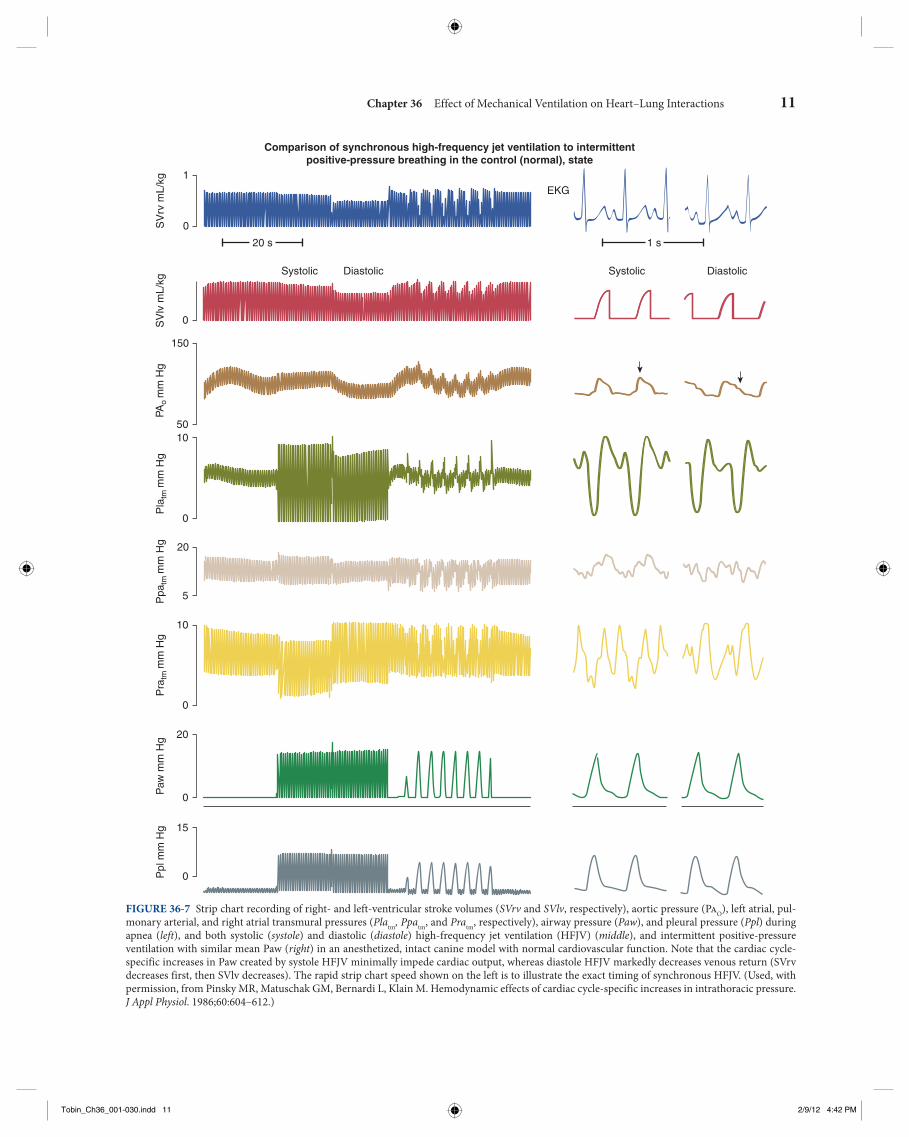
FIGURE 36-7 Strip chart recording of right- and left-ventricular stroke volumes ( SVrv and SVlv , respectively), aortic pressure (Pa O ), left atrial, pul-monary arterial, and right atrial transmural pressures ( Pla tm , Ppa tm , and Pra tm , respectively), airway pressure ( Paw ), and pleural pressure ( Ppl ) during apnea ( left ), and both systolic ( systole ) and diastolic ( diastole ) high-frequency jet ventilation (HFJV) ( middle ), and intermittent positive-pressure ventilation with similar mean Paw ( right ) in an anesthetized, intact canine model with normal cardiovascular function. Note that the cardiac cycle-specific increases in Paw created by systole HFJV minimally impede cardiac output, whereas diastole HFJV markedly decreases venous return (SVrv decreases first, then SVlv decreases). The rapid strip chart speed shown on the left is to illustrate the exact timing of synchronous HFJV. (Used, with permission, from Pinsky MR, Matuschak GM, Bernardi L, Klain M. Hemodynamic effects of cardiac cycle-specific increases in intrathoracic pressure. J Appl Physiol . 1986;60:604–612.)
Tobin_Ch36_001-030.indd 11Tobin_Ch36_001-030.indd 11 2/9/12 4:42 PM2/9/12 4:42 PM

Comparison of synchronous high-frequency jet ventilation to intermittentpositive-pressure breathing in acute ventricular failure
1
0SV
rv m
L/kg
1
0SV
lv m
L/kg
50
150
10
25
5
Pla
tm m
m H
gP
patm
mm
Hg
25
5Pra
tm m
m H
g
20
0
Paw
mm
Hg
15
0Ppl
mm
Hg
PAo
mm
Hg
30
Intermittentpositive-pressure
breathing
Intermittentpositive-pressure
breathingPresystolic Systolic Systolic ApneaPresystolic
20 s
FIGURE 36-8 Continuous strip chart recording of right- and left-ventricular stroke volumes ( SVrv and SVlv , respectively), aortic pressure (Pa O ), left atrial, pulmonary arterial, and right-atrial transmural pressures ( Pla tm , Ppa tm , and Pra tm , respectively), airway pressure ( Paw ), pleural pressure ( Ppl ), and right-atrial pressure ( Pra ) during intermittent positive-pressure ventilation (tidal volume [V T ] 10 mL/kg), apnea ( left ), and then both preejection systole (presystolic) and LV ejection (systolic) synchronous high-frequency jet ventilation (HFJV) ( middle ), and then intermittent positive-pressure ventilation again ( right ) in an anesthetized, intact canine model with fluid-resuscitated acute ventricular failure. Note that the cardiac cycle–specific increases in Paw created by both presystolic and systolic HFJV increase steady-state SVrv and SVlv (i.e., cardiac output), but affect Pa O differently. Presystolic HFJV does not change Pa O pulse pressure despite an increase in SVlv (reduced afterload), whereas systolic HFJV increases Pa O pulse pres-sure for a similar increase in SVlv. (Used, with permission, from Pinsky MR, Matuschak GM, Bernardi L, Klain M. Hemodynamic effects of cardiac cycle-specific increases in intrathoracic pressure. J Appl Physiol . 1986;60:604–612.)
Tobin_Ch36_001-030.indd 12Tobin_Ch36_001-030.indd 12 2/9/12 4:42 PM2/9/12 4:42 PM

Chapter 36 Effect of Mechanical Ventilation on Heart–Lung Interactions 13
DETECTION AND MONITORING
Weaning Failure
Ventilator-dependent patients who fail to wean often have impaired baseline cardiovascular performance that is read-ily apparent, 153 but commonly patients develop overt signs of heart failure during weaning, such as pulmonary edema, 153 , 174 myocardial ischemia, 175 – 178 tachycardia, and gut ischemia. 179 Pulmonary artery occlusion pressure may rise rapidly to nonphysiologic levels within 5 minutes of instituting wean-ing. 153 Although all patients increase their cardiac outputs in response to a weaning trial, those that subsequently fail to wean demonstrate a reduction in mixed venous O 2 satu-ration, consistent with a failing cardiovascular response to an increased metabolic demand. 180 Weaning from mechani-cal ventilation can be considered a cardiovascular stress test. Again, investigators have documented weaning-associated electrocardiogram and thallium cardiac blood flow scan-related signs of ischemia in both patients with known coronary artery disease 175 and in otherwise normal patients. 177 , 178 Using this same logic, placing patients with severe heart failure and/or ischemia on ventilator support, by either intubation and ventilation 181 or noninvasive CPAP 182 can reverse myocardial ischemia. Importantly, the increased work of breathing may come from the endotracheal tube flow resistance. 183 Thus, some patients who fail a spontaneous breathing trial may actually be able to breathe on their own if extubated. There is, however, no known method of identifying this subgroup.
Using Ventilation to Define Cardiovascular Performance
Because the cardiovascular response to positive-pressure breathing is determined by the baseline cardiovascular state, these responses can be used to define such cardiovascular states. Sustained increases in airway pressure will reduce venous return, allowing one to assess LV ejection over a range of end-diastolic volumes. If echocardiographic mea-sures of LV volumes are simultaneously made, then one can use an inspiratory-hold maneuver to measure cardiac con-tractility, as defined by the end-systolic pressure-volume relationship, 184 which is similar to those created by transient inferior vena-caval occlusion. 185 , 186 Furthermore, these mea-sures can be made during the ventilatory cycle to define dynamic interactions. 186
Patients with relative hypervolemia, a condition often associated with CHF, are at less risk of developing impaired venous return during initiation of mechanical ventilation, whereas hypovolemic patients are at increased risk. If posi-tive airway pressure augments LV ejection in heart failure states by reducing LV afterload, then systolic arterial pres-sure should not decrease but actually increase during inspira-tion, so-called reverse pulsus paradoxus. This was what Abel et al 187 saw in ten postcardiac surgery patients. Perel et al 188 – 190 suggested that the relationship between ventilatory efforts
differences between spontaneous ventilation and positive-pressure ventilation are caused by the changes in ITP and the muscular contraction needed to create these changes. Importantly, even if a patient is receiving ventilator support, spontaneous respiratory efforts can persist and may result in marked increases in metabolic load, and contribute to sus-tained respiratory muscle fatigue. 168 Still, a primary reason for instituting mechanical ventilation is to decrease the work of breathing. Normal spontaneous ventilation augments venous return and vigorous inspiratory efforts account for most of the increased blood flow seen in exercise. Conversely, positive-pressure ventilation may impair ventricular fill-ing and induce hypovolemic cardiac dysfunction in normal or hypovolemic subjects while augmenting LV function in patients with heart failure. Finally, heart failure, whether primary or induced by ventilation, may induce acute respira-tory muscle fatigue causing respiratory failure or failure to wean from mechanical ventilation, and overtax the ability of the circulation to deliver O 2 to the rest of the body.
Fundamental to this concept is the realization that spon-taneous ventilation is exercise. Spontaneous ventilatory efforts are induced by contraction of the respiratory muscles, mainly the diaphragm and intercostal muscles. 148 Although ventilation normally requires less than 5% of total O 2 deliv-ery to meet its demand 148 (and is difficult to measure at the bedside even when using calibrated metabolic measuring devices), in lung disease states in which work of breathing is increased, the metabolic demand for O 2 can increase to 30% of total O 2 delivery. 80 , 125 , 148 , 169 With marked hyperpnea, muscles of the abdominal wall and shoulder girdle function as accessory muscles. Blood flow to these muscles is derived from several arterial circuits, whose absolute flow exceeds the highest metabolic demand of maximally exercising skele-tal muscle under normal conditions. 148 , 170 , 171 Thus, blood flow is usually not the limiting factor determining maximal ven-tilatory effort. In severe heart failure states, however, blood flow constraints may limit ventilation because blood flow to other organs and to the respiratory muscles may be compro-mised, inducing both tissue hypoperfusion and lactic aci-dosis. 170 – 172 Aubier et al demonstrated that if cardiac output is severely limited by the artificial induction of tamponade in a canine model that respiratory muscle failure develops despite high central neuronal drive. 171 The animals die a respiratory death before cardiovascular standstill. The insti-tution of mechanical ventilation for ventilatory and hypox-emic respiratory failure may reduce metabolic demand on the stressed cardiovascular system increasing mixed venous oxygen saturation (SVO 2 ) for a constant cardiac output and arterial oxygen content (CaO 2 ). 172 Intubation and mechani-cal ventilation, when adjusted to the metabolic demands of the patient, may dramatically decrease the work of breath-ing, resulting in increased O 2 delivery to other vital organs and decreased serum lactic acid levels. Under conditions in which fixed right-to-left shunts exist, the obligatory increase in SVO 2 will result in an increase in the partial pressure of arterial oxygen (PaO2
), despite no change in the ratio of shunt blood flow to cardiac output.
Tobin_Ch36_001-030.indd 13Tobin_Ch36_001-030.indd 13 2/9/12 4:42 PM2/9/12 4:42 PM

14 Part IX Physiologic Effect of Mechanical Ventilation
LV volumes, as often occurs with CHF and afterload reduc-tion, may be quite volume responsive. Thus, preload does not equal preload responsiveness. Third, all the reported stud-ies used positive-pressure ventilation to vary venous return. For such changes in venous return, however, to induce LV output changes, the changes must be of sufficient enough magnitude to cause measurable changes in preload. 69 If the increase in lung volume with each tidal breath is either not great enough to induce changes in pulmonary venous flow, 198 or if the positive-pressure breath is associated with spontane-ous inspiratory efforts that minimize the changes in venous return, 199 then the cyclic perturbations to cardiac filling may not be great enough to induce the cyclic variations in LV fill-ing needed to identify preload responsiveness. Furthermore, the degree of pressure or flow variation will be proportional to tidal volume, with greater tidal volumes inducing greater changes for the same cardiovascular state. 192 , 195 , 200 Thus, the means by which cyclic changes in lung volume and ITP are induced will affect the magnitude of arterial pressure and flow variations. Fourth, although the primary determinant of arterial pulse-pressure variation over a single breath is LV stroke-volume variation, because changes in aortic imped-ance and arterial tone cannot change that rapidly 201 over time, this limitation no longer applies. As arterial tone decreases, for example, then for the same aortic flow and stroke volume both mean arterial pressure and pulse pressure will be less. Accordingly, flow variation becomes more sensitive than pulse pressure variation as hemorrhage progresses. 193
CLINICAL SCENARIOS
Initiating Mechanical Ventilation
NORMOVOLEMIC AND HYPOVOLEMIC PATIENTS
The process of initiating mechanical ventilation is a complex physiologic process for a variety of reasons. First, pharma-cologic factors needed to allow for endotracheal intubation also blunt sympathetic responses, exaggerating the hemo-dynamic effects induced by increasing airway pressure and defining tidal volume and ventilatory frequency. This point is clearly demonstrated by comparing the relative benign impact that reinstituting ventilator support in a patient with a preexistent tracheotomy, with the impact of the initial intubation and ventilation of the same patient a few days or weeks earlier. As noted above, positive-pressure ventilation increases ITP, which must alter venous return. If the patient has reduced vasomotor tone, as commonly exists during induction of anesthesia, the associated increase in Pra will induce a proportional decrease in venous return, pulmo-nary blood flow and subsequently cardiac output. 1 , 35 , 152 , 202 If the associated tidal volumes are excessive for the duration of expiratory time available to allow for passive deflation, then dynamic hyperinflation will occur, increasing pulmonary vascular resistance and compressing the heart in the cardiac fossa, further decreasing further biventricular volumes. 127 If one were to examine the dynamic effects of ventilation on
and systolic arterial pressure may be used to identify which patients may benefit from cardiac-assist maneuvers. Patients who increase their systolic arterial pressure during venti-lation, relative to an apneic baseline, tend to have a greater degree of volume overload 189 and heart failure, 190 whereas patients who decrease systolic arterial pressure tend to be volume responsive. Perhaps more relevant to usual clinical practice is the identification of patients whose cardiac output will increase if given a volume challenge. The identification of preload responsiveness is important because only half of the hemodynamically unstable patients studied in several clinical series were actually preload-responsive. 191 Thus, nonspecific fluid loading will not only be ineffective at restoring cardio-vascular stability in half the subjects, it will also both delay definitive therapy and may promote cor pulmonale or pulmo-nary edema. Finally, Michard et al 192 found, in a series of ven-tilator-dependent septic patients, that the greater the degree of arterial pulse-pressure variation during positive-pressure ventilation, the greater the subsequent increase in cardiac output in response to volume-expansion therapy. The recent literature has documented that both arterial pulse-pressure and LV stroke-volume variations 193 , 194 induced by positive-pressure ventilation are sensitive and specific markers of pre-load responsiveness. This literature was recently reviewed. 195 The greater the degree of flow or pressure variation over the course of the respiratory cycle for a fixed tidal volume, the more likely a patient is to increase cardiac output in response to a volume challenge, and the greater that increase. The over-arching principles of this clinical tool have only recently been described. 173 There are several important caveats and limita-tions to this approach that need to be considered before the clinician proceeds to monitoring arterial pulse pressure or stroke volume variation during ventilation as a routine assess-ment of preload responsiveness.
First, and perhaps most importantly, being preload-responsive does not mean that the patient should be given volume. Otherwise healthy subjects under general anesthe-sia without evidence of cardiovascular insufficiency are also preload-responsive, but do not need a volume challenge. The presence of positive-pressure-induced changes in aortic flow or arterial pulse pressure does not itself define therapy. Independent documentation of cardiovascular insufficiency needs to be sought before the clinician attempts fluid resus-citation based on these measures. Second, these indices, which quantify the variation in aortic flow, stroke volume, and arterial systolic and pulse pressures, have routinely been demonstrated to outperform more traditional measures of LV preload, such as pulmonary occlusion pressure, Pra, total tho-racic blood volume, RV end-diastolic volume, and LV end-diastolic area. 192 , 194 There appears to be little relation between ventricular preload and preload responsiveness. Ventricular filling pressures poorly reflect ventricular volumes, and mea-sures of absolute ventricular volumes do not define diastolic compliance. 196 , 197 Patients with small left ventricles that are also stiff, as may occur with acute cor pulmonale, tampon-ade, LV hypertrophy, and myocardial fibrosis, will show poor volume responsiveness. Conversely, patients with large
Tobin_Ch36_001-030.indd 14Tobin_Ch36_001-030.indd 14 2/9/12 4:42 PM2/9/12 4:42 PM
Chapter 36 Effect of Mechanical Ventilation on Heart–Lung Interactions 15
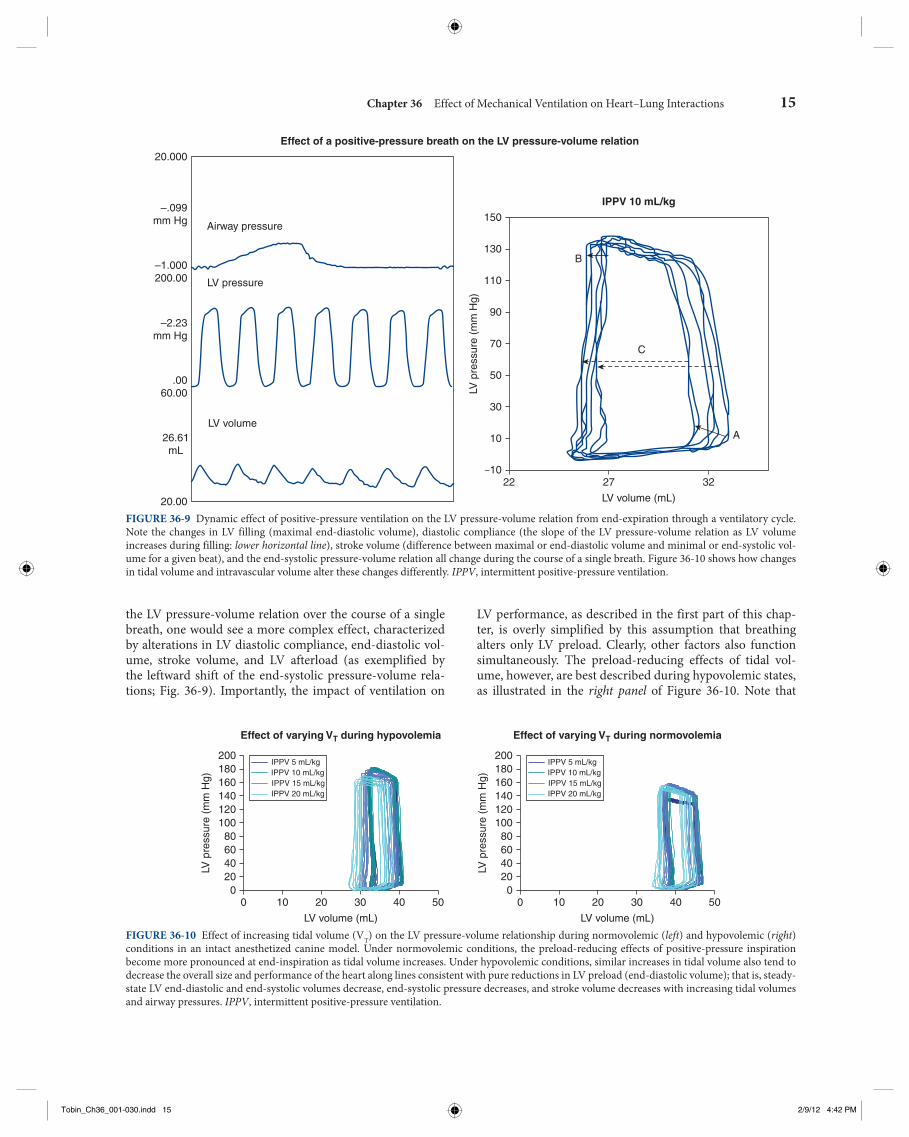
LV performance, as described in the first part of this chap-ter, is overly simplified by this assumption that breathing alters only LV preload. Clearly, other factors also function simultaneously. The preload-reducing effects of tidal vol-ume, however, are best described during hypovolemic states, as illustrated in the right panel of Figure 36-10 . Note that
the LV pressure-volume relation over the course of a single breath, one would see a more complex effect, characterized by alterations in LV diastolic compliance, end-diastolic vol-ume, stroke volume, and LV afterload (as exemplified by the leftward shift of the end-systolic pressure-volume rela-tions; Fig. 36-9 ). Importantly, the impact of ventilation on
Effect of a positive-pressure breath on the LV pressure-volume relation
–1022 27 32
A10
30
50
LV p
ress
ure
(mm
Hg)
70
90
110
130
150
IPPV 10 mL/kg
LV volume (mL)
B
C
20.00
26.61mL
.0060.00
–2.23mm Hg
–1.000200.00
Airway pressure
LV pressure
LV volume
–.099mm Hg
20.000
FIGURE 36-9 Dynamic effect of positive-pressure ventilation on the LV pressure-volume relation from end-expiration through a ventilatory cycle. Note the changes in LV filling (maximal end-diastolic volume), diastolic compliance (the slope of the LV pressure-volume relation as LV volume increases during filling: lower horizontal line ), stroke volume (difference between maximal or end-diastolic volume and minimal or end-systolic vol-ume for a given beat), and the end-systolic pressure-volume relation all change during the course of a single breath. Figure 36-10 shows how changes in tidal volume and intravascular volume alter these changes differently. IPPV , intermittent positive-pressure ventilation.
200180160140120100806040200
0 10 20 30 40 50
LV volume (mL)
Effect of varying VT during hypovolemia
LV p
ress
ure
(mm
Hg)
IPPV 5 mL/kgIPPV 10 mL/kgIPPV 15 mL/kgIPPV 20 mL/kg
200180160140120100806040200
0 10 20 30 40 50
LV volume (mL)
Effect of varying VT during normovolemia
LV p
ress
ure
(mm
Hg)
IPPV 5 mL/kgIPPV 10 mL/kgIPPV 15 mL/kgIPPV 20 mL/kg
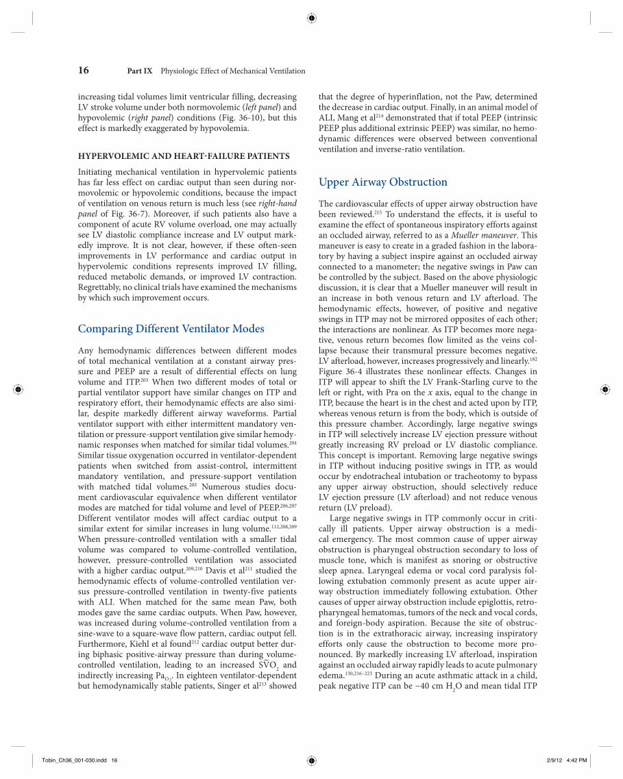
FIGURE 36-10 Effect of increasing tidal volume (V T ) on the LV pressure-volume relationship during normovolemic ( left ) and hypovolemic ( right ) conditions in an intact anesthetized canine model. Under normovolemic conditions, the preload-reducing effects of positive-pressure inspiration become more pronounced at end-inspiration as tidal volume increases. Under hypovolemic conditions, similar increases in tidal volume also tend to decrease the overall size and performance of the heart along lines consistent with pure reductions in LV preload (end-diastolic volume); that is, steady-state LV end-diastolic and end-systolic volumes decrease, end-systolic pressure decreases, and stroke volume decreases with increasing tidal volumes and airway pressures. IPPV , intermittent positive-pressure ventilation.
Tobin_Ch36_001-030.indd 15Tobin_Ch36_001-030.indd 15 2/9/12 4:42 PM2/9/12 4:42 PM
16 Part IX Physiologic Effect of Mechanical Ventilation
that the degree of hyperinflation, not the Paw, determined the decrease in cardiac output. Finally, in an animal model of ALI, Mang et al 214 demonstrated that if total PEEP (intrinsic PEEP plus additional extrinsic PEEP) was similar, no hemo-dynamic differences were observed between conventional ventilation and inverse-ratio ventilation.
Upper Airway Obstruction
The cardiovascular effects of upper airway obstruction have been reviewed. 215 To understand the effects, it is useful to examine the effect of spontaneous inspiratory efforts against an occluded airway, referred to as a Mueller maneuver . This maneuver is easy to create in a graded fashion in the labora-tory by having a subject inspire against an occluded airway connected to a manometer; the negative swings in Paw can be controlled by the subject. Based on the above physiologic discussion, it is clear that a Mueller maneuver will result in an increase in both venous return and LV afterload. The hemodynamic effects, however, of positive and negative swings in ITP may not be mirrored opposites of each other; the interactions are nonlinear. As ITP becomes more nega-tive, venous return becomes flow limited as the veins col-lapse because their transmural pressure becomes negative. LV afterload, however, increases progressively and linearly. 182 Figure 36-4 illustrates these nonlinear effects. Changes in ITP will appear to shift the LV Frank-Starling curve to the left or right, with Pra on the x axis, equal to the change in ITP, because the heart is in the chest and acted upon by ITP, whereas venous return is from the body, which is outside of this pressure chamber. Accordingly, large negative swings in ITP will selectively increase LV ejection pressure without greatly increasing RV preload or LV diastolic compliance. This concept is important. Removing large negative swings in ITP without inducing positive swings in ITP, as would occur by endotracheal intubation or tracheotomy to bypass any upper airway obstruction, should selectively reduce LV ejection pressure (LV afterload) and not reduce venous return (LV preload).
Large negative swings in ITP commonly occur in criti-cally ill patients. Upper airway obstruction is a medi-cal emergency. The most common cause of upper airway obstruction is pharyngeal obstruction secondary to loss of muscle tone, which is manifest as snoring or obstructive sleep apnea. Laryngeal edema or vocal cord paralysis fol-lowing extubation commonly present as acute upper air-way obstruction immediately following extubation. Other causes of upper airway obstruction include epiglottis, retro-pharyngeal hematomas, tumors of the neck and vocal cords, and foreign-body aspiration. Because the site of obstruc-tion is in the extrathoracic airway, increasing inspiratory efforts only cause the obstruction to become more pro-nounced. By markedly increasing LV afterload, inspiration against an occluded airway rapidly leads to acute pulmonary edema. 130 , 216 – 223 During an acute asthmatic attack in a child, peak negative ITP can be −40 cm H 2 O and mean tidal ITP
increasing tidal volumes limit ventricular filling, decreasing LV stroke volume under both normovolemic ( left panel ) and hypovolemic ( right panel ) conditions ( Fig. 36-10 ), but this effect is markedly exaggerated by hypovolemia.
HYPERVOLEMIC AND HEART-FAILURE PATIENTS
Initiating mechanical ventilation in hypervolemic patients has far less effect on cardiac output than seen during nor-movolemic or hypovolemic conditions, because the impact of ventilation on venous return is much less (see right-hand panel of Fig. 36-7 ). Moreover, if such patients also have a component of acute RV volume overload, one may actually see LV diastolic compliance increase and LV output mark-edly improve. It is not clear, however, if these often-seen improvements in LV performance and cardiac output in hypervolemic conditions represents improved LV filling, reduced metabolic demands, or improved LV contraction. Regrettably, no clinical trials have examined the mechanisms by which such improvement occurs.
Comparing Different Ventilator Modes
Any hemodynamic differences between different modes of total mechanical ventilation at a constant airway pres-sure and PEEP are a result of differential effects on lung volume and ITP. 203 When two different modes of total or partial ventilator support have similar changes on ITP and respiratory effort, their hemodynamic effects are also simi-lar, despite markedly different airway waveforms. Partial ventilator support with either intermittent mandatory ven-tilation or pressure-support ventilation give similar hemody-namic responses when matched for similar tidal volumes. 204 Similar tissue oxygenation occurred in ventilator-dependent patients when switched from assist-control, intermittent mandatory ventilation, and pressure-support ventilation with matched tidal volumes. 205 Numerous studies docu-ment cardiovascular equivalence when different ventilator modes are matched for tidal volume and level of PEEP. 206 , 207 Different ventilator modes will affect cardiac output to a similar extent for similar increases in lung volume. 112 , 208 , 209 When pressure-controlled ventilation with a smaller tidal volume was compared to volume-controlled ventilation, however, pressure-controlled ventilation was associated with a higher cardiac output. 209 , 210 Davis et al 211 studied the hemodynamic effects of volume-controlled ventilation ver-sus pressure-controlled ventilation in twenty-five patients with ALI. When matched for the same mean Paw, both modes gave the same cardiac outputs. When Paw, however, was increased during volume-controlled ventilation from a sine-wave to a square-wave flow pattern, cardiac output fell. Furthermore, Kiehl et al found 212 cardiac output better dur-ing biphasic positive-airway pressure than during volume-controlled ventilation, leading to an increased SVO 2 and indirectly increasing PaO2
. In eighteen ventilator-dependent but hemodynamically stable patients, Singer et al 213 showed
Tobin_Ch36_001-030.indd 16Tobin_Ch36_001-030.indd 16 2/9/12 4:42 PM2/9/12 4:42 PM
Chapter 36 Effect of Mechanical Ventilation on Heart–Lung Interactions 17
of mechanical ventilation, it is easy to set tidal volume too large and inspiratory time too long, promoting dynamic hyperinflation. 224 , 225 Every effort should be made to mini-mize this life-threatening complication. Importantly, the car-diovascular management of this life-threatening process is to reduce RV wall strain and maintain or improve RV coronary perfusion. These considerations are briefly summarized. First, one almost always sees acute elevations of Pra, often accompanied by acute tricuspid regurgitation at end-inspira-tion. If a pulmonary arterial catheter is present, Pra equal or exceed pulmonary artery occlusion pressure. Furthermore, if the catheter has the ability to measure RV ejection fraction, it almost always reduced (<40%). Importantly, acute volume infusions will only compromise the dilated right ventricle further, such that both stroke volume and RV ejection frac-tion are decreased. These are clear signs of impending or existing cardiovascular collapse secondary to acute cor pul-monale. Because most of RV myocardial blood flow occurs in systole, maintaining aortic pressure higher than pulmo-nary arterial pressure to sustain RV myocardial perfusion is an essential aspect of the initial cardiovascular management.
LEFT-VENTRICULAR FAILURE
Although COPD is usually characterized by right-sided dysfunction owing to the alterations in pulmonary vascular biology, these subjects also tend to be smokers, elderly, and male, three demographic qualities that place them at high risk of coronary artery disease. With the exception of intuba-tion-induced hypotension and reactive tachycardia, the risk of LV ischemia during intubation and sustained mechanical ventilation is relatively low in these patients. Because they are usually volume overloaded and have a reduced cardiac reserve before intubation, these patients usually benefit from a reduction in metabolic demands and reduced ventricular interdependence owing to the smaller RV volumes. Patients with COPD may fail weaning attempts because their work of breathing exceeds their cardiovascular reserve. 226 , 227 Just as these patients cannot climb two flights of stairs, they may wean because of impaired cardiovascular reserve. The com-bination of occult impaired cardiovascular reserve and the stress of spontaneous ventilation, with associated negative swings in ITP and positive swings intraabdominal pressure, which augment venous return, provide a primary reason for cases of weaning failure characterized by hypoxemia or transient pulmonary edema. Beach et al demonstrated many years ago that heart-failure patients who are ventilator-dependent may be weaned if pharmacologic support of the heart is subsequently introduced. 152
Auto–Positive End-Expiratory Pressure
The hemodynamic effects of positive-pressure ventilation are caused by changes in ITP, not airway pressure. This concept greatly influences the analysis of heart–lung interactions in patients with lung disease. As discussed above, the primary AQ6
maintained between −24 and −7 cm H 2 O, 13 increasing both LV afterload 130 , 131 and promoting pulmonary edema. 216
Chronic Obstructive Pulmonary Disease
The hemodynamic consequences of COPD reflect com-plex issues related to hyperinflation, a propensity to further dynamic hyperinflation and increased airway resistance. Hyperinflation within the context of preexisting reduced pulmonary vascular cross-sectional area and increased pulmonary vasomotor tone, are the primary reasons for ventilation-induced pulmonary hypertension and RV fail-ure. RV afterload is often increased owing to loss of pulmo-nary parenchyma and hyperinflation. Ventilation–perfusion mismatch can promote further increases in pulmonary vaso-motor tone through hypoxic pulmonary vasoconstriction. The ultimate effects of this process are to impede RV ejection and induce by RV dilation, and, if pulmonary hypertension persists, induce RV hypertrophy. An immediate increase in lung volume may decrease RV end-diastolic volume because of cardiac compression and an associated increase in Pra. Neurohumoral reflex mechanisms, however, acting through right atrial stretch receptors cause salt and water retention, causing blood volume and Pra to increase. The goal of this exercise is to restore venous return to its baseline level. Accordingly, the elevated Pra commonly seen in COPD patients reflects a survival strategy analogous to LV dilation in heart failure.
During exacerbations of COPD, hypoxemia, respiratory acidosis, increased intrinsic sympathetic tone, and increased, but inefficient, respiratory efforts combine to increase the work of breathing. The net result is often unpredictable, but certain scenarios often present themselves, which suggests the dominance of one process over the others. These differences are relevant because these identify specific, and often opposite, therapeutic strategies that are used to reverse the associated cardiovascular insufficiency. On a global level, however, any treatments that can reduce airway obstruction and broncho-spasm will reduce work of breathing, minimize hyperinflation, and reverse respiratory acidosis and hypoxemia, decreasing RV afterload. Accordingly, the aggressive use of supplemen-tal O 2 , bronchodilating agents, and antibiotics to reduce air-way infection and the volume and viscosity of secretion will all improve cardiovascular function. If mechanical ventilation can reverse hyperinflation and alveolar hypoxia, one will see reductions in Pra, increases in cardiac output, and less radical arterial pressure swings during ventilation. If, however, hyper-inflation persists or is exaggerated by mechanical ventilation, then acute RV failure may occur.
ACUTE COR PULMONALE
Hyperinflation, in the setting of preexisting pulmonary hypertension or decreased pulmonary vessel cross-sectional area, can induce profound increases in pulmonary arterial pressure, promoting acute cor pulmonale. With the initiation
Tobin_Ch36_001-030.indd 17Tobin_Ch36_001-030.indd 17 2/9/12 4:42 PM2/9/12 4:42 PM

18 Part IX Physiologic Effect of Mechanical Ventilation
significant auto-PEEP include increased anxiety and agita-tion during spontaneous breathing associated with a marked increase in respiratory efforts and paradoxical chest wall motion. Because changes in ITP are occurring even though no air movement initially takes place, an arterial pressure recording will show immediate decreases in diastolic arterial pressure without changes in pulse pressure until the inspi-ratory breath finally occurs. Finally, by adding progressive increases in extrinsic PEEP to the ventilator circuit during a spontaneous breathing trial, changes in arterial diastolic pressure and pulse pressure will start to occur in unison as ventilatory efforts recouple with the ability to cause airflow.
Acute Respiratory Distress Syndrome and Acute Lung Injury
Patients with ALI have decreased aerated lung volumes owing to alveolar collapse and flooding. Because lung expan-sion during positive-pressure inspiration pushes on the sur-rounding structures, distorting them, this expansion causes thoracic surface pressures to increase. The degree of lateral chest wall, diaphragmatic or juxtacardiac ITP increase, relative to each other as lung volume increases, will be a function of the compliance and inertance of their oppos-ing structures. 71 Changes in pleural pressure (Ppl) induced by positive-pressure inflation are different among differing lung regions ( Fig. 36-11 ). Pleural pressure close to the dia-phragm increases least during inspiration, and juxtacardiac Ppl increases most, presumably because the diaphragm is very compliant whereas the mediastinal contents are not. If abdominal distension develops, however, then the dia-phragm will become relatively noncompliant and ITP will increase similarly across the entire thorax. Increasing Paw to overcome chest wall stiffness (abdominal distension) in sec-ondary acute respiratory distress syndrome should produce a greater increase in ITP, with greater hemodynamic conse-quences, but it should not improve gas exchange, because the alveoli are not damaged. Conversely, if lung compliance is reduced, as in primary acute respiratory distress syndrome, then for a similar increase in Paw, ITP will increase less, cre-ating fewer hemodynamic effects, but also recruiting more collapsed and injured alveolar units, improving gas exchange. If lung injury induces alveolar flooding or increased pulmo-nary parenchyma stiffness, then greater increases in Paw will be required to distend the lungs to a constant end-inspiratory volume. Romand et al 82 demonstrated that although Paw increased more during ALI than under control conditions for a constant tidal volume, the increases in lateral chest wall Ppl and pericardial pressure were equivalent for both condi-tions if tidal volume was held constant (see Fig. 36-11 ). The primary determinant of the increase in Ppl and pericardial pressure during positive-pressure ventilation is lung volume change, not Paw change. 84
The distribution of alveolar collapse and lung compli-ance in ALI is nonhomogeneous. Accordingly, lung dis-tension during positive-pressure ventilation must reflect
determinants of the hemodynamic responses to ventilation are secondary to changes in ITP and lung volume, 3 not Paw. The relation between Paw, ITP, pericardial pressure, and lung volume varies with spontaneous ventilatory effort, lung compliance, and chest wall compliance. Lung and thoracic compliance determine the relation between end-expiratory Paw and lung volume in the sedated paralyzed patient. If a ventilated patient, however, actively resists lung inflation or sustains expiratory muscle activity at end-inspiration, then end-inspiratory Paw will exceed resting Paw for that lung volume. Similarly, if the patient activity prevents full exhala-tion by expiratory braking, then for the same end-expiratory Paw, lung volume may be much higher than predicted from end-expiratory Paw values alone. Finally, even if inspira-tion is passive and no increased airway resistance is present, Paw may rapidly increase over minutes as chest wall compli-ance decreases. During inspiration, positive-pressure Paw increases as a function of both total thoracic compliance and airway resistance. Patients with marked bronchospasm will display a peak Paw greater than end-inspiratory (plateau) Paw. The difference between measured Paw and Palv is called auto-PEEP . 224 , 225 Changes in transpulmonary pressure and total thoracic compliance alter FRC, and FRC is the primary volume about which all hemodynamic interactions revolve.
FRC is a nefarious value. When one reclines from a stand-ing position, FRC may decrease by as much as 500 mL in a 70-kg healthy male. PEEP and CPAP increase FRC by offset-ting Palv. If a subject does not have sufficient time to exhale completely to FRC, however, then the next breath will stack upon the extra lung volume present. Bergman described this concept of dynamic hyperinflation many years ago. 224 Pepe and Marini coined the term “auto-PEEP” to connote the sim-ilarities between dynamic hyperinflation (also called occult PEEP) and extrinsically applied PEEP. 225 Auto-PEEP is not measured by the ventilator, as part of its usual parameters, and may go unappreciated. Yet, it functions identically to extrinsic PEEP in altering pulmonary vascular resistance and recruiting alveoli. The hemodynamic effect of this hyperin-flation is to increase ITP and pulmonary vascular resistance, and compress the heart within the cardiac fossa. Thus, one may see Pra and pulmonary artery occlusion pressure pro-gressively increase as arterial pulse pressure and urine out-put decrease. One may then make the erroneous diagnosis of acute heart failure, when all that is occurring is hyperinfla-tion and the unaccounted increase in ITP. If one adds extrin-sic PEEP to the ventilator circuit of patients with auto-PEEP, no measurable hemodynamic effects are seen until extrinsic PEEP exceeds auto-PEEP levels. 228 These data suggest that auto-PEEP and extrinsic PEEP have identical hemodynamic effects during controlled mechanical ventilation.
During assisted ventilator support, however, or spon-taneous breathing, auto-PEEP adds an additional elastic workload on the respiratory muscles. This increased work-load is often the cause of failure to wean because sponta-neous breathing trials are often associated with tachypnea, which prevents adequate time for complete exhalation. Clinical signs and symptoms suggestive of hemodynamically
Tobin_Ch36_001-030.indd 18Tobin_Ch36_001-030.indd 18 2/9/12 4:42 PM2/9/12 4:42 PM
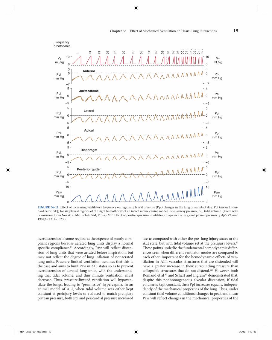
Chapter 36 Effect of Mechanical Ventilation on Heart–Lung Interactions 19
less as compared with either the pre–lung injury states or the ALI state, but with tidal volume set at the preinjury levels. 82 These points underlie the fundamental hemodynamic differ-ences seen when different ventilator modes are compared to each other. Important for the hemodynamic effects of ven-tilation in ALI, vascular structures that are distended will have a greater increase in their surrounding pressure than collapsible structures that do not distend. 229 However, both Romand et al 82 and Scharf and Ingram 83 demonstrated that, despite this nonhomogeneous alveolar distension, if tidal volume is kept constant, then Ppl increases equally, indepen-dently of the mechanical properties of the lung. Thus, under constant tidal volume conditions, changes in peak and mean Paw will reflect changes in the mechanical properties of the
overdistension of some regions at the expense of poorly com-pliant regions because aerated lung units display a normal specific compliance. 85 Accordingly, Paw will reflect disten-sion of lung units that were aerated before inspiration, but may not reflect the degree of lung inflation of nonaerated lung units. Pressure-limited ventilation assumes that this is the case and aims to limit Paw in ALI states so as to prevent overdistension of aerated lung units, with the understand-ing that tidal volume, and thus minute ventilation, must decrease. Thus, pressure-limited ventilation will hypoven-tilate the lungs, leading to “permissive” hypercapnia. In an animal model of ALI, when tidal volume was either kept constant at preinjury levels or reduced to match preinjury plateau pressure, both Ppl and pericardial pressure increased
10
Frequencybreaths/min
VTmL/kg
VTmL/kg
Anterior
5
10 15 20 25 30 35 40 45 50 60 70 80 90
100110120130140150
Juxtacardiac
Lateral
Apical
Diaphragm
Posterior gutter
Pplmm Hg
Pplmm Hg
Pplmm Hg
Pplmm Hg
Pplmm Hg
Pplmm Hg
Pawmm Hg
Pplmm Hg
Pplmm Hg
Pplmm Hg
Pplmm Hg
Pplmm Hg
Pplmm Hg
Pawmm Hg
0
30
–75
0
–5
5
0
–5
5
0
–5
5
0
–5
5
0
–5
10
0
10
0
30
–7
5
0
–5
5
0
–5
5
0
–5
5
0
–5
5
0
–5
10
0 FIGURE 36-11 Effect of increasing ventilatory frequency on regional pleural pressure (Ppl) changes in the lung of an intact dog. Ppl (mean ± stan-dard error [SE]) for six pleural regions of the right hemothorax of an intact supine canine model. Paw , airway pressure; V T , tidal volume. (Used, with permission, from Novak R, Matuschak GM, Pinsky MR. Effect of positive-pressure ventilatory frequency on regional pleural pressure. J Appl Physiol . 1988;65:1314–1323.)
Tobin_Ch36_001-030.indd 19Tobin_Ch36_001-030.indd 19 2/9/12 4:42 PM2/9/12 4:42 PM

20 Part IX Physiologic Effect of Mechanical Ventilation
These beneficial effects do not required endotracheal intubation. They are realized with the use of mask CPAP. In fact, CPAP levels as low as 5 cm H 2 O can increase cardiac out-put in patients with CHF. Cardiac output, however, decreases with similar levels of CPAP in both normal subjects and in patients with heart failure who are not volume overloaded. Nasal CPAP can also accomplish the same results in patients with obstructive sleep apnea and heart failure, 237 although the benefits do not appear to be related to changes in obstructive breathing pattern. 238 Prolonged nighttime nasal CPAP can selectively improve respiratory muscle strength and LV con-tractile function in the patients who have preexisting heart failure. 239 , 240 These benefits are associated with reductions of serum catecholamine levels. 241 There is no special effect of nonintubated CPAP on cardiac performance. In patients with hypovolemic CHF, as manifested by a pulmonary artery occlusion pressure equal to or less than 12 mm Hg, CPAP and biphasic positive airway pressure, at the same mean airway pressure, decrease cardiac outputs equally. 242
lungs and patient coordination, but may not reflect changes in ITP. Similarly, these changes in Paw may not alter global cardiovascular dynamics.
Positive-pressure ventilation can also have benefi-cial effects on hemodynamics in ALI patients. Intentional hyperinflation in subjects with ALI, induced by the use of supplemental PEEP to treat hypoxemia, may reduce pulmo-nary vascular resistance, if lung recruitment and aeration of hypoxic alveolar units reduces hypoxic pulmonary vasocon-striction; eventually, however, increase pulmonary vascular resistance must increase as all lung units are expanded above their normal resting volumes. 230 Applying the least amount of PEEP necessary to achieve an adequate PaO2
and fractional inspired oxygen concentration (FIO2) combination should be associated with the least-detrimental hemodynamic effects.
Congestive Heart Failure
Patients with CHF are difficult to wean from the ventilator because the increases in work of breathing, venous return, and intrathoracic blood volume during the transition from assisted to spontaneous ventilation may cause acute pulmo-nary edema. Rasanen et al documented that decreasing levels of ventilator support in patients with myocardial ischemia and acute LV failure worsened ischemia and promoted the development of pulmonary edema. 181 , 231 These effects could be minimized by preventing effort-induced negative swings in ITP by the use of CPAP while allowing the patient to con-tinue to breathe spontaneously. 182 Thus, in these patients, it is not the work-cost of breathing that is inducing heart fail-ure, but the negative swings in ITP. Presumably, the ability of CPAP to decrease ventricular explains the beneficial effects of CPAP during weaning trials.
If increases in ITP during positive-pressure ventilation decrease LV afterload, why then does positive-pressure ven-tilation not induce an increase in cardiac output in patients with CHF? The answer is that it does. Increases in cardiac output with Paw increases suggest the presence of CHF. 182 , 187 Grace and Greenbaum 232 noted that adding PEEP in patients with heart failure did not decrease cardiac output; cardiac output actually increased if pulmonary artery occlusion pres-sure exceeded 18 mm Hg. Similarly, Calvin et al 233 noted that patients with cardiogenic pulmonary edema had no decrease in cardiac output when given PEEP. 234 Finally, Pinsky et al demonstrated that ventilator-induced increases in ITP, using ventilatory frequencies of 12 to 20 breaths/min (phasic high intrathoracic pressure support; see Fig. 36-11 ) and increases in ITP synchronized to occur with each cardiac systole (car-diac cycle-specific where ventilator frequency equals heart rate; Fig. 36-12 ) greatly increased cardiac output in cardio-myopathy. 235 , 236 Note the similarities in the increase in mean cardiac output seen with systolic synchronized ventilation with the cardiovascular responses to similar systolic syn-chronized ventilation (see Fig. 36-8 ), which was derived in an acute animal model, wherein many more hemodynamic measures were made.
AQ7
15
10
5
05004003002001000
5004003002001000
1.5
1
0.5
0
–0.5
–1
–1.5
–2
–2.5
–3
–3.5
Control
ALI
Static lung volume above FRC (mL)
Static lung volume above FRC (mL)
Ple
ural
pre
ssur
e (m
m H
g)Tr
ansp
ulm
onar
y pr
essu
re(m
m H
g)
Control
ALI
FIGURE 36-12 Relation between transpulmonary pressure ( top ) and pleural pressure ( bottom ) and lung volume as lung volume is progres-sively increased above functional residual capacity (FRC) in control and oleic acid-induced acute lung injury (ALI) conditions in a canine model. Note that despite greater increases in transpulmonary pressure for the same increase in lung volume during ALI as compared to con-trol conditions, pleural pressure increases similarly during both control and ALI conditions for the same increase in lung volume. (Used, with permission, from Romand JA, Shi W, Pinsky MR. Cardiopulmonary effects of positive-pressure ventilation during acute lung injury. Chest . 1995;108:1041–1048.)
Tobin_Ch36_001-030.indd 20Tobin_Ch36_001-030.indd 20 2/9/12 4:42 PM2/9/12 4:42 PM
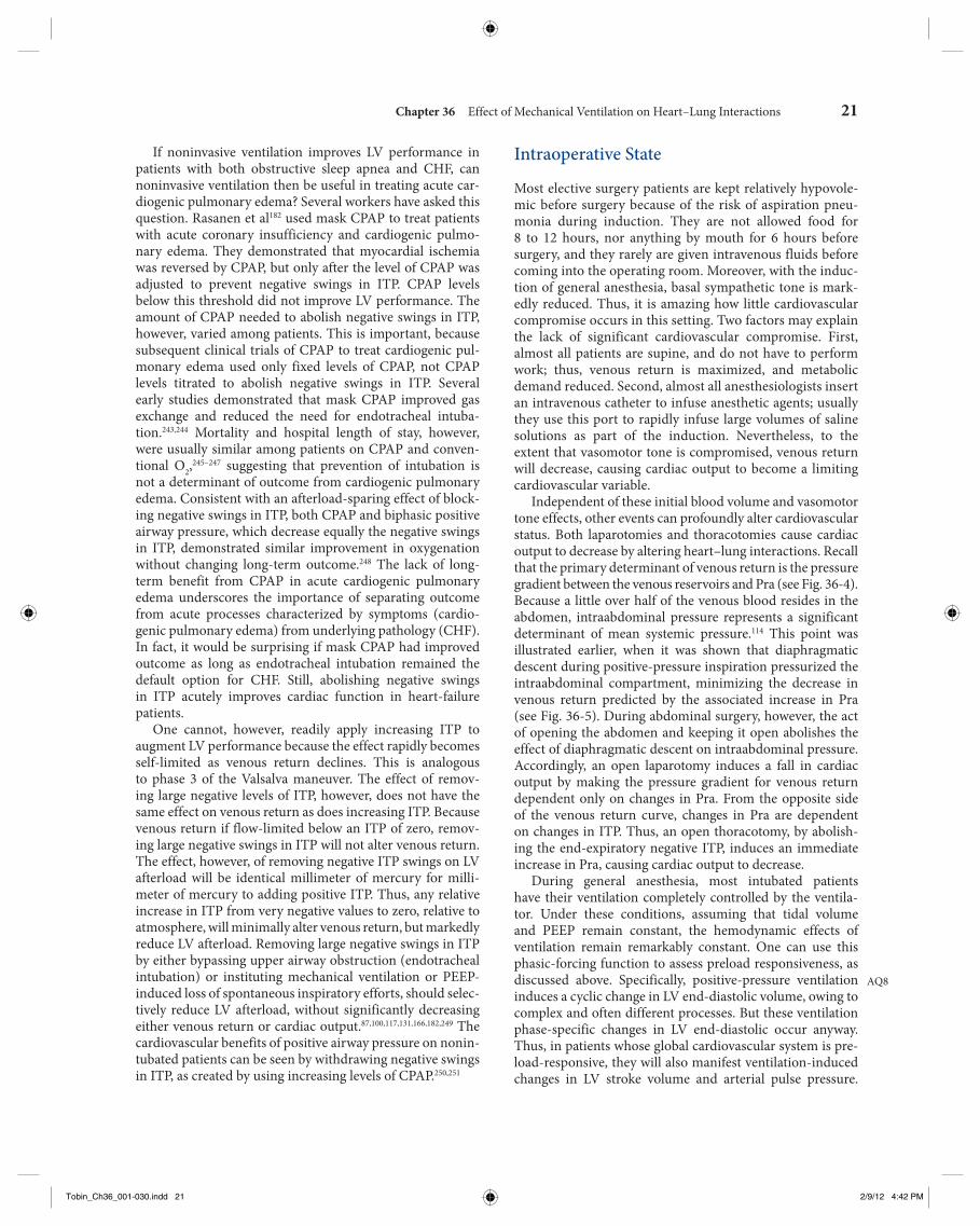
Chapter 36 Effect of Mechanical Ventilation on Heart–Lung Interactions 21
Intraoperative State
Most elective surgery patients are kept relatively hypovole-mic before surgery because of the risk of aspiration pneu-monia during induction. They are not allowed food for 8 to 12 hours, nor anything by mouth for 6 hours before surgery, and they rarely are given intravenous fluids before coming into the operating room. Moreover, with the induc-tion of general anesthesia, basal sympathetic tone is mark-edly reduced. Thus, it is amazing how little cardiovascular compromise occurs in this setting. Two factors may explain the lack of significant cardiovascular compromise. First, almost all patients are supine, and do not have to perform work; thus, venous return is maximized, and metabolic demand reduced. Second, almost all anesthesiologists insert an intravenous catheter to infuse anesthetic agents; usually they use this port to rapidly infuse large volumes of saline solutions as part of the induction. Nevertheless, to the extent that vasomotor tone is compromised, venous return will decrease, causing cardiac output to become a limiting cardiovascular variable.
Independent of these initial blood volume and vasomotor tone effects, other events can profoundly alter cardiovascular status. Both laparotomies and thoracotomies cause cardiac output to decrease by altering heart–lung interactions. Recall that the primary determinant of venous return is the pressure gradient between the venous reservoirs and Pra (see Fig. 36-4 ). Because a little over half of the venous blood resides in the abdomen, intraabdominal pressure represents a significant determinant of mean systemic pressure. 114 This point was illustrated earlier, when it was shown that diaphragmatic descent during positive-pressure inspiration pressurized the intraabdominal compartment, minimizing the decrease in venous return predicted by the associated increase in Pra (see Fig. 36-5 ). During abdominal surgery, however, the act of opening the abdomen and keeping it open abolishes the effect of diaphragmatic descent on intraabdominal pressure. Accordingly, an open laparotomy induces a fall in cardiac output by making the pressure gradient for venous return dependent only on changes in Pra. From the opposite side of the venous return curve, changes in Pra are dependent on changes in ITP. Thus, an open thoracotomy, by abolish-ing the end-expiratory negative ITP, induces an immediate increase in Pra, causing cardiac output to decrease.
During general anesthesia, most intubated patients have their ventilation completely controlled by the ventila-tor. Under these conditions, assuming that tidal volume and PEEP remain constant, the hemodynamic effects of ventilation remain remarkably constant. One can use this phasic-forcing function to assess preload responsiveness, as discussed above. Specifically, positive-pressure ventilation induces a cyclic change in LV end-diastolic volume, owing to complex and often different processes. But these ventilation phase-specific changes in LV end-diastolic occur anyway. Thus, in patients whose global cardiovascular system is pre-load-responsive, they will also manifest ventilation-induced changes in LV stroke volume and arterial pulse pressure.
AQ8
If noninvasive ventilation improves LV performance in patients with both obstructive sleep apnea and CHF, can noninvasive ventilation then be useful in treating acute car-diogenic pulmonary edema? Several workers have asked this question. Rasanen et al 182 used mask CPAP to treat patients with acute coronary insufficiency and cardiogenic pulmo-nary edema. They demonstrated that myocardial ischemia was reversed by CPAP, but only after the level of CPAP was adjusted to prevent negative swings in ITP. CPAP levels below this threshold did not improve LV performance. The amount of CPAP needed to abolish negative swings in ITP, however, varied among patients. This is important, because subsequent clinical trials of CPAP to treat cardiogenic pul-monary edema used only fixed levels of CPAP, not CPAP levels titrated to abolish negative swings in ITP. Several early studies demonstrated that mask CPAP improved gas exchange and reduced the need for endotracheal intuba-tion. 243 , 244 Mortality and hospital length of stay, however, were usually similar among patients on CPAP and conven-tional O 2 , 245 – 247 suggesting that prevention of intubation is not a determinant of outcome from cardiogenic pulmonary edema. Consistent with an afterload-sparing effect of block-ing negative swings in ITP, both CPAP and biphasic positive airway pressure, which decrease equally the negative swings in ITP, demonstrated similar improvement in oxygenation without changing long-term outcome. 248 The lack of long-term benefit from CPAP in acute cardiogenic pulmonary edema underscores the importance of separating outcome from acute processes characterized by symptoms (cardio-genic pulmonary edema) from underlying pathology (CHF). In fact, it would be surprising if mask CPAP had improved outcome as long as endotracheal intubation remained the default option for CHF. Still, abolishing negative swings in ITP acutely improves cardiac function in heart-failure patients.
One cannot, however, readily apply increasing ITP to augment LV performance because the effect rapidly becomes self-limited as venous return declines. This is analogous to phase 3 of the Valsalva maneuver. The effect of remov-ing large negative levels of ITP, however, does not have the same effect on venous return as does increasing ITP. Because venous return if flow-limited below an ITP of zero, remov-ing large negative swings in ITP will not alter venous return. The effect, however, of removing negative ITP swings on LV afterload will be identical millimeter of mercury for milli-meter of mercury to adding positive ITP. Thus, any relative increase in ITP from very negative values to zero, relative to atmosphere, will minimally alter venous return, but markedly reduce LV afterload. Removing large negative swings in ITP by either bypassing upper airway obstruction (endotracheal intubation) or instituting mechanical ventilation or PEEP-induced loss of spontaneous inspiratory efforts, should selec-tively reduce LV afterload, without significantly decreasing either venous return or cardiac output. 87 , 100 , 117 , 131 , 166 , 182 , 249 The cardiovascular benefits of positive airway pressure on nonin-tubated patients can be seen by withdrawing negative swings in ITP, as created by using increasing levels of CPAP. 250 , 251
Tobin_Ch36_001-030.indd 21Tobin_Ch36_001-030.indd 21 2/9/12 4:42 PM2/9/12 4:42 PM
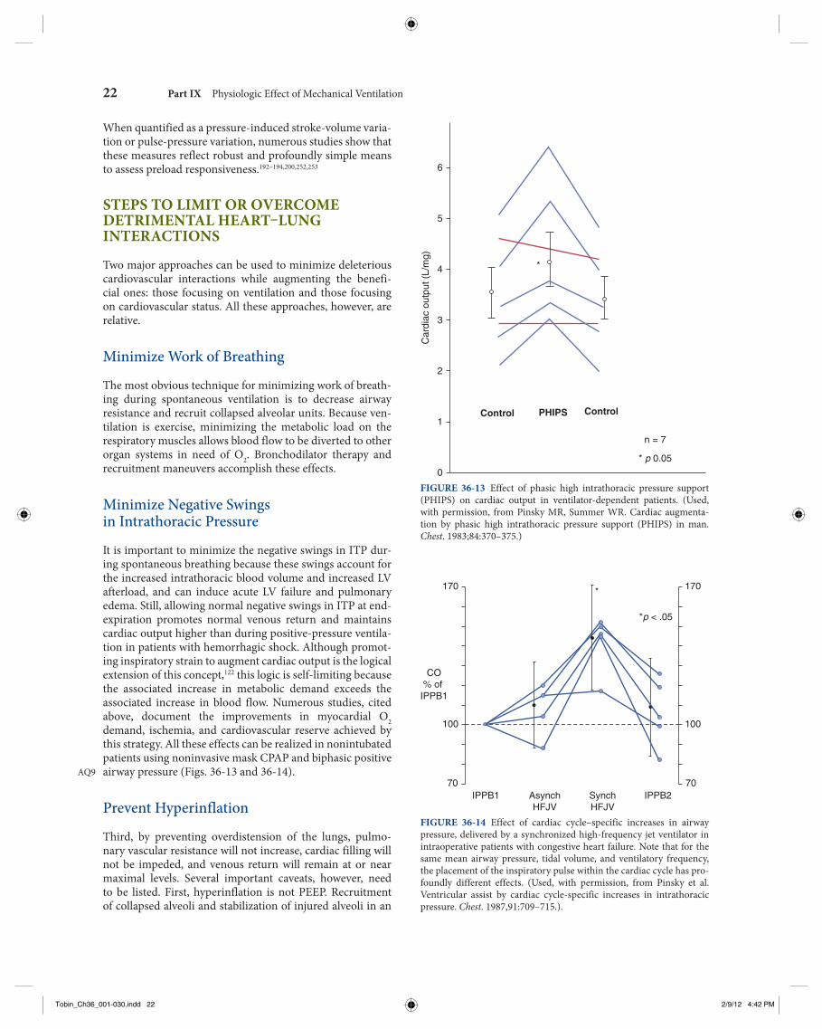
22 Part IX Physiologic Effect of Mechanical Ventilation
When quantified as a pressure-induced stroke-volume varia-tion or pulse-pressure variation, numerous studies show that these measures reflect robust and profoundly simple means to assess preload responsiveness. 192 – 194 , 200 , 252 , 253
STEPS TO LIMIT OR OVERCOME DETRIMENTAL HEART–LUNG INTERACTIONS
Two major approaches can be used to minimize deleterious cardiovascular interactions while augmenting the benefi-cial ones: those focusing on ventilation and those focusing on cardiovascular status. All these approaches, however, are relative.
Minimize Work of Breathing
The most obvious technique for minimizing work of breath-ing during spontaneous ventilation is to decrease airway resistance and recruit collapsed alveolar units. Because ven-tilation is exercise, minimizing the metabolic load on the respiratory muscles allows blood flow to be diverted to other organ systems in need of O 2 . Bronchodilator therapy and recruitment maneuvers accomplish these effects.
Minimize Negative Swings in Intrathoracic Pressure
It is important to minimize the negative swings in ITP dur-ing spontaneous breathing because these swings account for the increased intrathoracic blood volume and increased LV afterload, and can induce acute LV failure and pulmonary edema. Still, allowing normal negative swings in ITP at end-expiration promotes normal venous return and maintains cardiac output higher than during positive-pressure ventila-tion in patients with hemorrhagic shock. Although promot-ing inspiratory strain to augment cardiac output is the logical extension of this concept, 122 this logic is self-limiting because the associated increase in metabolic demand exceeds the associated increase in blood flow. Numerous studies, cited above, document the improvements in myocardial O 2 demand, ischemia, and cardiovascular reserve achieved by this strategy. All these effects can be realized in nonintubated patients using noninvasive mask CPAP and biphasic positive airway pressure (Figs. 36-13 and 36-14).
Prevent Hyperinflation
Third, by preventing overdistension of the lungs, pulmo-nary vascular resistance will not increase, cardiac filling will not be impeded, and venous return will remain at or near maximal levels. Several important caveats, however, need to be listed. First, hyperinflation is not PEEP. Recruitment of collapsed alveoli and stabilization of injured alveoli in an
AQ9
6
5
4
3
2
1
0
n = 7
* p 0.05
PHIPS ControlControlC
ardi
ac o
utpu
t (L/
mg)
*
FIGURE 36-13 Effect of phasic high intrathoracic pressure support (PHIPS) on cardiac output in ventilator-dependent patients. ( Used, with permission, from Pinsky MR, Summer WR. Cardiac augmenta-tion by phasic high intrathoracic pressure support (PHIPS) in man. Chest. 1983;84:370–375.)
170
CO% of IPPB1
70IPPB1 IPPB2Asynch
HFJVSynchHFJV
100
170
*p < .05
70
100
*
FIGURE 36-14 Effect of cardiac cycle–specific increases in airway pressure, delivered by a synchronized high-frequency jet ventilator in intraoperative patients with congestive heart failure. Note that for the same mean airway pressure, tidal volume, and ventilatory frequency, the placement of the inspiratory pulse within the cardiac cycle has pro-foundly different effects. ( Used, with permission, from Pinsky et al. Ventricular assist by cardiac cycle-specific increases in intrathoracic pressure. Chest. 1987,91:709–715.).
Tobin_Ch36_001-030.indd 22Tobin_Ch36_001-030.indd 22 2/9/12 4:42 PM2/9/12 4:42 PM
Chapter 36 Effect of Mechanical Ventilation on Heart–Lung Interactions 23
Augment Cardiac Contractility
Because spontaneous breathing trial is exercise, it may pre-cipitate acute coronary insufficiency, even in patients who are successfully weaned. Almost 40 years ago, Beach et al demonstrated that many ventilator-dependent patients can be successfully weaned if they are simultaneously given a positive inotropic agent, such as dobutamine. 152 Although no prospective clinical trial has ever been done to address this issue, many physicians support such patients with dobu-tamine infusions for 12 to 24 hours before a spontaneous breathing trial. If one were to use this approach, then the inotropic agent may still be needed following extubation and weaned thereafter, because the work of breathing may remain high even if the endotracheal tube is not contributing to the increase in airway resistance.
IMPORTANT UNKNOWNS
Perhaps the most important unknown in assessing the hemo-dynamic effect of ventilation is the assessment of cardiovas-cular reserve and the effects of breathing on hyperinflation and the swings in ITP. To date, no set of physiologic variables derived from measures of respiratory performance or ven-tilatory reserve have proven reliable in predicting weaning outcome in the setting of acute ventricular failure. In large part, we believe, this failure reflects an underappreciation of the role that cardiovascular responsiveness plays in wean-ing and the inadequate methods for assessing cardiovascular reserve. In essence, physicians have chosen an oversimplistic approach, and now institute blind daily spontaneous breath-ing trials to define when a patient is capable of weaning. Although this approach is commendable in its simplicity, it still places patients who would obviously fail such trials at risk of coronary ischemia, ongoing respiratory muscle fatigue, and impaired gas exchange secondary to LV failure and hydrostatic pulmonary edema. Because the breathing pattern can change in just one breath, and physiologically significant hyperinflation can occur in a single breath, it is probably impossible to predict with accuracy whether one can wean or not from mechanical ventilatory trials using static measures.
THE FUTURE
The future of heart–lung interactions is wedded to both new and evolving methods of mechanical ventilation and our increasing reliance on clinical techniques, like the spontane-ous breathing trial, to predict weaning success and the need for supplemental cardiovascular support. To the extent that new techniques of ventilator support follow the principle of proportional assist ventilation, they will limit the detrimental effects of positive-pressure ventilation and patient– ventilator dyssynchrony, minimizing the work of breathing. Mask CPAP will need to be titrated to minimize negative swings in
aerated state often requires the use of PEEP, which itself may reduce pulmonary vascular resistance. Although overdisten-sion of aerated alveoli will improve gas exchange further, it will also increase pulmonary vascular resistance. Thus, one should use the lowest level of PEEP required to create ade-quate oxygenation. Second, in CHF, lung inflation improves LV ejection effectiveness 254 and may itself reflect a type of ventricular support.
Fluid Resuscitation during Initiation of Positive-Pressure Ventilation
The act of endotracheal intubation is often accompanied by complex manipulations, including the use of anesthetic and analgesic agents, and institution of positive-pressure ventilation. The consequent reduction in sympathetic tone and increase in Pra act synergistically to reduce venous return. These combined effects can be lifesaving in patients with cardiogenic pulmonary edema. In otherwise healthy subjects or in patients with hypovolemia (e.g., trauma), however, these additive effects can induce hypovolemic car-diovascular collapse. Thus, the bedside caregiver should be prepared to rapidly infuse intravascular volume to poten-tially hypovolemic patients during the act of endotracheal intubation.
Prevent Volume Overload during Weaning
The transition from positive-pressure to spontaneous ven-tilation must reduce ITP and increase oxygen consump-tion. Thus, patients are at risk of developing or worsening pulmonary edema, myocardial ischemia, and acute LV failure during weaning trials. Before initiating a spontane-ous breathing trial, it is important to ensure that a patient is not volume overloaded. Steps to minimize volume overload include limiting fluids before weaning trials, forced diure-sis in the setting of overt volume overload and pleural effu-sion, and gradual reduction in the level of positive-pressure through the use of partial support modes, so as to allow fluid shifts to be excreted in urine. Furthermore, in patients with markedly increased total-body water, fluid resorption often accelerates as Pra decreases. Thus, attention to subsequent fluid overload over the days following extubation and the use of limited intravascular fluids or forced diuresis is often needed to prevent reintubation. A clinician cannot presume that a patient with clear evidence of increased extravascu-lar water has been successfully extubated when an endo-tracheal tube has been removed or even several hours later. Although detailed studies on the pathophysiology of extuba-tion failure have not been conducted, and while failure may be multifactorial, cardiovascular compromise secondary to subsequent volume overload from either fluid resorption or intravascular volume loading (often manifested by increased secretions, wheezing, and hypoxemia) is likely to be one important cause.
Tobin_Ch36_001-030.indd 23Tobin_Ch36_001-030.indd 23 2/9/12 4:42 PM2/9/12 4:42 PM
24 Part IX Physiologic Effect of Mechanical Ventilation
tamponade. As lung volume increases, so does the pressure difference between airway and Ppl. When this pressure dif-ference exceeds pulmonary artery pressure, pulmonary ves-sels collapse as they pass from the pulmonary arteries into the alveolar space, increasing pulmonary vascular resistance. Thus, hyperinflation increases pulmonary vascular resis-tance and pulmonary artery pressure, which impede RV ejection. Decreases in lung volume below FRC, as occurs in ALI and alveolar collapse, also increase pulmonary vasomo-tor tone by the process of hypoxic pulmonary vasoconstric-tion. Recruitment maneuvers, PEEP, and CPAP may reverse hypoxic pulmonary vasoconstriction and reduce pulmonary artery pressure.
Third, spontaneous inspiratory efforts decrease intra-thoracic pressure. Because diaphragmatic descent increases intraabdominal pressure, these combined effects cause Pra inside the thorax to decrease but venous pressure in the abdomen to increase, which markedly increases the pressure gradient for systemic venous return. Furthermore, the greater the decrease in intrathoracic pressure, the greater the increase in LV afterload for a constant arterial pressure. Mechanical ventilation, by abolishing the negative swings in intrathoracic pressure, selectively decreases LV afterload, as long as the increases in lung volume and intrathoracic pressure are small.
Finally, positive-pressure ventilation increases ITP. Because diaphragmatic descent increases intraabdominal pressure, the decrease in the pressure gradient for venous return is less than would otherwise occur if the only change were an increase in Pra. 3 In hypovolemic states, how-ever, positive-pressure ventilation can induce profound decreases in venous return. Increases in intrathoracic pres-sure decreases LV afterload and will augment LV ejection. In patients with hypervolemic heart failure, this afterload reducing effect can result in improved LV ejection, increased cardiac output, and reduced myocardial O 2 demand.
REFERENCES 1. Cournaud A, Motley HL, Werko L, et al. Physiologic studies of the
effect of intermittent positive pressure breathing on cardiac output in man. Am J Physiol. 1948;152:162–174.
2. Tyberg JV, Grant DA, Kingma I, et al. Effects of positive intrathoracic pressure on pulmonary and systemic hemodynamics. Respir Physiol . 2000;119:171–179.
3. Whittenberger JL, McGregor M, Berglund E, et al. Influence of state of inflation of the lung on pulmonary vascular resistance. J Appl Physiol. 1960;15:878–882.
4. Glick G, Wechsler AS, Epstein DE. Reflex cardiovascular depression produced by stimulation of pulmonary stretch receptors in the dog. J Clin Invest. 1969;48:467–472.
5. Painal AS. Vagal sensory receptors and their reflex effects. Physiol Rev. 1973;53:59–88.
6. Anrep GV, Pascual W, Rossler R. Respiratory variations in the heart rate in the reflex mechanism of the respiratory arrhythmia. Proc R Soc Lond B Biol Sci. 1936;119:191–217.
7. Taha BH, Simon PM, Dempsey JA, et al. Respiratory sinus arrhythmia in humans: an obligatory role for Vagal feedback from the lungs. J Appl Physiol. 1995;78:638–645.
8. Bernardi L, Calciati A, Gratarola A, et al. Heart rate-respiration rela-tionship: computerized method for early detection of cardiac auto-nomic damage in diabetic patients. Acta Cardiol. 1986;41:197–206.
ITP. To the extent that mask CPAP abolishes swings in ITP, it promotes LV ejection efficiency and minimizes LV failure. The use of pressure-limited ventilation in patients with ALI has resulted in decreases delivered tidal volume. Because increases in ITP are linked to changes in lung volume, these newer ventilatory strategies must result in less cardiovascu-lar dysfunction than were seen with use of higher tidal vol-umes and peak airway pressures used in the past.
Acute care medicine is evolving from a static treatment and monitoring center into a proactive diagnostic and treatment center. We now use pulse pressure and stroke volume variation during positive-pressure ventilation to identify those hemo-dynamically unstable patients who are likely to respond to a volume challenge with an increase in cardiac output, and by how much. In the future, patients would be better served if cli-nicians were to examine the immediate hemodynamic effects of spontaneous breathing trials before patients progressed to ventilatory and cardiovascular deterioration. Invasive and noninvasive measures of tissue oxygenation using data derived from pulmonary artery catheters, central venous catheters, pulse oximetry, and newer evolving noninvasive technologies will result in a dynamic assessment of impending respiratory failure and its causes. The future is upon us, and will be led by the firms that are developing newer noninvasive technologies that address these specific issues. 255
SUMMARY AND CONCLUSIONS
Our understanding of clinically relevant cardiopulmonary interactions has advanced far over the past 50 years. What was once cloaked with much mystery, now seems obvious. Still, complacency in the application of these principles at the bedside should be avoided. Just as we thought we knew all there was to know about COPD 20 years ago, the entire man-agement scheme was altered with a better understanding of dynamic hyperinflation and auto-PEEP. 224 , 225 Similarly, the exact nature by which heart–lung interactions define myo-cardial ejection efficiency and myocardial O 2 requirements in both ALI states and severe airflow obstruction remain to be defined.
The hemodynamic effects of ventilation are multiple and complex, but can be grouped into four clinically rele-vant concepts. First, spontaneous ventilation is exercise. In patients’ increased work of breathing, initiation of mechani-cal ventilation improves O 2 delivery to the remainder of the body by decreasing O 2 consumption. To the extent that mixed venous PO2
increases, arterial PO2 will also increase
without any improvement in gas exchange. Similarly, wean-ing from mechanical ventilatory support is a cardiovascular stress test. Patients who fail weaning exhibit cardiovascular insufficiency during the failed weaning attempts. Improving cardiovascular reserve or supplementing support with ino-tropic therapy may allow patients to wean.
Second, changes in lung volume alter autonomic tone and pulmonary vascular resistance, and high lung volumes compress the heart in the cardiac fossa, similarly to cardiac
Tobin_Ch36_001-030.indd 24Tobin_Ch36_001-030.indd 24 2/9/12 4:42 PM2/9/12 4:42 PM
Chapter 36 Effect of Mechanical Ventilation on Heart–Lung Interactions 25
35. Pinsky MR. Determinants of pulmonary arterial flow variation during respiration. J Appl Physiol. 1984;56:1237–1245.
36. Theres H, Binkau J, Laule M, et al. Phase-related changes in right ventricular cardiac output under volume-controlled mechanical ventilation with positive end-expiratory pressure. Crit Care Med. 1999;27:953–958.
37. Johnston WE, Vinten-Johansen J, Shugart HE, Santamore WP. Positive end-expiratory pressure potentates the severity of canine right ventricular ischemia-reperfusion injury. Am J Physiol. 1992;262:H168–H176.
38. Madden JA, Dawson CA, Harder DR. Hypoxia-induced activation in small isolated pulmonary arteries from the cat. J Appl Physiol. 1985;59:113–118.
39. Hakim TS, Michel RP, Chang HK. Effect of lung inflation on pul-monary vascular resistance by arterial and venous occlusion. J Appl Physiol. 1982;53:1110–1115.
40. Quebbeman EJ, Dawson CA. Influence of inflation and atelectasis on the hypoxic pressure response in isolated dog lung lobes. Cardiovasc Res . 1976;10:672–677.
41. Brower RG, Gottlieb J, Wise RA, et al. Locus of hypoxic vasoconstric-tion in isolated ferret lungs. J Appl Physiol. 1987;63:58–65.
42. Hakim TS, Michel RP, Minami H, Chang K. Site of pulmonary hypoxic vasoconstriction studied with arterial and venous occlusion. J Appl Physiol. 1983;54:1298–1302.
43. Marshall BE, Marshall C. Continuity of response to hypoxic pulmo-nary vasoconstriction. J Appl Physiol. 1980;49:189–196.
44. Marshall BE, Marshall C. A model for hypoxic constriction of the pulmonary circulation. J Appl Physiol. 1988;64:68–77.
45. Dawson CA, Grimm DJ, Linehan JH. Lung inflation and longitudinal distribution of pulmonary vascular resistance during hypoxia. J Appl Physiol. 1979;47:532–536.
46. Howell JBL, Permutt S, Proctor DF, et al. Effect of inflation of the lung on different parts of the pulmonary vascular bed. J Appl Physiol. 1961;16:71–76.
47. West JB, Dollery CT, Naimark A. Distribution of blood flow in iso-lated lung; relation to vascular and alveolar pressures. J Appl Physiol. 1964;19:713–724.
48. Canada E, Benumnof JL, Tousdale FR. Pulmonary vascular resistance correlated in intact normal and abnormal canine lungs. Crit Care Med. 1982;10:719–723.
49. Fuhrman BP, Everitt J, Lock JE. Cardiopulmonary effects of unilat-eral airway pressure changes in intact infant lambs. J Appl Physiol. 1984;56:1439–1448.
50. Fuhrman BP, Smith-Wright DL, Kulik TJ, Lock JE. Effects of static and fluctuating airway pressure on the intact, immature pulmonary circu-lation. J Appl Physiol. 1986;60:114–122.
51. Thorvalson J, Ilebekk A, Kiil F. Determinants of pulmonary blood vol-ume. Effects of acute changes in airway pressure. Acta Physiol Scand. 1985;125:471–479.
52. Hoffmann B, Jepsen M, Hachenberg T, et al. Cardiopulmonary effects of non-invasive positive pressure ventilation (NPPV)—a controlled, prospective study. Thorac Cardiovasc Surg. 2003;51:142–146.
53. Lopez-Muniz R, Stephens NL, Bromberger-Barnea B, et al. Critical closure of pulmonary vessels analyzed in terms of Starling resistor model. J Appl Physiol. 1968;24:625–635.
54. Block AJ, Boyson PG, Wynne JW. The origins of cor pulmonale, a hypothesis. Chest. 1979;75:109–114.
55. Vieillard-Baron A, Loubieres Y, Schmitt JM, et al. Cyclic changes in right ventricular output impedance during mechanical ventilation. J Appl Physiol. 1999;87:1644–1650.
56. Hoffman EA, Ritman EL. Heart-lung interaction: effect on regional lung air content and total heart volume. Ann Biomed Eng. 1987;15:241–257.
57. Olson LE, Hoffman EA. Heart-lung interactions determined by elec-tron beam x-ray CT in laterally recumbent rabbits. J Appl Physiol. 1995;78:417–427.
58. Grant BJB, Lieber BB. Compliance of the main pulmonary artery dur-ing the ventilatory cycle. J Appl Physiol. 1992;72:535–542.
59. Permutt S, Bromberger-Barnea B, Bane HN. Alveolar pressure, pul-monary venous pressure, and the vascular waterfall. Med Thorac . 1962;19:239–260.
60. Rothe CF. Venous system: physiology of the capacitance vessels. In: Shepherd JT, Abboud FM, eds. Handbook of Physiology: The
9. Persson MG, Lonnqvist PA, Gustafsson LE. Positive end- expiratory pressure ventilation elicits increases in endogenously formed nitric oxide as detected in air exhaled by rabbits. Anesthesiology. 1995;82:969–974.
10. Cassidy SS, Eschenbacher WI, Johnson RL Jr. Reflex cardiovascular depression during unilateral lung hyperinflation in the dog. J Clin Invest. 1979;64:620–626.
11. Daly MB, Hazzledine JL, Ungar A. The reflex effects of alterations in lung volume on systemic vascular resistance in the dog. J Physiol . 1967;188:331–351.
12. Shepherd JT. The lungs as receptor sites for cardiovascular regulation. Circulation. 1981;63:1–10.
13. Stalcup SA, Mellins RB. Mechanical forces producing pulmonary edema in acute asthma. N Engl J Med. 1977;297:592–596.
14. Vatner SF, Rutherford JD. Control of the myocardial contractile state by carotid chemo-and baroreceptor and pulmonary inflation reflexes in conscious dogs. J Clin Invest. 1978;63:1593–1601.
15. Karlocai K, Jokkel G, Kollai M. Changes in left ventricular contractility with the phase of respiration. J Auton Nerv Syst. 1998;73:86–92.
16. Rankin JS, Olsen CO, Arentzen CE et al. The effects of airway pressure on cardiac function in intact dogs and man. Circulation . 1982;66:108–120.
17. Ashton JH, SS Cassidy. Reflex depression of cardiovascular function during lung inflation. J Appl Physiol. 1985;58:137–145.
18. Fuhrman BP, Everitt J, Lock JE. Cardiopulmonary effects of unilat-eral airway pressure changes in intact infant lambs. J Appl Physiol. 1984;56:1439–1448.
19. Said SI, Kitamura S, Vreim C. Prostaglandins: release from the lung during mechanical ventilation at large tidal ventilation. J Clin Invest. 1972;51:83a.
20. Bedetti C, Del Basso P, Argiolas C, Carpi A. Arachidonic acid and pulmonary function in a heart-lung preparation of guinea-pig. Modulation by PCO2. Arch Int Pharmacodyn Ther. 1987;285:98–116.
21. Berend N, Christopher KL, Voelkel NF. Effect of positive end- expiratory pressure on functional residual capacity: role of prostaglan-din production. Am Rev Respir Dis. 1982;126:641–647.
22. Pattern MY, Liebman PR, Hetchman HG. Humorally-mediated decreases in cardiac output associated with positive end-expiratory pressure. Microvasc Res. 1977;13:137–144.
23. Berglund JE, Halden E, Jakobson S, Svensson J. PEEP ventilation does not cause humorally mediated cardiac output depression in pigs. Intensive Care Med. 1994;20:360–364.
24. Brandt RR, Wright RS, Redfield MM, Burnett JC Jr. Atrial natri-uretic peptide in heart failure. J Am Coll Cardiol. 1993;22(4 Suppl A):86A–92A.
25. Payen DM, Brun-Buisson CJL, Carli PA, et al. Hemodynamic, gas exchange, and hormonal consequences of LBPP during PEEP ventila-tion. J Appl Physiol. 1987;62:61–70.
26. Frage D, de la Coussaye JE, Beloucif S, et al. Interactions between hor-monal modifications during peep-induced antidiuresis and antinatri-uresis. Chest. 1995;107:1095–1100.
27. Frass M, Watschinger B, Traindl O, et al. Atrial natriuretic peptide release in response to different positive end-expiratory pressure levels. Crit Care Med. 1993;21:343–347.
28. Wilkins MA, Su XL, Palayew MD, et al. The effects of posture change and continuous positive airway pressure on cardiac natriuretic pep-tides in congestive heart failure. Chest. 1995;107:909–915.
29. Shirakami G, Magaribuchi T, Shingu K, et al. Positive end-expiratory pressure ventilation decreases plasma atrial and brain natriuretic pep-tide levels in humans. Anesth Analg. 1993;77:1116–1121.
30. Fletcher EC, Proctor M, Yu J, et al. Pulmonary edema develops after recurrent obstructive apneas. Am J Respir Crit Care Med. 1999;160:1688–1696.
31. Chen L, Shi Q, Scharf SM. Hemodynamic effects of periodic obstruc-tive apneas in sedated pigs with congestive heart failure. J Appl Physiol. 2000;88:1051–1060.
32. Maughan WL, Shoukas AA, Sagawa K, Weisfeldt ML. Instantaneous pressure-volume relationships of the canine right ventricle. Circ Res. 1979;44:309–315.
33. Sibbald WJ, Driedger AA. Right ventricular function in disease states: pathophysiologic considerations. Crit Care Med. 1983;11:339.
34. Piene H, Sund T. Does pulmonary impedance constitute the optimal load for the right ventricle? Am J Physiol. 1982;242:H154–H160.
Tobin_Ch36_001-030.indd 25Tobin_Ch36_001-030.indd 25 2/9/12 4:42 PM2/9/12 4:42 PM
26 Part IX Physiologic Effect of Mechanical Ventilation
87. Guyton AC, Lindsey AW, Abernathy B, et al. Venous return at vari-ous right atrial pressures and the normal venous return curve. Am J Physiol. 1957;189:609–615.
88. Goldberg HS, Rabson J. Control of cardiac output by systemic vessels: circulatory adjustments of acute and chronic respiratory failure and the effects of therapeutic interventions. Am J Cardiol. 1981;47:696–702.
89. Pinsky MR. Instantaneous venous return curves in an intact canine preparation. J Appl Physiol. 1984;56:765–771.
90. Kilburn KH. Cardiorespiratory effects of large pneumothorax in con-scious and anesthetized dogs. J Appl Physiol. 1963;18:279–283.
91. Chevalier PA, Weber KC, Engle JC, et al. Direct measurement of right and left heart outputs in Valsalva-like maneuver in dogs. Proc Soc Exp Biol Med . 1972;139:1429–1437.
92. Guntheroth WC, Gould R, Butler J, et al. Pulsatile flow in pulmonary artery, capillary and vein in the dog. Cardiovasc Res . 1974;8:330–337.
93. Guntheroth WG, Morgan BC, Mullins GL. Effect of respiration on venous return and stroke volume in cardiac tamponade. Mechanism of pulsus paradoxus. Circ Res. 1967;20:381–390.
94. Guyton AC. Effect of cardiac output by respiration, opening the chest, and cardiac tamponade. In: Circulatory Physiology: Cardiac Output and Its Regulation . Philadelphia, PA: Saunders; 1963:378–386.
95. Holt JP. The effect of positive and negative intrathoracic pres-sure on cardiac output and venous return in the dog. Am J Physiol. 1944;142:594–603.
96. Morgan BC, Abel FL, Mullins GL, et al. Flow patterns in cavae, pul-monary artery, pulmonary vein and aorta in intact dogs. Am J Physiol. 1966;210:903–909.
97. Morgan BC, Martin WE, Hornbein TF, et al. Hemodynamic effects of intermittent positive pressure respiration. Anesthesiology. 1960;27:584–590.
98. Scharf SM, Brown R, Saunders N, Green LH. Hemodynamic effects of positive pressure inflation. J Appl Physiol. 1980;49:124–131.
99. Jardin F, Vieillard-Baron A. Right ventricular function and positive-pressure ventilation in clinical practice: from hemodynamic subsets to respirator settings. Intensive Care Med. 2003;29:1426–1434.
100. Braunwald E, Binion JT, Morgan WL, Sarnoff SJ. Alterations in cen-tral blood volume and cardiac output induced by positive pressure breathing and counteracted by metaraminol (Aramine). Circ Res. 1957;5:670–675.
101. Scharf SM, Brown R, Saunders N, et al. Effects of normal and loaded spontaneous inspiration on cardiovascular function. J Appl Physiol. 1979;47:582–590.
102. Groeneveld AB, Berendsen RR, Schneider AJ, et al. Effect of the mechanical ventilatory cycle on thermodilution right ventricular volumes and cardiac output. J Appl Physiol. 2000;89:89–96.
103. Fessler HE, Brower RG, Wise RA, Permutt S. Effects of positive end-expiratory pressure on the canine venous return curve. Am Rev Respir Dis. 1992;146:4–10.
104. Takata M, Robotham JL. Effects of inspiratory diaphragmatic descent on inferior vena caval venous return. J Appl Physiol. 1992;72:597–607.
105. Takata M, Wise R, Robotham JL. Effects of abdominal pressure on venous return: abdominal vascular zone conditions. J Appl Physiol. 1990;69:1961–9172.
106. Nanas S, Magder S. Adaptations of the peripheral circulation to PEEP. Am Rev Respir Dis. 1992;146:688–693.
107. Jellinek H, Krenn H, Oczenski W, et al. Influence of positive airway pressure on the pressure gradient for venous return in humans. J Appl Physiol. 2000;88:926–932.
108. Peters J, Hecker B, Neuser D, Schaden W. Regional blood volume dis-tribution during positive and negative airway pressure breathing in supine humans. J Appl Physiol. 1993;75:1740–1747.
109. Matuschak GM, Pinsky MR, Rogers RM. Effects of positive end- expiratory pressure on hepatic blood flow and hepatic performance. J Appl Physiol. 1987;62:1377–1383.
110. Chihara E, Hasimoto S, Kinoshita T, et al. Elevated mean systemic filling pressure due to intermittent positive-pressure ventilation. Am J Physiol. 1992;262:H1116–H1121.
111. Takata M, Wise RA, Robotham JL. Effects of abdominal pressure on venous return: abdominal vascular zone conditions. J Appl Physiol. 1990;69:1961–1972.
112. Barnes GE, Laine GA, Giam PY, et al. Cardiovascular responses to elevation of intra-abdominal hydrostatic pressure. Am J Physiol. 1985;248:R208–R213.
AQ10
Cardiovascular System. Vol. 3: Peripheral Circulation and Organ Blood Flow. Part 1. Bethesda, MD: American Physiological Society; 1983:397–452.
61. Janicki JS, Weber KT. The pericardium and ventricular interaction, distensibility and function. Am J Physiol. 1980;238:H494–H503.
62. Taylor RR, Corell JW, Sonnenblick EH, Ross J Jr. Dependence of ven-tricular distensibility on filling the opposite ventricle. Am J Physiol. 1967;213:711–718.
63. Brinker JA, Weiss I, Lappe DL, et al. Leftward septal displacement dur-ing right ventricular loading in man. Circulation. 1980;61:626–633.
64. Wise RA, Robotham JL, Summer WR. Effects of spontaneous ventila-tion on the circulation. Lung. 1981;159:175–192.
65. Takata M, Harasawa Y, Beloucif S, Robotham JL. Coupled vs. uncou-pled pericardial restraint: effects on cardiac chamber interactions. J Appl Physiol. 1997;83:1799–1813.
66. Nielson J, Ostergaard M, Kjaegaad J, et al. Lung recruitment maneu-ver decreases central haemodynamics in patients after cardiac surgery. Intensive Care Med. 2005;31:1189–1194.
67. Mitchell JR, Whitelaw WA, Sas R, et al. RV filling modulates LV func-tion by direct ventricular interaction during mechanical ventilation. Am J Physiol. 2005;289:H549–H557.
68. Jardin F, Delorme G, Hardy A, et al. Reevaluation of hemodynamic consequences of positive pressure ventilation: emphasis on cyclic right ventricular afterloading by mechanical lung inflation. Anesthesiology. 1990;72:966–970.
69. Pinsky MR. Determinants of pulmonary artery flow variation during respiration. J Appl Physiol. 1984;56:1237–1245.
70. Butler J. The heart is in good hands. Circulation. 1983;67:1163–1168. 71. Novak RA, Matuschak GM, Pinsky MR. Effect of ventilatory frequency
on regional pleural pressure. J Appl Physiol. 1988;65:1314–1323. 72. Tsitlik JE, Halperin HR, Guerci AD, et al. Augmentation of pres-
sure in a vessel indenting the surface of the lung. Ann Biomed Eng . 1987;15:259–284.
73. Cassidy SS, Wead WB, Seibert GB, Ramanathan M. Changes in left ventricular geometry during spontaneous breathing. J Appl Physiol. 1987;63:803–811.
74. Marini JJ, Culver BN, Butler J. Mechanical effect of lung disten-tion with positive pressure on cardiac function. Am Rev Respir Dis. 1980;124:382–386.
75. Cassidy SS, Robertson CH, Pierce AK, et al. Cardiovascular effects of positive end-expiratory pressure in dogs. J Appl Physiol. 1978;4:743–749.
76. Conway CM. Hemodynamic effects of pulmonary ventilation. Br J Anaesth. 1975;47:761–766.
77. Jardin F, Farcot JC, Boisante L. Influence of positive end- expiratory pressure on left ventricular performance. N Engl J Med. 1981;304:387–392.
78. Berglund JE, Halden E, Jakobson S, Landelius J. Echocardiographic analysis of cardiac function during high PEEP ventilation. Intensive Care Med. 1994;20:174–180.
79. Pinsky MR, Vincent JL, DeSmet JM. Estimating left ventricular filling pressure during positive end-expiratory pressure in humans. Am Rev Respir Dis. 1991;143:25–31.
80. Shuey CB, Pierce AK, Johnson RL. An evaluation of exercise tests in chronic obstructive lung disease. J Appl Physiol. 1969;27:256–261.
81. Jayaweera AR, Ehrlich W. Changes of phasic pleural pressure in awake dogs during exercise: potential effects on cardiac output. Ann Biomed Eng. 1987;15:311–318.
82. Romand JA, Shi W, Pinsky MR. Cardiopulmonary effects of posi-tive pressure ventilation during acute lung injury. Chest. 1995;108:1041–1048.
83. Scharf SM, Ingram RH Jr. Effects of decreasing lung compliance with oleic acid on the cardiovascular response to PEEP. Am J Physiol. 1977;233:H635–H641.
84. O’Quinn RJ, Marini JJ, Culver BH, et al. Transmission of airway pres-sure to pleural pressure during lung edema and chest wall restriction. J Appl Physiol. 1985;59:1171–1177.
85. Gattinoni L, Mascheroni D, Torresin A, et al. Morphological response to positive end-expiratory pressure in acute respiratory failure. Intensive Care Med. 1986;12:137–142.
86. Guyton AC. Determination of cardiac output by equating venous return curves with cardiac response curves. Physiol Rev. 1955;35:123–129.
Tobin_Ch36_001-030.indd 26Tobin_Ch36_001-030.indd 26 2/9/12 4:42 PM2/9/12 4:42 PM
Chapter 36 Effect of Mechanical Ventilation on Heart–Lung Interactions 27
138. Ruskin J, Bache RJ, Rembert JC, Greenfield JC Jr. Pressure-flow studies in man: effect of respiration on left ventricular stroke volume. Circulation. 1973;48:79–85.
139. Olsen CO, Tyson GS, Maier GW, et al. Dynamic ventricular interac-tion in the conscious dog. Circ Res. 1983;52:85–104.
140. Tyberg JV, Taichman GC, Smith ER, et al. The relationship between pericardial pressure and right atrial pressure: an intraoperative study. Circulation. 1986;73:428–432.
141. Pinsky MR, Vincent JL, DeSmet JM. Effect of positive end-expiratory pressure on right ventricular function in man. Am Rev Respir Dis. 1992;146:681–687.
142. Dhainaut JF, Devaux JY, Monsallier JF, et al. Mechanisms of decreased left ventricular preload during continuous positive pressure ventila-tion in ARDS. Chest. 1986;90:74–80.
143. Rebuck AS, Read J. Assessment and management of severe asthma. Am J Med. 1971;51:788–792.
144. Beyar R, Goldstein Y. Model studies of the effects of the thoracic pres-sure on the circulation. Ann Biomed Eng. 1987;15:373–383.
145. Pinsky MR, Summer WR, Wise RA, et al. Augmentation of car-diac function by elevation of intrathoracic pressure. J Appl Physiol. 1983;54:950–955.
146. Pinsky MR, Matuschak GM, Bernardi L, Klain M. Hemodynamic effects of cardiac cycle-specific increases in intrathoracic pressure. J Appl Physiol. 1986;60:604–612.
147. Matuschak GM, Pinsky MR, Klain M. Hemodynamic effects of syn-chronous high-frequency jet ventilation during acute hypovolemia. J Appl Physiol. 1986;61:44–53.
148. Roussos C, Macklem PT. The respiratory muscles. N Engl J Med. 1982;307:786–797.
149. Cassidy SA, Wead WB, Seibert GB, Ramanathan M. Geometric left-ventricular responses to interactions between the lung and left ventricle: positive pressure breathing. Ann Biomed Eng. 1987;15:285–295.
150. Scharf SM, Brown R, Warner KG, Khuri S. Intrathoracic pressure and left ventricular configuration with respiratory maneuvers. J Appl Physiol. 1989;66:481–491.
151. Bromberger-Barnea B. Mechanical effects of inspiration on heart functions: a review. Fed Proc. 1981;40:2172–2177.
152. Beach T, Millen E, Grenvik A. Hemodynamic response to discontinu-ance of mechanical ventilation. Crit Care Med. 1973;1:85–90.
153. Lemaire F, Teboul JL, Cinoti L, et al. Acute left ventricular dysfunc-tion during unsuccessful weaning from mechanical ventilation. Anesthesiology. 1988;69:171–179.
154. Pinsky MR, Matuschak GM, Itzkoff JM. Respiratory augmenta-tion of left ventricular function during spontaneous ventilation in severe left ventricular failure by grunting: an auto-EPAP effect. Chest. 1984;86:267–269.
155. Blaustein AS, Risser TA, Weiss JW, et al. Mechanisms of pulsus para-doxus during resistive respiratory loading and asthma. J Am Coll Cardiol. 1986;8:529–536.
156. Strohl KP, Scharf SM, Brown R, Ingram RH Jr. Cardiovascular perfor-mance during bronchospasm in dogs. Respiration. 1987;51:39–48.
157. Scharf SM, Graver LM, Balaban K. Cardiovascular effects of peri-odic occlusions of the upper airways in dogs. Am Rev Respir Dis. 1992;146:321–329.
158. Viola AR, Puy RJM, Goldman E. Mechanisms of pulsus paradoxus in airway obstruction. J Appl Physiol. 1990;68:1927–1931.
159. Scharf SM, Graver LM, Khilnani S, Balaban K. Respiratory phasic effects of inspiratory loading on left ventricular hemodynamics in vagotomized dogs. J Appl Physiol. 1992;73:995–1003.
160. Latham RD, Sipkema P, Westerhof N, Rubal BJ. Aortic input imped-ance during Mueller maneuver: an evaluation of “effective strength.” J Appl Physiol. 1988;65:1604–1610.
161. Virolainen J, Ventila M, Turto H, Kupari M. Effect of negative intra-thoracic pressure on left ventricular pressure dynamics and relaxation. J Appl Physiol. 1995;79:455–460.
162. Gomez A, Mink S. Interaction between effects of hypoxia and hyper-capnia on altering left ventricular relaxation and chamber stiffness in dogs. Am Rev Respir Dis. 1992;146:313–320.
163. Kingma I, Smiseth OA, Frais MA, et al. Left ventricular external con-straint: relationship between pericardial, pleural and esophageal pres-sures during positive end-expiratory pressure and volume loading in dogs. Ann Biomed Eng . 1987;15:331–346.
113. Lichtwarck-Aschoff M, Zeravik J, Pfeiffer UJ. Intrathoracic blood volume accurately reflects circulatory volume status in critically ill patients with mechanical ventilation. Intensive Care Med. 1992;18:142–145.
114. van den Berg P, Jansen JRC, Pinsky MR. The effect of positive-pressure inspiration on venous return in volume loaded post-operative cardiac surgical patients. J Appl Physiol. 2002;92:1223–1231.
115. Brienza N, Revelly JP, Ayuse T, Robotham JL. Effects of PEEP on liver arterial and venous blood flows. Am J Respir Crit Care Med. 1995;152:504–510.
116. Brecher GA, Hubay CA. Pulmonary blood flow and venous return during spontaneous respiration. Circ Res. 1955;3:40–214.
117. Sibbald WH, Calvin J, Driedger AA. Right and left ventricular preload, and diastolic ventricular compliance: implications of therapy in criti-cally ill patients. In: Critical Care State of the Art , Vol. 3. Fullerton, CA: California Society of Critical Care; 1982.
118. Magder S, Georgiadis G, Cheong T. Respiratory variation in right atrial pressure predict the response to fluid challenge. J Crit Care. 1992;7:76–85.
119. Terada N, Takeuchi T. Postural changes in venous pressure gradients in anesthetized monkeys. Am J Physiol. 1993;264:H21–H25.
120. Scharf S, Tow DE, Miller MJ, et al. Influence of posture and abdomi-nal pressure on the hemodynamic effects of Mueller’s maneuver. J Crit Care. 1989;4:26–34.
121. Tarasiuk A, Scharf SM. Effects of periodic obstructive apneas on venous return in closed-chest dogs. Am Rev Respir Dis. 1993;148:323–329.
122. Convertino VA, Ratliff DA, Ryan KL, et al. Hemodynamics associated with breathing through an inspiratory impedance threshold device in human volunteers. Crit Care Med. 2004;32(9 Suppl):S381–S386.
123. Shekerdemian LS, Bush A, Shore DF, et al. Cardiorespiratory responses to negative pressure ventilation after tetralogy of Fallot repair: a hemo-dynamic tool for patients with low-output state. J Am Coll Cardiol. 1999;33:549–555.
124. Lores ME, Keagy BA, Vassiliades T, et al. Cardiovascular effects of pos-itive end-expiratory pressure (PEEP) after pneumonectomy in dogs. Ann Thorac Surg. 1985;40:464–473.
125. Grenvik A. Respiratory, circulatory and metabolic effects of respi-ratory treatment. A clinical study in postoperative thoracic surgical patients. Acta Anaesthesiol . 1966;(19 Suppl):1–122.
126. Harken AH, Brennan MF, Smith N, Barsamian EM. The hemody-namic response to positive end-expiratory ventilation in hypovolemic patients. Surgery. 1974;76:786–793.
127. Bell RC, Robotham JL, Badke FR, et al. Left ventricular geometry during intermittent positive pressure ventilation in dogs. J Crit Care. 1987;2:230–244.
128. Qvist J, Pontoppidan H, Wilson RS, et al. Hemodynamic responses to mechanical ventilation with PEEP: the effects of hypovolemia. Anesthesiology. 1975;42:45–53.
129. Denault AY, Gorcsan III J, Deneault LG, Pinsky MR. Effect of positive pressure ventilation on left ventricular pressure-volume relationship. Anesthesiology. 1993;79:A315.
130. Buda AJ, Pinsky MR, Ingels NB, et al. Effect of intrathoracic pressure on left ventricular performance. N Engl J Med. 1979;301:453–459.
131. Sharpey-Schaffer EP. Effects of Valsalva maneuver on the normal and failing circulation. Br Med J 1955;1:693–699.
132. Jardin FF, Farcot JC, Gueret P, et al. Echocardiographic evaluation of ventricles during continuous positive pressure breathing. J Appl Physiol. 1984;56:619–627.
133. Prec KJ, Cassels DE. Oximeter studies in newborn infants during cry-ing. Pediatrics . 1952;9:756–761.
134. Peters J, Kindred MK, Robotham JL. Transient analysis of car-diopulmonary interactions II. Systolic events. J Appl Physiol. 1988;64:1518–1526.
135. Pinsky MR, Matuschak GM, Klain M. Determinants of cardiac aug-mentation by increases in intrathoracic pressure. J Appl Physiol. 1985;58:1189–1198.
136. Rankin JS, Olsen CO, Arentzen CE, et al. The effects of airway pressure on cardiac function in intact dogs and man. Circulation. 1982;66:108–120.
137. Robotham JL, Rabson J, Permutt S, Bromberger-Barnea B. Left ventricular hemodynamics during respiration. J Appl Physiol. 1979;47:1295–1303.
Tobin_Ch36_001-030.indd 27Tobin_Ch36_001-030.indd 27 2/9/12 4:42 PM2/9/12 4:42 PM
28 Part IX Physiologic Effect of Mechanical Ventilation
187. Abel JG, Salerno TA, Panos A, et al. Cardiovascular effects of positive pressure ventilation in humans. Ann Thorac Surg. 1987;43:36–43.
188. Baeaussier M, Coriat P, Perel A, et al. Determinants of systolic pressure variation in patients ventilated after vascular surgery. J Cardiothorac Vasc Anesth. 1995;9:547–551.
189. Coriat P, Vrillon M, Perel A, et al. A comparison of systolic blood pressure variations and echocardiographic estimates of end-diastolic left ventricular size in patients after aortic surgery. Anesth Analg. 1994;78:46–53.
190. Szold A, Pizov R, Segal E, Perel A. The effect of tidal volume and intra-vascular volume state on systolic pressure variation in ventilated dogs. Intensive Care Med. 1989;15:368–371.
191. Michard F, Teboul JL. Predicting fluid responsiveness in ICU patients: a critical analysis of the evidence. Chest. 2002;121:2000–2008.
192. Michard F, Boussat S, Chemla D, et al. Relation between respiratory changes in arterial pulse pressure and fluid responsiveness in septic patients with acute circulatory failure. Am J Respir Crit Care Med. 2000;162:134–188.
193. Slama M, Masson H, Teboul JL, et al. Monitoring of respiratory varia-tions of aortic flow velocity using oesophageal Doppler. Intensive Care Med. 2004;30:1182–1187.
194. Reuter DA, Felbinger TW, Schmidt C, et al. Stroke volume varia-tion for assessment of cardiac responsiveness to volume loading in mechanically ventilated patients after cardiac surgery. Intensive Care Med. 2002;28:392–398.
195. Marik PE, Cavallazzi R, Vasu T, Hirani A. Dynamic changes in arterial waveform derived variables and fluid responsiveness in mechanically ventilated patients: a systematic review of the literature. Crit Care Med. 37:2642–2647, 2009.
196. Kumar A, Anel R, Bunnell E, et al. Pulmonary artery occlusion pres-sure and central venous pressure fail to predict ventricular volumes, cardiac performance, or the response to volume infusion in normal subjects. Crit Care Med. 2004;32:691–699.
197. Pinsky MR. Clinical significance of pulmonary artery occlusion pres-sure. Intensive Care Med. 2003;29:175–178.
198. Romand JA, Shi W, Pinsky MR. Cardiopulmonary effects of positive pressure ventilation during acute lung injury. Chest. 1995;108:1041–1048.
199. Pinsky MR. Instantaneous venous return curves in an intact canine preparation. J Appl Physiol. 1984;56:765–771.
200. Rooke GA, Schwid HA, Shapira Y. The effect of graded hemorrhage and intravascular volume replacement on systolic pressure variation in humans during mechanical and spontaneous ventilation. Anesth Analg. 1995;80:925–932.
201. Sylvester JT, Gilbert RD, Traystman RJ, Permutt S. Effects of hypoxia on the closing pressure of the canine systemic arterial circulation. Circ Res. 1981;49:980–987.
202. Pinsky, MR, JG Guimond. The effects of positive end-expiratory pressure on heart-lung interactions. J Crit Care. 1991;6:1–11.
203. Pinsky MR, Matuschak GM, Bernardi L, Klain M. Hemodynamic effects of cardiac cycle-specific increases in intrathoracic pressure. J Appl Physiol. 1986;60:604–612.
204. Dries DJ, Kumar P, Mathru M, et al. Hemodynamic effects of pressure support ventilation in cardiac surgery patients. Am Surg. 1991;57:122–125.
205. Sternberg R, Sahebjami H. Hemodynamic and oxygen transport characteristics of common ventilatory modes. Chest. 1994;105:1798–1803.
206. Lessard MR, Guerot E, Lorini H, et al. Effects of pressure-controlled with different I:E ratios versus volume-controlled ventilation on respiratory mechanics, gas exchange and hemodynamics in patients with adult respiratory distress syndrome. Anesthesiology. 1994;80:983–991.
207. Chan K, Abraham E. Effects of inverse ratio ventilation on cardio-respiratory parameters in severe respiratory failure. Chest. 1992;102:1556–1561.
208. Hartmann M, Rosberg B, Jonsson K. The influence of different lev-els of PEEP on peripheral tissue perfusion measured by subcutane-ous and transcutaneous oxygen tension. Intensive Care Med. 1992;18:474–478.
209. Abraham E, Yoshihara G. Cardiorespiratory effects of pressure controlled ventilation in severe respiratory failure. Chest. 1990;98:1445–1449.
164. Denault AY, Gorcsan J 3rd, Pinsky MR. Dynamic effects of positive-pressure ventilation on canine left ventricular pressure-volume rela-tions. J Appl Physiol. 2001;91:298–308.
165. Abel FL, Mihailescu LS, Lader AS, Starr RG. Effects of pericardial pressure on systemic and coronary hemodynamics in dogs. Am J Physiol. 1995;268:H1593–H1605.
166. Khilnani S, Graver LM, Balaban K, Scharf SM. Effects of inspiratory loading on left ventricular myocardial blood flow and metabolism. J Appl Physiol. 1992;72:1488–1492.
167. Satoh S, Watanabe J, Keitoku M, et al. Influences of pressure sur-rounding the heart and intracardiac pressure on the diastolic coro-nary pressure-flow relation in excised canine heart. Circ Res. 1988;63:788–797.
168. Marini JJ, Rodriguez RM, Lamb V. The inspiratory workload of patient-initiated mechanical ventilation. Am Rev Respir Dis. 1986;134:902–909.
169. Stock MC, David DW, Manning JW, Ryan ML. Lung mechanics and oxygen consumption during spontaneous ventilation and severe heart failure. Chest. 1992;102:279–283.
170. Kawagoe Y, Permutt S, Fessler HE. Hyperinflation with intrinsic PEEP and respiratory muscle blood flow. J Appl Physiol. 1994;77:2440–2448.
171. Aubier M, Vires N, Sillye G, et al. Respiratory muscle contribu-tion to lactic acidosis in low cardiac output. Am Rev Respir Dis. 1982;126:648–652.
172. Vires N, Sillye G, Rassidakis A, et al. Effect of mechanical ventila-tion on respiratory muscle blood flow during shock. Physiologist. 1980;23:1–8.
173. Mesquida J, Kim HK, Pinsky MR. Effect of tidal volume, intratho-racic pressure and cardiac contractility on variations in pulse pressure, stroke volume and intrathoracic blood volume. Intensive Care Med. 2011;37:1672–1679.
174. Richard C, Teboul JL, Archambaud F, et al. Left ventricular dysfunc-tion during weaning in patients with chronic obstructive pulmonary disease. Intensive Care Med. 1994;20:171–172.
175. Hurford WE, Lynch KE, Strauss HW, et al. Myocardial perfusion as assessed by thallium-201 scintigraphy during the discontinu-ation of mechanical ventilation in ventilator-dependent patients. Anesthesiology. 1991;74:1007–1016.
176. Abalos A. Leibowitz AB, Distefano D, et al. Myocardial ischemia during the weaning period. Am J Crit Care. 1992;1:32–36.
177. Chatila W, Ani S, Guaglianone D, et al. Cardiac ischemia during wean-ing from mechanical ventilation. Chest. 1996;109:1421–1422.
178. Srivastava S, Chatila W, Amoateng-Adjepong Y, et al. Myocardial isch-emia and weaning failure in patients with coronary disease: an update. Crit Care Med. 1999;27:2109–2112.
179. Mohsenifar Z, Hay A, Hay J, et al. Gastric intramural pH as a predictor of success or failure in weaning patients from mechanical ventilation. Ann Intern Med. 1993;119:794–798.
180. Jabran A, Mathru M, Dries D, Tobin MJ. Continuous recordings of mixed venous oxygen saturation during weaning from mechanical ventilation and the ramifications thereof. Am J Respir Crit Care Med. 1998;158:1763–1769.
181. Rasanen J, Nikki P, Heikkila J. Acute myocardial infarction compli-cated by respiratory failure. The effects of mechanical ventilation. Chest. 1984;85:21–28.
182. Rasanen J, Vaisanen IT, Heikkila J, et al. Acute myocardial infarc-tion complicated by left ventricular dysfunction and respiratory failure. The effects of continuous positive airway pressure. Chest. 1985;87:156–162.
183. Straus C, Lewis B, Isebey D, et al. Contribution of the endotracheal tube and the upper airway to breathing workload. Am J Respir Crit Care Med. 1998;157:23–30.
184. Suga H, Sagawa K. Instantaneous pressure-volume relationships and their ratio in the excised supported canine left ventricle. Circ Res. 1974;35:117–134.
185. Haney MF, Johansson G, Haggmark S, Biber B. Heart-lung interactions during positive-pressure ventilation: left ventricular pressure-volume momentary response to airway pressure elevation. Acta Anaesthesiol Scand. 2001;45:702–709.
186. Haney MF, Johansson G, Haggmark S, Biber B. Method of pre-load reduction during LVESPVR analysis of systolic function: air-way pressure elevations and vena caval occlusion. Anesthesiology. 2002;97:436–446.
Tobin_Ch36_001-030.indd 28Tobin_Ch36_001-030.indd 28 2/9/12 4:42 PM2/9/12 4:42 PM
Chapter 36 Effect of Mechanical Ventilation on Heart–Lung Interactions 29
234. Loick HM, Wendt M, Rotker J Theissen JL. Ventilation with positive end-expiratory airway pressure causes leukocyte retention in human lung. J Appl Physiol. 1993;75:301–306.
235. Pinsky MR, Summer WR. Cardiac augmentation by phasic high intra-thoracic support (PHIPS) in man. Chest. 1983;84:370–375.
236. Pinsky MR, Marquez J, Martin D, Klain M. Ventricular assist by cardiac cycle-specific increases in intrathoracic pressure. Chest. 1987;91:709–715.
237. Lin M, Yang Y-F, Chiang H-T, et al. Reappraisal of continuous posi-tive airway pressure therapy in acute cardiogenic pulmonary edema. Chest. 1995;107:1379–1386.
238. Buckle P, Millar T, Kryger M. The effect of short-term nasal CPAP on Cheyne-Stokes respiration in congestive heart failure. Chest. 1992;102:31–35.
239. Granton JT, Naughton MT, Benard DC, et al. CPAP improves inspira-tory muscle strength in patients with heart failure and central sleep apnea. Am J Respir Crit Care Med. 1996;153:277–282.
240. Kaneko Y, Floras JS, Usui K, et al. Cardiovascular effects of continuous positive airway pressure in patients with heart failure and obstructive sleep apnea. N Engl J Med. 2003;348:1233–1241.
241. Naughton MT, Benard DC, Liu PP, et al. Effects of nasal CPAP on sym-pathetic activity in patients with heart failure and central sleep apnea. Am J Respir Crit Care Med. 1995;152:473–479.
242. Joet-Philip FF, Paganelli FF, Dutau HL, Saadjian AY. Hemodynamic effects of bi-level nasal positive airway pressure ventilation in patients with heart failure. Respiration. 1999;66:136–143.
243. Bersten AD, Holt AW, Vedig AE, et al. Treatment of severe cardiogenic pulmonary edema with continuous positive airway pressure delivered by facemask. N Engl J Med. 1991;325:1825–1830.
244. Lin M, Yang YF, Chiang HT, et al. Reappraisal of continuous positive airway pressure therapy in acute cardiogenic pulmonary edema: short-term results and long term follow-up. Chest. 1995;107:1379–1386.
245. Masip J, Betbesé AJ, Páez J, et al. Non-invasive pressure support versus conventional oxygen therapy in acute cardiogenic pulmonary oedema: a randomized trial. Lancet. 2000;356:2126–2132.
246. Giacomini M, Iapichino G, Cigada M, et al. Short-term noninvasive pressure support prevents ICU admittance in patients with acute car-diogenic pulmonary edema. Chest. 2003;123:2057–2061.
247. Nava S, Carbone G, DiBattista N, et al. Noninvasive ventilation in cardiogenic pulmonary edema: a multicenter randomized trial. Am J Respir Crit Care Med. 2003;168:1432–1437.
248. Park M, Sangean MC, de Volpe M, et al. Randomized, prospective trial of oxygen, continuous positive airway pressure, and bilevel positive airway pressure by face mask in acute cardiogenic pulmonary edema. Crit Care Med. 2004;32:2407–2415.
249. Garpestad E, Parker JA, Katayama H, et al. Decrease in ventricular stroke volume at apnea termination is independent of oxygen desatu-ration. J Appl Physiol. 1994;77:1602–1608.
250. De Hoyos A, Liu PP, Benard DC, Bradley TD. Haemodynamic effects of continuous positive airway pressure in humans with normal and impaired left ventricular function. Clin Sci (Lond) . 1995;88:173–178.
251. Naughton MT, Rahman MA, Hara K, et al. Effect of continuous posi-tive airway pressure on intrathoracic and left ventricular transmu-ral pressures in patients with congestive heart failure. Circulation. 1995;91:1725–1731.
252. Pinsky MR. Functional hemodynamic monitoring: asking the right question. Intensive Care Med. 2002;28:386–388.
253. Pinsky MR. Probing the limits of arterial pulse contour analysis to predict preload-responsiveness. Anesth Analg . 2003;96:1245–1247.
254. Pinsky MR. The hemodynamic consequences of mechanical ventilation: an evolving story. Intensive Care Med. 1997;23:493–503.
255. Pinsky MR. Though the past darkly: ventilatory management of patients with chronic obstructive pulmonary disease. Crit Care Med. 1994;22:1714–1717.
210. Poelaert JI, Visser CA, Everaert JA, et al. Acute hemodynamic changes of pressure-controlled inverse ration ventilation in the adult respira-tory distress syndrome. A transesophageal echocardiographic and Doppler study. Chest. 1993;104:214–219.
211. Davis K Jr, Branson RD, Campbell R, Porembka DT. Comparison of volume control and pressure control ventilation: is flow waveform the difference? J Trauma. 1996;41:808–814.
212. Kiehl M, Schiele C, Stenzinger W, Kienast J. Volume-controlled versus biphasic pressure ventilation in leukopenic patients with severe respi-ratory failure. Crit Care Med. 1996;24:780–784.
213. Singer M, Vermaat J, Hall G, et al. Hemodynamic effects of manual hyperinflation in critically ill mechanically ventilated patients. Chest. 1994;106:1182–1187.
214. Mang H, Kacmarek RM, Ritz R, et al. Cardiorespiratory effects of volume-and pressure-controlled ventilation at various I/E ratios in an acute lung injury model. Am J Respir Crit Care Med. 1995;151:731–736.
215. Blum RH, McGowan FX Jr. Chronic upper airway obstruction and cardiac dysfunction: anatomy, pathophysiology and anesthetic impli-cations. Paediatr Anaesth. 2004;14:75–83.
216. Conzanitis DA, Leijala M, Pesoner E, Saki HA. Acute pulmonary edema due to laryngeal spasm. Anaesthesia . 1982;37:1198–1199.
217. Jackson FN, Rowland V, Corssen G. Laryngospasm-induced pulmo-nary edema. Chest. 1980;78:819–824.
218. Leatherman JW, Schwartz S. Pulmonary edema due to upper airway obstruction. South Med J. 1983;76:1058–1060.
219. Lee KWT, Downes JJ. Pulmonary edema secondary to laryngospasm in children. Anesthesiology. 1983;59:347–349.
220. Oswalt CE, Gates GA, Holstrom FMG. Pulmonary edema as a compli-cation of acute upper airway obstruction. JAMA. 1977;238:1833–1835.
221. Poulton TJ. Laryngospasm-induced pulmonary edema. Chest. 1981;80:762–763.
222. Stradling J, Bolton P. Upper airways obstruction as cause of pulmo-nary edema. Lancet. 1982;1:1353–1354.
223. Sofer S, Bar-Ziv J, Scharf SM. Pulmonary edema following relief of upper airway obstruction. Chest. 1984;86:401–403.
224. Bergman NA. Intrapulmonary gas trapping during mechanical ventilation at rapid frequencies. Anesthesiology. 1969;37:626–633.
225. Pepe P, Marini JJ. Occult positive end-expiratory pressure in mechani-cally ventilated patients with airflow obstruction: the auto-PEEP effect. Am Rev Respir Dis. 1982;126:166–170.
226. Frazier SK, Stone KS, Schertel ER, et al. A comparison of hemody-namic changes during the transition from mechanical ventilation to T-piece, pressure support, and continuous positive airway pressure in canines. Biol Res Nurs . 2000;1:253–264.
227. Magder S, Erian R, Roussos C. Respiratory muscle blood flow in oleic acid-induced pulmonary edema. J Appl Physiol . 1986;60:1849–1856.
228. Baigorri F, De Monte A, Blanch L, et al. Hemodynamic responses to external counterbalancing of auto-positive end-expiratory pressure in mechanically ventilated patients with chronic obstructive pulmonary disease. Crit Care Med. 1994;22:1782–1791.
229. Globits S, Burghuber OC, Koller J, et al. Effect of lung transplan-tation on right and left ventricular volumes and function mea-sured by magnetic resonance imaging. Am J Respir Crit Care Med. 1994;149:1000–1004.
230. Calvin JE, Driedger AA, Sibbald WJ. Positive end-expiratory pres-sure (PEEP) does not depress left ventricular function in patients with pulmonary edema. Am Rev Respir Dis. 1981;124:121–128.
231. Rasanen J. Respiratory failure in acute myocardial infarction. Appl Cardiopulm Pathophysiol. 1988;2:271–279.
232. Grace MP, Greenbaum DM. Cardiac performance in response to PEEP in patients with cardiac dysfunction. Crit Care Med. 1982;20:358–360.
233. Calvin JE, Driedger AA, Sibbald WJ. Positive end-expiratory pressure (PEEP) does not depress left ventricular function in patients with pul-monary edema. Am Rev Respir Dis. 1981;124:121–128.
Tobin_Ch36_001-030.indd 29Tobin_Ch36_001-030.indd 29 2/9/12 4:42 PM2/9/12 4:42 PM

Tobin_Ch36_001-030.indd 30Tobin_Ch36_001-030.indd 30 2/9/12 4:42 PM2/9/12 4:42 PM

AUTHOR QUERY
AQ1: Figure 36-1, 2, 3, 4, 5, 6, 12 and 13 are redrawn. Please check.AQ2: Please identify where by section title.AQ3: By “next section” do you mean “Effect of Intrathoracic Pressure” or the next major section, Spontaneous Breathing
Versus Mechanical Ventilation”? Please replace “in the next section” with “in “XYZ”” replacing XYZ with the section title.AQ4: Please provide listed author’s/editor’s initials and the names of 2 additional coauthors/editors, as well as the article title.
Also, confirm that cite is correct. A search of PubMed does not return any result for this author in this journal in this year.AQ5: Please identify where by section title.AQ6: Please identify where by section title.AQ7: Is this change OK?AQ8: Please identify where by section title.AQ9: Please check citation Fig. 36-13 and Fig. 36-14.AQ10: Please check page range in ref 105.
Tobin_Ch36_001-030.indd 31Tobin_Ch36_001-030.indd 31 2/9/12 4:42 PM2/9/12 4:42 PM