Embed Size (px)
Citation preview
EFFECT OF PMA ON AUTOPHAGY IN HUMAN MONOCYTE CELL-LINE, THP-1
THESIS SUBMITTED TO
NATIONAL INSTITUTE OF TECHNOLOGY, ROURKELA
FOR THE PARTIAL FULFILMENT
OF THE MASTER OF SCIENCE DEGREE IN LIFE SCIENCE
Submitted by ASSIRBAD BEHURA
ROLL NO – 413LS2024
Under the guidance of Dr. ROHAN DHIMAN
ASSISTANT PROFESSOR
DEPARTMENT OF LIFE SCIENCE NATIONAL INSTITUTE OF TECHNOLOGY
ROURKELA-769008, ODISHA, INDIA
ACKNOWLEDGEMENT
This project is by far the most significant accomplishment in my life and it
would have been impossible without people who supported me and believed in my
calibre.
I would like to extend my gratitude and sincere thanks to my honourable supervisor
Dr. Rohan Dhiman, Assistant Professor, Department of Life Science. He is not only a
great lecturer with deep vision but also most importantly a kind person. I sincerely
thank him for his exemplary guidance and encouragement. His trust and support
inspired me in the most important moments of making right decisions and I am glad to
work under his supervision.
I express my sincere thanks to Head of the Dept. Dr. S. K. Bhutia, and all other
faculties of Department of Life Science, NIT Rourkela for showing sustained interest
and providing help throughout the period of my work.
I express my heartfelt thanks to Miss Shradha Mawatwal, PhD scholar, Department of
life science, for her active cooperation and sincere help.
I am genuinely appreciative of my friends Uttam Chetan Muni, Pratibha
Kumari, Subhashree Priyadarshini and my entire batch mates for their
suggestions and moral support during my work.
Last, but not the least, I would thank the Almighty and my parents, whose dedicated
and untiring efforts towards me has brought me at this stage of my life.
Assirbad Behura
413LS2024
DECLARATION
I do hereby declare that the Project Work entitled “Effect of PMA on Autophagy in Human
Monocyte Cell line, THP-1”, submitted to the Department of Life Science, National Institute of
Technology, Rourkela is a faithful record of bonafied and original research work carried out by me
under the guidance and supervision of Dr. Rohan Dhiman, Asst Professor Department of Life
Science, NIT Rourkela
Date:
Place: Assirbad Behura
CONTENTS
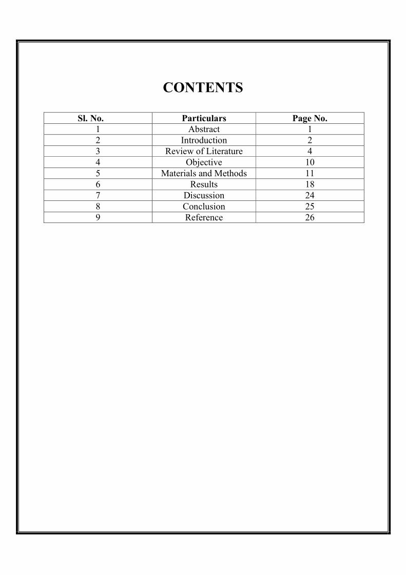
Sl. No. Particulars Page No. 1 Abstract 1 2 Introduction 2 3 Review of Literature 4 4 Objective 10 5 Materials and Methods 11 6 Results 18 7 Discussion 24 8 Conclusion 25 9 Reference 26
LIST OF FIGURES
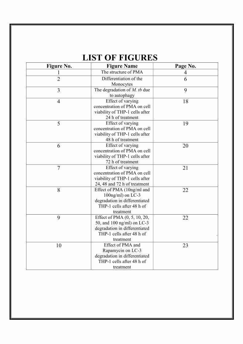
Figure No. Figure Name Page No. 1 The structure of PMA 4 2 Differentiation of the
Monocytes 6
3 The degradation of M. tb due to autophagy
9
4 Effect of varying concentration of PMA on cell viability of THP-1 cells after
24 h of treatment
18
5 Effect of varying concentration of PMA on cell viability of THP-1 cells after
48 h of treatment
19
6 Effect of varying concentration of PMA on cell viability of THP-1 cells after
72 h of treatment
20
7 Effect of varying concentration of PMA on cell viability of THP-1 cells after 24, 48 and 72 h of treatment
21
8 Effect of PMA (10ng/ml and 100ng/ml) on LC-3
degradation in differentiated THP-1 cells after 48 h of
treatment
22
9 Effect of PMA (0, 5, 10, 20, 50, and 100 ng/ml) on LC-3 degradation in differentiated
THP-1 cells after 48 h of treatment
22
10 Effect of PMA and Rapamycin on LC-3
degradation in differentiated THP-1 cells after 48 h of
treatment
23
List of abbreviations
7-IFN- 7 interferon
Atg- Autophagy genes
cAMP- Cyclic AMP
CLR- C-type lectin receptors
DAG- Diacylglycerol
IL-1- Interleukin 1
LPS- Lipopolysaccharide
M.tb- Mycobacterium tuberculosis
MAP1LC3- Microtubule associated protein 1 Light Chain 3
MDR TB- Multi Drug Resistant TB
MR- Mannose Receptor
NLRs- Nod like receptors
PGE2-Prostaglandin E2
PI3K- Phospho inositole 3 kinase
PKC- Protein Kinase C
PMA-Phorbol 12-Myristate 13-Acetate
RNI- Reactive Nitrogen Intermediates
ROI- Reactive oxygen intermediate
STING- Stimulator for Interferon Genes
TB- Tuberculosis
Th1- T helper- 1 type
TLR- Toll like receptors
TNF-α-tumour necrosis factor-a
XDR TB- Extensively drug resistant TB
1. ABSTRACT
THP-1 cells are human monocyte like cell line that is blocked at certain steps of the
differentiation process and its differentiation into macrophages can be induced by addition of
PMA. PMA activates Protein Kinase C (PKC) that mimics the physiological activator
diacylglycerol (DAG) thus differentiation into macrophage occurs. These cells mimic the
alveolar macrophages. So, is a good model to study tuberculosis. According to W.H.O.
around 1.3 million people die of this disease every year. Mycobacterium tuberculosis the
causative organism of this disease enters into the body through the airway passages. The
alveolar macrophages phagocyte the bacteria. Once inside they escape degradation by
preventing the phagosomes-lysosome fusion. Thus, they survive inside the phagosomes by
inhibiting autophagy. To curtail this bacterial load, various host defence mechanisms like
apoptosis, reactive oxygen and nitrogen species, phagosome-lysosome fusion and autophagy
play a very important role. Autophagy in the cells is induced by ATP, vitamin D, cytokines.
ATP is known to increase the intracellular calcium level that facilitates autophagy.
Calcimycin, an important calcium ionophore increases the intracellular calcium level thus
facilitates autophagy. Since autophagy plays an important role in curtailing bacterial load so
we attempted to study the role of PMA in inducing autophagy in THP-1 cells.
Keywords: THP-1, PMA, Autophagy, Tuberculosis, Macrophages, Phagosome-lysosome
2. INTRODUCTION:-
THP-1 cells are human monocyte leukemia cells that were obtained from a one year old
leukaemia patient (Tsuchiya et al., 1980). The differentiation of these cells is obstructed at
specific steps and the differentiation of these cells can only be brought about by several
stimuli that are available (Collins, 1987; Koeffler, 1988). They are morphologically similar,
express the same membrane antigens, have similar secretory products with human
monocytes. These cells express complement, Fc receptors, lack surface and cytoplasmic
immunoglobulin and are phagocytic in nature (Auwerx, 1990). They are not responsive
towards TLR antagonists in their undifferentiated stage but become highly responsive upon
getting differentiated. This is one of the broadly used cell line to study the regulation of
monocytes and macrophages, upon discovery 35 years back.
The differentiation of THP-1 monocytic cells into macrophages is mediated by Phorbol 12
Myristate 13-Acetate (PMA). This is a type of phorbol easter that mimics the activity of
diacylglycerol (DAG). Thus they artificially activate Protein Kinase C (PKC). On activation
of PKC monocyte to macrophage transition occurs. This differentiation occurs due to the
changes in the expression of PKC isozymes. Upon differentiation they actively mimic the
human alveolar macrophages thus, are a good model system for studying tuberculosis.
Tuberculosis (TB) is the single largest infectious disease causing nearly 1.3 million deaths
per year (WHO, 2013). This is caused by the pathogen Mycobacterium tuberculosis (M. tb).
The uniqueness of this pathogen is that it is able to survive in vivo for years before the
activation/reactivation of the disease occurs. Although a lot of people are exposed to this
pathogen, but the disease occurs only in few of them. This is because of a compromised
immune system. The pathogen enters into the body through the airway passages i.e. upon
inhaling aerosol droplets containing M. tb. When a person gets infected, the macrophages,
dendritic cells and lymphocytes are recruited to the site of infection. The accumulation of
these cells at the site of infection leads to the formation of granuloma (Ulrichs and
Kaufmann, 2006). On formation of granuloma, the occurrence of active infection is prevented
in most healthy individuals, but the infection is not completely eradicated (Saunders et al.,
1999). This phase is called the phase of latent TB where the host does not shows any
symptoms of infection but they still have M. tb inside their body that can reactivate into its
active form upon compromise of the host immune defence. Thus, complete eradication of the
pathogen from the body is very difficult and the appearance of the Multidrug resistant (MDR)
and Extensively drug resistant (XDR) TB has further aggravated the problem. To curtail this
bacterial load, various host defence mechanisms like apoptosis, reactive oxygen and nitrogen
species, phagosome-lysosome fusion and autophagy play a very important role.
Autophagy is predominantly a cytoprotective process (Kroemer and Levine, 2008). In this
particular cytoplasmic molecules are targeted and are separated from the rest of the
cytoplasm by development of a double membrane structure around them. This double
membraned structure called autophagosome fuse with lysosome on maturation. This leads to
the degradation of the sequestered molecules. Autophagy helps in generating substrates for
energy metabolism on nutrient scarcity. This is done by non-selective consumption of cellular
components (Deretic and Levine, 2009). Autophagy helps in the elimination of the
intracellular microbes that enters cytosol. Some intracellular pathogens like M. tb avoid
degradation by the host by preventing autophagosome lysosome fusion.
As THP-1 cells are monocytes, PMA treatment is necessary for the differentiation of these
cells. Differentiation of the cells increases the phagocytic index ultimately leading to efficient
internalization of mycobacteria. Since autophagy plays an important role in curtailing
bacterial load so we attempted to study the role of PMA in inducing autophagy in THP-1
cells.
3. REVIEW OF LITERATURE:-
3.1. PMA
On treatment of the THP-1 cells with PMA, the proliferation of the cells stop and they start to
differentiate into macrophages. This leads to the alternation of the cell morphology as they
acquire a variety of cell shapes, many phagocytic vacuoles arise in their cytoplasm and the
nucleus becomes more irregular. Upon differentiation the nucleus to cytoplasm ratio
decreases as the cytoplasmic volume increases (Sokol et al., 1987). Thus, due to the above
mentioned changes that takes place the cells shows adherence property, as they stick to the
surface of the culture flask. The flow cytometry results confirmed that the cell volume
decreases as these cells show a decrease in the forward scattering of the light. In macrophages
the number of membrane bound organelles increases, thus the granularity increases (Sokol et
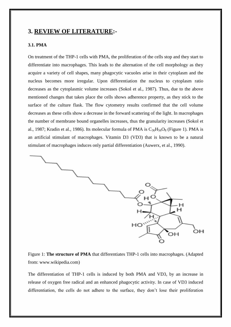
al., 1987; Kradin et al., 1986). Its molecular formula of PMA is C36H56O8 (Figure 1). PMA is
an artificial stimulant of macrophages. Vitamin D3 (VD3) that is known to be a natural
stimulant of macrophages induces only partial differentiation (Auwerx, et al., 1990).
Figure 1: The structure of PMA that differentiates THP-1 cells into macrophages. (Adapted
from: www.wikipedia.com)
The differentiation of THP-1 cells is induced by both PMA and VD3, by an increase in
release of oxygen free radical and an enhanced phagocytic activity. In case of VD3 induced
differentiation, the cells do not adhere to the surface, they don’t lose their proliferation
capability, don’t release Prostaglandin E2 (PGE2) and release very small amount of tumour
necrosis factor- (TNF-α). Whereas in contrast to VD3, the PMA differentiated cells adhere
to the surface, stop proliferating and produce a lot of PGE2 and TNF-α. The PMA
differentiated cells has an enhanced expression of CD11 while CD14 was more expressed in
the cells differentiated by VD3 (Schwende et al., 1996).
Not a single cytokine or growth factors that have been tested till date (TNF, IL-2, GM-CSF,
IL-1) can induce the differentiation of the monocytes by themselves. But all of them together
might mimic the activity of PMA. It has also been reported that the colony formation in THP-
1 cells can be inhibited by 7-IFN and TNF (Lubbert and Koeffier, 1988).
3.2. THP-1 (Monocytes and Macrophages)
Various substances like reactive oxygen species, hormones, enzymes etc. are secreted by
macrophages and the THP-1 cells are used to study the functions of the various secretions of
the macrophages. It has been reported that the several proteins that are secreted from the
mature macrophages are also secreted by the THP-1 cells (Johnston, 1988 and Nathan, 1987).
The THP-1 cells are mostly used to study the production of peptide hormones and cytokines.
The THP-1 cells activate IL-1β as they possess enzymatic activity (Kostura, 1989). The IL-1β
mRNA levels in THP-1 cells are induced by both PMA and Lipopolysaccharide (LPS)
(Fenton, 1988 and Turner, 1988). It has been reported that the level IL-1 is increased
significantly on infection with HIV (Molina et al., 1989).
The THP-1 cells are also used to study the production of apolipoprotein E (apoE) (the lipid
binding protein) and lipoprotein lipase (LPL). Various cells synthesize the glycoprotein
enzyme LPL that hydrolyzes the core triglycerides into triglyceride-rich lipoproteins to
glycerol and free fatty acids (Olivecrona and Olivecrona, 1987). LPL is only produced by
the THP-1 cells when they differentiate into macrophage that is on addition of PMA (Sudhof
et al., 1987; Auwerx et al., 1988, 1989). Amongst all the human leukemia cell line this
property is unique to THP-1 cells.
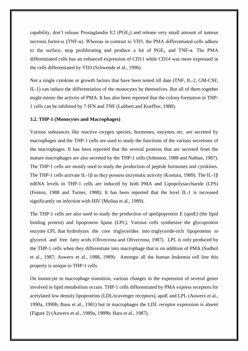
On monocyte to macrophage transition, various changes in the expression of several genes
involved in lipid metabolism occurs. THP-1 cells differentiated by PMA express receptors for
acetylated low density lipoproteins (LDL/scavenger receptors), apoE and LPL (Auwerx et al.,
1990a, 1990b; Basu et al., 1981) but in macrophages the LDL receptor expression is absent
(Figure 2) (Auwerx et al., 1989a, 1989b; Hara et al., 1987).
Figure 2: Differentiation of the Monocytes: - The expression of genes for lipid metabolism
changes on the differentiation of monocytes to macrophages. The expression of receptor for
LDL is absent on differentiation of monocytes. The differentiated cells rather possess a
receptor for acetyl LDL, HDL, apoE and LPL. (Adapted from: Auwerx, 1991)
On differentiation of the monocytes into macrophages, they acquire the property of
accumulating cholesterol inside them. This leads to the formation of foam cells and
atherosclerosis. For studying these changes, THP-1 cells are used as a model (Auwerx, 1991).
3.3. Macrophage Receptors Involved in Tuberculosis Pathogenesis
M. tb is detected by the innate immune system only when it binds to the receptors that are
available on the surface of macrophages and other myeloid cells. The receptors to which M.
tb binds are Toll like receptors (TLRs), Nod like receptors (NLRs), C-type lectin receptors
(CLRs) and Mannose receptors (MR). After this M. tb are engulfed by the macrophages.
Inside the phagosomes the pathogen prevents the phagosomes lysosome fusion. Through this
way it escapes from the immune system of our body. But when the activation of macrophage
occurs, the phagosome and lysosome fusion takes place. Along with that secretion of a
variety of cytokines, production of antimicrobial reactive nitrogen intermediates (RNI) and
ROI also occurs. This leads to the death of the M. tb present inside the body.
CD14, a cell surface glycoprotein induces a signalling cascade on detection of LPS
(lipopolysaccharide) by acting as a co-receptor for the TLR-4 (Tavera et al., 2006). In TB,
CD14 interacts with many M. tb surface components that mediates uptake of the bacteria and
releases the proinflamatory cytokines (Ponta et al., 2003; Drage et al., 2009). Another surface
glycoprotein CD44 similarly mediates mycobacterial phagocytosis and induces protective
immunity against M. tb (Leemans et al., 2003). CD44 is a receptor for glycoprotein and is
found in many immune cells where it activates lymphocytes. CD44 plays an important role
during the granuloma formation in the lungs infected with M. tb (Ponta et al., 2003). Another
receptor involved in the uptake of M. tb is MR (Mannose receptor). These binds to ManLAM
component present on the surface of M. tb and initiates the internalization of the pathogen
(Schlesinger, 1993).
3.4. Autophagy
Autophagy is a greek word meaning self eating (auto “self” and phagein “to eat”). This word
was coined by Dr. Christian de Duve, a Belgian biochemist in 1963. This involves the
degradation of unnecessary cellular components by the help of lysosome (Lin et al., 2012).
This degradation of cellular components is essential for survival during starvation conditions
as it maintains the cellular energy level. Initially the autophagosome formation starts that are
double membrane structure and isolate the targeted molecule from the rest of the cytoplasm.
The autophagosome mature once they fuse with the lysosome. On fusion the molecules
present inside the autophagosome gets degraded due to the acidic environment of lysosome
(Patel et al., 2012).
Autophagy is of three different types: macroautophagy, microautophagy and
chaperonemediated autophagy (Peracchio et al., 2012).
Macroautophagy occurs mainly to remove the damaged cell organelles from the cell (Levine
et al., 2011). Autophagosome formation takes that acquire the proteases required for
degradation of the separated material by fusion with lysosome.
Microautophagy is poorly understood in mammalian cells. Unlike macroautophagy, in
microautophagy the lysosomal membrane itself protrudes to sequester the cytosolic
components (Russell et al., 2013). The entire cytosolic region constitutes the cargo for
microautophagy. This pathway was important for the survival of cells under starvation
conditions. It regulates the composition of the lysosomal membrane by degrading the lipids
incorporated into the membrane (Li et al., 2012).
Chaperone-mediated autophagy (CMA), is a complex and specific pathway. This requires
the unfolding of the protein cargo before it gets internalized into the lysosome. It’s very
selective and occurs only for the soluble cytosolic proteins (Levine et al., 2011; Matsushita et
al., 2007). This is mostly activated when the cell is under stress. It transfers the protein cargo
one after another thus is very different from the other types of autophagy. It only allows
certain proteins to cross the lysosomal membrane barrier, thus are characterized as selective
(Levine et al., 2011).
It has been seen in neutrophils under different clinical conditions, that they are able to form
vacuoles. When neutophils are stimulated with PMA many cytoplasmic vacuoles were
observed. This effect of PMA was dose and time dependant. MDC staining was done to
identify the nature of the vacuoles formed and it confirmed the existence of acidified
autophagosomes. This confirmed the existence of autophagy. On incubation of cells with 3-
MA an autophagy inhibitor prior to addition of PMA, the vacuole formation was not seen.
The expression of ATG3 and LC3B mRNA increases after the treatment of the neutrophils
with PMA (Remijsen et al., 2011).
3.5. Autophagy and M.tb
Upon the entry of M. tb, macrophages form phagosomes. These phagosomes then fuse with
lysosome to form autolysosomes and thus M. tb gets degraded.
It has been observed that the M. tb growth is reduced in the THP-1 cells when AKT1 and
AKT2 are knocked down. SLRs (Sequestasome like receptors) target the microbes to
autophagosome (Semba, 1999).
In case of TB infection, the process of autophagy enhances the host resistance by encircling
the M. tb induced phagosomes maturation arrest (Jagannath et al., 2009; Gutierrez et al.,
2004). The immature phagosomes containing M. tb are tagged with the autophagy proteins
(Atg). First Atg6 i.e. Beclin-1 binds to the phagosomes. With the attachment of belcin-1,
many other Atg proteins bind to the membrane making a complex. Finally, the characteristic
feature of autophagy i.e. autophagosome formation starts. Then microtubule associated
protein 1 light chain 3 (LC3-I) gets degraded and is converted to LC3-II. The LC3-II then
binds to the membrane of autophagosome and fusion with lysosome occurs. (Figure-3)
(Stromhaug and Klionsky, 2001).
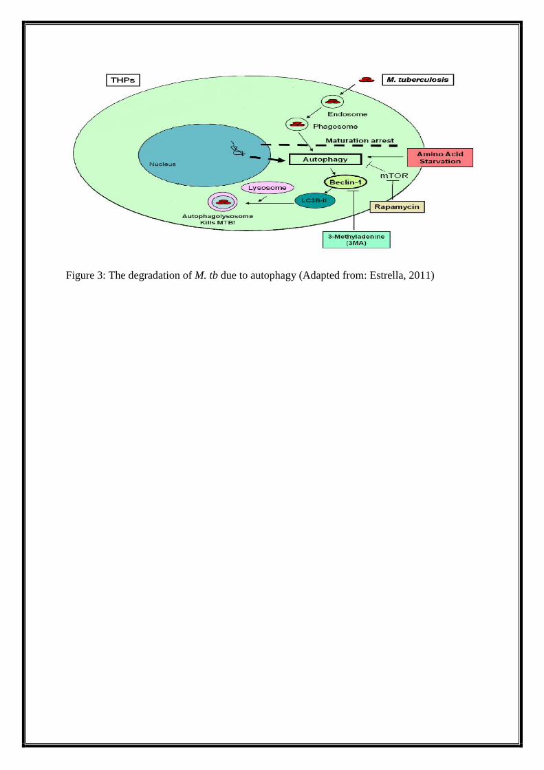
Figure 3: The degradation of M. tb due to autophagy (Adapted from: Estrella, 2011)
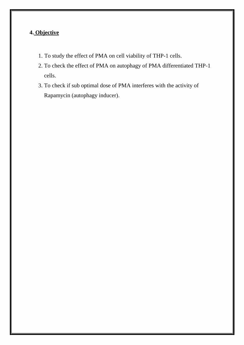
4. Objective
1. To study the effect of PMA on cell viability of THP-1 cells.
2. To check the effect of PMA on autophagy of PMA differentiated THP-1
cells.
3. To check if sub optimal dose of PMA interferes with the activity of
Rapamycin (autophagy inducer).
5. Materials and Methods
5.1. CELL LINES
THP-1 cell line that was used in the study was a kind gift from Dr. Vivek Rai, Institute of
Life Science (Bhubaneswar, India). RPMI-1640 growth medium was used to maintain the
cells. The contents of the media are; 10% heat inactivated Fetal Bovine Serum (FBS) and
antibiotic cocktail like streptomycin and penicillin. The cells were stored at 37°C with 5% of
CO2 .
5.2. CHEMICALS
The chemicals used were of analytical grade and were purchased from commercial sources.
The cell culture media RPMI-1640 and FBS were purchased from GIBCO (Grand Island,
NY)
The reagents used for detection and analysis during western blotting were purchased from GE
healthcare (UK).
The MTT cell assay kit was purchased from HiMedia Laboratory (Mumbai, India).
The BCA kit was purchased from Sigma Sigma-Aldrich.
Following reagents were purchased from HiMedia Laboratory (Mumbai, India): Sodium
Chloride (NaCl), Sodium Dodecyl Sulphate (SDS), Acrylamide, Dimethyl Sulfoxide
(DMSO), Hydrochloric acid (HCL), Tris base, Glycine, Potassium Chloride (KCl), Potassium
Dihydrogen Phosphate (KH2PO4), Disodium Phosphate (Na2HPO4),
Ethylenediaminetetraacetic acid (EDTA), Triton X-100, Glycerol, 2-mercaptoehtanol,
Bromophenol Blue, Sodium Hydroxide (NaOH), Phorbol-Myristate-Acetate (PMA) and
Bovine Serum Albumin (BSA). Following reagents were purchased from Sigma (St. Louis,
Mo, USA): Bisacrylamide, Sodium Deoxycholate. All media and reagents used were
endotoxin-free.
5.2.1. CELL CULUTURE MEDIA
Complete RPMI-1640 (with antibiotic)
To prepare complete RPMI-1640 media we need the following chemicals:-
500 ml of RPMI media
50 ml of FBS
5.5 ml of (1X) Penicillin- Streptomycin
To the 500 ml RPMI medium, 50 ml of heat inactivated FBS was added. This was followed
by addition of 5.5 ml of Penicillin-Streptomycin. Then this was stored at 4°C by preparing
aliquots of 100 ml each.
5.2.2 BUFFER/ SOLUTIONS
10 X Phosphate Buffered Saline (pH 7.4)
To prepare 10X Phosphate Buffer Saline (PBS) we need the following chemicals:-
80 g of NaCl
2 g of KCl
14.4 g of Na2HPO4
2.4 g of KH2PO4
In 800ml of distilled water, the components listed above were dissolved. Using HCl the pH
was adjusted to 7.4. The final volume was adjusted to 1 litre using distilled water.
1 M Tris (pH 6.8 or 7.4 or 8.0 or 9.0)
To prepare 1M Tris we need the following chemicals:-
121.1 g of Tris base
In 800ml of distilled water 121.1g of tris base was dissolved and the pH was adjusted by
using HCl. Finally the volume was adjusted to 1 litre by adding distilled water.
Reservoir Buffer 10X (For SDS-PAGE electrophoresis)
To prepare Reservoir Buffer 10X we need the following chemicals:-
3 g of Tris base
14.4 g of Glycine
10 ml of 10% SDS
In 990ml of distilled water the above listed components except 10% SDS were dissolved.
And finally 10% SDS was added.
4 X Protein Sample buffer (For SDS-PAGE electrophoresis)
The 4X Protein Sample Buffer was prepared using the following chemicals:-
2.4 ml of Upper Tris (1M, pH-6.8)
0.8 g of SDS
4.0 ml of Glycerol
0.5 ml of β-Mercaptoethanol
4 mg of Bromophenol blue
In 3.3 ml of distilled water the above listed components were added and the final volume was
made to 10 ml by adding distilled water. The solution was stored at -20°C after dispencing
into aliquots.
Transfer Buffer10X (For Western blotting)
The transfer Buffer 10X was prepared by using the following chemicals:-
10.8 g of Tris base
50.7 g of Glycine
In 250 ml of distilled water the above listed components were dissolved and the final volume
was adjusted to 360 ml by adding distilled water. Then 300 ml of methanol was added. The
solution was autoclaved.
Acrylamide - bis-Acrylamide mixture
The Acrylamide-bis-Acrylamide mixture was prepared by using the following chemicals:-
30 g of Acrylamide
0.8 g of Bis-Acrylamide
Both the components were dissolved in 70 ml o distilled water. The final volume was
adjusted to 100 ml. Then the solution was filtered and was stored at 4°C.
10% Sodium dodecyl sulfate (SDS)
10% SDS was prepared using the following chemicals:-
5 g of SDS
In 40 ml of distilled water 5 g of SDS was dissolved. The volume was then adjusted to 50 ml
by adding distilled water.
10% Ammonium Persulfate (APS)
10% APS was prepared using the following chemicals:-
0.1 g of APS
In 1 ml of distilled water 0.1 g of APS was dissolved.
PBS-T (For Western blotting)
PBS-T was prepared using the following chemicals:-
50 ml of 10 X PBS
2.5 ml of Tween-20
450 ml of Distilled water
These were mixed with distilled water.
Lower Tris (4 X) pH 8.8 (For SDS-PAGE electrophoresis)
Lower Tris was prepared by using the following chemicals:-
18.17 g of Tris base
4 ml of 10% SDS
In 80 ml of distilled water tris base was dissolved and the pH was adjusted to 8.8 by using
HCl. Finally 4 ml of 10% SDS was added and the final volume was made to 100 ml by
adding distilled water.
Upper Tris (4 X) pH 6.8 (For SDS-PAGE electrophoresis)
Upper Tris was prepared by using the following chemicals:-
6.06 g of Tris base
4 ml of 10% SDS
In 25 ml of distilled water, Tris base was dissolved and the pH was adjusted to 6.8 by using
HCl. Finally 4 ml of 10% SDS was added and the volume was adjusted to 100 ml by adding
distilled water.
RIPA buffer
RIPA buffer was prepared by using the following chemicals:-
1 ml of 1% Triton X – 100
1.5 ml of 150 mMNaCl
0.5 ml of 0.5% Sodium deoxycholate
0.5 ml of 50 mMTris, pH – 8.0
0.1 ml of 0.1 % SDS
0.1 ml of Protease inhibitor cocktail (1X) (PIC)
6.3 ml of Distilled water
All the components except PIC were dissolved in the distilled water and the final volume was
adjusted to 9.9 ml by adding distilled water. These were dispensed into aliquots and were
stored at -20°C. PIC was freshly added to the buffer when it was used.
5% SKIM MILK (For Western blotting)
Skim Milk was prepared by using the following chemicals:-
5 g of Skim milk
100 ml of PBS-T
In 100 ml of PBS-T 5 g of skim milk was dissolved.
5.3. METHODS
5.3.1. Cell culture
The THP-1 cells were cultured in RPMI-1640 media. The cells were stored at 37°C in
presence of 5% CO2. The media was changed periodically as the cell density increased and
the pH of the media became acidic. This was done by centrifuging the media at 1000 rpm for
5 minutes. The supernatant was discarded and the pellet was retained. The pellet was made to
dissolve in 5 ml of fresh culture media and was transferred to the culture flask. And the flask
was placed back in the CO2 incubator.
5.3.2. Cell viability
The MTT cell viability kit was used to check the cell viability. The procedure followed was
according to the protocol provided by the manufacturer.
2.5×10⁴ cells were seeded into 39 wells of 96 well plates. The cells were seeded in triplicates
and were incubated for 24 h in an incubator. After 24 h, different concentrations of PMA
were added to the wells. The plates were then kept in the incubator for varying time points
(24, 48, 72 h). At indicated time points, MTT reagent (10 μl, 5 mg/ml) was added to the
wells. The plate was gently swirled to mix the contents and was incubated for 2-4 h at 37°C.
Then the formation of Formazan crystals was checked and 100 μl lysis solution was added to
all wells except negative control well. The absorbance was taken under 570nm. And the
viability graph was plotted according to formula % Cell viability = (OD of Test sample/OD
of Control) x 100.
5.3.3. Western blotting
After protein estimation of the cell lysates was done by BCA assay. Electrophoresis
(15% SDS-PAGE) was done with 50 g of protein. The gel was then removed from the
electrophoresis unit and was then used in western blotting. The gel was placed carefully in
the blotting cassette such that no bubbles were formed. In the blotting cassette
sponge/paper/gel/membrane/paper/sponge were arranged accordingly. The nitrocellulose
membrane used was treated skim milk to block the non-specific binding site of antibodies.
After western blotting the bands got transferred from the gel into the membrane. Then the
membrane was probed with anti LC3 antibody (primary antibody) that recognises the
presence of LC3-I and LC3-II bands in the membrane. After overnight incubation the
membrane was washed 3 times with PBS-T and secondary antibody conjugated with HRP
was added. After 45 minuites the membrane was washed 3 times with PBS-T. The protein
bands were visualized using enhanced chemiluminescence kit (Amersham, USA) as per
manufacturer’s instructions. The membrane was then stripped and was reprobed with anti β-
actin antibody following same protocol as above.
Cell lysis
Fresh cell lysis buffer was made by adding 10 µl of PIC to 1 ml of RIPA buffer and was
mixed gently. The supernatant of the cultured cells were removed at appropriate time interval
and the adhered cells were washed twice with 1X PBS. Then the lysis buffer was added to the
cells in the well. The lysis buffer was gently mixed with the cells and the cell lysates were
transferred to pre chilled micro centrifuge tubes. Then it was vortexed 3 times at a constant
interval of 10 minutes. Then the lysates were centrifuged at 15000 rpm for 15 min at 4C.
The supernatant was made into aliquots and were store at -20C.
Protein estimation by BSA reagent
First BCA working solution was prepared by mixing 50:1 part of solution A & B. Then 5 µl
of BSA standard and the cell lysates were added to each well of a 96 well plate. Then, 200 µl
of BCA working solution was added to each well. The plate was incubated at 37 C for 10-15
min till purple colour developed. Then the absorbance was taken at 562 nm. The standard
curve for BSA was prepared and from that graph we calculated the concentration of protein
present in the unknown sample i.e. cell lysates according to the regression equation.
6. RESULT
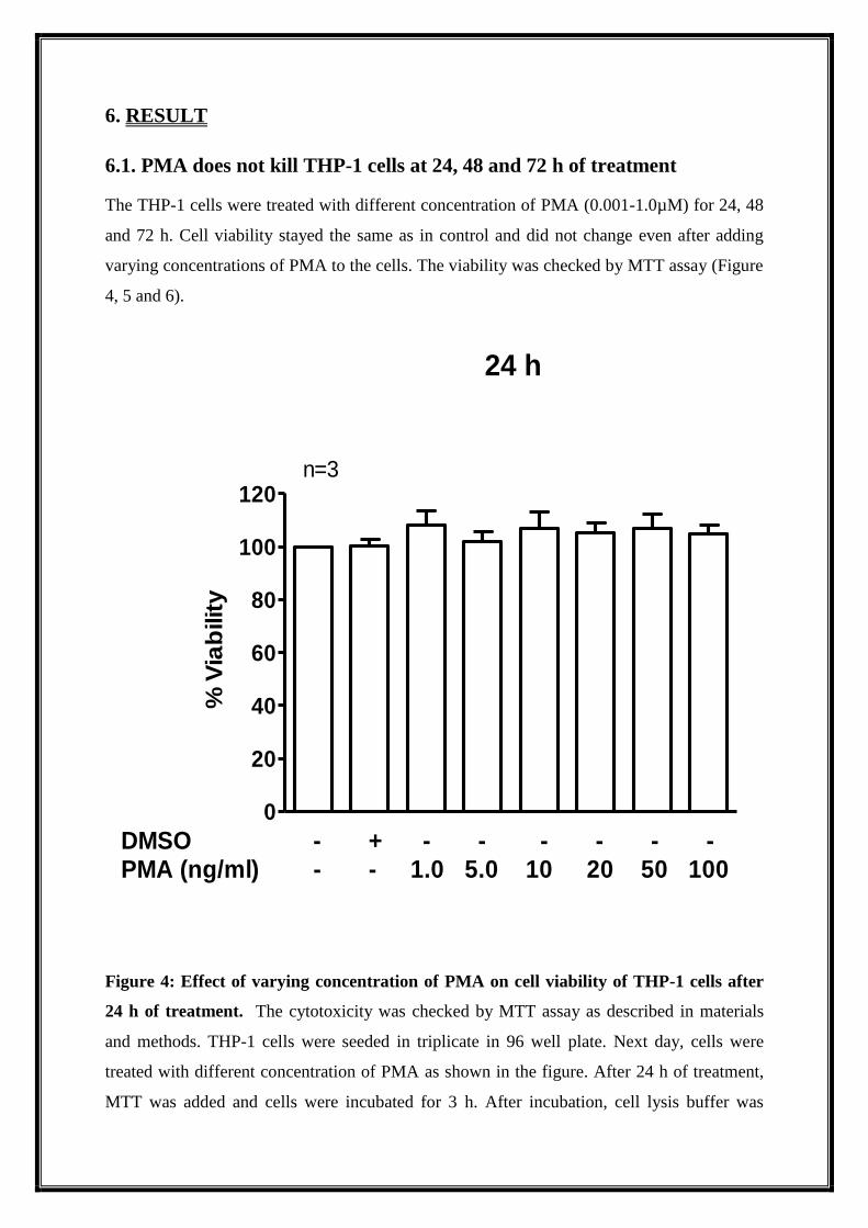
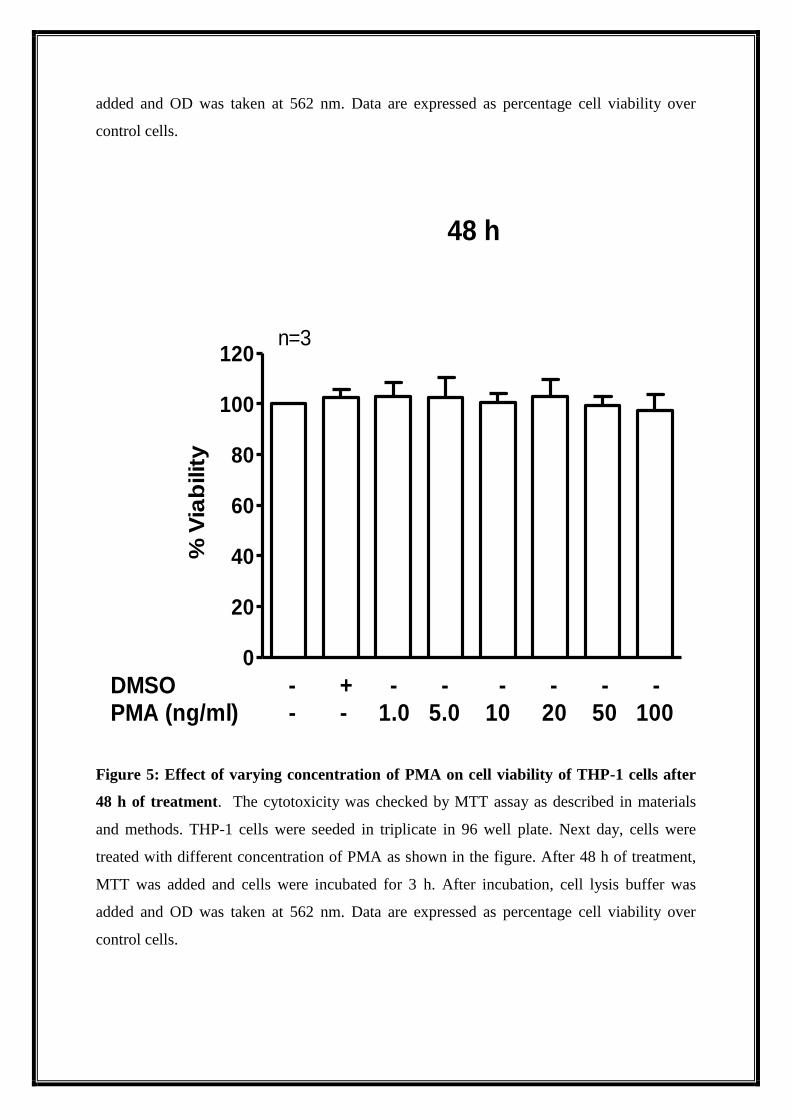
6.1. PMA does not kill THP-1 cells at 24, 48 and 72 h of treatment
The THP-1 cells were treated with different concentration of PMA (0.001-1.0µM) for 24, 48
and 72 h. Cell viability stayed the same as in control and did not change even after adding
varying concentrations of PMA to the cells. The viability was checked by MTT assay (Figure
4, 5 and 6).
0
20
40
60
80
100
120
DMSO - + - - - - - -PMA (ng/ml) - - 1.0 5.0 10 20 50 100
24 h
n=3
% V
iab
ilit
y
Figure 4: Effect of varying concentration of PMA on cell viability of THP-1 cells after
24 h of treatment. The cytotoxicity was checked by MTT assay as described in materials
and methods. THP-1 cells were seeded in triplicate in 96 well plate. Next day, cells were
treated with different concentration of PMA as shown in the figure. After 24 h of treatment,
MTT was added and cells were incubated for 3 h. After incubation, cell lysis buffer was
added and OD was taken at 562 nm. Data are expressed as percentage cell viability over
control cells.
0
20
40
60
80
100
120
DMSO - + - - - - - -PMA (ng/ml) - - 1.0 5.0 10 20 50 100
48 h
n=3
% V
iab
ilit
y
Figure 5: Effect of varying concentration of PMA on cell viability of THP-1 cells after
48 h of treatment. The cytotoxicity was checked by MTT assay as described in materials
and methods. THP-1 cells were seeded in triplicate in 96 well plate. Next day, cells were
treated with different concentration of PMA as shown in the figure. After 48 h of treatment,
MTT was added and cells were incubated for 3 h. After incubation, cell lysis buffer was
added and OD was taken at 562 nm. Data are expressed as percentage cell viability over
control cells.
0
20
40
60
80
100
120
DMSO - + - - - - - -PMA (ng/ml) - - 1.0 5.0 10 20 50 100
72 h
n=3%
Via
bilit
y
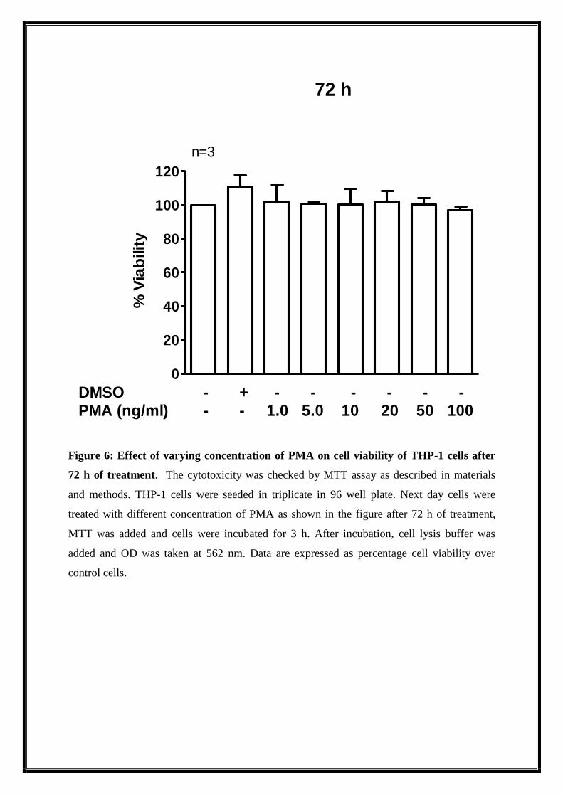
Figure 6: Effect of varying concentration of PMA on cell viability of THP-1 cells after
72 h of treatment. The cytotoxicity was checked by MTT assay as described in materials
and methods. THP-1 cells were seeded in triplicate in 96 well plate. Next day cells were
treated with different concentration of PMA as shown in the figure after 72 h of treatment,
MTT was added and cells were incubated for 3 h. After incubation, cell lysis buffer was
added and OD was taken at 562 nm. Data are expressed as percentage cell viability over
control cells.
0
20
40
60
80
100
120
Control cells
24 h
48 h
72 h
n=3
DMSO - + - - - - - -PMA (ng/ml) - - 1.0 5.0 10 20 50 100
% V
iab
ilit
y
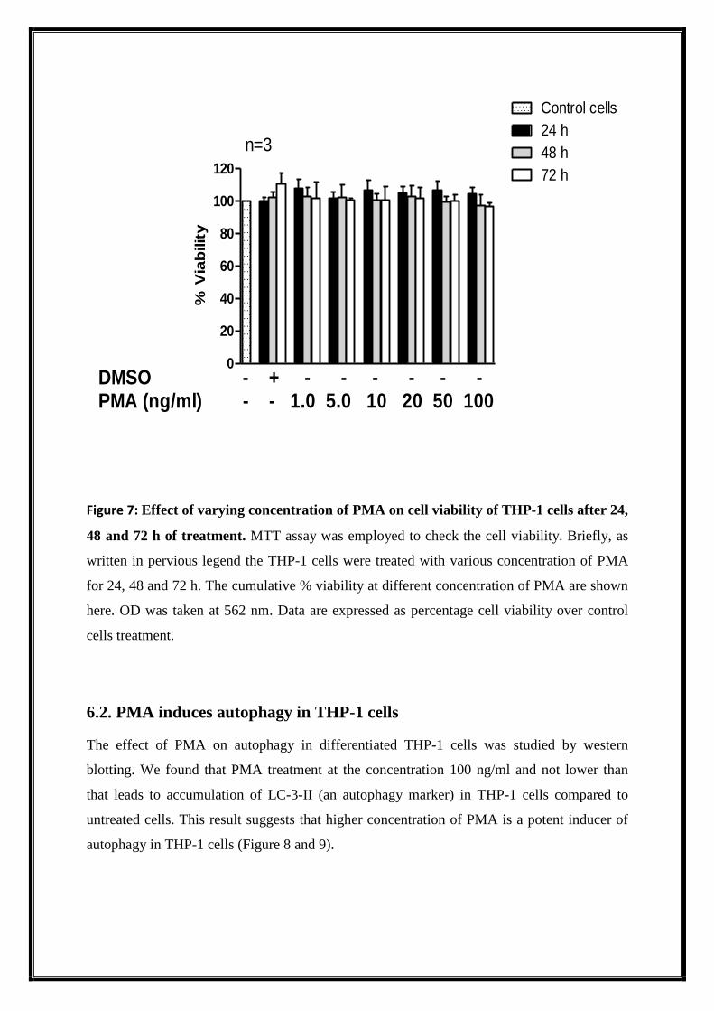
Figure 7: Effect of varying concentration of PMA on cell viability of THP-1 cells after 24,
48 and 72 h of treatment. MTT assay was employed to check the cell viability. Briefly, as
written in pervious legend the THP-1 cells were treated with various concentration of PMA
for 24, 48 and 72 h. The cumulative % viability at different concentration of PMA are shown
here. OD was taken at 562 nm. Data are expressed as percentage cell viability over control
cells treatment.
6.2. PMA induces autophagy in THP-1 cells
The effect of PMA on autophagy in differentiated THP-1 cells was studied by western
blotting. We found that PMA treatment at the concentration 100 ng/ml and not lower than
that leads to accumulation of LC-3-II (an autophagy marker) in THP-1 cells compared to
untreated cells. This result suggests that higher concentration of PMA is a potent inducer of
autophagy in THP-1 cells (Figure 8 and 9).
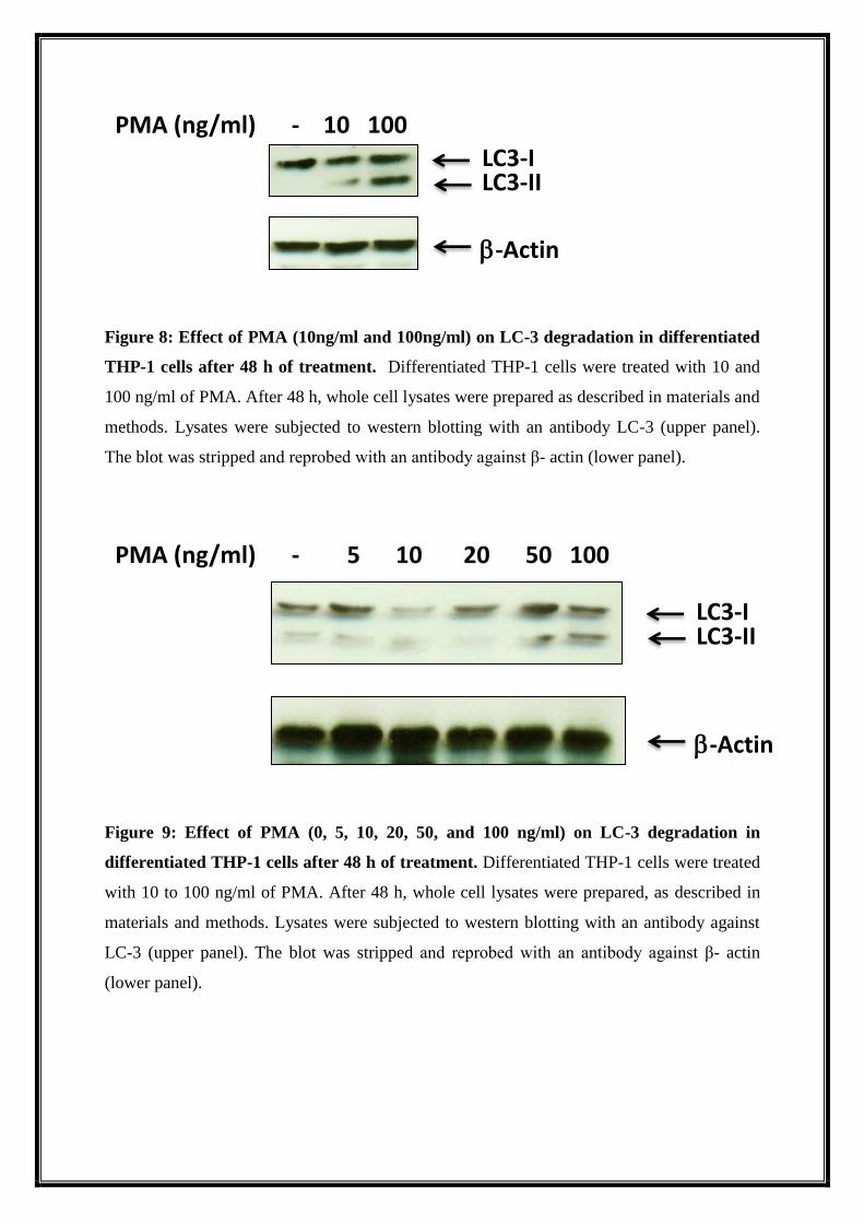
Figure 8: Effect of PMA (10ng/ml and 100ng/ml) on LC-3 degradation in differentiated
THP-1 cells after 48 h of treatment. Differentiated THP-1 cells were treated with 10 and
100 ng/ml of PMA. After 48 h, whole cell lysates were prepared as described in materials and
methods. Lysates were subjected to western blotting with an antibody LC-3 (upper panel).
The blot was stripped and reprobed with an antibody against β- actin (lower panel).
Figure 9: Effect of PMA (0, 5, 10, 20, 50, and 100 ng/ml) on LC-3 degradation in
differentiated THP-1 cells after 48 h of treatment. Differentiated THP-1 cells were treated
with 10 to 100 ng/ml of PMA. After 48 h, whole cell lysates were prepared, as described in
materials and methods. Lysates were subjected to western blotting with an antibody against
LC-3 (upper panel). The blot was stripped and reprobed with an antibody against β- actin
(lower panel).
PMA (ng/ml) - 10 100
LC3-I
LC3-II
-Actin
PMA (ng/ml) - 5 10 20 50 100
LC3-I
LC3-II
-Actin
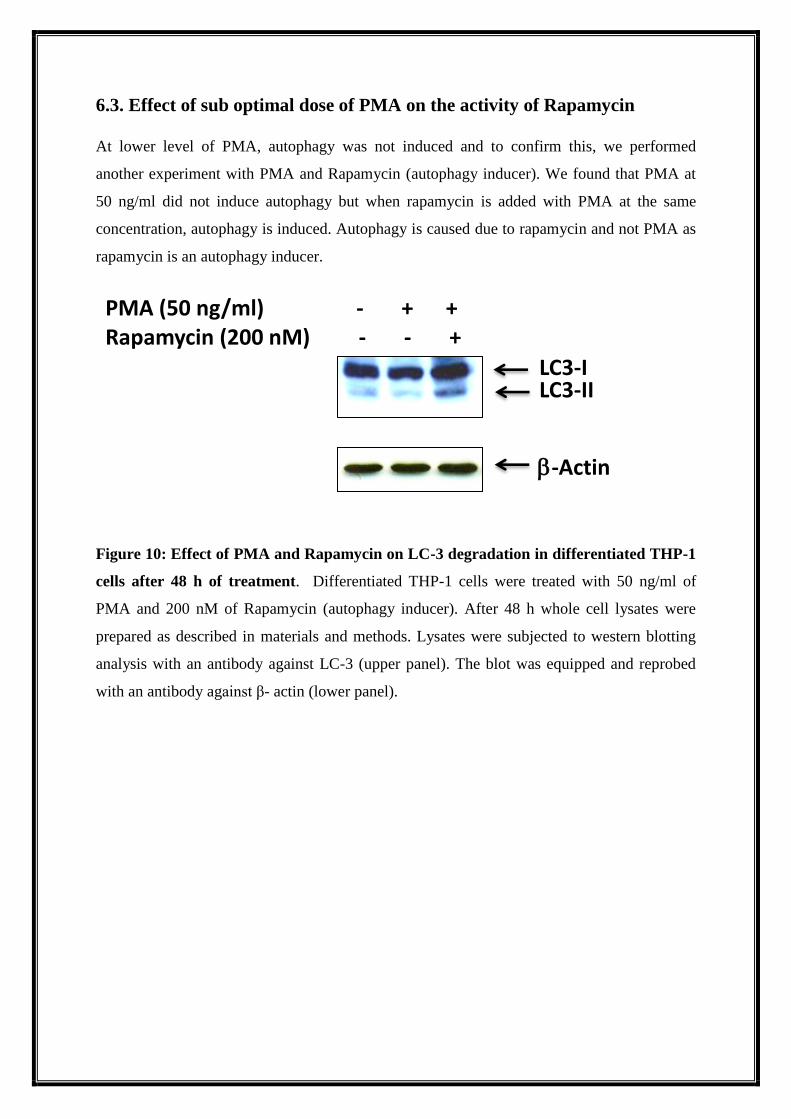
6.3. Effect of sub optimal dose of PMA on the activity of Rapamycin
At lower level of PMA, autophagy was not induced and to confirm this, we performed
another experiment with PMA and Rapamycin (autophagy inducer). We found that PMA at
50 ng/ml did not induce autophagy but when rapamycin is added with PMA at the same
concentration, autophagy is induced. Autophagy is caused due to rapamycin and not PMA as
rapamycin is an autophagy inducer.
Figure 10: Effect of PMA and Rapamycin on LC-3 degradation in differentiated THP-1
cells after 48 h of treatment. Differentiated THP-1 cells were treated with 50 ng/ml of
PMA and 200 nM of Rapamycin (autophagy inducer). After 48 h whole cell lysates were
prepared as described in materials and methods. Lysates were subjected to western blotting
analysis with an antibody against LC-3 (upper panel). The blot was equipped and reprobed
with an antibody against β- actin (lower panel).
PMA (50 ng/ml) - + +
Rapamycin (200 nM) - - +
LC3-I
LC3-II
-Actin
7. DISCUSSION
The factors leading to monocyte macrophage differentiation and the mode of autophagy
remains partially characterized. In this study we tried to study the effect of PMA on
autophagy of THP-1 cells.
First we tried to see the time kinetics cell viability of THP-1 cells under varying
concentrations of PMA (from 1 ng/ml to 100 ng/ml) and we found that PMA at a
concentration of 100 ng/ml were not at all toxic to THP-1 cells at 24,48 and 72 h after
treatment.
Then we tried to see the effect of two variable concentration of PMA, lowest (1 ng/ml) and
highest (100 ng/ml) on autophagy of THP-1 cells. We found that in comparison to the lowest
dose of PMA highest dose was potentially enough to convert LC-3 I to LC-3 II form. Further
to pinpoint the exact dose of PMA having ability to induce autophagy, we perform
concentration kinetics experiment and proved that no other dose lower than 100 ng/ml is
inducing autophagy in THP-1 cells. Since for further studies in our lab and as reported in
literature 50 ng/ml PMA will be used to differentiate THP-1 cells, we were interested to cross
check further the autophagy with that dose of PMA in the presence and absence of an
autophagy inducer, Rapamycin (200 nM). We found that 50 ng/ml of PMA is not inducing
any autophagy in comparison to Rapamycin. This result will give us a leverage of using PMA
at a dose of 50 ng/ml for differentiation of THP-1 cells for further experiments in our lab.
8. CONCLUSION
From the experiments that were conducted we concluded that
PMA does not have any cytotoxic effect on THP-1 cells, as all the cells were viable.
Higher dose of PMA i.e. 100 ng/ml induces autophagy in THP-1 cells.
Lower dose of PMA i.e. 1 ng/ml does not induce autophagy.
Out of the variable doses of PMA (0, 5, 10, 20, 50, 100 ng/ml) that were checked,
autophagy was induced only at a concentration of 100 ng/ml. Concentrations below
100 ng/ml could not induce autophagy.
Sub optimal dose of PMA used for the differentiation of the monocytes did not have
any interference with the effect of Rapamycin
9. REFERENCES
Auwerx, J., Chait, A., and Deeb, S. S., Transcriptional regulation of the LDL-receptor and
HMG-CoA reductase genes by protein kinase C and a putative negative regolatory
protein. Proc. natl Acad. Sci. USA 86 (1989) 1133-1137.
Auwerx, J., Chait, A., Wolfbauer, G., and Deeb, S., Involvement of second messengers in
regulation of the low-density lipoprotein recep- tor gene.Molec.cell. Biol. 9 (1989)
2298-2302.
Auwerx, J., Deeb, S., Brunzell, J. D., Peng, R., and Chait, A., Tran- scriptional activation of
the lipoprotein lipase and apolipoprotein E genes accompanies differentiation in some
human macrophage-like cell lines. Biochemistry 27 (1988) 2651 2655.
Auwerx, J., Deeb, S., Brunzell, J. D., Wolfbauer, G., and Chait, A., Lipoprotein lipase gene
expression in THP-1 cells. Biochemistry 28 (:1989) 4563-4567.
Auwerx, J., Staels, B., Van Vaeck, E, Verhoeven, G., and Ceuppens, J., IgG Fc receptor
expression during macrophage differentiation of the monocyticleukemia cell line,
THP-1. (1990)
Auwerx, J., Staels, B., and Sassone-Corsi, P., Coupled and uncou- pled induction of fos and
jun transcription by different second mes- sengers in ceils of hematopoietic origin.
Nucl. Acids Res. 18 (1990) 221 - 228.
Auwerx, J., Staels, B., Van Vaeck, E, Verhoeven, G., and Ceuppens, J., IgG Fc receptor
expression during macrophage differentiation of the monocyticleukemia cell line, THP-
1. (1990) submitted.
Auwerx, J., The human leukemia cell line, THP-1 : A multifacetted model for the study of
monocyte-macrophage differentiation. (1991)
Basu, S. K., Brown, M. S., Ho, Y. K., Havel, R. J., and Goldstein, J. L., Mouse macrophages
synthesize and secrete a protein resem- bling apolipoprotein E. Proc. natl Acad. Sci.
USA 78 (1981) 7545 7549.
Biddison, W. E., Rao, P. E., Talle, M. A., Goldstein, G., and Shaw, S., Possible
involvement of the OKT4 molecule in T cell recognition of class II HLA antigens. J.
exp. Med.156 (1982) 1065 1076.
Bishop, J. M., Viral oncogenes. Cell 42 (1985) .
Brown, M. S., and Goldstein, J. L., Lipoprotein metabolism in the macrophage: implications
for cholesterol deposition in atherosclero- sis. A. Rev. Biochem. 52 (1983) 223 261.
Brown, M. S., Goldstein, J. L., Krieger, M., Ho, Y. K., and Ander- son, R. G. W, Reversible
accumulation of cholesteryl esters in macrophages incubated with acetylated
lipoproteins. J. Cell Biol. 82 (1979) 597 613.
Collins, S. J., The HL-60 promyelocyticleukemia cell line: prolifera- tion, differentiation, and
cellular oncogene expression. Blood 70 (1987) 1233 1244.
Drage, M.G., Pecora, N.D., Hise, A.G., Febbraio, M., Silverstein, R.L., Golenbock, D.T.,
Boom, W.H., and Harding, C.V. 2009. TLR2 and its co-receptors determine responses
of macrophages and dendritic cells to lipoproteins of Mycobacterium tuberculosis. Cell
Immunol 258:29-37.
Fenton, M.J., Vermeulen, M. W., Clark, B. D., Webb, A. C., and Auron, P. E., Human pro-
IL-I beta gene expression in monocytic cells is regulated by two distinct pathways. J.
Immunol. 140 (1988) 2267-2273.
Global Tuberculosis Report (2013), W.H.O.
Gutierrez, M.G., Master, S.S., Singh, S.B., Taylor, G.A., Colombo, M.I., and Deretic, V.
2004. Autophagy is a defense mechanism inhibiting BCG and Mycobacterium
tuberculosis survival in infected macrophages. Cell 119:753-766.
Hara, H., Tanishita, H., Yokayana, S., Tajima, S., and Yamamoto, A., Induction of acetylated
low density lipoprotein receptor and suppression of low density Epoprotein receptor on
the cells of a human monocyticleukemia cell line.Biochem.biophys. Res. Com-
mun.146 (1987) 802 808.
Jagannath, C., Lindsey, D.R., Dhandayuthapani, S., Xu, Y., Hunter, R.L., Jr., and Eissa, N.T.
2009. Autophagy enhances the efficacy of BCG vaccine by increasing peptide
presentation in mouse dendritic cells. Nat Med 15:267-276.
Johnston, R. B., Monocytes and macrophages. N. Engl. J. Med. 318 (1988) 747-751.
Kodama, T, Reddy, P., Kishimoto, C., and Krieger, M., Purification and characterization of a
bovine acetyl low density lipoprotein re- ceptor. Proc. natl Acad. Sci. USA 85 (1988)
9238-9242.
Kodama, T., Freeman, M., Rohrer, L., Zabrecky, J., Matsudaira, P., and Krieger, M., Type I
macrophage scavenger receptor contains e-helical and collagen-like coiled coils. Nature
343 (1990) 531-535.
Kostura, M.J., Tocci, M. J., Limjucco, G., Chin, Ji, Cameron, P., Hillman, A. G., Chartrain,
N. A., and Schmidt, J. A., Identification of a monocyte specific pre-interleukin 1 beta
convertase activity. Proc. natl Acad. Sci. USA 86 (1989) 5227-5231.
Kradin RL, McCarthy KM, Preffer FI, Schneeberger EE (1986) Flow- cytometric and
ultrastructural analysis of alveolar macrophage maturation. J LeukocBiol 40: 407–417.
Leemans, J.C., Florquin, S., Heikens, M., Pals, S.T., van der Neut, R., and Van Der Poll, T.
2003. CD44 is a macrophage binding site for Mycobacterium tuberculosis that 102
mediates macrophage recruitment and protective immunity against tuberculosis. J Clin
Invest 111:681-689.
Lubbert, M., and Koeffler, H. P., Myeloid cell lines: tools for studying differentiation of
normal and abnormal hematopoietic cells. Blood. Rev 2 (1988) Lubbert, M., and
Koeffler, H. P., Myeloid cell lines: tools for study- ing differentiation of normal and
abnormal hematopoietic cells. Blood. Rev 2 (1988) 121 133.
Mitchinson, M. J., and Ball, R. Y., Macrophages and atherogenesis.Lance 2 (1987) 146 149.
Molina, J.M., Scadden, D.T., Byrn, R., Dinarello, C.A., and Groopman, J. E., Production of
tumor necrosis factor alpha and interleukin 1 beta by monocytic cells infected with
human immuno- deficiency virus. J. clin. Invest. 84 (1989) 733-737.
Nathan, C., Secretory products of macrophages. J. clin. Invest. 79 (1987) 319-326.
Olivecrona, T., and Bengtsson-Olivecrona, G., Lipoprotein lipase from milk-the model
enzyme in lipoprotein lipase research, in: Lipo- protein Lipase, pp. 15-58. Ed. J.
Borensztajn. Evener press. Chicago 1987.
Ponta, H., Sherman, L., and Herrlich, P.A. 2003. CD44: from adhesion molecules to
signalling regulators. Nat Rev Mol Cell Biol 4:33-45.
Remijsen, Q., Berghe, T. V., Wirawan, E., Asselbergh, B., Parthoens, E., De Rycke, R., ... &
Vandenabeele, P. (2011). Neutrophil extracellular trap cell death requires both
autophagy and superoxide generation. Cell research, 21(2), 290-304.
Ross, R., Atherosclerosis: a problem of the biology of the arterial wall cells and their
interactions with blood components. Arterioscle- rosis 1 (1981) 293-311.
Schlesinger, L.S. 1993. Macrophage phagocytosis of virulent but not attenuated strains of
Mycobacterium tuberculosis is mediated by mannose receptors in addition to
complement receptors. J Immunol 150:2920-2930.
Schwartz, S. M., and Reidy, M., Common mechanisms of prolifera- tion of smooth muscle in
atherosclerosis and hypertension. Hum. Path. 18 (1987) 240-247.
Schwende H., Fitzke E., Ambs P., and Dieter P., Differences in the state of differentiation of
THP-1 cells induced by phorbol ester and 1 ,25-dihydroxyvitamin D3 (1996)
Semba, R.D. 1999. Vitamin A as "anti-infective" therapy, 1920-1940.J Nutr 129:783- 791.
Sokol RJ, Hudson G, James NT, Frost IJ, Wales J (1987) Human macrophage development: a
morphometric study. J Anat 151: 27–35.
Stromhaug, P.E., and Klionsky, D.J. 2001. Approaching the molecular mechanism of
autophagy. Traffic 2:524-531.
Sudhof, T. C., Russell, D. W., Brown, M. S., and Goldstein, J. L., 42 bp element from LDL
receptor gene confers end-pr0duct repression by sterols when inserted into viral TK
promotor. Cell 48 (1987) 1061-1069.
Tavera-Mendoza, L., Wang, T.T., Lallemant, B., Zhang, R., Nagai, Y., Bourdeau, V.,
Ramirez-Calderon, M., Desbarats, J., Mader, S., and White, J.H. 2006. Convergence of
vitamin D and retinoic acid signalling at a common hormone response element. EMBO
Rep 7:180-185.
Tsuchiya, S., Yamabe, M., Yamaguchi, Y., Kobayashi, Y., Konno, T., and Tada, K.,
Establishment and characterization of a human acute monocytic leukemia cell line
(THP-1). Int. J. Cancer 26 (1980) 171 176.
Turner, M., Chantry, D., and Feldmann, M., Post-transcriptional control of IL-1 gene
expression in the acute monocyticleukemia line THP-1. Biochem.biophys.
Res.Commun.156 (1988) 830-839.


![Autophagy Precedes Apoptosis in Angiotensin II-Induced ... · apoptosis [10, 11]. Many stimuli can cause simultaneous apoptosis and autophagy. Ang II induces autophagy, which is further](https://img.pdfslide.net/doc/110x75/5f027da77e708231d4048618/autophagy-precedes-apoptosis-in-angiotensin-ii-induced-apoptosis-10-11-many.jpg)