Embed Size (px)
Citation preview

Experimental Hematology 31 (2003) 770–778
Effect of proinflammatory cytokines on PIGA� hematopoiesis
Shashikant Kulkarni and Monica BesslerDepartment of Internal Medicine, Division of Hematology, Washington University School of Medicine, St. Louis, Mo., USA
(Received 18 February 2003; revised 24 May 2003; accepted 3 June 2003)
Objective. Blood cells from patients with paroxysmal nocturnal hemoglobinuria lack glycosylphosphatidylinositol (GPI)-linked proteins, due to a somatic mutation in the X-linked PIGAgene. It is believed that clonal expansion of PIGA� blood cells is due to a survival advantagein the hostile marrow environment of aplastic anemia. Here we investigated the effectsof inhibitory cytokines in mice genetically engineered to have blood cells deficient in GPI-linked proteins.
Materials and Methods. The effect of inhibitory cytokines (tumor necrosis factor-a [TNF-a],interferon-g [IFN-g], macrophage inflammatory protein-1 alpha [MIP-1a], and transform-ing growth factor-b1 [TGF-b1]) was investigated, using clonogenic assays, competitiverepopulation, and in vivo induction of proinflammatory cytokines by double-stranded RNA.The expression of Fas on progenitor cells and its up-regulation by inhibitory cytokines wereanalyzed by flow cytometry.
Results. TNF-a, IFN-g, MIP-1a, and TGF-b1 suppressed colony formation in a dose-dependentfashion that was similar for PIGA� and PIGA� blood bone marrow cells. Competitiverepopulation of bone marrow cells cultured in IFN-g and TNF-a resulted in a comparable abilityof PIGA� and PIGA� hematopoietic stem cells to reconstitute hematopoiesis. Fas expressionwas minimal on PIGA� and PIGA� progenitor cells and was up-regulated to the same extentin response to IFN-g and TNF-a as assessed by Fas antibody-mediated apoptosis. Similarly, invivo induction of proinflammatory cytokines by double-stranded RNA had no effect on theproportion of circulating PIGA� blood cells.
Conclusions. These results indicate that PIGA� and PIGA� hematopoietic progenitor cellsrespond similarly to inhibitory cytokines, suggesting that other factors are responsible for theclonal expansion of paroxysmal nocturnal hemoglobinuria cells. � 2003 International Societyfor Experimental Hematology. Published by Elsevier Inc.
It is currently believed that in paroxysmal nocturnal hemo-globinuria (PNH), hematopoietic progenitor cells deficientin glycosyl phosphatidylinositol (GPI)-linked proteins havea growth or survival advantage in a bone marrow environ-ment of aplastic anemia (AA) [1]. PNH is a clonal disorderof the hematopoietic stem cell caused by expansion of hema-topoietic progenitor cells carrying a somatic mutation inthe X-linked phosphatidylinositol glycan class A (PIGA)gene [2,3]. The PIGA protein is essential in the synthesisof GPI anchors [4]. Thus, blood cells carrying the PIGAgene mutation are deficient in all cell surface proteins thatare attached to membrane by a GPI anchor [5,6]. Althoughthe lack of GPI-linked proteins explains some of the clinical
Offprint requests to: Monica Bessler, M.D., Ph.D., Division of Hematol-ogy, Department of Internal Medicine, Washington University School ofMedicine, Campus Box 8125, 660 S. Euclid Avenue, St. Louis, MO 63110;E-mail: [email protected]
0301-472X/03 $–see front matter. Copyright � 2003 International Society fordoi: 10.1016/S0301-472X(03)00189-9
features of PNH, the mechanism that leads to clonal expan-sion of the PNH progenitor cell remains elusive. PNH hasa close association with AA [7]. Most patients with PNHhave some clinical or laboratory signs of bone marrow fail-ure, and close to 50% of patients with acquired AA havecirculating PNH blood cells [8,9]. Based on this association,it is hypothesized that in an environment such as AA, wherenormal hematopoietic stem cells die, PNH hematopoieticprogenitors may survive and expand. We and others havepreviously shown that the PIGA gene mutation dose notalter the sensitivity of hematopoietic progenitor cells toapoptotic cell death [10,11], indicating that other factors,in addition to PIGA gene mutations, are required to cause theexpansion of cells deficient in GPI-linked proteins. Increasedlevels of inhibitory cytokines, specifically of interferon-γ(IFN-γ) and tumor necrosis factor-α (TNF-α) have beenfound in patients with AA [12–16] and have been implicatedto play a role in the pathogenesis of bone marrow failure.
Experimental Hematology. Published by Elsevier Inc.
S. Kulkarni and M. Bessler /Experimental Hematology 31 (2003) 770–778 771
Similarly, an increased level of Fas expression has beendetected on bone marrow progenitors from patients withinherited [17,18] and acquired forms of bone marrow failure[19]. Increased Fas expression has also recently been foundon bone marrow cells from patients with PNH [20,21]. Inter-estingly, increased Fas expression was only observed onPIGA� progenitor cells but not PIGA� progenitor cells,suggesting that the PNH cells might escape cell death dueto the lack of Fas expression. We previously reported onthe generation of mice that, due to targeted PIGA geneinactivation, have a proportion of blood cells deficient inGPI-linked proteins [22,23]. Under laboratory conditions,these mice do not develop signs of bone marrow failure,and the proportion of PIGA� blood cells is stable throughoutlife. Here we use our genetically engineered mice to testwhether an altered sensitivity to inhibitory cytokinesmight result in a growth advantage of hematopoietic cellscarrying a PIGA gene mutation. The response of hematopoi-etic progenitor cells carrying a PIGA gene mutation to in-flammatory cytokines has never been investigated. Ourfindings demonstrate that increasing concentrations of inhib-itory cytokines of TNF-α, IFN-γ, macrophage inflammatoryprotein-1 alpha (MIP-1α), and transforming growth factor-β1 (TGF-β1) suppressed in vitro colony formation in adose-dependent fashion that was similar for PIGA� andPIGA� blood bone marrow cells. Similarly, competitive re-population assays of TNF-α– or IFN-γ–treated bone marrowcells resulted in a comparable hematopoietic reconstitutionability of PIGA� and PIGA� cells. In our mice, however,only low levels of constitutive Fas expression were foundon both PIGA� and PIGA� progenitor cells, and after stimu-lation with TNF-α and IFN-γ, Fas expression increasedsimilarly on both progenitors cells, as assessed by Fasantibody-mediated apoptosis. Finally, the injection ofdouble-stranded RNA, a strong inducer of inflammatory cy-tokines, did not alter the contribution of PIGA� cells tothe circulating blood pool. These findings demonstrate thatPIGA� hematopoietic progenitors are not impaired in theirresponse to inhibitory cytokines.
Material and methods
MiceThe two mouse strains, LF and EL, both with PIGA� blood cells,have been previously described [22,23]. In LF mice, Cre expressionis controlled by the transcription regulatory sequences of thehuman c-fes gene, which inactivates the floxed PIGA gene in hema-topoietic stem cells [23]. The proportion of PIGA� cells in female LFmice used in these experiments was approximately 50% (becausein approximately half of female somatic cells the inactivated PIGAgene is on the inactive X chromosome), whereas in male miceit reached close to 100% [23]. No further Cre-mediated PIGAinactivation occurs in the blood cells of these animals [23]. In ELmice, Cre is expressed under the adenoviral EIIa promoter causingrecombination of the floxed PIGA gene in early embryogenesis
[24]. EL mice are mosaic for PIGA� cells in all blood lineages, andthe proportion of PIGA� cells is stable throughout the lifespan of theanimal [22]. No further Cre-mediated recombination occurs in adultlife [23]. Mice carrying only the floxed PIGA gene or only the Cretransgene were used as wild-type (WT) controls. All experimentswere performed with mice in a C57BL/6 background (N � 10).
Flow cytometry and monoclonal antibodiesFlow cytometric analysis and antibodies used for the analysis havebeen previously described [11,25]. Phycoerythrin-conjugated anti-mouse FAS monoclonal antibody (Jo2) was used to determine FASexpression. All antibodies were purchased from Pharmingen (SanDiego, CA, USA).
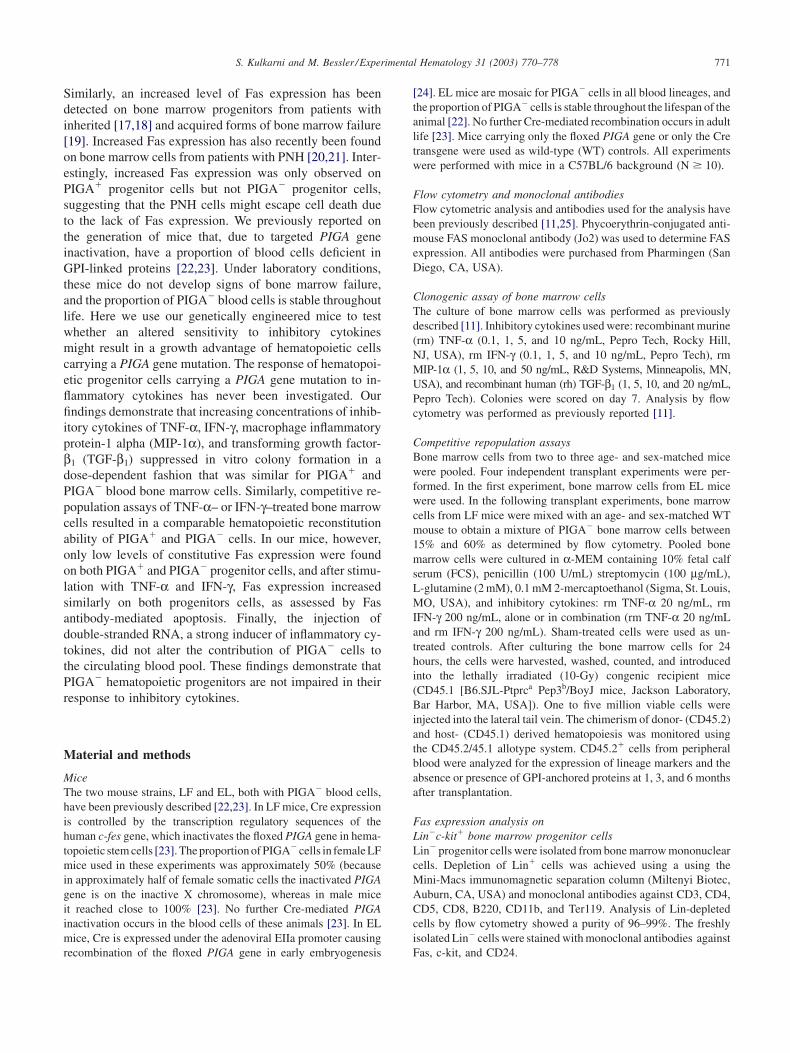
Clonogenic assay of bone marrow cellsThe culture of bone marrow cells was performed as previouslydescribed [11]. Inhibitory cytokines used were: recombinant murine(rm) TNF-α (0.1, 1, 5, and 10 ng/mL, Pepro Tech, Rocky Hill,NJ, USA), rm IFN-γ (0.1, 1, 5, and 10 ng/mL, Pepro Tech), rmMIP-1α (1, 5, 10, and 50 ng/mL, R&D Systems, Minneapolis, MN,USA), and recombinant human (rh) TGF-β1 (1, 5, 10, and 20 ng/mL,Pepro Tech). Colonies were scored on day 7. Analysis by flowcytometry was performed as previously reported [11].
Competitive repopulation assaysBone marrow cells from two to three age- and sex-matched micewere pooled. Four independent transplant experiments were per-formed. In the first experiment, bone marrow cells from EL micewere used. In the following transplant experiments, bone marrowcells from LF mice were mixed with an age- and sex-matched WTmouse to obtain a mixture of PIGA� bone marrow cells between15% and 60% as determined by flow cytometry. Pooled bonemarrow cells were cultured in α-MEM containing 10% fetal calfserum (FCS), penicillin (100 U/mL) streptomycin (100 µg/mL),L-glutamine (2 mM), 0.1 mM 2-mercaptoethanol (Sigma, St. Louis,MO, USA), and inhibitory cytokines: rm TNF-α 20 ng/mL, rmIFN-γ 200 ng/mL, alone or in combination (rm TNF-α 20 ng/mLand rm IFN-γ 200 ng/mL). Sham-treated cells were used as un-treated controls. After culturing the bone marrow cells for 24hours, the cells were harvested, washed, counted, and introducedinto the lethally irradiated (10-Gy) congenic recipient mice(CD45.1 [B6.SJL-Ptprca Pep3b/BoyJ mice, Jackson Laboratory,Bar Harbor, MA, USA]). One to five million viable cells wereinjected into the lateral tail vein. The chimerism of donor- (CD45.2)and host- (CD45.1) derived hematopoiesis was monitored usingthe CD45.2/45.1 allotype system. CD45.2� cells from peripheralblood were analyzed for the expression of lineage markers and theabsence or presence of GPI-anchored proteins at 1, 3, and 6 monthsafter transplantation.
Fas expression analysis onLin�c-kit� bone marrow progenitor cellsLin� progenitor cells were isolated from bone marrow mononuclearcells. Depletion of Lin� cells was achieved using a using theMini-Macs immunomagnetic separation column (Miltenyi Biotec,Auburn, CA, USA) and monoclonal antibodies against CD3, CD4,CD5, CD8, B220, CD11b, and Ter119. Analysis of Lin-depletedcells by flow cytometry showed a purity of 96–99%. The freshlyisolated Lin� cells were stained with monoclonal antibodies againstFas, c-kit, and CD24.

S. Kulkarni and M. Bessler /Experimental Hematology 31 (2003) 770–778772
Up-regulation of Fas expressionand Fas antibody-mediated apoptosis assaysFas was up-regulated on bone marrow cells with either TNF-α(1 and 10 ng/mL) or IFN-γ (1 and 10 ng/mL). The up-regulationof Fas was determined by assessing the sensitivity of the culturedbone marrow cells to cell death using monoclonal antibody againstFas. Cytokine-treated cells and untreated cells were cultured inmethylcellulose (Methocult M3434) for 7 days in the presence orabsence of Fas antibody (Jo2, 0.5 µg/mL). Colonies were scoredon day 7. Colony numbers were normalized to the colony numbersobtained from sham-treated cultures, not exposed to Fas antibody.After counting the colonies, the cells were harvested from the entireculture plate, counted, and analyzed by flow cytometry.
In vivo treatment with double-stranded RNADouble-stranded RNA (Poly I:C, Polyinosinic-polycytidylic acid,Sigma) was administered at 250 µg intraperitoneally, 3 days perweek for 10 weeks. The peripheral blood values and the proportionof cells deficient in GPI-linked proteins were determined by flowcytometry at 4, 8, 10, and 12 weeks after treatment.
Results
Growth response of PIGA� and PIGA�
hematopoietic progenitor cells to inhibitory cytokinesTo test whether PIGA� bone marrow cells respond less toinhibitory cytokines, we first cultured bone marrow cellsfrom mice with PIGA� and PIGA� blood cells in thepresence of increasing concentrations of TNF-α, IFN-γ, andMIP-1α. GPI-linked receptors that bind TGF-β1 have beenreported on keratinocytes, endometrium cells, and endothe-lial cells, but not on bone marrow cells.To investigate whetherthe lack of binding of TGF-β1 might result in the growthadvantage of PIGA� bone marrow cells, we also includedTGF-β1. The total number of colony forming units (CFUs)obtained after 7 days of culture was compared with the totalnumber of CFUs obtained from WT control mice culturedunder identical conditions. The percentage of PIGA� bonemarrow cells in LF mice used for bone marrow cultureexperiments varied between 50% and 60%. After 7 daysof methylcellulose culture, the total number of CFUs fromLF mice and WT control decreased with increased concentra-tions of IFN-γ, TNF-α, MIP-1 α, and TGF-β1 (Fig. 1A). Thedecrease in colony numbers was comparable (p � 0.05) inLF and WT mice for each cytokine and at each concentra-tion tested. To determine the proportion of PIGA� cells, weanalyzed pooled cells from individual plates for the expres-sion of GPI-linked proteins using flow cytometry. Antibod-ies to the GPI-linked protein CD24 were used to determinethe proportion of PIGA� and PIGA� erythroid progenitorcells, characterized by high expression of CD71 (CD71high)and myeloid progenitor cells determined by the expressionof CD11b. With the exception of one LF mouse whoseproportion of PIGA� erythroid cells increased, no significantincrease of PIGA� erythroid or myeloid cells was foundwith increasing concentrations of inhibitory cytokines when
compared to the untreated control samples (Fig. 1B). Theincrease of PIGA� erythroid and, to a lesser extent, inthe myeloid cells observed in the culture of bone marrowcells from one mouse lies far outside the normal range ofthe remainder of the data. We do not know the cause for theincrease of the proportion of PIGA� cells in this experiment.As in the four other bone marrow samples, no statisticallysignificant increase (p � 0.05) in PIGA� was documented;therefore, we conclude that a PIGA gene mutation by itselfdoes not alter the sensitivity of hematopoietic progenitorcells to the inhibitory cytokines IFN-γ, TNF-α, MIP-1α, andTGF-β1. We cannot exclude the possibility that in someindividuals there might be a differential effect of the cyto-kines due to some additional factors, such as occurred inthe “outlier mouse.”
Competitive repopulation of cytokine-treated PIGA� and PIGA� bone marrow cellsThe levels of TNF-α and IFN-γ are increased and have beenimplicated in the pathogenesis of bone marrow failure in thesepatients. Reasoning that there might be differences in theresponse to inhibitory cytokines at the stem cell level, we nextmeasured the repopulating ability of PIGA� and PIGA� bonemarrow cells after exposure to TNF-α (20 ng/mL), IFN-γ(200 ng/mL), and a combination of both (20 and 200 ng/mL). The concentrations of TNF-α and IFN-γ and the combi-nation of both were chosen after careful review of the litera-ture assessing the inhibition of colony formation by thesecytokines. Short exposure time of marrow cells to TNF-αhas shown maximal or near-maximal inhibition comparedwith cells exposed to TNF-α for the full culture incubationperiod [26,27]. Similarly, significant colony inhibition wasachieved after 24 hours of exposure to 2000 IU/mL of IFN-γ [28], which corresponds to the 200 ng/mL IFN-γ used inour experiments. The proportion of PIGA� cells in thepooled donor bone marrow varied between 5% and 60%.Four individual transplant experiments were performed, eachwith pooled bone marrow cells from two to three age-, sex-,and strain-matched donor mice; 31 mice were transplantedin total. The donor chimerism and proportion of PIGA�
cells in peripheral granulocytes and B and T cells weredetermined 3 and 6 months after transplantation. Donor-derived hematopoiesis was close to 100% for sham-treatedcells and above 80% in cytokine-treated cells (data notshown). The proportion of PIGA� cells in peripheral blooddid not change significantly between 3 and 6 months within anindividual animal. Figure 2 and Table 1 show the proportionofPIGA� cells at 6 months. The contribution of PIGA� bloodcells to the individual blood cell lineages was highly variablein the recipient mice receiving cytokine-treated bone marrowcells. No significant increase of the proportion of PIGA� cellswas observed in the myeloid lineage in all animals under allconditions tested. In contrast, for TNF-α–treated donor cells,the average contribution of PIGA� cells to myeloid cells(48.7 � 49, p � 0.03, n � 6) and B cells (41.2 � 28,

S. Kulkarni and M. Bessler /Experimental Hematology 31 (2003) 770–778 773
Figure 1. Effect of inhibitory cytokines on PIGA� and PIGA� bone marrow progenitors. (A) Number of colonies (CFUs) obtained after treatment withgraded concentration of IFN-γ, TNF-α, MIP-1α, and TGF-β1. Each data point indicates the number of CFUs obtained in a treated sample normalized tonumber of CFUs in the untreated sample from the same animal. In panel A, gray solid triangle and gray solid line represents total CFUs from LF mice.Wild-type (WT) mice are represented by open circles and dashed lines. LF (n � 5) and WT (n � 2) for IFN-γ, TNF-α, and MIP-1α LF (n � 7) and WT(n � 5) for TGF-β1 are shown. (B) Relative proportion of PIGA� CD11b� myeloid cells and PIGA� erythroid cells (CD 71high) as determined by flowcytometry, after treatment with inhibitory cytokines, compared to untreated samples. Each line represents values from one LF mouse (n � 5) for IFN-γ,TNF-α, and MIP-1α LF (n � 7) for TGF-β1.
p � 0.02, n � 6) were significantly lower compared to micetransplanted with sham-treated cells (polymorphonuclearleukocytes [PMN]: 100 � 33; B: 100 � 49). After IFN-γ(175.7 � 296, n � 11) and TNF-α (174.7 � 258, n � 6)treatment, the average contribution of PIGA� cells to T cellswas higher compared to mice transplanted with sham-treatedcells (T: 100 � 34, n � 10), but this did not reach statisticalsignificance (p � 0.05). The most likely explanation for thehigh variability in the proportion of PIGA� cells in the recipi-ent mice receiving cytokine-treated bone marrow cells isoligoclonal reconstitution. For the last set of transplantation(11 mice total), we therefore increased the number of trans-planted bone marrow cells to 5 million. Although the vari-ability was lower, this did not alter the repopulation abilityof PIGA� compared to PIGA� cells. Collectively, our long-term repopulation experiments demonstrate that PIGA� stemcells do not have a growth advantage when exposed to IFN-γ, TNF-α, or a combination of both compared to normalstem cells. On the contrary, PIGA� stem cells are at leastequally, if not more, sensitive to growth inhibitory cytokinescompared to normal hematopoietic stem cells derived fromthe same animal.
Fas expression on Lin�c-kit� bone marrow stem cellsWe next measured Fas expression on freshly isolated, Lin�c-kit� cells isolated from the bone marrow of our LF mice.Figure 3 shows a representative flow cytometric analysisfrom a control mouse and an LF mouse that has 60% ofbone marrow cells deficient in GPI-linked proteins. Fas ex-pression on PIGA�Lin�c-kit� was similar to the expressionfound on PIGA�Lin�c-kit� cells from the same mouse. Thelevel of Fas expression on Lin�c-kit� on LF mice was lowand not significantly different from Fas expression measuredon Lin�c-kit� cells from WT mice (Fig. 3).
Up-regulation of Fas expression and Fas antibody-mediated response on PIGA� and PIGA� stem cellsTNF-α and IFN-γ up-regulate the Fas expression on hemato-poietic progenitor cells [29]. We therefore next assessed Fasexpression on bone marrow cells cocultured with 1 and 10ng/mL of TNF-α and IFN-γ. The functional up-regulationof Fas expression was determined by measuring the enhance-ment of colony inhibition in the presence or absence of Fasantibody (Jo2) [29]. Colonies were counted after 7 days ofculture. In both LF and WT mice, the presence of Fas anti-body significantly enhanced colony inhibition induced by

S. Kulkarni and M. Bessler /Experimental Hematology 31 (2003) 770–778774
Figure 2. In vivo reconstitution activity of cytokines-treated PIGA� bonemarrow cells. Bone marrow cells were treated with either IFN-γ, TNF-α,or a combination of IFN-γ plus TNF-α as indicated and transplanted intolethally irradiated recipient mice. The y-axis represents the percentage ofdonor-derived PIGA� cells normalized to untreated controls. The analysiswas performed 6 month after transplantation (IFN-γ, n � 11; TNF-α, n � 6;IFN-γ and TNF-α, n � 4; untreated, n � 10). Four independent transplantexperiments were performed. The proportion of PIGA� bone marrow cellsin the myeloid, B-cell, and T-cell compartment were 4%, 8%, and 5% inthe first transplant experiment; 60%, 74%, and 88% in the second; 22%,13%, and 55% in the third; and 15%, 5%, and 21% in the fourth trans-plant experiment.
Figure 3. Fas expression on Lin�c-kit� PIGA� and PIGA� hematopoieticprogenitor cells. Lin-depleted bone marrow cells from a wild-type mouse(WT) and a mouse with PIGA� hematopoiesis (LF) were stained withantibodies against c-kit, Fas, and CD24 (GPI linked). Dot blot analysisshows c-kit� gated cells analyzed for the expression of CD24 (x-axis) andFas expression (y-axis). Numbers in the upper right quadrant represent theproportion of cells in each quadrant.
IFN-γ and TNF-α at all concentrations. The decrease ofcolony numbers in the presence of Fas antibody was similarfor LF and WT mice (Fig. 4A, p � 0.05). To determinea possible survival advantage of PIGA� progenitor cells,colonies from culture plates were pooled and analyzed forthe lack of GPI-linked proteins. No significant increase(p � 0.05) in the proportion of PIGA� myeloid cells orPIGA� erythroid cells was found compared to untreatedsamples (Fig. 4B). Thus, our results of enhanced colonyinhibition by Fas antibody suggest a similar induction ofFas expression by IFN-γ and TNF-α on PIGA� and PIGA�
bone marrow progenitor cells.
Effect of in vivo induction of inhibitory cytokinesFinally, to test the in vivo sensitivity of PIGA� and PIGA�
bone marrow cells to inflammatory cytokines, we injectedmice with PIGA� blood cells and WT control mice with
Table 1. Proportion of PIGA� donor cells in recipient mice transplanted with sham- or cytokine-treated bone marrow cells
Untreated (SD) INF-γ (SD) TNF-α (SD) INF-γ � TNF-α (SD)
Myeloid cellsExperiment 1 5 n � 1 4 n � 1 1(1) n � 2 3 n � 1Experiment 2 61 (29) n � 2 53 (2) n � 2 66 (23) n � 2 64 (34) n � 2Experiment 3 22 (4) n � 2 21 (17) n � 2 5 (1) n � 2 6 n � 1Experiment 4 16 (6) n � 5 15 (8) n � 6 ND ND
B cellsExperiment 1 9 n � 1 9 n � 1 4 (3) n � 2 1 n � 1Experiment 2 89 (15) n � 2 60 (4) n � 2 42 (26) n � 2 87 (18) n � 2Experiment 3 13 (13) n � 2 4 (3) n � 2 3 (4) n � 2 12 n � 1Experiment 4 5 (2) n � 5 9 (7) n � 6 ND ND
T cellsExperiment 1 4 n � 1 40 n � 1 14 (18) n � 2 2 n � 1Experiment 2 61 (10) n � 2 53 (7) n � 2 66 (15) n � 2 64 (7) n � 2Experiment 3 55 (42) n � 2 11 (4) n � 2 12 (11) n � 2 1 n � 1Experiment 4 21 (8) n � 5 35 (19) n � 6 ND ND
Data are expressed as mean (standard deviation) and the numbers of transplanted animals.ND � not done.

S. Kulkarni and M. Bessler /Experimental Hematology 31 (2003) 770–778 775
Figure 4. Augmented inhibition of colony formation of inhibitory cytokines by Fas antibody Jo2. (A) Colony assays were performed as described in theMaterials and methods section. The y-axis represents total colony-forming units (CFU) calculated as percentage of the untreated. LF and WT, n � 4. (B)Percentages of PIGA� myeloid (CD11b�) cells and erythroid cells (CD71high) as determined by flow cytometry normalized to the percentage of PIGA� cellsin the control sample without cytokines and without Fas antibody.
double-stranded RNA (Poly I:C). For these experiments, weused mice that are mosaic for PIGA� cells (EL mice).We previously showed that EL mice under normal conditionshave a stable proportion of PIGA� cell in all blood celllineages throughout adulthood[22]. EL and control mice wereinjected with 250 µg Poly I:C intraperitoneally three timesper week for a period of 10 weeks. Poly I:C leads to a dose-dependent induction of inflammatory cytokines with a ro-bust induction of interleukin-6 (IL-6), interleukin-12 (IL-12),TNF-α, and interferon-α at a dose of 100 µg Poly I:C permouse intraperitoneally [30–32]. Age- and sex-matched syn-geneic littermates were used as nontreated control animals.The proportion of circulating PIGA� blood cells was fol-lowed over a period of 3 months (Fig. 5). Injected animalswere somewhat more lethargic and hunched during the injec-tion period compared to noninjected animals. Two animalsdied (one EL animal and one normal control mouse). In ELmice, the proportion of PIGA� blood cells within red cellsdecreased in the first 4 weeks and was maintained at about50% throughout the remaining observation period of 8weeks. The percentage of PIGA� granulocytes decreasedas well, whereas the proportion of PIGA�cells was stablewithin the B cells and transiently increased within the Tcells. In none of the animals treated was a persistent increaseof PIGA� cells documented on all blood cell lineages, whichwould imply a resistance of PIGA� progenitor cells to in-flammatory cytokines.
DiscussionThe mechanism that allows the PNH clone to expand is oneof the most controversial topics in the field of PNH research.
We and others previously showed that in mice an acquiredPIGA gene mutation alone does not provide a proliferativeadvantage to PIGA� hematopoietic stem cells [22,33]. PNHhas a close association with AA. Based on this clinicalobservation, it is hypothesized that, due to the lack of GPI-linked proteins, PNH progenitor cells may have a growthor survival advantage in an environment that impairs the
Figure 5. Proportion of PIGA� cells in the peripheral blood after in vivoinduction of inhibitory cytokines. EL mice were injected intraperitoneallywith double-stranded RNA (Poly I:C 250 µg, n � 11, solid line). Nonin-jected mice (n � 9) are shown as the dashed line. The mice were followed-up for a period of 12 weeks. The proportion of PIGA� cells normalized tothe basal level (before injection) is shown on the y-axis. The ranges ofproportion of PIGA� cells at the basal level were 1–11% red cells, 7–20%granulocytes, 0.5–1.3% B cells, and 4–32% T cells.

S. Kulkarni and M. Bessler /Experimental Hematology 31 (2003) 770–778776
growth or survival of normal hematopoietic stem cells [1,34].Using our mouse model, we previously showed that a so-matic PIGA gene mutation does not alter the response to avariety of apoptotic stimuli and that PIGA� hematopoieticcells in vitro and in vivo have a sensitivity to cell death thatis comparable to normal hematopoietic cells from the sameanimal or to hematopoietic cells from a normal controlmouse [11]. Based on the generally accepted hypothesis thatPIGA� hematopoietic cells prosper in the environment ofAA, we have now investigated the response of PIGA� andPIGA� hematopoietic cells to inhibitory cytokines. IFN-γ,TNF-α, MIP-1α, and TGF-β1 are known to have a growthinhibitory effect on hematopoietic progenitor cells [26,35],and elevated levels of IFN-γ and TNF-α are found inpatients with AA [12–16] and are thought to play an im-portant role in its pathogenesis [36–38]. A decreased sensi-tivity of PIGA� hematopoietic progenitor cells to thesecytokines would elegantly explain the growth advantage ofPIGA� cells in the environment of AA. The responseof PIGA� progenitor cells to these cytokines has never beeninvestigated. In contrast to patients with PNH, mice withPIGA� blood cells have only the PIGA gene abnormalitybut no additional bone marrow failure; therefore, mice withPIGA� blood cells are the ideal tools to investigate whetherthe PIGA gene mutation alters the growth inhibition ofhematopoietic blood cells in response to these cytokines.Female mice with PIGA� blood cells are mosaic for PIGA�
and PIGA� blood cells. Competition of PIGA� and PIGA�
bone marrow cells, which, with the exception of thePIGA gene mutation, are genetically identical and are ex-posed to inhibitory cytokines under identical conditions,provides a highly sensitive assay to identify even subtledifferences in proliferation and survival. Coculture of PIGA�
and PIGA� bone marrow cells with various concentrationsof IFN-γ, TNF-α, MIP-1α, and TGF-β1 demonstrated a sig-nificant inhibition of colony formation in clonogenic pro-genitor assays. However, flow cytometric analysis of thecolonies demonstrated that the growth inhibition of PIGA�
progenitor cells was comparable to the inhibition of normalPIGA� progenitor cells. In patients with AA, the levels ofTGF-β1 levels are decreased [39]. In contrast, the levelsof TNF-α and IFN-γ are increased and have been implicatedin the pathogenesis of bone marrow failure in these patients.We therefore chose to further investigate TNF-α and IFN-γ using competitive repopulation. Competitive repopulationof bone marrow cells exposed to IFN-γ, TNF-α, and thecombination of both did not reveal any advantage in favorof the PIGA� hematopoiesis, indicating that at the commit-ted and long-term repopulating progenitor cell level, theresponse to IFN-γ and TNF-α is comparable for PIGA� andPIGA� cells.
We next investigated the expression of Fas on hematopoi-etic progenitor cells in our mice with PIGA� blood cells.Increased levels of Fas expression on bone marrow progeni-tor cells has been directly linked to the increased levels of
cell death in various forms of bone marrow failure. Forexample, increased Fas expression was described on bonemarrow cells from patients with Fanconi anemia [40] andfrom patients with Shwachman-Diamond syndrome [18].Similarly, increased levels of functional Fas expression alsohas been detected on bone marrow cells from the Fanconianemia mouse model carrying a mutation in the Fancc gene(Fancc�/� mice) [41]. Increased Fas expression also hasbeen described in bone marrow cells of patients with PNH.However, the expression of Fas was increased only in thepatient’s phenotypically normal cells but not on the PNH-cells derived from the same patient [20,21]. In our animalswith PIGA� blood cells, Fas expression on bone marrowprogenitor cells was not different from the expression of Fason bone marrow progenitor cells from a normal mouse.Moreover, Fas expression on PIGA� was equivalent to Fasexpression on PIGA� progenitor cells isolated from the sameanimal, indicating that, in contrast to humans, mice withPIGA� blood cells have normal Fas expression. This sug-gests that the increased levels of Fas expression observedon bone marrow cells from PNH patients might be caused bythe underlying bone marrow failure that is absent in our micewith PIGA� blood cells. Postulating that Fas up-regulationin response to inflammatory cytokines might be impaired onPIGA� progenitor cells, we tested the up-regulation of Fason hematopoietic progenitor cells in response to 1 and 10ng/mL IFN-γ or TNF-α. The up-regulation of Fas was as-sessed by the augmentation of colony inhibition when thecells were cultured in the presence of Fas agonistic antibody.Wild-type mice and mice with PIGA� blood cells showeda similar enhancement in the inhibition of colony formationin the presence of Fas antibody and cytokines, and the Fas-mediated inhibition of colony formation affected PIGA� andPIGA� progenitor cells similarly, indicating that the up-regulation of Fas in response to the cytokines was com-parable on PIGA� and PIGA� cells. This suggests that thedifference in Fas expression observed on PIGA� and PIGA�
bone marrow cells from patients with PNH is not causedby a difference in the response to cytokines.
Finally, we tested whether in vivo induction of proin-flammatory cytokines over a prolonged time period wouldaffect the proportion of PIGA� blood cells in our animals.Double-stranded RNA at the dose of 250 µg is a potentinducer of inflammatory cytokines, particularly of type Iinterferons [42] but also of other cytokines including TNF-α [43]. A 10-week course of Poly I:C did not cause anincrease in the proportion of circulating PIGA� blood cells,with the possible exception of the T cells. In contrast, asignificant decrease in the proportion of PIGA� red cellsand, although to a lesser degree, PIGA� granulocytes wasobserved. The nature of the decrease in PIGA� red cells andgranulocytes in response to proinflammatory cytokines isunclear. The most likely explanation is that this decreaseis caused by the activation of complement and by the in-creased sensitivity of PIGA� blood cells to complement(unpublished observations).

S. Kulkarni and M. Bessler /Experimental Hematology 31 (2003) 770–778 777
Collectively, our data demonstrate that PIGA� hemato-poietic progenitor cells have a similar ability to respondto inhibitory cytokines as normal PIGA� cells, suggestingthat in patients with PNH, inhibitory cytokines might con-tribute to the pathogenesis of bone marrow failure but arenot responsible for the growth advantage of deficient hema-topoietic cells carrying a PIGA gene mutation. This findingis supported by the clinical observation that in many patientswith PNH, the proportion of PIGA� cells is stable [44,45] andthat immune suppression with steroid, antilymphocyte glob-ulin, and cyclosporine does not affect the size of the PNHclone [46], suggesting that the factor that spares the PIGA�
cells but injures normal hematopoietic cells lies in the earlysteps of the biogenesis of bone marrow failure. Acquired bonemarrow failure is thought to be autoimmune mediated [47].Using our genetically engineered mice, we have excludedintrinsic growth advantage [22], decreased intrinsic sensitiv-ity to apoptotic cell death [11], and altered response to inhibi-tory cytokines as possible mechanisms causing the clonalexpansion of PIGA� hematopoietic stem cells. It remainsthe possibility that PNH stem cells, due to the lack of oneor more GPI-linked protein, escape the immune attack medi-ated by T cells and/or natural killer (NK) cells. Indeed, aGPI-linked protein has been identified to be the ligand ofthe NKG2D activating receptor of NK cells [48,49], and arecent report by Nagakura et al. [50] showed that leukemiccell lines deficient in GPI-linked protein are less responsiveto NK-mediated killing in vitro. Thus, using our mouse modelit will be interesting to next test whether PIGA� hematopoi-etic stem cells are impaired in their ability to activate NKcells and whether this ability to activate NK cells will resultin the clonal expansion of PIGA� cells in the environment ofbone marrow failure.
AcknowledgmentsSupported by National Institutes of Health Grant RO1-CA-89091and HL063208, the Barnes Jewish Hospital Foundation, the Mal-linckrodt Foundation, and American Cancer Society IRG-58-010-41.S.K. is supported by the Lady Tata Foundation, London, UnitedKingdom. The authors thank Martin Rogers and Deborah LaFlamefor technical assistance and Bing Han for helpful suggestions.The authors also thank Amgen for the kind gift of recombinanthuman erythropoietin.
References1. Luzzatto L, Bessler M, Rotoli B. Somatic mutations in paroxysmal
nocturnal hemoglobinuria: a blessing in disguise? Cell. 1997;88:1–4.2. Takeda J, Miyata T, Kawagoe K, et al. Deficiency of the GPI anchor
caused by a somatic mutation of the PIG-A gene in paroxysmal noc-turnal hemoglobinuria. Cell. 1993;73:703–711.
3. Bessler M, Mason PJ, Hillmen P, et al. Paroxysmal nocturnal haemoglo-binuria (PNH) is caused by somatic mutations in the PIG-A gene.EMBO J. 1994;13:110–117.
4. Armstrong C, Schubert J, Ueda E, et al. Affected paroxysmal nocturnalhemoglobinuria T lymphocytes harbor a common defect in assembly
of N-acetyl-D-glucosamine inositol phospholipid corresponding to thatin class A Thy-1-murine lymphoma mutants. J Biol Chem. 1992;267:25347–25351.
5. Davitz MA, Low MG, Nussenzweig V. Release of decay-acceleratingfactor (DAF) from the cell membrane by phosphatidylinositol-specificphospholipase C (PIPLC). Selective modification of a complementregulatory protein. J Exp Med. 1986;163:1150–1161.
6. Rosse WF. Phosphatidylinositol-linked proteins and paroxysmal noc-turnal hemoglobinuria. Blood. 1990;75:1595–1601.
7. Dacie JV, Lewis SM. Paroxysmal nocturnal hemoglobinuria: variationin clinical severity and association with bone marrow hypoplasia. BrJ Haematol. 1961;7:442–457.
8. Schrezenmeier H, Hertenstein B,Wagner B, Raghavachar A, Heimpel H.A pathogenetic link between aplastic anemia and paroxysmal nocturnalhemoglobinuria is suggested by a high frequency of aplastic anemiapatients with a deficiency of phosphatidylinositol glycan anchoredproteins. Exp Hematol. 1995;23:81–87.
9. Maciejewski JP, Rivera C, Kook H, Dunn D, Young NS. Relationshipbetween bone marrow failure syndromes and the presence of glycophos-phatidyl inositol-anchored protein-deficient clones. Br J Haematol.2001;115:1015–1022.
10. Horikawa K, Nakakuma H, Kawaguchi T, et al. Apoptosis resistanceof blood cells from patients with paroxysmal nocturnal hemoglobinuria,aplastic anemia, and myelodysplastic syndrome. Blood. 1997;90:2716–2722.
11. Kulkarni S, Bessler M. The effect of GPI-anchor deficiency on apoptosisin mice carrying a Piga gene mutation in hematopoietic cells. J LeukocBiol. 2002;72:1228–1233.
12. Hinterberger W, Adolf G, Aichinger G, et al. Further evidence forlymphokine overproduction in severe aplastic anemia. Blood. 1988;72:266–272.
13. Schultz JC, Shahidi NT. Detection of tumor necrosis factor-α in bonemarrow plasma and peripheral blood plasma from patients with aplasticanemia. Am J Hematol. 1994;45:32–38.
14. Rosselli F, Sanceau J, Gluckman E, Wietzerbin J, Moustacchi E. Abnor-mal lymphokine production: a novel feature of the genetic diseaseFanconi anemia. II. In vitro and in vivo spontaneous overproductionof tumor necrosis factor alpha. Blood. 1994;83:1216–1225.
15. Hsu HC, Tsai WH, Chen LY, et al. Production of hematopoietic regula-tory cytokines by peripheral blood mononuclear cells in patients withaplastic anemia. Exp Hematol. 1996;24:31–36.
16. Dufour C, Corcione A, Svahn J, et al. Interferon gamma and tumournecrosis factor alpha are overexpressed in bone marrow T lymphocytesfrom paediatric patients with aplastic anaemia. Br J Haematol.2001;115:1023–1031.
17. Rathbun RK, Faulkner GR, Ostroski MH, et al. Inactivation of theFanconi anemia group C gene augments interferon-γ-induced apoptoticresponses in hematopoietic cells. Blood. 1997;90:974–985.
18. Dror Y, Freedman MH. Shwachman-Diamond syndrome marrow cellsshow abnormally increased apoptosis mediated through the Fas path-way. Blood. 2001;97:3011–3016.
19. Maciejewski JP, Selleri C, Sato T, Anderson S, Young NS. Increasedexpression of Fas antigen on bone marrow CD34� cells of patientswith aplastic anaemia. Br J Haematol. 1995;91:245–252.
20. Chen R, Nagarajan S, Prince GM, et al. Impaired growth and elevatedfas receptor expression in PIGA(�) stem cells in primary paroxysmalnocturnal hemoglobinuria. J Clin Invest. 2000;106:689–696.
21. Chen G, Kirby M, Zeng W, Young NS, Maciejewski JP. Superiorgrowth of glycophosphatidyl inositol-anchored protein-deficient pro-genitor cells in vitro is due to the higher apoptotic rate of progenitorswith normal phenotype in vivo. Exp Hematol. 2002;30:774–782.
22. Tremml G, Dominguez C, Rosti V, et al. Increased sensitivity to comple-ment and a decreased red blood cell life span in mice mosaic for anonfunctional Piga gene. Blood. 1999;94:2945–2954.
23. Keller P, Payne JL, Tremml G, et al. FES-Cre targets phosphatidylinosi-tol glycan class A (PIGA) inactivation to hematopoietic stem cells inthe bone marrow. J Exp Med. 2001;194:581–589.

S. Kulkarni and M. Bessler /Experimental Hematology 31 (2003) 770–778778
24. Lakso M, Pichel JG, Gorman JR, et al. Efficient in vivo manipulationof mouse genomic sequences at the zygote stage. Proc Natl Acad SciUSA. 1996;93:5860–5865.
25. Rosti V, Tremml G, Soares V, et al. Murine embryonic stem cellswithout pig-a gene activity are competent for hematopoiesis with thePNH phenotype but not for clonal expansion. J Clin Invest. 1997;100:1028–1036.
26. Broxmeyer HE, Williams DE, Lu L, et al. The suppressive influencesof human tumor necrosis factors on bone marrow hematopoietic progen-itor cells from normal donors and patients with leukemia: synergismof tumor necrosis factor and interferon-γ. J Immunol. 1986;136:4487–4495.
27. Wiesmann A, Kim M, Georgelas A, et al. Modulation of hematopoieticstem/progenitor cell engraftment by transforming growth factor beta.Exp Hematol. 2000;28:128–139.
28. Selleri C, Maciejewski JP, Sato T, Young NS. Interferon-γ constitutivelyexpressed in the stromal microenvironment of human marrow culturesmediates potent hematopoietic inhibition. Blood. 1996;87:4149–4157.
29. Maciejewski J, Selleri C, Anderson S, Young NS. Fas antigen expres-sion on CD34� human marrow cells is induced by interferon gammaand tumor necrosis factor alpha and potentiates cytokine-mediatedhematopoietic suppression in vitro. Blood. 1995;85:3183–3190.
30. Turner W, Chan SP, Chirigos MA. Stimulation of humoral and cellularantibody formation in mice by poly Ir:Cr. Proc Soc Exp Biol Med.1970;133:334–338.
31. Pruett SB, Fan R, Zheng Q. Acute ethanol administration profoundlyalters poly I:C-induced cytokine expression in mice by a mechanismthat is not dependent on corticosterone. Life Sci. 2003;72:1825–1839.
32. Alexopoulou L, Holt AC, Medzhitov R, Flavell RA. Recognition ofdouble-stranded RNA and activation of NF-kappaB by Toll-like recep-tor 3. Nature. 2001;413:732–738.
33. Murakami Y, Kinoshita T, Maeda Y, et al. Different roles of glycosyl-phosphatidylinositol in various hematopoietic cells as revealed bya mouse model of paroxysmal nocturnal hemoglobinuria. Blood.1999;94:2963–2970.
34. Rotoli B, Luzzatto L. Paroxysmal nocturnal haemoglobinuria. Bail-lieres Clin Haematol. 1989;2:113–138.
35. Broxmeyer HE, Sherry B, Lu L, et al. Enhancing and suppressingeffects of recombinant murine macrophage inflammatory proteins oncolony formation in vitro by bone marrow myeloid progenitor cells.Blood. 1990;76:1110–1116.
36. Zoumbos NC, Gascon P, Djeu JY, Young NS. Interferon is a mediatorof hematopoietic suppression in aplastic anemia in vitro and possiblyin vivo. Proc Natl Acad Sci USA. 1985;82:188–192.
37. Whitney MA, Royle G, Low MJ, et al. Germ cell defects and hematopoi-etic hypersensitivity to gamma-interferon in mice with a targeted dis-ruption of the Fanconi anemia C gene. Blood. 1996;88:49–58.
38. Nishimura J, Smith CA, Phillips KL, Ware RE, Rosse WF. Paroxysmalnocturnal hemoglobinuria: molecular pathogenesis and molecular ther-apeutic approaches. Hematopathol Mol Hematol. 1998;11:119–146.
39. Rizzo S, Killick SB, Patel S, et al. Reduced TGF-β1 in patients withaplastic anaemia in vivo and in vitro. Br J Haematol. 1999;107:797–803.
40. Ridet A, Guillouf C, Duchaud E, et al. Deregulated apoptosis is ahallmark of the Fanconi anemia syndrome. Cancer Res. 1997;57:1722–1730.
41. Otsuki T, Nagakura S, Wang J, et al. Tumor necrosis factor-α andCD95 ligation suppress erythropoiesis in Fanconi anemia C gene knock-out mice. J Cell Physiol. 1999;179:79–86.
42. Finkelman FD, Svetic A, Gresser I, et al. Regulation by interferon alphaof immunoglobulin isotype selection and lymphokine production inmice. J Exp Med. 1991;174:1179–1188.
43. Wathelet MG, Berr PM, Huez GA. Regulation of gene expression bycytokines and virus in human cells lacking the type-I interferon locus.Eur J Biochem. 1992;206:901–910.
44. Nishimura Ji J, Hirota T, Kanakura Y, et al. Long-term support ofhematopoiesis by a single stem cell clone in patients with paroxysmalnocturnal hemoglobinuria. Blood. 2002;99:2748–2751.
45. Araten DJ, Bessler M, McKenzie S, et al. Dynamics of hematopoiesis inparoxysmal nocturnal hemoglobinuria (PNH): no evidence for intrinsicgrowth advantage of PNH clones. Leukemia. 2002;16:2243–2248.
46. Paquette RL, Yoshimura R, Veiseh C, et al. Clinical characteristicspredict response to antithymocyte globulin in paroxysmal nocturnalhaemoglobinuria. Br J Haematol. 1997;96:92–97.
47. Young NS, Maciejewski J. The pathophysiology of acquired aplasticanemia. N Engl J Med. 1997;336:1365–1372.
48. Cerwenka A, Bakker AB, McClanahan T, et al. Retinoic acid earlyinducible genes define a ligand family for the activating NKG2D recep-tor in mice. Immunity. 2000;12:721–727.
49. Diefenbach A, Jamieson AM, Liu SD, Shastri N, Raulet DH. Ligandsfor the murine NKG2D receptor: expression by tumor cells and activa-tion of NK cells and macrophages. Nat Immunol. 2000;1:119–126.
50. Nagakura S, Ishihara S, Dunn DE, et al. Decreased susceptibility ofleukemic cells with PIG-A mutation to natural killer cells in vitro.Blood. 2002;100:1031–1037.