Embed Size (px)
Citation preview
Effect of transport enrichment media, transport time, and growth media on the detection of
Campylobacter fetus subsp. venerealis
Holly Monke1
Senior Honors Research Project
Project Advisors: Brenda C. Love, DVM, Ph.D.2, Thomas E. Wi1:tun\ Ph.D.3
fae HON:ANI 2000 M664
Academic Advisor: Peter W. Spike, Ph.D. 1
May25, 2000
1. The Ohio State University Department of Animal Sciences, Columbus, Ohio 2. Ohio Department of Agriculture Animal Disease Diagnostic Laboratory,
Reynoldsburg, Ohio 3. The Ohio State University Department ofVeterinary Preventive Medicine,
Columbus, Ohio
Abstract
Bovine genital campylobacteriosis is a contagious venereal disease of cattle caused by
Campylobacter fetus subsp. venerealis. Semen collected from a bull infected with C. fetus
subsp. venerealis can be contaminated and the bacteria transmitted to thousands of cows by
artificial insemination. Reliable diagnostic procedures are required to accurately test semen
donor bulls and to prevent seminal transmission of disease.
The purpose of this study was to determine a combination of microbiological growth
conditions such as transport enrichment media (TEM), transport time, TEM incubation, and
growth media that best yields C. fetus subsp. venerealis while inhibiting contaminants.
Transport enrichment medias evaluated include Weybridge, Cary and Blair, and 0.85% saline
solution. Each TEM was inoculated with preputial smegma spiked with C. fetus subsp.
venerealis and transported for 4 or 24 hours before being inoculated onto growth media with and
without overnight incubation at 37 C. C. fetus subsp. venerealis and contamination growths
were evaluated on a scale of 0-4. Median scores of C. fetus subsp. venerealis and microbial
contamination were compared within TEM, transport time, overnight incubation, and growth
media groups using the Mann-Whitney U test and Kruskal-Wallis test. The proportion of
samples with any C. fetus subsp. venerealis growth or microbial contamination within each
group was compared using the chi square test.
The results suggest that C. fetus subsp. venerealis growth was significantly influenced by
three of the four criteria. Weybridge TEM more effectively promoted growth than either Cary
and Blair TEM or 0.85% saline solution (P<O.OOO 1 ). Transport time of 4 hours rather than 24
hours was superior (P<O.OOOl). Benefits were associated with avoiding overnight TEM
incubation at 37 C (P=0.0002). Significant differences were not identified for growth media;
however, Skirrow's Campylobacter Agar yielded slightly better growth than either blood agar or
Greenbriar Plus Agar.
Contaminant growth was also significantly influenced by three of the four variables.
Differences associated with TEM indicated Weybridge TEM inhibited contaminant growth more
effectively than either Cary and Blair TEM or 0.85% saline solution (P<O.OOOI). Transport
times of 4 and 24 hours did not significantly influence contaminant growth. Abstaining from
overnight incubation of TEM was preferred for reduction of contaminant growth (P=0.0032).
Skirrow's Agar was preferred to both blood agar and Greenbriar Plus Agar (P<O.OOOl).
These results suggest that the detection of C. fetus subsp. venerealis is enhanced when
preputial smegma samples arrive at the diagnostic laboratory within 4 hours of collection using
Weybridge TEM followed by direct inoculation onto Skirrow's Agar the day samples arrive.
Adherence to these guidelines will fucilitate accurate diagnosis of bovine genital
campylobacteriosis in bulls, thereby reducing the potential for seminal transmission of C. fetus
subsp. venerealis and subsequent occurrence of infertility, early embryonic death, and abortion.
Introduction
Bovine genital campylobacteriosis is caused by the gram-negative, microaerophilic,
motile, spiral-shaped rod bacterium Campylobacter fetus subsp. venerealis that is harbored in the
proximal preputial cavity ofbulls and in the genital tract of cows (Fig. 1).2
Transmission ofthe
bacterium occurs mechanically between cows and bulls during coitus, by contaminated semen
used for artificial insemination, or between bulls through contact with either contaminated
bedding or contact with contaminated semen-collecting equipment. 1
~ .. ~ ,( .. ,71 ~. 1e/l!_§ subsp.
venereal is ~ -
Figure I. C. fetus subsp. venerealis appears as non-hemolytic, grayish-white convex colonies. The S-shaped morphology is apparent when counter-stained with carbol fuchsin (I OOOx oil immersion). Smegma samples are collected from a hull's preputial cavity. (Schematic from Monke, I998)
Campylobacteriosis in the cow or heifer causes irregular estrous cycles, uterine infections
that prevent conception, and early embryonic death. 5
'7
Most cows recover from infection and
begin estrous cycles after several months, but the herd experiences a decreased pregnancy rate
and a prolonged calving season. 7
Subsequently, reproductive efficiency is decreased and if
carriers are not identified and management actions taken, the disease can persist in the herd for
years.
Disease control may be assisted by artificially inseminating cows with semen collected
from bulls diagnosed free of C. fetus subsp. venerealis infection. In addition, international
semen export requirements mandate that individual bulls or the entire bull herd be tested
negative for C. fetus subsp. venerealis. 12
'13
It is important, therefore, that the diagnostic test for
C. fetus subsp. venerealis be able to accurately detect and identifY infected bulls.
Preputial samples require careful handling because the bacterium has limited viability
outside the host animal due to toxic effects of prolonged exposure to atmospheric levels of
oxygen. 3 Furthermore, faster growing, non-pathogenic microorganisms ubiquitously present in
the sample, such as Pseudomonas spp. and Proteus spp., may overgrow and contaminate the
culture media, thus reducing the ability to identifY C. fetus subsp. venerealis. 3
'10
To minimize
these problems and enhance bacterial viability, specimens are inoculated in a transport
enrichment medium (TEM), transported to the diagnostic laboratory as soon as possible, cultured
on selective growth media, and microaerophilically incubated. 4•8
•9
Each variable used in this study is currently employed in diagnostic procedures for
culturing C. fetus subsp. venerea/is. However, the optimal combination of TEM, growth media,
transit time, and post-transit incubation that results in satisfactory recovery of C. fetus subsp.
venerealis has not been determined.
Objectives
The purpose is to determine the combination of TEM, transit time, laboratory processing
procedure, and growth media that best maximizes growth of C. fetus subsp. venerealis while
inhibiting growth of microbial contaminants.
Materials and methods
Transport and culture media
Cary and Blair transport media was prepared according to manufacturer's directions.8
Prepared saline solution and Weybridge transport media were also used.b Blood agar contained
Tryptic Soy Agar and 5% sterile defibrinated sheep blood.c Greenbriar Plus contained Eugon
Agar, 5% sterile defibrinated sheep blood, albamycin, bacitracin, polymixin B, and
cycloheximide.d Skirrow's media contained Bacto Campylobacter Agar Base, Bacto
Campylobacter Antimicrobic SupplementS, and sterile defibrinated sheep blood.e'6
Preputial sample collection
Preputial samples were collected on several occasions from 6 adult semen-donor bulls
previously diagnosed negative for C. fetus subsp. venerealis and residing in a herd tested
negative for C. fetus subsp. venerealis. r A disposable plastic pipette fitted with a sterile rubber
bulb and containing 4 ml of 0.85% sterile saline solution was inserted into the prepuce. Smegma
from the region near the preputial fornix was collected using a combination of scraping and
aspirating. 11 Samples were placed into sterile 13xl00mm glass tubes fitted with metal closures
for transport to the diagnostic laboratory at 25 C within one hour. Upon arrival at the laboratory,
300J.l.l of the sample were inoculated onto blood agar, Greenbriar Plus Agar, and Skirrow's
Campylobacter Media to confirm the bulls' negative status.
Inoculation of preputial material
Confluent colonies of C. fetus subsp. venerea/is ATCC strain 19438 were propagated on
blood agar plates and microaerophilically incubated for 48 hours at 37 C. Smegma was pooled
into 20ml mixtures in 50ml plastic centrifuge tubes and spiked with C. fetus subsp. venerealis.
Colonies were removed from blood agar using sterile cotton tipped swabs dipped in sterile
0.85% saline and swiped across the media. The swabs were swirled into the pooled preputial
smegma samples to inoculate them with C. fetus subsp. venereal is colonies.
Serial dilutions of the spiked sample were used to determine the concentration of C. fetus
subsp. venerealis present. Spiked smegma was diluted to mixtures of lxl0-1 through lxl0-5
colony forming units (cfu)/ml and 300.J.1l of each dilution was spread with a sterile glass hockey
stick onto blood agar plates and microaerophilically incubated for 48 hours at 37 C.
Enumeration plates with 30-300 cfu's were used to calculate that spiked samples contained
between 6.9xi05 to 6.lxl06 organisms/mi.
Inocula preparation
Experiment 1. Three TEM (Cary and Blair, 0.85% saline solution, and Weybridge TEM)
were inoculated with smegma spiked with C. fetus subsp. venerealis (Fig. 2). Cary and Blair
transport media was inoculated by inserting a sterile cotton-tipped swab into the spiked smegma
to thoroughly wet the cotton, placing the swab into a 13xl00mm glass tube containing the media,
and leaving the swab in the media to be covered with a stainless steel closure. One milliliter of
spiked smegma was added to 9ml of 0.85% sterile saline. Weybridge media was inoculated with
300.f.ll of spiked smegma and gently mixed.
Smegma spiked with Campy/obacter fetus subsp. venerea/is
TEM TEM TEM TEM
+ + I
~ t 4 hours, 25 C 24 hours, 25 C 4 hours, 25 C 24 hours, 25 C
I
~ ~ I • I
~
Growth media Growth media Incubate overnight, 37 C Incubate overnight, 37 C
+ + + I ~
Incubate 48-72 hours Incubate 48-72 hours Growth media Growth media
37C 37C • t Incubate 48-72 hours Incubate 48-72 hours
I 37C 37C + ~
~ Evaluate Evaluate ~
• Evaluate Evaluate ~
~ i Stain Stain • Stain Stain
Figure 2. Sample processing procedure.
The three TEM were divided into two groups. These were held in the laboratory at either
25 C for 4 hours or 25 C for 24 hours to represent the times a sample would typically be in
transit from collection site to the diagnostic laboratory.
Three growth media (blood agar, Greenbriar Plus, and Skirrow's Campylobacter Media)
were inoculated with each TEM and transport time combination. The swab in the Cary and Blair
media was swirled in 300J.1l of 0.85% sterile saline in 1 Ox75mm sterile glass tubes to remove the
colonies and allow uniform distribution of colonies. The 300J.1l of saline was spread onto each
growth media using a sterile glass hockey stick. Each growth media was inoculated with 0.85%
saline solution and Weybridge TEM by spreading 300J.1l of each solution onto each media using
a sterile glass hockey stick. Growth media were microaerophilically incubated at 37 C for 48-72
hours.
Experiment 2. Transport enrichment media in experiment 2 were inoculated and
remained at 25 C for 4 and 24 hours as were those in experiment 1. Following the pseudo
transport interval, TEM were aerobically incubated at 37 C overnight. Growth media were
inoculated and incubated as in experiment 1.
Culture analysis
Cultures were visually examined and evaluated for growth of C. fetus subsp. venerealis
on a scale of 0-4 according to the number of colonies recovered. Inhibition of contaminant
growth was also visually evaluated and ranked on a scale of 0-4 according to the percentage of
growth media covered by microbial contaminant growth.
Diagnosis of C. fetus subsp. venerealis was confirmed by microscopic examination of
colonies stained with crystal violet followed by iodine, decolorized with alcohol, and
counterstained with carbol fuchsin then viewed at 1 OOOx oil immersion.
Statistical analysis
Median scores of C. fetus subsp. venerealis and microbial contamination were compared
within TEM and growth media groups using the Mann-Whitney U test. Median scores were
compared within transport time and overnight incubation groups using the Kruskal-Wallis test.
The proportion of samples with any C. fetus subsp. venerealis growth or contamination within
each of the four groups was compared using the chi square test.
Results
Visual evaluation of samples suggests that microbial contaminants may adversely affect
the detection of C. fetus subsp. venerealis. Fast-growing, large, or swarming contaminants such
as Pseudomonas, Proteus, and mold reduce the growth media area available for C. fetus subsp.
venerealis to be detected (Fig. 3).
The frequency of C. fetus subsp. venerealis growth in each score category indicates the
efficacy of growth promotion (Table 1). Likewise, frequency of contaminant growth in each
score category denotes the extent of overgrowth (Table 2). Analysis of the microbial growth
associated with the variables of TEM, transit time, incubation, and growth media indicates the
relative magnitude of C. fetus subsp. venerealis and contaminant growth. Variables that
facilitate C. fetus subsp. venerealis culture growth are compared to variables that inhibit
contamination in Figure 4.
Analysis of median score results using the Mann-Whitney U test and Kruskal-Wallis test
suggest that C. fetus subsp. venerealis growth is significantly influenced by three of the four
criteria. Weybridge TEM more effectively promotes growth than either Cary and Blair TEM or
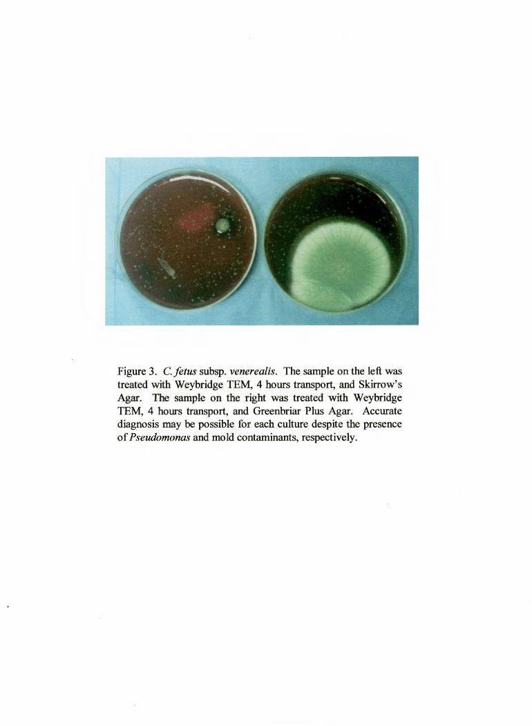
Figure 3. C. fetus subsp. venerealis. The sample on the left was treated with Weybridge TEM, 4 hours transport, and Skirrow's Agar. The sample on the right was treated with Weybridge TEM, 4 hours transport, and Greenbriar Plus Agar. Accurate diagnosis may be possible for each culture despite the presence of Pseudomonas and mold contaminants, respectively.
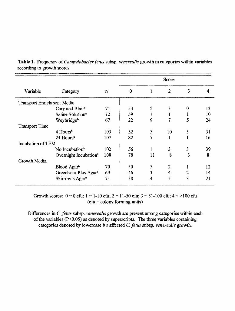
Table I. Frequency of Campylobacter fetus subsp. venerealis growth in categories within variables according to growth scores.
Score
Variable Category n 0 1 2 3
Transport Enrichment Media Cary and Blair' 71 53 2 3 0 Saline Solutiona 72 59 1 1 1 Weybridgeb 67 22 9 7 5
Transport Time 4 Hoursb 103 52 5 10 5 24 Hoursa 107 82 7 1 1
Incubation ofTEM No Incubationb 102 56 1 3 3 Overnight Incubationa 108 78 11 8 3
Growth Media Blood Agar 70 50 5 2 1 Greenbriar Plus Agar 69 46 3 4 2 Skirrow' s Agar 71 38 4 5 3
Growth scores: 0 = 0 cfu; 1 1-10 cfu; 2 = 11-50 cfu; 3 51-100 cfu; 4 > 100 cfu (cfu =colony forming units)
Differences in C. fetus subsp. venerealis growth are present among categories within each of the variables (P<0.05) as denoted by superscripts. The three variables containing
categories denoted by lowercase b's affected C. fetus subsp. venerealis growth.
4
13 10 24
31 16
39 8
12 14 21
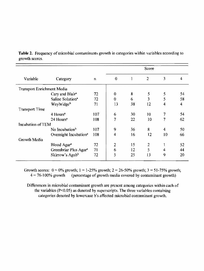
Table 2. Frequency of microbial contaminants growth in categories within variables according to growth scores.
Score
Variable Category n 0 1 2 3 4
Transport Enrichment Media Cary and Blair 72 0 8 5 5 54 Saline Solutiona 72 0 6 3 5 58 Weybridgeb 71 13 38 12 4 4
Transport Time 4 Hoursa 107 6 30 10 7 54 24 Hoursa 108 7 22 10 7 62
Incubation ofTEM No Incubationb 107 9 36 8 4 50 Overnight Incubationa 108 4 16 12 10 66
Growth Media BloodAg~ 72 2 15 2 1 52 Greenbriar Plus Agar 71 6 12 5 4 44 Skirrow's Agabb 72 5 25 13 9 20
Growth scores: 0 = 0% growth; 1 1-25% growth; 2 = 26-50% growth; 3 = 51-75% growth; 4 = 76-1 00% growth (percentage of growth media covered by contaminant growth)
Differences in microbial contaminant growth are present among categories within each of the variables (P<0.05) as denoted by superscripts. The three variables containing
categories denoted by lowercase b's affected microbial contaminant growth.
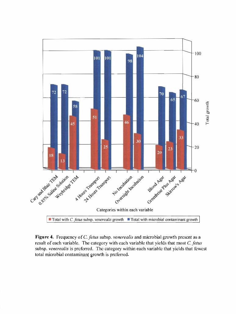
Categories within each variable
I• Total with C. fetus subsp. venerealis growth • Total with microbial con tam in ant growth I
Figure 4. Frequency of C. fetus subsp. venerealis and microbial growth present as a result of each variable. The category with each variable that yields that most C. f etus subsp. venerealis is preferred. The category within each variable that yields that fewest total microbial contaminant growth is preferred.
0.85% saline solution (P<O.OOOl). Transport time of 4 hours rather than 24 hours are superior
(P<O.OOOI). Benefits are associated with avoiding overnight TEM incubation (P=0.0002).
Significant differences are not identified for growth media; however, Skirrow's Agar yields
slightly better growth than either blood agar or Greenbriar Plus Agar.
The Mann-Whitney U test and Kruskal-Wallis test indicate that contaminant growth is
also significantly influenced by three of the four variables. Differences associated with TEM
indicate Weybridge TEM inhibits contaminant growth more effectively than either Cary and
Blair TEM or 0.85% saline solution (P<O.OOOI). Transport times of 4 and 24 hours do not
significantly influence contaminant growth. Abstaining from overnight incubation of TEM is
preferred for reduction of contaminant growth (P=0.0032). Skirrow's Agar is preferred to both
blood agar and Greenbriar Plus Agar (P<O.OOOI).
Discussion
These results suggest that the diagnosis of campylobacteriosis is enhanced when preputial
smegma samples arrive at the diagnostic laboratory within 4 hours of collection using Weybridge
TEM and when direct inoculation of Skirrow's Agar occurs the day samples arrive. Adherence
to these guidelines will facilitate accurate diagnosis of bovine genital campylobacteriosis in
bulls, thereby reducing the potential for seminal transmission of C. fetus subsp. venerealis and
subsequent occurrence of infertility, early embryonic death, and abortion.
Acknowledgements
The authors appreciate the financial support provided by COBA-Kellogg Honors
Research Project Grant and Select Sires, Inc. in Plain City, Ohio. Donald R. Monke, DVM of
Select Sires' Veterinary Department generously provided assistance with sample collection and
project guidance. The contributions of Anne Parkinson, Mary Beth Weisner, and Troy Farrell of
the Ohio Department of Agriculture- Animal Disease Diagnostic Laboratory's bacteriology
section are gratefully appreciated.
Sources and manufacturers
a. Becton Dickinson and Company, Cockeysville, MD.
b. Central Animal Health Laboratory, Madison, WI.
c. Difco Laboratories, Inc., Detroit, MI.
d. Greenbriar Veterinary Services, Inc., Delaware, OH.
e. RemeL Inc., Lenexa, KS.
£ Select Sires, Inc., Plain City, OH.
References
1. Acha P, and Szyfres B: 1980, Zoonoses and communicable diseases to man and animals.
pp. 46-47,49. Pan American Health Organization, Washington, D.C.
2. Bruner DW, Gillespie lli: 1973, Hagan's infectious diseases of domestic animals. pp. 125-
130. Cornell University Press, Ithaca, NY.
3. Clark BL, Dufty lli, Monsbourgh MJ: 1972, A method for maintaining the viability of
Vibrio fetus var. venerealis in samples of preputial secretions collected from carrier bulls.
Austr Vet J 48:462-464.
4. Clark BL, and Dufty lli: 1978, Isolation ofCampylobacter fetus from bulls. Austr Vet J
54:262-263.
5. Dekeyser PJ: 1986, Bovine genital campylobacteriosis. In: Current therapy in
theriogenology. Morrow DA, pp. 263-266. W. B. Saunders Company, Philadelphia, P A.
6. Difco Laboratories: 1984. Campylobacter media. In: Difco Manual- Dehydrated culture
media and reagents for microbiology. Difco Manual 1 Otb ed., pp. 192-196. Difco
Laboratories, Detroit MI.
7. Hoerlein AB: 1986, Vibriosis. In: Current veterinary therapy. Howard JL, pp. 596-598.
W. B. Saunders Company, Philadelphia, P A.
8. HumS, Brunner J, Mcinnes A, et al: 1994. Evaluation of cultural methods and selective
media for the isolation ofCampylobacter fetus susp. venerealis from cattle. Austr Vet J 71:
184-186.
9. Lander KP: 1990, The development of a transport and enrichment medium for
Campylobacter fetus. Br Vet J 146: 327-333.
10. Lander KP: 1990, The application of a transport enrichment medium to the diagnosis of
Campylobacter fetus infection in bulls. Br Vet J 146: 334-340.
11. Monke DR: 1998, Risk analysis: CSS testing protocol for trichomoniasis. Proc 17th Tech
Conf Artificial Insemination & Reprod. pp. 37-42. National Association of Animal
Breeders, Columbia, MO.
12. Office International Des Epizooties: 1999, Bovine genital campylobacteriosis. In:
International animal health code. Office International Des Epizooties, 8th ed., pp. 157-158.
Office International Des Epizooties, Paris, France.
13. Office International Des Epizooties: 1999, Bovine semen. In: International animal health
code. Office International Des Epizooties, 8th ed., pp. 297-299. Office International Des
Epizooties, Paris, France.