Embed Size (px)
Citation preview
International Journal of
Molecular Sciences
Article
Effect of Unloading Condition on the Healing Processand Effectiveness of Platelet Rich Plasma as aCountermeasure: Study on In Vivo and In VitroWound Healing Models
Francesca Cialdai 1,†, Alessandra Colciago 2,†, Desiré Pantalone 3, Angela Maria Rizzo 2 ,Stefania Zava 2, Lucia Morbidelli 4 , Fabio Celotti 2 , Daniele Bani 5 and Monica Monici 1,*
1 ASA campus Joint Laboratory, ASA Res. Div., Department of Experimental and Clinical Biomedical Sciences“Mario Serio”, University of Florence, 50139 Florence, Italy; [email protected]
2 Department of Pharmacological and Biomolecular Sciences, University of Milan, 20133 Milan, Italy;[email protected] (A.C.); [email protected] (A.M.R.); [email protected] (S.Z.);[email protected] (F.C.)
3 Unit of Surgery and Trauma Care, Department of Clinical and Experimental Medicine, University ofFlorence, 50134 Florence, Italy; [email protected]
4 Department of Life Sciences, University of Siena, 53100 Siena, Italy; [email protected] Research Unit of Histology & Embryology, Department of Experimental and Clinical Medicine, University of
Florence, 50139 Florence, Italy; [email protected]* Correspondence: [email protected]; Tel.: +39-055-275-8366† These authors contributed equally to this work.
Received: 30 October 2019; Accepted: 23 December 2019; Published: 9 January 2020�����������������
Abstract: Wound healing is a very complex process that allows organisms to survive injuries. It isstrictly regulated by a number of biochemical and physical factors, mechanical forces included.Studying wound healing in space is interesting for two main reasons: (i) defining tools, procedures,and protocols to manage serious wounds and burns eventually occurring in future long-lasting spaceexploration missions, without the possibility of timely medical evacuation to Earth; (ii) understandingthe role of gravity and mechanical factors in the healing process and scarring, thus contributing tounravelling the mechanisms underlying the switching between perfect regeneration and imperfectrepair with scarring. In the study presented here, a new in vivo sutured wound healing model inthe leech (Hirudo medicinalis) has been used to evaluate the effect of unloading conditions on thehealing process and the effectiveness of platelet rich plasma (PRP) as a countermeasure. The resultsreveal that microgravity caused a healing delay and structural alterations in the repair tissue, whichwere prevented by PRP treatment. Moreover, investigating the effects of microgravity and PRP on anin vitro wound healing model, it was found that PRP is able to counteract the microgravity-inducedimpairment in fibroblast migration to the wound site. This could be one of the mechanisms underlyingthe effectiveness of PRP in preventing healing impairment in unloading conditions.
Keywords: wound healing; Hirudo medicinalis; platelet rich plasma; microgravity
1. Introduction
Wound healing is the process that makes organisms resilient to injuries, allowing survival. Beinga process of fundamental importance for life, it has been conserved throughout evolution. Woundhealing is classically divided into three phases: inflammation, proliferation, and remodeling. Indeed,it consists of a very complex series of events and mechanisms, which partially overlap both spatiallyand temporally. After an injury, the blood clotting process starts immediately. Clot formation
Int. J. Mol. Sci. 2020, 21, 407; doi:10.3390/ijms21020407 www.mdpi.com/journal/ijms

Int. J. Mol. Sci. 2020, 21, 407 2 of 26
requires interactions among endothelial cells, platelets, and coagulation factors. Trapped cellsand platelets within the clot trigger an inflammatory response by the release of vasodilators andchemoattractants and activation of the complement cascade [1]. The recruited neutrophils andmacrophages produce pro-inflammatory cytokines and growth factors, resulting in migration andactivation of (myo)fibroblasts, endothelial, and epithelial cells in the wound site, which are responsiblefor extracellular matrix deposition, neoangiogenesis, and re-epithelialization, respectively. Clot anddebris are then removed by macrophages and extracellular hydrolases—matrix metalloproteases(MMPs), elastase, and plasmin protease—and the repair proceeds with wound contraction, extracellularmatrix (ECM) secretion, and remodeling due to myofibroblasts and secreted metalloproteases.
Many factors can impair wound healing. A crucial factor is tissue hypoxia, which may be causedby primary vascular diseases, metabolic diseases such as diabetes, local and systemic infections,malnutrition, and persistent local pressure. In skin ulcers, the persistence of the inflammatory phasecan lead to high protease activity, with degradation of growth factors and other molecular stimuliinvolved in tissue repair. Growth factors may be trapped by extracellular matrix molecules or degradedby proteases. Imbalance between extracellular hydrolases and their inhibitors results in abnormalECM degradation [1,2]. Other alterations of the repair mechanisms, such as the persistence of stromalactivation, can cause fibrotic scars and keloids. Moreover, pathogenic microorganisms can infect thewound (contaminated wounds and surgical site infections), stimulating a further immune response,inflammation, and tissue damage, as well as slowing the healing process and promoting abnormaltissue remodeling.
Skin wounds and compromised wound healing are major public health concerns. Complex andlengthy treatments cause an increasing burden on healthcare expenses. Even in uncomplicated cases,burns, chronic, and complicated wounds can require surgery and extended hospitalization periods.In the United States (US) alone, millions of patients need treatments for chronic wounds and anestimated US$ 50 billion is spent annually (data published by ‘Wound Care Awareness Week-2017’,including direct and indirect costs). More worryingly, the burden is growing year by year mainlyowing to the increasing prevalence of obesity and diabetes. This problem is of similar importance inEurope; in the United Kingdom (UK), about 200,000 patients have a chronic wound [3], and in theEuropean Community, the prevalence is expected to be over 1.5 million people [4,5].
Various medical approaches and therapeutic interventions can affect the different processesinvolved in wound healing. Topical application of growth factors and protease inhibitors, incisionpriming with platelet derived growth factor (PDGF) or interleukin-1 (IL-1), electric and magneticfield stimulation, exposure to laser radiation, negative pressure therapy, use of prosthetic materials,and gene and stem cell therapies have all been exploited to improve healing [6–11]. When a sutureis needed, proper suture techniques and materials can strongly affect healing time and scar quality.In fact, the suture facilitates wound closure by obliteration of dead space, distribution of tension alongdeep suture lines, and maintenance of tensile strength in the injured tissue.
Among the therapeutic interventions, the application of platelet rich plasma (PRP) is one of themost widely used. PRP is a mixture of growth factors and cytokines, released by platelet alpha-granulesdegranulation. It is obtained from total blood by centrifugation and filtering; the fraction enrichedin platelets is then activated using batroxobine or thrombin, thus degranulating [12]. PRP use is asimple and cheap method to promote tissue regeneration using autologous growth factors. Whena tissue is damaged, the rapid initiation of the repairing phase is crucial to assure regeneration;the release of growth factors and cytokines stored in platelet alpha-granules, occurring within a fewminutes of the damaging insult, represents a rapid support to tissue healing. For regeneration tooccur, migration, proliferation, and differentiation of various cell types such as fibroblasts, endothelialcells, and mesenchymal stem cells are needed [12,13]. In particular, PRP efficacy in tissue regenerationis mainly thanks to its effects on the migration, proliferation, and biosynthetic activity of dermalfibroblasts, thus promoting their differentiation into myofibroblasts [14].

Int. J. Mol. Sci. 2020, 21, 407 3 of 26
PRP has been used in different clinical conditions since the 1990s, starting from its first use asgrafting for dental implants [15], and following with other more recent applications in diabetic ulcers,burns, and tissue recovery after surgery [12,16,17]. The interest in PRP’s clinical uses is still increasing,as demonstrated by a number of registered clinical trials where PRP is tested alone or in combinationwith scaffold materials or stem cells [18]. Nonetheless, the molecular basis underlying the use of PRPhas not yet been fully elucidated. The management of serious wounds—for example, those resultingfrom trauma or emergency surgery—during space missions is a very challenging issue. Critical factorsare, on one side, the limited availability of diagnostic and therapeutic tools, as well as the lack of aspecialized medical staff on board space vehicles; and on the other side, the microgravity (µg)-inducedalterations of human physiology, which can affect patient conditions and evolution of the healingprocess. Moreover, the microbial load and microorganism behavior onboard space vehicles should beconsidered to prevent/minimize the risk of wound infection and, in case an infection occurs, to haveadequate countermeasures.
In current missions, the management of traumatic and surgical emergencies consists of patientstabilization and rapid return to Earth. In future interplanetary missions, timely medical evacuationto Earth would not be feasible and the guide of crew actions remotely would be useless because of thecommunication lag. Therefore, studies on the behavior and healing of sutured/non-sutured wounds inspace environment are needed to understand possible problems and define adequate countermeasures [19].The literature on wound healing in weightlessness is relatively poor. Studies on animal models inunloading conditions have given controversial results and no definite conclusions [20–25].
In vitro studies on immune cells, fibroblasts, endothelial, and epithelial cells cultured both inreal and modeled µg conditions show alterations of functions involved in wound healing, such asphagocytosis, adhesion/migration, apoptosis, proliferation, intercellular cross-talking, production ofinflammatory mediators, ECM molecules, growth factors, and so on [26–32].
In astronauts, deficient immune function, signs of chronic inflammation, metabolic alterations, andskin atrophy have been observed [33–35], and could affect the efficiency of tissue repair mechanisms.
In order to expand the knowledge on the behavior and healing of wounds and sutures in spaceenvironment, suitable models are needed to perform experiments onboard space vehicles as well as inmodeled µg and hypergravity conditions on ground.
Recently, we developed an in vivo model of sutured and not sutured wound healing inweightlessness based on the use of leeches (Hirudo medicinalis), which is an adaptation of a widelyaccepted model for the study of basic tissue repair events in normal gravity (1× g) [36]. The leech isconsidered a reliable model because, despite being an invertebrate, it displays a wound healing processcharacterized by the same sequence of events (i.e., fibroplasia, angiogenesis, and remodeling) observedin vertebrates in response to injury. Indeed, the process involves the same cellular mechanisms and alsothe same types of molecules to regulate cell behavior in the different phases of healing [37]. Moreover,the leech is a particularly suitable model for experiments in space flight and in µg-modeling facilitiesowing to its small size and very long resistance to fasting [38].
The present paper describes the results of a study on the healing of sutured wounds inleeches exposed to weightlessness, modeled by a random positioning machine (RPM). Because,in weightlessness, the healing resulted delayed, the effectiveness of platelet rich plasma (PRP) inpromoting repair mechanisms in µg was tested. Finally, PRP treatment was applied to an in vitrowound healing model, consisting of a scratch assay on fibroblast monolayers exposed or not to modeledµg. The effect obtained in vitro was analyzed and compared to that observed in vivo, with the aim toshed some light on the molecular and cellular mechanisms underlying PRP action in µg conditions.
Int. J. Mol. Sci. 2020, 21, 407 4 of 26
2. Results
2.1. Effect of Modeled µg on In Vivo Model of Sutured Wound Healing
2.1.1. Histological Analysis
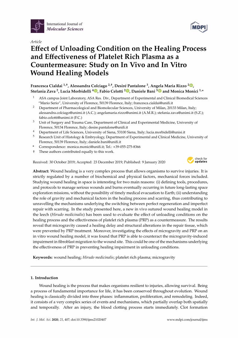
To evaluate the effect of µg on the healing progress, the morphological features at the wound sitewere analyzed by histological techniques. In hematoxylin–eosin stained cross sections from animalsexposed to modeled µg, light microscopical analysis of the tissues at the wound site (dorsal skin)showed a delayed healing in comparison with 1× g controls. At any time of exposure to µg, that is,48 h, 96 h (Figure 1), and 144 h (Figure 4), the wounds appeared only partially covered by a looseepithelium characterized by poorly adherent cells, while control samples showed signs of a moreorganized re-epithelialization. The healing impairment was particularly evident after 144 h exposure;as shown in Figure 4, despite the considerable time elapsed from surgery, the dorsal muscle tissue wasstill adjacent to the wound margins, likely owing to the very scarce de novo formation of reparativeconnective tissue in the leeches longer exposed to µg.
2.1.2. Analysis of Collagen Fibres
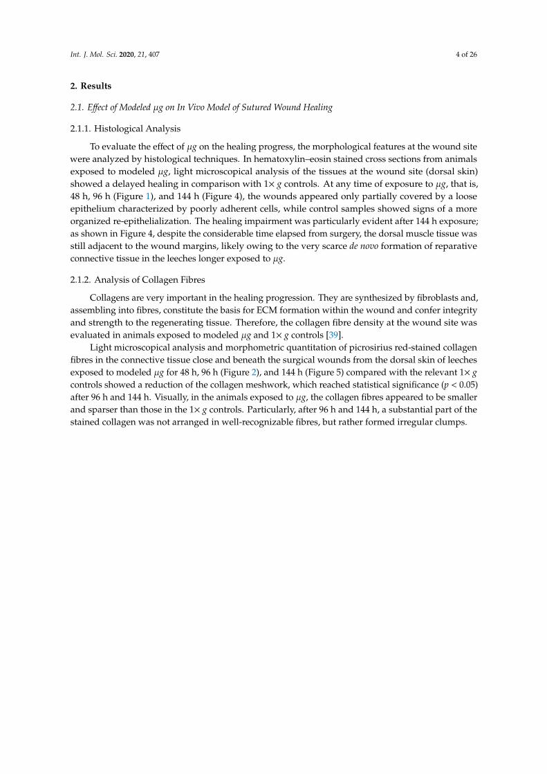
Collagens are very important in the healing progression. They are synthesized by fibroblasts and,assembling into fibres, constitute the basis for ECM formation within the wound and confer integrityand strength to the regenerating tissue. Therefore, the collagen fibre density at the wound site wasevaluated in animals exposed to modeled µg and 1× g controls [39].
Light microscopical analysis and morphometric quantitation of picrosirius red-stained collagenfibres in the connective tissue close and beneath the surgical wounds from the dorsal skin of leechesexposed to modeled µg for 48 h, 96 h (Figure 2), and 144 h (Figure 5) compared with the relevant 1× gcontrols showed a reduction of the collagen meshwork, which reached statistical significance (p < 0.05)after 96 h and 144 h. Visually, in the animals exposed to µg, the collagen fibres appeared to be smallerand sparser than those in the 1× g controls. Particularly, after 96 h and 144 h, a substantial part of thestained collagen was not arranged in well-recognizable fibres, but rather formed irregular clumps.
Int. J. Mol. Sci. 2020, 21, 407 5 of 26Int. J. Mol. Sci. 2020, 21, x FOR PEER REVIEW 5 of 26
Figure 1. Effect of modeled μg on in vivo model of sutured wound healing (Hirudo medicinalis):
state of the wound after 48 h and 96 h exposure. Hematoxylin–eosin stained cross sections. The
panels show the state of the wounds on the back of the leeches (arrows) in the 1× g controls and
in the animals exposed to modeled μg for 48 h or 96 h. Letterings are as follows: e, epidermis; d,
dermis; mf, muscle fibers; ne, neo-epidermis; gt, granulation tissue. Calibration bars are in μm,
as indicated.
2.1.2. Analysis of Collagen Fibres
Collagens are very important in the healing progression. They are synthesized by
fibroblasts and, assembling into fibres, constitute the basis for ECM formation within the wound
and confer integrity and strength to the regenerating tissue. Therefore, the collagen fibre density
at the wound site was evaluated in animals exposed to modeled μg and 1× g controls [39].
Light microscopical analysis and morphometric quantitation of picrosirius red-stained
collagen fibres in the connective tissue close and beneath the surgical wounds from the dorsal
Figure 1. Effect of modeled µg on in vivo model of sutured wound healing (Hirudo medicinalis): state ofthe wound after 48 h and 96 h exposure. Hematoxylin–eosin stained cross sections. The panels showthe state of the wounds on the back of the leeches (arrows) in the 1× g controls and in the animalsexposed to modeled µg for 48 h or 96 h. Letterings are as follows: e, epidermis; d, dermis; mf, musclefibers; ne, neo-epidermis; gt, granulation tissue. Calibration bars are in µm, as indicated.
Int. J. Mol. Sci. 2020, 21, 407 6 of 26
Int. J. Mol. Sci. 2020, 21, x FOR PEER REVIEW 6 of 26
skin of leeches exposed to modeled μg for 48 h, 96 h (Figure 2), and 144 h (Figure 5) compared
with the relevant 1× g controls showed a reduction of the collagen meshwork, which reached
statistical significance (p < 0.05) after 96 h and 144 h. Visually, in the animals exposed to μg, the
collagen fibres appeared to be smaller and sparser than those in the 1× g controls. Particularly,
after 96 h and 144 h, a substantial part of the stained collagen was not arranged in well-
recognizable fibres, but rather formed irregular clumps.
Figure 2. Effect of modeled μg on in vivo model of sutured wound healing (Hirudo medicinalis):
collagen fibres content in the connective tissue at the wound site. Light microscopical features
and morphometric quantitation of picrosirius red-stained collagen fibres at the wound site after
48 h and 96 h exposure to modeled μg. C = 1× g controls; * p < 0.05 vs. C-96 h (n = 3); n.s. = not
significant.
2.1.3. Analysis of Elastic Fibres
Elastin fibre density was analyzed in the wounded tissues of leeches exposed and not
exposed to μg because elastin has an important structural role in the skin, imparting recoil and
resistance. Elastin signaling in tissue repair is still underexplored, but it fits with the hallmarks
of fetal scarless wound healing. In wounds of adult organisms, the appearance of elastin is
delayed and the elastic fibre network is disorganized [40]. The effect of μg on elastin fibre
formation is unknown.
Light microscopical analysis and morphometric quantitation of paraldehyde fuchsin-
stained elastic fibres in the connective tissue close and beneath the surgical wounds from the
dorsal skin of leeches exposed to modeled μg for 48 h, 96 h (Figure 3), and 144 h (Figure 6)
showed no significant differences in the elastic meshwork compared with the relevant 1× g
controls. Visually, in the animals subjected to μg, the elastic fibres appeared slightly smaller and
Figure 2. Effect of modeled µg on in vivo model of sutured wound healing (Hirudo medicinalis): collagenfibres content in the connective tissue at the wound site. Light microscopical features and morphometricquantitation of picrosirius red-stained collagen fibres at the wound site after 48 h and 96 h exposure tomodeled µg. C = 1× g controls; * p < 0.05 vs. C-96 h (n = 3); n.s. = not significant.
2.1.3. Analysis of Elastic Fibres
Elastin fibre density was analyzed in the wounded tissues of leeches exposed and not exposedto µg because elastin has an important structural role in the skin, imparting recoil and resistance.Elastin signaling in tissue repair is still underexplored, but it fits with the hallmarks of fetal scarlesswound healing. In wounds of adult organisms, the appearance of elastin is delayed and the elasticfibre network is disorganized [40]. The effect of µg on elastin fibre formation is unknown.
Light microscopical analysis and morphometric quantitation of paraldehyde fuchsin-stainedelastic fibres in the connective tissue close and beneath the surgical wounds from the dorsal skin ofleeches exposed to modeled µg for 48 h, 96 h (Figure 3), and 144 h (Figure 6) showed no significantdifferences in the elastic meshwork compared with the relevant 1× g controls. Visually, in the animalssubjected to µg, the elastic fibres appeared slightly smaller and sparser than those in the 1× g controls,but the measured differences did not reach statistical significance. Both in 1× g controls and in theanimals exposed to µg conditions, the content in elastic fibres significantly increased with time (to notethat the wounds were inflicted immediately before the exposure to µg; therefore, the exposure timecoincided with the time elapsed from the wounding).

Int. J. Mol. Sci. 2020, 21, 407 7 of 26
Int. J. Mol. Sci. 2020, 21, x FOR PEER REVIEW 7 of 26
sparser than those in the 1× g controls, but the measured differences did not reach statistical
significance. Both in 1× g controls and in the animals exposed to μg conditions, the content in
elastic fibres significantly increased with time (to note that the wounds were inflicted
immediately before the exposure to μg; therefore, the exposure time coincided with the time
elapsed from the wounding).
Figure 3. Effect of modeled μg on in vivo model of sutured wound healing (Hirudo medicinalis):
elastic fibre content in the connective tissue at the wound site. Light microscopical features and
morphometric quantitation of paraldehyde fuchsin-stained elastic fibres at the wound site after
48 h and 96 h exposure to modeled μg (b,d), compared with 1× g controls (a,c). In the graph C =
1× g; bar = 20 μm; * p < 0.05 vs. C-48h, ++ p < 0.01 vs. μg-48 h, (n = 3); n.s. = not significant.
2.2. Effect of Modeled μg and PRP on In Vivo Model of Sutured Wound Healing
In this paper, PRP was evaluated as a possible countermeasure against the wound healing
impairment in unloading conditions. Therefore, the effects of PRP treatment on morphology,
Figure 3. Effect of modeled µg on in vivo model of sutured wound healing (Hirudo medicinalis): elasticfibre content in the connective tissue at the wound site. Light microscopical features and morphometricquantitation of paraldehyde fuchsin-stained elastic fibres at the wound site after 48 h and 96 h exposureto modeled µg (b,d), compared with 1× g controls (a,c). In the graph C = 1× g; bar = 20 µm; * p < 0.05vs. C-48h, ++ p < 0.01 vs. µg-48 h, (n = 3); n.s. = not significant.
2.2. Effect of Modeled µg and PRP on In Vivo Model of Sutured Wound Healing
In this paper, PRP was evaluated as a possible countermeasure against the wound healingimpairment in unloading conditions. Therefore, the effects of PRP treatment on morphology, collagen,and elastic fibre content at the wound site were analyzed in animals exposed/not exposed to µg.
2.2.1. Morphological Analysis
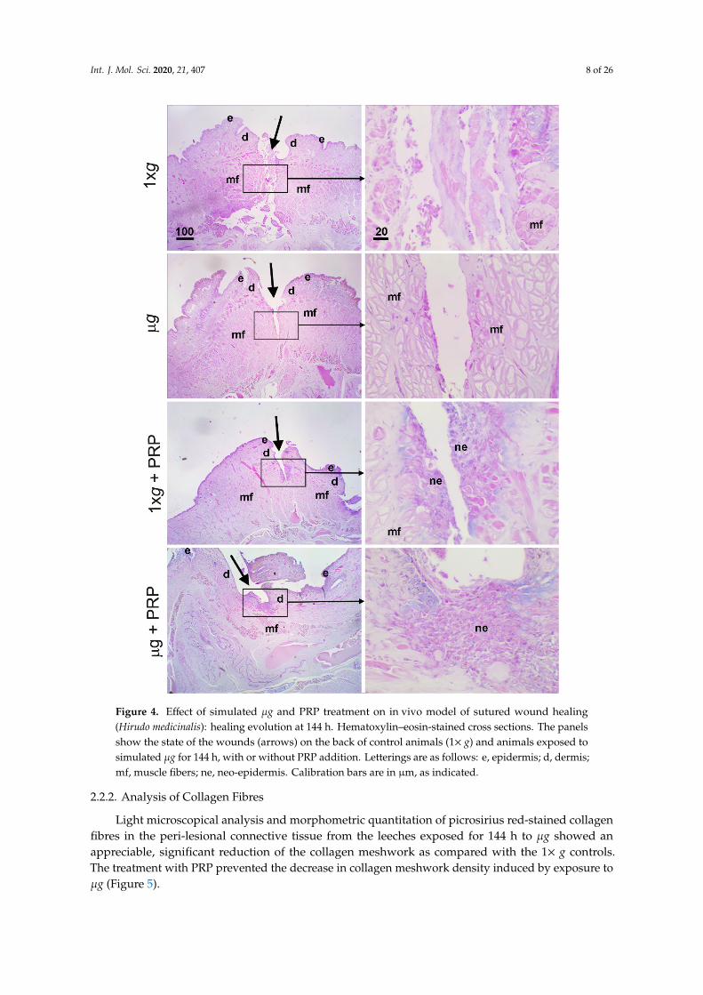
In both controls (1× g) and µg-exposed animals (144 h), PRP promoted wound healing bynarrowing the surgical wound and enhancing re-epithelialization (Figure 4); in all the PRP-treatedanimals, exposed or not to modeled µg, the dorsal lesion was covered by a thick regenerative epithelium.
Int. J. Mol. Sci. 2020, 21, 407 8 of 26
Int. J. Mol. Sci. 2020, 21, x FOR PEER REVIEW 8 of 26
collagen, and elastic fibre content at the wound site were analyzed in animals exposed/not
exposed to μg.
2.2.1. Morphological Analysis
In both controls (1× g) and μg-exposed animals (144 h), PRP promoted wound healing by
narrowing the surgical wound and enhancing re-epithelialization (Figure 4); in all the PRP-
treated animals, exposed or not to modeled μg, the dorsal lesion was covered by a thick
regenerative epithelium.
Figure 4. Effect of simulated μg and PRP treatment on in vivo model of sutured wound healing
(Hirudo medicinalis): healing evolution at 144 h. Hematoxylin–eosin-stained cross sections. The
panels show the state of the wounds (arrows) on the back of control animals (1× g) and animals
exposed to simulated μg for 144 h, with or without PRP addition. Letterings are as follows: e,
epidermis; d, dermis; mf, muscle fibers; ne, neo-epidermis. Calibration bars are in μm, as
indicated.
Figure 4. Effect of simulated µg and PRP treatment on in vivo model of sutured wound healing(Hirudo medicinalis): healing evolution at 144 h. Hematoxylin–eosin-stained cross sections. The panelsshow the state of the wounds (arrows) on the back of control animals (1× g) and animals exposed tosimulated µg for 144 h, with or without PRP addition. Letterings are as follows: e, epidermis; d, dermis;mf, muscle fibers; ne, neo-epidermis. Calibration bars are in µm, as indicated.
2.2.2. Analysis of Collagen Fibres
Light microscopical analysis and morphometric quantitation of picrosirius red-stained collagenfibres in the peri-lesional connective tissue from the leeches exposed for 144 h to µg showed anappreciable, significant reduction of the collagen meshwork as compared with the 1× g controls.The treatment with PRP prevented the decrease in collagen meshwork density induced by exposure toµg (Figure 5).

Int. J. Mol. Sci. 2020, 21, 407 9 of 26
Int. J. Mol. Sci. 2020, 21, x FOR PEER REVIEW 9 of 26
2.2.2. Analysis of Collagen Fibres
Light microscopical analysis and morphometric quantitation of picrosirius red-stained
collagen fibres in the peri-lesional connective tissue from the leeches exposed for 144 h to μg
showed an appreciable, significant reduction of the collagen meshwork as compared with the
1× g controls. The treatment with PRP prevented the decrease in collagen meshwork density
induced by exposure to μg (Figure 5).
Figure 5. Effect of simulated μg and PRP treatment on in vivo model of sutured wound healing
(Hirudo medicinalis): Collagen fibre content after 144 h. Light microscopical analysis and
morphometric quantitation of picrosirius red-stained collagen fibres in the connective tissue at
the wound site in control and μg-exposed animals, treated/untreated with PRP. C = 1× g; * p <
0.05 vs. all the other groups (n = 3); n.s. = not significant. To note the significantly higher collagen
fiber density in μg-exposed animals treated with PRP compared with the untreated.
2.2.3. Analysis of Elastic Fibres
Light microscopical analysis and morphometric quantitation of paraldehyde fuchsin-
stained elastic fibres in the connective tissue close to the surgical wounds on the back of leeches
exposed to μg for 144 h (Figure 6) showed no significant changes of the elastic meshwork
compared with the relevant controls (1× g). The treatment with PRP did not result in appreciable,
significant changes in both control and μg-exposed animals.
Figure 5. Effect of simulated µg and PRP treatment on in vivo model of sutured wound healing(Hirudo medicinalis): Collagen fibre content after 144 h. Light microscopical analysis and morphometricquantitation of picrosirius red-stained collagen fibres in the connective tissue at the wound site incontrol and µg-exposed animals, treated/untreated with PRP. C = 1× g; * p < 0.05 vs. all the other groups(n = 3); n.s. = not significant. To note the significantly higher collagen fiber density in µg-exposedanimals treated with PRP compared with the untreated.
2.2.3. Analysis of Elastic Fibres
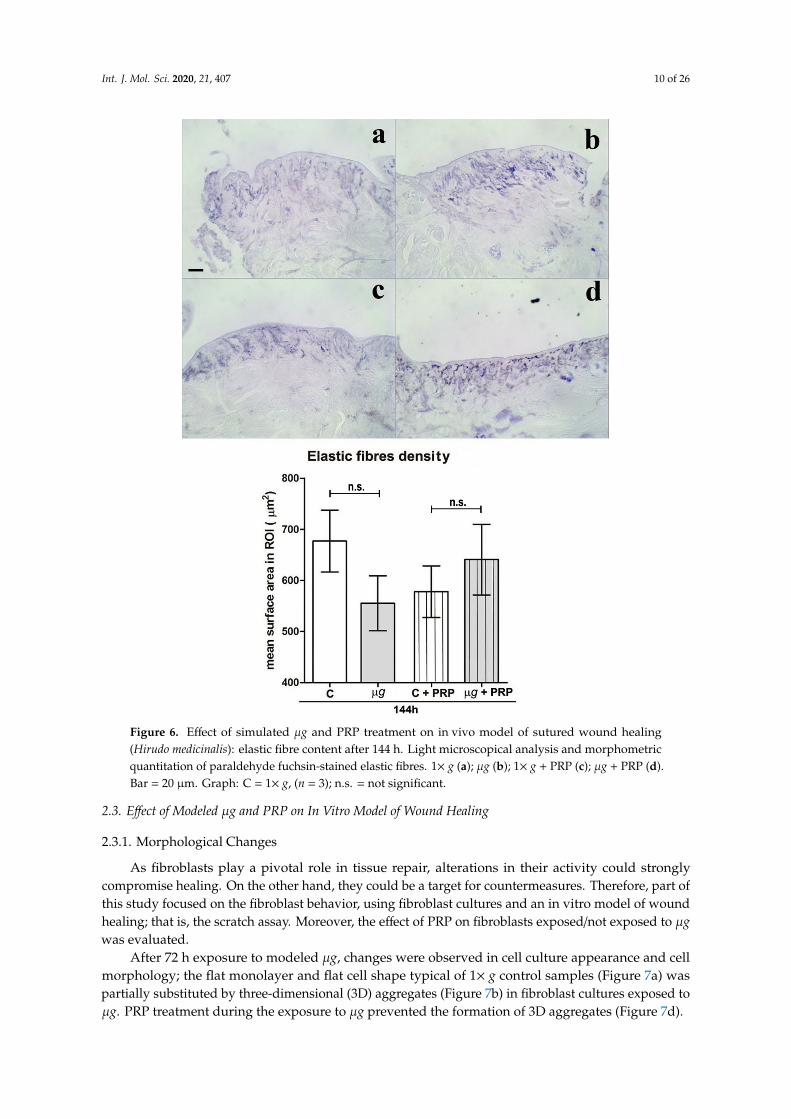
Light microscopical analysis and morphometric quantitation of paraldehyde fuchsin-stainedelastic fibres in the connective tissue close to the surgical wounds on the back of leeches exposed toµg for 144 h (Figure 6) showed no significant changes of the elastic meshwork compared with therelevant controls (1× g). The treatment with PRP did not result in appreciable, significant changes inboth control and µg-exposed animals.
Int. J. Mol. Sci. 2020, 21, 407 10 of 26Int. J. Mol. Sci. 2020, 21, x FOR PEER REVIEW 10 of 26
Figure 6. Effect of simulated μg and PRP treatment on in vivo model of sutured wound healing
(Hirudo medicinalis): elastic fibre content after 144 h. Light microscopical analysis and
morphometric quantitation of paraldehyde fuchsin-stained elastic fibres. 1× g (a); μg (b); 1× g +
PRP (c); μg + PRP (d). Bar = 20 μm. Graph: C = 1× g, (n = 3); n.s. = not significant.
2.3. Effect of Modeled μg and PRP on in Vitro Model of Wound Healing
2.3.1. Morphological Changes
As fibroblasts play a pivotal role in tissue repair, alterations in their activity could strongly
compromise healing. On the other hand, they could be a target for countermeasures. Therefore,
part of this study focused on the fibroblast behavior, using fibroblast cultures and an in vitro
model of wound healing; that is, the scratch assay. Moreover, the effect of PRP on fibroblasts
exposed/not exposed to μg was evaluated.
Figure 6. Effect of simulated µg and PRP treatment on in vivo model of sutured wound healing(Hirudo medicinalis): elastic fibre content after 144 h. Light microscopical analysis and morphometricquantitation of paraldehyde fuchsin-stained elastic fibres. 1× g (a); µg (b); 1× g + PRP (c); µg + PRP (d).Bar = 20 µm. Graph: C = 1× g, (n = 3); n.s. = not significant.
2.3. Effect of Modeled µg and PRP on In Vitro Model of Wound Healing
2.3.1. Morphological Changes
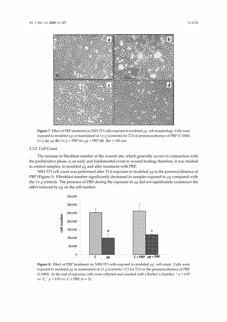
As fibroblasts play a pivotal role in tissue repair, alterations in their activity could stronglycompromise healing. On the other hand, they could be a target for countermeasures. Therefore, part ofthis study focused on the fibroblast behavior, using fibroblast cultures and an in vitro model of woundhealing; that is, the scratch assay. Moreover, the effect of PRP on fibroblasts exposed/not exposed to µgwas evaluated.
After 72 h exposure to modeled µg, changes were observed in cell culture appearance and cellmorphology; the flat monolayer and flat cell shape typical of 1× g control samples (Figure 7a) waspartially substituted by three-dimensional (3D) aggregates (Figure 7b) in fibroblast cultures exposed toµg. PRP treatment during the exposure to µg prevented the formation of 3D aggregates (Figure 7d).
Int. J. Mol. Sci. 2020, 21, 407 11 of 26
Int. J. Mol. Sci. 2020, 21, x FOR PEER REVIEW 11 of 26
After 72 h exposure to modeled μg, changes were observed in cell culture appearance and
cell morphology; the flat monolayer and flat cell shape typical of 1xg control samples (Figure
7a) was partially substituted by three-dimensional (3D) aggregates (Figure 7b) in fibroblast
cultures exposed to μg. PRP treatment during the exposure to μg prevented the formation of 3D
aggregates (Figure 7d).
Figure 7. Effect of PRP treatment on NIH-3T3 cells exposed to modeled μg: cell morphology.
Cells were exposed to modeled μg or maintained at 1× g (controls) for 72 h in presence/absence
of PRP (1:1000). 1× g (a); μg (b); 1× g + PRP (c); μg + PRP (d). Bar = 100 μm.
2.3.2. Cell Count
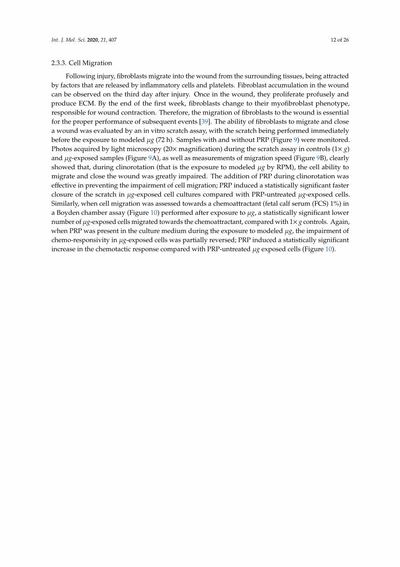
The increase in fibroblast number at the wound site, which generally occurs in conjunction
with the proliferative phase, is an early and fundamental event in wound healing; therefore, it
was studied in control samples, in modeled μg and after treatment with PRP.
NIH-3T3 cell count was performed after 72 h exposure to modeled μg in the
presence/absence of PRP (Figure 8). Fibroblast number significantly decreased in samples
exposed to μg compared with the 1× g controls. The presence of PRP during the exposure to μg
did not significantly counteract the effect induced by μg on the cell number.
Figure 7. Effect of PRP treatment on NIH-3T3 cells exposed to modeled µg: cell morphology. Cells wereexposed to modeled µg or maintained at 1× g (controls) for 72 h in presence/absence of PRP (1:1000).1× g (a); µg (b); 1× g + PRP (c); µg + PRP (d). Bar = 100 µm.
2.3.2. Cell Count
The increase in fibroblast number at the wound site, which generally occurs in conjunction withthe proliferative phase, is an early and fundamental event in wound healing; therefore, it was studiedin control samples, in modeled µg and after treatment with PRP.
NIH-3T3 cell count was performed after 72 h exposure to modeled µg in the presence/absence ofPRP (Figure 8). Fibroblast number significantly decreased in samples exposed to µg compared withthe 1× g controls. The presence of PRP during the exposure to µg did not significantly counteract theeffect induced by µg on the cell number.
Int. J. Mol. Sci. 2020, 21, x FOR PEER REVIEW 12 of 26
Figure 8. Effect of PRP treatment on NIH-3T3 cells exposed to modeled μg: cell count. Cells were
exposed to modeled μg or maintained at 1× g (controls = C) for 72 h in the presence/absence of
PRP (1:1000). At the end of exposure, cells were collected and counted with a Burker’s chamber.
* p < 0.05 vs. C, ^ p < 0.05 vs. C + PRP, (n = 3).
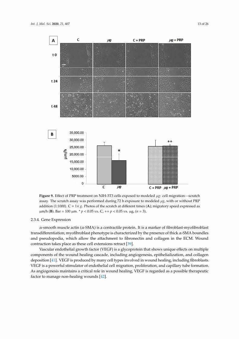
2.3.3. Cell Migration
Following injury, fibroblasts migrate into the wound from the surrounding tissues, being
attracted by factors that are released by inflammatory cells and platelets. Fibroblast
accumulation in the wound can be observed on the third day after injury. Once in the wound,
they proliferate profusely and produce ECM. By the end of the first week, fibroblasts change to
their myofibroblast phenotype, responsible for wound contraction. Therefore, the migration of
fibroblasts to the wound is essential for the proper performance of subsequent events [39]. The
ability of fibroblasts to migrate and close a wound was evaluated by an in vitro scratch assay,
with the scratch being performed immediately before the exposure to modeled μg (72 h).
Samples with and without PRP (Figure 9) were monitored. Photos acquired by light microscopy
(20× magnification) during the scratch assay in controls (1× g) and μg-exposed samples (Figure
9A), as well as measurements of migration speed (Figure 9B), clearly showed that, during
clinorotation (that is the exposure to modeled μg by RPM), the cell ability to migrate and close
the wound was greatly impaired. The addition of PRP during clinorotation was effective in
preventing the impairment of cell migration; PRP induced a statistically significant faster
closure of the scratch in μg-exposed cell cultures compared with PRP-untreated μg-exposed
cells. Similarly, when cell migration was assessed towards a chemoattractant (fetal calf serum
(FCS) 1%) in a Boyden chamber assay (Figure 10) performed after exposure to μg, a statistically
significant lower number of μg-exposed cells migrated towards the chemoattractant, compared
with 1× g controls. Again, when PRP was present in the culture medium during the exposure to
modeled μg, the impairment of chemo-responsivity in μg-exposed cells was partially reversed;
PRP induced a statistically significant increase in the chemotactic response compared with PRP-
untreated μg exposed cells (Figure 10).
Figure 8. Effect of PRP treatment on NIH-3T3 cells exposed to modeled µg: cell count. Cells wereexposed to modeled µg or maintained at 1× g (controls = C) for 72 h in the presence/absence of PRP(1:1000). At the end of exposure, cells were collected and counted with a Burker’s chamber. * p < 0.05vs. C, ˆ p < 0.05 vs. C + PRP, (n = 3).
Int. J. Mol. Sci. 2020, 21, 407 12 of 26
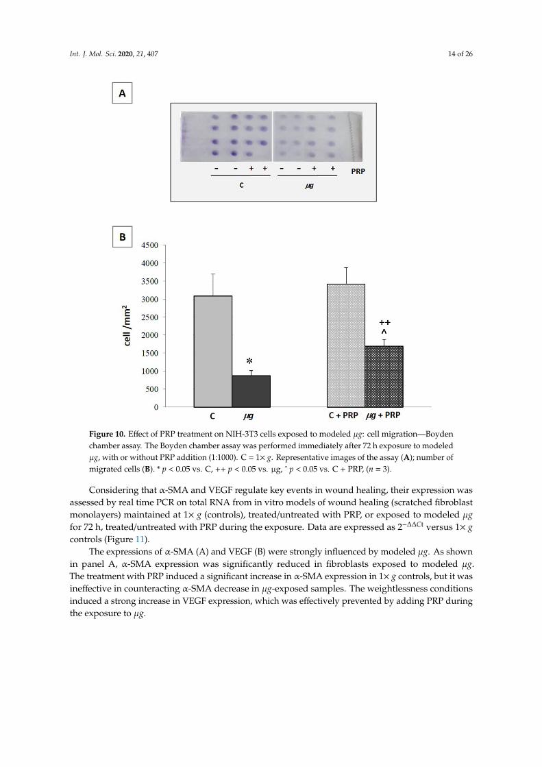
2.3.3. Cell Migration
Following injury, fibroblasts migrate into the wound from the surrounding tissues, being attractedby factors that are released by inflammatory cells and platelets. Fibroblast accumulation in the woundcan be observed on the third day after injury. Once in the wound, they proliferate profusely andproduce ECM. By the end of the first week, fibroblasts change to their myofibroblast phenotype,responsible for wound contraction. Therefore, the migration of fibroblasts to the wound is essentialfor the proper performance of subsequent events [39]. The ability of fibroblasts to migrate and closea wound was evaluated by an in vitro scratch assay, with the scratch being performed immediatelybefore the exposure to modeled µg (72 h). Samples with and without PRP (Figure 9) were monitored.Photos acquired by light microscopy (20×magnification) during the scratch assay in controls (1× g)and µg-exposed samples (Figure 9A), as well as measurements of migration speed (Figure 9B), clearlyshowed that, during clinorotation (that is the exposure to modeled µg by RPM), the cell ability tomigrate and close the wound was greatly impaired. The addition of PRP during clinorotation waseffective in preventing the impairment of cell migration; PRP induced a statistically significant fasterclosure of the scratch in µg-exposed cell cultures compared with PRP-untreated µg-exposed cells.Similarly, when cell migration was assessed towards a chemoattractant (fetal calf serum (FCS) 1%) ina Boyden chamber assay (Figure 10) performed after exposure to µg, a statistically significant lowernumber of µg-exposed cells migrated towards the chemoattractant, compared with 1× g controls. Again,when PRP was present in the culture medium during the exposure to modeled µg, the impairment ofchemo-responsivity in µg-exposed cells was partially reversed; PRP induced a statistically significantincrease in the chemotactic response compared with PRP-untreated µg exposed cells (Figure 10).
Int. J. Mol. Sci. 2020, 21, 407 13 of 26Int. J. Mol. Sci. 2020, 21, x FOR PEER REVIEW 13 of 26
Figure 9. Effect of PRP treatment on NIH-3T3 cells exposed to modeled μg: cell migration—
scratch assay. The scratch assay was performed during 72 h exposure to modeled μg, with or
without PRP addition (1:1000). C = 1× g. Photos of the scratch at different times (A); migratory
speed expressed as μm/h (B). Bar = 100 μm. * p < 0.05 vs. C, ++ p < 0.05 vs. μg, (n = 3).
Figure 9. Effect of PRP treatment on NIH-3T3 cells exposed to modeled µg: cell migration—scratchassay. The scratch assay was performed during 72 h exposure to modeled µg, with or without PRPaddition (1:1000). C = 1× g. Photos of the scratch at different times (A); migratory speed expressed asµm/h (B). Bar = 100 µm. * p < 0.05 vs. C, ++ p < 0.05 vs. µg, (n = 3).
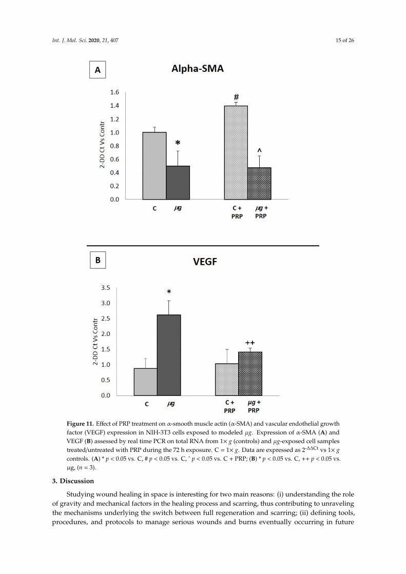
2.3.4. Gene Expression
α-smooth muscle actin (α-SMA) is a contractile protein. It is a marker of fibroblast-myofibroblasttransdifferentiation; myofibroblast phenotype is characterized by the presence of thick α-SMA boundlesand pseudopodia, which allow the attachment to fibronectin and collagen in the ECM. Woundcontraction takes place as these cell extensions retract [39].
Vascular endothelial growth factor (VEGF) is a glycoprotein that shows unique effects on multiplecomponents of the wound healing cascade, including angiogenesis, epithelialization, and collagendeposition [41]. VEGF is produced by many cell types involved in wound healing, including fibroblasts.VEGF is a powerful stimulator of endothelial cell migration, proliferation, and capillary tube formation.As angiogenesis maintains a critical role in wound healing, VEGF is regarded as a possible therapeuticfactor to manage non-healing wounds [42].
Int. J. Mol. Sci. 2020, 21, 407 14 of 26Int. J. Mol. Sci. 2020, 21, x FOR PEER REVIEW 14 of 26
Figure 10. Effect of PRP treatment on NIH-3T3 cells exposed to modeled μg: cell migration—
Boyden chamber assay. The Boyden chamber assay was performed immediately after 72 h
exposure to modeled μg, with or without PRP addition (1:1000). C = 1× g. Representative images
of the assay (A); number of migrated cells (B). * p < 0.05 vs. C, ++ p < 0.05 vs. μg, ^ p < 0.05 vs. C
+ PRP, (n = 3).
2.3.4. Gene Expression
α-smooth muscle actin (α-SMA) is a contractile protein. It is a marker of fibroblast-
myofibroblast transdifferentiation; myofibroblast phenotype is characterized by the presence of
thick α-SMA boundles and pseudopodia, which allow the attachment to fibronectin and
collagen in the ECM. Wound contraction takes place as these cell extensions retract [39].
Vascular endothelial growth factor (VEGF) is a glycoprotein that shows unique effects on
multiple components of the wound healing cascade, including angiogenesis, epithelialization,
and collagen deposition [41]. VEGF is produced by many cell types involved in wound healing,
including fibroblasts. VEGF is a powerful stimulator of endothelial cell migration, proliferation,
and capillary tube formation. As angiogenesis maintains a critical role in wound healing, VEGF is regarded as a possible therapeutic factor to manage non-healing wounds [42].
Considering that α-SMA and VEGF regulate key events in wound healing, their expression
was assessed by real time PCR on total RNA from in vitro models of wound healing (scratched
fibroblast monolayers) maintained at 1× g (controls), treated/untreated with PRP, or exposed to
Figure 10. Effect of PRP treatment on NIH-3T3 cells exposed to modeled µg: cell migration—Boydenchamber assay. The Boyden chamber assay was performed immediately after 72 h exposure to modeledµg, with or without PRP addition (1:1000). C = 1× g. Representative images of the assay (A); number ofmigrated cells (B). * p < 0.05 vs. C, ++ p < 0.05 vs. µg, ˆ p < 0.05 vs. C + PRP, (n = 3).
Considering that α-SMA and VEGF regulate key events in wound healing, their expression wasassessed by real time PCR on total RNA from in vitro models of wound healing (scratched fibroblastmonolayers) maintained at 1× g (controls), treated/untreated with PRP, or exposed to modeled µgfor 72 h, treated/untreated with PRP during the exposure. Data are expressed as 2−∆∆Ct versus 1× gcontrols (Figure 11).
The expressions of α-SMA (A) and VEGF (B) were strongly influenced by modeled µg. As shownin panel A, α-SMA expression was significantly reduced in fibroblasts exposed to modeled µg.The treatment with PRP induced a significant increase in α-SMA expression in 1× g controls, but it wasineffective in counteracting α-SMA decrease in µg-exposed samples. The weightlessness conditionsinduced a strong increase in VEGF expression, which was effectively prevented by adding PRP duringthe exposure to µg.
Int. J. Mol. Sci. 2020, 21, 407 15 of 26
Int. J. Mol. Sci. 2020, 21, x FOR PEER REVIEW 15 of 26
modeled μg for 72 h, treated/untreated with PRP during the exposure. Data are expressed as
2−∆∆Ct versus 1× g controls (Figure 11).
The expressions of α-SMA (A) and VEGF (B) were strongly influenced by modeled μg. As
shown in panel A, α-SMA expression was significantly reduced in fibroblasts exposed to
modeled μg. The treatment with PRP induced a significant increase in α-SMA expression in 1xg
controls, but it was ineffective in counteracting α-SMA decrease in μg-exposed samples. The
weightlessness conditions induced a strong increase in VEGF expression, which was effectively
prevented by adding PRP during the exposure to μg.
Figure 11. Effect of PRP treatment on α-smooth muscle actin (α-SMA) and vascular endothelial
growth factor (VEGF) expression in NIH-3T3 cells exposed to modeled μg. Expression of α-SMA
Figure 11. Effect of PRP treatment on α-smooth muscle actin (α-SMA) and vascular endothelial growthfactor (VEGF) expression in NIH-3T3 cells exposed to modeled µg. Expression of α-SMA (A) andVEGF (B) assessed by real time PCR on total RNA from 1× g (controls) and µg-exposed cell samplestreated/untreated with PRP during the 72 h exposure. C = 1× g. Data are expressed as 2-∆∆Ct vs 1× gcontrols. (A) * p < 0.05 vs. C, # p < 0.05 vs. C, ˆ p < 0.05 vs. C + PRP; (B) * p < 0.05 vs. C, ++ p < 0.05 vs.µg, (n = 3).
3. Discussion
Studying wound healing in space is interesting for two main reasons: (i) understanding the roleof gravity and mechanical factors in the healing process and scarring, thus contributing to unravelingthe mechanisms underlying the switch between full regeneration and scarring; (ii) defining tools,procedures, and protocols to manage serious wounds and burns eventually occurring in future

Int. J. Mol. Sci. 2020, 21, 407 16 of 26
long-lasting space exploration missions. However, owing to the constraints imposed by the use ofdevices to model µg or platforms allowing experiments in real µg, these studies need models withspecific requirements. The leech is accepted as a reliable in vivo model to study wound healing;although it is a relatively simple organism, its tissue repair processes show a striking similaritywith those of vertebrates. Moreover, the leech is considered an effectual model to test the action ofpharmacological and non-pharmacological treatments [43].
We hypothesized that the leech could also be a reliable model to study wound healing in alteredgravity conditions as well as for experiments to be performed in space. Indeed, the present resultsdemonstrate that the sutured wound model proposed here is sensitive to µg and PRP treatment, testedas a countermeasure for µg-induced anomalies in the healing process.
In the animals wounded, sutured, and exposed to µg for times varying from 48 h to 144 h, healingwas delayed in comparison with 1× g controls. Histology revealed wounds with newly formedconnective and epithelial tissues, which appeared poorly organized. These structural alterations werefurther confirmed by the decrease in collagen fibre density in µg-exposed animals, while elastic fibresdid not differ significantly from 1× g controls. It is well known that elastic fibres appear at a latestage of healing. Therefore, the relatively short exposure to µg (max 144 h), which, in the describedexperiments, was performed immediately after wounding, apparently affected far more early thanlate events.
So far, relatively few studies on wound healing have been conducted in animal models exposedto unloading conditions [20,21,23–25,44]. Most of them focused on healing of bone fractures andligaments in rodent models; therefore, there is a lack of information on soft tissue repair and thecomparison with our study is quite difficult. However, in full agreement with our results, most ofthe studies reported healing delay characterized by reduced ECM deposition, which consequentlyjeopardized repair mechanisms in connective tissues [45,46].
The addition of PRP to the medium during exposure of the wounded leeches to µg preventedboth healing delay and alterations in tissue structure. On Earth, PRP is widely used to favour healingin wounds and ulcers. The results of this study demonstrate that PRP is also effective in promotingwound healing in unloading conditions. Therefore, in view of future long-lasting space explorationmissions, PRP should be considered among the possible countermeasures to manage wound healingin space.
In a previous in vitro study [31], we observed that PRP treatment, performed in fibroblast culturesafter exposure to µg, modeled by a rotating cell culture system (RCCS), was able to partially counteractµg-induced alterations in fibroblast functions involved in wound healing. As fibroblasts play a crucialrole in wound healing, we hypothesized that the effect of PRP on fibroblasts could be one of themechanisms underlying the PRP effectiveness in promoting wound healing in the µg-exposed leeches.To verify this hypothesis, in the present study, we also evaluated the response of fibroblasts to PRP,adding it to the culture medium during the exposure of the samples to µg. After 72 h exposure, weobserved clear changes in cell morphology as well as in cell distribution, with formation of aggregatesinstead of an ordered monolayer. PRP treatment prevented the µg-induced formation of 3D aggregates.The three-dimensional growth observed after 72 h RPM exposure is a known effect of unloadingconditions on different cell types [47–49]. During rotation, cells are not driven against a solid surfaceand do not grow across a solid–liquid interface, but rather tend to form 3D aggregates. This behavioris also associated to cytoskeletal disruption and reorganization [49]. PRP ability to counteract theµg-induced formation of 3D aggregates might be related to its cytoskeletal remodeling abilities [50].
In modeled µg conditions, cell count revealed a decrease in fibroblast number and PRP treatmentwas unable to counteract this effect, which could be because of cell death/apoptosis or the reducedproliferation rate. Theµg-induced decrease in proliferation [47,49] as well as increase in apoptosis [28,29]have been described in different cell types. To our knowledge, an increase in apoptosis has neverbeen described in fibroblasts cultured in real or modeled µg, and we did not observe necrosis and/orapoptotic bodies in fibroblast cultures (see Figure 7). Sun et al. [51] reported that µg-induced reduction

Int. J. Mol. Sci. 2020, 21, 407 17 of 26
in osteoblast proliferation is because of an arrest in cell cycle progression. In agreement with Sun, inexperiments on fibroblasts cultured in µg, modeled by a rotating cell culture system (RCCS), we founda temporary arrest of the cell cycle, with repercussion on proliferation (data not yet published). On thebasis of the above reports in the literature and our observations, it could be speculated that the decreasein fibroblast number was the result of a reduced proliferation rather than an increased apoptosis.However, considering that proliferation and apoptosis have key roles in healing, they need to bestudied in depth in fibroblasts and other cells involved in tissue repair process.
In the in vitro wound healing models (scratched fibroblast monolayers) exposed to unloadingconditions, the ability of fibroblasts to migrate was significantly decreased, as assessed by both scratchand Boyden chamber assays. Previous studies demonstrated an altered production of ECM molecules,such as fibronectin and collagen I, in fibroblasts and endothelial cells cultured in µg simulated byRPM [30]. A relationship could be speculated between inability to migrate and altered ECM production,which could be also a cause of the scarce de novo formation of reparative connective tissue observed inthe leeches exposed to µg (Figure 4).
Moreover, in µg-exposed models, the expression of α-SMA, a key marker of fibroblast activation,decreased while the expression of VEGF increased as compared with the 1× g controls.
Cytoskeletal remodeling, formation of 3D-aggregates, and proliferation decrease might be tightlyrelated to α-SMA expression modification. α-SMA is an actin isoform strictly related to myofibroblastactivation [52], as its basal expression (absence of tissue injuries) is markedly increased during tissuerepair. Indeed, the activated myofibroblasts can contract, merging wound edges and speeding uphealing [53]. In Earth gravity conditions (1× g), PRP-induced α-SMA expression might represent oneof the multiple mechanisms by which PRP facilitates tissue regeneration [54]. In unloading conditions,PRP was ineffective in preventing the decrease in α-SMA, suggesting a weaker or slower effect onmyofibroblast activation. The present results largely confirm what we observed in a previous studyon fibroblasts cultured for 72 h in RCCS [31]. A significant difference concerns the VEGF expression;following exposure to simulated µg, it increased in the present study, while it was found to be decreasedin the previous one. This different behaviour could reflect the differences in the models used; an in vitrowound healing model (scratch on fibroblast monolayer) exposed to µg simulated by RPM and aculture of fibroblasts adherent on beads exposed to µg simulated by RCCS, respectively. It has beendemonstrated that µg per se induces an increase in inflammatory signals [31]. Therefore, in the presentstudy, the stimulus due to the scratch may have reinforced the inflammatory response, which wouldjustify the increase in VEGF transcription. Moreover, it is known that VEGF transcription is induced byhypoxia [55]. In the internal core of 3D aggregates observed in this study following the exposure to µg,modeled by RPM, hypoxia was probably present and may have induced VEGF expression. The abilityof PRP to prevent 3D aggregates formation might be related to the reduced VEGF expression obtainedafter PRP treatment.
During the exposure of the samples to unloading conditions, PRP treatment was effective inpreserving the fibroblast ability to migrate, as demonstrated by the scratch and Boyden chamber assays.PRP, as a concentrated mixture of growth factors, is known for its ability to induce proliferation andmigration in many cell types [55]. Thus, the experiments described here were done in low serumconditions to reduce the possible proliferative effect of PRP on healing closure velocity. Moreover,as stated above, PRP did not influence fibroblast proliferation either in 1× g control or inµg-exposed cells.PRP was also effective in preventing VEGF increase. On the contrary, PRP was ineffective in preventingthe decrease in α-SMA, which would suggest a weaker or slower effect on fibroblast activation.
In conclusion, to the best of our knowledge, this is the first report about the use of an in vivosutured wound healing model in the leech to study soft tissue wound repair in unloading conditions.The present results demonstrate that modeled µg delays the wound healing process both in vitro andin vivo, and that PRP treatment is effective in counteracting some of the µg-induced alterations inthe healing process, both in vitro and in vivo. PRP action seems mostly the result of its effectivenessin preserving the ability of fibroblast to migrate at the wound site. The weaker/slower effect of PRP
Int. J. Mol. Sci. 2020, 21, 407 18 of 26
on fibroblast activation observed in µg conditions raises intriguing questions. Currently, the mainhypothesis on the mechanisms that switch from full regeneration to scarring argues that those favoringfaster, albeit qualitatively inferior, tissue repair more commonly occur because they offer an evolutionaryadvantage over those inducing a slower, albeit better, tissue regeneration [56,57]. Studies on woundhealing and tissue regeneration in µg can help understand this important biological problem and pavethe way for the development of therapeutic strategies to favor regeneration instead of scarring.
4. Materials and Methods
4.1. Animals and Surgical Treatment
Leeches (Hirudo medicinalis, Annelida, Hirudinea) measuring about 7.00 cm in length and 1.00 cmin diameter were purchased by Ricarimpex (Eysines, France). In 2004, this company received Foodand Drug Administration (FDA) clearance to market leeches as medical devices to favor skin graftshealing, reattachment surgery, and the restoration of blood circulation by removing pooled blood.
Leeches were housed in aerated aquaria containing mineral water at a temperature of about 20 ◦C.The leeches were fed 15 days before the experiment. As leeches can last for more than one monthwithout food, it was not necessary to feed them during the experiment.
Before surgical procedures, leeches were anesthetized by immersion in 10% ethanol (AppliChemGmbH, Darmstadt, Germany). A surgical lesion (length 10 mm, depth ~2 mm) was performed on thedorsal skin (at about the 14th metamere) of each leech with a 15◦ scalpel (Benefis srl, Genova, Italy).The incision was made, taking care to avoid the complete incision of the outer longitudinal musclelayer. Three stitches in a non-absorbable 4-0 polyamide monofilament suture with a 3/8 cutting needle(B. Braun Surgical, Rubi, Spain) were made to close the wounds.
4.2. Exposure to Modeled µg—Leeches
After the surgical procedure, the leeches were individually placed into T25 cell culture flasks(Corning Life Sciences, Tewksbury MA, USA) and randomly divided into separate experimentalgroups, as follows:
Group C—control animals maintained for 48 h, 96 h, or 144 h at 1× g;Group µg—animals exposed for 48 h, 96 h, or 144 h to µg modeled by RPM;Because, as expected, 144 h exposure better evidenced the effect of µg on the healing process,
in these experiments, the effectiveness of PRP as a countermeasure was tested by including thefollowing groups:
Group C + PRP—animals treated with PRP and maintained for 144 h at 1× gGroup µg + PRP—animals treated with PRP and exposed for 144 h to µg modeled by RPM.For the exposure to modeled µg, the T25 flasks containing leeches were completely filled with
mineral water in order to avoid air bubbles and shear stress. The exposure to µg coincided with theevents occurring in the first week of the wound healing process.
In particular, the leech is an excellent model for studying fibroblast function and ECM production;a few hours after injury, fibroblasts in the surrounding tissues are stimulated to proliferate and thenmigrate to the wound site. Fibroblasts accumulate in the wound (third day), where they continue toproliferate and produce ECM and biochemical factors, such as VEGF, which regulate the activity ofother cell populations. By the end of the first week, they change to their myofibroblast phenotype.
4.3. PRP Preparation
Platelet rich plasma was obtained from blood of healthy volunteers and prepared as previouslydescribed [58]. Briefly, blood was collected in citrate phosphate dextrose (Merck KGaA, Darmstadt,Germany) as anti-coagulant. The whole blood was centrifuged at 180× g for 20 min to separate platelets(upper layer) from red and white blood cells (lower layer). The upper layer was transferred into cleantubes and centrifuged at 580× g for 15 min; the platelet pellet was resuspended in a small volume of

Int. J. Mol. Sci. 2020, 21, 407 19 of 26
supernatant giving the final PRP fraction (platelet enrichment of about 4–5-fold, resulting in about1.0–1.2 × 10⁶ platelets/µL). PRP was then activated with calcium gluconate/batroxobine (Pentapharm,Basel, Switzerland), giving an insoluble gel, where the activated platelets degranulate and releasegrowth factors and cytokines. Platelet gel was centrifuged (1400× g, 10 min, room temperature) andthe growth factors-enriched liquid phase was frozen at −20 ◦C till use (PRP). Under these storageconditions, the preparation remains effective for many months. For all the experiments described inthis study, PRP was quickly thawed and added to mineral water (leeches) or culture medium (cells) at1:1000 final dilution.
4.4. Random Positioning Machine Exposure
Simulation of µg was achieved by a random positioning machine (RPM, Dutch, Space, Leiden,The Netherlands). This is a three-axis clinostat, in which the weight vector is continuously reorientedas in traditional clinorotation, but with increased directional randomization. In the RPM, samples arefixed as close as possible to the center of the platform consisting of two frames rotating one insidethe other, driven by separate motors. The rotation of each frame is random and autonomous undercomputer control. The µg conditions are modeled by averaging the gravity vector via the independentrotation of the two frames. The outer frame rotated perpendicular to the inner frame, which causedthe samples to move randomly in three axes.
To simulate µg, the instrument was set in real random mode (random intervals, time, and rotation);the maximal angular velocity of rotation was set at 60◦/s (1 rads/s). The samples contained in a T25flask, filled with mineral water or medium (depending on experiments with leeches or cells), werefixed in the center of the inner frame reaching 10−3
× g [59,60].The RPM was accommodated in a temperature-controlled room, set at 20 ◦C for the exposure of
leeches and 37 ◦C for the exposure of cells. Then, 1× g controls were placed on the fixed base of theRPM, facing the same vibrations and temperature as the rotating ones.
4.5. Histological and Morphometrical Analysis
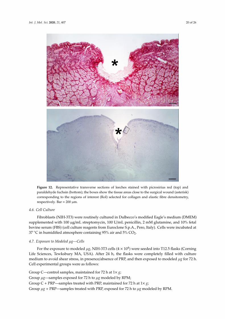
After exposure to modeled µg, leeches from the different experimental groups were anesthetizedby immersion in a solution of 10% ethanol (AppliChem GmbH, Darmstadt, Germany) in water andthen sacrificed by immersion in 95% ethanol for 15 min. The mid part of the body with the surgicalwound was dissected, fixed in 4% paraformaldehyde in phosphate buffer (Bio Optica Milano s.p.a.,Milano, Italy) for 24 h, dehydrated in graded ethanol, and embedded in paraffin (Diapath S.p.A.,Bergamo, Italy). Histological cross sections, 5 µm thick, were cut with a MR2 microtome (RMCBoeckeler, Tucson, AR, USA). Some sections were stained with hematoxylin and eosin (Bio OpticaMilano s.p.a., Milano, Italy) for conventional histological observation. Other sections were stainedwith Picrosirius red (Sigma-Aldrich, St. Louis, MO, USA) for assessment of collagen fibres contentor with paraldehyde fuchsin (Acros Organics, Geel, Belgium) for assessment of elastic fibre contentin the tissues around and beneath the surgical wound. Stainings were made in a single session tominimize artifactual staining differences. Digital photomicrographs were taken with a Nikon EclipseE200 light microscope with a ×40 objective, equipped with a Nikon DS Fi2 digital camera and NISElements image acquisition software (all from Nikon, Florence, Italy). Surface area measurements ofthe red-stained collagen fibers or the violet-stained elastic fibres were carried out using the ImageJ 1.33image analysis program (National Institutes of Health, USA; http://rsb.info.nih.gov/ij) upon selectionof four regions of interest (RoI) for each image and an appropriate threshold to only include the stainedfibres. The regions of interest (RoI) used for collagen and elastic fibre morphometry were selected fromthe sub-epidermal and inter-muscular stroma flanking the margins and bed of the wound (Figure 12).
Int. J. Mol. Sci. 2020, 21, 407 20 of 26
Int. J. Mol. Sci. 2020, 21, x FOR PEER REVIEW 20 of 26
Louis, MO, USA) for assessment of collagen fibres content or with paraldehyde fuchsin (Acros
Organics, Geel, Belgium) for assessment of elastic fibre content in the tissues around and
beneath the surgical wound. Stainings were made in a single session to minimize artifactual
staining differences. Digital photomicrographs were taken with a Nikon Eclipse E200 light
microscope with a ×40 objective, equipped with a Nikon DS Fi2 digital camera and NIS Elements
image acquisition software (all from Nikon, Florence, Italy). Surface area measurements of the
red-stained collagen fibers or the violet-stained elastic fibres were carried out using the ImageJ
1.33 image analysis program (National Institutes of Health, USA; http://rsb.info.nih.gov/ij) upon
selection of four regions of interest (RoI) for each image and an appropriate threshold to only
include the stained fibres. The regions of interest (RoI) used for collagen and elastic fibre
morphometry were selected from the sub-epidermal and inter-muscular stroma flanking the
margins and bed of the wound (Figure 12).
Figure 12. Representative transverse sections of leeches stained with picrosirius red (top) and
paraldehyde fuchsin (bottom); the boxes show the tissue areas close to the surgical wound
(asterisk) corresponding to the regions of interest (RoI) selected for collagen and elastic fibre
densitometry, respectively. Bar = 200 μm.
4.6. Cell Culture
Fibroblasts (NIH-3T3) were routinely cultured in Dulbecco’s modified Eagle’s medium
(DMEM) supplemented with 100 μg/mL streptomycin, 100 U/mL penicillin, 2 mM glutamine,
and 10% fetal bovine serum (FBS) (cell culture reagents from Euroclone S.p.A., Pero, Italy). Cells
were incubated at 37 °C in humidified atmosphere containing 95% air and 5% CO2.
Figure 12. Representative transverse sections of leeches stained with picrosirius red (top) andparaldehyde fuchsin (bottom); the boxes show the tissue areas close to the surgical wound (asterisk)corresponding to the regions of interest (RoI) selected for collagen and elastic fibre densitometry,respectively. Bar = 200 µm.
4.6. Cell Culture
Fibroblasts (NIH-3T3) were routinely cultured in Dulbecco’s modified Eagle’s medium (DMEM)supplemented with 100 µg/mL streptomycin, 100 U/mL penicillin, 2 mM glutamine, and 10% fetalbovine serum (FBS) (cell culture reagents from Euroclone S.p.A., Pero, Italy). Cells were incubated at37 ◦C in humidified atmosphere containing 95% air and 5% CO2.
4.7. Exposure to Modeled µg—Cells
For the exposure to modeled µg, NIH-3T3 cells (4 × 10⁴) were seeded into T12.5 flasks (CorningLife Sciences, Tewksbury MA, USA). After 24 h, the flasks were completely filled with culturemedium to avoid shear stress, in presence/absence of PRP, and then exposed to modeled µg for 72 h.Cell experimental groups were as follows:
Group C—control samples, maintained for 72 h at 1× g;Group µg—samples exposed for 72 h to µg modeled by RPM;Group C + PRP—samples treated with PRP, maintained for 72 h at 1× g;Group µg + PRP—samples treated with PRP, exposed for 72 h to µg modeled by RPM.

Int. J. Mol. Sci. 2020, 21, 407 21 of 26
4.8. Cell Count
At the end of the exposure to modeled µg, medium was removed and cells were washed withCa2+- and Mg2+-containing PBS (pH 7.4) (Euroclone S.p.A., Pero, Italy). Cells were then detachedby trypsinization (Trypsin 0.05%–EDTA 0.02%, Euroclone S.p.A., Pero, Italy) and counted with aBurker’s chamber under an inverted light microscope (Nikon, Amsterdam, The Netherlands). The sameprocedure was applied to 1× g control samples.
4.9. Migration Assays
4.9.1. Scratch Test
Scratch assay is an in vitro model of wound healing used to study cell migration in response toinjury. Cells of 8.5 × 10⁵ were seeded in T12.5 flasks and allowed to adhere. After the cells reachedsubconfluence, they were incubated for 24 h in DMEM + 0.5% FBS (Euroclone S.p.A., Pero, Italy)to reduce proliferation. Then, the culture medium was removed and a scratch was made on themonolayers with a plastic tip. Wounded monolayers were washed with Ca2+- and Mg2+-containingPBS (pH 7.4) to remove dead cells, and fresh complete medium, in presence/absence of PRP, was added.Experimental groups of samples were prepared as previously described. Wounded monolayers wereobserved and photographed immediately after the scratch (before being placed on RPM) (t0) andafter 24 h and 48 h of exposure to modeled µg. A scanning microscope (Axiovert 200 Zeiss, Jena,Germany) and the MetaVue software (Molecular Devices, Sunnyvale, CA, USA) were used. Distancesbetween cell fronts were measured with Image-ProPlus 6.0 (MediaCybernetics, Bethesda, MA, USA),considering at least six measurements from the top to the bottom. To capture images at 24 h and 48 hof exposure, RPM was stopped for a few minutes to allow samples displacement to the microscopeand repositioning on the machine.
4.9.2. Boyden Chamber Assay
The microchemotaxis assay was performed using a 48-well Boyden’s chamber according tomanufacturer’s instructions (Neuroprobe, Cabin John, MD, USA). NIH-3T3 cells previously exposedto modeled µg or maintained at 1× g for 72 h, in presence/absence of PRP, were used for the assay(see experimental groups described above). At the end of the exposure, cells were immediately collectedby trypsin, resuspended in DMEM + 0.1% bovine serum albumin (BSA, Merck KGaA, Darmstadt,Germany), and used to fill the upper compartment (3.5 × 10⁴ cells/well) of the chamber. FBS 1%was used as chemoattractant in the lower compartment of the chamber. Cells migrated througha polyvinylpyrrolidone-free polycarbonate porous membrane (8 µm pores, Biomap, Milano, Italy)pre-coated with gelatin (0.2 mg/mL in PBS, 5 days at 4 ◦C, Merck KGaA, Darmstadt, Germany).
After migration (overnight, 37 ◦C), cells adherent to the underside of the membrane were fixed bymethanol (Merck KGaA, Darmstadt, Germany) and stained according to the Diff-Quik kit (Biomap,Milano, Italy). For quantitative analysis, cells were photographed using an optical microscopewith a digital camera and counted using Image J software (National Institutes of Health, USA;http://rsb.info.nih.gov/ij). Three random objective fields were counted for each well and the meannumber of migrating cells was calculated.
4.9.3. Gene Expression
The expression of alpha-SMA and VEGF proteins was assessed by qRT-PCR on total RNAobtained from samples belonging to the four different experimental groups: NIH-3T3 cells culturedat 1× g (controls), treated/untreated with PRP, and NIH-3T3 cells exposed for 72 h to modeled µg,treated/untreated with PRP. Before exposure to µg and PRP treatment, a scratch was performed onthe fibroblast monolayers to stimulate cell activation. The same procedure was performed on therelated controls.

Int. J. Mol. Sci. 2020, 21, 407 22 of 26
4.9.4. RNA Extraction, Purification and Quantitation
Total RNA was extracted with RNeasy Mini kit (Qiagen, Hilden, Germany) according to themanufacturer’s instructions and then quantified with Nano-Drop2000 (Thermo Scientific, Waltham,MA, USA).
4.9.5. Quantitative Real-Time PCR
Reverse transcription was performed on 1 microgram of total RNA from each sample accordingto the manufacturer’s protocol (iScript cDNA synthesis kit, BioRad, Segrate, Italy) using randomprimers. qPCR was performed in singleplex in CFX96 Touch™ Real-Time PCR Detection System(BioRad, Segrate, Italy) using SYBR Green dye (SsoAdvanced SYBR Green Supermix, Bio-Rad, Segrate,Italy) and specific sets of primers as follows:
Alpha-SMA: 5′-CCCTGAAGAGCATCCGACAC-3′ and 5′ GCATAGCCCTCATAGATAGGCA-3′;VEGF: 5′-AAAACACAGACTCGCGTTGC-3′ and 5′-CTCCTAGGCCCCTCAGAAGT-3′;GAPDH: 5′-CCTGCGACTTCAACAGCAAC-3′ and 5′-TAGGGCCTCTCTTGCTCAGT-3′.
Data analysis was performed using the CFX Manager 2.0 software (Bio-Rad, Segrate, Italy).Each sample was analyzed in triplicate. Data were normalized for GAPDH Ct value. Relative mRNAlevels were then calculated by the comparative Ct method (2−∆∆Ct) and data were expressed as foldinduction versus control.
4.10. Statistical Analysis
Animal and cell experiments were carried out in triplicate.Morphometrical data were represented as means ± SEM of the measurements of four individual
animals, with at least five regions of interest (ROI, about 10,000 µm2 each), from the differentexperimental groups. Statistical comparison of differences between groups was carried out usingone-way analysis of variance (ANOVA) followed by Student–Newman–Keuls multiple comparisontest. A p-value ≤ 0.05 was considered significant. Calculations were done using GraphPad Prism 5.0statistical program (GraphPad Software, San Diego, CA, USA).
The statistical analysis of migration assays was carried out with the Prism4 software for Macintosh(GraphPad Software, San Diego, CA, USA) and represented as means ± SD. Comparisons among theexperimental groups were performed by Tukey’s multiple comparison test. A p value ≤ 0.05 wasconsidered significant.
Author Contributions: The authors’ contributions are as follows: conceptualization, M.M., F.C. (Fabio Celotti);data curation, F.C. (Francesca Cialdai), A.C., and D.B.; formal analysis, F.C. (Francesca Cialdai), A.C., and D.B.;funding acquisition, M.M., F.C. (Fabio Celotti), L.M., D.B., and D.P.; investigation, F.C. (Francesca Cialdai), A.C.,D.P., A.M.R., S.Z., F.C. (Fabio Celotti), D.B., L.M., and M.M.; methodology, M.M., D.P., D.B., F.C. (Fabio Celotti),and A.M.R.; project administration, M.M., F.C. (Fabio Celotti), and D.B.; resources, M.M., D.P., D.B., A.M.R., F.C.(Fabio Celotti), and L.M., supervision, M.M. and F.C. (Fabio Celotti), validation, F.C. (Francesca Cialdai) andA.C.; visualization, F.C. (Francesca Cialdai), A.C., D.P., A.M.R., S.Z., F.C. (Fabio Celotti), D.B., L.M., and M.M.,writing—original draft preparation, F.C. (Francesca Cialdai), A.C., and M.M., writing—review and editing, D.P.,A.M.R., S.Z., L.M., F.C. (Fabio Celotti), D.B., M.M., F.C. (Francesca Cialdai), and A.C. All authors have read andagreed to the published version of the manuscript.
Funding: This research was funded by the Italian Space Agency (Tissue Repair in Microgravity ASI N. 2013-090-R.O,Wound Healing and Sutures in Unloading Conditions ASI N. 2018-14-U.0).
Acknowledgments: The authors thank Laura Calosi, Stefano Catarinicchia, and Daniele Guasti for their technicalsupport in histological sample preparation.
Conflicts of Interest: The authors declare no conflict of interest.

Int. J. Mol. Sci. 2020, 21, 407 23 of 26
Abbreviations
PRP Platelet rich plasmaMMPs Matrix metalloproteasesRPM Random positioning machineNIH-3T3 Fibroblasts (mouse, NIH Swiss, embryo)DMEM Dulbecco’s modified Eagle mediumFCS Fetal calf serumFBS Fetal bovine serumBSA Bovine serum albuminαSMA Alfa smooth muscle actinVEGF Vascular endothelial growth factorECM Extracellular matrixRCCS Rotating cell culture system
References
1. Yanez, D.A.; Lacher, R.K.; Vidyarthi, A.; Colegio, O.R. The role of macrophages in skin homeostasis. PflügersArch. 2017, 469, 455–463. [CrossRef]
2. Sorg, H.; Tilkorn, D.J.; Hager, S.; Hauser, J.; Mirastschijski, U. Skin Wound Healing: An Update on theCurrent Knowledge and Concepts. Eur. Surg. Res. 2017, 58, 81–94. [CrossRef]
3. Posnett, J.; Franks, P. The burden of chronic wounds in the UK. Nurs. Times 2008, 104, 44–45.4. Posnett, J.; Gottrup, F.; Lundgren, H.; Saal, G. The resource impact of wounds on health-care providers in
Europe. J. Wound Care 2009, 18, 154–161. [CrossRef]5. Järbrink, K.; Ni, G.; Sönnergren, H.; Schmidtchen, A.; Pang, C.; Bajpai, R.; Car, J. Prevalence and incidence
of chronic wounds and related complications: A protocol for a systematic review. Syst. Rev. 2016, 5, 152.[CrossRef]
6. Tuner, J.; Hode, L. Laser Therapy Handbook: A Guide for Research Scientists, Doctors, Dentists, Veterinarians andother Interested Parties within the Medical Field; Prima Books: Grangesberg, Sweden, 2004.
7. Alster, T.; Zaulyanov-Scanlon, L. Laser scar revision: A review. Dermatol. Surg. 2007, 33, 131–140. [PubMed]8. Shah, J.B. The history of wound care. J. Am. Coll. Certif. Wound Spec. 2011, 3, 65–66. [CrossRef] [PubMed]9. Dreifke, M.B.; Jayasurya, A.A.; Ambalangodage, C.J. Current wound healing procedures and potential care.
Mater. Sci. Eng. C Mater. Biol. Appl. 2015, 48, 651–662. [CrossRef] [PubMed]10. Franca, C.M.; Anders, J.J.; Lanzafame, R.J. Photobiomodulation in wound healing: What are we not
considering? Photomed. Laser Surg. 2016, 34, 51–52. [CrossRef] [PubMed]11. Nesi-Reis, V.; Lera-Nonose, D.S.S.L.; Oyama, J.; Paula Silva-Lalucci, M.P.; Demarchi, I.G.; Aristides, S.M.A.;
Teixeira, J.J.V.; Silveira, T.G.V.; Lonardoni, M.V.C. Contribution of phodynamic therapy in wound healing:A systematic review. Photodiagn. Photodyn. Ther. 2018, 21, 294–305. [CrossRef]
12. Etulain, J. Platelets in wound healing and regenerative medicine. Platelets 2018, 29, 556–568. [CrossRef][PubMed]
13. Colciago, A.; Celotti, F.; Casati, L.; Giancola, R.; Castano, S.M.; Antonini, G.; Sacchi, M.C.; Negri-Cesi, P.In Vitro Effects of PDGF Isoforms (AA, BB, AB and CC) on Migration and Proliferation of SaOS-2 Osteoblastsand on Migration of Human Osteoblasts. Int. J. Biomed. Sci. 2009, 5, 380–389. [PubMed]
14. Anitua, E.; Pino, A.; Orive, G. Plasma rich in growth factors promotes dermal fibroblast proliferation,migration and biosynthetic activity. J. Wound Care 2016, 25, 680–687. [CrossRef]
15. Anitua, E. Plasma rich in growth factors: Preliminary results of use in the preparation of future sites forimplants. Int. J. Oral Maxillofac. Implant. 1999, 14, 529–535.
16. De Pascale, M.R.; Sommese, L.; Casamassimi, A.; Napoli, C. Platelet derivatives in regenerative medicine:An update. Transfus. Med. Rev. 2015, 29, 52–61. [CrossRef] [PubMed]
17. Hesseler, M.J.; Shyam, N. Platelet-rich plasma and its utility in medical dermatology: A systematic review.J. Am. Acad. Dermatol. 2019, 81, 834–846. [CrossRef]
18. Lana, J.F.; Huber, S.B.; Purita, J.; Tambeli, C.H.; Santos, G.S.; Paulus, C.; Annichino-Bizzacchi, J.M.Leukocyte-rich PRP versus leukocyte-poor PRP-The role of monocyte/macrophage function in the healingcascade. J. Clin. Orthop. Trauma 2019, 10, S7–S12. [CrossRef]

Int. J. Mol. Sci. 2020, 21, 407 24 of 26
19. Drudi, L.; Ball, C.G.; Kirkpatrick, A.W.; Saary, J.; Grenon, S.M. Surgery in space: where are we at now?Acta. Astronaut. 2012, 79, 61–66. [CrossRef]
20. Davidson, J.M.; Aquino, A.M.; Woodward, S.C.; Wilfinger, W.W. Sustained microgravity reduces intrinsicwound healing and growth factor responses in rat. FASEB J. 1999, 13, 325–329. [CrossRef]
21. Delp, M.D. Unraveling the complex web of impaired wound healing with mechanical unloading and physicaldeconditioning. J. Appl. Physiol. 2008, 104, 1262–1263. [CrossRef]
22. Radek, K.A.; Baer Lisa, A.; Eckhardt, J.; DiPietro, L.A.; Wade, C.E. Mechanical unloading impairs keratinocytemigration and angiogenesis during cutaneous wound healing. J. Appl. Physiol. 2008, 104, 1295–1303.[CrossRef] [PubMed]
23. Campbell, M.R.; Williams, D.R.; Buckey, J.C.J.; Kirkpatrick, A.W. Animal surgery during spaceflight on theNeurolab Shuttle mission. Aviat. Space Environ. Med. 2005, 76, 589–593.
24. Midura, R.J.; Su, X.; Androjna, C. A simulated weightlessness state diminishes cortical bone healing responses.J. Musculoskelet. Neuronal. Interact. 2006, 6, 327–328. [PubMed]
25. Kirchen, M.E.; O’Connor, K.M.; Gruber, H.E.; Sweeney, J.R.; Fras, I.A.; Stover, S.J.; Sarmiento, A.; Marshall, G.J.Effects of microgravity on bone healing in a rat fibular osteotomy model. Clin. Orthop. Relat. Res. 1995, 318,231–242.
26. Infanger, M.; Kossmehl, P.; Shakibaei, M.; Bauer, J.; Kossmehl-Zorn, S.; Cogoli, A.; Curcio, F.; Oksche, A.;Wehland, M.; Kreutz, R.; et al. Simulated weightlessness changes the cytoskeleton and extracellular matrixproteins in papillary thyroid carcinoma cells. Cell Tissue Res. 2006, 324, 267–277. [CrossRef] [PubMed]
27. Pietsch, J.; Bauer, J.; Egli, M.; Infanger, M.; Wise, P.; Ulbrich, C.; Grimm, D. The Effects of Weightlessness onthe Human Organism and Mammalian Cells. Curr. Mol. Med. 2011, 11, 350–364. [CrossRef] [PubMed]
28. Morbidelli, L.; Monici, M.; Marziliano, N.; Cogoli, A.; Fusi, F.; Waltenberger, J.; Ziche, M. Simulatedhypogravity impairs the angiogenic response of endothelium by up-regulating apoptotic signals. Biochem.Biophys. Res. Com. 2005, 334, 491–499. [CrossRef]
29. Monici, M.; Fusi, F.; Paglierani, M.; Marziliano, N.; Cogoli, A.; Pratesi, R.; Bernabei, P.A. Modeled gravitationalunloading triggers differentiation and apoptosis in preosteoclastic cells. J. Cell Biochem. 2006, 98, 65–80.[CrossRef]
30. Monici, M.; Cialdai, F.; Romano, G.; Fusi, F.; Egli, M.; Pezzatini, S.; Morbidelli, L. An in Vitro Study on TissueRepair: Impact of unloading on cells Involved in the remodelling phase. Microgravity Sci. Technol. 2011, 23,391–401. [CrossRef]
31. Cialdai, F.; Vignali, L.; Morbidelli, L.; Colciago, A.; Celotti, F.; Santi, A.; Caselli, A.; Cirri, P.; Monici, M.Modeled microgravity affects fibroblast function related to wound healing. Microgravity Sci. Technol. 2017,29, 121–132. [CrossRef]
32. Chung, C.Y.; Erickson, H.P. Glycosaminoglycans modulate fibronectin matrix assembly and are essential formatrix incorporation of tenascin-C. J. Cell Sci. 1997, 110, 1413–1419. [PubMed]
33. Strollo, F.; Gentile, S.; Strollo, G.; Mambro, A.; Vernikos, J. Recent progress in Space Physiology and Aging.Front. Physiol. 2018, 9, 1551. [CrossRef]
34. Strollo, F.; Macchi, C.; Eberini, I.; Masini, M.A.; Botta, M.; Vassilieva, G.; Nichiporuk, I.; Monici, M.;Santucci, D.; Celotti, F.; et al. Body composition and metabolic changes during a 520-day mission simulationto Mars. J. Endocrinol. Investig. 2018, 41, 1267–1273. [CrossRef]
35. Neutelings, T.; Nusgens, B.V.; Liu, Y.; Tavella, S.; Ruggiu, A.; Cancedda, R.; Gabriel, M.; Colige, A.; Lambert, C.Skin physiology in microgravity: a 3-month stay aboard ISS induces dermal atrophy and affects cutaneousmuscle and hair follicles cycling in mice. NPJ Microgravity 2015, 1, 15002. [CrossRef]
36. Tettamanti, G.; Grimaldi, A.; Rinaldi, L.; Arnaboldi, F.; Congiu, T.; Valvassori, R.; de Eguileor, M.The multifunctional role of fibroblasts during wound healing in Hirudo medicinalis (Annelida, Hirudinea).Biol. Cell 2004, 96, 443–455. [CrossRef] [PubMed]
37. Tettamanti, G.; Grimaldi, A.; Congiu, T.; Perletti, G.; Raspanti, M.; Valvassori, R.; de Eguileor, M. Collagenreorganization in leech wound healing. Biol. Cell 2005, 97, 557–568. [CrossRef] [PubMed]
38. Lotz, R.G.; Baum, P.; Bowman, G.H.; Klein, K.D.; von Lohr, R.; Schrotter, L. Test of a life support system withHirudo medicinalis in a sounding rocket. Life Sci. Space Res. 1972, 10, 133–143.
39. Velnar, T.; Bailet, T.; Smrkolj, V. The wound healing process: An overview of the cellular and molecularmechanisms. J. Int. Med. Res. 2009, 37, 1528–1542. [CrossRef]
Int. J. Mol. Sci. 2020, 21, 407 25 of 26
40. Stojadinovic, O.K.A.; Golinko, M.; Tomic-Canic, M.; Brem, H. A novel, non-angiogenic mechanism of VEGF:Stimulation of keratinocyte and fibroblast migration. Wound Repair Regen. 2007, 15, A30.
41. Bao, P.; Kodra, A.; Tomic-Canic, M.; Golinko, M.S.; Ehrlich, H.P.; Brem, H. The role of vascular endothelialgrowth factor in wound healing. J. Surg. Res. 2009, 153, 347–358. [CrossRef]
42. Almina, J.F.; Wise, S.G.; Weiss, A.S. Elastin signaling in wound repair. Birth Defects Res. (Part C) 2012, 96,248–257. [CrossRef] [PubMed]
43. Grimaldi, A.; Banfi, S.; Bianchi, C.; Gabriella, G.; Tettamanti, G.; Noonan, D.M.; Valvassori, R.; de Eguileor, M.The leech: A novel invertebrate model for studying muscle regeneration and diseases. Curr. Pharm. Des.2010, 16, 968–977. [CrossRef] [PubMed]
44. Heinemeier, K.M.; Olesen, J.L.; Haddad, F.; Schjerling, P.; Baldwin, K.M.; Kjaer, M. Effect of unloadingfollowed by reloading on expression of collagen and related growth factors in rat tendon and muscle. J. Appl.Physiol. 2009, 106, 178–186. [CrossRef]
45. Provenzano, P.P.; Martinez, D.A.; Grindeland, R.E.; Dwyer, K.W.; Turner, J.; Vailas, A.C.; Vanderby, R., Jr.Hindlimb unloading alters ligament healing. J. Appl. Physiol. 2003, 94, 314–324. [CrossRef]
46. Martinez, D.A.; Vailas, A.C.; Vanderby, R.J.; Grindeland, R.E. Temporal extracellular matrix adaptations inligament during wound healing and hindlimb unloading. Am. J. Physiol. Regul. Integr. Comp. Physiol. 2007,293, 1552–1560. [CrossRef]
47. Krüger, M.; Pietsch, J.; Bauer, J.; Kopp, S.; Carvalho, D.T.O.; Baatout, S.; Moreels, M.; Melnik, D.; Wehland, M.;Egli, M.; et al. Growth of Endothelial Cells in Space and in Simulated Microgravity—A Comparison on theSecretory Level. Cell Physiol. Biochem. 2019, 52, 1039–1060.
48. Chen, Z.Y.; Guo, S.; Li, B.; Jiang, N.; Li, A.; Yan, H.F.; Yang, H.M.; Zhou, J.L.; Li, C.L.; Cui, Y. Effect ofWeightlessness on the 3D Structure Formation and Physiologic Function of Human Cancer Cells. BioMed Res.Int. 2019, 2019, 1–17. [CrossRef] [PubMed]
49. Aleshcheva, G.; Sahana, J.; Ma, X.; Hauslage, J.; Hemmersbach, R.; Egli, M.; Infanger, M.; Bauer, J.; Grimm, D.Changes in Morphology, Gene Expression and Protein Content in Chondrocytes Cultured on a RandomPositioning Machine. PLoS ONE 2013, 8, e79057. [CrossRef]
50. Casati, L.; Celotti, F.; Negri-Cesi, P.; Sacchi, M.; Castano, P.; Colciago, A. Platelet derived growth factor(PDGF) contained in Platelet Rich Plasma (PRP) stimulates migration of osteoblasts by reorganizing actincytoskeleton. Cell Adhes. Migr. 2014, 8, 595–602. [CrossRef]
51. Sun, Z.; Li, Y.; Wang, H.; Cai, M.; Gao, S.; Liu, J.; Tong, L.; Hu, Z.; Wang, Y.; Wang, K.; et al. MiR-181c-5pmediates simulated microgravity-induced impaired osteoblast proliferation by promoting cell cycle arrestedin the G2 phase. J. Cell Mol. Med. 2019, 23, 3302–3316. [CrossRef] [PubMed]
52. Cherng, S.; Young, J.; Ma, H. Alpha-Smooth Muscle Actin (α-SMA). J. Am. Sci. 2008, 4, 7–9.53. Park, S.; Bivona, B.J.; Harrison-Bernard, L.M. Compromised renal microvascular reactivity of angiotensin
type 1 double null mice. Am. J. Physiol. Renal. Physiol. 2007, 293, F60–F67. [CrossRef]54. Kushida, S.; Kakudo, N.; Suzuki, K.; Kusumoto, K. Effects of Platelet-Rich Plasma on Proliferation and
Myofibroblastic Differentiation in Human Dermal Fibroblasts. Ann. Plast. Surg. 2013, 71, 219–224. [CrossRef]55. Ahluwalia, A.; Tarnawski, A.S. Critical Role of Hypoxia Sensor—HIF-1alpha in VEGF Gene Activation.
Implications for Angiogenesis and Tissue Injury Healing. Curr. Med. Chem. 2012, 19, 90–97. [CrossRef]56. Redd, M.J.; Cooper, L.; Wood, W.; Stramer, B.; Martin, P. Wound healing and inflammation: embryos reveal
the way to perfect repair. Philos. Trans. R. Soc. B 2004, 359, 777–784. [CrossRef]57. Bani, D.; Nistri, S. New insights into the morphogenic role of stromal cells and their relevance for regenerative
medicine. Lessons from the heart. J. Cell. Mol. Med. 2014, 18, 363–370. [CrossRef]58. Celotti, F.; Colciago, A.; Negri-Cesi, P.; Pravettoni, A.; Zaninetti, R.; Sacchi, M.C. Effect of platelet-rich
plasma on migration and proliferation of SaOS-2 osteoblasts: Role of platelet-derived growth factor andtransforming growth factor-beta. Wound Repair Regen. 2006, 14, 195–202. [CrossRef]

Int. J. Mol. Sci. 2020, 21, 407 26 of 26
59. Hammond, T.; Allen, P. The Bonn criteria: Minimal experimental parameter reporting for clinostat andrandom positioning machine experiments with cells and tissues. Microgravity Sci. Technol. 2011, 23, 271–275.[CrossRef]
60. van Loon, J. Some history and use of the Random Positioning machine, RPM, in gravity related research.Adv. Space Res. 2007, 39, 1161–1165. [CrossRef]
© 2020 by the authors. Licensee MDPI, Basel, Switzerland. This article is an open accessarticle distributed under the terms and conditions of the Creative Commons Attribution(CC BY) license (http://creativecommons.org/licenses/by/4.0/).