Embed Size (px)
Citation preview
doi: 10.2522/ptj.20140361 Published online November 13, 2014PHYS THER.
Taylor-Vaisey, Sean Abdulla and Yaadwinder Shergillder Velde, Linda Carroll, Craig L. Jacobs, Anne L. Ameis, Maja Stupar, Margareta Nordin, Gabreille M. vanVaratharajan, Danielle Southerst, Silvano A. Mior, Arthur
SharanyaWong, Deborah A. Sutton, Kristi A. Randhawa, Hainan Yu, Pierre Côté, Heather M. Shearer, Jessica J.Protocol for Traffic Injury Management CollaborationShoulder Pain: A Systematic Review by the Ontario Effectiveness of Passive Physical Modalities for
http://ptjournal.apta.org/content/early/2014/11/12/ptj.20140361found online at: The online version of this article, along with updated information and services, can be
E-mail alerts to receive free e-mail alerts hereSign up
corrections and replace the original author manuscript. : edited and typeset versions of articles that incorporate any authorPage proofs
readers almost immediate access to accepted papers.
PTJaccepted for publication but have not yet been copyedited or typeset. This allows : PDF versions of manuscripts that have been peer-reviewed andAuthor manuscripts
publishes 2 types of Online First articles: PTJ). PTJ (Therapy
PhysicalOnline First articles are published online before they appear in a regular issue of
by Louis Houle on November 21, 2014http://ptjournal.apta.org/Downloaded from by Louis Houle on November 21, 2014http://ptjournal.apta.org/Downloaded from
1
Running head: Passive Physical Modalities for Shoulder Pain
Research Report
Effectiveness of Passive Physical Modalities for Shoulder Pain: A Systematic Review by the
Ontario Protocol for Traffic Injury Management Collaboration
Hainan Yu, Pierre Côté, Heather M. Shearer, Jessica J. Wong, Deborah A. Sutton, Kristi A.
Randhawa, Sharanya Varatharajan, Danielle Southerst, Silvano A. Mior, Arthur Ameis, Maja
Stupar, Margareta Nordin, Gabreille M. van der Velde, Linda Carroll, Craig L. Jacobs, Anne L.
Taylor-Vaisey, Sean Abdulla, Yaadwinder Shergill
H. Yu, MBBS, MSc, UOIT-CMCC Centre for the Study of Disability Prevention and
Rehabilitation, University of Ontario Institute of Technology (UOIT) and Canadian Memorial
Chiropractic College (CMCC), 6100 Leslie St, Toronto, Ontario, Canada, M2H 3 J1. Address all
correspondence to Dr Yu at: [email protected]
P. Côté, PhD, UOIT-CMCC Centre for the Study of Disability Prevention and Rehabilitation,
UOIT and CMCC.
H.M. Shearer, DC, MSc, FCCS(C), UOIT-CMCC Centre for the Study of Disability Prevention
and Rehabilitation, UOIT and CMCC.
J.J. Wong, BSc, DC, FCCS(C), UOIT-CMCC Centre for the Study of Disability Prevention and
Rehabilitation, UOIT and CMCC.
by Louis Houle on November 21, 2014http://ptjournal.apta.org/Downloaded from
2
D.A. Sutton, MEd, MSc, UOIT-CMCC Centre for the Study of Disability Prevention and
Rehabilitation, UOIT and CMCC.
K.A. Randhawa, MPH, UOIT-CMCC Centre for the Study of Disability Prevention and
Rehabilitation, UOIT and CMCC.
S. Varatharajan, MSc, UOIT-CMCC Centre for the Study of Disability Prevention and
Rehabilitation, UOIT and CMCC.
D. Southerst, BScH, DC, FCCS(C), UOIT-CMCC Centre for the Study of Disability Prevention
and Rehabilitation, UOIT and CMCC.
S.A. Mior, DC, PhD, Division of Graduate Education and Research, CMCC.
A. Ameis, MD, FRCPC, DESS, DABPM&R, University of Montreal, Quebec, Canada.
M. Stupar, DC, PhD, UOIT-CMCC Centre for the Study of Disability Prevention and
Rehabilitation, UOIT and CMCC.
M. Nordin, DrMedSci, Department of Orthopedic Surgery, New York University, New York,
New York.
G.M. van der Velde, DC, PhD, Toronto Health Economics and Technology Assessment
(THETA) Collaborative, University of Toronto, Toronto, Ontario, Canada, and Institute for
Work and Health, Toronto, Ontario, Canada.
L. Carroll, PhD, School of Public Health, University of Alberta, Alberta, Canada.
C.L. Jacobs, BFA, DC, MSc, FCCS(C), UOIT-CMCC Centre for the Study of Disability
Prevention and Rehabilitation, UOIT and CMCC.
by Louis Houle on November 21, 2014http://ptjournal.apta.org/Downloaded from
3
A.L. Taylor-Vaisey, MLS, UOIT-CMCC Centre for the Study of Disability Prevention and
Rehabilitation, UOIT and CMCC.
S. Abdulla, BA, MSc, DC, Department of Graduate Studies, CMCC.
Y. Shergill, BSc, DC, Department of Graduate Studies, CMCC, and Department of Anesthesia,
the Ottawa Hospital, Ottawa, Canada.
[Yu H, Côté P, Shearer H, et al. Effectiveness of passive physical modalities for shoulder pain: a
systematic review by the Ontario Protocol for Traffic Injury Management Collaboration. Phys
Ther. 2015;95:xxx–xxx.]
© 2014 American Physical Therapy Association
Published Ahead of Print: xxxx
Accepted: November 3, 2014
Submitted: August 25, 2014
by Louis Houle on November 21, 2014http://ptjournal.apta.org/Downloaded from

4
Background: Shoulder pain is a common musculoskeletal condition in the general population.
Passive physical modalities are commonly used to treat shoulder pain. However, previous
systematic reviews report conflicting results.
Purpose: To evaluate the effectiveness of passive physical modalities for the management of
soft tissue injuries of the shoulder.
Data Sources: MEDLINE, EMBASE, CINAHL, PsycINFO, and the Cochrane Central Register
of Controlled Trials from January 1st, 1990 to April 18th, 2013.
Study Selection: Randomized controlled trials (RCTs), cohort and case-control studies were
eligible. Random pairs of independent reviewers screened 1470 of 1760 retrieved articles after
removing 290 duplicates. Twenty-two articles were eligible for critical appraisal. We critically
appraised the eligible studies using the Scottish Intercollegiate Guidelines Network criteria. Of
those, 11 studies had a low risk of bias.
Data Extraction: The lead author extracted data from low risk of bias studies and built evidence
tables. A second reviewer independently checked the extracted data.
Data Synthesis: We synthesized the findings of low risk of bias studies according to principles
of best evidence synthesis. We found that pre-tensioned tape, ultrasound and interferential
current are not effective to manage shoulder pain. However, diathermy and corticosteroid
injections lead to similar outcomes. Low-level laser therapy provides short-term pain reduction
by Louis Houle on November 21, 2014http://ptjournal.apta.org/Downloaded from
5
for subacromial impingement syndrome. Extracorporeal shock-wave therapy is not effective for
subacromial impingement syndrome but it provides benefits for persistent shoulder calcific
tendonitis.
Limitations: Non-English studies excluded.
Conclusions: Most passive physical modalities do not benefit patients with subacromial
impingement syndrome. However, low-level laser therapy is more effective than placebo or
ultrasound for subacromial impingement syndrome. Similarly, shock-wave therapy is more
effective than sham for persistent shoulder calcific tendinitis.
by Louis Houle on November 21, 2014http://ptjournal.apta.org/Downloaded from

6
INTRODUCTION
Shoulder pain is common in the general population, ranking fourth behind low back pain, knee
pain and neck pain as the most prevalent musculoskeletal conditions.1-3 One third of adults
experience shoulder pain every year.2 Shoulder complaints place a significant burden upon the
health care system.4, 5 In the United Kingdom, 2.4% of the population consult general
practitioners for shoulder pain each year.6 In the United States, more than 500,000 rotator cuff
surgical repairs and shoulder arthroscopies are performed annually.7 Moreover, shoulder injuries
are associated with a substantial economic burden costing an average of €326 per patient during
six months in Sweden.8
Musculoskeletal conditions are commonly managed with passive physical modalities.9, 10
Despite being commonly used, large insurers such as the Ontario Workplace Safety and
Insurance Board (WSIB) do not recommend passive physical modalities for the management of
shoulder pain.11 Such recommendation is consistent with the results of several systematic
reviews suggesting that ultrasound, interferential current therapy and kinesiotaping are equal to
placebo or other interventions for the management of shoulder disorders.12-15 In addition,
evidence on the effectiveness of electromagnetic field therapy, low-level laser therapy and
shock-wave therapy is conflicting.14, 16-18 However, these reviews suffer from methodological
limitations that may have biased their conclusions. Specifically, the reviews pooled quantitative
results from heterogeneous studies13, 14, 16, 17 and synthesized the evidence from studies with
small sample sizes and/or a high risk of bias.12-18
by Louis Houle on November 21, 2014http://ptjournal.apta.org/Downloaded from
7
Therefore, an up-to-date systematic review is needed to evaluate the effectiveness of passive
physical modalities for soft tissue shoulder injuries. We aim to address the limitations of
previous reviews by assessing the homogeneity of samples across studies. Moreover, our review
aims to minimize bias by restricting our synthesis to high-quality evidence. The purpose of our
review is to determine the effectiveness of passive physical modalities on self-rated recovery,
functional recovery, pain intensity, health-related quality of life, psychological outcomes, and
adverse events in patients with soft tissue injuries of the shoulder.
METHODS
Registration
We registered this review with the International Prospective Register of Systematic Reviews
(PROSPERO) on April 18th, 2013 (CRD42013004854).
Eligibility Criteria
Population: Our review targeted studies of adults and children with soft tissue injuries of the
shoulder. We included grade I-II sprains/strains, nonspecific diffuse shoulder pain, shoulder
tendinitis, impingement syndromes, bursitis, and other soft tissue injuries of the shoulder.19-22 We
excluded studies of shoulder pain due to pathology (e.g., fractures, dislocations, infections,
neoplasms, frozen shoulder or systemic disease). The principles outlined in the Declaration of
Helsinki were followed.
Interventions: We restricted our review to studies on the effectiveness of passive physical
modalities. Passive physical modalities include physical modalities or devices that do not require
by Louis Houle on November 21, 2014http://ptjournal.apta.org/Downloaded from
8
the active participation of patients (including rest). We divided passive physical modalities into
two categories: physico-chemical and structural.9, 10 Physico-chemical modalities use thermal or
electromagnetic effect, such as cold, heat or light application at the skin level, or light, ultrasonic
or electromagnetic radiation affecting structures beneath the skin. Structural modalities include
non-functional assistive devices that encourage rest in anatomic positions (e.g., pillows, seat
cushions) or actively inhibit or prevent movement (e.g., collars, corsets, casts, slings, and rest
splints); and functional assistive devices that align, support or indirectly facilitate function in the
affected region (e.g., tenodesis splints, taping, and assistive braces).
Comparison groups: We included studies that compared passive physical modalities to other
types of conservative care, waiting list, sham/placebo or no intervention.
Outcomes: We aimed to capture both specific and generic health outcomes. Eligible studies had
to include one of the following outcomes: 1) self-rated recovery (e.g., self-reported on Likert
Scale); 2) functional recovery (e.g., range of motion measured with a goniometer; function
measured with the Constant-Murley Scale; disability measured with the Shoulder Pain and
Disability Index; self-reported return to activities, work or school); 3) pain intensity (e.g.,
measured with the Visual Analog Scale or Numerical Rating Scale); 4) health-related quality of
life (e.g., measured with EuroQol EQ-5D or the SF-36); 5) psychological outcomes (e.g.,
depression measured with the CES-D or Beck Depression Inventory); or 6) adverse events.
Study characteristics: Eligible studies met the following criteria: 1) English language; 2)
published between January 1st, 1990 to April 18th, 2013; 3) randomized controlled trials (RCTs),
by Louis Houle on November 21, 2014http://ptjournal.apta.org/Downloaded from
9
cohort studies, or case-control studies; and 4) included an inception cohort of a minimum of 30
participants per treatment arm with a soft tissue shoulder injury in RCTs or 100 subjects per
group with the specified condition in cohort studies or case-control studies. We excluded studies
with the following characteristics: 1) letters, editorials, commentaries, unpublished manuscripts,
dissertations, government reports, books and book chapters, conference proceedings, meeting
abstracts, lectures and addresses, consensus development statements, or guideline statements; 2)
pilot studies, cross-sectional studies, case reports, case series, qualitative studies, narrative
reviews, systematic reviews, clinical practice guidelines, biomechanical studies, or laboratory
studies; or 3) cadaveric or animal studies.
Data Sources and Searches
We developed our search strategy with a health sciences librarian (Appendix I). A second
librarian reviewed the search strategy for completeness and accuracy using the Peer Review of
Electronic Search Strategies (PRESS) Checklist.23, 24 We searched MEDLINE, EMBASE,
CINAHL, PsycINFO, and the Cochrane Central Register of Controlled Trials from January 1st,
1990 to April 18th, 2013.
We developed the search strategy in MEDLINE, which was subsequently adapted to the other
bibliographic databases. The search terms included subject headings specific to each database
(e.g., MeSH in MEDLINE) and free text words relevant to passive physical modalities and soft
tissue injuries of the shoulder. We downloaded the search results into a database created using
EndNote X6 (http://endnote.com/if/online-user-manual).
by Louis Houle on November 21, 2014http://ptjournal.apta.org/Downloaded from
10
Study Selection
We used a two-phase screening process. In phase one, random pairs of independent trained
reviewers (from a pool of eight reviewers) screened citation titles and abstracts to determine
eligibility and classified citations as relevant, possibly relevant or irrelevant. In phase two, the
same pairs of reviewers independently reviewed possibly relevant manuscripts to make a final
determination. Reviewers met to resolve disagreements. If consensus could not be reached, a
third reviewer was used.
Quality Assessment and Data Extraction
Eligible studies were critically appraised by random pairs of independent reviewers (from a pool
of ten reviewers). We assessed the internal validity of studies using the Scottish Intercollegiate
Guidelines Network (SIGN) criteria (Table 1).25 The SIGN criteria were used to qualitatively
evaluate the impact of selection bias, information bias, and confounding on study results. We did
not use a quantitative score or a cutoff point to determine the internal validity of studies.26
Rather, the SIGN criteria were used to assist reviewers in making an informed judgment on the
internal validity of studies.
Specifically, we critically appraised the following methodological aspects: 1) clarity of the
research question; 2) randomization method; 3) concealment of treatment allocation; 4) blinding
of treatment and outcomes; 5) similarity of baseline characteristics between/among treatment
arms; 6) co-intervention contamination; 7) validity and reliability of outcome measures; 8)
follow-up rates; 9) analysis according to intention to treat principles; and 10) comparability of
results across study sites (where applicable). All reviewers were trained to critically appraise
by Louis Houle on November 21, 2014http://ptjournal.apta.org/Downloaded from
11
studies using the SIGN criteria. Consensus between two reviewers in each pair was reached
through discussion with the involvement of an independent third reviewer where necessary. We
contacted authors when we needed additional information for the critical appraisal to be accurate
and valid. After critical appraisal, studies with a low risk of bias were included in our synthesis.27
The lead author extracted data from studies with a low risk of bias and built evidence tables
(Table 2). A second reviewer independently checked the extracted data. Edits were made using
“Track Changes” in Microsoft Word by the second reviewer and incorporated by the lead author.
Disagreements were resolved through discussion.
Data Synthesis and Analysis
We assessed the clinical homogeneity of studies by comparing the diagnoses, characteristics of
the study samples and the parameters of the interventions. We considered conducting a meta-
analysis if the studies were homogeneous. However, a qualitative best evidence synthesis was
performed if the studies were clinically heterogeneous.27 We used minimal clinically important
differences (MCIDs) to determine the clinical significance of outcome measures. These include a
between-group difference of 1.4/10 cm on the Visual Analog Scale (VAS),28 18/100 on the
Shoulder Pain and Disability Index (SPADI),29 and 8/100 on the short form of the Disabilities of
the Arm, Shoulder, and Hand questionnaire (QuickDASH).30 The MCIDs for range of motion
(ROM), the Simple Shoulder Test (SST), the Constant-Murley Score (CMS) and the University
of California/Los Angeles (UCLA) scales have not been defined in the literature. We synthesized
the evidence according to the outcome measures. Specifically, we report the effectiveness of
interventions according to their impact on specific (e.g., shoulder function) or generic (e.g.,
by Louis Houle on November 21, 2014http://ptjournal.apta.org/Downloaded from
12
health-related quality of life) outcomes. We stratified our results by shoulder diagnosis and
duration [i.e., recent (< 3 months), persistent (≥ 3 months) or variable (all durations included)].
We computed reviewer agreement for the screening of titles and abstracts and reported kappa
statistics (k) with 95% confidence interval (CI).31 The percentage agreement for critical appraisal
of articles was calculated for the studies with high and low risk of bias. We calculated
differences in mean change from baseline between groups with 95% CI where data were
available to quantify the effectiveness of interventions. We based the computation of the 95% CI
on the assumption that the pre- and post-intervention outcomes were highly correlated (r= 0.8).32,
33
Reporting
This systematic review complies with the Preferred Reporting Items for Systematic Reviews and
Meta-Analyses (PRISMA) statement.34
RESULTS
Study Selection
We retrieved 1760 articles, removed 290 duplicates and screened 1470 articles for eligibility
(Figure 1). Twenty-two articles were eligible for critical appraisal.35-56 Of those, 11 studies
(reported in 12 articles) had a low risk of bias and were included in our synthesis.35-46 Two of the
articles with a low risk of bias (Engebretsen et al., 2009/2011) reported outcomes from different
follow-ups from one study.39, 40 The inter-rater agreement for the screening of articles was
k=0.94 (95% CI 0.88, 1.00). The percentage agreement for the critical appraisal of studies was
by Louis Houle on November 21, 2014http://ptjournal.apta.org/Downloaded from
13
81.0% (17/21 studies). For the four studies where reviewers disagreed, consensus was reached
through discussion.
Study Characteristics
All 11 low risk of bias studies were RCTs conducted in adults.35-46 Of these, six RCTs addressed
subacromial impingement syndrome,35, 39, 40, 42-45 two investigated nonspecific shoulder pain,36, 46
and three addressed shoulder calcific tendinitis.37, 38, 41 Of the 11 RCTs, five investigated the
effectiveness of shock-wave therapy,37-41, 45 three evaluated the effectiveness of ultrasound,36, 44,
46 and two examined the effectiveness of low-level laser therapy.35, 44 The remaining studies
compared the effectiveness of bipolar interferential current therapy,46 local microwave
diathermy43 and tape.42 The parameters of the passive physical modalities are described in Table
3. The studies were clinically heterogeneous and could not be pooled in a meta-analysis.
Risk of Bias
We critically appraised 21 studies reported in 22 articles. Of these, 11 studies (52%)35-46 had a
low risk of bias and 10 were judged to have a high risk of bias.47-56 All 11 studies with a low risk
of bias blinded data collection, used valid and reliable outcome measures and performed an
intention-to-treat analysis (Table 1).35-46 Eighty-two percent (9/11) of studies with a low risk of
bias used appropriate randomization 35, 36, 38-44, 46 and allocation concealment procedures.35-41, 43,
44, 46 Balance in baseline characteristics was reported in 10/11 studies.35-45 The remaining study
statistically controlled for differences in baseline characteristics.46 The follow-up rate was greater
than 70% in all studies with most (8/11) achieving a follow-up rate of at least 80%.35, 37-40, 42-44, 46
by Louis Houle on November 21, 2014http://ptjournal.apta.org/Downloaded from
14
Of the 21 studies critically appraised, 10 studies had a high risk of bias and important
limitations.47-56 These limitations related to the methods of randomization (6/10),47-50, 52, 53, 55
concealment of treatment allocation (9/10),47-56 or blinding where possible (4/10).48, 50, 53, 55
Baseline characteristics were not reported in 5/10 studies48, 49, 53, 54, 56 and 3/10 trials reported
clinically important differences between groups at baseline.50, 51, 55 Co-interventions were not
described or accounted for in 8/10 studies.48-51, 53-56 Outcomes were not measured by using a
valid and reliable instrument in one study.47 Most studies (7/10) reported high attrition or
differential attrition between treatment arms, or did not report attrition by groups.48-50, 52, 53, 55, 56
Intention to treat analyses were not conducted or could not be confirmed in all but one study
(9/10).47
Summary of Evidence for Soft Tissue Injuries of the Shoulder
1. Subacromial Impingement Syndrome57 of Variable Duration
Low-level Laser Therapy
Consistent evidence from two RCTs suggests that low-level laser therapy (LLLT) is effective in
providing short-term pain reduction for subacromial impingement syndrome of variable duration.
However, the long-term benefits of LLLT are unknown. Abrisham et al. randomized participants
with subacromial impingement syndrome (rotator cuff and biceps tendinitis) to 10 sessions over
two weeks of: 1) LLLT (wavelength of 890 nm, pulsed mode) and exercise (strengthening,
stretching and mobilization exercises), or 2) placebo laser and the same exercise (Table 2).35
Participants in the LLLT group reported a clinically important reduction in shoulder pain
immediately post-intervention [between group mean change from baseline: VAS 1.6 (95% CI
1.27; 1.93)]. The LLLT group also showed greater improvement in active shoulder flexion
by Louis Houle on November 21, 2014http://ptjournal.apta.org/Downloaded from

15
(17.8°) and abduction (17.9°) immediately post-intervention. However, the improvements of
shoulder range of motion (ROM) are within the standard error of the measurement (flexion: 25°;
abduction: 21°; as measured by goniometer).58 Similarly, Santamato et al. randomized
participants with subacromial impingement syndrome (≥ 4 weeks duration) to 10 sessions over
two weeks of: 1) high-intensity laser over the upper trapezius, deltoid, and pectoralis minor
muscles; or 2) continuous ultrasound over the superior and anterior peri-articular regions of the
glenohumeral joint and trigger points (Table 2).44 They reported a clinically important difference
in shoulder pain favouring LLLT immediately post-intervention [difference between group mean
change from baseline: VAS 1.69 (95% CI 1.12, 2.27)]. They also reported significant differences
in shoulder function (CMS) favouring LLLT, however the clinical importance of these
differences is unclear because there is no known MCID.
Scapular and Thoracic Pre-tensioned Taping
Evidence from one placebo-controlled crossover RCT suggests that one application of pre-
tensioned tape does not improve pain over placebo tape immediately post-intervention in patients
with subacromial impingement syndrome of variable duration. Lewis et al. randomized
participants with subacromial impingement syndrome (≥1 week duration) to one session of 20-30
minutes: 1) pre-tensioned tape (3.8 cm wide) or 2) placebo (not pre-tensioned) tape (5 cm wide).
A one-hour washout period occurred between interventions (Table 2).42 Both interventions were
applied bilaterally from T1 to T12 and from the center of the spine of the scapula to the T12
spinous process. No differences in pain between the two groups were reported immediately post-
intervention. Although the difference in ROM (measured by inclinometer) was statistically
by Louis Houle on November 21, 2014http://ptjournal.apta.org/Downloaded from
16
significant, we cannot comment on the clinical importance of these differences because there is
no known MCID.
2. Persistent Subacromial Impingement Syndrome
Shock-Wave Therapy
Evidence from two RCTs suggests that shock-wave therapy is not effective for the management
of persistent subacromial impingement syndrome. Speed et al. randomized participants with
shoulder pain (≥ 3 months) to three sessions (1 session per month over 3 months) of: 1)
extracorporeal shock-wave therapy (dose of 1500 pulses/session, energy of 0.12mJ/mm2) at the
site of maximal tenderness or 2) placebo shock-wave therapy (Table 2).45 No differences in
shoulder pain and disability were found between groups at three months. In a second RCT,
Engebretsen et al. randomized patients with subacromial shoulder pain (≥ 3 months) to: 1) radial
extracorporeal shock-wave therapy (1 session per week / 4-6 weeks), or 2) a multimodal program
of care (supervised clinic and home based posture and endurance exercises and soft tissue
therapy) (2 sessions per week / 12 weeks) (Table 2).39, 40 No differences were found between
groups in shoulder pain (at rest or during activity), function or range of motion. However,
participants receiving a multimodal program of care were more likely than participants receiving
radial extracorporeal shock-wave therapy to report improvement in shoulder pain and disability
(≥19.6 on SPADI) [odds ratio (OR) 3.2 (95% CI 1.3, 7.8)] and to return to work [relative risk
(RR) 1.46 (95% CI 1.06, 2.00)] at 18 weeks.
Diathermy
by Louis Houle on November 21, 2014http://ptjournal.apta.org/Downloaded from
17
Evidence from one RCT suggests that local microwave diathermy and subacromial corticosteroid
injections lead to similar outcomes in shoulder disability, pain, and function in adults with
persistent subacromial impingement syndrome. Rabini et al. randomized participants to: 1) local
microwave diathermy (3 sessions per week over 4 weeks); or 2) three local posterolateral
subacromial corticosteroid injections (1 injection every 2 weeks at baseline, 2 and 4 weeks)
(Table 2).43 They reported no differences between groups for shoulder disability, pain, and
function at 4, 12, or 24 weeks post-intervention.
3. Nonspecific Shoulder Pain of Variable Duration
Ultrasound
Evidence from two RCTs suggests that ultrasound is not effective for the management of
nonspecific shoulder pain of variable duration. Ainsworth et al., compared: 1) ultrasound
combined with education, exercises and manual therapy to 2) placebo ultrasound, education,
exercises and manual therapy for the management of unilateral nonspecific shoulder pain
(including upper arm pain) aggravated by movement (Table 2).36 Education, exercises and
manual therapy were identical in both groups. There were no differences between groups for
shoulder disability, average pain, global improvement or quality of life up to six months follow-
up. van der Heijen et al. randomized participants with pain in the deltoid region or restricted
glenohumeral range of motion to a six weeks program of: 1) ultrasound plus exercise; 2) placebo
ultrasound plus exercise; or 3) exercise alone (Table 2).46 The exercise program (active and
passive shoulder range of motion) was identical in all groups. There were no differences between
groups in self-perceived recovery, pain or functional capacity at short- and long-term follow-up.
by Louis Houle on November 21, 2014http://ptjournal.apta.org/Downloaded from
18
Bipolar Interferential Current Therapy
Evidence from one RCT suggests that bipolar interferential current is not effective for the
management of nonspecific shoulder pain of variable duration. van der Heijen et al. randomized
participants with pain in the deltoid region or restricted glenohumeral range of motion to a six-
week program of: 1) bipolar interferential current plus exercise (active and passive shoulder
range of motion); 2) placebo bipolar interferential current plus exercise; or 3) exercise alone
(Table 2).46 The exercise program was standardized across treatment groups. There were no
differences between groups in self-perceived recovery, pain or functional capacity at short- and
long-term follow-up.
4. Persistent Shoulder Pain with Calcific Tendinitis
Shock-wave Therapy
Evidence from three RCTs suggests that shock-wave therapy is effective for improving shoulder
pain and disability in adults with persistent calcific tendinitis. Cacchio et al. randomized
participants with calcific tendinitis and shoulder pain lasting at least six months to four sessions
over four weeks of: 1) radial shock-wave therapy or 2) sham shock-wave therapy (Table 2).38
Clinically important differences in shoulder pain favoured the radial shock-wave therapy
[between group mean change from baseline post-intervention: VAS 5.19 (95% CI 4.7, 5.68) and
at six months: VAS 6.13 (95% CI 5.60, 6.66)]. Participants in the radial shock-wave therapy
group showed larger reductions in calcification size post-intervention. The authors reported
by Louis Houle on November 21, 2014http://ptjournal.apta.org/Downloaded from
19
statistically significant differences in shoulder function (UCLA) favouring the radial shock-wave
therapy, but their clinical importance is unclear because there is no known MCID.
Two other trials support the results of Cacchio et al. In the first study, Albert et al. evaluated the
effectiveness of varied energy levels of shock-wave therapy for the management of calcific
tendinitis with shoulder discomfort (≥3 months) (Table 2).37 Participants were randomized to two
sessions with a 14 days interval of: 1) high-energy extracorporeal shock-wave therapy; or 2) low-
energy extracorporeal shock-wave therapy. Participants receiving high-energy shock-wave were
more likely to report improvement in shoulder pain and function (≥15 points on CMS) [RR 2.5
(95%CI 1.3, 5.0)] and to perceive treatment efficacy [RR 2.45 (95%CI 1.42, 4.24)] three months
post-intervention. There was no difference between groups for shoulder pain three months post-
intervention. In the second study, Gerdesmeyer et al. evaluated the effectiveness of high-energy,
low-energy and sham extracorporeal shock-wave therapy for the management of calcific
tendinitis with shoulder pain (≥ 6 months).41 Participants were randomized to two 1-hour
sessions with a 12-16 days interval of: 1) high-energy, 2) low-energy, or 3) sham extracorporeal
shock-wave therapy. All groups received ten sessions of physiotherapy following the
interventions (active and passive exercise, mobilization, massage and manual therapy) and pain
medication as needed. Clinically important differences in pain reduction favoured high-energy
over low-energy shock-wave therapy in the short and long-term [between group mean change
from baseline in VAS: 2.3 (95% CI 0.5, 1.3) at three months, 3.1 (95% CI 2.5, 4.3) at six
months, and 3.0 (95% CI 2.3, 3.7) at 12 months]. There were also reductions in the size of
calcifications favoring high-energy over low-energy shock-wave therapy in the short and long-
term. There is no difference in shoulder pain between low-energy and sham extracorporeal
by Louis Houle on November 21, 2014http://ptjournal.apta.org/Downloaded from
20
shock-wave therapy. There were statistically significant differences in shoulder function (CMS)
among three groups, however the clinical importance of these differences is unclear because
there is no known MCID
5. Adverse Events
Eight of the 11 RCTs with a low risk of bias reported adverse events.35, 37-41, 43, 45, 46 The rate of
non-serious adverse events ranged from 3% for extracorporeal shock-wave therapy (energy=0.12
mJ/mm2)45 to 75% for high-energy extracorporeal shock-wave (energy=0.32 mJ/mm2).41 Non-
serious events included pain, erythema or bleeding (petechiae), hematoma, and aggravation of
presenting pain. No studies reported serious adverse events.
DISCUSSION
Summary of Evidence
We examined the effectiveness of passive physical modalities for the management of soft tissue
injuries of the shoulder. We found that pre-tensioned tape and shock-wave therapy are not more
effective than placebo for the management of subacromial impingement syndrome. Local
microwave diathermy and subacromial corticosteroid injections lead to similar outcomes for the
management of persistent subacromial impingement syndrome. Moreover, ultrasound and
interferential current therapy are not more effective than placebo for nonspecific shoulder pain of
variable duration. However, we found that LLLT is more effective than placebo or ultrasound in
providing short-term (two weeks) pain reduction for subacromial impingement syndrome of
by Louis Houle on November 21, 2014http://ptjournal.apta.org/Downloaded from
21
variable duration. The long-term benefits of LLLT have not been investigated. Finally, we found
that shock-wave therapy is more effective than sham in improving short- and long-term (over
one year period) shoulder pain and disability for the treatment of persistent calcific tendinitis.
Furthermore, individuals receiving high-energy shock wave therapy reported more adverse
events.
Our review provides insight into the effective range of parameters of passive physical modalities
(Table 3). For laser, the reported parameters include wavelengths of 890nm and 1064nm, power
of 2-4J/cm2 and 0.76J/cm2 and irradiation time of 120s/point.35, 44 These parameters overlap with
the effective range of 820-830nm, 0.8-9.0J/ cm2 and 15-180s; or 904nm, 0.8-4.2J/ cm2 and 100-
600s, proposed by Chow et al.59 For shock-wave therapy, the range for radial medium-energy is
0.08mJ/mm2-0.28mJ/mm2 60 and for focused high-energy is 0.28mJ/mm2-0.60mJ/mm2 60 for the
effective management of persistent calcific tendonitis.37, 38, 41 For ultrasound, diathermy and tape,
we cannot comment on their effective parameters based on the comparison interventions used,
limited information of parameters and the study designs. More research is needed to validate the
effective range of parameters.
Previous Systematic Reviews
We did not identify systematic reviews that comprehensively investigated the effectiveness of
passive physical modalities for soft tissue injuries of the shoulder. However, many systematic
reviews focused on individual passive physical modalities.12-18, 61-65
by Louis Houle on November 21, 2014http://ptjournal.apta.org/Downloaded from
22
Our results agree with two reviews that ultrasound is not effective for the management of soft
tissue shoulder injuries.12, 14 Our review strengthens these conclusions with the inclusion of a
recent RCT.44 Our conclusion that LLLT is effective for short-term relief of subacromial
impingement syndrome is consistent with von der Heyde et al. but not with Kromer et al.14, 18
This may be explained by the methodology used by Kromer et al. who performed a meta-
analysis of heterogeneous studies.14 We found that extracorporeal shock-wave therapy is
effective for the management of persistent calcific shoulder tendinitis. Our conclusion supports
the review by Ioppolo et al. but contradicts the findings of Lee et al. which was based on a meta-
analysis of outdated heterogeneous studies.16, 17 Our conclusion agrees with Fuentes et al. that
interferential current alone is not better than placebo or other therapies.13 Our review supports
the conclusions of Morris et al. who reported that taping is not more effective than sham or usual
care.15
Generalizability of Passive Physical Modalities in Body Regions
Our review highlights that the effectiveness of passive physical modalities may be modality and
condition specific. For example, previous reviews suggest that LLLT may be effective for the
management of neck pain, but not for persistent low back pain.59, 66-69 In our reviews, we found
evidence that LLLT is effective for the management of subacromial impingement syndrome.
Furthermore, one review found that shock-wave therapy is not effective for treating low back
pain.70 This conclusion agrees with our finding that it is not effective for the management of
subacromial impingement syndrome. However, we found that shock-wave therapy is effective
by Louis Houle on November 21, 2014http://ptjournal.apta.org/Downloaded from

23
for shoulder calcific tendinitis. Therefore, broad generalizations about the effectiveness of
passive physical modalities cannot be made.
Strengths and Limitations
Our review has several strengths. First, we developed a comprehensive search strategy that was
reviewed by a second independent librarian to minimize errors. Second, we defined an explicit
set of inclusion and exclusion criteria to identify all possibly relevant literature. Third, we used
two independent reviewers for screening and critical appraisal in order to minimize error and
bias. Our methodology was standardized and all reviewers were trained in critical appraisal prior
to commencing the systematic review. Fourth, we used a well-accepted and valid set of criteria
(SIGN) for critical appraisal. Fifth, we performed a best evidence synthesis by including low risk
of bias studies to minimize bias. Finally, we assessed the clinical homogeneity of included
studies.
Our review has limitations. First, we restricted our search to studies published in the English
language. However, this is an unlikely source of bias because most large RCT’s are published in
English.71 Also, previous reviews reported that the restriction of systematic reviews to English
language studies does not lead to bias.72-75 Second, our search may have missed potentially
relevant studies. This may be due to inconsistently indexed terms of passive physical modalities.
Third, critical appraisal requires scientific judgment. However, we trained reviewers to use a
standardized critical appraisal tool and used a consensus to reach decisions regarding the quality
by Louis Houle on November 21, 2014http://ptjournal.apta.org/Downloaded from
24
of studies. Finally, we did not include qualitative studies that explored the lived experience of
patients treated with passive physical modalities.
CONCLUSIONS
We found evidence that low-level laser therapy is more effective than placebo or ultrasound for
subacromial impingement syndrome at two weeks follow-up. We also found that shock-wave
therapy is more effective than sham for persistent calcific tendinitis over one year follow-up.
However, pre-tensioned tape and shock-wave therapy are not more effective than placebo for
subacromial impingement syndrome. Similarly, ultrasound and interferential current therapy are
not more effective than placebo for nonspecific shoulder pain. Our review challenges the role of
several passive physical modalities for the management of shoulder pain. Clinicians should
select interventions with demonstrated effectiveness.
by Louis Houle on November 21, 2014http://ptjournal.apta.org/Downloaded from
25
Dr Yu, Dr Côté, Dr Shearer, Dr Wong, Ms Sutton, Ms Randhawa, Ms Varatharajan, Dr
Southerst, Dr Mior, Dr Ameis, Dr Stupar, Dr Nordin, Dr van der Velde, Dr Carroll, and Dr
Jacobs provided concept/idea/research design. Dr Yu, Dr Côté, Dr Wong, and Ms Sutton
provided writing. Dr Yu, Dr Côté, Dr Shearer, Dr Wong, Ms Sutton, Ms Randhawa, Ms
Varatharajan, Dr Southerst, Dr Mior, Dr van der Velde, Ms Taylor-Vaisey, Dr Abdulla, and Dr
Shergill provided data collection. Dr Yu, Dr Côté, and Dr Stupar provided data analysis. Dr Yu,
Dr Côté, Dr Shearer, and Dr Jacobs provided project management. Dr Côté provided fund
procurement, facilities/equipment, and institutional liaisons. Dr Yu provided administrative
support. Dr Côté, Dr Shearer, Dr Wong, Ms Sutton, Ms Randhawa, Ms Varatharajan, Dr
Southerst, Dr Mior, Dr Ameis, Dr Stupar, Dr Nordin, Dr van der Velde, Dr Carroll, Dr Jacobs,
Ms Taylor-Vaisey, Dr Abdulla, and Dr Shergill provided consultation (including review of
manuscript before submission).
This study was funded by the Ontario Ministry of Finance and the Financial Services
Commission of Ontario (RFP# No.: OSS_00267175). This research was undertaken, in part,
thanks to funding from the Canada Research Chairs program (#950-228941). The funding
agency was not involved in the collection of data, data analysis, interpretation of data, or drafting
of the manuscript.
Systematic Review Registration Number: CRD42013004854.
by Louis Houle on November 21, 2014http://ptjournal.apta.org/Downloaded from
26
DOI: 10.2522/ptj.20140361
by Louis Houle on November 21, 2014http://ptjournal.apta.org/Downloaded from
27
References
1. Hill CL, Gill TK, Shanahan EM and Taylor AW. Prevalence and correlates of shoulder
pain and stiffness in a population-based study: the North West Adelaide Health Study. Int J
Rheum Dis. 2010; 13: 215-22.
2. Picavet HS and Schouten JS. Musculoskeletal pain in the Netherlands: prevalences,
consequences and risk groups, the DMC(3)-study. Pain. 2003; 102: 167-78.
3. CDC and NCHS (National Center for Health Statistics). 2010. Health, United States,
2010. Chart-book, Special feature on death and dying. Hyattsville, MD: CDC and NCHS.
4. Ostor AJ, Richards CA, Prevost AT, Speed CA and Hazleman BL. Diagnosis and relation
to general health of shoulder disorders presenting to primary care. Rheumatology (Oxford). 2005;
44: 800-5.
5. van der Heijden GJ. Shoulder disorders: a state-of-the-art review. Baillieres Best Pract
Res Clin Rheumatol. 1999; 13: 287-309.
6. Linsell L, Dawson J, Zondervan K, et al. Prevalence and incidence of adults consulting
for shoulder conditions in UK primary care; patterns of diagnosis and referral. Rheumatology
(Oxford). 2006; 45: 215-21.
7. Jain NB, Higgins LD, Losina E, Collins J, Blazar PE and Katz JN. Epidemiology of
musculoskeletal upper extremity ambulatory surgery in the United States. BMC Musculoskelet
Disord. 2014; 15: 4.
8. Virta L, Joranger P, Brox JI and Eriksson R. Costs of shoulder pain and resource use in
primary health care: a cost-of-illness study in Sweden. BMC Musculoskelet Disord. 2012; 13: 17.
by Louis Houle on November 21, 2014http://ptjournal.apta.org/Downloaded from
28
9. Lindsay DM, Dearness J and McGinley CC. Electrotherapy usage trends in private
physiotherapy practice in Alberta. Physiother Can. 1995; 47: 30-4.
10. Chipchase LS, Williams MT and Robertson VJ. A national study of the availability and
use of electrophysical agents by Australian physiotherapists. Physiother Theory Pract. 2009; 25:
279-96.
11. The Ontario Workplace Safety and Insurance Board. The Shoulder Program of Care
(Shoulder POC). Toronto, Ontario: WSIB, 2012. Available at:
http://www.wsib.on.ca/en/community/WSIB/ArticleDetail?vgnextoid=9bcdb0f3c956a310VgnV
CM100000469c710aRCRD, Accessed: May 02, 2014.
12. Alexander LD, Gilman DR, Brown DR, Brown JL and Houghton PE. Exposure to low
amounts of ultrasound energy does not improve soft tissue shoulder pathology: a systematic
review. Phys Ther. 2010; 90: 14-25.
13. Fuentes JP, Armijo Olivo S, Magee DJ and Gross DP. Effectiveness of interferential
current therapy in the management of musculoskeletal pain: a systematic review and meta-
analysis. Phys Ther. 2010; 90: 1219-38.
14. Kromer TO, Tautenhahn UG, de Bie RA, Staal JB and Bastiaenen CH. Effects of
physiotherapy in patients with shoulder impingement syndrome: a systematic review of the
literature. J Rehabil Med. 2009; 41: 870-80.
15. Morris D, Jones D, Ryan H and Ryan CG. The clinical effects of Kinesio(R) Tex taping:
A systematic review. Physiother Theory Pract. 2013; 29: 259-70.
16. Ioppolo F, Tattoli M, Di Sante L, et al. Clinical improvement and resorption of
calcifications in calcific tendinitis of the shoulder after shock wave therapy at 6 months' follow-
up: a systematic review and meta-analysis. Arch Phys Med Rehabil. 2013; 94: 1699-706.
by Louis Houle on November 21, 2014http://ptjournal.apta.org/Downloaded from
29
17. Lee SY, Cheng B and Grimmer-Somers K. The midterm effectiveness of extracorporeal
shockwave therapy in the management of chronic calcific shoulder tendinitis. J Shoulder Elbow
Surg. 2011; 20: 845-54.
18. von der Heyde RL. Occupational therapy interventions for shoulder conditions: a
systematic review. Am J Occup Ther. 2011; 65: 16-23.
19. AAOS. http://orthoinfo.aaos.org/topic.cfm?topic=A00551 2014. Available from:
http://orthoinfo.aaos.org/topic.cfm?topic=A00551.
20. Chan O, Del Buono A, Best TM and Maffulli N. Acute muscle strain injuries: a proposed
new classification system. Knee Surg Sports Traumatol Arthrosc. 2012; 20: 2356-62.
21. Noonan TJ and Garrett WE, Jr. Muscle strain injury: diagnosis and treatment. J Am Acad
Orthop Surg. 1999; 7: 262-9.
22. Woodward TW and Best TM. The painful shoulder: part II. Acute and chronic disorders.
Am Fam Physician. 2000; 61: 3291-300.
23. McGowan J, Sampson M and Lefebvre C. An Evidence Based Checklist for the Peer
Review of Electronic Search Strategies (PRESS EBC). 2010.
24. Sampson M, McGowan J, Cogo E, Grimshaw J, Moher D and Lefebvre C. An evidence-
based practice guideline for the peer review of electronic search strategies. J Clin Epidemiol.
2009; 62: 944-52.
25. Harbour R and Miller J. A new system for grading recommendations in evidence based
guidelines. BMJ. 2001; 323: 334-6.
26. van der Velde G, van Tulder M, Cote P, et al. The sensitivity of review results to methods
used to appraise and incorporate trial quality into data synthesis. Spine (Phila Pa 1976). 2007;
32: 796-806.
by Louis Houle on November 21, 2014http://ptjournal.apta.org/Downloaded from

30
27. Slavin RE. Best evidence synthesis: an intelligent alternative to meta-analysis. J Clin
Epidemiol. 1995; 48: 9-18.
28. Tashjian RZ, Deloach J, Porucznik CA and Powell AP. Minimal clinically important
differences (MCID) and patient acceptable symptomatic state (PASS) for visual analog scales
(VAS) measuring pain in patients treated for rotator cuff disease. J Shoulder Elbow Surg. 2009;
18: 927-32.
29. Breckenridge JD and McAuley JH. Shoulder Pain and Disability Index (SPADI). J
Physiother. 2011; 57: 197.
30. Mintken PE, Glynn P and Cleland JA. Psychometric properties of the shortened
disabilities of the Arm, Shoulder, and Hand Questionnaire (QuickDASH) and Numeric Pain
Rating Scale in patients with shoulder pain. J Shoulder Elbow Surg. 2009; 18: 920-6.
31. Cohen J. A coefficient of agreement for nominal scales. Educational and pscyhological
measurement. 1960; 20: 37-46.
32. Abrams KR, Gillies CL and Lambert PC. Meta-analysis of heterogeneously reported
trials assessing change from baseline. Stat Med. 2005; 24: 3823-44.
33. Follmann D, Elliott P, Suh I and Cutler J. Variance imputation for overviews of clinical
trials with continuous response. J Clin Epidemiol. 1992; 45: 769-73.
34. Moher D, Liberati A, Tetzlaff J, Altman DG and Group P. Preferred reporting items for
systematic reviews and meta-analyses: the PRISMA statement. BMJ. 2009; 339: b2535.
35. Abrisham SMK-A, M.; Ghahramani, R.; Jabbari, L.; Jomeh, H.; Zare, M. Additive effects
of low-level laser therapy with exercise on subacromial syndrome: a randomised, double-blind,
controlled trial. Clinical Rheumatology. 2011; 30: 1341-6.
by Louis Houle on November 21, 2014http://ptjournal.apta.org/Downloaded from
31
36. Ainsworth RD, K.; Hiller, L.; Daniels, J.; Bruton, A.; Broadfield, J. A prospective double
blind placebo-controlled randomized trial of ultrasound in the physiotherapy treatment of
shoulder pain. Rheumatology. 2007; 46: 815-20.
37. Albert JDM, J.; Guggenbuhl, P.; Marin, F.; Benkalfate, T.; Thomazeau, H.; Chalès, G.
High-energy extracorporeal shock-wave therapy for calcifying tendinitis of the rotator cuff: a
randomised trial. The Journal of bone and joint surgery British volume 89: 335-41 (2007).
38. Cacchio AP, M.; Barile, A.; Don, R.; de Paulis, F.; Calvisi, V.; Ranavolo, A.; Frascarelli,
M.; Santilli, V.; Spacca, G. Effectiveness of radial shock-wave therapy for calcific tendinitis of
the shoulder: single-blind, randomized clinical study. Physical Therapy. 2006; 86: 672-82.
39. Engebretsen KG, M.; Bautz-Holter, E.; Sandvik, L.; Juel, N. G.; Ekeberg, O. M.; Brox, J.
I. Radial extracorporeal shockwave treatment compared with supervised exercises in patients
with subacromial pain syndrome: single blind randomised study. BMJ (Clinical research ed).
2009; 339: b3360.
40. Engebretsen KG, M.; Bautz-Holter, E.; Ekeberg, O. M.; Juel, N. G.; Brox, J. I.
Supervised exercises compared with radial extracorporeal shock-wave therapy for subacromial
shoulder pain: 1-year results of a single-blind randomized controlled trial. Physical Therapy.
2011; 91: 37-47.
41. Gerdesmeyer LW, S.; Haake, M.; Maier, M.; Loew, M.; Wörtler, K.; Lampe, R.; Seil, R.;
Handle, G.; Gassel, S.; Rompe, J. D. Extracorporeal shock wave therapy for the treatment of
chronic calcifying tendonitis of the rotator cuff: a randomized controlled trial. JAMA : the
journal of the American Medical Association 290: 2573-80 (2003).
by Louis Houle on November 21, 2014http://ptjournal.apta.org/Downloaded from
32
42. Lewis JSW, C.; Green, A. Subacromial impingement syndrome: the effect of changing
posture on shoulder range of movement. The Journal of orthopaedic and sports physical therapy.
2005; 35: 72-87.
43. Rabini AP, D. B.; Bertolini, C.; Deriu, L.; Saccomanno, M. F.; Santagada, D. A.; Sgadari,
A.; Bernabei, R.; Fabbriciani, C.; Marzetti, E.; Milano, G. Effects of local microwave diathermy
on shoulder pain and function in patients with rotator cuff tendinopathy in comparison to
subacromial corticosteroid injections: a single-blind randomized trial. Journal of Orthopaedic &
Sports Physical Therapy. 2012; 42: 363-70.
44. Santamato AS, V.; Panza, F.; Tondi, G.; Frisardi, V.; Leggin, B. G.; Ranieri, M.; Fiore, P.
Short-term effects of high-intensity laser therapy versus ultrasound therapy in the treatment of
people with subacromial impingement syndrome: a randomized clinical trial.[Erratum appears in
Phys Ther. 2009 Sep;89(9):999]. Physical Therapy. 2009; 89: 643-52.
45. Speed CAR, C.; Nichols, D.; Burnet, S.; Wies, J. T.; Humphreys, H.; Hazleman, B. L.
Extracorporeal shock-wave therapy for tendonitis of the rotator cuff. A double-blind,
randomised, controlled trial. Journal of Bone & Joint Surgery - British Volume. 2002; 84: 509-
12.
46. Van Der Heijden GJL, P.; Wolters, P. J.; Verheijden, J. J.; van Mameren, H.; Houben, J.
P.; Bouter, L. M.; Knipschild, P. G. No effect of bipolar interferential electrotherapy and pulsed
ultrasound for soft tissue shoulder disorders: a randomised controlled trial. Annals of the
Rheumatic Diseases. 1999; 58: 530-40.
47. Johansson KMA, L. E.; Foldevi, M. O. Effects of acupuncture versus ultrasound in
patients with impingement syndrome: randomized clinical trial. Physical Therapy. 2005; 85:
490-501.
by Louis Houle on November 21, 2014http://ptjournal.apta.org/Downloaded from
33
48. Baskurt ZB, F.; Ozcan, A.; Yilmaz, O. The immediate effects of heat and TENS on
pressure pain threshold and pain intensity in patients with Stage I shoulder impingement
syndrome. Pain Clinic 18: 81-5 (2006).
49. Cosentino RDS, R.; Selvi, E.; Frati, E.; Manca, S.; Frediani, B.; Marcolongo, R.
Extracorporeal shock wave therapy for chronic calcific tendinitis of the shoulder: single blind
study. Annals of the Rheumatic Diseases. 2003; 62: 248-50.
50. Melegati GT, D.; Bandi, M. Effectiveness of extracorporeal shock wave therapy
associated with kinesitherapy in the treatment of subacromial impingement: A randomised,
controlled study; Efficacia della terapia con onde d'urto extracorporee associata a chinesiterapia
nel trattamento della sindrome da conflitto subacromiale: Studio randomizzato controllato.
Journal of Sports Traumatology and Related Research. 2000; 22: 58-64.
51. Montes-Molina RP-B, A.; Martinez-Rodriguez, M. E.; Romojaro-Rodriguez, A. B.;
Gallego-Mendez, V.; Martinez-Ruiz, F. Interferential laser therapy in the treatment of shoulder
pain and disability from musculoskeletal pathologies: a randomised comparative study.
Physiotherapy. 2012; 98: 143-50.
52. Nykanen M. Pulsed ultrasound treatment of the painful shoulder a randomized, double-
blind, placebo-controlled study. Scandinavian Journal of Rehabilitation Medicine. 1995; 27:
105-8.
53. Perlick LL, C.; Bathis, H.; Perlick, C.; Kraft, C.; Diedrich, O. Efficacy of extracorporal
shock-wave treatment for calcific tendinitis of the shoulder: experimental and clinical results.
Journal of Orthopaedic Science. 2003; 8: 777-83.
54. Peters JL, W.; Schwarz, W.; Jacobi, V.; Herzog, C.; Vogl, T. J. Extracorporeal shock
wave therapy in calcific tendinitis of the shoulder. Skeletal Radiology 33: 712-8 (2004).
by Louis Houle on November 21, 2014http://ptjournal.apta.org/Downloaded from
34
55. Rompe JDB, R.; Hopf, C.; Eysel, P. Shoulder function after extracorporal shock wave
therapy for calcific tendinitis. Journal of Shoulder & Elbow Surgery. 1998; 7: 505-9.
56. Yeldan IC, E.; Razak Ozdincler, A. The effectiveness of low-level laser therapy on
shoulder function in subacromial impingement syndrome. Disability and Rehabilitation. 2009;
31: 935-40.
57. Umer M, Qadir I and Azam M. Subacromial impingement syndrome. Orthop Rev
(Pavia). 2012; 4: e18.
58. Hayes K, Walton JR, Szomor ZR and Murrell GA. Reliability of five methods for
assessing shoulder range of motion. Aust J Physiother. 2001; 47: 289-94.
59. Chow RT, Johnson MI, Lopes-Martins RA and Bjordal JM. Efficacy of low-level laser
therapy in the management of neck pain: a systematic review and meta-analysis of randomised
placebo or active-treatment controlled trials. Lancet. 2009; 374: 1897-908.
60. Harniman E, Carette S, Kennedy C and Beaton D. Extracorporeal shock wave therapy for
calcific and noncalcific tendonitis of the rotator cuff: a systematic review. J Hand Ther. 2004;
17: 132-51.
61. Grant HJ, Arthur A and Pichora DR. Evaluation of interventions for rotator cuff
pathology: a systematic review. J Hand Ther. 2004; 17: 274-99.
62. Green S, Buchbinder R and Hetrick S. Physiotherapy interventions for shoulder pain.
Cochrane Database Syst Rev. 2003: CD004258.
63. Michener LA, Walsworth MK and Burnet EN. Effectiveness of rehabilitation for patients
with subacromial impingement syndrome: a systematic review. J Hand Ther. 2004; 17: 152-64.
64. Robertson VJ and Baker KG. A review of therapeutic ultrasound: effectiveness studies.
Phys Ther. 2001; 81: 1339-50.
by Louis Houle on November 21, 2014http://ptjournal.apta.org/Downloaded from

35
65. van der Windt DA, van der Heijden GJ, van den Berg SG, ter Riet G, de Winter AF and
Bouter LM. Ultrasound therapy for musculoskeletal disorders: a systematic review. Pain. 1999;
81: 257-71.
66. Yousefi-Nooraie R, Schonstein E, Heidari K, et al. Low level laser therapy for
nonspecific low-back pain. Cochrane Database Syst Rev. 2008: CD005107.
67. Low Back Pain: Early Management of Persistent Non-specific Low Back Pain. London:
Royal College of General Practitioners, 2009.
68. Hurwitz EL, Carragee EJ, van der Velde G, et al. Treatment of neck pain: noninvasive
interventions: results of the Bone and Joint Decade 2000-2010 Task Force on Neck Pain and Its
Associated Disorders. Spine (Phila Pa 1976). 2008; 33: S123-52.
69. Airaksinen O, Brox JI, Cedraschi C, et al. Chapter 4. European guidelines for the
management of chronic nonspecific low back pain. Eur Spine J. 2006; 15 Suppl 2: S192-300.
70. Seco J, Kovacs FM and Urrutia G. The efficacy, safety, effectiveness, and cost-
effectiveness of ultrasound and shock wave therapies for low back pain: a systematic review.
Spine J. 2011; 11: 966-77.
71. Juni P, Holenstein F, Sterne J, Bartlett C and Egger M. Direction and impact of language
bias in meta-analyses of controlled trials: empirical study. Int J Epidemiol. 2002; 31: 115-23.
72. Moher D, Fortin P, Jadad AR, et al. Completeness of reporting of trials published in
languages other than English: implications for conduct and reporting of systematic reviews.
Lancet. 1996; 347: 363-6.
73. Moher D, Pham B, Lawson ML and Klassen TP. The inclusion of reports of randomised
trials published in languages other than English in systematic reviews. Health Technol Assess.
2003; 7: 1-90.
by Louis Houle on November 21, 2014http://ptjournal.apta.org/Downloaded from
36
74. Morrison A, Polisena J, Husereau D, et al. The effect of English-language restriction on
systematic review-based meta-analyses: a systematic review of empirical studies. Int J Technol
Assess Health Care. 2012; 28: 138-44.
75. Sutton AJ, Duval SJ, Tweedie RL, Abrams KR and Jones DR. Empirical assessment of
effect of publication bias on meta-analyses. BMJ. 2000; 320: 1574-7.
by Louis Houle on November 21, 2014http://ptjournal.apta.org/Downloaded from
38
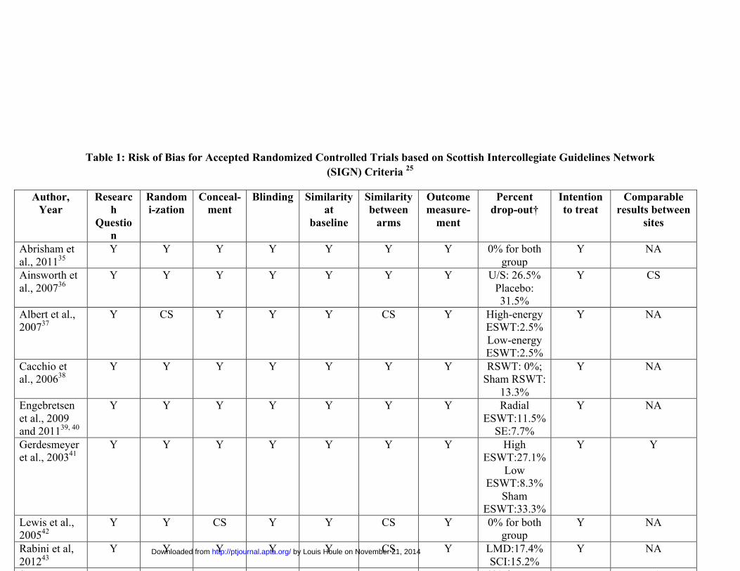
Table 1: Risk of Bias for Accepted Randomized Controlled Trials based on Scottish Intercollegiate Guidelines Network (SIGN) Criteria 25
Author, Year
Research
Question
Randomi-zation
Conceal-ment
Blinding Similarity at
baseline
Similarity between
arms
Outcome measure-
ment
Percent drop-out†
Intention to treat
Comparable results between
sites
Abrisham et al., 201135
Y Y Y Y Y Y Y 0% for both group
Y NA
Ainsworth et al., 200736
Y Y Y Y Y Y Y U/S: 26.5% Placebo: 31.5%
Y CS
Albert et al., 200737
Y CS Y Y Y CS Y High-energy ESWT:2.5% Low-energy ESWT:2.5%
Y NA
Cacchio et al., 200638
Y Y Y Y Y Y Y RSWT: 0%; Sham RSWT:
13.3%
Y NA
Engebretsen et al., 2009 and 201139, 40
Y Y Y Y Y Y Y Radial ESWT:11.5%
SE:7.7%
Y NA
Gerdesmeyer et al., 200341
Y Y Y Y Y Y Y High ESWT:27.1%
Low ESWT:8.3%
Sham ESWT:33.3%
Y Y
Lewis et al., 200542
Y Y CS Y Y CS Y 0% for both group
Y NA
Rabini et al, 201243
Y Y Y Y Y CS Y LMD:17.4% SCI:15.2%
Y NA
Santamato et Y Y Y Y Y Y Y 0% drop out Y NA
by Louis Houle on November 21, 2014http://ptjournal.apta.org/Downloaded from
39
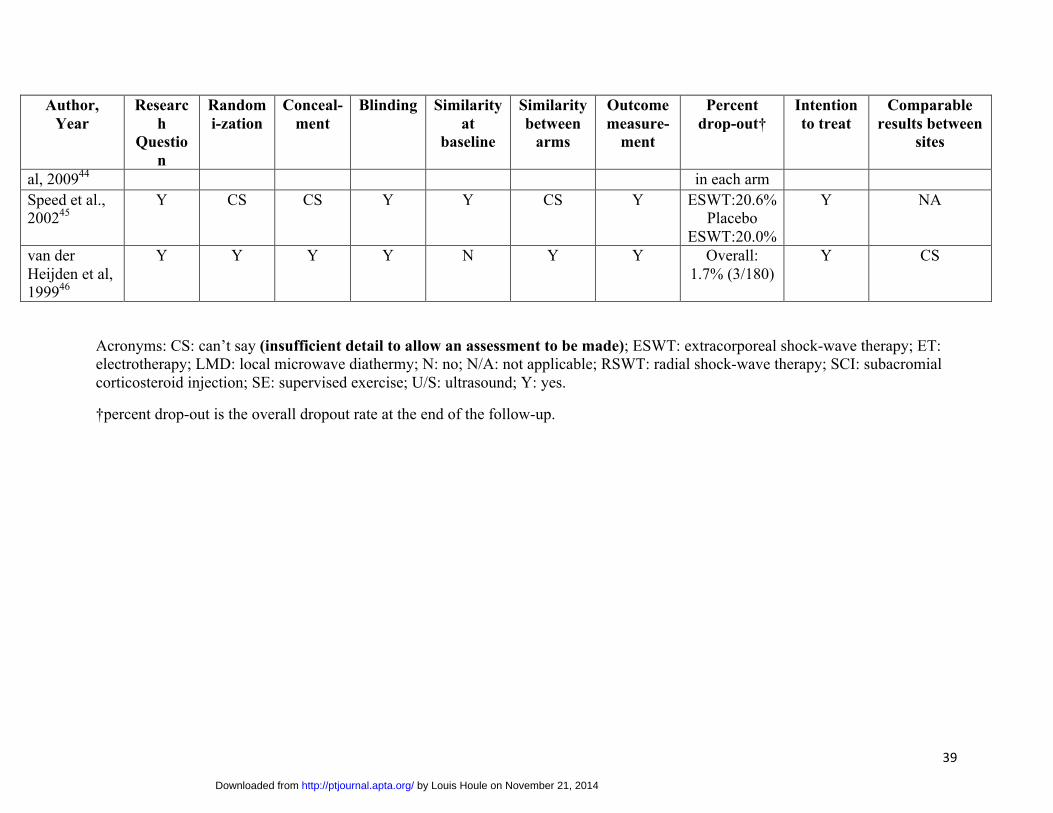
Author, Year
Research
Question
Randomi-zation
Conceal-ment
Blinding Similarity at
baseline
Similarity between
arms
Outcome measure-
ment
Percent drop-out†
Intention to treat
Comparable results between
sites
al, 200944 in each arm Speed et al., 200245
Y CS CS Y Y CS Y ESWT:20.6% Placebo
ESWT:20.0%
Y NA
van der Heijden et al, 199946
Y Y Y Y N Y Y Overall: 1.7% (3/180)
Y CS
Acronyms: CS: can’t say (insufficient detail to allow an assessment to be made); ESWT: extracorporeal shock-wave therapy; ET: electrotherapy; LMD: local microwave diathermy; N: no; N/A: not applicable; RSWT: radial shock-wave therapy; SCI: subacromial corticosteroid injection; SE: supervised exercise; U/S: ultrasound; Y: yes.
†percent drop-out is the overall dropout rate at the end of the follow-up.
by Louis Houle on November 21, 2014http://ptjournal.apta.org/Downloaded from
40
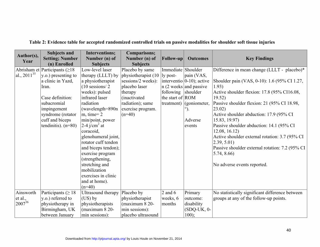
Table 2: Evidence table for accepted randomized controlled trials on passive modalities for shoulder soft tissue injuries
Author(s), Year
Subjects and Setting; Number
(n) Enrolled
Interventions; Number (n) of
Subjects
Comparisons; Number (n) of
Subjects Follow-up Outcomes Key Findings
Abrisham et al., 201135
Participants (≥18 y.o.) presenting toa clinic in Yazd, Iran.
Case definition: subacromial impingement syndrome (rotator cuff and biceps tendinitis). (n=80)
Low-level laser therapy (LLLT) by a physiotherapist (10 sessions/ 2 weeks): pulsed infrared laser radiation (wavelength=890nm, time= 2 min/point, power 2-4 j/cm2 at coracoid, glenohumeral joint, rotator cuff tendon and biceps tendon); exercise program (strengthening, stretching and mobilization exercises in clinic and at home). (n=40)
Placebo by same physiotherapist (10 sessions/2 weeks): placebo laser therapy (inactivated radiation); same exercise program. (n=40)
Immediately post-intervention (2 weeks following the start of treatment)
Shoulder pain (VAS, 0-10); active and passive shoulder ROM (goniometer, °).
Adverse events
Difference in mean change (LLLT - placebo)*
Shoulder pain (VAS, 0-10): 1.6 (95% CI 1.27, 1.93) Active shoulder flexion: 17.8 (95% CI16.08, 19.52) Passive shoulder flexion: 21 (95% CI 18.98, 23.02) Active shoulder abduction: 17.9 (95% CI 15.83, 19.97) Passive shoulder abduction: 14.1 (95% CI 12.08, 16.12) Active shoulder external rotation: 3.7 (95% CI 2.39, 5.01) Passive shoulder external rotation: 7.2 (95% CI 5.74, 8.66)
No adverse events reported.
Ainsworth et al., 200736
Participants (≥ 18 y.o.) referred tophysiotherapy in Birmingham, UK between January
Ultrasound therapy (US) by physiotherapists (maximum 8 20-min sessions):
Placebo by physiotherapist (maximum 8 20-min sessions): placebo ultrasound
2 and 6 weeks, 6 months
Primary outcome: disability (SDQ-UK, 0-100);
No statistically significant difference between groups at any of the follow-up points.
by Louis Houle on November 21, 2014http://ptjournal.apta.org/Downloaded from

41
Author(s), Year
Subjects and Setting; Number
(n) Enrolled
Interventions; Number (n) of
Subjects
Comparisons; Number (n) of
Subjects Follow-up Outcomes Key Findings
1999 and September 2001.
Case definition: unilateral shoulder pain exacerbated by active or passive shoulder movement. (n=221)
varied dose of pulsed ultrasound, exercise, manual therapy (no acupuncture and other electrotherapy modalities), and education (advice sheet). (n=113)
(inactivated), exercise, manual therapy (no acupuncture and other electrotherapy modalities), and education (advice sheet). (n=108)
Secondary outcome: global assessment of improvement (5-point scale); average pain during previous 24h (VAS 0-10); global perception of shoulder problem affection (VAS 0-10); global HRQoL (EuroQol EQ-5D and EuroQol health thermometer); range of movement.
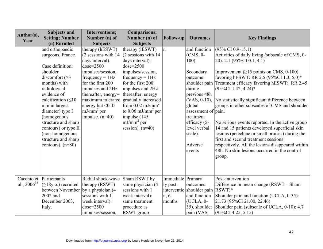
Albert et al., 200737
Participants (18-75 y.o.) referred byrheumatologists
High-energy extracorporeal shock-wave
Low-energy extracorporeal shock-wave
3 months post-interventio
Primary outcome: shoulder pain
Difference in mean change (hESWT – lESWT)
Shoulder pain and function (CMS, 0-100): 8.0
by Louis Houle on November 21, 2014http://ptjournal.apta.org/Downloaded from
42
Author(s), Year
Subjects and Setting; Number
(n) Enrolled
Interventions; Number (n) of
Subjects
Comparisons; Number (n) of
Subjects Follow-up Outcomes Key Findings
and orthopaedic surgeons, France.
Case definition: shoulder discomfort (≥3 months) with radiological evidence of calcification (≤10 mm in largest diameter) type I (homogenous structure and sharp contours) or type II (non-homogenous structure and sharp contours). (n=80)
therapy (hESWT) (2 sessions with 14 days interval): dose=2500 impulses/session, frequency = 1Hz for the first 200 impulses and 2Hz thereafter, energy= maximum tolerated energy but <0.45 mJ/mm2 per impulse. (n=40)
therapy (lESWT) (2 sessions with 14 days interval): dose=2500 impulses/session, frequency = 1Hz for the first 200 impulses and 2Hz thereafter, energy gradually increased from 0.02 mJ/mm2
to 0.06 mJ/mm2 per impulse (145 mJ/mm2 per session). (n=40)
n and function (CMS, 0-100);
Secondary outcome: shoulder pain during previous 48h (VAS, 0-10), global assessment of treatment efficacy (5-level verbal scale).
Adverse events
(95% CI 0.9-15.1) Activities of daily living (subscale of CMS, 0-20): 2.1 (95%CI 0.1, 4.1)
Improvement (≥15 points on CMS, 0-100) favoring hESWT: RR 2.5 (95%CI 1.3, 5.0)* Treatment efficacy favoring hESWT: RR 2.45 (95%CI 1.42, 4.24)*
No statistically significant difference between groups in other subscales of CMS and shoulder pain.
No serious events reported. In the active group 14 and 15 patients developed superficial skin lesions (petechiae or small bruises) during the first and second treatment sessions respectively. All the lesions disappeared within 48h. No skin lesions occurred in the control group.
Cacchio et al., 200638
Participants (≥18y.o.) recruited between November 2002 and December 2003, Italy.
Radial shock-wave therapy (RSWT) by a physician (4 sessions with 1 week interval): dose=2500 impulses/session,
Sham RSWT by same physician (4 sessions with 1 week interval): same treatment procedure as RSWT group
Immediately post-intervention, 6 months
Primary outcomes: shoulder pain and function (UCLA, 0-35), shoulder pain (VAS,
Post-intervention Difference in mean change (RSWT – Sham RSWT)* Shoulder pain and function (UCLA, 0-35): 21.73 (95%CI 21.00, 22.46) Shoulder pain (subscale of UCLA, 0-10): 4.7 (95%CI 4.25, 5.15)
by Louis Houle on November 21, 2014http://ptjournal.apta.org/Downloaded from
43
Author(s), Year
Subjects and Setting; Number
(n) Enrolled
Interventions; Number (n) of
Subjects
Comparisons; Number (n) of
Subjects Follow-up Outcomes Key Findings
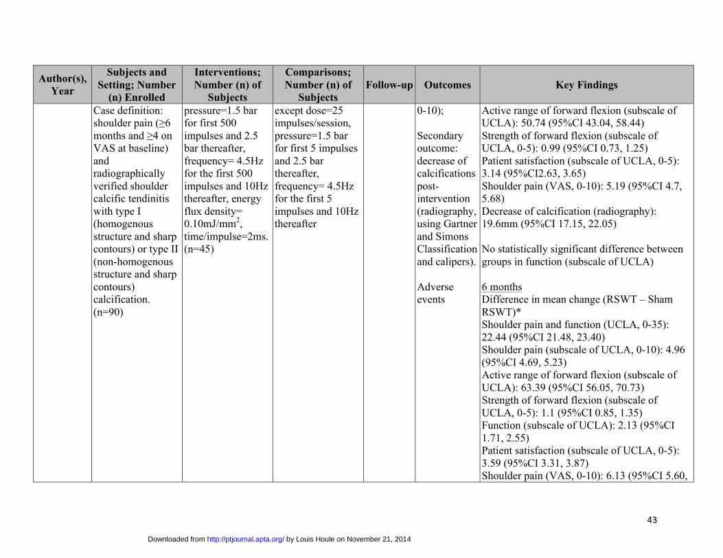
Case definition: shoulder pain (≥6 months and ≥4 on VAS at baseline) and radiographically verified shoulder calcific tendinitis with type I (homogenous structure and sharp contours) or type II (non-homogenous structure and sharp contours) calcification. (n=90)
pressure=1.5 bar for first 500 impulses and 2.5 bar thereafter, frequency= 4.5Hz for the first 500 impulses and 10Hz thereafter, energy flux density= 0.10mJ/mm2, time/impulse=2ms. (n=45)
except dose=25 impulses/session, pressure=1.5 bar for first 5 impulses and 2.5 bar thereafter, frequency= 4.5Hz for the first 5 impulses and 10Hz thereafter
0-10);
Secondary outcome: decrease of calcifications post-intervention (radiography, using Gartner and Simons Classification and calipers).
Adverse events
Active range of forward flexion (subscale of UCLA): 50.74 (95%CI 43.04, 58.44) Strength of forward flexion (subscale of UCLA, 0-5): 0.99 (95%CI 0.73, 1.25) Patient satisfaction (subscale of UCLA, 0-5): 3.14 (95%CI2.63, 3.65) Shoulder pain (VAS, 0-10): 5.19 (95%CI 4.7, 5.68) Decrease of calcification (radiography): 19.6mm (95%CI 17.15, 22.05)
No statistically significant difference between groups in function (subscale of UCLA)
6 months Difference in mean change (RSWT – Sham RSWT)* Shoulder pain and function (UCLA, 0-35): 22.44 (95%CI 21.48, 23.40) Shoulder pain (subscale of UCLA, 0-10): 4.96 (95%CI 4.69, 5.23) Active range of forward flexion (subscale of UCLA): 63.39 (95%CI 56.05, 70.73) Strength of forward flexion (subscale of UCLA, 0-5): 1.1 (95%CI 0.85, 1.35) Function (subscale of UCLA): 2.13 (95%CI 1.71, 2.55) Patient satisfaction (subscale of UCLA, 0-5): 3.59 (95%CI 3.31, 3.87) Shoulder pain (VAS, 0-10): 6.13 (95%CI 5.60,
by Louis Houle on November 21, 2014http://ptjournal.apta.org/Downloaded from
44
Author(s), Year
Subjects and Setting; Number
(n) Enrolled
Interventions; Number (n) of
Subjects
Comparisons; Number (n) of
Subjects Follow-up Outcomes Key Findings
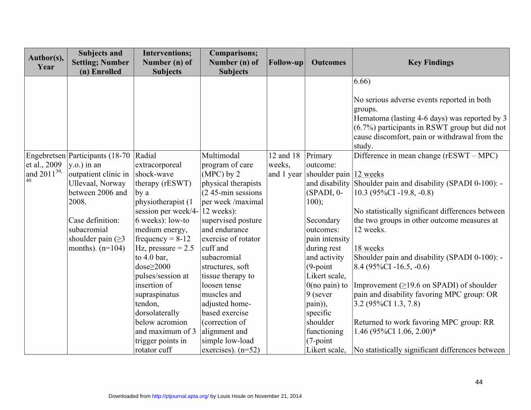
6.66)
No serious adverse events reported in both groups. Hematoma (lasting 4-6 days) was reported by 3 (6.7%) participants in RSWT group but did not cause discomfort, pain or withdrawal from the study.
Engebretsen et al., 2009 and 201139,
40
Participants (18-70 y.o.) in anoutpatient clinic in Ullevaal, Norway between 2006 and 2008.
Case definition: subacromial shoulder pain (≥3 months). (n=104)
Radial extracorporeal shock-wave therapy (rESWT) by a physiotherapist (1 session per week/4-6 weeks): low-to medium energy, frequency = 8-12 Hz, pressure = 2.5 to 4.0 bar, dose≥2000 pulses/session at insertion of supraspinatus tendon, dorsolaterally below acromion and maximum of 3 trigger points in rotator cuff
Multimodal program of care (MPC) by 2 physical therapists (2 45-min sessions per week /maximal 12 weeks): supervised posture and endurance exercise of rotator cuff and subacromial structures, soft tissue therapy to loosen tense muscles and adjusted home-based exercise (correction of alignment and simple low-load exercises). (n=52)
12 and 18 weeks, and 1 year
Primary outcome: shoulder pain and disability (SPADI, 0-100);
Secondary outcomes: pain intensity during rest and activity (9-point Likert scale, 0(no pain) to 9 (sever pain)), specific shoulder functioning (7-point Likert scale,
Difference in mean change (rESWT – MPC)
12 weeks Shoulder pain and disability (SPADI 0-100): -10.3 (95%CI -19.8, -0.8)
No statistically significant differences between the two groups in other outcome measures at 12 weeks.
18 weeks Shoulder pain and disability (SPADI 0-100): -8.4 (95%CI -16.5, -0.6)
Improvement (≥19.6 on SPADI) of shoulder pain and disability favoring MPC group: OR 3.2 (95%CI 1.3, 7.8)
Returned to work favoring MPC group: RR 1.46 (95%CI 1.06, 2.00)*
No statistically significant differences between
by Louis Houle on November 21, 2014http://ptjournal.apta.org/Downloaded from

45
Author(s), Year
Subjects and Setting; Number
(n) Enrolled
Interventions; Number (n) of
Subjects
Comparisons; Number (n) of
Subjects Follow-up Outcomes Key Findings
muscles. (n=52) 1 (easy) to 7 (impossible)), active ROM, returned to work, daily and weekly medication used.
Adverse events
the two groups in pain during rest and activity, function, active ROM, and medication use.
1 year: Returned to work favoring MPC group: OR 1.1 (95%CI 1.0, 1.2)
No statistically significant differences between the two groups in the shoulder pain and disability (SPADI), pain intensity during rest and activity, function and medication use.
Adverse events: 2 participants reported aggravation of pain in rESWT group.
Gerdesmeyer et al., 200341
Participants (≥18 y.o.) referred byprimary care physicians, orthopedic surgeons and sports physicians from 7 orthopedic departments in Germany and Austria between February 1997 and March 2001.
Case definition: shoulder symptoms
High-energy extracorporeal shock-wave therapy (hESWT) (2 1-hour sessions with 12 to16 days interval followed by 10 physiotherapy sessions): dose=1500 impulses/session, energy=0.32mJ/mm2, frequency=120 impulses/minute, total energy
Low-energy extracorporeal shock-wave therapy (lESWT) (2 1-hour sessions with 12 to 16 days interval followed by 10 physiotherapy sessions): Dose=6000 impulses/session, energy=0.08mJ/mm2, frequency=120 impulses/minute, total energy
3, 6 and 12 months post-intervention
Primary outcome: shoulder pain and function (CMS, 0-100) at 6 months post-intervention;
Secondary outcomes: shoulder pain and function (CMS, 0-100) at 3 and 12 months
3 months Difference in mean change (hESWT – sESWT) Shoulder pain and function (CMS, 0-100): 16.4 (95%CI 10.3, 22.5) Proportion of patients with 30% improvement on CMS: 0.56 (95%CI 0.36, 0.71) Pain (VAS, 0-10): 3.2 (95%CI 2.2, 4.2) Decrease of calcific deposit (mm2): 98.6 (95%CI 51.8, 145.4)
Difference in mean change (lESWT – sESWT) Pain and function (CMS, 0-100): 6.6 (95%CI 0.1, 13.1) Shoulder ADL (subscale of CMS, 0-20): 2.0 (95%CI 0.2, 3.7)
by Louis Houle on November 21, 2014http://ptjournal.apta.org/Downloaded from
46
Author(s), Year
Subjects and Setting; Number
(n) Enrolled
Interventions; Number (n) of
Subjects
Comparisons; Number (n) of
Subjects Follow-up Outcomes Key Findings
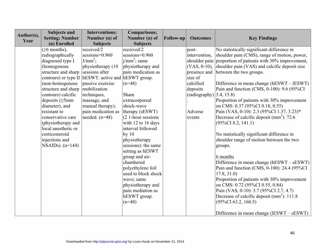
(≥6 months), radiographically diagnosed type I (homogenous structure and sharp contours) or type II (non-homogenous structure and sharp contours) calcific deposits (≥5mm diameter), and resistant to conservative care (physiotherapy and local anesthetic or corticosteroid injections and NSAIDs). (n=144)
received/2 sessions=0.960 J/mm2; physiotherapy (10 sessions after hESWT: active and passive exercise mobilization techniques, massage, and manual therapy); pain medication as needed. (n=48)
received/2 sessions=0.960 j/mm2; same physiotherapy and pain medication as hESWT group. (n=48)
Sham extracorporeal shock-wave therapy (sESWT) (2 1-hour sessions with 12 to 16 days interval followed by 10 physiotherapy sessions): the same setting as hESWT group and air-chambered polyethylene foil used to block shock wave; same physiotherapy and pain mediation as hESWT group. (n=48)
post-intervention, shoulder pain (VAS, 0-10), presence and size of calcified deposits (radiography).
Adverse events
No statistically significant difference in shoulder pain (CMS), range of motion, power, proportion of patients with 30% improvement, shoulder pain (VAS) and calcific deposit size between the two groups.
Difference in mean change (hESWT – lESWT) Pain and function (CMS, 0-100): 9.6 (95%CI 3.4, 15.8) Proportion of patients with 30% improvement on CMS: 0.37 (95%CI 0.18, 0.55) Pain (VAS, 0-10): 2.3 (95%CI 1.37, 3.23)* Decrease of calcific deposit (mm2): 72.6 (95%CI 8.2, 141.1)
No statistically significant difference in shoulder range of motion between the two groups.
6 months Difference in mean change (hESWT – sESWT) Pain and function (CMS, 0-100): 24.4 (95%CI 17.8, 31.0) Proportion of patients with 30% improvement on CMS: 0.72 (95%CI 0.55, 0.84) Pain (VAS, 0-10): 3.7 (95%CI 2.7, 4.7) Decrease of calcific deposit (mm2): 111.8 (95%CI 63.2, 160.5)
Difference in mean change (lESWT – sESWT)
by Louis Houle on November 21, 2014http://ptjournal.apta.org/Downloaded from

47
Author(s), Year
Subjects and Setting; Number
(n) Enrolled
Interventions; Number (n) of
Subjects
Comparisons; Number (n) of
Subjects Follow-up Outcomes Key Findings
Pain and function (CMS, 0-100): 8.4 (95%CI 1.4, 15.4) Proportion of patients with 30% improvement on CMS: 0.24 (95%CI 0.05, 0.42) Pain (VAS, 0-10): 1.3 (95%CI 0.4, 2.2)
No statistically significant difference in decrease of calcific deposits between the two groups.
Difference in mean change (hESWT – lESWT) Pain and function (CMS, 0-100): 16.0 (95%CI 10.8, 22.9) Proportion of patients with 30% improvement on CMS: 0.48 (95%CI 0.30, 0.63) Pain (VAS, 0-10): 3.1 (95%CI 2.5, 4.3) Decrease of calcific deposit (mm2): 75.1 (95%CI 9.0, 144.3)
12 months Difference in mean changes (hESWT – sESWT) Pain and function (CMS, 0-100): 17.9 (95%CI 11.1, 24.7) Proportion of patients with 30% improvement on CMS: 0.72 (95%CI 0.53, 0.85) Pain (VAS, 0-10): 3.7 (95%CI 2.7, 4.7) Decrease of calcific deposit (mm2): 115.4 (95%CI 65.4, 165.4)
by Louis Houle on November 21, 2014http://ptjournal.apta.org/Downloaded from

48
Author(s), Year
Subjects and Setting; Number
(n) Enrolled
Interventions; Number (n) of
Subjects
Comparisons; Number (n) of
Subjects Follow-up Outcomes Key Findings
Difference in mean change (lESWT – sESWT) Proportion of patients with 30% improvement on CMS: 0.23 (95%CI 0.01, 0.43)
No statistically significant difference in shoulder pain and function, shoulder pain (CMS), activities of daily living, range of motion, power, shoulder pain (VAS) and calcific deposit size between the two groups.
Difference in mean change (hESWT – lESWT) Pain and function (CMS, 0-100): 13.9 (95%CI 8.3, 19.7) Proportion of patients with 30% improvement on CMS: 0.49 (95%CI 0.31, 0.64) Pain (VAS, 0-10): 3.0 (95%CI 2.3, 3.7) Decrease of calcific deposit (mm2): 70.7 (95%CI 1.9, 139.5)
No serious adverse events except severe pain. 20 participants reported moderate pain and 16 reported severe pain in the hESWT group. 22 participants reported moderate pain and 5 reported severe pain in the lESWT group. 25 participants reported some sensation of pain in the sESWT group.
Petechiae, bleeding, hematoma, or erythema were found in three groups (36 in hESWT, 32 in lESWT and 8 in sESWT).
by Louis Houle on November 21, 2014http://ptjournal.apta.org/Downloaded from
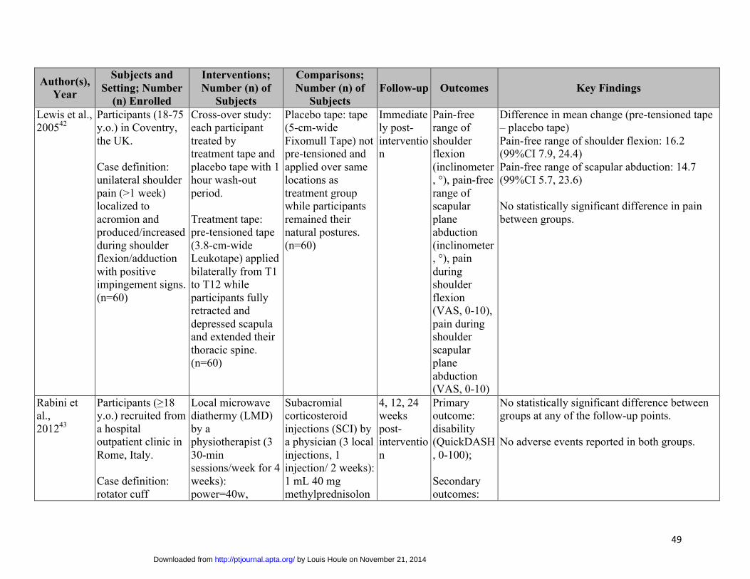
49
Author(s), Year
Subjects and Setting; Number
(n) Enrolled
Interventions; Number (n) of
Subjects
Comparisons; Number (n) of
Subjects Follow-up Outcomes Key Findings
Lewis et al., 200542
Participants (18-75 y.o.) in Coventry, the UK. Case definition: unilateral shoulder pain (>1 week) localized to acromion and produced/increased during shoulder flexion/adduction with positive impingement signs. (n=60)
Cross-over study: each participant treated by treatment tape and placebo tape with 1 hour wash-out period. Treatment tape: pre-tensioned tape (3.8-cm-wide Leukotape) applied bilaterally from T1 to T12 while participants fully retracted and depressed scapula and extended their thoracic spine. (n=60)
Placebo tape: tape (5-cm-wide Fixomull Tape) not pre-tensioned and applied over same locations as treatment group while participants remained their natural postures. (n=60)
Immediately post-intervention
Pain-free range of shoulder flexion (inclinometer, °), pain-free range of scapular plane abduction (inclinometer, °), pain during shoulder flexion (VAS, 0-10), pain during shoulder scapular plane abduction (VAS, 0-10)
Difference in mean change (pre-tensioned tape – placebo tape) Pain-free range of shoulder flexion: 16.2 (99%CI 7.9, 24.4) Pain-free range of scapular abduction: 14.7 (99%CI 5.7, 23.6) No statistically significant difference in pain between groups.
Rabini et al., 201243
Participants (≥18 y.o.) recruited from a hospital outpatient clinic in Rome, Italy. Case definition: rotator cuff
Local microwave diathermy (LMD) by a physiotherapist (3 30-min sessions/week for 4 weeks): power=40w,
Subacromial corticosteroid injections (SCI) by a physician (3 local injections, 1 injection/ 2 weeks): 1 mL 40 mg methylprednisolon
4, 12, 24 weeks post-intervention
Primary outcome: disability (QuickDASH, 0-100); Secondary outcomes:
No statistically significant difference between groups at any of the follow-up points. No adverse events reported in both groups.
by Louis Houle on November 21, 2014http://ptjournal.apta.org/Downloaded from
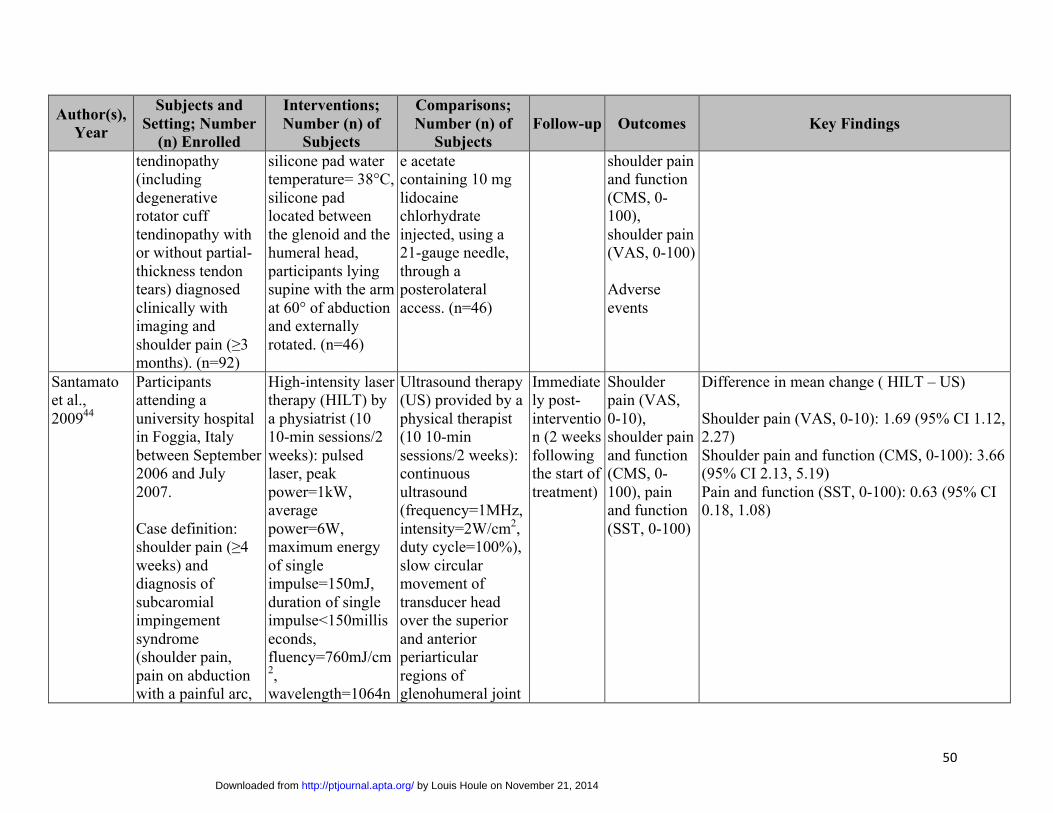
50
Author(s), Year
Subjects and Setting; Number
(n) Enrolled
Interventions; Number (n) of
Subjects
Comparisons; Number (n) of
Subjects Follow-up Outcomes Key Findings
tendinopathy (including degenerative rotator cuff tendinopathy with or without partial-thickness tendon tears) diagnosed clinically with imaging and shoulder pain (≥3 months). (n=92)
silicone pad water temperature= 38°C, silicone pad located between the glenoid and the humeral head, participants lying supine with the arm at 60° of abduction and externally rotated. (n=46)
e acetate containing 10 mg lidocaine chlorhydrate injected, using a 21-gauge needle, through a posterolateral access. (n=46)
shoulder pain and function (CMS, 0-100), shoulder pain (VAS, 0-100)
Adverse events
Santamato et al., 200944
Participants attending a university hospital in Foggia, Italy between September 2006 and July 2007.
Case definition: shoulder pain (≥4 weeks) and diagnosis of subcaromial impingement syndrome (shoulder pain, pain on abduction with a painful arc,
High-intensity laser therapy (HILT) by a physiatrist (10 10-min sessions/2 weeks): pulsed laser, peak power=1kW, average power=6W, maximum energy of single impulse=150mJ, duration of single impulse<150milliseconds, fluency=760mJ/cm2, wavelength=1064n
Ultrasound therapy (US) provided by a physical therapist (10 10-min sessions/2 weeks): continuous ultrasound (frequency=1MHz, intensity=2W/cm2, duty cycle=100%), slow circular movement of transducer head over the superior and anterior periarticular regions of glenohumeral joint
Immediately post-intervention (2 weeks following the start of treatment)
Shoulder pain (VAS, 0-10), shoulder pain and function (CMS, 0-100), pain and function (SST, 0-100)
Difference in mean change ( HILT – US)
Shoulder pain (VAS, 0-10): 1.69 (95% CI 1.12, 2.27) Shoulder pain and function (CMS, 0-100): 3.66 (95% CI 2.13, 5.19) Pain and function (SST, 0-100): 0.63 (95% CI 0.18, 1.08)
by Louis Houle on November 21, 2014http://ptjournal.apta.org/Downloaded from
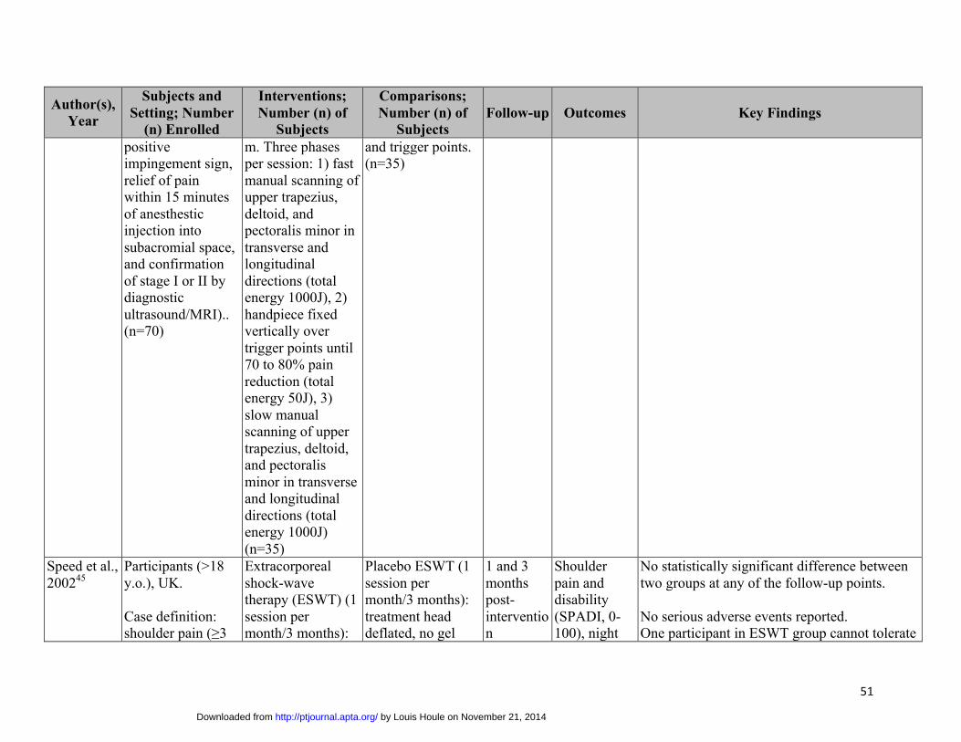
51
Author(s), Year
Subjects and Setting; Number
(n) Enrolled
Interventions; Number (n) of
Subjects
Comparisons; Number (n) of
Subjects Follow-up Outcomes Key Findings
positive impingement sign, relief of pain within 15 minutes of anesthestic injection into subacromial space, and confirmation of stage I or II by diagnostic ultrasound/MRI).. (n=70)
m. Three phasesper session: 1) fast manual scanning of upper trapezius, deltoid, and pectoralis minor in transverse and longitudinal directions (total energy 1000J), 2) handpiece fixed vertically over trigger points until 70 to 80% pain reduction (total energy 50J), 3) slow manual scanning of upper trapezius, deltoid, and pectoralis minor in transverse and longitudinal directions (total energy 1000J) (n=35)
and trigger points. (n=35)
Speed et al., 200245
Participants (>18 y.o.), UK.
Case definition: shoulder pain (≥3
Extracorporeal shock-wave therapy (ESWT) (1 session per month/3 months):
Placebo ESWT (1 session per month/3 months): treatment head deflated, no gel
1 and 3 months post-intervention
Shoulder pain and disability (SPADI, 0-100), night
No statistically significant difference between two groups at any of the follow-up points.
No serious adverse events reported. One participant in ESWT group cannot tolerate
by Louis Houle on November 21, 2014http://ptjournal.apta.org/Downloaded from
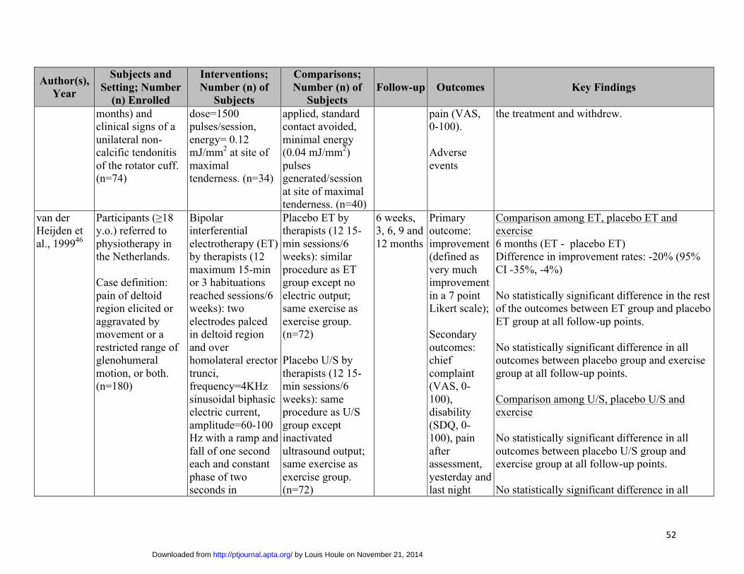
52
Author(s), Year
Subjects and Setting; Number
(n) Enrolled
Interventions; Number (n) of
Subjects
Comparisons; Number (n) of
Subjects Follow-up Outcomes Key Findings
months) and clinical signs of a unilateral non-calcific tendonitis of the rotator cuff. (n=74)
dose=1500 pulses/session, energy= 0.12 mJ/mm2 at site of maximal tenderness. (n=34)
applied, standard contact avoided, minimal energy (0.04 mJ/mm2) pulses generated/session at site of maximal tenderness. (n=40)
pain (VAS, 0-100).
Adverse events
the treatment and withdrew.
van der Heijden et al., 199946
Participants (≥18 y.o.) referred tophysiotherapy in the Netherlands.
Case definition: pain of deltoid region elicited or aggravated by movement or a restricted range of glenohumeral motion, or both. (n=180)
Bipolar interferential electrotherapy (ET) by therapists (12 maximum 15-min or 3 habituations reached sessions/6 weeks): two electrodes palced in deltoid region and over homolateral erector trunci, frequency=4KHz sinusoidal biphasic electric current, amplitude=60-100 Hz with a ramp and fall of one second each and constant phase of two seconds in
Placebo ET by therapists (12 15-min sessions/6 weeks): similar procedure as ET group except no electric output; same exercise as exercise group. (n=72)
Placebo U/S by therapists (12 15-min sessions/6 weeks): same procedure as U/S group except inactivated ultrasound output; same exercise as exercise group. (n=72)
6 weeks, 3, 6, 9 and 12 months
Primary outcome: improvement (defined as very much improvement in a 7 point Likert scale);
Secondary outcomes: chief complaint (VAS, 0-100), disability (SDQ, 0-100), pain after assessment, yesterday and last night
Comparison among ET, placebo ET and exercise 6 months (ET - placebo ET) Difference in improvement rates: -20% (95% CI -35%, -4%)
No statistically significant difference in the rest of the outcomes between ET group and placebo ET group at all follow-up points.
No statistically significant difference in all outcomes between placebo group and exercise group at all follow-up points.
Comparison among U/S, placebo U/S and exercise
No statistically significant difference in all outcomes between placebo U/S group and exercise group at all follow-up points.
No statistically significant difference in all
by Louis Houle on November 21, 2014http://ptjournal.apta.org/Downloaded from
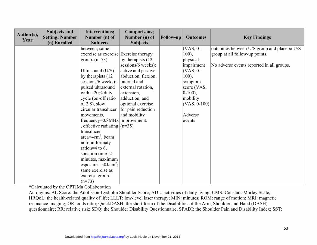
53
Author(s), Year
Subjects and Setting; Number
(n) Enrolled
Interventions; Number (n) of
Subjects
Comparisons; Number (n) of
Subjects Follow-up Outcomes Key Findings
between; same exercise as exercise group. (n=73)
Ultrasound (U/S) by therapists (12 sessions/6 weeks): pulsed ultrasound with a 20% duty cycle (on-off ratio of 2:8), slow circular transducer movements, frequency=0.8MHz, effective radiating transducer area=4cm2, beam non-uniformaty ration=4 to 6, sonation time=2 minutes, maximum esposure= 50J/cm2; same exercise as exercise group. (n=73)
Exercise therapy by therapists (12 sessions/6 weeks): active and passive abduction, flexion, internal and external rotation, extension, adduction, and optional exercise for pain reduction and mobility improvement. (n=35)
(VAS, 0-100), physical impairment (VAS, 0-100), symptom score (VAS, 0-100), mobility (VAS, 0-100)
Adverse events
outcomes between U/S group and placebo U/S group at all follow-up points.
No adverse events reported in all groups.
*Calculated by the OPTIMa CollaborationAcronyms: AL Score: the Adolfsson-Lysholm Shoulder Score; ADL: activities of daily living; CMS: Constant-Murley Scale; HRQoL: the health-related quality of life; LLLT: low-level laser therapy; MIN: minutes; ROM: range of motion; MRI: magnetic resonance imaging; OR: odds ratio; QuickDASH: the short form of the Disabilities of the Arm, Shoulder and Hand (DASH) questionnaire; RR: relative risk; SDQ: the Shoulder Disability Questionnaire; SPADI: the Shoulder Pain and Disability Index; SST:
by Louis Houle on November 21, 2014http://ptjournal.apta.org/Downloaded from

54
the Simple Shoulder Test; UCLA: the University of California-Los Angeles shoulder Rating Scale; VAS: Visual Analogue Scale; y.o.: years old.
by Louis Houle on November 21, 2014http://ptjournal.apta.org/Downloaded from

56
Table 3: Parameters of passive physical modalities in accepted randomized controlled trials
Author(s), Year Diagnoses Interventions Comparisons
Calcific shoulder pain Albert et al., 200737
Persistent shoulder calcific tendonitis
High-energy extracorporeal shock-wave therapy (hESWT); • Focused• dose=2500 impulses/session, frequency = 1Hz for the
first 200 impulses and 2Hz thereafter, energy= maximumtolerated energy but <0.45 mJ/mm2 per impulse
Low-energy extracorporeal shock-wave therapy (lESWT):
• Pulsed• dose=2500 impulses/session,
frequency = 1Hz for the first 200impulses and 2Hz thereafter,energy gradually increased from0.02 mJ/mm2 to 0.06 mJ/mm2 perimpulse (145 mJ/mm2 per session)
Cacchio et al., 200638
Persistent shoulder calcific tendonitis
Radial shock-wave therapy (RSWT) : • Radial• dose=2500 impulses/session, pressure=1.5 bar for first
500 impulses and 2.5 bar thereafter, frequency= 4.5Hzfor the first 500 impulses and 10Hz thereafter, energyflux density= 0.10mJ/mm2, time/impulse=2ms
Sham RSWT: same treatment procedure as RSWT group except dose=25 impulses/session, pressure=1.5 bar for first 5 impulses and 2.5 bar thereafter, frequency= 4.5Hz for the first 5 impulses and 10Hz thereafter
Gerdesmeyer et al., 200341
Persistent shoulder calcific tendonitis
High-energy extracorporeal shock-wave therapy (hESWT) : • Focused• dose=1500 impulses/session, energy=0.32mJ/mm2,
frequency=120 impulses/minute, total energy received/2sessions=0.960 J/mm2
Low-energy extracorporeal shock-wave therapy (lESWT):
• Pulsed• Dose=6000 impulses/session,
energy=0.08mJ/mm2,frequency=120 impulses/minute,total energy received/2sessions=0.960 j/mm2
Sham extracorporeal shock-wave therapy (sESWT); the same setting as hESWT group and air-chambered polyethylene foil used to block
by Louis Houle on November 21, 2014http://ptjournal.apta.org/Downloaded from
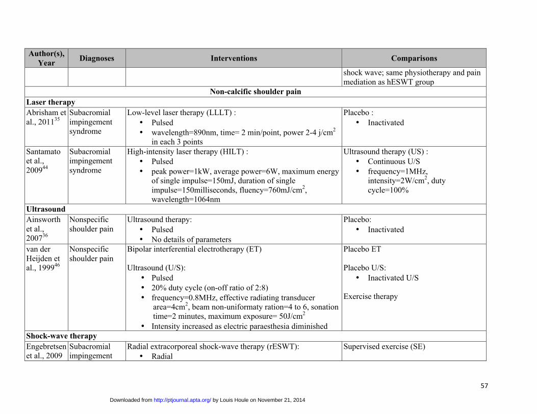
57
Author(s), Year Diagnoses Interventions Comparisons
shock wave; same physiotherapy and pain mediation as hESWT group
Non-calcific shoulder pain Laser therapy Abrisham et al., 201135
Subacromial impingement syndrome
Low-level laser therapy (LLLT) : • Pulsed• wavelength=890nm, time= 2 min/point, power 2-4 j/cm2
in each 3 points
Placebo : • Inactivated
Santamato et al., 200944
Subacromial impingement syndrome
High-intensity laser therapy (HILT) : • Pulsed• peak power=1kW, average power=6W, maximum energy
of single impulse=150mJ, duration of singleimpulse=150milliseconds, fluency=760mJ/cm2,wavelength=1064nm
Ultrasound therapy (US) : • Continuous U/S• frequency=1MHz,
intensity=2W/cm2, dutycycle=100%
Ultrasound Ainsworth et al., 200736
Nonspecific shoulder pain
Ultrasound therapy: • Pulsed• No details of parameters
Placebo: • Inactivated
van der Heijden et al., 199946
Nonspecific shoulder pain
Bipolar interferential electrotherapy (ET)
Ultrasound (U/S): • Pulsed• 20% duty cycle (on-off ratio of 2:8)• frequency=0.8MHz, effective radiating transducer
area=4cm2, beam non-uniformaty ration=4 to 6, sonationtime=2 minutes, maximum exposure= 50J/cm2
• Intensity increased as electric paraesthesia diminished
Placebo ET
Placebo U/S: • Inactivated U/S
Exercise therapy
Shock-wave therapy Engebretsen et al., 2009
Subacromial impingement
Radial extracorporeal shock-wave therapy (rESWT): • Radial
Supervised exercise (SE)
by Louis Houle on November 21, 2014http://ptjournal.apta.org/Downloaded from

58
Author(s), Year Diagnoses Interventions Comparisons
and 201139,
40syndrome • frequency = 8-12 Hz, pressure = 2.5 to 4.0 bar,
dose≥2000 pulses/session• Low to medium energy shock-wave (no number
provided)Speed et al., 200245
Subacromial impingement syndrome
Extracorporeal shock-wave therapy (ESWT): • Focused• dose=1500 pulses/session, energy= 0.12 mJ/mm2 at site
of maximal tenderness
Placebo ESWT: treatment head deflated, no gel applied, standard contact avoided, minimal energy (0.04 mJ/mm2) pulses generated/session at site of maximal tenderness
Diathermy Rabini et al., 201243
Subacromial impingement syndrome
Local microwave diathermy (LMD): power=40w, silicone pad water temperature= 38°C
Subacromial corticosteroid injections (SCI)
Efficacy study Lewis et al., 200542
Subacromial impingement syndrome
Pre-tensioned tape Placebo tape
by Louis Houle on November 21, 2014http://ptjournal.apta.org/Downloaded from
60
Figure Legends
Figure 1: Identification and Selection of Articles
by Louis Houle on November 21, 2014http://ptjournal.apta.org/Downloaded from

61
Figure 1: Identification and Selection of Articles
Citations identified through database searching: 1760
Duplicates removed: 290
Citations screened: 1470
(Title/abstracts screening)
Citations excluded due to ineligibility: 1427
Eligible for critical appraisal in full text: 22
Articles with a high risk of bias: 10
Articles with a low risk of bias: 12 (reporting 11 randomized
controlled trials)
Citations screened: 43
(Full-text screening) Full-text articles excluded: 20
Primary reasons for exclusion: - Small sample size = 9 - Ineligible design = 5 - Shoulder specific results not reported = 3 - Ineligible intervention of interest = 2 - Non-English article = 1
Article could not be retrieved: 1
by Louis Houle on November 21, 2014http://ptjournal.apta.org/Downloaded from
62
Appendix I: MEDLINE through OVID search Strategy
1. exp Hydrotherapy/
2. Laser Therapy, Low-Level/
3. Cryotherapy/
4. Magnetic Field Therapy/
5. exp Electric Stimulation Therapy/
6. exp Orthotic Devices/
7. exp Diathermy/
8. Hot Temperature/tu [Therapeutic Use]
9. Casts, Surgical/
10. Fluid Therapy/
11. Magnetics/tu [Therapeutic Use]
12. "Bedding and Linens"/
13. High-Energy Shock Waves/tu [Therapeutic Use]
14. Bed Rest/
15. Rest/
16. Self-Help Devices/
17. Restraint, Physical/
18. or/1-17
19. (cold and (therap* or pack* or compress or massage or immersion or soak or treatment or
therap*)).ab,ti.
20. (ice and (therap* or pack* or compress or massage or immersion or soak or treatment or
therap*)).ab,ti.
21. (heat* and (therap* or pack* or compress or massage or lamp or pad or bath or soak or tub or
bottle or superficial or therapeutic)).ab,ti.
22. (hot and (therap* or pack* or compress or massage or lamp or pad or bath or soak or tub or
bottle or superficial or therapeutic)).ab,ti.
23. ((shockwave* or shock wave* or shock-wave*) and (ultrasonic or therap* or radiation)).ab,ti.
by Louis Houle on November 21, 2014http://ptjournal.apta.org/Downloaded from
63
24. "assistive device*".ab,ti.
25. (athletic and (tape or taping)).ab,ti.
26. "back belt*".ab,ti.
27. (braces or brace or bracing).ab,ti.
28. (cast or casts).ab,ti.
29. (collar or collars).ab,ti.
30. (corset or corsets).ab,ti.
31. "cryotherap*".ab,ti.
32. diathermy.ab,ti.
33. (electric* and (stimulation or EMS or heating pad*)).ab,ti.
34. electroanalgesia.ab,ti.
35. (electrogalvanic stimulation or EGS).ab,ti.
36. (electromagnet* and (radiation or therap*)).ab,ti.
37. "electromodalit*".ab,ti.
38. electrotherapy.ab,ti.
39. "fluidotherap*".ab,ti.
40. galvanic stimulation.ab,ti.
41. (guard* and (teeth or night or mouth or wrist or knee)).ab,ti.
42. (high energy shock wave* or high-energy shock wave* or HESW).ab,ti.
43. (H-Wave Device Stimulation or HWDS).ab,ti.
44. "hydrocollar*".ab,ti.
45. "hydrotherap*".ab,ti.
46. infrared.ab,ti.
47. (interferential current* or ICS or IFC).ab,ti.
48. iontophoresis.ab,ti.
49. "kinesiotap*".ab,ti.
50. (laser* and (phototherapy or irradiation or biostimulation or light or therap*)).ab,ti.
51. "low level laser*".ab,ti.
52. "lumbar support*".ab,ti.
53. (magnetic and (necklace* or therap* or bracelet*)).ab,ti.
54. Microcurrent Electrical Neuromuscular Stimulation.ab,ti.
by Louis Houle on November 21, 2014http://ptjournal.apta.org/Downloaded from

64
55. "microwave*".ab,ti.
56. "moist air bath*".ab,ti.
57. muscle activation.ab,ti.
58. myofascial release.ab,ti.
59. (Neuromuscular Electrical Stimulation or NMES).ab,ti.
60. "orthotic*".ab,ti.
61. (paraffin and (treatment* or therap*)).ab,ti.
62. "passive modalit*".ab,ti.
63. "Percutaneous Electric* Nerve Stimulation".ab,ti.
64. "pillow*".ab,ti.
65. (pulsed and (electromagnetic or magnetic or radio frequency or energy)).ab,ti.
66. radiant light.ab,ti.
67. Russian stimulation.ab,ti.
68. "seat cushion*".ab,ti.
69. (short wave* or short-wave*).ab,ti.
70. (sling or slings).ab,ti.
71. (splint or splinting or splints).ab,ti.
72. "spray and stretch".ab,ti.
73. (tape or taping).ab,ti.
74. (transcutaneous electrical stimulation or TENS).ab,ti.
75. ultrasound.ab,ti.
76. vapocoolant spray.ab,ti.
77. "vibration therap*".ab,ti.
78. "warm compress*".ab,ti.
79. "wax treatment*".ab,ti.
80. whirlpool.ab,ti.
81. or/19-80
82. 18 or 81
83. Shoulder Pain/
84. Shoulder Impingement Syndrome/
85. exp Shoulder Joint/in [Injuries]
by Louis Houle on November 21, 2014http://ptjournal.apta.org/Downloaded from

65
86. Rotator Cuff/
87. Shoulder/in [Injuries]
88. "Sprains and Strains"/
89. "shoulder*".ab,ti.
90. 88 and 89
91. (shoulder* and (pain or sprain* or strain* or injur* or impair* or impingement)).ab,ti.
92. (shoulder* and (tendinopathy or tendinitis or tendonitis or capsulitis)).ab,ti.
93. ((glenohumeral or scapul* or acromioclavicular) and (pain or sprain* or strain* or
injur*)).ab,ti.
94. (rotator cuff* and (sprain* or strain* or tear* or bursitis tendinitis or impingement)).ab,ti.
95. ((supraspinatus or infraspinatus or subscapularis or teres minor or teres major or trapezius or
deltoid or bicep* or bicipital) and (impingement or strain* or tear*)).ab,ti.
96. biceps tendinitis.ab,ti.
97. painful arc.ab,ti.
98. (shoulder and capsul* and (sprain* or tear*)).ab,ti.
99. 83 or 84 or 85 or 86 or 87 or 90 or 91 or 92 or 93 or 94 or 95 or 96 or 97 or 98
100. Randomized Controlled Trials as Topic/
101. exp Controlled Clinical Trials as Topic/
102. exp Case-Control Studies/
103. exp Cohort Studies/
104. exp Double-Blind Method/
105. exp Single-Blind Method/
106. Placebos/
107. randomized controlled trial.pt.
108. controlled clinical trial.pt.
109. (meta analys* or meta-analys* or metaanalys*).ab,ti.
110. (cohort adj4 (study or studies or analys*)).ab,ti.
111. (random* adj4 (control* or clinical or allocat*)).ab,ti.
112. (case adj control*).ab,ti.
113. ((double or single) adj4 blind*).ab,ti.
114. "placebo*".ab,ti.
by Louis Houle on November 21, 2014http://ptjournal.apta.org/Downloaded from

66
115. or/100-114
116. 82 and 99 and 115
117. 82 and 99 and 115
118. limit 117 to (english language and humans and yr="2000 – April 15, 2013")
by Louis Houle on November 21, 2014http://ptjournal.apta.org/Downloaded from
doi: 10.2522/ptj.20140361 Published online November 13, 2014PHYS THER.
Taylor-Vaisey, Sean Abdulla and Yaadwinder Shergillder Velde, Linda Carroll, Craig L. Jacobs, Anne L. Ameis, Maja Stupar, Margareta Nordin, Gabreille M. vanVaratharajan, Danielle Southerst, Silvano A. Mior, Arthur
SharanyaWong, Deborah A. Sutton, Kristi A. Randhawa, Hainan Yu, Pierre Côté, Heather M. Shearer, Jessica J.Protocol for Traffic Injury Management CollaborationShoulder Pain: A Systematic Review by the Ontario Effectiveness of Passive Physical Modalities for
Information Subscription http://ptjournal.apta.org/subscriptions/
Permissions and Reprints http://ptjournal.apta.org/site/misc/terms.xhtml
Information for Authors http://ptjournal.apta.org/site/misc/ifora.xhtml
by Louis Houle on November 21, 2014http://ptjournal.apta.org/Downloaded from