Embed Size (px)
Citation preview
Effects of antioxidant supplementation on cancer
progression
Kristell Le Gal Beneroso
Department of Medical Chemistry and Cell Biology
Institute of Biomedicine
Sahlgrenska Academy, University of Gothenburg
Gothenburg 2018
Cover illustration: “Not all heroes wear capes” by Kristell Le Gal. Picture of
a NAC-treated BPT mouse in the style of “The starry night” by Van Gogh.
Image generated with Deep Dream Generator.
Effects of antioxidant supplementation on cancer progression
© Kristell Le Gal 2018
ISBN 978-91-7833-033-1 (PRINT)
ISBN 978-91-7833-034-8 (PDF)
Printed in Gothenburg, Sweden 2018
Printed by BrandFactory

True science teaches us, above all, to doubt and to be ignorant – Miguel de Unamuno

ABSTRACT
Popular wisdom holds that antioxidants protect against cancer because they
neutralize reactive oxygen species (ROS) and other free radicals which can
otherwise cause cancer by damaging DNA. This has been the rationale behind
many clinical trials with antioxidants, which in most cases failed to show a
beneficial effect and in others even increased cancer incidence. Our group
believes that these inconsistencies can be explained by the idea that
antioxidants have opposite effects on tumor initiation and progression, and
that tumor cells benefit from low ROS levels which is facilitated by
antioxidant supplementation. In this thesis we describe the effects of two
widespread antioxidants, N-acetylcysteine and vitamin E, on malignant
melanoma progression, a cancer known to be sensitive to redox alterations,
using a transgenic mouse model and a panel of human cell lines. Because
strong evidence links mitochondria-associated ROS to tumor progression,
we also define the impact of targeting mitochondrial ROS on malignant
melanoma and lung cancer progression. The results show that dietary
antioxidant supplementation increases metastasis in malignant melanoma, and
that this is dependent on new glutathione synthesis and activated RHOA.
The data also indicates that mitochondria-targeted antioxidants do not inhibit
cancer progression. These results suggest that cancer patients and people
with high risk of developing cancer should avoid the use of antioxidant
supplements.
Keywords: antioxidants, ROS, cancer, metastasis
ISBN 978-91-7833-033-1 (PRINT)
ISBN 978-91-7833-034-8 (PDF)
SAMMANFATTNING PÅ SVENSKA
Det är allmänt vedertaget att antioxidanter skyddar mot cancer eftersom de
neutraliserar reaktiva syreföreningar (ROS) och andra fria radikaler som
annars kan orsaka cancer genom att skada DNA. Detta har varit grunden till
många kliniska prövningar med antioxidanter, vilka i de flesta fall misslyckades
med att visa en fördelaktig effekt och där vissa även ökade cancerincidensen.
Vår grupp anser att dessa inkonsekvenser kan förklaras av att antioxidanter
har motsatta effekter på tumörinitiering och progression, och att
tumörcellerna drar nytta av låga ROS-nivåer, vilket underlättas av
antioxidanttillskott. I denna avhandling beskrivs effekterna av två väl använda
antioxidanter, acetylcystein och E-vitamin, på malignt melanomprogression,
en cancer som är känd för att vara känslig för redoxförändringar, genom att
använda en transgen musmodell och en panel av humana cellinjer. Eftersom
starka bevis kopplar mitokondrie-associerade ROS till tumörprogression
definierar vi också effekten av att rikta antioxidanter specifikt mot
mitokondriella ROS på malignt melanom och lungcancerprogression.
Resultaten visar att kosttillskott av antioxidanter ökar metastasering i malignt
melanom och att detta är beroende av ny glutationsyntes och aktiverad
RHOA. Uppgifterna indikerar också att mitokondrie-riktade antioxidanter
inte hämmar cancerprogression. Dessa resultat tyder på att cancerpatienter
och personer med hög risk att utveckla cancer bör undvika användning av
kosttillskott som innehåller antioxidanter.
i
LIST OF PAPERS
This thesis is based on the following studies, referred to in the text by their
Roman numerals.
I. Le Gal, K. et al. Antioxidants can increase melanoma metastasis in
mice.
Science Translational Medicine 2015; volume 7, issue 308.
II. Le Gal, K. et al. Mitochondria-targeted antioxidants do not influence
malignant melanoma and lung cancer progression in mice.
Manuscript.
CONTENT
ABBREVIATIONS ................................................................................................. V
INTRODUCTION ....................................................................................................
ANTIOXIDANTS AND ROS: REACHING FOR THE GOLDEN MEAN ................................ 1
ROS can cause cancer ................................................................................. 2
ROS localization affects their role ........................................................... 3
Antioxidant supplements affect cell signaling by targeting ROS .................. 4
Choosing cancer models to define effects of antioxidants on cancer .......... 6
RESEARCH METHODS ..........................................................................................
THE MOUSE AS A RESEARCH TOOL ...................................................................... 9
Mice are valuable in cancer research ........................................................... 9
The Cre-loxP system allows for genome editing ................................... 10
A mouse model to study metastasis ..................................................... 10
The Kraslsl model recapitulates events in human lung cancer .............. 12
Ethical considerations are needed when working with animal models ...... 13
iii
ANALYZING ROS IN CELLS: WHEN AND WHERE?................................................... 14
Fluorescent probes facilitate monitoring of ROS in cell cultures ................ 14
Genetically encoded biosensors increase spatio-temporal resolution ........ 15
RATIONALE, RESULTS & DISCUSSION .....................................................................
RATIONALE ...................................................................................................... 17
PAPER I: ANTIOXIDANTS CAN INCREASE MELANOMA METASTASIS IN MICE ............. 18
The general antioxidants NAC and vitamin E accelerate metastasis ......... 18
The increased migration depended on gsh synthesis ........................... 21
Cancer patients and survivors should avoid antioxidant supplements ....... 23
PAPER II: MITOCHONDRIA-TARGETED ANTIOXIDANTS DO NOT INFLUENCE
MALIGNANT MELANOMA AND LUNG CANCER PROGRESSION IN MICE ...................... 25
Mitochondria-targeted antioxidants do not inhibit cancer progression ....... 26
Mito-TEMPO increases cytosolic oxidation ........................................... 32
Mitochondria-targeted antioxidants: these are not the compounds you are
looking for .................................................................................................... 33
GENERAL DISCUSSION & FUTURE WORK .................................................................

THE ANTIOXIDANT/ROS DOGMA NEEDS TO BE RECONSIDERED ............................. 35
Are hypothetical benefits of antioxidants worth the risk? ........................... 35
FUTURE WORK ................................................................................................ 36
ACKNOWLEDGEMENT ....................................................................................... 39
REFERENCES .................................................................................................. 42
v
ABBREVIATIONS
BSO Buthionine sulfoximine
COPD Chronic obstructive pulmonary disease
DHE Dihydroethidium
DHR Dihydrorodamine
DNA Deoxyribonucleic acid
EGF Epidermal growth factor
ETC Electron transport chain
GFP Green fluorescent protein
roGFP Redox-sensitive green fluorescent protein
GRX Glutaredoxin
GSH Reduced glutathione
GSSG Oxidized glutathione
H2DCF 2´,7´-dihydrodichlorofluorescein
4-HT 4-hydroxytamoxifen
MAPK Mitogen-activated protein kinase
NAC N-acetylcysteine
NOX NAD(P)H oxidase
PDGF Platelet-derived growth factor
PRX Peroxiredoxin
PTP Protein tyrosine phosphatase
ROCK Rho-associated protein kinase
ROS Reactive oxygen species
SOD Superoxide dismutase
dTPP Decyltriphenylphosphonium
TRX Thioredoxin
UV Ultra-violet

INTRODUCTION
If I had a world of my own, everything would be nonsense. Nothing would be what
it is, because everything would be what it isn't. And contrary wise, what is, it
wouldn't be. And what it wouldn't be, it would. You see? – Lewis Carroll

1
ANTIOXIDANTS AND ROS: REACHING FOR THE GOLDEN MEAN
The concept of balance as a centerpiece of harmony and wellbeing is common
to most societies and cultures. And thus we read of virtue and aurea
mediocritas or Golden Mean from classic Greek philosophers like Aristotle,
the Middle Path from Buddha, moderation in all monotheistic religions, and
we even encounter the notion of “lagom” in the everyday Swedish life.
This idea is but a reflection of life itself where organisms adapt to their
environment and find stability to exist, and where cells regulate their internal
state in search for an equilibrium or homeostasis. This homeostasis however,
is not static and it is subjected to necessary fluctuations; hence, there is a
need for systems with the ability to detect these alterations in the equilibrium
and counteract the extremes.
An example of environmental adaptation would be the evolutionary selective
pressure occurred during the “Great Oxidation” around 2.4 billion years ago,
which favored life adjusted to the presence of oxygen [1, 2]. Although a toxic
agent, oxygen increased the production of energy in aerobic organisms by
becoming the final electron acceptor in the electron transport chain (ETC),
and consequently some oxygen biproducts were generated during the
process [3]. These agents are known as reactive oxygen species (ROS)
because they have the capability to interact with other molecules and alter
their oxidative status and their function [4]. Upon excessive activity of these
ROS, a situation known as oxidative stress, and in order to reach back to
homeostasis, cells have developed antioxidant defenses that neutralize ROS
by giving back the electrons taken [5, 6]. These antioxidants can be produced
endogenously, but they can also be supplied in the diet [7].
ROS were initially regarded as purely damaging agents, the toll we paid for
using oxygen to produce more energy. But thanks to advances in the redox
field, in charge of studying reduction-oxidation reactions, we now know that
they also regulate a wide variety of cell signaling events that are essential to
the normal function of cells and organisms [8]. Therefore, understanding
their role in health and disease is of great interest in medicine.
ROS CAN CAUSE CANCER
As previously mentioned ROS can modify proteins and DNA and therefore
regulate signaling pathways. For example, they can inhibit or activate them by
reversible oxidation of cysteine residues in proteins. The advantage of this
type of regulation is that ROS have a short half-life and are able to easily
diffuse across membranes, making available both intra- and intercellular
control [9]. Examples of ROS mediated signaling are the response to growth
factors, such as EGF or PDGF, which upon binding to their receptors increase
ROS production through NAD(P)H oxidases (NOXes) located in the cellular
membrane or the response to steroid hormones, which can change
intracellular levels of calcium and dephosphorylate cytochrome c oxidase,
thereby increasing the mitochondrial membrane potential and consequently,
the production of superoxide (O2· ) [10-12]. Conversely, antioxidants can
inhibit growth factor signaling. One way in which ROS regulate these signaling
cascades is the inhibition of neighboring phosphatases. Hydrogen peroxide
(H2O2) is a well-known inhibitor of protein tyrosine phosphatases (PTPs),
such as RPTP-α, PTP-1B, SHP-2 and MKPs. ROS can also inhibit antioxidant
proteins that are normally bound to kinases, like thioredoxins (Trx) or
peroxiredoxins (Prx)[13].
Increased ROS production has been observed in a variety of cancers [14, 15].
This exacerbated production can come from NOXes in the cellular
3
membrane, the ETC in the mitochondria or from xanthine oxidase [6]. A
decrease in endogenous antioxidant activity can also increase ROS content
in the cell. The exact mechanisms that trigger the uncontrolled production
of ROS remain largely unknown. Nevertheless, altered ROS levels can cause
mitochondrial and genomic DNA damage. It also affects the regulation of
transcription factors that are involved in apoptotic signaling by regulating
their DNA binding activity. For instance, the tumor suppressor p53 requires
its reactive cysteines to be reduced in order to bind to DNA [16].
Since ROS can modify proteins and DNA, they can cause the formation of
protein and DNA adducts, that in turn favor the propagation of mutations in
the highly proliferative cancer environment [17, 18]. The formation of these
adducts can affect gene expression by interfering with methyltransferases and
producing hypomethylation of promoters, such as those of oncogenes [19,
20]. Combined with the mutational silencing of tumor suppressor genes,
there is no question that ROS can contribute to carcinogenesis [21].
ROS LOCALIZATION AFFECTS THEIR ROLE
Mitochondria largely contribute to the production of ROS in the cells; in fact,
they are the major source due to the production of O2· in the ETC from
complexes I, II and III [3]. The O2· produced is taken care of by the
antioxidant enzymes superoxide dismutases (SODs) and rapidly turned into
H2O2 [22]. This mitochondrial-associated H2O2 can diffuse from the
mitochondria into the cytosol and the extracellular environment and trigger
signaling pathways [9]. In addition to cell signaling, mitochondrial ROS
contribute to carcinogenesis by mutating mitochondrial DNA (mtDNA). This
mtDNA is susceptible to mutations because of its proximity to the source of
ROS, lower level of histones and limited proofreading [23-26]. Mutations in
the ETC have been reported in many forms of cancer [27, 28].
Hence, cancer cells could use mitochondrial ROS production to their
advantage [29]. Along those lines, several mitochondria-targeting antioxidant
compounds have been developed and some promising results have been
reported [30-32]. However, their impact on endogenous mouse models of
cancer with an intact immune system has yet to be evaluated.
ANTIOXIDANT SUPPLEMENTS AFFECT CELL SIGNALING BY TARGETING ROS
As presented so far, the dual character of ROS, cell signaling molecules vs
damaging agents, requires some fine tuning to keep cellular balance. It has
also been shown that high oxidative stress levels correlate with malignant
progression. Thus it was thought that antioxidant supplementation would
counteract the damaging effects of ROS and promote a healthy cellular state.
In addition, several epidemiological studies show an inverse correlation
between cancer and antioxidant-rich diets [33].
To that end, numerous clinical trials have been conducted to test whether
antioxidant supplementation could be used to fight and prevent cancer. But
the results are somewhat mixed and it would seem that general conclusions
cannot be drawn. The effects varied depending on the population, the type
of cancer and the type of antioxidant used. For instance, the Linxian Nutrition
Intervention Trial showed a decrease in gastric cancer incidence for
participants who were supplemented with beta-carotene, vitamin E and
selenium, but not with retinol and zinc, riboflavin and niacin, or vitamin C and
molybdenum [34, 35]. However, the protective effect of beta-carotene,
vitamin E and selenium was lost after 10 years post-intervention and
increased risk for esophageal cancer was observed in participants who were
55 years old or above at the time of inclusion [36]. In an independent study
where Finnish male smokers were given alpha-tocopherol and beta-carotene
5
(ATBC trial), higher incidence of lung cancer was observed in the beta-
carotene treated group [37, 38]. The results were additionally confirmed in
another large trial involving men and women at risk of developing lung cancer
who were given beta-carotene and retinol (CARET trial); the trial had to be
prematurely stopped due to significantly higher incidence and death rate in
the antioxidant-supplemented group [39]. In a third study where apparently
healthy women were given beta-carotene to assess its usefulness in
preventing cancer and cardiovascular diseases, no harm nor benefit was
observed [40]. In another large trial where the effects of selenium and vitamin
E on prostate cancer prevention were assessed (SELECT trial) no significant
differences were seen at first between treatment groups. However a
statistically significant increase in tumor incidence was later observed in the
vitamin E treated group [41, 42].
These inconsistencies are perhaps a result of a vague scientific question: “are
antioxidants beneficial in fighting cancer?” which we think should be split into
two different ones:
1. Can antioxidants prevent tumor initiation?
2. Do antioxidants hinder tumor progression?
The answer to these questions is not an easy one. Tumor cells do have
elevated levels of ROS in comparison to normal cells, but they are also
vulnerable to further increases, and therefore are dependent on the use of
antioxidant defenses. In addition, decreases in reduced glutathione (GSH) and
increases in ROS have been shown to delay cell cycle progression through
G1 & S phases and led to G2 cycle arrest [43]. Nonetheless, the metabolic
plasticity of cancer cells allows them to adjust pathways to ensure the supply
of antioxidant molecules and regulate multiple antioxidant enzymes [44, 45].
CHOOSING CANCER MODELS TO DEFINE EFFECTS OF ANTIOXIDANTS ON CANCER
Melanoma is the deadliest form of skin cancer and its prevalence has
increased over the past decades [46, 47]. It can develop anywhere in the body
and most commonly does in the skin (cutaneous melanoma). However, it is
the metastases that arise from the primary skin tumor which determine
patient prognosis and survival [48-50].
Our current knowledge and understanding of the genetic changes present in
melanoma is vast, but the molecular mechanisms that trigger and regulate the
progression of the disease remain largely unknown [51]. Some oncogenic
mutations have been well described; For instance, the BRAF p.V600E mutation
that leads to the activation of the mitogen-activated protein kinase (MAPK)
pathway is present in roughly 50% of all cutaneous melanomas. Another
classical melanoma oncogene is NRAS, which is found mutated in 15-20% of
melanomas; In addition to activating the MAPK pathway, oncogenic NRAS also
triggers the phosphatidyl-inositol 3-kinase (PI3K) pathway [52, 53]. However,
expression of mutant BRAF alone does not progress into melanoma unless
accompanied by other events [54], such as loss or alteration of tumor
suppressors like PTEN or CDKN2A [55].
The primary identified mutagen in malignant melanoma is UV light exposure,
but it does not account for the driving mutations that regulate known
oncogenes in melanoma at the molecular level, leaving room for other
processes such as oxidative stress to have an important role in the
development of the disease [56, 57]. In addition, the skin can be exposed to
antioxidant supplementation from different sources, such as topical and
dietary [58].
Lung cancer has also caught the attention of the antioxidant field. Being the
deadliest and most common form of cancer, it is not strange that one of the
7
largest clinical trials on antioxidant supplementation ever conducted assessed
their efficacy in preventing it. Although its incidence among men has declined
over the years, it is still the leading cause of cancer death among this gender
[59].
The use of tobacco is the main risk factor associated with the disease [59],
and longtime smokers are at high risk of developing chronic obstructive
pulmonary disease (COPD) [60, 61]. To those affected, N-acetylcysteine
(NAC) is often prescribed as a mucolytic to facilitate respiration.
In order to evaluate the impact of antioxidant supplementation and redox
modulation on these forms of cancer, we need to make use of specific
research tools.

RESEARCH METHODS
Climate is what we expect, weather is what we get. – Mark Twain

9
THE MOUSE AS A RESEARCH TOOL Since the times of Ancient Greece, scientists have used animal
experimentation to study and understand the complexity of life and biological
processes. As early as in the 4th century BC, Aristotle observed differences
in the anatomical content and placement of organs across species through
dissections, and Erasistratus was the first to document experiments on living
organisms. Science and medicine have been able to develop to their current
state thanks to the use of animal models. These organisms have offered the
possibility of researching questions that were relevant to another species
without direct intervention, and they have contributed to the validation of
the scientific method in multiple disciplines [62]. However, the model chosen
to answer to a specific physiological or pathological question should be
carefully considered and should be relevant to the research problem at hand
[63].
For this theis, I used one particular and well-known model organism to
understand and monitor key events in cancer progression: Mus musculus,
commonly known as the house mouse.
Humans and mice have shared habitats since about 12,000 years ago, by the
time of the Neolithic Revolution. It is not surprising then that these animals
were picked as research models in the early stages of science. They are small,
easy to breed, strains can be highly standardized through inbreeding, and their
genetic mutations often represent human disease.
MICE ARE VALUABLE IN CANCER RESEARCH
Given that around 99% of the mouse genes have a human homologue, we
can model a large variety of human pathologies by altering the mouse genome
[64]. Additionally, although a rare event in wildlife, every mouse tissue is
potentially subjected to the development of neoplastic events, just like their
human counterparts. In order for that to happen, two types of genes can be
manipulated: tumor suppressor genes (loss of function) and oncogenes (gain
of function) [65].
THE CRE-LOXP SYSTEM ALLOWS FOR GENOME EDITING
One of the most common methods used to modify genes is the Cre-loxP
technique, which relies on the use of the bacteriophage P1 cyclic recombinase
(Cre) which recognizes DNA sequences called locus of crossing over (loxP).
The loxP sites consist of 34 base pair (bp) long DNA fragments formed by
two 13 bp inverted repeats separated by an 8 bp spacer region. The enzyme
Cre cleaves sequences of DNA flanked by two loxP sites with the same
orientation, and the resulting cleaved sequence is excised in a circular loop
of DNA. The expression of Cre can be regulated temporally and/or spatially
by exogenous Cre expressing vectors (plasmid or viral particles) or by
inserting Cre behind tissue-specific promoters.
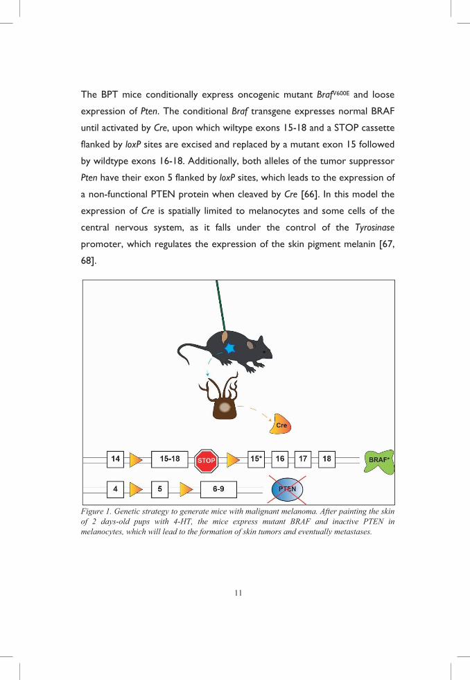
A MOUSE MODEL TO STUDY METASTASIS
With the aim of studying the effects of antioxidant supplementation on
metastasis, we used the Braf CA/+ Pten f/f Tyrosinase-Cre (BPT) mouse model of
malignant melanoma. This model is used in both paper I and II.
11
The BPT mice conditionally express oncogenic mutant BrafV600E and loose
expression of Pten. The conditional Braf transgene expresses normal BRAF
until activated by Cre, upon which wiltype exons 15-18 and a STOP cassette
flanked by loxP sites are excised and replaced by a mutant exon 15 followed
by wildtype exons 16-18. Additionally, both alleles of the tumor suppressor
Pten have their exon 5 flanked by loxP sites, which leads to the expression of
a non-functional PTEN protein when cleaved by Cre [66]. In this model the
expression of Cre is spatially limited to melanocytes and some cells of the
central nervous system, as it falls under the control of the Tyrosinase
promoter, which regulates the expression of the skin pigment melanin [67,
68].
Figure 1. Genetic strategy to generate mice with malignant melanoma. After painting the skin of 2 days-old pups with 4-HT, the mice express mutant BRAF and inactive PTEN in melanocytes, which will lead to the formation of skin tumors and eventually metastases.
By painting the right flank of the animals at postnatal day 2 with 4-
hydroxytamoxifen (4-HT), Cre is induced in melanocytes and thus mutant
protein BRAF is expressed and PTEN is lost; all of which leads to the
formation of skin tumors that eventually metastasize to regional lymph nodes
and in some cases lungs. Despite recapitulating most of the events leading to
the development of the disease in humans, this model is limited by the fact
that the mice often come to a humane endpoint due to the size of the primary
tumor and not due to the metastatic burden, which is the leading cause of
death in humans.
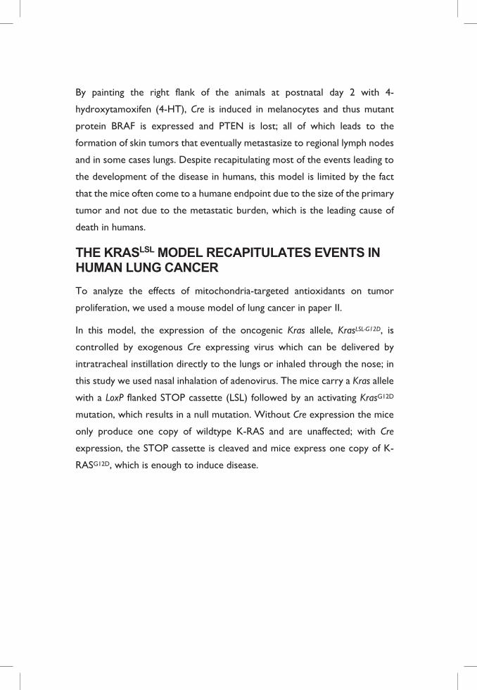
THE KRASLSL MODEL RECAPITULATES EVENTS IN HUMAN LUNG CANCER
To analyze the effects of mitochondria-targeted antioxidants on tumor
proliferation, we used a mouse model of lung cancer in paper II.
In this model, the expression of the oncogenic Kras allele, KrasLSL-G12D, is
controlled by exogenous Cre expressing virus which can be delivered by
intratracheal instillation directly to the lungs or inhaled through the nose; in
this study we used nasal inhalation of adenovirus. The mice carry a Kras allele
with a LoxP flanked STOP cassette (LSL) followed by an activating KrasG12D
mutation, which results in a null mutation. Without Cre expression the mice
only produce one copy of wildtype K-RAS and are unaffected; with Cre
expression, the STOP cassette is cleaved and mice express one copy of K-
RASG12D, which is enough to induce disease.
13
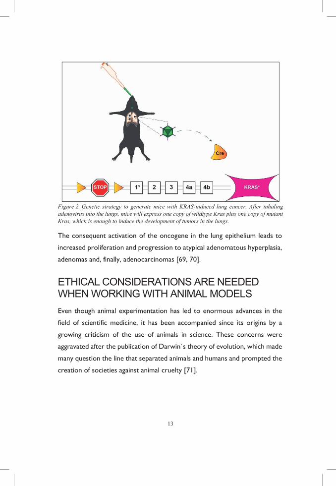
Figure 2. Genetic strategy to generate mice with KRAS-induced lung cancer. After inhaling adenovirus into the lungs, mice will express one copy of wildtype Kras plus one copy of mutant Kras, which is enough to induce the development of tumors in the lungs.
The consequent activation of the oncogene in the lung epithelium leads to
increased proliferation and progression to atypical adenomatous hyperplasia,
adenomas and, finally, adenocarcinomas [69, 70].
ETHICAL CONSIDERATIONS ARE NEEDED WHEN WORKING WITH ANIMAL MODELS
Even though animal experimentation has led to enormous advances in the
field of scientific medicine, it has been accompanied since its origins by a
growing criticism of the use of animals in science. These concerns were
aggravated after the publication of Darwin´s theory of evolution, which made
many question the line that separated animals and humans and prompted the
creation of societies against animal cruelty [71].
Nevertheless, in comparison to other industries where animals are exploited
for human benefit, such as farming, the use of animals in experimental
research is tightly regulated and controlled at several levels.
All animal experiments performed during the development of this thesis were
evaluated and approved by the Research Animal Ethics Committee in
Gothenburg, and all researchers involved strived to follow the 3Rs principle.
ANALYZING ROS IN CELLS: WHEN AND WHERE? Contrary to popular belief, redox couples are not found in thermodynamic
equilibrium in cells; they vary in their subcellular localization and differ in their
kinetics [72]. Hence, it is necessary to use tools that allow us to gain a better
understanding of the context in which redox reactions occur. However,
whole-cell extract based assays can be useful to obtain an overall look and
determine whether certain conditions are pro-oxidative or reducing at a
general level, for example, by measuring glutathione; and even though they
are usually specific, reproducible and sensitive, they do not give any
information about specific compartments.
FLUORESCENT PROBES FACILITATE MONITORING OF ROS IN CELL CULTURES
A variety of redox-active fluorescent probes that are triggered by different
oxidative species are commercially available. They enable monitoring of
redox processes in the cell through microscopy techniques, and can be
combined with compartment-specific dyes to increase spatial-specificity of
the reactions studied. They are easy to use in culture and some of them can
be used to stain tissues too. The use of general probes, such as 2´,7´-
dihydrodichlorofluorescein (H2DCF), dihydroethidium (DHE), cellROX,
15
dihydrorodamine (DHR) or mitochondria-targeting ones, like mitoSOX or
mitoPY1, are widely spread in the literature. Though useful, a major caveat is
their partial non-specific behavior, meaning that they can be triggered by
several oxidative reactions, and their activation is irreversible, making the
analysis of redox kinetics impossible.
GENETICALLY ENCODED BIOSENSORS INCREASE SPATIO-TEMPORAL RESOLUTION
In order to define redox processes in their natural context, genetically
encoded redox probes based on green fluorescent protein (GFP) were
developed. In this thesis redox-sensitive GFP (roGFP) biosensors were used,
but there are other biosensors available, such as redox-sensitive yellow FP
(rxYFP) and HyPer. Some of the major advantages of roGFP is its ratiometric
fluorogenic behavior, and the possibility of engineering redox relays between
redox enzymes and roGFPs to increase its specificity and sensitivity, and equal
response of the fluorescent protein in different tissues.
In papers I and II we used biosensors based on enhanced GFP (EGFP)
developed by Tobias Dick´s lab [73]. Briefly, two reactive cysteines were
engineered in positions S147 and Q204, located on β-strands 7 and 10 of
EGP. Excitation maxima from GFP are preserved (400 nm for A-band and
475-490 nm for B-band), but oxidation results in an increase in excitability in
the A-band and a decrease in the B-band and a reverse behavior during
reducing conditions. Analyzing the ratio of fluorescence intensity between
the 405 and 488 excitation maxima, one can conveniently visualize oxidative
processes (increased ratio) or reducing reactions (decreased ratio). By fusing
roGFP with human glutaredoxin-1 (Grx1) real-time equilibration between
the sensor protein (Grx1-roGFP) and the glutathione redox
couple (GSH/GSSG) is facilitated, [74], and fusion to the yeast peroxidase
Orp1 mediates oxidation of roGFP by H2O2 [75]. Versions of the probes that
target specifically to the mitochondrial matrix are also available.
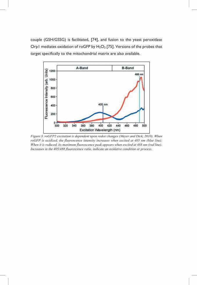
Figure 3. roGFP2 excitation is dependent upon redox changes (Meyer and Dick, 2010). When roGFP is oxidized, the fluorescence intensity increases when excited at 405 nm (blue line). When it is reduced, its maximum fluorescence peak appears when excited at 488 nm (red line). Increases in the 405/488 fluorescence ratio, indicate an oxidative condition or process.

RATIONALE, RESULTS & DISCUSSION
Trust those who seek the truth, doubt those who found it; doubt everything; but don’t doubt yourself. – André Gide

17
RATIONALE The overall aim of this thesis was to evaluate the effect of antioxidant
supplementation in the progression of cancer, with special focus on malignant
melanoma.
The specific aims of the two papers included in the thesis were:
I. Antioxidants can increase melanoma metastasis in mice
The rationale behind this first paper was to assess the impact of NAC
and vitamin E as dietary antioxidants on the progression of a
malignant melanoma mouse model, in order to validate the
hypothesis that tumors, with high endogenous ROS levels, benefit
from additional antioxidant supplementation.
II. Mitochondria-targeted antioxidants do not influence
malignant melanoma and lung cancer progression in mice
The aim of this second study was to determine whether targeting
mitochondria, the main source of cellular ROS, with antioxidant
compounds would hinder cancer progression in mouse models of
lung cancer and malignant melanoma.
PAPER I: ANTIOXIDANTS CAN INCREASE MELANOMA METASTASIS IN MICE Following up on a study published by Sayin and colleagues in 2014 [76], we
decided to investigate whether the accelerated proliferation observed upon
antioxidant treatment was exclusive to lung cancer or if it could be
extrapolated to other forms of cancer.
THE GENERAL ANTIOXIDANTS NAC AND VITAMIN E ACCELERATE METASTASIS
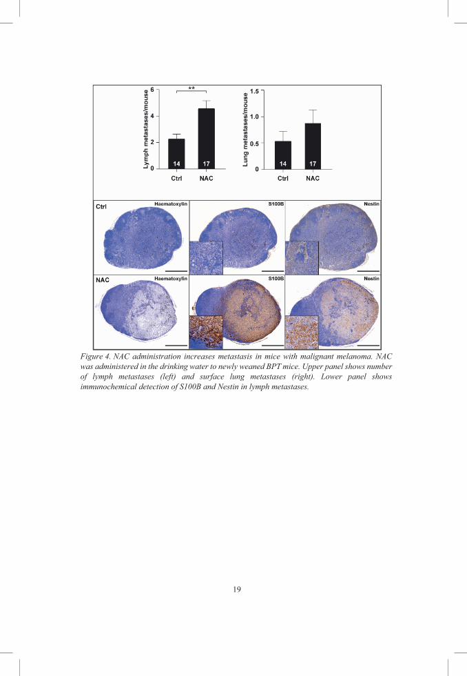
In this study we show that, dietary supplementation of NAC in the drinking
water doubled the number of lymph metastases in BPT mice [77]. In addition,
these metastases showed increased S100B and Nestin staining, both markers
of malignancy [78, 79].
Concordant to our in vivo observations, NAC and Trolox, an analogue of
vitamin E, increased migrating and invasive properties in a panel of human
melanoma cell lines.
19
Figure 4. NAC administration increases metastasis in mice with malignant melanoma. NAC was administered in the drinking water to newly weaned BPT mice. Upper panel shows number of lymph metastases (left) and surface lung metastases (right). Lower panel shows immunochemical detection of S100B and Nestin in lymph metastases.
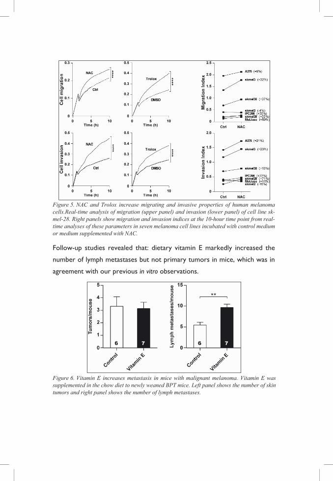
Figure 5. NAC and Trolox increase migrating and invasive properties of human melanoma cells.Real-time analysis of migration (upper panel) and invasion (lower panel) of cell line sk-mel-28. Right panels show migration and invasion indices at the 10-hour time point from real-time analyses of these parameters in seven melanoma cell lines incubated with control medium or medium supplemented with NAC.
Follow-up studies revealed that: dietary vitamin E markedly increased the
number of lymph metastases but not primary tumors in mice, which was in
agreement with our previous in vitro observations.
Figure 6. Vitamin E increases metastasis in mice with malignant melanoma. Vitamin E was supplemented in the chow diet to newly weaned BPT mice. Left panel shows the number of skin tumors and right panel shows the number of lymph metastases.
21
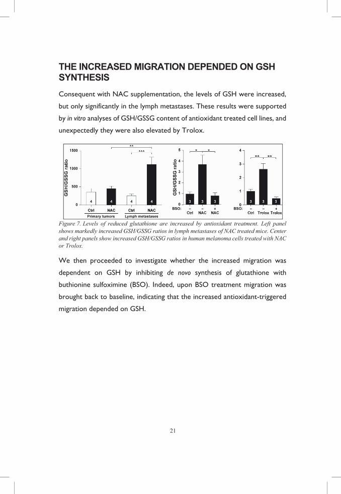
THE INCREASED MIGRATION DEPENDED ON GSH SYNTHESIS
Consequent with NAC supplementation, the levels of GSH were increased,
but only significantly in the lymph metastases. These results were supported
by in vitro analyses of GSH/GSSG content of antioxidant treated cell lines, and
unexpectedly they were also elevated by Trolox.
Figure 7. Levels of reduced glutathione are increased by antioxidant treatment. Left panel shows markedly increased GSH/GSSG ratios in lymph metastases of NAC treated mice. Center and right panels show increased GSH/GSSG ratios in human melanoma cells treated with NAC or Trolox.
We then proceeded to investigate whether the increased migration was
dependent on GSH by inhibiting de novo synthesis of glutathione with
buthionine sulfoximine (BSO). Indeed, upon BSO treatment migration was
brought back to baseline, indicating that the increased antioxidant-triggered
migration depended on GSH.
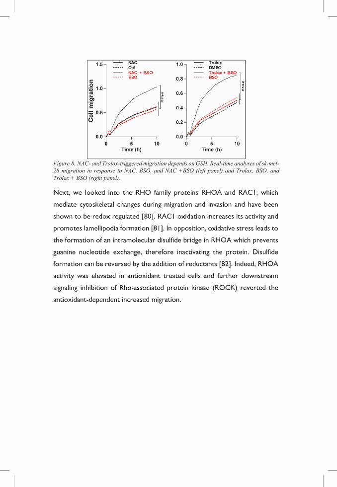
Figure 8. NAC- and Trolox-triggered migration depends on GSH. Real-time analyses of sk-mel-28 migration in response to NAC, BSO, and NAC +BSO (left panel) and Trolox, BSO, and Trolox + BSO (right panel).
Next, we looked into the RHO family proteins RHOA and RAC1, which
mediate cytoskeletal changes during migration and invasion and have been
shown to be redox regulated [80]. RAC1 oxidation increases its activity and
promotes lamellipodia formation [81]. In opposition, oxidative stress leads to
the formation of an intramolecular disulfide bridge in RHOA which prevents
guanine nucleotide exchange, therefore inactivating the protein. Disulfide
formation can be reversed by the addition of reductants [82]. Indeed, RHOA
activity was elevated in antioxidant treated cells and further downstream
signaling inhibition of Rho-associated protein kinase (ROCK) reverted the
antioxidant-dependent increased migration.
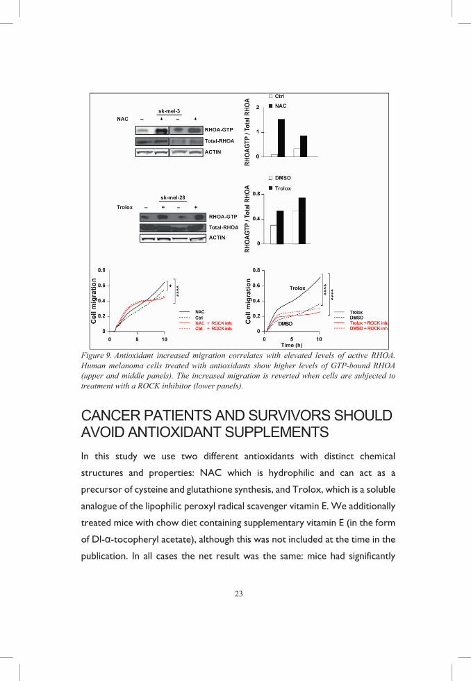
23
Figure 9. Antioxidant increased migration correlates with elevated levels of active RHOA. Human melanoma cells treated with antioxidants show higher levels of GTP-bound RHOA (upper and middle panels). The increased migration is reverted when cells are subjected to treatment with a ROCK inhibitor (lower panels).
CANCER PATIENTS AND SURVIVORS SHOULD AVOID ANTIOXIDANT SUPPLEMENTS
In this study we use two different antioxidants with distinct chemical
structures and properties: NAC which is hydrophilic and can act as a
precursor of cysteine and glutathione synthesis, and Trolox, which is a soluble
analogue of the lipophilic peroxyl radical scavenger vitamin E. We additionally
treated mice with chow diet containing supplementary vitamin E (in the form
of Dl-α-tocopheryl acetate), although this was not included at the time in the
publication. In all cases the net result was the same: mice had significantly
more metastases at endpoint, and human malignant melanoma cells migrated
and invaded more.
In order to ensure that the doses administered in vivo were in accordance
to human doses, we used a body surface area conversion [83]. NAC
supplementation was in range of what it is prescribed to COPD patients and
vitamin E doses were adjusted to 20 times the recommended daily intake,
which can be found in vitamin supplements.
Shortly after the release of this article, other publications showed that
oxidative stress limits metastasis of human malignant melanoma cells injected
into immunocompromised mice [84], and it also impairs tumor invasion in
vivo by suppressing Rho-ROCK activity through mechanisms involving p53
[85], all of which further supported our findings. Additionally, another group
reported that several antidiabetic drugs with antioxidant properties
accelerated metastasis in mouse models of cancer [86].
Although it remains to be seen whether these results can be directly
translated into the context of human health care, all of the studies above
mentioned together with the lack of evidence showing beneficial effects of
antioxidant supplementation in the vast majority of cancer clinical trials
suggest that cancer patients and people at risk of developing cancer should
avoid the use of antioxidant supplementation [29].
25
PAPER II: MITOCHONDRIA-TARGETED ANTIOXIDANTS DO NOT INFLUENCE MALIGNANT MELANOMA AND LUNG CANCER PROGRESSION IN MICE Following our observations that dietary antioxidant supplementation
accelerated proliferation and metastasis in lung cancer and malignant
melanoma respectively, we decided to target ROS at its main production site.
Previous studies hypothesize that mitochondria-associated and not cytosolic
ROS are responsible for the pro-tumorigenic signaling [87-89]. This raises
the possibility of using mitochondria-targeted antioxidants to inhibit tumor
growth.
In order to target mitochondrial ROS we used two different antioxidant
compounds conjugated to a lipophilic cation, which ensures uptake through
the phospholipid bilayer and mitochondrial accumulation by plasma
membrane potential.
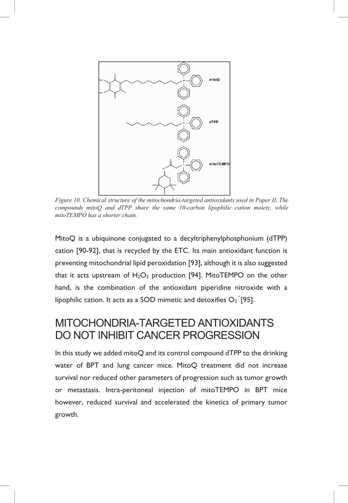
Figure 10. Chemical structure of the mitochondria-targeted antioxidants used in Paper II. The compounds mitoQ and dTPP share the same 10-carbon lipophilic cation moiety, while mitoTEMPO has a shorter chain.
MitoQ is a ubiquinone conjugated to a decyltriphenylphosphonium (dTPP)
cation [90-92], that is recycled by the ETC. Its main antioxidant function is
preventing mitochondrial lipid peroxidation [93], although it is also suggested
that it acts upstream of H2O2 production [94]. MitoTEMPO on the other
hand, is the combination of the antioxidant piperidine nitroxide with a
lipophilic cation. It acts as a SOD mimetic and detoxifies O2· [95].
MITOCHONDRIA-TARGETED ANTIOXIDANTS DO NOT INHIBIT CANCER PROGRESSION
In this study we added mitoQ and its control compound dTPP to the drinking
water of BPT and lung cancer mice. MitoQ treatment did not increase
survival nor reduced other parameters of progression such as tumor growth
or metastasis. Intra-peritoneal injection of mitoTEMPO in BPT mice
however, reduced survival and accelerated the kinetics of primary tumor
growth.
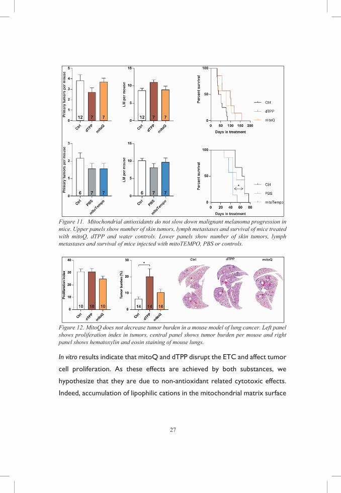
27
Figure 11. Mitochondrial antioxidants do not slow down malignant melanoma progression in mice. Upper panels show number of skin tumors, lymph metastases and survival of mice treated with mitoQ, dTPP and water controls. Lower panels show number of skin tumors, lymph metastases and survival of mice injected with mitoTEMPO, PBS or controls.
Figure 12. MitoQ does not decrease tumor burden in a mouse model of lung cancer. Left panel shows proliferation index in tumors, central panel shows tumor burden per mouse and right panel shows hematoxylin and eosin staining of mouse lungs.
In vitro results indicate that mitoQ and dTPP disrupt the ETC and affect tumor
cell proliferation. As these effects are achieved by both substances, we
hypothesize that they are due to non-antioxidant related cytotoxic effects.
Indeed, accumulation of lipophilic cations in the mitochondrial matrix surface
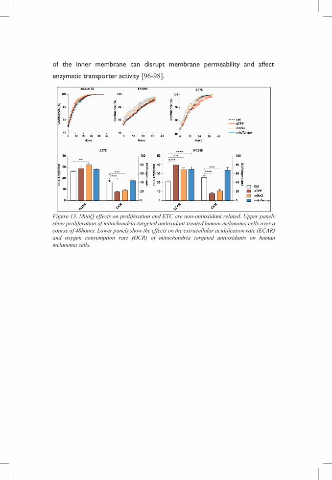
of the inner membrane can disrupt membrane permeability and affect
enzymatic transporter activity [96-98].
Figure 13. MitoQ effects on proliferation and ETC are non-antioxidant related. Upper panels show proliferation of mitochondria-targeted antioxidant-treated human melanoma cells over a course of 48hours. Lower panels show the effects on the extracellular acidification rate (ECAR) and oxygen consumption rate (OCR) of mitochondria targeted antioxidants on human melanoma cells.
29
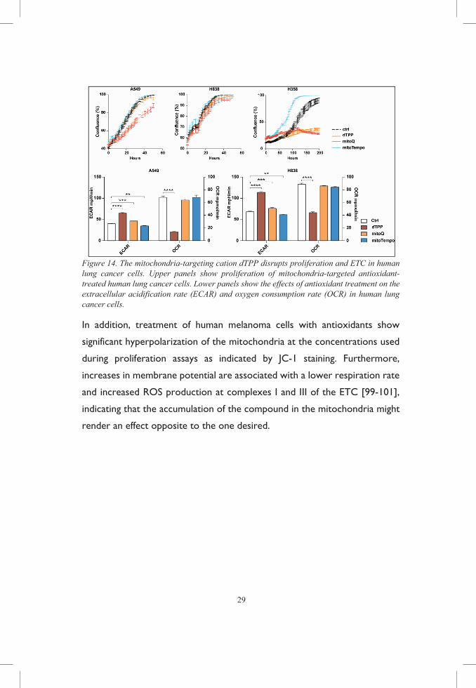
Figure 14. The mitochondria-targeting cation dTPP disrupts proliferation and ETC in human lung cancer cells. Upper panels show proliferation of mitochondria-targeted antioxidant-treated human lung cancer cells. Lower panels show the effects of antioxidant treatment on the extracellular acidification rate (ECAR) and oxygen consumption rate (OCR) in human lung cancer cells.
In addition, treatment of human melanoma cells with antioxidants show
significant hyperpolarization of the mitochondria at the concentrations used
during proliferation assays as indicated by JC-1 staining. Furthermore,
increases in membrane potential are associated with a lower respiration rate
and increased ROS production at complexes I and III of the ETC [99-101],
indicating that the accumulation of the compound in the mitochondria might
render an effect opposite to the one desired.
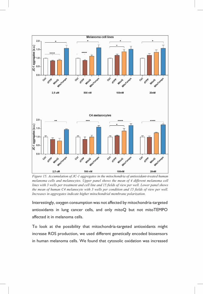
Figure 15. Accumulation of JC-1 aggregates in the mitochondria of antioxidant-treated human melanoma cells and melanocytes. Upper panel shows the mean of 4 different melanoma cell lines with 3 wells per treatment and cell line and 15 fields of view per well. Lower panel shows the mean of human C4 melanocyte with 3 wells per condition and 15 fields of view per well. Increases in aggregates indicate higher mitochondrial membrane polarization.
Interestingly, oxygen consumption was not affected by mitochondria-targeted
antioxidants in lung cancer cells, and only mitoQ but not mitoTEMPO
affected it in melanoma cells.
To look at the possibility that mitochondria-targeted antioxidants might
increase ROS production, we used different genetically encoded biosensors
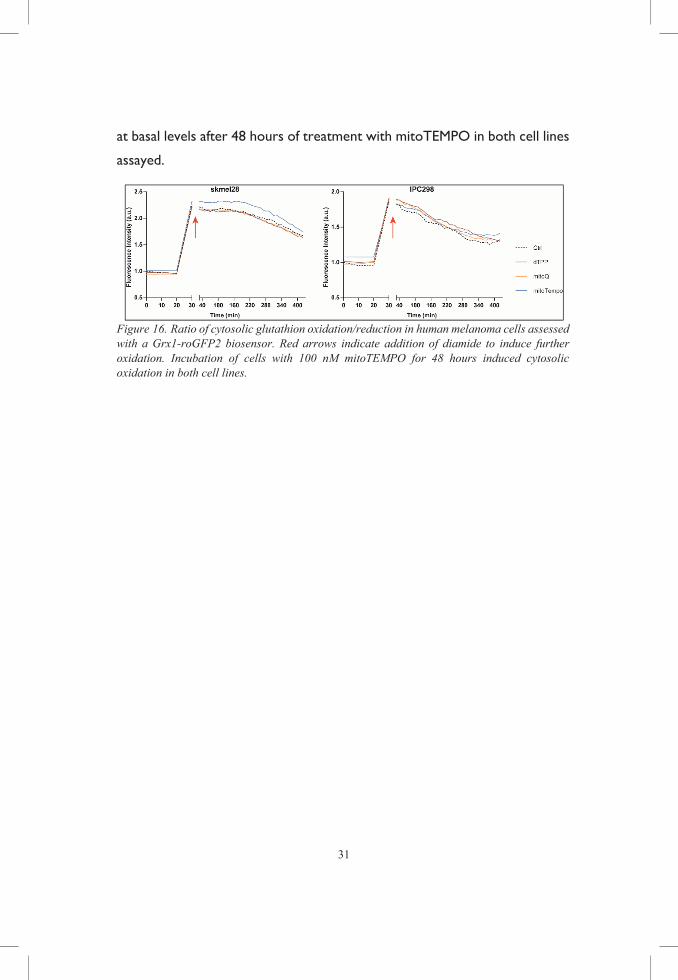
in human melanoma cells. We found that cytosolic oxidation was increased
31
at basal levels after 48 hours of treatment with mitoTEMPO in both cell lines
assayed.
Figure 16. Ratio of cytosolic glutathion oxidation/reduction in human melanoma cells assessed with a Grx1-roGFP2 biosensor. Red arrows indicate addition of diamide to induce further oxidation. Incubation of cells with 100 nM mitoTEMPO for 48 hours induced cytosolic oxidation in both cell lines.
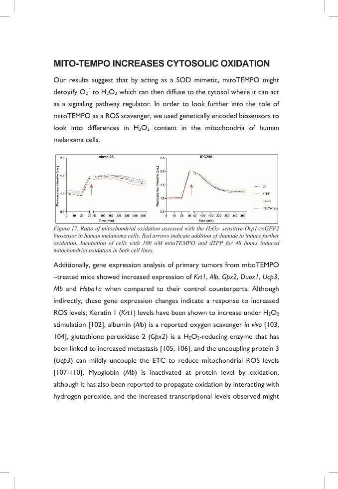
MITO-TEMPO INCREASES CYTOSOLIC OXIDATION
Our results suggest that by acting as a SOD mimetic, mitoTEMPO might
detoxify O2· to H2O2 which can then diffuse to the cytosol where it can act
as a signaling pathway regulator. In order to look further into the role of
mitoTEMPO as a ROS scavenger, we used genetically encoded biosensors to
look into differences in H2O2 content in the mitochondria of human
melanoma cells.
Figure 17. Ratio of mitochondrial oxidation assessed with the H2O2- sensitive Orp1-roGFP2 biosensor in human melanoma cells. Red arrows indicate addition of diamide to induce further oxidation. Incubation of cells with 100 nM mitoTEMPO and dTPP for 48 hours induced mitochondrial oxidation in both cell lines.
Additionally, gene expression analysis of primary tumors from mitoTEMPO
–treated mice showed increased expression of Krt1, Alb, Gpx2, Duox1, Ucp3,
Mb and Hspa1a when compared to their control counterparts. Although
indirectly, these gene expression changes indicate a response to increased
ROS levels; Keratin 1 (Krt1) levels have been shown to increase under H2O2
stimulation [102], albumin (Alb) is a reported oxygen scavenger in vivo [103,
104], glutathione peroxidase 2 (Gpx2) is a H2O2-reducing enzyme that has
been linked to increased metastasis [105, 106], and the uncoupling protein 3
(Ucp3) can mildly uncouple the ETC to reduce mitochondrial ROS levels
[107-110]. Myoglobin (Mb) is inactivated at protein level by oxidation,
although it has also been reported to propagate oxidation by interacting with
hydrogen peroxide, and the increased transcriptional levels observed might
33
be a compensatory mechanism to such protein inhibition [111, 112]. The heat
shock protein family A member 1A (Hspa1a) has been reported to participate
in the removal of proteins damaged by oxidation [113]. Interestingly, the
H2O2 producing dual oxidase 1 (Duox1) was also overexpressed. This result
is in opposition to what Dikalov and colleagues have previously described on
how blocking mitochondrial O2· production with mitoTEMPO
downregulated cytosolic O2· production by NOXes, breaking a forward feed
loop [114].
Overall we conclude that mitoTEMPO acts as a mitochondrial
antioxidant/cytosolic pro-oxidant in our system. To validate such hypothesis
we could isolate mitochondria and look at excreted H2O2 upon mitoTEMPO
treatment. If our hypothesis was confirmed, we could conditionally
overexpress catalase in human melanoma cells in vitro or in BPT mice in vivo
to see whether the phenotypes observed can be reverted.
MITOCHONDRIA-TARGETED ANTIOXIDANTS: THESE ARE NOT THE COMPOUNDS YOU ARE LOOKING FOR
Previous studies have shown that mitochondria-targeted antioxidants could
potentially inhibit tumor development. Indeed, combined inhibition of
mitochondrial ROS and glycolysis successfully decreased ATP production and
induced apoptosis in hepatocellular carcinoma [30]; targeting mitochondrial
ROS decreased KRAS-mediated tumorigenicity by increasing ERK 1/2
signaling [32]: it also reversed superoxide-dependent migration upon partial
ETC inhibition [31].
Although promising, none of these studies have evaluated the impact of such
compounds in transgenic mouse models. Even with similar doses, neither
mitoQ nor mitoTEMPO blocked disease progression. In fact, mitoTEMPO
decreased survival which is in concordance with the work of Wang and
colleagues, where mitochondria-targeted antioxidants aggravated
tumorigenesis by affecting DNA-damage repair in a chemically induced model
of hepatocellular carcinoma [86].
In addition, our in vitro results show that no direct translation can be drawn
to an in vivo context, which might explain the conflict with previous studies.
Furthermore, the effects observed with mitoQ treatment were recapitulated
by the control substance, suggesting that the decrease in proliferation
observed is related to cytotoxic effects coupled to the targeting moiety
rather than to antioxidant properties of the ubiquinone. It has been proposed
that genetic therapy with alternative oxidase, an enzyme present in plants and
lower animals, could potentially reduce mitochondrial ROS formation by
bypassing the ETC when disrupted and maintaining the electron flow and
redox homeostasis in the cell [115, 116]. However, preliminary histological
data indicates that the ETC complexes remain unaltered in the mitochondria-
targeted antioxidant-treated mice, questioning the usefulness of such
treatment in our model (data not shown).
Mitochondrial antioxidants have been successfully used in other areas and
models, such as in acute hypoxia, inflammation, cardiovascular diseases, and
ischemia reperfusion [117-120], but our results demonstrate that they are
unlikely to be useful in cancer therapy.

GENERAL DISCUSSION
&
FUTURE WORK
One never notices what has been done; one can only see what remains to be done. – Marie Sklodowska-Curie

35
THE ANTIOXIDANT/ROS DOGMA NEEDS TO BE RECONSIDERED ROS are not only damaging products, they are important players in the
maintenance of cell signaling and homeostasis.
Antioxidant supplementation has been traditionally seen as a way to protect
against oxidative stress-related damage. However, antioxidants protect both
healthy and tumor cells. The latter have elevated levels of ROS and rely on
antioxidant defenses to protect themselves from further damage.
Antioxidants give them the additional help they need.
In paper I we show that general antioxidants supplied in the diet accelerate
metastasis in in vivo and in vitro models of malignant melanoma.
In paper II we show that mitochondria-targeted antioxidants did not inhibit
cancer progression in vivo. In fact, one of the compounds, mitoTEMPO,
reduced survival of mice with malignant melanoma and this was accompanied
by increased levels of cytosolic H2O2.
ARE HYPOTHETICAL BENEFITS OF ANTIOXIDANTS WORTH THE RISK?
Clinical trials have consistently failed at showing the value of antioxidant
supplementation for the prevention and treatment of cancer. In fact, meta-
analysis studies of clinical trials show that antioxidant supplementation lacks
support for beneficial effects and may increase mortality of certain forms of
cancer [121-124].
In addition, there is a widespread use of antioxidant supplements by cancer
patients [125-128] partially to alleviate toxic radiotherapy and chemotherapy
side-effects, but also prompted by the popular conception that antioxidants
help fighting cancer. Interestingly, the published literature suggests growing
concern and debate amongst clinicians on the potential interference of such
supplements with therapy that relies on the production of ROS and induction
of apoptosis [129-133].
Furthermore, although some experimental studies of chemically- and
radiation-induced cancers have displayed potential therapeutic effects of the
use of antioxidants [134, 135], there is an increasing body of evidence
showing their role in the acceleration of progression [136-138].
Overall, there is no doubt that redox regulation plays an important role in
the development and progression of cancer. We therefore think that the
study of redox-regulated pathways, proteins and genes might reveal new drug
targets and offer new and reliable therapeutic possibilities [139].
FUTURE WORK One of the main difficulties in the field is the study of redox reactions in vivo.
Indeed, we have to rely on methods that can give an overall idea of whether
certain conditions are pro-oxidative or reductive. As described in the
methods section, the use of genetic encoded biosensors has revolutionized
the field by giving the possibility to analyze when and where in the cell these
redox reactions occur. This tool is now being expanded to in vivo models in
Tobias Dick´s group, where genetically encoded biosensors have been stably
expressed in mouse tissues [140]. This opens many possibilities if combined
with our cancer models, since it would be easier to pinpoint where and when
during the development of the disease redox alterations occur with and
without the use of antioxidants.
For instance, in Paper I we saw that the increased migrating and invasive
properties of cancer cells were dependent on new synthesis of GSH [141].
We also saw that the GSH/GSSG ratio was increased in lymph metastases of
NAC treated mice according to a whole cell extract assay. It would be
37
interesting to combine the BPT model with the expression of a glutathione
biosensor to observe whether this increase is particular to the cells that
survive transit from the primary tumor to the lymph nodes, or whether it is
only those cells within the primary tumor that show increased GSH that
migrate. It would also allow us to see what happens with the rest of the cells
in the neoplastic niche. For instance, it is well known that redox regulation
plays an important role in the vascularization of tissue. It has been proposed
that low levels of ROS can stimulate angiogenesis and therefore influence
tumor progression [142]. Indeed, the accelerated growth kinetics observed
in mitoTEMPO-treated mice might be related not so much to tumor cell
proliferation in itself as to a better vascularization of the neoplastic tissue,
prompted by the excretion of H2O2.
Another important point from Paper I was that migration was also dependent
on RHOA signaling, and we hypothesize that this increase in signaling is due
to either the inhibition by reduction of RAC1 (and hence de-repression of
RHOA) or activation of RHOA by reduction. But we cannot rule out other
effects of redox regulation of the cytoskeleton. One way of analyzing this
would be to study the thiol proteome by mass spectrometry and study
potentially GSH-regulated cysteines.
We could combine these results with RNAseq analysis of primary tumors
and lymph metastases from NAC and vitamin E treated mice, to get a better
landscape of redox regulation by antioxidant supplementation. Although the
phenotype exhibited by both treatments is the same, we cannot rule out that
the underlying mechanisms are different.
To that end, it would be interesting to perform the same experiments in
immunodeficient mice to rule out the possibility that effects on the immune
system are responsible for the observed metastasis.
In Paper II we observed a decreased survival by mitoTEMPO and we argue
that growth kinetics were affected by the treatment. To challenge this idea,
we are now repeating a new study where mice will be sacrificed after five
weeks of treatment. We also observed that scavenging of mitochondrial O2·
resulted in increased levels of cytosolic ROS and we hypothesize that this in
turn triggers cellular signaling cascades that accelerate growth. To verify the
hypothesis we could overexpress a mitochondrial catalase to decrease pro-
tumorigenic signaling from mitochondrial H2O2 or isolate mitochondria and
measure H2O2 excretion.
In addition to these studies, we are interested in comparing different methods
of antioxidant delivery to the skin in the context of malignant melanoma. Skin
lotions often contain different forms of antioxidants, whether with the
purpose of stabilizing formulation and avoiding rancidity or with the promise
of improving skin texture and condition. We are interested in studying how
these will affect malignant melanoma progression. I have administered vitamin
E in the diet, and as a lotion to BPT mice, and I am going to test dietary
mitochondrial targeting with mitoE.
39
ACKNOWLEDGEMENT What is considered the end of the world for a caterpillar is the beginning of
Mariposa´s story – Sayin, 2018.
All good stories come to an end, and what a journey this one was! I would
like to thank, in no particular order, the following people for keeping me good
company along the way:
My supervisor, Martin Bergö, for believing in my potential when I was about
to give up and allowing me to develop into an independent researcher.
My co-supervisors, Levent Akyürek and Per Lindahl, for all the fruitful
scientific discussions and career advice over the years.
The current and previous members in the Bergö lab, for all the time (and it´s
been a lot of hours) shared together. Martin Dalin, for introducing me to
the BPT model, always with a smile on your face, even though you had to
write your own thesis. Christin, for making sure I´m feeling fine and I have
everything I need to do my work. You´re the best lab-mum ever! Volkan
“Just” Sayin, you have been a friend, a mentor, and one of my biggest
supports during these years. Thank you for the laughs, the hugs, and the Hello
Kitty candy! Clotilde, thank you for walking the redox rocky road with me,
even though we get mixed up sometimes. Your talent and ambition inspire us
all, but what I like the most is your sarcasm and availability to get drinks with
me! Ella, for always making sure everyone is feeling well and included. You´re
beautiful both outside and inside. Mohamed, for welcoming me to the group
before it was even official. I sincerely don´t know what´s bigger, your brain
or your heart. Murali “baba”, for singing, huffing and puffing around the lab,
and bringing joy to our lives! Jaroslaw, for the scientific discussions and
twisted sense of humor. We definitely need the same centrifuge as in CSI.
Tony, for all the spontaneous neck massages, your energy, your friendship
and trust. We will for sure fix your “problem”. Oskar “den starke”, for all
your hard work and help with the mice and stubborn bottles. We wouldn’t
be able to do all this without you! Julia, Josefin, and Louise, for lightening
up the mood in the lab! Your enthusiasm and hunger for knowledge is
contagious.
The members of SCC for creating such a great research family! Camilla, for
the words of wisdom (and the heavy metal!). Mattias, for the math lessons:
double nothing is still nothing. André, for all the insightful conversations. It´s
gonna be great to have you back. Patricia, for all the advice and the
WhatsApp chats! I really wish you had stayed around longer. Jonas, Lisa,
Berglind, Som, Joy, Elin, Gülay, Emman, Yvonne, Tobias, Gautam,
Dorota, Agnieszka, and Toshima for the fun times at the sixth floor.
Emma, Paloma, Tajana, and Marta, for being awesome office mates and
cheering up my days. Anna, Karoline, Christoffer, Sara, Emma, Stefan,
Mamen, Elena, Gustav, Pernilla, Malin, Shawn, Valerio, Ágota,
Daniel, Sara, Andreas, and Mila, for the lunch and corridor talks. Paul,
for the singing, the documentary suggestions, and for sharing a dark sense of
humor with me. You make me feel a little bit more normal (but not too
much).
Aditi, Ahmed, Octavia, Sally, Jonathan and Peidi, for being friends with
this lab-addict and all the happy moments shared over the years. Let’s create
some more soon?
Lydia and Dzeneta, for inspiration. You two are amazing in every aspect
possible. Never forget that, and never let anyone take that away from you.
Amiko and Tomiko, for being extraordinary friends despite the distance.
Thinking about all the moments we had together in the lab always warms my
heart.
Soheila, for friendship and opening the doors to your “fun place”. Thanks
for trusting me despite my proficiency at winning lying games! Maryam, for

41
being kindness personified. I´m blessed to count you among my friends.
Ángela and Esther, for always being there for me no matter where, when
or why. Words cannot tell how thankful I am for having you both in my life.
Roberto, Llanos, and Sabrina, for always taking the time to meet and listen
to me whenever I travel back home. You guys are gold.
My parents, Michel and María Teresa, for being the best teachers one
could ever wish for. I owe you so very much, and I hope I will be able to repay
you some day. My grandparents, Encarnación and José, and my auntie,
Tere, for all the love and care. My brother, Mikael, for being my first lab
assistant and for your unconditional support. You know you will always have
mine. I love you all more than words can tell.
And last, but absolutely not least: Emil. Thank you for shining bright when
I’m at my darkest, and making me laugh when I don’t even feel like smiling.
For your patience with this free and radical “Spanish temper” of mine. For
love.
REFERENCES
1. Holland, H.D., The oxygenation of the atmosphere and oceans. Philos Trans R Soc Lond B Biol Sci, 2006. 361(1470): p. 903-15.
2. Sessions, A.L., et al., The continuing puzzle of the great oxidation event. Curr Biol, 2009. 19(14): p. R567-74.
3. Murphy, M.P., How mitochondria produce reactive oxygen species. Biochem J, 2009. 417(1): p. 1-13.
4. Halliwell, B. and J.M. Gutteridge, Free radicals in Biology and Medicine. Fourth edition ed. 2007: Oxford Biosciences.
5. Cadenas, E., Basic mechanisms of antioxidant activity. Biofactors, 1997. 6(4): p. 391-7.
6. Curtin, J.F., M. Donovan, and T.G. Cotter, Regulation and measurement of oxidative stress in apoptosis. J Immunol Methods, 2002. 265(1-2): p. 49-72.
7. Sies, H., W. Stahl, and A.R. Sundquist, Antioxidant functions of vitamins. Vitamins E and C, beta-carotene, and other carotenoids. Ann N Y Acad Sci, 1992. 669: p. 7-20.
8. Holmstrom, K.M. and T. Finkel, Cellular mechanisms and physiological consequences of redox-dependent signalling. Nat Rev Mol Cell Biol, 2014. 15(6): p. 411-21.
9. D'Autreaux, B. and M.B. Toledano, ROS as signalling molecules: mechanisms that generate specificity in ROS homeostasis. Nat Rev Mol Cell Biol, 2007. 8(10): p. 813-24.
10. Catarzi, S., et al., Redox regulation of platelet-derived-growth-factor-receptor: role of NADPH-oxidase and c-Src tyrosine kinase. Biochim Biophys Acta, 2005. 1745(2): p. 166-75.
11. Lee, I., E. Bender, and B. Kadenbach, Control of mitochondrial membrane potential and ROS formation by reversible
43
phosphorylation of cytochrome c oxidase. Mol Cell Biochem, 2002. 234-235(1-2): p. 63-70.
12. Truong, T.H. and K.S. Carroll, Redox regulation of epidermal growth factor receptor signaling through cysteine oxidation. Biochemistry, 2012. 51(50): p. 9954-65.
13. Tonks, N.K., Redox redux: revisiting PTPs and the control of cell signaling. Cell, 2005. 121(5): p. 667-70.
14. Schafer, Z.T., et al., Antioxidant and oncogene rescue of metabolic defects caused by loss of matrix attachment. Nature, 2009. 461(7260): p. 109-13.
15. Szatrowski, T.P. and C.F. Nathan, Production of large amounts of hydrogen peroxide by human tumor cells. Cancer Res, 1991. 51(3): p. 794-8.
16. Rainwater, R., et al., Role of cysteine residues in regulation of p53 function. Mol Cell Biol, 1995. 15(7): p. 3892-903.
17. Klaunig, J.E. and L.M. Kamendulis, The role of oxidative stress in carcinogenesis. Annu Rev Pharmacol Toxicol, 2004. 44: p. 239-67.
18. Shibutani, S., M. Takeshita, and A.P. Grollman, Insertion of specific bases during DNA synthesis past the oxidation-damaged base 8-oxodG. Nature, 1991. 349(6308): p. 431-4.
19. Baylin, S.B., Tying it all together: epigenetics, genetics, cell cycle, and cancer. Science, 1997. 277(5334): p. 1948-9.
20. Counts, J.L. and J.I. Goodman, Alterations in DNA methylation may play a variety of roles in carcinogenesis. Cell, 1995. 83(1): p. 13-5.
21. Greger, V., et al., Frequency and parental origin of hypermethylated RB1 alleles in retinoblastoma. Hum Genet, 1994. 94(5): p. 491-6.
22. Fridovich, I., Superoxide anion radical (O2-.), superoxide dismutases, and related matters. J Biol Chem, 1997. 272(30): p. 18515-7.
23. Yakes, F.M. and B. Van Houten, Mitochondrial DNA damage is more extensive and persists longer than nuclear DNA damage in human cells following oxidative stress. Proc Natl Acad Sci U S A, 1997. 94(2): p. 514-9.
24. Kujoth, G.C., et al., The role of mitochondrial DNA mutations in mammalian aging. PLoS Genet, 2007. 3(2): p. e24.
25. Brown, W.M., M. George, Jr., and A.C. Wilson, Rapid evolution of animal mitochondrial DNA. Proc Natl Acad Sci U S A, 1979. 76(4): p. 1967-71.
26. Choi, Y.S., et al., Shot-gun proteomic analysis of mitochondrial D-loop DNA binding proteins: identification of mitochondrial histones. Mol Biosyst, 2011. 7(5): p. 1523-36.
27. Chatterjee, A., E. Mambo, and D. Sidransky, Mitochondrial DNA mutations in human cancer. Oncogene, 2006. 25(34): p. 4663-74.
28. Chen, E.I., Mitochondrial dysfunction and cancer metastasis. J Bioenerg Biomembr, 2012. 44(6): p. 619-22.
29. Chandel, N.S. and D.A. Tuveson, The promise and perils of antioxidants for cancer patients. N Engl J Med, 2014. 371(2): p. 177-8.
30. Dilip, A., et al., Mitochondria-targeted antioxidant and glycolysis inhibition: synergistic therapy in hepatocellular carcinoma. Anticancer Drugs, 2013. 24(9): p. 881-8.
31. Porporato, P.E., et al., A mitochondrial switch promotes tumor metastasis. Cell Rep, 2014. 8(3): p. 754-66.
32. Weinberg, F., et al., Mitochondrial metabolism and ROS generation are essential for Kras-mediated tumorigenicity. Proc Natl Acad Sci U S A, 2010. 107(19): p. 8788-93.
45
33. Hercberg, S., et al., The potential role of antioxidant vitamins in preventing cardiovascular diseases and cancers. Nutrition, 1998. 14(6): p. 513-20.
34. Blot, W.J., et al., Nutrition intervention trials in Linxian, China: supplementation with specific vitamin/mineral combinations, cancer incidence, and disease-specific mortality in the general population. J Natl Cancer Inst, 1993. 85(18): p. 1483-92.
35. Li, B., et al., Linxian nutrition intervention trials. Design, methods, participant characteristics, and compliance. Ann Epidemiol, 1993. 3(6): p. 577-85.
36. Wang, S.M., et al., Effects of Nutrition Intervention on Total and Cancer Mortality: 25-Year Post-trial Follow-up of the 5.25-Year Linxian Nutrition Intervention Trial. J Natl Cancer Inst, 2018.
37. The effect of vitamin E and beta carotene on the incidence of lung cancer and other cancers in male smokers. The Alpha-Tocopherol, Beta Carotene Cancer Prevention Study Group. N Engl J Med, 1994. 330(15): p. 1029-35.
38. Albanes, D., et al., Alpha-Tocopherol and beta-carotene supplements and lung cancer incidence in the alpha-tocopherol, beta-carotene cancer prevention study: effects of base-line characteristics and study compliance. J Natl Cancer Inst, 1996. 88(21): p. 1560-70.
39. Omenn, G.S., et al., Risk factors for lung cancer and for intervention effects in CARET, the Beta-Carotene and Retinol Efficacy Trial. J Natl Cancer Inst, 1996. 88(21): p. 1550-9.
40. Lee, I.M., et al., Beta-carotene supplementation and incidence of cancer and cardiovascular disease: the Women's Health Study. J Natl Cancer Inst, 1999. 91(24): p. 2102-6.
41. Lippman, S.M., et al., Effect of selenium and vitamin E on risk of prostate cancer and other cancers: the Selenium and Vitamin E Cancer Prevention Trial (SELECT). JAMA, 2009. 301(1): p. 39-51.
42. Klein, E.A., et al., Vitamin E and the risk of prostate cancer: the Selenium and Vitamin E Cancer Prevention Trial (SELECT). JAMA, 2011. 306(14): p. 1549-56.
43. Li, J., et al., Andrographolide induces cell cycle arrest at G2/M phase and cell death in HepG2 cells via alteration of reactive oxygen species. Eur J Pharmacol, 2007. 568(1-3): p. 31-44.
44. DeBerardinis, R.J. and N.S. Chandel, Fundamentals of cancer metabolism. Sci Adv, 2016. 2(5): p. e1600200.
45. Shen, Y.A., et al., Metabolic reprogramming orchestrates cancer stem cell properties in nasopharyngeal carcinoma. Cell Cycle, 2015. 14(1): p. 86-98.
46. Erdmann, F., et al., International trends in the incidence of malignant melanoma 1953-2008--are recent generations at higher or lower risk? Int J Cancer, 2013. 132(2): p. 385-400.
47. National Cancer Institute, SEER Cancer Statistics Review (CSR) 1975-2010. 2013.
48. DeSantis, C.E., et al., Cancer treatment and survivorship statistics, 2014. CA Cancer J Clin, 2014. 64(4): p. 252-71.
49. Miller, K.D., et al., Cancer treatment and survivorship statistics, 2016. CA Cancer J Clin, 2016. 66(4): p. 271-89.
50. Siegel, R., et al., Cancer treatment and survivorship statistics, 2012. CA Cancer J Clin, 2012. 62(4): p. 220-41.
51. Bandarchi, B., et al., Molecular biology of normal melanocytes and melanoma cells. J Clin Pathol, 2013. 66(8): p. 644-8.
52. Eggermont, A.M., A. Spatz, and C. Robert, Cutaneous melanoma. Lancet, 2014. 383(9919): p. 816-27.
53. Kunz, M., Oncogenes in melanoma: an update. Eur J Cell Biol, 2014. 93(1-2): p. 1-10.
47
54. Michaloglou, C., et al., BRAFE600-associated senescence-like cell cycle arrest of human naevi. Nature, 2005. 436(7051): p. 720-4.
55. Chin, L., L.A. Garraway, and D.E. Fisher, Malignant melanoma: genetics and therapeutics in the genomic era. Genes Dev, 2006. 20(16): p. 2149-82.
56. Hodis, E., et al., A landscape of driver mutations in melanoma. Cell, 2012. 150(2): p. 251-63.
57. Denat, L., et al., Melanocytes as instigators and victims of oxidative stress. J Invest Dermatol, 2014. 134(6): p. 1512-8.
58. Godic, A., S. Haque-Hussain, and N.P. Burrows, Neonatal lupus erythematosus: the use of telephone images in diagnosis. Clin Exp Dermatol, 2014. 39(7): p. 852-3.
59. GLOBOCAN. 2012.
60. Laniado-Laborin, R., Smoking and chronic obstructive pulmonary disease (COPD). Parallel epidemics of the 21 century. Int J Environ Res Public Health, 2009. 6(1): p. 209-24.
61. Vestbo, J., COPD: definition and phenotypes. Clin Chest Med, 2014. 35(1): p. 1-6.
62. Fox, J.G., Laboratory animal medicine. Changes and challenges. Cornell Vet, 1985. 75(1): p. 159-70.
63. Bernard, C., Introduction à l'étude de la médecine expérimentale. 1865.
64. Mouse Genome Sequencing, C., et al., Initial sequencing and comparative analysis of the mouse genome. Nature, 2002. 420(6915): p. 520-62.
65. Jonas, A.M., The mouse in biomedical research. Physiologist, 1984. 27(5): p. 330-46.
66. Dankort, D., et al., Braf(V600E) cooperates with Pten loss to induce metastatic melanoma. Nat Genet, 2009. 41(5): p. 544-52.
67. Bosenberg, M., et al., Characterization of melanocyte-specific inducible Cre recombinase transgenic mice. Genesis, 2006. 44(5): p. 262-7.
68. Tonks, I.D., et al., Tyrosinase-Cre mice for tissue-specific gene ablation in neural crest and neuroepithelial-derived tissues. Genesis, 2003. 37(3): p. 131-8.
69. Jackson, E.L., et al., Analysis of lung tumor initiation and progression using conditional expression of oncogenic K-ras. Genes Dev, 2001. 15(24): p. 3243-8.
70. Nikitin, A.Y., et al., Classification of proliferative pulmonary lesions of the mouse: recommendations of the mouse models of human cancers consortium. Cancer Res, 2004. 64(7): p. 2307-16.
71. Loew, F.M., Animal experimentation. Bull Hist Med, 1982. 56(1): p. 123-6.
72. Kemp, M., Y.M. Go, and D.P. Jones, Nonequilibrium thermodynamics of thiol/disulfide redox systems: a perspective on redox systems biology. Free Radic Biol Med, 2008. 44(6): p. 921-37.
73. Meyer, A.J. and T.P. Dick, Fluorescent protein-based redox probes. Antioxid Redox Signal, 2010. 13(5): p. 621-50.
74. Gutscher, M., et al., Real-time imaging of the intracellular glutathione redox potential. Nat Methods, 2008. 5(6): p. 553-9.
75. Gutscher, M., et al., Proximity-based protein thiol oxidation by H2O2-scavenging peroxidases. J Biol Chem, 2009. 284(46): p. 31532-40.
76. Sayin, V.I., et al., Antioxidants accelerate lung cancer progression in mice. Sci Transl Med, 2014. 6(221): p. 221ra15.
49
77. Aruoma, O.I., et al., The antioxidant action of N-acetylcysteine: its reaction with hydrogen peroxide, hydroxyl radical, superoxide, and hypochlorous acid. Free Radic Biol Med, 1989. 6(6): p. 593-7.
78. Chen, P.L., et al., Diagnostic utility of neural stem and progenitor cell markers nestin and SOX2 in distinguishing nodal melanocytic nevi from metastatic melanomas. Mod Pathol, 2013. 26(1): p. 44-53.
79. Fusi, A., et al., Expression of the stem cell markers nestin and CD133 on circulating melanoma cells. J Invest Dermatol, 2011. 131(2): p. 487-94.
80. Nimnual, A.S., L.J. Taylor, and D. Bar-Sagi, Redox-dependent downregulation of Rho by Rac. Nat Cell Biol, 2003. 5(3): p. 236-41.
81. Hobbs, G.A., et al., Redox regulation of Rac1 by thiol oxidation. Free Radic Biol Med, 2015. 79: p. 237-50.
82. Heo, J., et al., Redox regulation of RhoA. Biochemistry, 2006. 45(48): p. 14481-9.
83. Nair, A.B. and S. Jacob, A simple practice guide for dose conversion between animals and human. J Basic Clin Pharm, 2016. 7(2): p. 27-31.
84. Piskounova, E., et al., Oxidative stress inhibits distant metastasis by human melanoma cells. Nature, 2015. 527(7577): p. 186-91.
85. Herraiz, C., et al., Reactivation of p53 by a Cytoskeletal Sensor to Control the Balance Between DNA Damage and Tumor Dissemination. J Natl Cancer Inst, 2016. 108(1).
86. Wang, H., et al., NRF2 activation by antioxidant antidiabetic agents accelerates tumor metastasis. Sci Transl Med, 2016. 8(334): p. 334ra51.
87. Wheaton, W.W., et al., Metformin inhibits mitochondrial complex I of cancer cells to reduce tumorigenesis. Elife, 2014. 3: p. e02242.
88. Diebold, L. and N.S. Chandel, Mitochondrial ROS regulation of proliferating cells. Free Radic Biol Med, 2016. 100: p. 86-93.
89. Goh, J., et al., Mitochondrial targeted catalase suppresses invasive breast cancer in mice. BMC Cancer, 2011. 11: p. 191.
90. Murphy, M.P., Selective targeting of bioactive compounds to mitochondria. Trends Biotechnol, 1997. 15(8): p. 326-30.
91. Murphy, M.P. and R.A. Smith, Drug delivery to mitochondria: the key to mitochondrial medicine. Adv Drug Deliv Rev, 2000. 41(2): p. 235-50.
92. Smith, R.A., et al., Using mitochondria-targeted molecules to study mitochondrial radical production and its consequences. Biochem Soc Trans, 2003. 31(Pt 6): p. 1295-9.
93. Kelso, G.F., et al., Selective targeting of a redox-active ubiquinone to mitochondria within cells: antioxidant and antiapoptotic properties. J Biol Chem, 2001. 276(7): p. 4588-96.
94. Cocheme, H.M., et al., Mitochondrial targeting of quinones: therapeutic implications. Mitochondrion, 2007. 7 Suppl: p. S94-102.
95. Dikalova, A.E., et al., Therapeutic targeting of mitochondrial superoxide in hypertension. Circ Res, 2010. 107(1): p. 106-16.
96. Azzone, G.F., V. Petronilli, and M. Zoratti, 'Cross-talk' between redox- and ATP-driven H+ pumps. Biochem Soc Trans, 1984. 12(3): p. 414-6.
97. Bakeeva, L.E., et al., Conversion of biomembrane-produced energy into electric form. II. Intact mitochondria. Biochim Biophys Acta, 1970. 216(1): p. 13-21.
98. Brand, M.D. and A. Kesseler, Control analysis of energy metabolism in mitochondria. Biochem Soc Trans, 1995. 23(2): p. 371-6.
51
99. Korshunov, S.S., V.P. Skulachev, and A.A. Starkov, High protonic potential actuates a mechanism of production of reactive oxygen species in mitochondria. FEBS Lett, 1997. 416(1): p. 15-8.
100. Nicholls, D.G., Mitochondrial membrane potential and aging. Aging Cell, 2004. 3(1): p. 35-40.
101. Skulachev, V.P., Role of uncoupled and non-coupled oxidations in maintenance of safely low levels of oxygen and its one-electron reductants. Q Rev Biophys, 1996. 29(2): p. 169-202.
102. Choi, H., et al., Hydrogen peroxide generated by DUOX1 regulates the expression levels of specific differentiation markers in normal human keratinocytes. J Dermatol Sci, 2014. 74(1): p. 56-63.
103. Brown, J.M., et al., Albumin decreases hydrogen peroxide and reperfusion injury in isolated rat hearts. Inflammation, 1989. 13(5): p. 583-9.
104. Finch, J.W., et al., Mass spectrometric identification of modifications to human serum albumin treated with hydrogen peroxide. Arch Biochem Biophys, 1993. 305(2): p. 595-9.
105. Belanova, A.A., et al., Individual expression features of GPX2, NQO1 and SQSTM1 transcript variants induced by hydrogen peroxide treatment in HeLa cells. Genet Mol Biol, 2017. 40(2): p. 515-524.
106. Emmink, B.L., et al., GPx2 suppression of H2O2 stress links the formation of differentiated tumor mass to metastatic capacity in colorectal cancer. Cancer Res, 2014. 74(22): p. 6717-30.
107. Brand, M.D. and T.C. Esteves, Physiological functions of the mitochondrial uncoupling proteins UCP2 and UCP3. Cell Metab, 2005. 2(2): p. 85-93.
108. Barreiro, E., et al., UCP3 overexpression neutralizes oxidative stress rather than nitrosative stress in mouse myotubes. FEBS Lett, 2009. 583(2): p. 350-6.
109. Anderson, E.J., H. Yamazaki, and P.D. Neufer, Induction of endogenous uncoupling protein 3 suppresses mitochondrial oxidant emission during fatty acid-supported respiration. J Biol Chem, 2007. 282(43): p. 31257-66.
110. Nabben, M., et al., The effect of UCP3 overexpression on mitochondrial ROS production in skeletal muscle of young versus aged mice. FEBS Lett, 2008. 582(30): p. 4147-52.
111. Kelman, D.J., J.A. DeGray, and R.P. Mason, Reaction of myoglobin with hydrogen peroxide forms a peroxyl radical which oxidizes substrates. J Biol Chem, 1994. 269(10): p. 7458-63.
112. Wazawa, T., et al., Hydrogen peroxide plays a key role in the oxidation reaction of myoglobin by molecular oxygen. A computer simulation. Biophys J, 1992. 63(2): p. 544-50.
113. Reeg, S., et al., The molecular chaperone Hsp70 promotes the proteolytic removal of oxidatively damaged proteins by the proteasome. Free Radic Biol Med, 2016. 99: p. 153-166.
114. Dikalov, S., Cross talk between mitochondria and NADPH oxidases. Free Radic Biol Med, 2011. 51(7): p. 1289-301.
115. El-Khoury, R., et al., Engineering the alternative oxidase gene to better understand and counteract mitochondrial defects: state of the art and perspectives. Br J Pharmacol, 2014. 171(8): p. 2243-9.
116. Szibor, M., et al., Broad AOX expression in a genetically tractable mouse model does not disturb normal physiology. Dis Model Mech, 2017. 10(2): p. 163-171.
117. Formentini, L., et al., Mitochondrial ROS Production Protects the Intestine from Inflammation through Functional M2 Macrophage Polarization. Cell Rep, 2017. 19(6): p. 1202-1213.
118. Santini, E., et al., Mitochondrial Superoxide Contributes to Hippocampal Synaptic Dysfunction and Memory Deficits in Angelman Syndrome Model Mice. J Neurosci, 2015. 35(49): p. 16213-20.
53
119. Smith, R.A. and M.P. Murphy, Animal and human studies with the mitochondria-targeted antioxidant MitoQ. Ann N Y Acad Sci, 2010. 1201: p. 96-103.
120. Supinski, G.S., M.P. Murphy, and L.A. Callahan, MitoQ administration prevents endotoxin-induced cardiac dysfunction. Am J Physiol Regul Integr Comp Physiol, 2009. 297(4): p. R1095-102.
121. Bjelakovic, G., et al., Mortality in randomized trials of antioxidant supplements for primary and secondary prevention: systematic review and meta-analysis. JAMA, 2007. 297(8): p. 842-57.
122. Bjelakovic, G., et al., Antioxidant supplements for prevention of gastrointestinal cancers: a systematic review and meta-analysis. Lancet, 2004. 364(9441): p. 1219-28.
123. Miller, E.R., 3rd, et al., Meta-analysis: high-dosage vitamin E supplementation may increase all-cause mortality. Ann Intern Med, 2005. 142(1): p. 37-46.
124. Myung, S.K., et al., Effects of antioxidant supplements on cancer prevention: meta-analysis of randomized controlled trials. Ann Oncol, 2010. 21(1): p. 166-79.
125. Miller, M.F., et al., Dietary supplement use in individuals living with cancer and other chronic conditions: a population-based study. J Am Diet Assoc, 2008. 108(3): p. 483-94.
126. Miller, P.E., et al., Dietary supplement use in adult cancer survivors. Oncol Nurs Forum, 2009. 36(1): p. 61-8.
127. Velicer, C.M. and C.M. Ulrich, Vitamin and mineral supplement use among US adults after cancer diagnosis: a systematic review. J Clin Oncol, 2008. 26(4): p. 665-73.
128. Burden, M.J., et al., The effects of maternal binge drinking during pregnancy on neural correlates of response inhibition and memory in childhood. Alcohol Clin Exp Res, 2011. 35(1): p. 69-82.
129. Nakayama, A., et al., Systematic review: generating evidence-based guidelines on the concurrent use of dietary antioxidants and chemotherapy or radiotherapy. Cancer Invest, 2011. 29(10): p. 655-67.
130. Lawenda, B.D., et al., Should supplemental antioxidant administration be avoided during chemotherapy and radiation therapy? J Natl Cancer Inst, 2008. 100(11): p. 773-83.
131. Moss, R.W., Should patients undergoing chemotherapy and radiotherapy be prescribed antioxidants? Integr Cancer Ther, 2006. 5(1): p. 63-82.
132. D'Andrea, G.M., Use of antioxidants during chemotherapy and radiotherapy should be avoided. CA Cancer J Clin, 2005. 55(5): p. 319-21.
133. Gadjeva, V., A. Dimov, and N. Georgieva, Influence of therapy on the antioxidant status in patients with melanoma. J Clin Pharm Ther, 2008. 33(2): p. 179-85.
134. Bjorkhem-Bergman, L., et al., Selenium prevents tumor development in a rat model for chemical carcinogenesis. Carcinogenesis, 2005. 26(1): p. 125-31.
135. Cotter, M.A., et al., N-acetylcysteine protects melanocytes against oxidative stress/damage and delays onset of ultraviolet-induced melanoma in mice. Clin Cancer Res, 2007. 13(19): p. 5952-8.
136. Binker, M.G., et al., EGF promotes invasion by PANC-1 cells through Rac1/ROS-dependent secretion and activation of MMP-2. Biochem Biophys Res Commun, 2009. 379(2): p. 445-50.
137. Burns, E.M., et al., Differential effects of topical vitamin E and C E Ferulic(R) treatments on ultraviolet light B-induced cutaneous tumor development in Skh-1 mice. PLoS One, 2013. 8(5): p. e63809.
138. Gill, J.G., E. Piskounova, and S.J. Morrison, Cancer, Oxidative Stress, and Metastasis. Cold Spring Harb Symp Quant Biol, 2017.
55
139. Panieri, E. and M.M. Santoro, ROS homeostasis and metabolism: a dangerous liason in cancer cells. Cell Death Dis, 2016. 7(6): p. e2253.
140. Fujikawa, Y., et al., Mouse redox histology using genetically encoded probes. Sci Signal, 2016. 9(419): p. rs1.
141. Benlloch, M., et al., Acceleration of glutathione efflux and inhibition of gamma-glutamyltranspeptidase sensitize metastatic B16 melanoma cells to endothelium-induced cytotoxicity. J Biol Chem, 2005. 280(8): p. 6950-9.
142. Santoro, M.M., Fashioning blood vessels by ROS signalling and metabolism. Semin Cell Dev Biol, 2017.