Embed Size (px)
Citation preview

Effects of Cerium Oxide Nanoparticles on the Proliferation,Differentiation, and Mineralization Function of PrimaryOsteoblasts In Vitro
Guoqiang Zhou & Guangqi Gu & Yang Li & Qun Zhang &
Wenying Wang & Shuxiang Wang & Jinchao Zhang
Received: 7 February 2013 /Accepted: 27 March 2013# Springer Science+Business Media New York 2013
Abstract The effects of cerium oxide nanoparticles on theproliferation, differentiation, and mineralization function ofprimary osteoblasts in vitro were evaluated. The resultsshowed that the cell biological effects of cerium oxidenanoparticles varied with different diameters. The cytotoxicityof cerium oxide nanoparticles on primary osteoblasts varieswith the size and incubation time. Sixty-nanometer ceriumoxide nanoparticles show significant cytotoxicity on primaryosteoblasts at 48 h exposure. Cerium oxide nanoparticleswith diameters of 40 nm promoted the differentiation ofosteoblasts and the promotion rate was enhanced withincreasing concentration. Cerium oxide nanoparticles withdiameters of 60 nm promoted the differentiation of osteo-blasts at lower concentrations, but turned to inhibit thedifferentiation at higher concentrations. Cerium oxidenanoparticles promoted the adipogenic transdifferentiationof osteoblasts at all tested concentrations. Moreover, theeffects of 60-nm cerium oxide nanoparticles were strongerthan that of 40-nm cerium oxide nanoparticles. Ceriumoxide nanoparticles promoted the formation of mineralizedmatrix nodules of osteoblasts at all tested concentrations ina dose-dependent manner and the promotion rate increasedwith decreasing size. The results showed that cerium oxidenanoparticles had no acute cytotoxic effects on osteoblasts
and could promote the osteogenic differentiation and min-eralization of osteoblasts. Moreover, the size, concentra-tion, and culture time of nanoparticles have significantinfluence on the proliferation, differentiation, and mineral-ization of osteoblasts.
Keywords Cerium oxide nanoparticles . Primaryosteoblasts . Proliferation . Differentiation . Mineralization
Introduction
As a kind of rare earth nanomaterial, cerium oxidenanoparticles have been widely used in various fieldsincluding catalysis, luminescence, fuel cells, cosmetics,and medical materials [1]. For example, cerium oxidenanoparticles can be used as a luminescent material ma-trix in fluorescent materials and an active component insolar cells [2, 3]. It has been demonstrated that ceriumoxide nanoparticles can act as direct antioxidants to limitthe amount of reactive oxygen species required to kill thehippocampal nerve cells [4]. The following researchfound that cerium oxide nanoparticles exhibit catalasemimetic activity and can act as a catalyst that mimicssuperoxide dismutase and catalase [5]. Consequently, in-creasing amount of cerium oxide nanoparticles has beenentering the environment and food chains, including wa-ter, soil, and food. Are the rare earth nanomaterialsbeneficial or harmful to mankind? This question deservesto be answered urgently.
It has been reported that nanomaterials can transport intohuman body easily and cause toxic effects [6–8]. Previousbiodistribution studies of cerium oxide nanoparticles in vivohave shown that they are accumulated in the bone of miceafter intravenous administration, and the content in bone
G. Zhou (*) : J. Zhang (*)Key Laboratory of Medicinal Chemistry and Molecular Diagnosisof Ministry of Education, Key Laboratory of Chemical Biology ofHebei Province, College of Chemistry and Environmental Science,Hebei University, Baoding, People’s Republic of Chinae-mail: [email protected]: [email protected]
G. Gu :Y. Li :Q. Zhang :W. Wang : S. WangCollege of Chemistry and Environmental Science, HebeiUniversity, Baoding, People’s Republic of China
Biol Trace Elem ResDOI 10.1007/s12011-013-9655-2

was about 20 % of the total intake [9]. Until now, whetherand how cerium oxide nanoparticles have the potentialeffects on bone metabolism by affecting the proliferation,differentiation, and mineralization function of primary oste-oblasts (OBs) in vitro have not been reported. In this paper,the effects of cerium oxide nanoparticles on the prolifera-tion, differentiation, and mineralization of primary OBswere studied for the first time. The results of the experimentshowed that cerium oxide nanoparticles have no acutecytotoxic effects on OBs and have the potential to promotethe adipogenic differentiation and mineralization of OBs.The size plays a key role in the effects of cerium oxidenanoparticles on proliferation, differentiation, and miner-alization of OBs.
Materials and Methods
Materials and Reagents
Cerium oxide nanoparticles (purity >99.9 %, particle size<40 nm, and particle size <60 nm; Brunauer, Emmett, andTeller (BET) method) were obtained from Sigma-Aldrich Co.(St. Louis, USA). Kunming (KM) mice were purchased fromthe Animal Center of Hebei Medical University. 3-(4,5-Dimethylthiazol-2-yl)-2,5-diphenyltetrazolium bromide(MTT), β-glycerophosphate, penicillin, streptomycin, dexa-methasone, collagen II, ascorbic acid, insulin, oil red O, alizarinred S (ARS), and cetylpyridinium chloride were from Sigma-Aldrich (St. Louis, USA). Dulbecco’s modified Eagle’s medi-um (DMEM) and trypsin were purchased from Gibco.Neonatal bovine serum was purchased from HangzhouSijiqing Organism Engineering Institute. An alkaline phospha-tase (ALP) activity kit was obtained from the NanjingJiancheng Biological Engineering Institute (Jiangsu, China).A micro-protein assay kit was purchased from BeyotimeBiotechnology (Jiangsu, China).
Nanoparticle Characterization
The particle size and shape of cerium oxide nanoparticleswere measured by transmission electron microscope (TEM)(JEM-2010, JEOL, Japan). Crystal structure and puritywere characterized by X-ray powder diffraction (XRD)(D8 Advance, Bruker, Germany). The 2θ angle wasvaried from 20 to 100 °C. For surface area measurements,the BET method was used with TriStar II 3020, a volumetricadsorption apparatus (TriStar II 3020, Micromeritics, USA).The size distribution of the nanoparticles in medium wasevaluated using a dynamic light scattering spectrometer(DLS) (Delsa Nano C, Beckman, USA). The test solutionwas prepared in the cell culture medium and dispersed for20 min using a sonicator to prevent aggregation.
Isolation and Culture of Primary OBs
The primary OBs were prepared mechanically from 3-day-old KM mouse calvarias following the sequential enzymaticdigestion method as previously described [10]. Briefly, theskulls were dissected, and then, the endosteum and perios-teum were stripped off and the bone was cut into approxi-mately 1∼2 mm2 pieces and sequentially digested withtrypsin (2.5 mg/ml) for 30 min and collagenase II(1.0 mg/ml) twice for 1 h each time. The cells were collectedand cultured in DMEM with 10 % heat-inactivated neonatalbovine serum, benzylpenicillin (50 U/ml), and streptomycin(50 μg/ml) for 24 h in a humidified atmosphere of 5 % CO2
in air at 37 °C (Sanyo, Model MCO-18AIC, Japan). Theculture medium was changed every 3 days during theexperiments.
Osteoblast Proliferation Assay
The proliferation of OBs was measured according to theMTT method. In brief, OBs were seeded in 96-wellculture plates at a density of 2×10⁴ cells/well andincubated for 24 or 48 h. After incubation, cerium oxidenanoparticles were added to the wells at concentrationsof 2.5, 5, 10, 20, and 40 μg/ml. Nanoparticles werevortexed and sonicated for 3 min before being added tothe cells. The bath-type ultrasonicator (Kunshan, KQ5200DA, China) was used and the frequency was set40 kHz. Cells without cerium oxide nanoparticles treat-ment were used as control group. After 4 h of treat-ment, MTT dye solution (20 μl, 5 mg/ml) was added toeach well. Cells were further incubated for another 4 hand then analyzed using a microplate spectrophotometer(BioRad Model 3550, USA) at 570 nm. The prolifera-tion rate (in percent) was calculated according to theformula: ODsample ODcontrol � 100= .
ALP Activity Assay
OBs (2×10⁴ cells/well) were seeded in 48-well cultureplates then treated with cerium oxide nanoparticles at finalconcentrations of 2.5, 5, 10, 20, and 40 μg/ml for 24 or 48 hat 37 °C in an atmosphere containing 5 % CO2. After theOBs were administrated with cerium oxide nanoparticles,the plates were washed twice with an ice-cold phosphatebuffer solution (PBS) and lysed for two freeze and thawcycles. Aliquots of supernatants were subjected to ALPactivity and protein content measurements by an ALP kitand a micro-protein assay kit. All results were normalizedby protein content. The differentiation promotion rate (inpercent) was calculated according to the following formula:
ALP activitysample � ALP activitycontrol
� �ALP activitycontrol�= 100.
Zhou et al.
Adipocytic Transdifferentiation Assay
OBs were seeded in 48-well culture plates at a density of 3×10⁴ cells/well and cultured for 16 days. The adipogenic sup-plement (10 mg/l insulin, 1.00×10⁻⁷ mol/l dexamethasone)and cerium oxide nanoparticles were added to the culturemedium at concentrations of 2.5, 5, 10, 20, and 40 μg/ml.The medium was replaced every 3 days. Fat dropletswithin differentiated adipocytes from OBs were evaluatedby the oil red O staining method. Cells were fixed in ice-cold 4 % formalin then washed by PBS twice and stainedwith a 0.6 % (w/v) oil red O solution (60 % isopropanol,40 % water) for 15 min at room temperature. The pictureswere observed with inverted phase contrast microscopy(Olympus IX 51). For quantification of the oil red Ocontent, isopropyl alcohol was added to the culture platesand the absorbance of the extract was measured using amicroplate spectrophotometer (BioRad Model 3550, USA)at 490 nm. The adipogenic transdifferentiation promotingrate (in percent) was calculated according to the formula:ODsample � ODcontrol
� �ODcontrol � 100= .
Mineralized Matrix Formation Assay
OBs were seeded into 24-well culture plates with density of3×104 cells/well and cultured overnight at 37 °C in a 5 %CO2 humidified incubator for 21 days. The medium wassubsequently changed to a differentiation medium containing10 mmol/l β-glycerophosphate and 50 μg/ml ascorbic acid.Cerium oxide nanoparticles were added to the cells at concen-trations of 2.5, 5, 10, 20, and 40 μg/ml. The medium waschanged every 3 days with cerium oxide nanoparticlesremaining at the same concentration. The formation ofmineralized matrix nodules was determined by ARSstaining method [11]. Quantification of ARS staining wasperformed by elution with 10 % (w/v) cetylpyridiniumchloride for 10 min at room temperature and the absor-bance was measured at 570 nm. The mineralized matrix
promoting rate (in percent) was calculated according to theformula: ODsample � ODcontrol
� �ODcontrol � 100= .
Statistical Analysis
Data were expressed as mean±standard deviation (SD) fromthree independent experiments. The statistical differenceswere analyzed by one-tailed unpaired Student’s t test. Pvalues less than 0.05 were regarded as indicating statisticaldifferences.
Results
Nanoparticle Characterization
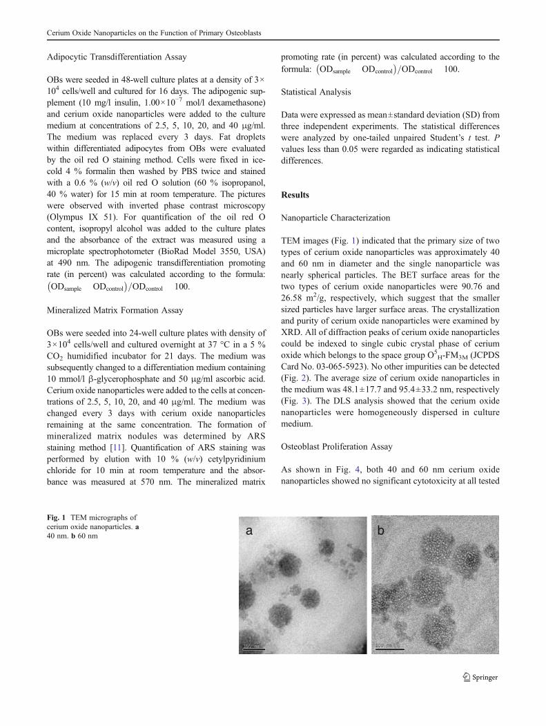
TEM images (Fig. 1) indicated that the primary size of twotypes of cerium oxide nanoparticles was approximately 40and 60 nm in diameter and the single nanoparticle wasnearly spherical particles. The BET surface areas for thetwo types of cerium oxide nanoparticles were 90.76 and26.58 m2/g, respectively, which suggest that the smallersized particles have larger surface areas. The crystallizationand purity of cerium oxide nanoparticles were examined byXRD. All of diffraction peaks of cerium oxide nanoparticlescould be indexed to single cubic crystal phase of ceriumoxide which belongs to the space group O5
H-FM3M (JCPDSCard No. 03-065-5923). No other impurities can be detected(Fig. 2). The average size of cerium oxide nanoparticles inthe medium was 48.1±17.7 and 95.4±33.2 nm, respectively(Fig. 3). The DLS analysis showed that the cerium oxidenanoparticles were homogeneously dispersed in culturemedium.
Osteoblast Proliferation Assay
As shown in Fig. 4, both 40 and 60 nm cerium oxidenanoparticles showed no significant cytotoxicity at all tested
baFig. 1 TEM micrographs ofcerium oxide nanoparticles. a40 nm. b 60 nm
Cerium Oxide Nanoparticles on the Function of Primary Osteoblasts

concentrations for 24 h. Cerium oxide nanoparticles (60 nm)inhibited the proliferation of OBs significantly at 48 h. AfterOBs were exposed to cerium oxide nanoparticles (60 nm) at2.5, 5, 10, 20, and 40 μg/ml for 48 h, cell viability decreasedto 89.1, 89.2, 86.3, 87.7, and 90.8 %, respectively, com-pared to the control. Interestingly, the inhibition effects ofthe 40-nm cerium oxide nanoparticles were lower than thatof the 60-nm cerium oxide nanoparticles at 48 h. No dose-dependent effects were observed.
Osteoblast Differentiation Assay
As shown in Fig. 5, cerium oxide nanoparticles (40 nm)promoted the differentiation of OBs at all tested concentra-tions for 24 and 48 h. The promotion effects were increasedwith increasing concentration. Cerium oxide nanoparticles(60 nm) promoted the differentiation of OBs at concentra-tions of 2.5 and 5 μg/ml, but inhibited the differentiation atconcentrations of 10, 20, and 40 μg/ml for 24 h. Theinfluence of the differentiation of OBs with cerium oxidenanoparticles (60 nm) at 48 h was similar with that at 24 h.The promotion effects of OB differentiation with cerium
oxide nanoparticles vary with different size and have dose-dependent manner.
Adipogenic Transdifferentiation Assay
As shown in Fig. 6, after OBs were exposed to cerium oxidenanoparticles (60 nm) at 2.5, 5, 10, 20, and 40 μg/ml for16 days, cell adipogenic differentiation-promoting rateswere 16.9, 14.9, 28.7, 27.4, and 33.1 %, respectively. Thecell adipogenic differentiation-promoting rate increased as afunction of dosage levels. The adipogenic differentiation-promoting rates of 40 nm cerium oxide nanoparticles werelower than that of 60 nm nanoparticles at the same concen-trations. After OBs were exposed to cerium oxidenanoparticles (40 nm) at 2.5, 5, 10, 20, and 40 μg/ml forthe same days, cell adipogenic differentiation-promotingrates were 13.4, 17.5, 15.1, 14.3, and 10.7 %, respectively.The morphological observation was in accordance with theabove results (Fig. 7). In brief, cerium oxide nanoparticlespromoted the adipogenic transdifferentiation of OBs at alltested concentrations. For the majority of concentrations, thepromotion effects were enhanced with the increasing size ofnanoparticles.
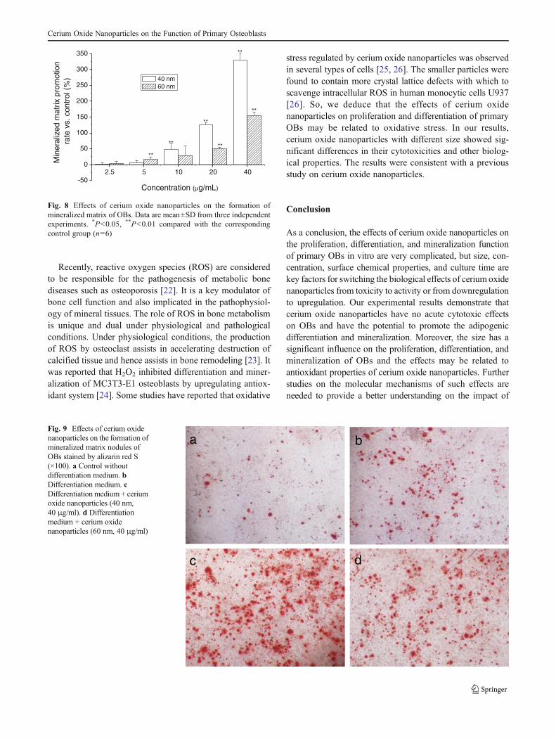
Mineralized Matrix Formation Assay
As shown in Fig. 8, cerium oxide nanoparticles (40 nm)promoted the formation of mineralized matrix of OBs at alltested concentrations and had a significant effect at theconcentrations of 10, 20, and 40 μg/ml. After the OBs wereexposed to cerium oxide nanoparticles (40 nm) at 10, 20,and 40 μg/ml, the promoting rates were 48.5, 125.7, and329.8 %, respectively. Cerium oxide nanoparticles (60 nm)also promoted the formation of mineralized matrix of OBs.However, the promotion effects of 60 nm cerium oxidenanoparticles were lower than that of 40 nm nanoparticlesat the same concentrations. For example, after OBs wereexposed to cerium oxide nanoparticles (60 nm) at 10, 20,and 40 μg/ml, the promoting rates were 27.6, 50.9, and154.3 %, respectively. The above results were also in
20 40 60 80 100
b
a
2θ (degree)
Inte
nsity
(Cou
nts)
Fig. 2 XRD patterns of cerium oxide nanoparticles. a 40 nm. b 60 nm
30.2 36.6 44.2 53.4 64.5 78 94.2 113.9 137.6 166.40
2
4
6
8
10
12
14
Num
ber
of p
artic
les
(%)
Diameter (nm)
a
59.3 68.3 78.8 90.9 104.8 120.8 139.3 160.6 185.2 213.5 246.2 283.80
2
4
6
8
10
Num
ber
of p
artic
les
(%)
Diameter (nm)
bFig. 3 Size distribution ofcerium oxide nanoparticles inmedium obtained by DLS. a40 nm. b 60 nm
Zhou et al.

accordance with the morphological observation (Fig. 9).Cerium oxide nanoparticles promoted the formation of min-eralized matrix of OBs at all tested concentrations and thepromotion effects were enhanced with the decreasing size ofnanoparticles.
Discussion
Recently, the potentially biological effects of nanoparticles onbone have been the attention issues of toxicologists.Publications on the bone cell biological effects of nanoparticlesare rapidly increasing. It has been demonstrated thatnanoparticles of various chemical compositions and sizescould promote or inhibit the differentiation and mineralizationof rat OBs and mesenchymal stem cells. Au nanoparticlespromoted differentiation and mineralization of the murinepreosteoblast MC3T3-E1 cells and the differentiationpromotion rate was enhanced with the decreasing sizeof nanoparticles [12]. Shi et al. showed that osteoblast-like cell proliferation and apoptosis were related to thesize of the hydroxyapatite nanoparticles. The hydroxy-apatite nanoparticles (20 nm) were the most effective atpromoting cell growth and inhibiting cell apoptosis [13].However, the effects of cerium oxide nanoparticles onprimary OBs are still unknown at this moment.
OBs cultured from newborn mouse skulls retain moresomatic cell function characteristics than cell lines and cansimulate biological changes in vivo. We investigated the ef-fects of cerium oxide nanoparticles on the proliferation,
differentiation, and mineralization of primary OBs. The re-sults indicate that the effects of cerium oxide nanoparticles onthe proliferation, differentiation, and mineralization of OBsdepend on size, concentration, and exposure time. The resultsof proliferation tests showed that toxicities of 60-nm ceriumoxide nanoparticles appeared in the cells at 48 h exposure. Tocompare the toxicity of different sized nanoparticles, we treat-ed 40 and 60 nm cerium oxide particles. The viability wasdecreased as size and exposure time increased. In our study,cerium oxide nanoparticles did not show significant cytotox-icity on primary OBs. ALP activity is a marker of osteo-genic differentiation and plays a key role in bone healingafter fracture by initiating and promoting the formation ofhyaluronic acid in osteoblast matrix vesicles [14]. Theexperimental results showed that dose is a key factorwhich affects the differentiation of OBs. Cerium oxidenanoparticles (40 nm) promoted the differentiation ofOBs and the promotion rate was enhanced with increasingconcentration. Cerium oxide nanoparticles (60 nm) pro-moted the differentiation of OBs at lower concentrations,but turned to inhibit the differentiation at higher concen-trations. From the results, we know that the ALP activityof OBs treated with 60-nm cerium oxide was significantlydifferent from that treated with 40-nm cerium oxide at thesame concentrations and time points. So, the size also playsa critical role in the differentiation of OBs. The increase ofALP activity is an early characteristic marker of osteogenicdifferentiation and mineral production is a later marker ofosteogenic differentiation. Mineralization function is a neces-sary condition of OBs to form bone calcification eventually. If
2.5 5 10 20 4040
50
60
70
80
90
100
110 40 nm 60 nm
Cel
l via
bilit
y vs
con
trol
(%
)
Concentration (μg/ml) Concentration (μg/ml)
a
2.5 5 10 20 4040
50
60
70
80
90
100
110
******
****
*
Cel
l via
bilit
y vs
con
trol
(%
)
40 nm 60 nm
*
bFig. 4 Viability of primary OBsafter exposure to cerium oxidenanoparticles. a 24 h. b 48 h.Values are mean±SD from threeindependent experiments.*P<0.05, **P<0.01 comparedwith the corresponding controlgroup (n=6)
2.5 5 10 20 40
-50
0
50
100
150a
****
*
*
**
****
**
Diff
eren
tiatio
n pr
omot
ion
ra
te v
s. c
ontr
ol (
%)
Concentration (μg/mL) Concentration (μg/mL)
40 nm 60 nm
2.5 5 10 20 40
-50
0
50
100
150b
*
**
****
**
****
****
Diff
eren
tiatio
n pr
omot
ion
ra
te v
s. c
ontr
ol (
%)
40 nm 60 nm
Fig. 5 Effects of cerium oxidenanoparticles on the ALPactivity of primary OBs. a 24 h.b 48 h. Values are mean±SDfrom three independentexperiments. *P<0.05,**P<0.01 compared with thecorresponding control group(n=6)
Cerium Oxide Nanoparticles on the Function of Primary Osteoblasts

the mineralization ability is reduced, the collagen cannotcalcify, leading to the formation of fibrous tissue. WhenOBs were mineralized, a large number of mineralizationnodes were formed [15]. So, the differentiation of OBs invitro can be characterized by matrix maturation and minerali-zation. In this paper, we found that cerium oxide nanoparticlespromoted the mineralization function of OBs in a dose-dependent manner and the promotion rate increased with adecreasing size.
The biological effects of nanomaterials depend on vari-ous factors, including chemical composition, size, shape,crystallinity, surface activity, solubility in aqueous media,
and aggregation state. We found that the differentiation andmineralization promotion rate of OBs with cerium oxidenanoparticles was enhanced with decreasing size. It hasbeen known that the toxicity of nanoparticles is closelyrelated to the surface area of the particles [16]. It may beexpected that the small-sized particles with more surfacearea are more active to the cells than large-sized particles.The surface chemistry properties mediate the protein-adsorbing capacity of nanoparticles, which determinesthe cellular binding of nanoparticles [17]. After exposingto serum, particles were rapidly coated with proteins inthe serum. A positive correlation was observed betweenprotein adsorption to particle surfaces and cellular binding.Cerium oxide nanoparticles which have different surfaceareas may influence the protein adsorption and cellularuptake of the nanoparticles [18, 19]. So, surface areamay play a critical role to determine the biological effectsof nanomaterials. However, it is controversial that surfacearea is used as the dose metric to judge the toxicity ofnanomaterials. Wittmaack addressed that the particle num-ber may be the best choice of the dose metric [20].Debate around the dose metric of nanoparticles is ongo-ing. The effects of cerium ion (Ce3+) on the proliferation,differentiation, and mineralization function of primaryOBs in vitro have been previously reported [21]. Theresults demonstrated that the behavior of cerium oxidenanoparticles on OBs was significantly different from thatof the Ce3+. The cerium oxide nanoparticles in cell me-tabolism system may not transform to release or releasevery few Ce3+ and biological results attributed to thebehavior of nanoparticles.
2.5 5 10 20 40-10
0
10
20
30
40
50
**
**
**
**
**
**
**
**
**
**
Adi
poge
nic
diffe
rent
iatio
n p
rom
otin
g ra
te (
%)
Concentration (μg/ml)
40 nm 60 nm
Fig. 6 Effects of cerium oxide nanoparticles on the adipogenictransdifferentiation of primary OBs. Values are mean±SD from threeindependent experiments. *P<0.05, **P<0.01 compared with the cor-responding control group (n=6)
a b
c d
Fig. 7 Effects of cerium oxidenanoparticles on the adipogenicdifferentiation of OBs by oil redO stain (×100). a Controlwithout any adipogenicsupplement. b Adipogenicsupplement. c Adipogenicsupplement + cerium oxidenanoparticles (40 nm, 40 μg/ml).d Adipogenic supplement +cerium oxide nanoparticles(60 nm, 40 μg/ml)
Zhou et al.
Recently, reactive oxygen species (ROS) are consideredto be responsible for the pathogenesis of metabolic bonediseases such as osteoporosis [22]. It is a key modulator ofbone cell function and also implicated in the pathophysiol-ogy of mineral tissues. The role of ROS in bone metabolismis unique and dual under physiological and pathologicalconditions. Under physiological conditions, the productionof ROS by osteoclast assists in accelerating destruction ofcalcified tissue and hence assists in bone remodeling [23]. Itwas reported that H2O2 inhibited differentiation and miner-alization of MC3T3-E1 osteoblasts by upregulating antiox-idant system [24]. Some studies have reported that oxidative
stress regulated by cerium oxide nanoparticles was observedin several types of cells [25, 26]. The smaller particles werefound to contain more crystal lattice defects with which toscavenge intracellular ROS in human monocytic cells U937[26]. So, we deduce that the effects of cerium oxidenanoparticles on proliferation and differentiation of primaryOBs may be related to oxidative stress. In our results,cerium oxide nanoparticles with different size showed sig-nificant differences in their cytotoxicities and other biolog-ical properties. The results were consistent with a previousstudy on cerium oxide nanoparticles.
Conclusion
As a conclusion, the effects of cerium oxide nanoparticles onthe proliferation, differentiation, and mineralization functionof primary OBs in vitro are very complicated, but size, con-centration, surface chemical properties, and culture time arekey factors for switching the biological effects of cerium oxidenanoparticles from toxicity to activity or from downregulationto upregulation. Our experimental results demonstrate thatcerium oxide nanoparticles have no acute cytotoxic effectson OBs and have the potential to promote the adipogenicdifferentiation and mineralization. Moreover, the size has asignificant influence on the proliferation, differentiation, andmineralization of OBs and the effects may be related toantioxidant properties of cerium oxide nanoparticles. Furtherstudies on the molecular mechanisms of such effects areneeded to provide a better understanding on the impact of
a b
c d
Fig. 9 Effects of cerium oxidenanoparticles on the formation ofmineralized matrix nodules ofOBs stained by alizarin red S(×100). a Control withoutdifferentiation medium. bDifferentiation medium. cDifferentiation medium + ceriumoxide nanoparticles (40 nm,40 μg/ml). d Differentiationmedium + cerium oxidenanoparticles (60 nm, 40 μg/ml)
2.5 5 10 20 40-50
0
50
100
150
200
250
300
350
**
**
**
** **
**M
iner
aliz
ed m
atrix
pro
mot
ion
rate
vs.
con
trol
(%
)
Concentration (μg/mL)
40 nm 60 nm
Fig. 8 Effects of cerium oxide nanoparticles on the formation ofmineralized matrix of OBs. Data are mean±SD from three independentexperiments. *P<0.05, **P<0.01 compared with the correspondingcontrol group (n=6)
Cerium Oxide Nanoparticles on the Function of Primary Osteoblasts
cerium oxide nanoparticles on biological systems and impor-tant information for future safe applications and the design ofbiocompatible nanomaterials.
Acknowledgments This research was supported by the NationalNatural Science Foundation of China (21001038 and 21271059) andthe Research Fund for the Doctoral Program of Higher Education ofChina (20111301110004).
References
1. Dong XT, Hong GY, Yu DC, Yu DS (1997) Synthesis and proper-ties of cerium oxide nanometer powder by pyrolysis of amorphouscitrate. J Mater Sci Technol 13:113–116
2. Zhai YQ, Zhang SY, Pang H (2007) Preparation, characterizationand photocatalytic activity of cerium oxide nanocrystalline usingammonium bicarbonate as precipitan. Mater Lett 61:1863–1866
3. Corma A, Atienzar P, Garcia H, Chane-Ching JY (2004)Hierarchically mesostructured doped cerium oxide with potentialfor solar-cell use. Nat Mater 3:394–397
4. Schubert D, Dargusch R, Raitano J, Chan SW (2006) Cerium andyttrium oxide nanoparticles are neuroprotective. Biochem BiophysRes Commun 342:86–91
5. Pirmohamed T, Dowding JM, Singh S, Wasserman B, Heckert E,Karakoti AS, King JE, Seal S, Self WT (2010) Nanoceria exhibitredox state-dependent catalase mimetic activity. Chem Commun46:2736–2738
6. Tsuji JS, Maynard AD, Howard PC, James JT, Lam CW, WarheitDB, Santamaria AB (2006) Research strategies for safety evalua-tion of nanomaterials, part IV: risk assessment of nanoparticles.Toxicol Sci 89:42–50
7. Mandeh M, Omidi M, Rahaie M (2012) In vitro influences of TiO2
nanoparticles on barley (Hordeum vulgare L.) tissue culture. BiolTrace Elem Res 150:376–380
8. Jin C, Tang Y, Yang FG, Li XL, Xu S, Fan XY, Huang YY, YangYJ (2011) Cellular toxicity of TiO2 nanoparticles in anatase andrutile crystal phase. Biol Trace Elem Res 141:3–15
9. Robert AY, Tu CA, Robert MP (2012) Distribution, elimination,and biopersistence to 90 days of a systemically introduced 30 nmceria-engineered nanomaterial in rats. Toxicol Sci 127:256–268
10. Nishimori S, Tanaka Y, Chiba T, Fujii M, Imamura T, Miyazono K,Ogasawara T, Kawaguchi H, Igarashi T, Fujita T, Tanaka K,Toyoshima H (2001) Smad-mediated transcription is required fortransforming growth factor-β1-induced p57Kip2 proteolysis inosteoblastic cells. J Biol Chem 276:10700–10705
11. Wang D, Christensen K, Chawla K, Xiao GZ, Krebsbach PH,Franceschi RT (1999) Isolation and characterization of MC3T3-E1
preosteoblast subclones with distinct in vitro and in vivo differenti-ation/mineralization potential. J Bone Miner Res 14:893–903
12. Liu DD, Zhang JC, Yi CQ, Yang MS (2010) The effects of goldnanoparticles on the proliferation, differentiation, and mineralizationfunction of MC3T3-E1 cells in vitro. Chin Sci Bull 55:1013–1019
13. Shi ZL, Huang X, Cai YR, Tang RK, Yang DS (2009) Size effectof hydroxyapatite nanoparticles on proliferation and apoptosis ofosteoblast-like cells. Acta Biomater 5:338–345
14. Tran N, Webster TJ (2011) Increased osteoblast functions in thepresence of hydroxyapatite-coated iron oxide nanoparticles. ActaBiomater 7:1298–1306
15. Birmingham E, Niebur GL, McHugh PE, Shaw G, Barry FP,McNamara LM (2012) Osteogenic differentiation of mesenchymalstem cells is regulated by osteocyte and osteoblast cells in asimplified bone niche. Eur Cells Mater 23:13–27
16. Oberdorster G, Oberdoster E, Oberdorster J (2005) Nanotoxicology:an emerging discipline evolving from studies of ultrafine particles.Environ Health Perspect 113:823–829
17. EhrenbergMS, FriedmanAE, Finkelstein JN, Oberdörster G,McGrathJL (2009) The influence of protein adsorption on nanoparticle associ-ation with cultured endothelial cells. Biomaterials 30:603–610
18. Patil S, Sandberg A, Heckert E, Self W, Seal S (2007) Proteinadsorption and cellular uptake of cerium oxide nanoparticles as afunction of zeta potential. Biomaterials 28:4600–4607
19. Asati A, Santra S, Kaittanis C, Perez JM (2010) Surface-charge-dependent cell localization and cytotoxicity of cerium oxidenanoparticles. ACS Nano 4:5321–5331
20. Wittmaack K (2007) In search of the most relevant parameter forquantifying lung inflammatory response to nanoparticle exposure:particle number, surface area, or what? Environ Health Perspect115:187–194
21. Zhang JC, Liu CC, Li YP, Sun J, Wang P, Di KQ, Zhao YY (2010)Effect of cerium ion on the proliferation, differentiation and min-eralization function of primary mouse osteoblasts in vitro. J RareEarths 28:138–142
22. Isomura H, Fujie K, Shibata K, Inoue N, Iizuka T, Takebe G,Takahashi K, Nishihira J, Izumi H, Sakamoto W (2004) Bonemetabolism and oxidative stress in postmenopausal rats with ironoverload. Toxicology 197:93–100
23. Filaire E, Toumi H (2012) Reactive oxygen species and exercise onbone metabolism: friend or enemy? Joint Bone Spine 79:341–346
24. Arai M, Shibata Y, Pugdee K, Abiko Y, Ogata Y (2007) Effects ofreactive oxygen species on antioxidant system and osteoblasticdifferentiation in MC3T3-E1 cells. IUBMB Life 59:27–33
25. Park EJ, Choi J, Park YK, Park K (2008) Oxidative stress inducedby cerium oxide nanoparticles in cultured BEAS-2B cells.Toxicology 245:90–100
26. Lord MS, Jung M, Teoh WY, Gunawan C, Vassie JA, Amal R,Whitelock JM (2012) Cellular uptake and reactive oxygen speciesmodulation of cerium oxide nanoparticles in human monocyte cellline U937. Biomaterials 33:7915–7924
Zhou et al.


![Indigenous Enhanced Mineralization Pyrene, Benzo[a]pyrene ...Indigenous soil microorganism mineralization experiments. All of the mineralization experiments were performed by using](https://img.pdfslide.net/doc/110x75/5e7c41b0b7c4ef64181e5e16/indigenous-enhanced-mineralization-pyrene-benzoapyrene-indigenous-soil-microorganism.jpg)