Embed Size (px)
Citation preview
Journal of Dental Sciences (2012) 7, 20e25
Available online at www.sciencedirect.com
journal homepage: www.e- jds.com
ORIGINAL ARTICLE
Effects of different surface treatments on the bondstrength of glass fiber-reinforced composite rootcanal posts to composite core material
Murat Kurt a, Ahmet Umut Guler a*, _Ibrahim Duran a, Altay Uludamar b,Ozgur _Inan c
aDepartment of Prosthodontics, Faculty of Dentistry, Ondokuz Mayıs University, Samsun, Turkeyb Private Practice, Ankara, TurkeycDepartment of Prosthodontics, Faculty of Dentistry, Selcuk University, Konya, Turkey
Final revision received 6 September 2011; accepted 6 December 2011Available online 19 February 2012
KEYWORDSacid etching;airborne-particleabrasion;
bond strength;core material;Er:YAG laser;FRC post
* Corresponding author. DepartmentE-mail address: [email protected]
1991-7902/$36 Copyrightª 2012, Assocdoi:10.1016/j.jds.2012.01.003
Abstract Background/purpose: The purpose of this study was to investigate the effects ofdifferent surface treatments on the bond strength of glass fiber-reinforced composite (FRC)posts to composite core material.Materials and methods: A total of 18 FRC posts were randomly divided into six groups(n Z 3), one of which was the untreated control group. Surface treatment of other groupswere as follows: airborne particle abrasion with 50-mm Al2O3 powder at 60 psi for 10 secondsthrough a nozzle distance of 10 mm; etching with 4% hydrofluoric (HF) acid; and surfacepreparation with an Er:YAG laser under three different power settings (of 300, 400, and500 mJ, at 2 Hz and 100 mS). A cylindrical Teflon mold was used to surround the treatedposts, and the mold was filled with dual-cure composite core material. All samples werelight-cured for 60 seconds through the top of the mold. After 24 hours of storage in water,specimens were sectioned perpendicular to the bonded interface under water cooling toobtain 2-mm thick post-and-core specimens. Each group consisted of 12 specimens. Push-out tests were performed at a cross-head speed of 0.5 mm/minute using a universal testingmachine. Data were analyzed by one-way analysis of variance followed by Tukey’s honestlysignificant difference test (a Z 0.05).Results: The lowest bond strength was observed in the Er:YAG 500-mJ group (6.14 �0.94 MPa). The acid-etched group revealed a higher bond strength (15.08 � 0.92 MPa) thanthe control group. The highest bond strength was observed in the airborne-particle abrasiongroup [18.89 � 0.83 MPa (P < 0.05)].
of Prosthodontics, Faculty of Dentistry, Ondokuz Mayıs University, Kurupelit, Samsun 55139, Turkey..tr (A.U. Guler).
iation for Dental Sciences of the Republic of China. Published by Elsevier Taiwan LLC. All rights reserved.
Surface treatments on FRC posts to core material 21
Conclusion: Er:YAG laser treatments on the FRC post surface decreased the bond strength.Airborne-particle abrasion and HF acid etching are alternative methods for increasing bondstrength of FRC posts to composite core material.Copyright ª 2012, Association for Dental Sciences of the Republic of China. Published byElsevier Taiwan LLC. All rights reserved.
Introduction
Endodontically treated teeth are often severely damagedby decay, excessive wear, or previous restorations, result-ing in a lack of coronal tooth structure.1 The longitudinalsuccess of restorative or prosthetic rehabilitations of anendodontically treated tooth depends on the quality of therestoration, on its clinical adaptation, and on the health ofthe supporting tissue.2 Most clinical failures involvingendodontically treated teeth reconstructed with posts aredue to cementation failure of the posts, whereas rootfractures are the most serious type of failure.3,4
Prefabricated glass fiber-reinforced composite (FRC)posts have been used since the beginning of the 1990s withthe introduction of carbon-fiber posts.5 Other types of FRCposts were developed in an attempt to improve aesthetics,thanks to the development of glass or white-quartz fibersand translucent resinous matrices.6 FRC posts are essen-tially composite materials composed of fibers of silica sur-rounded by a matrix of polymer resin, usually an epoxyresin. FRC posts are translucent and therefore haveaesthetic advantages. Currently, a wide variety of FRCposts are available with different sizes, tapers, andshapes.7,8 FRC posts also more closely match the modulusof elasticity of sound root dentin, and numerous in vitrostudies showed that the posts distribute occlusal stressesmore evenly in the root dentin, usually leading to fewerand less-catastrophic root fractures, which are oftenrepairable.2,7,9
Generally, retention is affected by the post type, theproperties of the cement, and the cement bond to the postand root canal dentin.10 FRC post placement involves theformation of two equally important interfaces, i.e., at thedentin/resin composite and resin composite/fiber level,where a failure can eventually occur.11 In publishedresearch12 on luting fiber posts to hybridized root canals,60% of failures during push-out testing occurred betweenthe fiber post and cement. The durability of a compositeresin core restoration depends on the formation of a strongbond between the core material and residual dentin, aswell as between the core and post material, enabling theinterface to transfer stresses under functional loading.13,14
A number of studies particularly focused on the possi-bility of improving adhesion at the fiber post-compositeinterface through various treatments of the postsurface.4,15e18 In an attempt to maximize resin bonding toFRC posts, several surface treatments were recently sug-gested. These procedures can be divided into three cate-gories: (1) silanization and/or adhesive application, (2) acidetching, sandblasting, and silica coating, and (3) alterna-tive etching techniques (i.e., treatments that combine botha micromechanical and chemical component).5 Due toimprovements in lasers used in dentistry, erbium: yttrium-
aluminum-garnet (Er:YAG) laser treatment is consideredan alternative method to other surface treatment methodsbecause of its optical penetration depth.19 As far as lasertreatment on FRC posts, no experimental research has beenundertaken to date.
The purpose of this study was to investigate the effectsof different surface treatment procedures on the bondstrength of FRC root canal posts to composite core mate-rial. It was hypothesized that the bond strength achieved atthe post-core interface would be affected by sandblasting,acid-etching, and the Er:YAG laser under different powersettings.
Materials and methods
A total of 18 FRC root canal posts (FRC Postec Plus, size 3,Ivoclar Vivadent, Schaan, Liechtenstein) were randomlydivided into six groups (n Z 3), with one being a controlgroup to which no surface treatment was applied. Surfacetreatment of the other groups were as follows: airborneparticle abrasion with 50-mm aluminum oxide (Korox 50,Bego, Bremen, Germany) at 60 psi for 10 seconds througha nozzle distance of 10 mm; etching with 4% hydrofluoric(HF) acid (Porcelain Etchant, Bisco, Schaumburg, IL USA)for 60 seconds; and surface preparation using an Er:YAGlaser (Fotona AT Fidelis, Ljubljana, Slovenia) under threedifferent power settings (of 300, 400, and 500 mJ, at 2 Hzand 100 mS) for 10 seconds. The specimens were treatedwith an Er:YAG laser working at 2940 nm. A 90�-angleddental hand-piece (R14-C) was used with a cylindricalsapphire (1.3 � 12 mm) fiber-optic tip. The tip was used atan incidence angle of 45� under water irrigation. The airand water pressure was set to two bars. The application tipwas moved from the bottom to the top and maintained inslight contact with the FRC post surface.
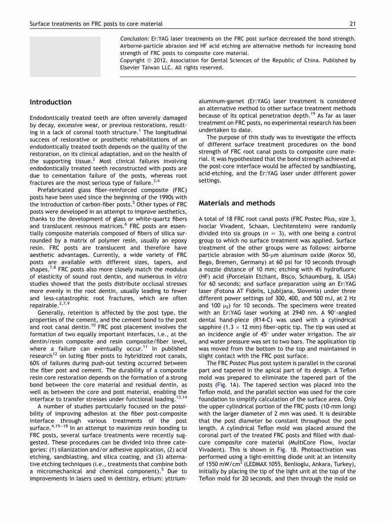
The FRC Postec Plus post system is parallel in the coronalpart and tapered in the apical part of its design. A Teflonmold was prepared to eliminate the tapered part of theposts (Fig. 1A). The tapered section was placed into theTeflon mold, and the parallel section was used for the corefoundation to simplify calculation of the surface area. Onlythe upper cylindrical portion of the FRC posts (10-mm long)with the larger diameter of 2 mm was used. It is desirablethat the post diameter be constant throughout the postlength. A cylindrical Teflon mold was placed around thecoronal part of the treated FRC posts and filled with dual-cure composite core material (MultiCore Flow, IvoclarVivadent). This is shown in Fig. 1B. Photoactivation wasperformed using a light-emitting diode unit at an intensityof 1550 mW/cm2 (LEDMAX 1055, Benlioglu, Ankara, Turkey),initially by placing the tip of the light unit at the top of theTeflon mold for 20 seconds, and then through the mold on

Figure 1 (A) Teflon mold for preparing the specimens;(B) core foundation preparation.
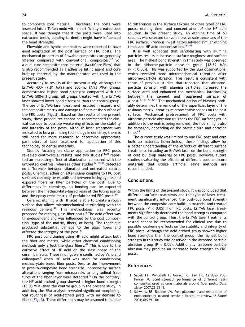
Figure 2 Schematic drawing of push-out test of thespecimens.
Table 1 Mean bond strengths (MPa), SD values, anddifferences for between the study groups.
Groups n Mean(MPa)
SD Differences*
Er:YAG 500 mJ 12 6.14 0.94 AEr:YAG 400 mJ 12 7.81 0.80 BEr:YAG 300 mJ 12 7.93 0.73 BControl 12 11.67 1.41 CHydrofluoric acid-etched 12 15.08 0.92 DAirborne-particle abrasion 12 18.89 0.83 E
* The different letters indicate dissimilarity of groups(P < 0.05).
22 M. Kurt et al
each side for 20 seconds for a total exposure of 60 seconds.After 24 h of storage in water, specimens were attached tothe arm of a low-speed saw (IsoMet; Buehler, Lake Bluff, IL,USA) and sectioned perpendicular to the bonded interfaceinto 2-mm-thick post and core segments under watercooling. Four segments were obtained from each post-and-core specimen, and therefore, each group of 12 post andcore specimens provided a total of 72 post and coresegments. The exact thickness of each post-and-coresegment was measured using a digital micrometer (Mitu-toyo, Tokyo, Japan) with 0.01-mm accuracy. The totalbonding area of each FRC post segment was calculatedusing the formula: A Z 2r � P � h, where r is the postradius,P is the constant 3.14, and h is the thickness of eachpost section. Because parallel-sided coronal sections wereused for the core foundations, the bonding area was equalfor all post segments and calculated to be A Z 2 � 3.14 � 1x 2 Z 12.56 mm2.
Push-out tests were performed at a cross-head speed of0.5-mm/minute using a universal testing machine (LloydLRX, Lloyd Instruments, Fareham, UK). After attaching thespecimens to a loading installment, the FRC post wasloaded with a 1.5-mm-diameter cylindrical stainless-steelplunger. The tip of the equipment was positioned such thatit only contacted the FRC post without contacting thecomposite core (Fig. 2). The peak force, at the point ofextrusion of the post segment from the test specimen, was
taken as the point of bond failure and recorded in Newtons(N). Push-out bond strength values in MPa were thencalculated by dividing this force by the bonded area of thepost segment.
One-way analysis of variance (ANOVA) in statisticalsoftware (SPSS for Windows, version 12.0.1; SPSS, Chicago,IL, USA) was used to evaluate the effects of surface treat-ment procedures on the bond strength between FRC postsand composite core material. The means were thencompared using Tukey’s honestly significant difference(HSD) test (a Z 0.05).
In addition, one FRC post specimen from each group wasprepared and evaluated by scanning electron microscopy[(SEM) JSM 6335-F, Jeol, Tokyo, Japan). Specimens wereobserved for surface irregularities under SEM at magnifi-cations of �250 and �1000.
Results
Bond strengths were shown to significantly differ by one-way ANOVA (P Z 0.001). The mean bond strengths, stan-dard deviations, and group differences for the six differentsurface-treatment groups are shown in Table 1.
In the study groups, the lowest bond strength wasobserved for the Er:YAG laser at 500 mJ (6.14 MPa). Nostatistically significant difference was observed betweenthe Er:YAG laser at 400 (7.81 MPa) and 300 mJ [7.93 MPa(P Z 0.752)], and these groups demonstrated higher bondstrengths compared with the Er:YAG laser at 500 mJ(P < 0.05). The control group demonstrated a statisticallysignificantly higher bond strength value (11.67 MPa)

Surface treatments on FRC posts to core material 23
compared with the above-mentioned groups (P < 0.05). Theacid-etched group showed a higher bond strength(15.08 MPa) than the control group. The highest bondstrength in this study was observed in the airborne-particleabrasion group [18.89 MPa (P < 0.05)]. Differences amonggroups are listed in Table 1.
The SEM studies revealed that the surface irregularitiesof the FRC root canal post corresponded to the results ofthe bond-strength study (Fig. 3).
Discussion
Within the limitations of the present study, it wasconcluded that our hypothesis was confirmed, i.e., bondstrengths of core build-up material to FRC posts weresignificantly affected by the investigated surface treat-ments. A number of studies particularly focused on thepossibility of improving adhesion at the fiber post-composite interface through various treatments of thepost surface.4,15e17,20,21
Laser applications for dental practice have beena research interest for the past 35 years. By varyinga number of parameters (pulse mode, irradiation time,frequency, and energy outputs), several types of lasers[neodymium: yttrium-aluminum: garnet (Nd:YAG), carbondioxide (CO2), Er:YAG, and semiconductor diode lasers]were indicated for dental treatments.22e24 The wavelengthof the Nd:YAG laser penetrates into water to a depth of60 mm, and the energy is scattered in soft tissues ratherthan being absorbed on the tissue surface. It is highlyabsorbed by black color; therefore, this laser is commonlyused for cutting and coagulation of oral soft tissues withgood hemostasis. However, due to its scattering effect, it isdifficult to judge the depth of penetration of this laser.25
Characteristics of the Er:YAG laser completely differ fromthose of the Nd:YAG laser. It is also applicable to both hardand soft tissues without carbonization.26 The wavelength ofthe Er:YAG laser lies near the boundary of the invisiblenear- and midinfrared portion of the spectrum. Thecoherent and collimated light of this laser with a wave-length of 2940 nm is highly absorbed by water. Theoreti-cally, its absorption coefficient by water is 10 times higherthan that of the CO2 laser (at a wavelength of 10,600 nm)and 15,000w20,000-times higher than that of the Nd:YAG
Figure 3 Scanning electron microscopy images of treated fi
laser (at a wavelength of 1064 nm).27 Due to its highabsorption by water, less tissue degeneration with a verythin surface interaction occurs with Er:YAG laser irradia-tion. Also, the temperature rise is minimal in the presenceof water irrigation, which makes hard substrate prepara-tions, caries removal, and scaling treatment possible withthis laser, with no carbonization.26,28 Various reportsconfirmed the safety and efficacy of CO2 and Nd:YAG lasers,which are the most commonly used lasers for soft-tissueapplications.29 However, when these lasers are applied todental hard tissues, thermal adverse events can be a majorproblem. The thermal effect of the laser beam is based onthe absorption of radiation by tissue and subsequenttransformation of laser energy into heat.30 Heat generationduring laser irradiation often causes carbonization, andmelting and cracking of the tooth structure. However, theEr:YAG laser showed satisfactory results for hard-tissueablatio, due to its characteristic wavelength that is wellabsorbed by water. So, Er:YAG laser treatment wasselected due to the reasons mentioned above. The use ofwater spray minimizes the heat generated by cooling theirradiated area and absorbing excessive laser energy.27,31,32
Water irrigation effectively prevented thermal damage andany major compositional or chemically deleterious changesdue to the irradiation.33
Murray and colleagues34 indicated that laser treatmentmay be a suitable alternative to airborne-particle abrasionor other surface pretreatment techniques for enhancing thebond strength of dental materials to metal surfaces. As forlaser treatment of FRC posts, no experimental research wasundertaken to date. Zhang and others35 reported thatcavity surfaces irradiated at 10 Hz with energies of 200 and300 mJ were similar, but those irradiated at 10 Hz with anenergy of 400 mJ showed cracks and melting of the dentin,indicating that the use of 4 W may damage the dentin.Gokce and colleagues36 reported that the shear bondstrength after laser treatment at 300 mJ was higher thanthat after treatment at 600w900 mJ. According to them,the reason for the low bond strengths observed at highpower settings may have been related to the observedheat-damaged layer.36 The current investigation focused onevaluating the effects of different surface treatmentprocedures, including Er:YAG laser under three differentpower settings [300 (0.6 W), 400 (0.8 W), and 500 mJ (1 W);2 Hz for 100 mS] on the bond strength of FRC root canal posts
ber post specimens at magnifications of �250 and �1000.
24 M. Kurt et al
to composite core material. Therefore, the posts wereinserted into a Teflon mold with an artificially created postspace. It was thought that if the posts were luted intoextracted teeth, bonding to dentin might have influencedthe bond strengths.
Flowable and hybrid composites were reported to havegood adaptation at the post surface of FRC posts. Themechanical properties of flowable composites are generallyinferior compared with conventional composites.37 So,a dual-cure composite core material (MultiCore Flow) thatis also recommended as a adhesive luting agent and corebuilt-up material by the manufacturer was used in thepresent study.
According to results of the present study, although theEr:YAG 400- (7.81 MPa) and 300-mJ (7.93 MPa) groupsdemonstrated higher bond strengths compared with theEr:YAG 500-mJ group, all groups treated with the Er:YAGlaser showed lower bond strengths than the control group.The use of Er:YAG laser treatment resulted in exposure ofthe composite matrix and damage to fibers at the surface ofthe FRC posts (Fig. 3). Based on the results of the presentstudy, these procedures cannot be recommended for clin-ical use due to possible weakening effects on the stabilityand integrity of the posts. Although laser treatment wasindicated to be a promising technology in dentistry, there isstill need for more research to determine appropriateparameters of laser treatment for application of thistechnology to dental materials.
Studies focusing on silane application to FRC postsrevealed controversial results, and two studies14,20 repor-ted an increasing effect of silanization compared with theuntreated controls, whereas other studies12,18,38 detectedno difference between silanated and untreated controlposts. Chemical adhesion after silane coupling to FRC postsurfaces can only be established between luting agents andexposed fibers or filler particles of the post. Due todifferences in chemistry, no bonding can be expectedbetween the methacrylate-based resin of the luting agentsand the epoxy resin matrix of prefabricated FRC posts.39
Ceramic etching with HF acid is able to create a roughsurface that allows micromechanical interlocking with theresinous cement.40 This methodology was recentlyproposed for etching glass-fiber posts.5 The acid effect wastime-dependent and was influenced by the post composi-tion (type of the matrix, fibers, or both). The techniqueproduced substantial damage to the glass fibers andaffected the integrity of the post.41
FRC post conditioning using HF acid might attack boththe fiber and matrix, while other chemical conditioningmethods only affect the glass fibers.42 This is due to thecorrosive effect of HF acid on the glass phase of theceramic matrix. These findings were confirmed by Vano andcolleagues5 when HF acid was used for conditioningmethacrylate-based fiber posts. Despite the improvementin post-to-composite bond strengths, noteworthy surfacealterations ranging from microcracks to longitudinal frac-tures of the fiber layer were detected.5 On the contrary,the HF acid-etched group showed a higher bond strength(15.08 MPa) than the control group in the present study. Inaddition, the SEM analysis revealed significant morpholog-ical roughness of acid-etched posts with no demage tofibers (Fig. 3). These differences may be assumed to be due
to differences in the surface texture of other types of FRCposts, etching time, and concentration of the HF acidsolution. In the present study, an etching time of 60seconds was selected to avoid massive substance loss of theFRC surface. Previous investigators advised similar etchingtimes and HF acid concentrations.41,42
It is well accepted that sandblasting with aluminaparticles results in increased surface roughness and surfacearea. The highest bond strength in this study was observedin the airborne-particle abrasion group [18.89 MPa(P < 0.05)]. This was supported by the SEM observations,which revealed more micromechanical retention afterairborne-particle abrasion. This result is consistent withthose of previous studies that reported that airborne-particle abrasion with alumina particles increased thesurface area and enhanced the mechanical interlockingbetween the cement and roughened surface ofa post.5,15,17,18,41 The mechanical action of blasting prob-ably determines the removal of the superficial layer of theresinous matrix, creating microretentive spaces on the postsurface. Mechanical pretreatment of FRC posts withairborne-particle abrasion roughens the FRC surface; yet, inaddition to the matrix being removed, the fibers might alsobe damaged, depending on the particle size and abrasiontime.43
The current study was limited to one FRC post and corebuild-up material. Nevertheless, these findings allow fora better understanding of the effects of different surfacetreatments including an Er:YAG laser on the bond strengthof core build-up material to FRC posts. However, futurestudies evaluating the effects of different post and corematerials that utilize artificial aging methods arerecommended.
Conclusions
Within the limits of the present study, it was concluded thatdifferent surface treatments and the type of laser treat-ment significantly influenced the push-out bond strengthbetween the composite core build-up material and treatedFRC posts (P < 0.05). For FRC posts, Er:YAG laser treat-ments significantly decreased the bond strengths comparedwith the control group. Thus, the Er:YAG laser treatmentstested cannot be recommended for clinical use due topossible weakening effects on the stability and integrity ofFRC posts. Although the acid-etched group showed higherbond strengths than the control group, the highest bondstrength in this study was observed in the airborne-particleabrasion group (P < 0.05). Additionally, airborne-particleabrasion may produce an increased bond strength to FRCposts.
References
1. Sadek FT, Monticelli F, Goracci C, Tay FR, Cardoso PEC,Ferrari M. Bond strength performance of different resincomposites used as core materials around fiber posts. DentMater 2007;23:95e9.
2. Schwartz RS, Robbins JW. Post placement and restoration ofendodontically treated teeth: a literature review. J Endod2004;30:289e301.

Surface treatments on FRC posts to core material 25
3. Testori T, Badino M, Castagnola M. Vertical root fractures inendodontically treated teeth: a clinical survey of 36 cases.J Endod 1993;19:87e91.
4. Bitter K, Priehn K, Martus P, Kielbassa AM. In vitro evaluation ofpush-out bond strengths of various luting agents to tooth-colored posts. J Prosthet Dent 2006;95:302e10.
5. Monticelli F, Ferrari M, Toledano M. Cement system and surfacetreatment selection for fiber post luting. Med Oral Patol OralCir Bucal 2008;13:214e21.
6. Ferrari M, Vichi A, Grandini S, Goracci C. Efficacy of a self-curing adhesive-resin cement system on luting glass-fiberposts into root canals: an SEM investigation. Int J Prostho-dont 2001;14:543e9.
7. Bateman G, Ricketts DNJ, Saunders WP. Fibre-based postsystems: A review. Br Dent J 2003;195:43e8.
8. Teixeira ECN, Teixeira FB, Piasick JR, Thompson JY. An in vitroassessment of prefabricated fiber post systems. J Am DentAssoc 2006;137:1006e12.
9. Wang VJJ, Chen YM, Yip KHK, Smales RJ, Meng QF, Chen L.Effect of two fiber post types and two luting cement systemson regional post retention using the push-out test. Dent Mater2008;24:372e7.
10. Albashaireh ZS, Ghazal M, Kern M. Effects of endodontic postsurface treatment, dentin conditioning, and artificial aging onthe retention of glass fiber-reinforced composite resin posts.J Prosthet Dent 2010;103:31e9.
11. Radovic I, Monticelli F, Cury AH, Bertelli E, Vulicevic ZR,Ferrari M. Coupling of composite resin cements to quartz fiberposts: a comparison of industrial and “Chairside” treatments ofthe post surface. J Adhes Dent 2008;10:57e66.
12. Perdigao J, Gomes G, Lee IK. The effect of silane on the bondstrengths of fiber posts. Dent Mater 2006;22:752e8.
13. Akgungor G, Sen D, Aydin M. Influence of different surfacetreatments on the short-term bond strength and durabilitybetween a zirconia post and a composite resin core material.J Prosthet Dent 2008;99:388e99.
14. Aksornmuang J, Foxton RM, Nakajima M, Tagami J. Micro-tensile bond strength of a dual-cure resin core material to glassand quartz fibre posts. J Dent 2004;32:443e50.
15. Radovic I, Monticelli F, Goracci C, et al. The effect of sand-blasting on adhesion of a dual-cured resin composite tomethacrylic fiber posts: microtensile bond strength and SEMevaluation. J Dent 2007;35:496e502.
16. Asmussen E, Peutzfeldt A, Sahafi A. Bonding of resin cementsto post materials: influence of surface energy characteristics.J Adhes Dent 2005;7:231e4.
17. Balbosh A, Kern M. Effect of surface treatment on retention ofglass-fiber endodontic posts. J Prosthet Dent 2006;95:218e23.
18. Sahafi A, Peutzfeldt A, Asmussen E, Gotfredsen K. Bondstrength of resin cement to dentin and to surface-treated postsof titanium alloy, glass fiber, and zirconia. J Adhes Dent 2003;5:153e62.
19. Shiu PD, Souza-Zaroni WC, Eduardo CP, Youssef MN. Effect offeldspathic ceramic surface treatments on bond strength toresin cement. Photomed Laser Surg 2007;25:291e6.
20. Goracci C, Raffaelli O, Monticelli F, Balleri B, Bertelli E,Ferrari M. The adhesion between prefabricated FRC posts andcomposite resin cores: microtensile bond strength with andwithout post-silanization. Dent Mater 2005;21:437e44.
21. Sahafi A, Peutzfeld A, Asmussen E, Gotfredsen K. Effect ofsurface treatment of prefabricated posts on bonding of resincement. Oper Dent 2004;29:60e8.
22. Gurgan S, Alpaslan T, Kiremitci A, Cakir FY, Yazıcı E, Gorucu J.Effect of different adhesive systems and laser treatment onthe shear bond strength of bleached enamel. J Dent 2009;37:527e34.
23. Van Meerbeek B, Munck JD, Mattar D, Landuyt KV,Lambrechts P. Microtensile bond strengths of an etch & rinse
and self-etch adhesive to enamel and dentin as a function ofsurface treatment. Oper Dent 2003;28:647e60.
24. Kim JT, Cho SA. The effects of laser etching on shear bondstrength at the titanium ceramic interface. J Prosthet Dent2009;101:101e6.
25. AAP. The Research, Science and Therapy Committee of theAmerican Academy of Periodontology: lasers in periodontics.J Periodontol 2002;73:1231e9.
26. Ishikawa I, Sasaki KM, Aoki A, Watanabe H. Effects of Er:YAGlaser on periodontal therapy. J Int Acad Periodontol 2003;5:23e8.
27. Hale GM, Querry MR. Optical constants of water in the 200-nmto 200-lm wavelength region. Appl Optics 1973;12:555e63.
28. Aoki A, Miura M, Akiyama F, et al. In vitro evaluation of Er:YAGlaser scaling of subgingival calculus in comparison with ultra-sonic scaling. J Periodont Res 2000;35:266e77.
29. White JM, Chaudhry SI, Kudler JJ, Sekandari N, Schoelch ML,Silverman Jr S. Nd:YAG and CO2 laser therapy of oral mucosallesions. J Clin Laser Med Surg 1998;16:299e304.
30. Rossmann JA, Cobb CM. Lasers in periodontal therapy. Perio-dontol 2000;1995(9):150e64.
31. Theodoro LH, Haypek P, Bachmann L, et al. Effect of Er:YAGand diode laser irradiation on the root surface: morphologicaland thermal analysis. J Periodontol 2003;74:838e43.
32. Burkes Jr EJ, Hoke J, Gomes E, Wolbarsht M. Wet versus dryenamel ablation by Er:YAG laser. J Prosthet Dent 1992;67:847e51.
33. Sasaki KM, Aoki A, Masuno H, Ichinose S, Yamada S, Ishikawa I.Compositional analysis of root cementum and dentin after Er:YAG laser irradiation compared with CO2 lased and intact rootsusing Fourier transformed infrared spectroscopy. J PeriodontalRes 2002;37:50e9.
34. Murray AK, Atrill DC, Dickinson MR. The effects of XeCl laseretching of Ni-Cr alloy on bond strengths to composite resin:a comparison with sandblasting procedures. Dent Mater 2005;21:538e44.
35. Zang S, Chen T, Ge L. Scanning electron microscopy study ofcavity preparation in deciduous teeth using the Er:YAG laserwith different powers. Lasers Med Sci 2012;27:141e4.
36. Gokce B, Ozpinar B, Dundar M, Comlekoglu E, Sen BH,Gungor MA. Bond strengths of all-ceramics: acid vs laseretching. Oper Dent 2007;32:173e8.
37. Wrbas KT, Schirrmeister JF, Altenburger MJ, Agrafioti A,Hellwig E. Bond strength between fibre posts and compositeresin cores: effect of post surface silanization. Int Endod J2007;40:538e43.
38. Bitter K, Meyer-Lueckel H, Priehn K, Martus P, Kielbassa AM.Bond strengths of resin cements to fiber-reinforced compositeposts. Am J Dent 2006;19:138e42.
39. Monticelli F, Toledano M, Tay FR, Sadek FT, Goracci C,Ferrari M. A simple etching technique for improving theretention of fiber posts to resin composites. J Endod 2006;32:44e7.
40. Guler AU, Yilmaz F, Yenisey M, Guler E. Ural C. Effect of acidetching time and a self etching adhesive on the shear bondstrength of composite resin to porcelain. J Adhes Dent 2006;8:21e5.
41. Valandro LF, Yoshiga S, De Melo RM, et al. Microtensile bondstrength between a quartz fiber post and a resin cement:effect of post surface conditioning. J Adhes Dent 2006;8:105e11.
42. Schmage P, Cakir FY, Nergiz I, Pfeiffer P. Effect of surfaceconditioning on the retentive bond strengths of fiber rein-forced composite posts. J Prosthet Dent 2009;102:368e77.
43. Magni E, Mazzitelli C, Papacchini F, et al. Adhesion betweenfiber posts and resin luting agents: a microtensile bondstrength test and an SEM investigation following differenttreatments of the post surface. J Adhes Dent 2007;9:195e202.