Embed Size (px)
Citation preview
Effects of epigallocatechin gallate on regulatory T cell number and function
in obese v. lean volunteers
Jung-Mi Yun, Ishwarlal Jialal and Sridevi Devaraj*
Department of Pathology and Laboratory Medicine, University of California, Davis, Medical Center, Sacramento, CA, USA
(Received 12 August 2009 – Revised 9 November 2009 – Accepted 17 December 2009 – First published online 23 February 2010)
Obesity predisposes to an increased incidence of diabetes and CVD. Also, obesity is a pro-inflammatory state. Regulatory T cells (Tregs) are
essential negative regulators of inflammation and are down-regulated in pro-inflammatory states. Animal models of obesity are associated with
decreased Tregs. The dietary modulation of Tregs could be used as a therapeutic strategy to control inflammation. Epigallocatechin gallate
(EGCG) is a potent anti-inflammatory agent and an active ingredient of green tea and is suggested to have a role as a preventive agent in obesity,
diabetes and CVD. The role of EGCG in the modulation of Tregs has, however, not been studied. Thus, the aim of the present study was to
determine the effect of EGCG on the number and function of Tregs in obese and lean human subjects in vitro, and to delineate its specific
regulation mechanisms. Tregs were isolated from normal-weight and obese subjects. Tregs were cultured in the absence or presence of EGCG
(20mM) for 24 h. Foxp3-expressing Tregs were enumerated using flow cytometry. Histone deacetylase (HDAC) activity and nuclear NF-kBp65
level were measured by ELISA and Western blots. Obese subjects had lower Tregs and IL-10 production than lean subjects. EGCG treatment
significantly enhanced the number of Foxp3-expressing Tregs and IL-10 production in vitro (P,0·05) in both groups. Also, EGCG decreased
NF-kB activity and increased HDAC activity and HDAC-2 expression in Tregs (P,0·05) in both groups. Thus, in part, EGCG enhances the
functionality of Tregs, i.e. IL-10 production and number by suppressing the NF-kB signalling pathway via inducing epigenetic changes.
Regulatory T cells: Inflammation: Green tea: Epigenetics
Obesity is increasingly recognised as a public health burden,because it is associated with an increased risk for manydiseases, including the metabolic syndrome, i.e. hypertension,insulin resistance or type 2 diabetes mellitus, arteriosclerosisand CHD(1). Obesity is associated with increased inflam-mation, as evidenced by increased levels of C-reactive protein,and decreased anti-inflammatory cytokines, such as IL-10(2).Obese individuals exhibit impaired immune responses.Immune and autoimmune responses are controlled by a finebalance between effector T cells (Teffs) and regulatory Tcells (Tregs). Tregs are essential negative regulators ofimmune responses. CD4þCD25þ Treg lymphocytes produceIL-10 and transforming growth factor-b; these are importantin controlling inflammatory T cells (both Th1 and Th2)(3). Itis now evident that Tregs are important in immunity andinflammation. Sakaguchi demonstrated that depletion ofCD4þCD25þ Tregs results in systemic autoimmune diseasein mice(4). CD4þCD25þ Tregs with a reduced in vitrosuppressive function were found in some studies performedon patients with type 1 diabetes mellitus and atherosclerosis,especially those with acute coronary syndromes(5 – 7). Further-more, recent studies have demonstrated that the forkheadfamily transcription factor (Foxp3) is predominantly expressedin CD4þCD25þ Tregs and plays a critical role for their
development and function. Mutations in the transcriptionfactor Foxp3 are associated with severe autoimmunediseases in man and the mouse(8 – 10). Thus, enhancement ofCD4þCD25þFoxp32 Treg cell activity may be important incontrolling immunity and inflammation.
Histone acetylation–deacetylation is an important epige-netic event that plays an important role in inflammation(11).Acetylation by histone acetyltransferases of specific lysineresidues on the N-terminal tail of core histones results inuncoiling of the DNA and increased accessibility to transcrip-tion factor binding. In contrast, histone deacetylation byhistone deacetylases (HDAC) represses gene transcriptionby promoting DNA winding, thereby limiting access to trans-cription factors. Furthermore, inhibiting HDAC resulted inenhanced activation of NF-kB(12) and increased histone acety-lation, culminating in increased pro-inflammatory cytokines.HDAC-2 has been reported to function in a corticosteroid-mediated anti-inflammatory mechanism(13). HDAC-1 and -2can interact directly with the p65 subunit of NF-kB to exertits co-repressor function in the nucleus(14). There is increasingevidence to suggest that HDAC plays an important role inregulating the pro-inflammatory response.
(2 )-Epigallocatechin gallate (EGCG) is one of the majorgreen tea catechins that are suggested to have a role as a
* Corresponding author: Dr Sridevi Devaraj, fax þ1 91 67346593, email [email protected]
Abbreviations: APC, allophycocyanin; EGCG, epigallocatechin gallate; Foxp3, forkhead family transcription factor; HDAC, histone deacetylases; PE, phycoerythrin;
Tregs, regulatory T cells.
British Journal of Nutrition (2010), 103, 1771–1777 doi:10.1017/S000711451000005Xq The Authors 2010
British
Journal
ofNutrition
preventive agent in cancer, obesity, diabetes and CVD(15).Due to the dominant role of CVD and the dramatic rise ofobesity and type 2 diabetes mellitus as major and interlinkedhealthcare problems, green tea and EGCG are increasinglybeing investigated in these areas(15). Additionally, there arenumerous in vivo studies demonstrating that EGCG exertcardiovascular and metabolic benefits in these model systems.Therefore, EGCG, a food component, can be regarded asuseful for the maintenance of cardiovascular and metabolichealth(15). However, there is a paucity of data examining themodulation of the number and function of Tregs with nutri-tional supplements.
The antioxidant and/or anti-inflammatory effects of dietarypolyphenols (curcumin and resveratrol) have all been shownto play a role in either controlling NF-kB activation or chro-matin remodelling through the modulation of HDAC activityand subsequently inflammatory gene expression(15 – 16). How-ever, there is a paucity of data on EGCG and HDAC activityin relation to inflammation and the function of Tregs.Also, there is a paucity of data on Tregs in obesity. Obesityis associated with increased inflammation and decreasedanti-inflammatory cytokines, such as IL-10(2). Thus, we hypoth-esise that EGCG enhances the functionality and number ofTregs through suppressing the NF-kB signalling pathway viainducing epigenetic changes, i.e. enhanced HDAC activity inobese human subjects.
Experimental methods
Materials
Anti-HDAC-2 and anti-HDAC-3 were procured from ActiveMotif (Carlsbad, CA, USA) and anti-NF-kBp65 antibodieswere procured from Cell Signaling Technology (Beverly,MA, USA). A CD4þCD25þ Regulatory T Cell Isolation kitwas procured from Miltenyi Biotec (Auburn, CA, USA).CD25-allophycocyanin (APC)-Cy7, CD4-phycoerythrin (PE)-Cy7 and Foxp3-Alexa Fluor antibodies were purchased fromBD Biosciences (San Jose, CA, USA). A transcriptionELISA kit (TransAM NF-kBp65) and HDAC assay kit(colorimetric) were purchased from Active Motif. A HumanIL-10 Quantikine HS ELISA kit was purchased from R&DSystems (Minneapolis, MN, USA). All other chemicals,unless otherwise stated, were obtained from Sigma (St Louis,MO, USA). The present study was conducted according to theguidelines laid down in the Declaration of Helsinki and allprocedures involving human subjects were approved by theUniversity of California Davis Institutional Review Board.Written informed consent was obtained from all subjects.
Subject selection and regulatory T cell isolation
Fasting blood (40 ml) was obtained from normal-weighthealthy donors (BMI , 25 kg/m2) and obese donors(BMI . 30 kg/m2) (twelve donors per group). Blood wasobtained in heparinised vacutainers and all complete bloodcell counts were normal. Twelve lean and twelve obesedonors were age-, sex- and race-matched and recruited. Leanand obese subjects were recruited according to the selectioncriteria (Table 1).
Peripheral blood mononuclear cells from lean and obesesubjects were isolated by Ficoll-Hypaque gradient(Sigma)(17) and then CD4þCD25þ Tregs were purified fromisolated peripheral blood mononuclear cells using aCD4þCD25þ Regulatory T Cell Isolation kit according tothe manufacturer’s protocols (Miltenyi Biotec). Briefly,CD4þT cells were negatively selected from the total periph-eral blood mononuclear cells, yielding a population of CD4þ
cells with purity of 92–98 %. Positive selection on anti-CD25 magnetic microbeads was then used to separate thenegative fraction containing CD4þCD252 T cells fromthe CD4þCD25þT cell fraction, using the CD4þCD25þ
Regulatory T Cell Isolation kit from Miltenyi Biotec. Thepurities of the sorted CD4þCD25þ populations werealways . 95 % as confirmed by flow cytometry. Cells werethen applied to a second magnetic column, washed, and elutedagain. Tregs were stained using three different antibodies suchas CD4-PE-Cy7, CD25-APC-Cy7 and Foxp3-Alexa Fluorwith a different colour using flow cytometry. CD4 and CD25antibodies were used for the surface staining of the Tregs.The Foxp3 antibody was used for the intracellular staining ofthe Tregs.
Regulatory T cell staining in whole blood
Tregs were stained using two different antibodies, i.e.CD4-PE-Cy7 and CD25-APC-Cy7, in whole blood by flowcytometry. CD4 and CD25 were used for the surface staining
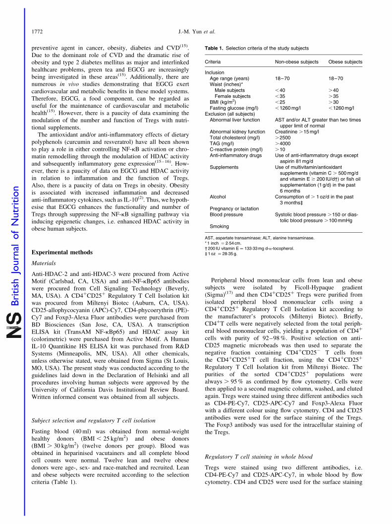
Table 1. Selection criteria of the study subjects
Criteria Non-obese subjects Obese subjects
InclusionAge range (years) 18–70 18–70Waist (inches)*
Male subjects ,40 .40Female subjects ,35 .35
BMI (kg/m2) ,25 .30Fasting glucose (mg/l) ,1260 mg/l ,1260 mg/l
Exclusion (all subjects)Abnormal liver function AST and/or ALT greater than two times
upper limit of normalAbnormal kidney function Creatinine .15 mg/lTotal cholesterol (mg/l) .2500TAG (mg/l) .4000C-reactive protein (mg/l) .10Anti-inflammatory drugs Use of anti-inflammatory drugs except
aspirin 81 mg/dSupplements Use of multivitamin/antioxidant
supplements (vitamin C . 500 mg/dand vitamin E $ 200 IU/d†) or fish oilsupplementation (1 g/d) in the past6 months
Alcohol Consumption of . 1 oz/d in the past3 months‡
Pregnancy or lactationBlood pressure Systolic blood pressure .150 or dias-
tolic blood pressure .100 mmHgSmoking
AST, aspartate transaminase; ALT, alanine transaminase.* 1 inch ¼ 2·54 cm.† 200 IU vitamin E ¼ 133·33 mg d-a-tocopherol.‡ 1 oz ¼ 28·35 g.
J.-M. Yun et al.1772
British
Journal
ofNutrition
of the Tregs. To 100ml of whole blood from both groups,10ml of Fcy blocking reagent were added. Blood sampleswere incubated with 10ml isotype control and/or 10ml appro-priate antibodies (CD25-APC-Cy7 and CD4-PE-Cy7) for30 min at 48C in the dark. After incubation, lysis buffer wasadded to the whole blood. Cells were centrifuged andwashed with PBS and re-suspended in 1 % paraformaldehyde(PFA) buffer. At least 10 000 cells were acquisitioned usingflow cytometry (BD Biosciences).
Cell culture and treatment with epigallocatechin gallate
Tregs were isolated from normal-weight and obese subjects.Tregs were cultured in Roswell Park Memorial Institute(RPMI) medium containing 10 % heat-inactivated fetalcalf serum, 1 % antibiotics and IL-2 (10 ng/ml) for the main-tenance of Tregs at 378C and 5 % CO2. EGCG (dissolved inRPMI medium) was used for the treatment of cells. Tregs(50–60 % confluent) were cultured in the absence orpresence of EGCG (20mM) for 24 h. After 24 h incubation,the medium was saved for the measurement of IL-10and cells were washed with PBS and then harvested forfurther studies.
Surface and intracellular staining of regulatory T cells
Tregs were stained using three different antibodies such asCD4-PE-Cy7, CD25-APC-Cy7 and Foxp3-Alexa Fluor byflow cytometry. For Foxp3 intracellular staining, cells wereincubated with CD25-APC-Cy7 and CD4-PE-Cy7 antibodiesat first for 30 min at 48C in the dark. After incubation,these cells were washed and then fixed and stained withthe Foxp3-Alexa Fluor antibody according to the manu-facturer’s instructions (BD Biosciences). Cells were washedthree times and re-suspended in 1 % PFA buffer. Atleast 10 000 cells were assayed by flow cytometry (BDBiosciences).
Measurement of IL-10 production in regulatory T cells
After treatment of cells with EGCG and without EGCG for24 h, the supernatant fraction was saved and stored at2808C. IL-10 levels were assayed in the supernatant fractionof treated cells using the Human IL-10 Quantikine HSELISA kit according to the manufacturer’s protocols (R&DSystems).
Preparation of nuclear lysates
After treatment of cells with and without EGCG, the cellswere washed twice in PBS (10 mM; pH 7·4). Nuclear lysateswere prepared using NE-PER Nuclear and CytoplasmicExtraction Reagents (Pierce, Thermo Scientific, Rockford,IL, USA). The lysates were collected and cleared by centri-fugation and the supernatant fraction sampled and storedat 2808C. The protein concentration in the lysates wasmeasured by BCA protein assay (Pierce) according to themanufacturer’s protocols.
Western blot analysis
For Western blot analysis, 12mg protein was resolvedover 4–20 % 2-amino-2-(hydroxymethyl)propane-1,3-diol(Tris)–glycine polyacrylamide gels (Invitrogen, Carlsbad,CA, USA), transferred onto nitrocellulose membranes, andsubsequently incubated in blocking buffer (5 % non-fatdry milk/1 % Tween 20; in 20 mM-Tris-buffered saline(pH 7·6)) for 2 h. The blots were incubated with appropriateprimary antibodies (HDAC-2 and NF-kBp65, 1:1000dilution), washed, and incubated with the appropriate second-ary horseradish peroxidase–conjugated antibody (1:2000dilution). The blots were detected with chemiluminescence(ECL kit; Amersham Biosciences, Piscataway, NJ, USA)and autoradiography, using XAR-5 film (Eastman Kodak,Rochester, NY, USA). Equal loading of protein wasconfirmed by stripping the blots and reprobing withlamin (Sigma).
Measurement of histone deacetylase activity using ELISA
Following treatment of cells with and without 20mM-EGCGfor 24 h, cells were harvested and nuclear lysates wereprepared. A quantity of 10mg nuclear lysate protein fromeach group was taken for determination of HDAC activity.The experiment was done according to the manufacturer’sinstructions (HDAC assay kit; Active Motif). Absorbancewas taken at 405 nm by using an ELISA reader (MultiscanMCC/340; Fisher Scientific, Rockford, IL, USA).
Detection of transcription factor NF-kBp65 using ELISA
The commercially available kit for NF-kBp65 (Active Motif)contains the specific oligos with the specific consensussequence for NF-kBp65 binding. Nuclear lysate protein(5 mg) from each group was taken for quantification ofNF-kB activity. The experiment was done according to themanufacturer’s instructions. Absorbance was taken at 450 nmby using the ELISA reader (Multiscan MCC/340; FisherScientific).
Immunocytochemical staining of histone deacetylase
After 24 h incubation in the absence or presence of EGCG,cells (1 £ 104) were placed on slides, the slides were airdried, fixed with 4 % formaldehyde for 30 min at 48C, andthen stained with HDAC-2 and -3 antibodies (1:1000) for18 h at 48C. Slides were washed first in PBS (pH 7·2) withTween 20 and then in PBS (10 min each). After being airdried, slides were incubated with the appropriate secondaryantibody (1:2000) for 60 min. The slides were washedas described above, air dried, mounted with mountingmedium, and then examined with a fluorescence microscopeat £ 400 magnification.
Statistical analysis
All in vitro experiments were conducted at least fivetimes in duplicate. Results were expressed as mean valuesand standard deviations. Statistical analysis was performed
Epigallocatechin gallate and T cell number 1773
British
Journal
ofNutrition
using ANOVA followed by Wilcoxon tests. P,0·05 wasconsidered significant.
Results
Foxp3-positive regulatory T cell numbers are decreased inobese compared with lean human subjects
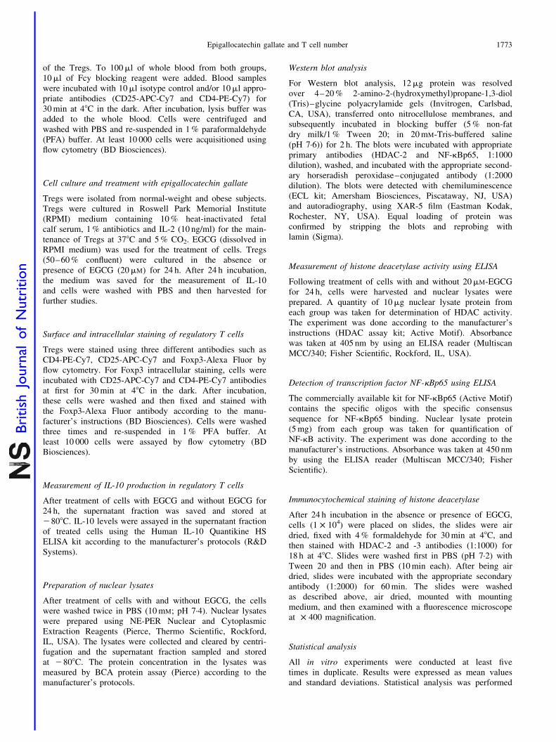
As shown in Fig. 1(a), the CD4þCD25þ Tregs in whole bloodwere 10·2 (SD 6·78) and 17·25 (SD 6·15) % in obese and leansubject groups, respectively (Fig. 1(a)). Thus, CD4þCD25þ
Tregs were significantly decreased in obese subjects comparedwith lean subjects (P,0·05) (Fig. 1(a)). We then evaluated theexpression of Foxp3 in CD4þCD25þ Tregs in obese v. leansubjects using flow cytometry. The purity of positivelyselected CD4þCD25þ Tregs was . 96 % as confirmed byflow cytometry in both groups. At least 10 000 cells wereassayed using flow cytometry (BD Bioscience). Half of thepurified CD4þCD25þ Tregs were Foxp3-positive. Foxp3-expressing CD4þCD25þ Tregs were significantly decreasedin obese subjects compared with lean subjects (P,0·05)(Fig. 1(b)).
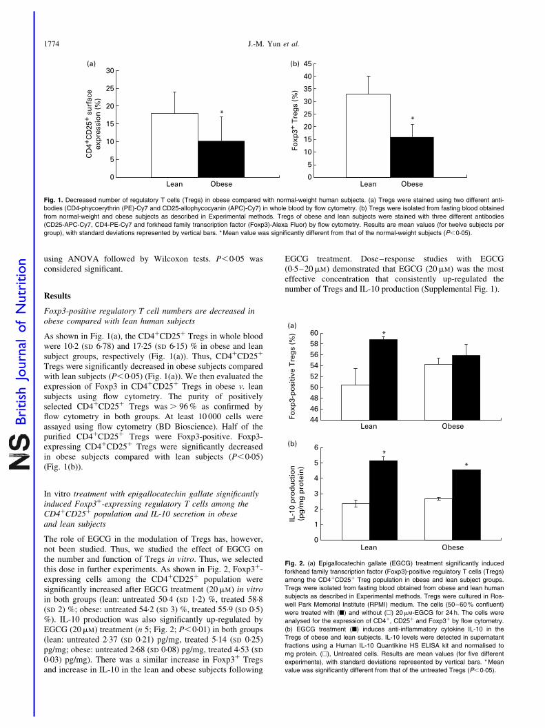
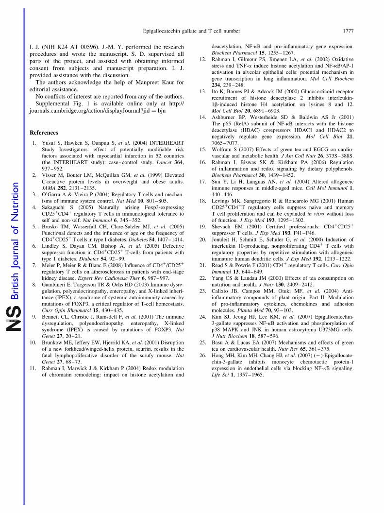
In vitro treatment with epigallocatechin gallate significantlyinduced Foxp3þ-expressing regulatory T cells among theCD4þCD25þ population and IL-10 secretion in obeseand lean subjects
The role of EGCG in the modulation of Tregs has, however,not been studied. Thus, we studied the effect of EGCG onthe number and function of Tregs in vitro. Thus, we selectedthis dose in further experiments. As shown in Fig. 2, Foxp3þ-expressing cells among the CD4þCD25þ population weresignificantly increased after EGCG treatment (20mM) in vitroin both groups (lean: untreated 50·4 (SD 1·2) %, treated 58·8(SD 2) %; obese: untreated 54·2 (SD 3) %, treated 55·9 (SD 0·5)%). IL-10 production was also significantly up-regulated byEGCG (20mM) treatment (n 5; Fig. 2; P,0·01) in both groups(lean: untreated 2·37 (SD 0·21) pg/mg, treated 5·14 (SD 0·25)pg/mg; obese: untreated 2·68 (SD 0·08) pg/mg, treated 4·53 (SD
0·03) pg/mg). There was a similar increase in Foxp3þ Tregsand increase in IL-10 in the lean and obese subjects following
EGCG treatment. Dose–response studies with EGCG(0·5–20mM) demonstrated that EGCG (20mM) was the mosteffective concentration that consistently up-regulated thenumber of Tregs and IL-10 production (Supplemental Fig. 1).
444648505254565860
Foxp
3-p
osi
tive
Tre
gs
(%)
0
1
2
3
4
5
6
IL-1
0 p
rod
uct
ion
(pg
/mg
pro
tein
)
Lean Obese
Lean Obese
*
*
*
(a)
(b)
Fig. 2. (a) Epigallocatechin gallate (EGCG) treatment significantly induced
forkhead family transcription factor (Foxp3)-positive regulatory T cells (Tregs)
among the CD4þCD25þ Treg population in obese and lean subject groups.
Tregs were isolated from fasting blood obtained from obese and lean human
subjects as described in Experimental methods. Tregs were cultured in Ros-
well Park Memorial Institute (RPMI) medium. The cells (50–60 % confluent)
were treated with (B) and without (A) 20mM-EGCG for 24 h. The cells were
analysed for the expression of CD4þ, CD25þ and Foxp3þ by flow cytometry.
(b) EGCG treatment (B) induces anti-inflammatory cytokine IL-10 in the
Tregs of obese and lean subjects. IL-10 levels were detected in supernatant
fractions using a Human IL-10 Quantikine HS ELISA kit and normalised to
mg protein. (A), Untreated cells. Results are mean values (for five different
experiments), with standard deviations represented by vertical bars. * Mean
value was significantly different from that of the untreated Tregs (P,0·05).
CD
4+C
D25
+ su
rfac
eex
pre
ssio
n (
%)
0
5
10
15
20
25
30
Lean Obese
Foxp
3+ T
reg
s (%
)
*
0
5
10
15
20
25
30
35
40
45
Lean
*
Obese
(a) (b)
Fig. 1. Decreased number of regulatory T cells (Tregs) in obese compared with normal-weight human subjects. (a) Tregs were stained using two different anti-
bodies (CD4-phycoerythrin (PE)-Cy7 and CD25-allophycocyanin (APC)-Cy7) in whole blood by flow cytometry. (b) Tregs were isolated from fasting blood obtained
from normal-weight and obese subjects as described in Experimental methods. Tregs of obese and lean subjects were stained with three different antibodies
(CD25-APC-Cy7, CD4-PE-Cy7 and forkhead family transcription factor (Foxp3)-Alexa Fluor) by flow cytometry. Results are mean values (for twelve subjects per
group), with standard deviations represented by vertical bars. * Mean value was significantly different from that of the normal-weight subjects (P,0·05).
J.-M. Yun et al.1774
British
Journal
ofNutrition
NF-κBp65
HD
AC
act
ivit
y (µ
M)
NF-
κBp
65 a
ctiv
ity
(op
tica
l den
sity
450
nm
)
0
0·1
0·2
0·3
0·4
0·5
0·6
0
10
20
30
40
50
60
Lean Obese
*
*
Lean Obese
* *
HDAC-2
Lamin
Untreated 20 µM-EGCG
NF-κBp65
HDAC-2
Lamin
Untreated 20 µM-EGCG
(b)(a)
(c)
Untreated
20 µM-EGCG
HDAC-2DAPI HDAC-3
Lean
Lean
Obese Untreated
Obese20 µM-EGCG
(d)
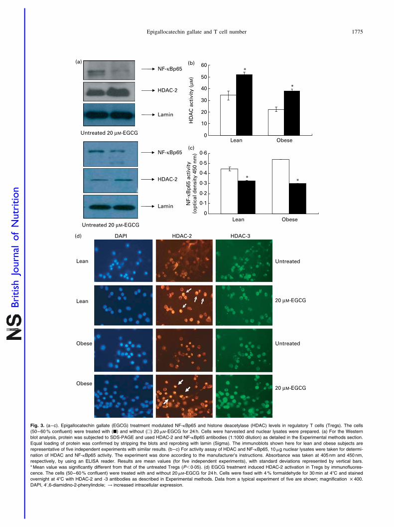
Fig. 3. (a–c). Epigallocatechin gallate (EGCG) treatment modulated NF-kBp65 and histone deacetylase (HDAC) levels in regulatory T cells (Tregs). The cells
(50–60 % confluent) were treated with (B) and without (A) 20mM-EGCG for 24 h. Cells were harvested and nuclear lysates were prepared. (a) For the Western
blot analysis, protein was subjected to SDS-PAGE and used HDAC-2 and NF-kBp65 antibodies (1:1000 dilution) as detailed in the Experimental methods section.
Equal loading of protein was confirmed by stripping the blots and reprobing with lamin (Sigma). The immunoblots shown here for lean and obese subjects are
representative of five independent experiments with similar results. (b–c) For activity assay of HDAC and NF-kBp65, 10mg nuclear lysates were taken for determi-
nation of HDAC and NF-kBp65 activity. The experiment was done according to the manufacturer’s instructions. Absorbance was taken at 405 nm and 450 nm,
respectively, by using an ELISA reader. Results are mean values (for five independent experiments), with standard deviations represented by vertical bars.
* Mean value was significantly different from that of the untreated Tregs (P,0·05). (d) EGCG treatment induced HDAC-2 activation in Tregs by immunofluores-
cence. The cells (50–60 % confluent) were treated with and without 20mM-EGCG for 24 h. Cells were fixed with 4 % formaldehyde for 30 min at 48C and stained
overnight at 48C with HDAC-2 and -3 antibodies as described in Experimental methods. Data from a typical experiment of five are shown; magnification £ 400.
DAPI, 40,6-diamidino-2-phenylindole; ! increased intracellular expression.
Epigallocatechin gallate and T cell number 1775
British
Journal
ofNutrition
Effect of epigallocatechin gallate on activation of NF-kB andhistone deacetylase levels in regulatory T cells
Next, we studied specific regulation mechanisms. Acetylation ofhistone protein is associated with increased binding of the tran-scription factor NF-kB(12). We studied the effects of EGCG ondeacetylation of histone proteins, HDAC activity, transactiva-tion and translocation of NF-kB in Tregs of both subjectgroups. Using immunoblot analysis, we observed that EGCGtreatment (20mM) of cells resulted in decreased NF-kBp65 inthe nuclear fraction in Tregs of the obese and lean subjectgroups (Fig. 3(a)). We further confirmed the inhibition ofNF-kBp65 transactivation (lean: untreated 0·446 (SD 0·019),EGCG treated 0·329 (SD 0·049) v. obese: untreated 0·54 (SD
0·02), EGCG treated 0·3 (SD 0·05) at 450 nm optical densityvalue) by performing ELISA (Fig. 3(c)). Also, EGCG enhancedtotal HDAC activity (lean: untreated 34·22 (SD 3·87)mM, EGCGtreated 51·78 (SD 2·31) mM v. obese: untreated 22·1 (SD 2·1) mM,EGCG treated 38·08 (SD 1·5) mM) and HDAC-2 levelsin the Tregs of obese and lean subject groups (Fig. 3(b) and(d) – immunofluorescence). There was no detectable significantdifference for the HDAC-3 in the Tregs of lean subjects(Fig. 3(d)). But EGCG increased HDAC-3 levels in theTregs of obese subjects (Fig. 3(d)). Thus, EGCG appears toenhance IL-10 production and Treg number through, at leastin part, suppressing the NF-kB signalling pathway via inducingHDAC activity in the Tregs of both groups.
Discussion
Obesity predisposes to increased diabetes and atherosclerosisand related complications, which represent the major causeof morbidity and mortality in the Western world. Obeseindividuals exhibit impaired immune responses. Immune andautoimmune responses are controlled by a fine balancebetween effector T cells (Teffs) and Tregs(3). Tregs constitu-tively express high levels of the IL-2 receptor a chain(CD25) and specifically express the forkhead/winged helixtranscription factor Foxp3 which acts in a regulatory capacityby inhibiting the activation and function of both self-antigen-and foreign-antigen-reactive T cells(18,19). Moreover, Foxp3plays a critical role for the development and function ofTregs(8 – 10). The critical physiological role of CD4þCD25þ-
Foxp3 Tregs is to control autoimmune diseases(20,21). Tregsmay suppress by a contact-dependent mechanism or throughthe secretion of anti-inflammatory cytokines IL-10 and trans-forming growth factor-b(1). These cells are a low-frequencysubpopulation of CD4þ cells, representing 1 to 2 % oftotal lymphocytes. We hypothesised that enhancement ofCD4þCD25þ Treg cell activity may protect individuals fromimpaired immune responses in patients with obesity.
Since ancient times, green tea has been considered a health-promoting beverage. Health-promoting effects of green tea aremainly attributed to its polyphenol content. Green tea is arich source of polyphenols, especially flavanols and flavonols.Catechins are the predominant flavanols and are mainlycomprised of EGCG, epigallocatechin, epicatechin gallate andepicatechin(15,22). Several intervention studies have demon-strated that green tea cathechins containing 200–300 mgEGCG exert beneficial effects on cardiovascular and meta-bolic health. Also, EGCG supplementation has been shown
to be anti-inflammatory as evidenced by decreased cytokinerelease and NF-kB activity(23 – 26). However, there are no dataexamining the effect of EGCG on the number and functionof Tregs in the obese.
In the present study, we provide important and novel pre-liminary data showing (1) decreased number and function ofTregs (decreased IL-10) in obese individuals in vivo comparedwith matched lean controls (Fig. 1) and (2) in vitro treatmentwith EGCG enhances the number of Foxp3-positive Tregs inobese and lean subjects (Fig. 2); this is associated withincreased IL-10 release (Fig. 2), decreased NF-kB activityand up-regulation of HDAC-2 (Fig. 3) in support of the earlierobservations(23 – 26). NF-kB plays a pivotal role in inflammation,and EGCG has been shown to down-regulate NF-kB in otherstudies; however, the effect of EGCG on Tregs has not beenstudied. Similar to other previous reports(23 – 26), in the presentstudy we show that EGCG promotes the number and functionof Tregs (IL-10 release) in both lean and obese subjectsin vitro. Furthermore, since Foxp3-expressing Tregs are moreeffective in affecting the functionality of Tregs, we examinedthe effect of EGCG treatment in vitro on Foxp3þ Tregs andsimilar findings were observed, i.e. enhancement of Foxp3þ
Tregs and IL-10 release in both lean and obese subjects.Epigenetics refers to heritable changes in phenotype (appear-
ance) or gene expression caused by mechanisms other thanchanges in the underlying DNA sequence. Histone acety-lation–deacetylation is an important epigenetic event thatplays an important role in inflammation(11). Acetylation ofhistone protein is associated with increased binding of the tran-scription factor NF-kB(12). While several other nutritional strat-egies such as curcumin and resveratrol have been shown topromote epigenetic events resulting in decreased inflammation,the effect of EGCG on chromatin remodelling has not beenstudied. Furthermore, in monocytes, inflammation and NF-kBappear to be regulated by changes in histone acetylation–deacetylation. Thus, we tested if EGCG up-regulates thenumber and anti-inflammatory activity of Tregs (by suppressingNF-kB) through chromatin remodelling. Here, we show thatEGCG alters HDAC activity and severely suppresses NF-kB,leading to increased IL-10 release in both obese and lean subjects.
In conclusion, EGCG up-regulates the number and anti-inflammatory activity of Tregs through chromatin remodellingby alteration of histone acetylation–deacetylation andsuppression of NF-kB, leading to the induction of IL-10release. However, the molecular mechanism of chromatinremodelling for pro-inflammatory genes in Tregs is not yetunderstood well and will be the focus of future studies.Furthermore, it will be important to confirm these in vitro find-ings in a placebo-controlled supplementation study of EGCGin obese human volunteers. Understanding of gene expressionand epigenetic modulation of Tregs by EGCG may be, at leastin part, a promising strategy to modulate the immune responseof the complications of obesity and its associated diseases andfuture studies will examine the effect of EGCG supplemen-tation in obese human subjects.
Acknowledgements
These studies were supported in part from discretionaryresearch funding available to the corresponding authorS. D. and through a mentoring award to the co-investigator
J.-M. Yun et al.1776
British
Journal
ofNutrition
I. J. (NIH K24 AT 00596). J.-M. Y. performed the researchprocedures and wrote the manuscript. S. D. supervised allparts of the project, and assisted with obtaining informedconsent from subjects and manuscript preparation. I. J.provided assistance with the discussion.
The authors acknowledge the help of Manpreet Kaur foreditorial assistance.
No conflicts of interest are reported from any of the authors.Supplemental Fig. 1 is available online only at http://
journals.cambridge.org/action/displayJournal?jid ¼ bjn
References
1. Yusuf S, Hawken S, Ounpuu S, et al. (2004) INTERHEART
Study Investigators: effect of potentially modifiable risk
factors associated with myocardial infarction in 52 countries
(the INTERHEART study): case–control study. Lancet 364,
937–952.
2. Visser M, Bouter LM, McQuillan GM, et al. (1999) Elevated
C-reactive protein levels in overweight and obese adults.
JAMA 282, 2131–2135.
3. O’Garra A & Vieira P (2004) Regulatory T cells and mechan-
isms of immune system control. Nat Med 10, 801–805.
4. Sakaguchi S (2005) Naturally arising Foxp3-expressing
CD25þCD4þ regulatory T cells in immunological tolerance to
self and non-self. Nat Immunol 6, 345–352.
5. Brusko TM, Wasserfall CH, Clare-Salzler MJ, et al. (2005)
Functional defects and the influence of age on the frequency of
CD4þCD25þ T cells in type 1 diabetes. Diabetes 54, 1407–1414.
6. Lindley S, Dayan CM, Bishop A, et al. (2005) Defective
suppressor function in CD4þCD25þ T-cells from patients with
type 1 diabetes. Diabetes 54, 92–99.
7. Meier P, Meier R & Blanc E (2008) Influence of CD4þ/CD25þ
regulatory T cells on atherosclerosis in patients with end-stage
kidney disease. Expert Rev Cadiovasc Ther 6, 987–997.
8. Gambineri E, Torgerson TR & Ochs HD (2003) Immune dysre-
gulation, polyendocrinopathy, enteropathy, and X-linked inheri-
tance (IPEX), a syndrome of systemic autoimmunity caused by
mutations of FOXP3, a critical regulator of T-cell homeostasis.
Curr Opin Rheumatol 15, 430–435.
9. Bennett CL, Christie J, Ramsdell F, et al. (2001) The immune
dysregulation, polyendocrinopathy, enteropathy, X-linked
syndrome (IPEX) is caused by mutations of FOXP3. Nat
Genet 27, 20–21.
10. Brunkow ME, Jeffery EW, Hjerrild KA, et al. (2001) Disruption
of a new forkhead/winged-helix protein, scurfin, results in the
fatal lymphopoliferative disorder of the scrufy mouse. Nat
Genet 27, 68–73.
11. Rahman I, Marwick J & Kirkham P (2004) Redox modulation
of chromatin remodeling: impact on histone acetylation and
deacetylation, NF-kB and pro-inflammatory gene expression.
Biochem Pharmacol 15, 1255–1267.
12. Rahman I, Gilmour PS, Jimenez LA, et al. (2002) Oxidative
stress and TNF-a induce histone acetylation and NF-kB/AP-1
activation in alveolar epithelial cells: potential mechanism in
gene transcription in lung inflammation. Mol Cell Biochem
234, 239–248.
13. Ito K, Barnes PJ & Adcock IM (2000) Glucocorticoid receptor
recruitment of histone deacetylase 2 inhibits interleukin-
1b-induced histone H4 acetylation on lysines 8 and 12.
Mol Cell Biol 20, 6891–6903.
14. Ashburner BP, Westerheide SD & Baldwin AS Jr (2001)
The p65 (RelA) subunit of NF-kB interacts with the histone
deacetylase (HDAC) corepressors HDAC1 and HDAC2 to
negatively regulate gene expression. Mol Cell Biol 21,
7065–7077.
15. Wolfram S (2007) Effects of green tea and EGCG on cardio-
vascular and metabolic health. J Am Coll Nutr 26, 373S–388S.
16. Rahman I, Biswas SK & Kirkham PA (2006) Regulation
of inflammation and redox signaling by dietary polyphenols.
Biochem Pharmacol 30, 1439–1452.
17. Sun Y, Li H, Langnas AN, et al. (2004) Altered allogeneic
immune responses in middle-aged mice. Cell Mol Immunol 1,
440–446.
18. Levings MK, Sangregorio R & Roncarolo MG (2001) Human
CD25þCD4þT regulatory cells suppress naive and memory
T cell proliferation and can be expanded in vitro without loss
of function. J Exp Med 193, 1295–1302.
19. Shevach EM (2001) Certified professionals: CD4þCD25þ
suppressor T cells. J Exp Med 193, F41–F46.
20. Jonuleit H, Schmitt E, Schuler G, et al. (2000) Induction of
interleukin 10-producing, nonproliferating CD4þ T cells with
regulatory properties by repetitive stimulation with allogeneic
immature human dendritic cells. J Exp Med 192, 1213–1222.
21. Read S & Powrie F (2001) CD4þ regulatory T cells. Curr Opin
Immunol 13, 644–649.
22. Yang CS & Landau JM (2000) Effects of tea consumption on
nutrition and health. J Nutr 130, 2409–2412.
23. Calixto JB, Campos MM, Otuki MF, et al. (2004) Anti-
inflammatory compounds of plant origin. Part II. Modulation
of pro-inflammatory cytokines, chemokines and adhesion
molecules. Planta Med 70, 93–103.
24. Kim SJ, Jeong HJ, Lee KM, et al. (2007) Epigallocatechin-
3-gallate suppresses NF-kB activation and phosphorylation of
p38 MAPK and JNK in human astrocytoma U373MG cells.
J Nutr Biochem 18, 587–596.
25. Basu A & Lucas EA (2007) Mechanisms and effects of green
tea on cardiovascular health. Nutr Rev 65, 361–375.
26. Hong MH, Kim MH, Chang HJ, et al. (2007) (2)-Epigallocate-
chin-3-gallate inhibits monocyte chemotactic protein-1
expression in endothelial cells via blocking NF-kB signaling.
Life Sci 1, 1957–1965.
Epigallocatechin gallate and T cell number 1777
British
Journal
ofNutrition


![Epigallocatechin gallate affects glucose metabolism and ... · The consumption of green tea (Camellia sinensis) has been associated with various health benefits [1–4]. The leaves](https://img.pdfslide.net/doc/110x75/5f5f07ebe6e36d6b2e185236/epigallocatechin-gallate-affects-glucose-metabolism-and-the-consumption-of-green.jpg)