Embed Size (px)
Citation preview
Effects of Guanfacine and Phenylephrine on a Spiking Neuron Model of Working
Memory
Peter Duggins ([email protected])
Terrence C. Stewart ([email protected])
Xuan Choo ([email protected])
Chris Eliasmith ([email protected])
Centre for Theoretical Neuroscience, University of Waterloo200 University Avenue West, Waterloo, ON, N2L 3G1, Canada
Abstract
We utilize a spiking neural network model of working mem-ory (WM) capable of performing the spatial delayed responsetask (DRT) to investigate the functional effects of two drugsthat affect WM: guanfacine (GFC); and phenylephrine (PHE).In this model, the loss of information over time results fromchanges in the spiking neural activity due to recurrent con-nections. We reproduce the standard forgetting curve, thenshow that this curve changes in the presence of GFC and PHE,whose application is simulated by manipulating various neuronproperties. In particular, applying GFC causes increased firingin neurons that are sensitive to the information currently be-ing remembered, while applying PHE leads to decreased firingin these same neurons. Interestingly, these memory-specificeffects emerge from network-level interactions, because GFCand PHE affect all neurons equally. We compare our model toboth electrophysiological data from neurons in monkey dorso-lateral prefrontal cortex and to behavioral evidence from mon-keys performing the DRT.Keywords: working memory; delayed response task; guan-facine; phenylephine; Neural Engineering Framework
Introduction
Working memory (WM) is a central component of cognitivesystems which use it to temporarily store information duringthe execution of complex tasks. Models of WM differ greatlybetween contemporary cognitive architectures, leading to di-verse predictions about how information is represented andaltered over time. Because WM is biologically realized innetworks of neurons, one goal for researchers studying WMis to understand how networks of spiking neurons implementinformation storage and retrieval in the brain. In so doing,such models can be used to characterize deficits of WM asso-ciated with mental disorders, such as attention deficit hyper-activity disorder (ADHD) and Tourettes syndrome (Scahill etal., 2014), and be used to understand the biochemical mech-anisms behind drugs used to treat such deficits (Avery, Fra-nowicz, Studholme, van Dyck, & Arnsten, 2000). Due tothe complexity of the interactions involved, few studies havecharacterized the relationships between drug chemistry, neu-robiology, and cognitive abilities, including working mem-ory.
In this paper we present a spiking neural network modelof WM and action selection applied to a mnemonic cognitivetest, the delayed response task (DRT). Computational modelsare well-suited to investigate multilevel interactions, includ-ing those between drugs that alter the brain’s biochemistryand the resulting disruptions in cognitive abilities. We con-struct such a model using the Neural Engineering Framework
(NEF) (Eliasmith & Anderson, 2003), a general method forbuilding cognitive models from spiking neurons. The NEFhas previously been used to create biologically-constrainedmodels of list memory (Choo & Eliasmith, 2010) and actionselection (Stewart, Choo, & Eliasmith, 2010) that are con-sistent with neural and behavioral data. This paper extendsthese models by simulating the effects of two drugs, guan-facine (GFC) and phenylephrine (PHE), which enhance andinhibit WM respectively.
In the next sections we describe the biological and com-putational basis of WM in the brain, examine the biophysi-cal mechanisms of the applied drugs on neural activity, andadvance a hypothesis for the relationship between them. Wethen present our model, describing how information is stored,forgotten, and retrieved in the delayed response task. WhenGFC (PHE) is applied to the model, we observe a shiftedfiring rate in those neurons whose spatial mnemonic tuning(preferred space/time direction) is aligned with the cue’s lo-cation. This in turn affects the value stored in WM, leading toan increase (decrease) in performance on the DRT. The mag-nitude and timing of this effect is comparable to empiricaldata from monkeys. We conclude by proposing biophysicaland anatomical extensions of the model.
Biological Background
WM is realized in the prefrontal cortex (PFC), a brain regionwhose prominent size in highly-evolved primates suggests itsimportance in complex cognitive tasks that require a flexi-ble mental workspace. The PFC represents information thatis temporarily held in mind and used to guide behavior anddecision-making, and is thought to be maintained through re-current excitatory connections between neurons with similartuning properties (Goldman-Rakic, 1995). Computationally,this recurrence realizes an extended temporal integration thatpreserves the represented item without external stimulation(Singh & Eliasmith, 2006).
The stable representation of items stored in WM is particu-larly sensitive to the synaptic connections of intra-PFC loopsand the biochemical environment of PFC neurons. Drugsthat are used to treat WM disorders such as ADHD andTourette’s Syndrome target these biophysical mechanismsand have been shown to affect WM in healthy animals (Averyet al., 2000; Scahill et al., 2014). For example, guanfacine(GFC), an agonist for the a2A-adrenoreceptor, influences
In D. Reitter & F. E. Ritter (Eds.), Proceedings of the 14th International Conference on Cognitive Modeling (ICCM 2016). University Park, PA: Penn State.
157
WM in PFC neurons expressing Hyperpolarization-activatedCyclic Nucleotide-gated (HCN) ion channels (Franowicz etal., 2002). At rest, HCN channels permit the influx of non-specific cations, but deactivate in response to depolarization.HCN channels are unevenly distributed along the dendritictree, with an almost sevenfold increase in density from thesomatic to distal end of the dendrites. These properties allowHCN channels to reduce the temporal variability of dendriticexcitatory postsynaptic potentials (EPSPs) that exists due tospatial distribution along the dendritic tree (Magee, 1999). Itis believed that these channels control the excitability of pyra-midal neurons in PFC by modulating the temporal dendriticsummation and resting potential (Poolos, Migliore, & John-ston, 2002).
GFC, acting through a cAMP-mediated intracellular sig-nalling cascade, closes HCN channels, resulting in less damp-ing of excitatory dendritic spikes and increasing the overallexcitability of the neuron. A study by Wang et al. (2007)showed that GFC increased the firing rate of PFC neuronswith weak mnemonic tuning in the direction of spatial cueson the delayed response task, while having no effect oncells tuned in the opposite direction. Similarly, the a2A-adrenoreceptor antagonist PHE opened HCN channels anddecreased firing rates of preferred-direction cells. These re-sults are consistent with increased (decreased) behavioral per-formance on the delay-response task (Mao, Arnsten, & Li,1999; Ramos, Stark, Verduzco, van Dyck, & Arnsten, 2006).We hypothesize that GFC raises the firing rate of spatiallytuned neurons, causes a slower decay of items stored in PFCneural integrators, induces lower rates of forgetting, and con-sequently increases performance on the delay-response task.We test this hypothesis in a spiking neuron model.
A Spiking Neuron Model of Working Memory
The core requirement in a neural model of WM is a popu-lation of neurons that can maintain its state over time. Thatis, given a brief input, the internal connectivity should causethe neural activity pattern that results from that input to per-sist after the input has stopped. This persistence will not beperfect – over time the neural activity will drift away from itsinitial value.
However, this population of neurons cannot maintain any
possible pattern of firing: we expect there to be correlationsin the structure of this neural activity. Indeed, it has becomecommon to analyze neural activity in WM areas (and else-where in the brain) by performing dimensionality reductionthrough techniques such as jPCA (Shenoy, Sahani, & Church-land, 2013). These approaches characterize the underlyingpatterns of correlation between the spiking neurons, identi-fying a lower-dimensional subspace that the neural activityrepresents. That is, rather than treating each neuron indepen-dently, we assume there is some vector x that is being repre-sented by the population of neurons. The dimensionality ofthis vector is much smaller than the number of neurons, whichmeans the information is redundantly encoded across these
neurons. In particular, each neuron i will have some particularvector e
i
for which that neuron fires most strongly (these areoften known as “preferred direction vectors” or “encoders”and have been widely used as a useful way of characterizingcortical activity (e.g. Georgopoulos, Kalaska, Caminiti, andMassey (1982)). We can consider the total overall current go-ing into a neuron to be proportional to e
i
·x (the similarity be-tween x and the preferred vector e
i
). To produce a variety oftuning curves and firing rates that matches those in PFC, werandomly chose a gain a
i
and bias current bi
for each neuron,resulting in a total input current of a
i
e
i
· x+bi
. This currentcan be fed into any neuron model, but here we simply use thestandard leaky integrate-and-fire (LIF) model.
Given that the neural spiking activity encodes some vectorx, it should be possible to recover that information by ob-serving the spikes. The simplest method is to “decode” thisspiking information via a weighted sum of the spikes, suchthat x(t) = Â
i
a
i
(t)di
⇤h(t), where a
i
(t) is the spiking activityof the ith neuron, h(t) is the shape of the post-synaptic cur-rent1 caused by the spikes, and d
i
is the weighting factor foreach neuron. The decoder (i.e., d
i
) values can be found byperforming a least-squares optimization that minimizes thedifference between x (the original vector) and x (the vectorrecovered by observing the spiking activity). This methodof characterizing neural representation is the first principle ofthe Neural Engineering Framework (NEF) (Eliasmith & An-derson, 2003).
Now that we have defined how a population of neurons canrepresent a value x, we can construct recurrent connectionswithin this population such that the neural activity continuesto represent x over time. To realize such a WM, we must findrecurrent connection weights that stabilize dynamical neuralactivity, regardless of the value x being represented. Usingthe third principle of the NEF, this can be characterized asanother least-squares minimization problem: previous workhas shown that the optimal weights from neuron i to neuron j
are w
i j
=aj
e
j
Nd
i
(Eliasmith & Anderson, 2003). The resultis a population of spiking neurons that maintains its activityover time, and has been the basis of multiple WM models(Singh & Eliasmith, 2006; Choo & Eliasmith, 2010).
To simulate the WM component of the delayed responsetask, we let x be two-dimensional, where the first dimensionis the value to be remembered, and the second dimension isthe amount of time it has been remembered for. Empiricaland modeling evidence are consistent with the claim that PFCneurons explicitly encode the passage of time (Lewis & Mi-all, 2006; Bekolay, Laubach, & Eliasmith, 2014; Singh &Eliasmith, 2006). For example, some PFC neurons start fir-ing only after a given amount of time has passed, while othersgradually decrease their firing rate over time (Romo, Brody,Hernandez, & Lemus, 1999). These “positive monotonic”and “negative monotonic” neurons can be thought of as neu-rons that are sensitive to both the value being represented and
1For this model, we use an exponential synapse with a decayconstant of 100 ms consistent with NMDA-type glutamate receptors.
158
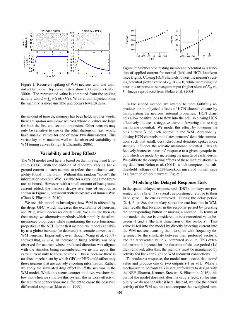
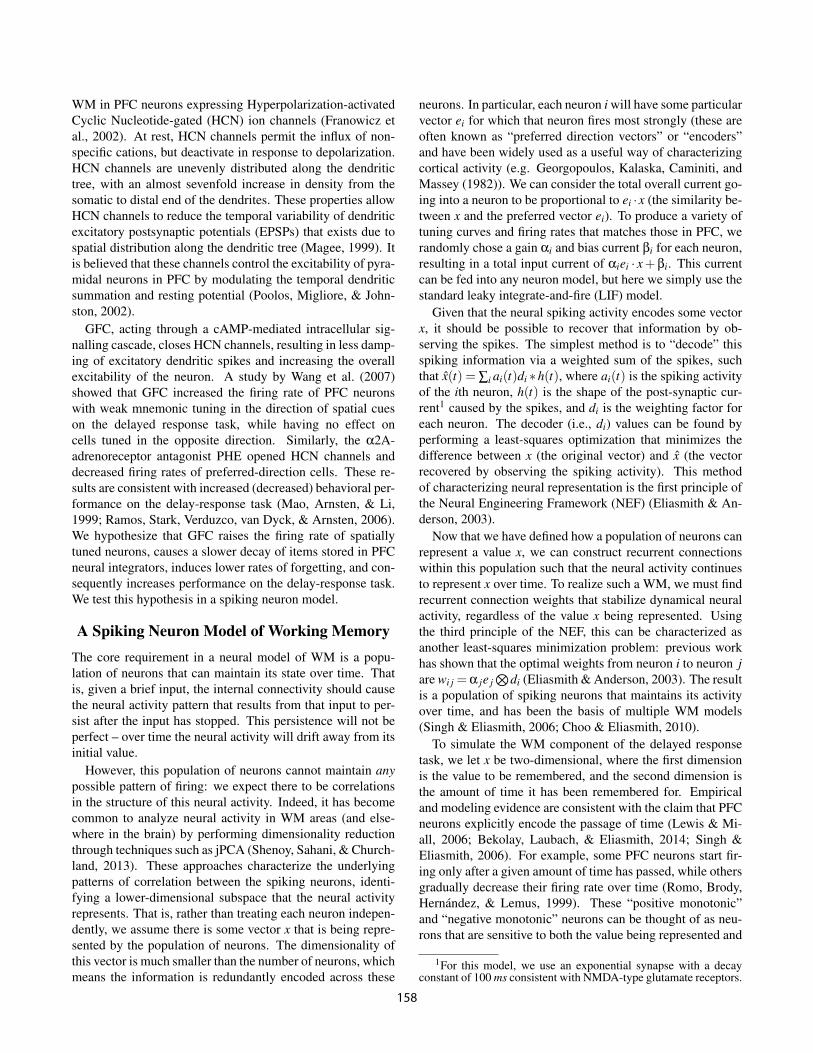
Figure 1: Recurrent spiking of WM neurons with and with-out added noise. Top spike rasters show 100 neurons (out of3000). The represented value is computed from the spikingactivity with x = Â
i
a
i
(t)di
⇤h(t). With random injected noisethe memory is more unstable and decays towards zero.
the amount of time the memory has been held; in other words,these are spatial mnemonic neurons whose e
i
values are largefor both the first and second dimension. Other neurons mayonly be sensitive to one or the other dimension (i.e. wouldhave small e
i
values for one of those two dimensions). Thisvariability in e
i
matches well to the observed variability inWM tuning curves (Singh & Eliasmith, 2006).
Variability and Drug Effects
The WM model used here is based on that in Singh and Elia-smith (2006), with the addition of randomly varying back-ground current to each neuron, to reflect the stochastic vari-ability found in the brain. Without this random “noise”, theinformation stored in WM is stable for a very long time (min-utes to hours). However, with a small amount of backgroundcurrent added, the memory decays over tens of seconds asshown in Figure 1, consistent with decay rates of human WM(Choo & Eliasmith, 2010)
We use this model to investigate how WM is affected bythe drugs GFC, which increases the excitability of neurons,and PHE, which decreases excitability. We simulate their ef-fects using two alternative methods which simplify the afore-mentioned biophysics while maintaining the core functionalproperties in the NEF. In the first method, we model excitabil-ity as a global increase (or decrease) in somatic current to allWM neurons. Importantly, even though Wang et al. (2007)showed that, in vivo, an increase in firing activity was onlyobserved for neurons whose preferred direction was alignedwith the stimulus being remembered, we do not apply thisextra current only to those neurons. This is because there isno direct mechanism by which GFC or PHE could affect onlythose neurons that are actively encoding information. Rather,we apply the simulated drug effect to all the neurons in theWM model. While this seems counter-intuitive, we show be-low that when we simulate this system, the network effects ofthe recurrent connections are sufficient to cause the observeddifferential response (Mao et al., 1999).
Figure 2: Subtheshold resting membrane potential as a func-tion of applied current for normal (left) and HCN-knockoutmice (right). Closing HCN channels lowers the neuron’s rest-ing potential (lower value of E
m
at I = 0) while increasing theneuron’s response to subsequent input (higher slope of E
m
vs.I). Image reproduced from Nolan et al. (2004).
In the second method, we attempt to more faithfully re-produce the biophysical effects of HCN channel closure bymanipulating the neurons’ internal properties. HCN chan-nels allow positive ions to flow into the cell, so closing HCNeffectively induces a negative current, lowering the restingmembrane potential. We model this effect by lowering thebias current b
i
of each neuron in the WM. Additionally,closing HCN channels modulates neurons’ dendritic summa-tion, such that small, desynchronized dendritic spikes morestrongly influence the somatic membrane potential. This ef-fectively increases neurons’ response to a given synaptic in-put, which we model by increasing the gain a
i
of each neuron.We calibrate the competing effects of these manipulations us-ing data from Nolan et al. (2004), which compares the sub-threshold voltages of HCN-knockout mice and normal miceas a function of input current, Figure 2.
Modeling the Delayed Response Task
In the spatial delayed-response task (DRT), monkeys are pre-sented with a brief (1s) visual cue positioned relative to theirfixed gaze. The cue is removed. During the delay period(2, 4, 6, or 8s), the monkey stores the cue location in WM,then recalls that location in the response period by pressingthe corresponding button or making a saccade. In terms ofour model, the cue is considered to be a numerical value be-tween -1 and 1 (the first dimension of the vector x). Thisvalue is fed into the model by directly injecting current intothe WM neurons, causing them to spike with frequency de-termined by the similarity between their preferred vector e
i
and the represented value x, computed as e
i
· x. This exter-nal current is injected for the duration of the cue period (1s)then removed; after this, the memory must be maintained byactivity fed back through the WM recurrent connections.
To produce a response, the model must access that storedvalue and produce one of two outputs (-1 or +1). While amechanism to perform this is straightforward to design withthe NEF (Sharma, Kromer, Stewart, & Eliasmith, 2016), thispart of the model does not alter the drug effects, so for sim-plicity we do not consider it here. Instead, we take the neuralactivity of the WM neurons and compute their weighted sum,
159
giving an estimate of the original value (x(t) = Âi
a
i
(t)di
⇤h(t)). Since a neural mechanism to convert this value into adecision will include some degree of variability, we approxi-mate this by adding normally distributed noise to this value.If the result is above zero we interpret this as the model giv-ing the first response, and if it is below zero we interpret it asgiving the second response.
Results
To simulate the cellular effects of GFC and PHE, we testedtwo methods for perturbing the neurons, as described above.In the first, we injected a noisy signal2 into neurons in theWM population, essentially using additive bias to increase(decrease) neural excitability. In the second, we manipulatedthe gains and biases of the LIF neurons used in the simu-lation, effectively decreasing each neuron’s resting potentialwhile increasing its gain to synaptic inputs3. Both perturba-tions produced the desired effects; we hereafter report resultsfrom the first method.
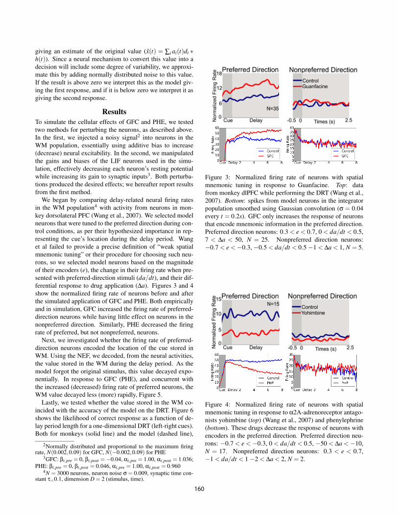
We began by comparing delay-related neural firing ratesin the WM population4 with activity from neurons in mon-key dorsolateral PFC (Wang et al., 2007). We selected modelneurons that were tuned to the preferred direction during con-trol conditions, as per their hypothesized importance in rep-resenting the cue’s location during the delay period. Wanget al failed to provide a precise definition of “weak spatialmnemonic tuning” or their procedure for choosing such neu-rons, so we selected model neurons based on the magnitudeof their encoders (e), the change in their firing rate when pre-sented with preferred-direction stimuli (da/dt), and their dif-ferential response to drug application (Da). Figures 3 and 4show the normalized firing rate of neurons before and afterthe simulated application of GFC and PHE. Both empiricallyand in simulation, GFC increased the firing rate of preferred-direction neurons while having little effect on neurons in thenonpreferred direction. Similarly, PHE decreased the firingrate of preferred, but not nonpreferred, neurons.
Next, we investigated whether the firing rate of preferred-direction neurons encoded the location of the cue stored inWM. Using the NEF, we decoded, from the neural activities,the value stored in the WM during the delay period. As themodel forgot the original stimulus, this value decayed expo-nentially. In response to GFC (PHE), and concurrent withthe increased (decreased) firing rate of preferred neurons, theWM value decayed less (more) rapidly, Figure 5.
Lastly, we tested whether the value stored in the WM co-incided with the accuracy of the model on the DRT. Figure 6shows the likelihood of correct response as a function of de-lay period length for a one-dimensional DRT (left-right cues).Both for monkeys (solid line) and the model (dashed line),
2Normally distributed and proportional to the maximum firingrate, N(0.002,0.09) for GFC, N(�0.002,0.09) for PHE
3GFC: bi,pre
= 0, bi,post
=�0.04, ai,pre
= 1.00, ai,post
= 1.036;PHE: b
i,pre
= 0, bi,post
= 0.046, ai,pre
= 1.00, ai,post
= 0.9604N = 3000 neurons, neuron noise s = 0.009, synaptic time con-
stant t=0.1, dimension D = 2 (stimulus, time).
Figure 3: Normalized firing rate of neurons with spatialmnemonic tuning in response to Guanfacine. Top: datafrom monkey dlPFC while performing the DRT (Wang et al.,2007). Bottom: spikes from model neurons in the integratorpopulation smoothed using Gaussian convolution (s = 0.04every t = 0.2s). GFC only increases the response of neuronsthat encode mnemonic information in the preferred direction.Preferred direction neurons: 0.3 < e < 0.7, 0 < da/dt < 0.5,7 < Da < 50, N = 25. Nonpreferred direction neurons:�0.7 < e <�0.3, �0.5 < da/dt < 0.5 �1 < Da < 1, N = 5.
Figure 4: Normalized firing rate of neurons with spatialmnemonic tuning in response to a2A-adrenoreceptor antago-nists yohimbine (top) (Wang et al., 2007) and phenylephrine(bottom). These drugs decrease the response of neurons withencoders in the preferred direction. Preferred direction neu-rons: �0.7 < e <�0.3, 0 < da/dt < 0.5, �50 < Da <�10,N = 17. Nonpreferred direction neurons: 0.3 < e < 0.7,�1 < da/dt < 1 �2 < Da < 2, N = 2.
160
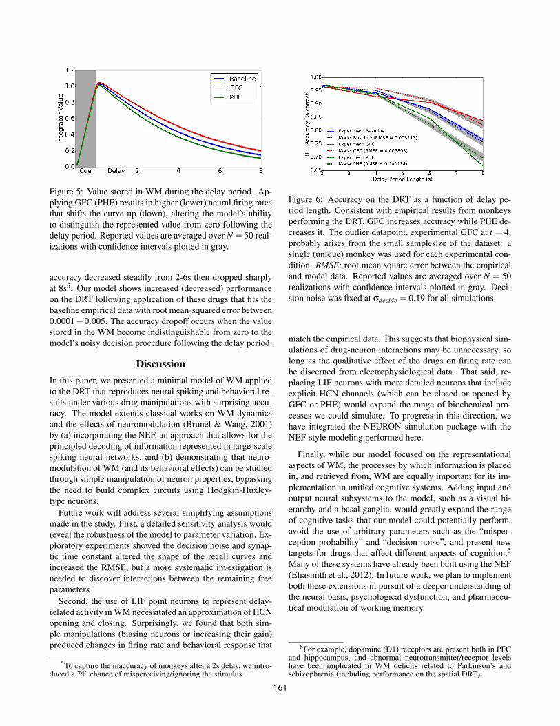
Figure 5: Value stored in WM during the delay period. Ap-plying GFC (PHE) results in higher (lower) neural firing ratesthat shifts the curve up (down), altering the model’s abilityto distinguish the represented value from zero following thedelay period. Reported values are averaged over N = 50 real-izations with confidence intervals plotted in gray.
accuracy decreased steadily from 2-6s then dropped sharplyat 8s5. Our model shows increased (decreased) performanceon the DRT following application of these drugs that fits thebaseline empirical data with root mean-squared error between0.0001�0.005. The accuracy dropoff occurs when the valuestored in the WM become indistinguishable from zero to themodel’s noisy decision procedure following the delay period.
Discussion
In this paper, we presented a minimal model of WM appliedto the DRT that reproduces neural spiking and behavioral re-sults under various drug manipulations with surprising accu-racy. The model extends classical works on WM dynamicsand the effects of neuromodulation (Brunel & Wang, 2001)by (a) incorporating the NEF, an approach that allows for theprincipled decoding of information represented in large-scalespiking neural networks, and (b) demonstrating that neuro-modulation of WM (and its behavioral effects) can be studiedthrough simple manipulation of neuron properties, bypassingthe need to build complex circuits using Hodgkin-Huxley-type neurons.
Future work will address several simplifying assumptionsmade in the study. First, a detailed sensitivity analysis wouldreveal the robustness of the model to parameter variation. Ex-ploratory experiments showed the decision noise and synap-tic time constant altered the shape of the recall curves andincreased the RMSE, but a more systematic investigation isneeded to discover interactions between the remaining freeparameters.
Second, the use of LIF point neurons to represent delay-related activity in WM necessitated an approximation of HCNopening and closing. Surprisingly, we found that both sim-ple manipulations (biasing neurons or increasing their gain)produced changes in firing rate and behavioral response that
5To capture the inaccuracy of monkeys after a 2s delay, we intro-duced a 7% chance of misperceiving/ignoring the stimulus.
Figure 6: Accuracy on the DRT as a function of delay pe-riod length. Consistent with empirical results from monkeysperforming the DRT, GFC increases accuracy while PHE de-creases it. The outlier datapoint, experimental GFC at t = 4,probably arises from the small samplesize of the dataset: asingle (unique) monkey was used for each experimental con-dition. RMSE: root mean square error between the empiricaland model data. Reported values are averaged over N = 50realizations with confidence intervals plotted in gray. Deci-sion noise was fixed at s
decide
= 0.19 for all simulations.
match the empirical data. This suggests that biophysical sim-ulations of drug-neuron interactions may be unnecessary, solong as the qualitative effect of the drugs on firing rate canbe discerned from electrophysiological data. That said, re-placing LIF neurons with more detailed neurons that includeexplicit HCN channels (which can be closed or opened byGFC or PHE) would expand the range of biochemical pro-cesses we could simulate. To progress in this direction, wehave integrated the NEURON simulation package with theNEF-style modeling performed here.
Finally, while our model focused on the representationalaspects of WM, the processes by which information is placedin, and retrieved from, WM are equally important for its im-plementation in unified cognitive systems. Adding input andoutput neural subsystems to the model, such as a visual hi-erarchy and a basal ganglia, would greatly expand the rangeof cognitive tasks that our model could potentially perform,avoid the use of arbitrary parameters such as the “misper-ception probability” and “decision noise”, and present newtargets for drugs that affect different aspects of cognition.6Many of these systems have already been built using the NEF(Eliasmith et al., 2012). In future work, we plan to implementboth these extensions in pursuit of a deeper understanding ofthe neural basis, psychological dysfunction, and pharmaceu-tical modulation of working memory.
6For example, dopamine (D1) receptors are present both in PFCand hippocampus, and abnormal neurotransmitter/receptor levelshave been implicated in WM deficits related to Parkinson’s andschizophrenia (including performance on the spatial DRT).
161
Acknowledgments
This work was supported by CFI and OIT infrastructure fund-ing as well as the Canada Research Chairs program, NSERCDiscovery grant 261453, ONR grant N000141310419,AFOSR grant FA8655-13-1-3084 and OGS graduate fund-ing.
References
Avery, R. A., Franowicz, J. S., Studholme, C., van Dyck,C. H., & Arnsten, A. F. (2000). The alpha-2a-adrenoceptoragonist, guanfacine, increases regional cerebral blood flowin dorsolateral prefrontal cortex of monkeys performing aspatial working memory task. Neuropsychopharmacology,23(3), 240–249.
Bekolay, T., Laubach, M., & Eliasmith, C. (2014). A spikingneural integrator model of the adaptive control of action bythe medial prefrontal cortex. The Journal of Neuroscience,34(5), 1892–1902.
Brunel, N., & Wang, X.-J. (2001). Effects of neuromodula-tion in a cortical network model of object working mem-ory dominated by recurrent inhibition. Journal of compu-
tational neuroscience, 11(1), 63–85.Choo, F.-X., & Eliasmith, C. (2010). A spiking neuron model
of serial-order recall. In Proceedings of the 32nd annual
conference of the cognitive science society (pp. 2188–93).Eliasmith, C., & Anderson, C. H. (2003). Neural engineer-
ing: Computation, representation, and dynamics in neuro-
biological systems. MIT press.Eliasmith, C., Stewart, T. C., Choo, X., Bekolay, T., DeWolf,
T., Tang, Y., & Rasmussen, D. (2012). A large-scale modelof the functioning brain. science, 338(6111), 1202–1205.
Franowicz, J. S., Kessler, L. E., Borja, C. M. D., Kobilka,B. K., Limbird, L. E., & Arnsten, A. F. (2002). Mutationof the a2a-adrenoceptor impairs working memory perfor-mance and annuls cognitive enhancement by guanfacine.The Journal of neuroscience, 22(19), 8771–8777.
Georgopoulos, A. P., Kalaska, J. F., Caminiti, R., & Massey,J. T. (1982). On the relations between the direction of two-dimensional arm movements and cell discharge in primatemotor cortex. The Journal of Neuroscience, 2(11), 1527–1537.
Goldman-Rakic, P. (1995). Cellular basis of working mem-ory. Neuron, 14(3), 477–485.
Lewis, P. A., & Miall, R. C. (2006). Remembering the time:a continuous clock. Trends in cognitive sciences, 10(9),401–406.
Magee, J. C. (1999). Dendritic ih normalizes temporal sum-mation in hippocampal ca1 neurons. Nature neuroscience,2(6), 508–514.
Mao, Z.-M., Arnsten, A. F., & Li, B.-M. (1999). Local infu-sion of an a-1 adrenergic agonist into the prefrontal corteximpairs spatial working memory performance in monkeys.Biological psychiatry, 46(9), 1259–1265.
Nolan, M. F., Malleret, G., Dudman, J. T., Buhl, D. L., San-toro, B., Gibbs, E., . . . others (2004). A behavioral role
for dendritic integration: Hcn1 channels constrain spatialmemory and plasticity at inputs to distal dendrites of ca1pyramidal neurons. Cell, 119(5), 719–732.
Poolos, N. P., Migliore, M., & Johnston, D. (2002). Pharma-cological upregulation of h-channels reduces the excitabil-ity of pyramidal neuron dendrites. Nature neuroscience,5(8), 767–774.
Ramos, B. P., Stark, D., Verduzco, L., van Dyck, C. H., &Arnsten, A. F. (2006). a2a-adrenoceptor stimulation im-proves prefrontal cortical regulation of behavior throughinhibition of camp signaling in aging animals. Learning
& Memory, 13(6), 770–776.Romo, R., Brody, C. D., Hernandez, A., & Lemus, L. (1999).
Neuronal correlates of parametric working memory in theprefrontal cortex. Nature, 399(6735), 470–473.
Scahill, L., Chappell, P. B., Kim, Y. S., Schultz, R. T., Katso-vich, L., Shepherd, E., . . . Leckman, J. F. (2014). Aplacebo-controlled study of guanfacine in the treatment ofchildren with tic disorders and attention deficit hyperactiv-ity disorder. American Journal of Psychiatry.
Sharma, S., Kromer, B., Stewart, T., & Eliasmith, C. (2016).A neural model of context dependent decision making inthe prefrontal cortex. In Proceedings of the 38th annual
conference of the cognitive science society.
Shenoy, K. V., Sahani, M., & Churchland, M. M. (2013).Cortical control of arm movements: a dynamical systemsperspective. Neuroscience, 36(1), 337.
Singh, R., & Eliasmith, C. (2006). Higher-dimensional neu-rons explain the tuning and dynamics of working memorycells. The journal of neuroscience, 26(14), 3667–3678.
Stewart, T. C., Choo, X., & Eliasmith, C. (2010). Dy-namic behaviour of a spiking model of action selection inthe basal ganglia. In Proceedings of the 10th international
conference on cognitive modeling (pp. 235–40).Wang, M., Ramos, B. P., Paspalas, C. D., Shu, Y., Simen,
A., Duque, A., . . . others (2007). a2a-adrenoceptorsstrengthen working memory networks by inhibiting camp-hcn channel signaling in prefrontal cortex. Cell, 129(2),397–410.
162