Embed Size (px)
Citation preview

Life Sciences, Vol. 32, pp. 181-190 Pergamon Press Printed in the U.S.A.
EFFECTS OF LEUCINE 5 - AND METHIONINE 5 - B h - ENDORPHIN ON BEHAVIOR AND ELECTROENCEPHALOGRAM IN CATS 1
Pablo Pacheco 2, Frank R. Ervin, and James C. Hodgdon
Department of Psychiatry, McGill University, Montreal, Quebec, CANADA H3A IAI
(Received in final form September 27, 1982)
Summary
Intracerebroventricular administration of leu 5 - and met 5 - ~h - endorphin produces sequential behavioral changes characterized by restlessness, eye fixation, head tremor, and exaggerated orienting reaction to external auditory stimuli, accompanied by EEG alterations. These changes take place during three stages. MetS- produces an increase in amplitude and reactivity of the theta rhythm in the hippocampus. Leu5- produces a masking of the theta rhythm by the appearance of spiking activity. Leu 5- produces a loss of reactivity to visual stimuli not observed with met5-. The exaggerated reaction to auditory stimuli accompanied by an increased response of the reticular formation indicates a sensitivity of some mesencephalic structures (concerned with the modulation of sensory input) to both endorphins. Naloxone blockade of the subcortical (limbic) activity prominent in Stage II reveals the persistence of the first seen Stage I characteristics. These reticular-neocortical effects persist also into Stage III where they are seen intermixed with the limbic effects most prominent in Stage II. This suggests the presence of two endorphin-sensitive neural systems, only one of which is Naloxone reversible, and is that which in the cat covaries with the excited, hyperattentive state rather than the sedation and immobility phase seen in the rat. This dual system is compared to that described by Jacquet (13).
Direct application of met5-B - Endorphin produces a reduction in neuronal sensitivity in all brain structures studied with the exception of the hippo- campal pyramidal cells, where it has a potent excitatory effect (I). When injected into the cerebral ventricular fluid of cats, B-endorphin affects several behavioral and physiological measures, e.g. head shakes, fixation of the eyes, diminished responsiveness to external stimuli, analgesia, mydriasis, vocalization, shivering and panting (2,3,4). Additionally when injected into the cerebral ventricular fluid of rats a marked "catatonic" state lasting for several hours is produced (5).
It is also observed that seizures are provoked following an intracerebro- ventricular (i.c.v.) or intracerebral application of B-endorphin or enkephalins (6,7,8). A non-convulsive EEG epileptiform activity, primarily limited to limbic brain areas (9), but also recorded on surface EEG (I0) after i.c.v. B-endorphin has been commonly described in recent studies. Apparently it has been concluded that enkephalin (ll) or B-endorphin (i0) produce seizures only when injected in or near the dorsomedial nucleus of the thalamus. The import- ance is that the electrophysiological changes, which are observed at doses well
iStudy was supported by the Medical Research Council of Canada, and by generous gifts of peptide from Peninsula Laboratories, San Carlos, CA. 2Visiting Scientist from Universidad Nacional Aut6noma de M~xico.
002"4--3205/83/030181-10503.00/0 Copyright (c) 1983 Pergamon Press Ltd.

182 Effects of Leu5- and Met’-Bh-endorphin Vol. 32, No. 3, 1983
below those required to produce overt behavioral changes, are a quantifiable index of the actions of the endogenous endorphins and similar substances.
Considering our previous study on the neurophysiological and behavioral activities produced by the i.c,v. administration of these two synthetic congeners of Bh-endorphin (12) and the recent report of Jacquet (13)) which shows that in the rat vas deferens and likely in the isolated guinea pig ileum, peptides produce a dual action - excitatory and inhibitory - mediated by two endorphin receptors, we decided to study with a different approach the effects of i.c.v. leu5- and met5-Bh-endorphin on the electroencephalographic and behavioral activity of cats. The present experiments provided evidence that these agents influence not only the EEG epileptiform activity in different proportion but also the visual and auditory sensory mechanisms.
Materials and Methods
From an extended study of behavioral and electrophysiological effects of i.c.v. injected endorphins, the following details are selected for the present study.
Four adult female cats of 2.5 - 3.0 kg are reported here. Surgical preparation of the animals was carried out under Nembutal anesthesia. With the aid of a Kopf stereotaxic apparatus, chronic stereotaxic placements based on the atlas of Jasper and Ajmone-Marsan (14) were: cannula, A 12, L 0, D +5; bipolar electrodes, N. accumbens (Acb) A 16, L 2.5, D 0; right globus pallidus (GP) A 13, L 9, D -2; left dorsal hippocampus (Hpp) A 0.5, L 10, D -6; left amygdala (Abm) A 12, L 9, D -7; and right reticular formation (Ret Mes) A 2, L 2.5, D -3. Cortical electrodes were placed in the area of the primary left visual cortex (VCx) . Positions were confirmed histologically by the method of Guzmsn-Flores et al. (15). The position of the ventricular cannula was verified during the implantation by the extraction of cerebral ventricular fluid. Gross behavioral observations were made in a 59 x 74 x 56 cm steel cage with a plexiglass viewing door. EEG recordings were made on a Grass model 78 D polygraph. Multi-unit activity from the Ret Mes was processed on an IDS Gating- Frequency Counter Rd-1.
The animals were allowed to recover from the implantation surgery for ten days prior to experimental manipulation. The experimental sequence was as follows: 1) 5 days habituation to cage and recording. 2) i.c.v. application of 0.05 ml of sterile water. 3) 48 hours later first injection of met5- (two cats) or leu5-Bh-endorphin (two different cats). 4) one week later, repeat i.c.v. endorphin+Naloxone 20 mg i.m. 5) one week later, repeat i.c.v. endorphin for comparison with “3”. 6) one week later administration of Naloxone 20 mg i.m. alone. Thus, we are reporting for each endorphin four replications of the effect and two replications of Naloxone interactions. As the observed results of the Naloxone administration are consistent with those seen in our other studies (12) the small n serves as an adequate example of the - present point.
All experimental manipulation was conducted between 13:00 and 17:00 hours in the observation cage. Drug injection was preceded by 30 min of control behavioral observation and recording. Once injected with the drug analysis was performed for three consecutive hours. Periods of one hour of observation and recording were done on three subsequent days. The injection of 0.05 ml of sterile water permitted us to determine possible volume effects on behavior and electrical recording.
The leu5- and met5- Bh-endorphins were purchased from Peninsula Laborat- ories, San Carlos, CA. The i.c.v. injections in doses of 50 vg dissolved in 0.05 ml sterile water, were administered through a 27 gauge needle, extended 2 mm beyond the tip of the guide cannula, that was connected to a small plastic microliter syringe. This apparatus was allowed to remain in place throughout

Vol. 32, No. 3, 1983 Effects of Leu 5- and MetS-Sh-endorphin 183
the observation period. Naloxone hydrochloride, obtained from Endo Laboratories, was administered in 20 mg i.m. injections in a volume of I ml.
The behavioral criteria were based on the assessment of the gross behavior (e.g., exploring, grooming and playing), posture, eye movement (e.g., opening, closing or closed), pupil size, head movement, inappropriate behavior (i.e., characterized by the animal seemingly visually tracking objects in space not visible to the observer) and the responsiveness of the animal to auditory stimuli (e.g., a sharp tap on the wall of the observation cage) and visual stimuli (e.g., a pencil movement or a I0 per second flash stimulation provided by a Grass model PS2 Photo Stimulator set on maximum intensity).
Results
A single i.c.v, dose of 50 ~g of leu 5- or met5-Sh-endorphin characteristic- ally produce the following gross behavioral changes: restlessness (the most prominent feature of which is the crouching position), eye fixation and assoc- iated constant lateral head movements, tremor of the head often occurring during the lateral movements, open eyes, reduction in responsiveness to external visual stimuli with a concurrent exaggerated orienting reaction to auditory stimuli, and occasional circling behavior. Also observed was pupillary dilation and transitory constriction occurring immediately after an auditory stimulus.
The duration as well as the quality of the behavioral and electrical changes observed following administration of either Bh-endorphin permitted us to consider three major states which can be delineated as follows:
State I. State I, which is induced immediately upon injection has a duration of five minutes when the cat is under the influence of leu 5- and ten minutes when under the influence of met 5-. Initially, with both endorphins, the visual cortex is invaded by short bursts of 5-6 Hz synchronic activity (Figs. 1,3,4). The intensity and frequency of the synchronic activity is dependent on the particular cat being tested, not on the agent used. Met5-~h-endorphin induced light spiking activity in the Ret Mes during the latter portion of State I (Fig. 3 - after ten minutes). This spiking activity was accompanied by an increment in the multiunit activity. Throughout the initial two minutes of State I the animals characteristically paced nervously around the observation cage (this effect also occurred when the animals were injected with the placebo) after which they took up the crouching position.
State II. The start of State II is signaled by the disappearance of the visual synchronic activity. It has a duration of 90 min when produced by met 5- and three hours when produced by leu5-~h-endorphin. During the initial 45 min cats displayed restlessness, fixation of the eyes, very little lateral head movement, and alert open eyes. All structures analyzed showed spiking activity that increased over time. Towards the end of the first 45 min portion of State II the animals typically started to move around the cage and engage in circling activity for a short time, after which they returned to the crouching position. The lateral head movements then became more frequent as the animal appeared to be tracking objects that didn't exist. The animal would occasionally hiss at or retreat from, or strike at such "hallucinated" targets.
The overall responsiveness of the animal to external stimuli during State II becomes exaggerated. The appearance of the exaggerated responsiveness is characterized by a pupillary constriction and clear orienting reaction directed toward the source of an auditory stimulus. When an animal is under the influ- ence of met5-Bh-endorphin a high voltage theta rhythm appears in the Hpp (Fig. 3) and intense spiking activity with associated multiunit response appears in the Ret Mes during auditory stimulation. Leu 5- also produces this increased activity in the Ret Mes as a response to auditory stimuli. With this endorphin, however, the hippocampal theta rhythm is masked by spiking activity. A striking difference between the respective action of leu5- and metb-Bh-endorphin during

co~m
l
~O
nl
>','>.,.-':,.,::,:-,;:.,;:..:.~;:::::::: :::::. ::
:: ::,?/~,: ..... :..
:
: :
; ,.;.:, ,
: :.:::.,
180 ra
in
,--~-
: t~
'-~' '.
~:
:~'~
'" ;
:W:,:
:-: :
~ ~
;",~
: ;:
-'.
~'
~l~
,~i~
vlll
lr'~
-'::
:-
:: ~
:.
~,~
,~,-
~.~
,~,~
.+~
'~
, ,
, ',
, ,
~i
~,
~,
~,
~4
~,
i
i
r,~ ~J~% :J.: - .; .:
"':-:,: ~.%y~:~/Y,.>S~.~i~/ii~..~i~/~s~{~i~,~/~iL~..;.~i~'i...~iiii/~d/~/(r
Fig. I.
Sequ
enti
al ef
fect
s of
SO
~g
leuS-Bh-endorphin i.c.v.
S
of 5-6
Hz ac
tivi
ty which di
sapp
ears
during th
e 60-90 min pe
riod
.
5rai
n af
ter
leu
S-~
-~
"Par
da"
u
~O~i
4Shr
l
:'/,st
: ~.:" :
{~ : :
:- ~4-.~/
t ,; ":
,, :.: D :,~ ; ,-
:<.'/:< "
: ~', ) i
i : ]
, : :
-
,.,- 4
% J'
:.]i.- ,{."..>/H.
Isec
min following injection tk
e vi
sual
cortex sh
ows
bursts
It re
appe
ars
after 180
min an
d persists 48
h
later.
Hypersynchronous non-convulsive li
mbic
activity is ev
iden
t.
(This
and
following Fi
gs.:
Acb: R. ac
cumb
ens;
Abm: L.
amyg
dala
; G.P.:
R. gl
obus
pa
llid
us;
Hpp: L. do
rsal
hi
ppoc
ampu
s; Re
t Mes: R. reticular fo
rmat
ion;
V. Cx:
visu
al cortex.
Final
trac
e is cu
mula
tive
multiunit Ret
Mes
activity with 1
sec
rese
t time.)
O0
l-h
0 iO on
I iO
I I iO
0 <~
0 0 Oo

Vol. 32, No. 3, 1983 Effects of Leu 5- and Met5-Sh-endorphin 185
S t a t e I I i s t h a t wh i l e under t he i n f l u e n c e o f l eu 5- t he c a t s e x h i b i t a 10 min p e r i o d dur ing which they do not respond b e h a v i o r a l l y to e x t e r n a l v i s u a l s t imu- l a t i o n . Recupe ra t ion from t h i s e f f e c t i s slow. When animals were t e s t e d dur ing t h i s p e r i o d with 10 Hz f l a s h s t i m u l a t i o n , i t was observed t h a t t he f l a s h f o l l o w i n g re sponse o f the v i s u a l c o r t e x was s i m i l a r to t h a t r eco rded dur ing the c o n t r o l S t a t e , but the Ret ~es showed enhanced d r i v i n g (F ig . 2) .
S t a t e I I I . The beg inn ing o f S t a t e I I I was c h a r a c t e r i z e d by changes in the e l e c t r i c a l p o t e n t i a l s o f the b r a i n a reas r eco rded . With the e x c e p t i o n o f the V Cx, t he animals e x h i b i t e d more n o n - c o n v u l s i v e a c t i v i t y . The V Cx produced the same b u r s t s o f hype r synchron ic a c t i v i t y a t 5-6 Hz t h a t were observed dur ing S t a t e I (F igs . 1 , 2 , 3 ) . When under the i n f l u e n c e o f met 5- t he ca t s e n t e r i n t o S t a t e I I I e a r l y , t y p i c a l l y only 90 min f o l l o w i n g i n j e c t i o n , and con t inue to be in t h i s S t a t e (a t s l i g h t l y reduced l e v e l ) a t the end o f the t h r e e - h o u r o b s e r - v a t i o n p e r i o d . When r e t e s t e d 24 h a f t e r i n j e c t i o n n e i t h e r the n o n - c o n v u l s i v e
control "Palda"
v o , # t i l i ~ d ~ l , " - - - - - - , , . - -
N P F F
6 0 r a n - f r e t l e u ~ - ~ - e ~ l o r p h i n
. .......... . . . . . . . , ~ . . . . . . . . . . . . . . . . . . . . . . . . . , - ~ - - . . . . . . . . . . . . . . . . . . . . . ~ _ , i l i l l , t ~
F N/q
o,i I m ~
lle
N ~ I ~
Fig . 2. R e a c t i v i t y o f EEG to n o i s e and lO-pe r - second f l a s h s t i m u l a t i o n dur ing t e-h--6-~ntrol p e r i o d and f o l l o w i n g the i . c . v , i n j e c t i o n o f 50 ~g leuS-Sh-endorph in . Flash dur ing the c o n t r o l i s fo l lowed only by the v i s u a l c o r t e x . During a c t i o n o f the endorphin both the v i s u a l c o r t e x and the r e t i c u l a r fo rma t ion respond. Noise dur ing the c o n t r o l p e r i o d o f EEG synchrony produces d e s y n c h r o n i z a t i o n o f a l l r eco rded s t r u c t u r e s . During a c t i o n o f the endorphin n o i s e a c t i v a t e s p r im a r - i l y the r e t i c u l a r fo rma t ion . (F: f l a s h , N: no i se )
186 Effects of Leu 5- and MetS-Bh-endorphin Vol. 32, No. 3, 1983
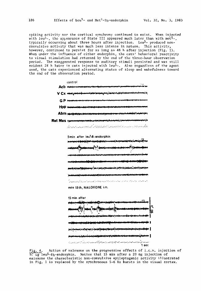
spiking activity nor the cortical synchrony continued to exist. When injected with leuS-, the appearance of State III appeared much later than with met 5-, typically occurring about three hours after injection. Leu 5- produced non- convulsive activity that was much less intense in nature. This activity, however, continued to persist for as long as 48 h after injection (Fig. l). When under the influence of either endorphin, the cats' behavioral reactivity to visual stimulation had returned by the end of the three-hour observation period. The exaggerated response to auditory stimuli persisted and was still evident 24 h later in cats injected with leu 5-. Also regardless of the agent used, the cats experienced alternating states of sleep and wakefulness toward the end of the observation period.
control ?]3
V C x , , * , ~ ~ , , ~ , , ~ * ~ # . ~ ' ~ , ' , ~ ~ " - ~ " ~
H P P :: ::.:-:: :::::: : : :.':.:: : : : : : :: ;:,: : : . : : : ; :::: : : : ::; :.-::::: :::::
R e t M e s ~ . , ~ , ~ . ~ w ~ , ~ . ~ , . ~ . w ; '~
5min after leu~8-endorphin
min t5th, NALOXONE i.m.
15 rain after I
I
I sec
Fig. 4. Action of naloxone on the progressive effects of i.c.v, injection of S$ pg leuS-Sh-endorphin. Notice that 1S min after a 20 mg injection of naloxone the characteristic non-convulsive epileptogenic activity illustrated in Fig. 1 is replaced by the synchronous 5-6 Hz bursts in the visual cortex.

Vol. 32, No. 3, 1983 Effects of Leu 5- and MetS-Bh-endorphin 187
control ~7~la"
V Cx . . . . . . . . . . . . . . . . . . . . . . . . . . .
G P . . . . . o , , . - ~ . . . . . . . . . . ' " ' " ~ . . . . . . . . . . . . . . . . . . . ' . . . . . . . . . . . . . . r . . . . . . . . . . . . . . . . . . . . . . . . . . . . ' . . . . . r . . . . . . . . . .
H ~ ~.~.~¢,r,~,,,~l.#~m~m~4~ ~ #¢~,~r~m.m,..~,~m.,~,~,,,~,r,.~o
,> ,%~, i i ( . ' , / / l ~ / , ~ - : l { : ~ ¢ L , ' . ~ ' t . / < ' v , d , , ' / / i . : / , , . 7 v / / , i ) . . , i r~ i / , / / ¢ , , , , , , < , , , + , , , . . . . . . . . , 1+ , , , "
lOmin after n~t5.- 8 - ~
i
!' , 7 , ' , ,
60rail
I
I
N ~
~Fig. 3. P r o g r e s s i v e e f f e c t s and r e a c t i v i t y o f EEG to n o i s e during the con tro l ~ a n d f o l l o w i n g the i . c . v , i n j e c t i o n o f 50 ug met5-Sh-endorphi n. Noise during the control period of EEG desynchrony produces a discrete reactivity of hippocampus and reticular formation. During action of the endorphin the react- ivity to noise is increased. The progressive effects of this endorphin are more intense than those described in Fig. 1 for leuS-Bh-endorph in. (N: noise)
188 Effects of Leu 5- and MetS-Bh-endorphin Vol. 32, No. 3, 1983
Naloxone effect
Because the effects induced by endorphin in this study were most pronounced during State II, Naloxone was administered during this phase. Naloxone was thus administered at 20 mg i.m. 15 min following the i.c.v, injection of the endor- phins. The results illustrated in figure 4 are exemplary of the Naloxone effects upon EEG activity. Approximately 15 min after the injection the char- acteristic non-convulsive epileptogenic activity of State II was replaced by synchronous 5-6 Hz bursts in the V Cx characteristic of "State I" and which reappear in State III. Animals under the influence of the endorphins alone did not exhibit this cortical activity for an additional one to two hours, depending upon the agent. The expected spiking activity was also clearly reduced.
The effects of Naloxone were also correlated with changes in the behavioral state of the animals. The cats displayed a loss of the restlessness and eye fixation into space that was associated with State II. The cats' return to normal gross behavior was evident by the exhibition of the sleep position with eyes closed alternating with periods of wakefulness. The effects of Naloxone upon the cats were the same regardless of the type of endorphin with which the animals had previously been injected.
Discussion
The altered gross behavior, which persists for about three hours in the cats, is followed by a gradual return to the normal behavior state. This observation was quite similar for the action of the two endorphins used in this study. However, when the subjective impression was combined with the testing of both the visual and the auditory orienting reactions, a clear difference between the two agents was observed.
Examples have been presented of the three states of pronounced changes that are provoked by the two compounds in both behavior and EEG activity. During the first and third states bursts of synchronic activity are evident in the visual cortex. Also present are variations in the subcortical activity which are not correlated with the cortical changes.
Of the subcortical structures, one of the most affected was the hippo- campus. The well characterized theta rhythm evoked in the control state by external stimuli becomes exaggerated during the second state, as observed by an increase in amplitude and reactivity when under the influence of met5-Bh - endorphin. The leu5-Bh-endorphin, on the other hand, produces a masking of the theta rhythm by the appearance of spiking activity. This hippocampal spiking always precedes the non-convulsive epileptiform activity of the amygdala and reticular formation.
During the non-convulsive electrical pattern the cats, when under the influence of leu5-Bh-endorphin, experience a I0 min period with loss of the orienting reaction to visual stimuli. Their reaction to external auditory stimuli, however, becomes exaggerated just as is seen in cats with morphine intoxication. This auditory response is accompanied by a clear pupillary contraction and distraction of eye fixation that is observed while the auditory hyper-reactivity continues to persist after 24 h. When cats are injected with metS-Bh-endorphin they exhibit this peculiarity of the auditory sector but not the accompanying loss of visual reactivity. During the period of loss of the orienting reaction it was observed that the "flash following response" of the visual cortex was present. Thus it was assumed that there was an alteration in the integrative mechanism of the orienting reaction that spared the specific visual pathway.
The three structures, hippocampus, amygdala, and reticular formation are involved in the modulation of sensory input. Under normal circumstances the

Vol. 32, No. 3, 1983 Effects of Leu 5- and MetS-Sh-endorphin 189
hippocampus respmlds to attention by producing theta rhythms (16). The amyg- dala has a clear convergence of sensory inputs (17), the inputs being under the modulation of the reticular formation (18). Present results, and others (4), demonstrate that the behavior of eye fixation into space resembles an exagger- ated state of attention. The increased response of the reticular formation concurrent with this exaggerated orienting response - including the pupillary reaction - to auditory stimuli also indicates a clear hyper-reactivity of some mesencephalic structures to endorphin. Thus, the above discussed actions in- duced by agents in the present study implies that the structures affected by endorphins are concerned with mechanisms of sensory input modulation.
Naloxone blocks the effects of both leu 5- and met5-Sh-endorphin seen in the State II described above. The effects of Naloxone are correlated with a change in the behavioral state of the animals, i.e., there is a loss of the restless- ness and the fixation of the eyes into space and a return to normal gross be- havior as evidenced by the frequent exhibition of the sleep position with closed eyes.
In the transitional period during which the effects of Naloxone become apparent and prior to the animal's falling asleep, bursts of synchronic activity appear in the visual cortex just as were seen in State I and would reappear in an additional 2 h in "State III", then persisting for an additional 24 h. That is to say, the Naloxone blockade of the dramatic limbic neural activity which dominates the EEG activity and the behavior of State II, reveals an apparently intact neural organization characteristic of the endorphin effect seen at 5-10 min and persistent for at least 24 h or longer. This condition, dominated by changes in reticular formation and cortex is a) apparently not blocked by Naloxone and b) seems to disappear (in State II only because it is dominated by the profound limbic activity), then reappear (in State III)o The State II effect, of longer latency and shorter duration than the reticular- cortical effect of State I, is Naloxone reversible and is that described in some detail in earlier work (6,12). State III is obviously a period when the two systems (the reticular-cortical of State I and the limbic of State II) are both active, but in some balance, so that the cortical-reticular effects can be identified even though there is a persistence of the epileptiform depth activity. We postulate, therefore, the existence of two endorphin-activated neural mechanisms, one Naloxone reversible, the other not; the one concomitant with restlessness and "hallucinatory" behavior, the other with quiesence and immobility. This notion is consistent with the hypothesis and supporting evidence of Jacquet (13), suggesting the presence of both inhibitory and excit- atory receptors for morphine, the former, Naloxone reversible (and stereo- specific). For the rat, it is the dominant sedation-immobility response to (-) morphine which is Naloxone reversible and assumed to be mediated by "inhibitory" receptors. In our cats, whose species-typical response to morphine is predom- inantly excitating, the Naloxone-reversible effect of i.c.v, endorphin is the more aroused of the two simultaneously induced states.
~ether this is at variance with the Jacquet hypothesis, or simply repre- sents differing species receptor organization, is not clear. Whether an excitatory behavioral state should be considered the consequence of activation of "excitatory" receptors, or the effect of "inhibitory" receptor activation on a system of inhibitory cells might help clarify this matter. However, a pre- liminary receptor analysis of cat brain as compared with that of rat, does suggest some important differences in distribution and ratio of various opiate receptors (20).
190 Effects of Leu 5- and Met5-Bh-endorphin Vol. 32, No. 3, 1983
References
I. R. NICOLL, G. SIGGINS, N. LING, F.E. BLOOM and R. GUILLEMIN, Proco Natl. Acad. Sci. (USA) 74, 2584-2588 (1977).
2. W. FELDBURG and D.G. SMYTH, Br. J. Pharmac. 60, 445-453 (1977). 3. Y. HOSOBUCHI, Proc. Natl. Acad. Sci. (lISA) 74, 774-776 (1977). 4. M. MEGLIO, Y. HOSOBUCHI, H.H. LOH, J.E. ADAMS and C.H. LI, Proc. Natl.
Acad. Sci. (USA) 74, 4017-4019. 5. F.E. BLOOM, D. SEGAL, N. LING and R. GUILLEMIN, Science 194, 630-632 (1976). 6. S.J. HENRIKSEN, F.E. BLOOM, N. LING and R. GUILLEMIN, Neuroscience Abstr.
3, 293 (1977). 7. ~o HAVLICEK, L. LEYBIN, M. REZEK and C. PINSKY, Abstr. of the 59th Annu.
Meet. of the Endocrine Society, p. 178 (1977). 8. G. URCA, H. FRENK, J.C. LIEBESKIND and A.N. TAYLOR, Science 197, 83-86
(1977). 9. S.J. HENRIKSEN, F.E. BLOOM, F. MCKOY, N. LING and R. GUILLEMIN, Proc. Natl.
Acad. Sci. (USA) 75, 5221-5225 (1978). I0. H.M. FIREMARK and R.E. WEITZMAN, Neuroscience 4, 1895-1902 (1979). II. H. FRENK, B.C. MCCARTHY and J.C. LIEBESKIND, S~ience 200, 335-337 (1978). 12. FoR. ERVIN, R.M. PALMOUR, C. GUZMAN-FLORES and P. PACHECO, Biological
Psychiatry Today, Vol. 2, J. Obiols, C. Ballus, E. Gonzalezmonclus and J. Pujol (Eds.), pp. 720-727, Elsevier/North-Holland Biomedical Press, Amsterdam (1979).
13. Y.F. JACQUET, Science, 210, 95-97 (1980). 14. H.H. JASPER, A Stereotaxic Atlas of the Diencephalon of the Cat, National
Research Council, Ottawa (1954). 15. Co GUZMAN-FLORES, M. ALCARAZ and A. FERNANDEZ-GUARDIOLA, Bol. Med. Biol.
(Mex) 16, 29-31 (1958). 16. J.D. GREEN, Physiol. Rev. 44, 561-608 (1964). 17. O.D. CREUTZFELDT, F.R. BELL and R. ADEY, Progress in Brain Research,Vol. 3,
W. Bargmann, J.P. Schade (Eds.), pp. 31-49, Elsevier Publishing Co., Amsterdam (1963).
18. H.W. MAGOUN, Biological and Biochemical Bases of Behavior, Harlow and Woolsey (Eds.), pp. 25-36, University of Wisconsin Press (1958).
19. Y.F. JACQUET, W.A. KLEE, K.C. RICE, I. IIJIMA and J. MINAMIKAWA, Science 198, 842-845 (1977).
20. R.M. PALMOUR, 1982, in preparation.