Embed Size (px)
Citation preview
Effects of Subthalamic Nucleus Lesion
and Electrical Stimulation in
6-OHDA-Induced Rat Parkinsonian
Model
Yong Sup Hwang
Department of Medical Science
The Graduate School, Yonsei University

Effects of Subthalamic Nucleus Lesion
and Electrical Stimulation in
6-OHDA-Induced Rat Parkinsonian
Model
Yong Sup Hwang
Department of Medical Science
The Graduate School, Yonsei University
Effects of Subthalamic Nucleus Lesion
and Electrical Stimulation in
6-OHDA-Induced Rat Parkinsonian
Model
Directed by Professor Jin Woo Chang
The Doctoral Dissertation submitted to the Department
of Medical Science, the Graduate School of Yonsei
University in partial fulfillment of the requirements for
the degree of Doctor of Philosophy of Medical Science
Yong Sup Hwang
December 2008
This certifies that the Doctoral
Dissertation of Yong Sup Hwang is
approved
Thesis Supervisor : Jin Woo Chang
Thesis Committee Member#1 : Sung June Kim
Thesis Committee Member#2 : Jong Doo Lee
Thesis Committee Member#3 : Young Ho Sohn
Thesis Committee Member#4 : Bae Hwan Lee
The Graduate School
Yonsei University
December 2008

Acknowledgements
Thanks to the wind ......
The wind is blowing strongly enough for you to strengthen. The wind
would not blow, if you might fall over as a result of the root of the weak.
Because the wind comes strongly your roots would not go down deeply
and deeply not to come down. This one's for you is that all the wind.
Actually, we have to thank the wind.『Ho Seung Jeong, the fairy tale of
love for age of twenty』
This thesis would not have been completed without the help and support
from many people. Remarkably I should like to avail myself of this
opportunity of expressing special thanks to my supervisor Prof. Jin-Woo
Chang for his excellent guidance and tireless support throughout my
doctoral degree. I admire his passion and devotion toward the basic
science researching which encouraged me to study and research more.
I thank my committee members, Prof. Sung-June Kim, Prof. Jong-Doo
Lee, Prof. Young-Ho Sohn and Prof. Bae-Hwan Lee not only for their
helpful suggestions but also for giving many inspiring discussions to my
research. I also wish to express my gratitude to Prof. Yong-Gou Park,
Prof. Jong-Eun Lee, Prof. In-sup Shim, Prof. Do-Heum Yoon, Associate
Prof. Jong-Hee Chang, Dr. Zang-Hee Cho, Choong-Jae Lee, Dr.
Bom-Bee Lee, and Tae-Hyoung Lee. I want to thank them for all their
help, support, interest and valuable hints. Particularly I would like to
express great appreciation to Dr. Se-Ik Park for his extraordinary
contribution and faithful friendship to my work from its beginning to
completion.

I would like to express thanks to my friends and colleagues; Jin-Hwan
Oh, Yoon-Hee Cho, Mi-Fa Jeon, Sung-Tae Kim, Bo-Young Lee,
Kyoung-Min Yang, Won-Ik Choi, Jae-Hwan Kim, Jeong-Hoon Kim,
Seong-Young Oh, Se-Ho Jin, Hyoun-Pyo Yang, Se-Uk Hwang, In-Kwan
Hwang, Jun-Jae Jeong, Seung-Gu Lee, Jae-Young Oh, Seung-Ryoung
Kang, Young-Won Seo, Dong-Yoon Lee, Myoung-Hoon Lee,
Jeong-Woo Lee, Seung-Ho Lee, Ji-Ung Chang, In-Hwan Moon and lab
members(Jae-Hyoung Kim, Da-Un Jeong, Jin-Hyoung Kim, Dong-Kyu
Lee). I also wish to express my appreciation to the members of
department of Neurosurgery; Yong-Sook Park, Hyeon-Ho Jeong,
Jeong-Han Kang, Dong-Wan Kang, Hae-Yu Kim, Ki-Hong Kim, Ha-Na
Kang and Eun-Jeong Kwon. It was a great pleasure to work with them.
Finally, I hope that I will continue an in-depth study without indolence
and the study can be consistently fascinating to me and that every person
around me will encourage me to step forward.
My parents and sister, who have not only made me grow up and stand but
also actually renounced much of their rights for the sake of my
development, will always stay inscribed deep in my mind, as the names
of the most respectful beings always just giving.
To beg my parents' pardon, I first wish to express the most of my
appreciation and respect to my girlfriend, Yeon-Woo Kim, who has
always been beside, waiting without a single word of complaints she
deserves to make, for my finishing the selfish exploration of the world. I
honestly want to say to love you through this dissertation. May God’s
blessing overflow to all people who have cherished me. Thank you.
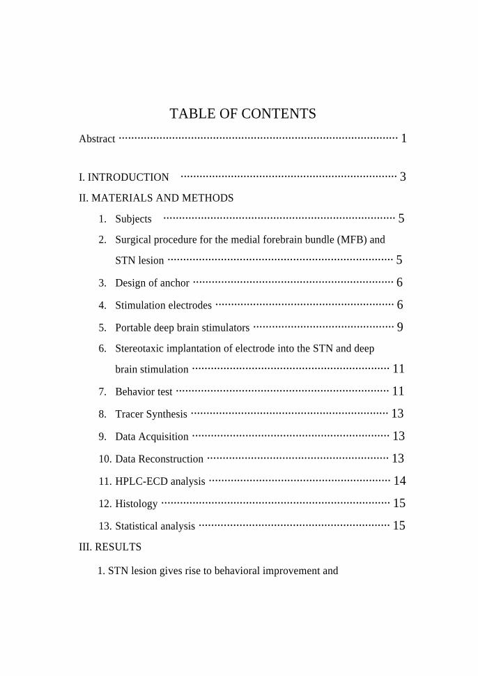
TABLE OF CONTENTS
Abstract ························································································· 1
I. INTRODUCTION ····································································· 3
II. MATERIALS AND METHODS
1. Subjects ·········································································· 5
2. Surgical procedure for the medial forebrain bundle (MFB) and
STN lesion ········································································ 5
3. Design of anchor ································································ 6
4. Stimulation electrodes ························································· 6
5. Portable deep brain stimulators ············································· 9
6. Stereotaxic implantation of electrode into the STN and deep
brain stimulation ······························································· 11
7. Behavior test ···································································· 11
8. Tracer Synthesis ······························································· 13
9. Data Acquisition ······························································· 13
10. Data Reconstruction ·························································· 13
11. HPLC-ECD analysis ·························································· 14
12. Histology ········································································· 15
13. Statistical analysis ····························································· 15
III. RESULTS
1. STN lesion gives rise to behavioral improvement and
dopamine elevation in rat parkinsonian model ······························ 16
2. STN-DBS gives rise to spontaneous and drug-induced
behavioral improvement in rat parkinsonian model ························ 24
3. STN-DBS does induce striatal dopamine release of rat
parkinsonian model in micro PET study ······································· 38
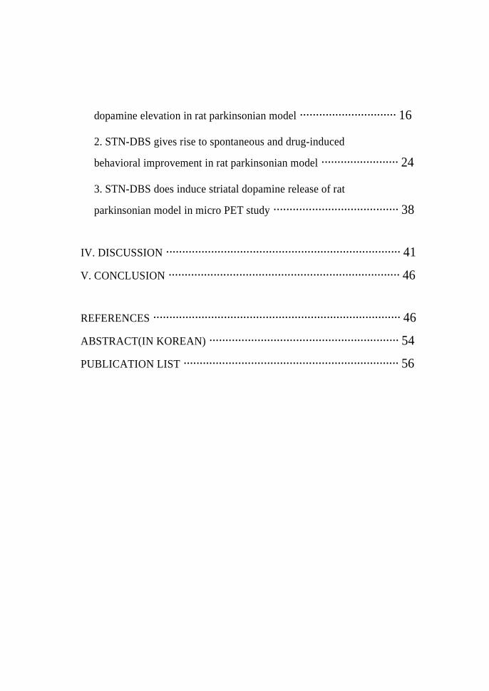
IV. DISCUSSION ········································································· 41
V. CONCLUSION ········································································ 46
REFERENCES ············································································· 46
ABSTRACT(IN KOREAN) ··························································· 54
PUBLICATION LIST ··································································· 56

LIST OF FIGURES
Figure 1. Photograph of electrode and diagram of implantation
on of electrode ··························································· 8
Figure 2. Bright field photomicrographs of histological section
obtained in rat PD models ······································· 17
Figure 3. Cresyl violet-stained sections illustrating a unilateral
kainic acid lesion in the STN ································ 18
Figure 4. Apomorphine-induced rotational behavior in rats
after 6-OHDA lesioning and ipsilateral kainic
acid lesioning of the STN ········································ 22
Figure 5. The average number of forepaw-adjusting steps in
rats with unilateral 6-OHDA lesioning and kainic
acid lesioning of the STN ········································ 23
Figure 6. Portable brain stimulator and biphasic output pulse
of the stimulator ······················································· 25
Figure 7. Photograph of the metal anchor implantation ··········· 26
Figure 8. The average number of forepaw-adjusting steps in
rats with unilateral 6-OHDA lesioning and DBS of
the STN ···································································· 28
Figure 9. Effect of rotarod motor test during DBS in rat
PD models ································································ 30
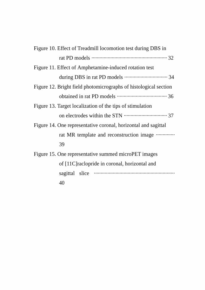
Figure 10. Effect of Treadmill locomotion test during DBS in
rat PD models ························································ 32
Figure 11. Effect of Amphetamine-induced rotation test
during DBS in rat PD models ································ 34
Figure 12. Bright field photomicrographs of histological section
obtained in rat PD models ····································· 36
Figure 13. Target localization of the tips of stimulation
on electrodes within the STN ································ 37
Figure 14. One representative coronal, horizontal and sagittal
rat MR template and reconstruction image ··············
39
Figure 15. One representative summed microPET images
of [11C]raclopride in coronal, horizontal and
sagittal slice ····························································
40
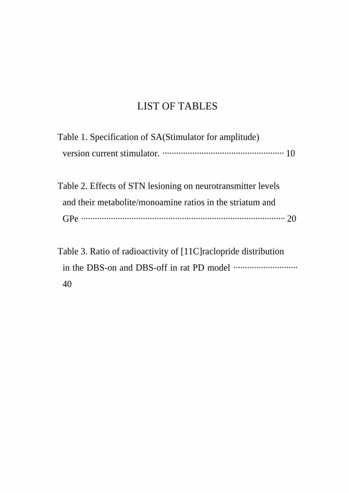
LIST OF TABLES
Table 1. Specification of SA(Stimulator for amplitude)
version current stimulator. ····················································· 10
Table 2. Effects of STN lesioning on neurotransmitter levels
and their metabolite/monoamine ratios in the striatum and
GPe ························································································· 20
Table 3. Ratio of radioactivity of [11C]raclopride distribution
in the DBS-on and DBS-off in rat PD model ····························
40
1
<ABSTRACT>
Effects of Subthalamic Nucleus Lesion and Electrical Stimulation in
6-OHDA-Induced Rat Parkinsonian Model
Yong Sup Hwang
Department of Medical Science
The Graduate School, Yonsei University
(Directed by Professor Jin Woo Chang)
Parkinson’s disease (PD) is a neurodegenerative motor disease
characterized by progressive loss of dopaminergic (DA) neurons in the
substantia nigra pars compacta (SNpc) and a concomitant reduction of striatal
dopamine. Abnormal activity of the STN, which sends hyperactivated
glutamatergic neurotransmission to the substantia nigra pars reticular (SNpr)
and globus pallidus internal segment (GPi), has been considered to be a
pivotal function in the expression of PD symptoms. The present study was to
investigate lesion and electrical stimulation of the STN ameliorated
behavioral and electrophysiological functions with 6-OHDA lesioned rats.
Behavioral changes were investigated after STN lesion by kainic acid in
6-hydroxydopamine (6-OHDA)-lesioned rats and measured levels of
dopamine (DA) and its metabolites using tissue dissection. The results
asserted that STN ablation induced behavioral improvement of parkinsonian
motor deficit and DA was increased in the striatum and globus pallidus
external segment (GPe).
The portable stimulators were made for animal PD model. STN-DBS

2
induced behavior improvement in PD models through the several behavioral
tests. STN-DBS also induced change with distributions of ligands for
dopamine D2 receptors ([11C]raclopride).
In the present study, The PD animals showed significant behavior
improvements tested the several behavioral changes under the STN-DBS in
PD models. A portable DBS stimulator and electrode fixation structure that
could be useful tools to investigate the behavioral changes after STN-DBS in
freely movable rat parkinsonian models. The results indicate that STN lesion
in a 6-OHDA induced hemipakinsonian rat model may counteract some of the
neurochemical changes within the striatum and GPe, and influence striatal
dopaminergic metabolism.
Key words: Parkinson’s disease, subthalamic nucleus, deep brain stimulation, High
performance liquid chromatography, microPET

3
Effects of Subthalamic Nucleus Lesion and Electrical Stimulation in
6-OHDA-Induced Rat Parkinsonian Model
Yong Sup Hwang
Department of Medical Science
The Graduate School, Yonsei University
(Directed by Professor Jin Woo Chang)
I. INTRODUCTION
Parkinson’s disease (PD) is a neurodegenerative motor disease
characterized by progressive loss of dopaminergic (DA) neurons in the
substantia nigra pars compacta (SNpc) and a concomitant reduction of striatal
dopamine.1, 2 The subthalamic nucleus (STN) is an important structure in the
basal ganglia circuitry and plays a critical role in regulating motor function.
Abnormal activity of the STN, which sends hyperactivated glutamatergic
neurotransmission to the substantia nigra pars reticular (SNpr) and globus
pallidus internal segment (GPi), has been considered to be a pivotal function
in the expression of PD symptoms such as akinesia, rigidity, and tremor.3
During the past decades, ablation of the STN by lesions or high frequency
stimulation has been shown to ameliorate motor dysfunction nonhuman
primate and rodent models of PD.4-6
Various experimental studies indicated therapeutic lesion of the STN
ameliorated behavioral and electrophysiological functions with 6-OHDA

4
lesioned rats.6-8 But, Lesions of the STN, a considerably vascularized and
small structure, may give rise to hemiballism and morbidity. During last years
neurosurgeons have therefore developed deep brain stimulation (DBS)
technique which become a fascinating intervention alternative in PD.
Although the basic mechanisms underlying DBS are still unknown, the
evidence that DBS of the STN has been shown to produce a dramatic
alleviation of motor symptoms of PD, in both animals and humans
experiments have been accumulated.5, 9-16 During the recent years, deep brain
stimulation (DBS) within basal ganglia has become a powerful therapeutic
tool with advanced Parkinson disease (PD) in contrast to other stereotactic
ablative surgery.17, 18
The advantages of DBS implicate its flexibility, the individual adjustment
of stimulation parameter; safety, no permanent damage to brain tissue; and
reversibility, the reduction of side effect and morbidity.19, 20 In addition, it has
been lately reported that STN-DBS seems to have neuroprotective effects as
well as improving dopaminergic activity in experimental parkinsonian
models.21-23
The aim of the present study was to investigate comparison between STN
lesion and STN stimulation therapy in 6-OHDA lesioned rats. We provided
comprehensive behavioral characterization of motor deficits by practicing
variable behavioral tests, such as forepaw-adjusting steps, rotarod motor test,
treadmill locomotor test, and amphetamine-induced rotational test.
In the present study, we report a portable DBS stimulator and electrode
fixation structure that could be useful tools to investigate the behavioral
changes after STN-DBS in freely movable rat parkinsonian models. As well,
we tested the several behavioral changes under the STN-DBS in PD models.
We also studied the tracer distributions of ligands for dopamine D2 receptors
([11C]raclopride) in the 6-OHDA lesioned rat during subthalamic electrical
5
stimulation to evaluate molecular changes for DA transmission in DBS.
Although a few clinical studies were performed using radiolignad
[11C]raclopride during DBS inpatients with Parkinson’s disease,24-26 their
PET data didn’t see any significant increased dopamine release under
STN-DBS. However, none of in vivo PET studies on DBS in the 6-OHDA
lesioned rats have elucidated how behavioral improvement relates to the
alteration of DA transmissions for electrical stimulation in quantitative
manner. To our knowledge, these are the first examples of [11C]raclopride
PET imaging between DBS-ON and DBS-Off state in the rat brain in the
progression of PD pathology.
II. MATERIALS AND METHODS
1. Subjects
Twenty five male Sprague-Dawley rats, weighing 230 to 250g each, were
used at the beginning of the experiment. The rats were allowed to feed ad
libitum during the experiment, and free access was given to water throughout
the study. The cages were kept in a temperature- and humidity-controlled
room with a 12 hour light-dark circle. Animal care and experiments were
performed in a facility certified by the American Association for the
Accreditation of Laboratory Animal Care. Rats were divided into 4 groups;
(A) a control group (n=5); (B) a PD group (n=6) having a lesion of
dopaminergic neurons caused by the 6-OHDA; (C) a STN group (n=7)
receiving kainic acid in the STN with 6-OHDA lesioned rats; (D) a DBS
group (n=7) subjected to electrical stimulation while the stimulation electrode
was placed in the STN with 6-OHDA lesioned rats.
2. Surgical procedure for the medial forebrain bundle (MFB) and STN
6
lesion
The rats were anesthetized with a mixture of ketamine (75mg/kg),
acepromazine (0.75 mg/kg), and rompun (4mg/kg). As previously described,27
8 µg(free base weight) of 6-OHDA(Sigma Chemical Co., St. Louis, MO) in
0.2% ascorbic acid with 0.9% normal saline was infused in 2 µl at a rate of
0.5 µl/min at the following coordinate of the medial forebrain bundle: -4.4mm
AP, 1.2mm relative to the bregma ML, and 7.5mm from the dura mater DV.
All Coordinates were taken from the atlas of Paxinos & Watson.28 After
injection of 6-OHDA, the cannula was left in place for 5 minutes before
slowly retracting it. To prevent destruction of adrenergic neurons, 12.5 mg/kg
desipramine was administered intraperitoneally 30 minutes before the infusion
of 6-OHDA. The STN lesion was made with 2 µg kainic acid (Sigma) in 1 µl
of saline solution injected at a rate of 0.5 µl/min (total 1 µl) into the STN
(coordinates: AP -3.7 mm, ML 2.5 mm relative to bregma, and DV –8.0 mm
from the dura). Upon recovery from the anaesthesia, many rats with STN
lesions were diazepam (10 mg/kg, i.p.) to prevent seizures dissolved in
sesame oil by warming and shaking. In the group of rats with paired lesions,
the STN lesion was performed one week after the injection of 6-OHDA into
the MFB.
3. Design of anchor
We design unique metal anchor to fix the electrode into skull of rat. We
make side ditches to fix stimulation electrode to the anchor by dental cement
and enable to bend the electrode and to connect with the connector cable.
With this approach, we can close the scalp of the rat after the insertion of the
electrode to minimize the inflammation.
4. Stimulation electrodes
7
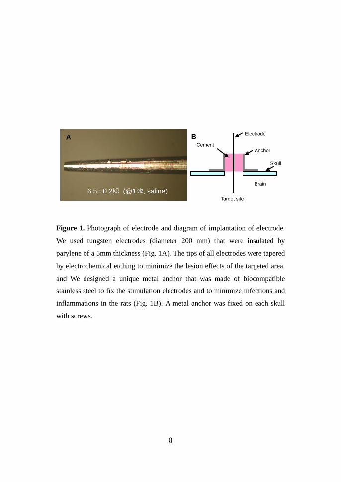
We used tungsten electrodes (diameter 200µm) which are insulated by
parylene coating of 5µm thickness for stimulation (Fig. 1). The tip of the all
electrodes is tapered by electro-chemical etching to reduce the lesion effect of
the target area. The tapered length of the electrode is 2mm and the area of the
stimulation site is about 0.035mm2, respectively. The impedance of the
electrode is 15±1㏀ in saline and 1㎑ with potentiostat. (Zahner Elektrik
IM6e, Germany)
8
Figure 1. Photograph of electrode and diagram of implantation of electrode.
We used tungsten electrodes (diameter 200 mm) that were insulated by
parylene of a 5mm thickness (Fig. 1A). The tips of all electrodes were tapered
by electrochemical etching to minimize the lesion effects of the targeted area.
and We designed a unique metal anchor that was made of biocompatible
stainless steel to fix the stimulation electrodes and to minimize infections and
inflammations in the rats (Fig. 1B). A metal anchor was fixed on each skull
with screws.
A
6.5±0.2㏀ (@1㎑, saline)
Electrode
Anchor Cement
Skull
Brain
Target site
B
9
5. Portable deep brain stimulators
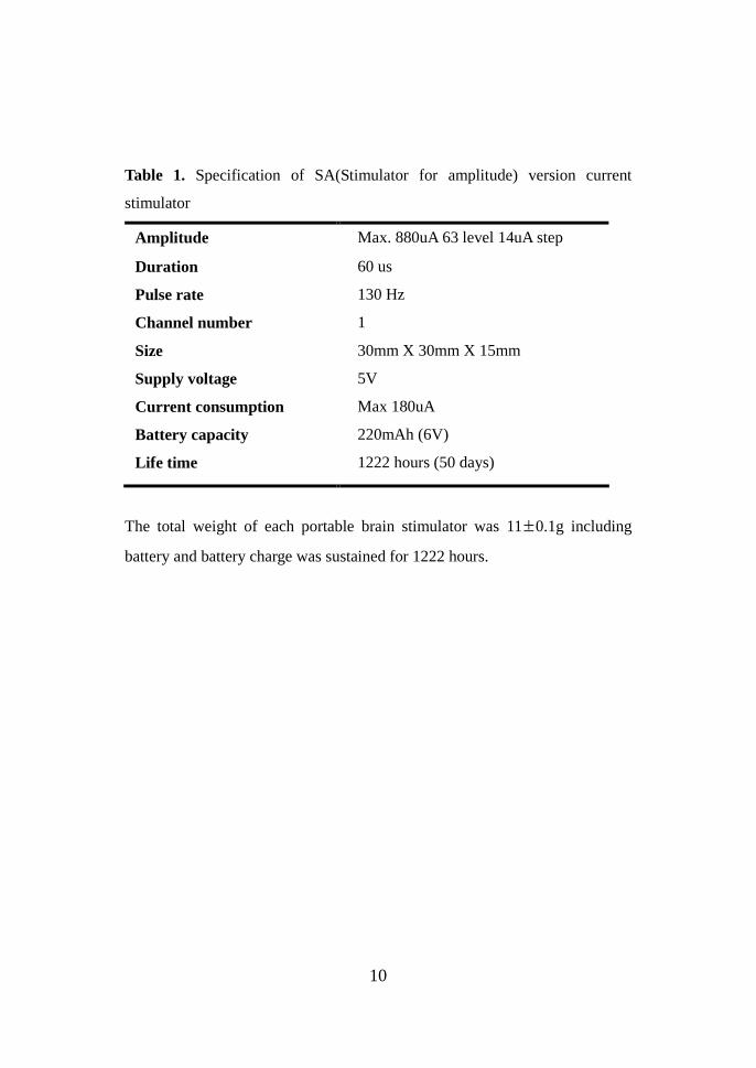
We used newly developed, small-sized portable stimulators to generate the
current stimulus pulses (Table 1.). The main chip of the stimulator was an
SX18AC microcontroller from Scenix. Stimulation parameters, such as
duration and stimulation rate, could be changed with PC-based software. The
parameters were stored in the internal ROM (Read Only Memory) of a chip
through the RS-232C serial communication port. The amplitude of the
stimulation voltage was controlled by the precision potentiometer. By
adjusting the knob position of the potentiometer, the output current could be
controlled from 14uA to 880uA. Finally, the stimulator was connected to the
stimulation electrodes through the percutaneous connector that was the
terminal of the subcutaneously implanted extension cable.
10
Table 1. Specification of SA(Stimulator for amplitude) version current
stimulator
Amplitude Max. 880uA 63 level 14uA step
Duration 60 us
Pulse rate 130 Hz
Channel number 1
Size 30mm X 30mm X 15mm
Supply voltage 5V
Current consumption Max 180uA
Battery capacity 220mAh (6V)
Life time 1222 hours (50 days)
The total weight of each portable brain stimulator was 11±0.1g including
battery and battery charge was sustained for 1222 hours.
11
6. Stereotaxic implantation of electrode into the STN and deep brain
stimulation
Selected Animals were further unilaterally implanted with a metal electrode
(tip diameter: 100µm) at the STN stereotaxic coordinates (AP-3.7mm, ML
2.5mm relative to bregma, and DV -8.2 mm from the dura) according to the
stereotaxic atlas of Paxino & Watson.28
Stimulation for DBS group was applied one week after implantation of the
electrode into STN. The stimulation parameters was determined on the basis
of previous DBS studies by Maesawa et al.23 Briefly, only current intensity
was altered from 128 µA to 896 µA. The optimal intensity was chosen
according to behavioral changes of each rat response below the threshold for
apparent dyskinetic movement of contralateral forepaw, which was
approximately 450 µA. Other parameters were fixed at a pulse rate 130 Hz
and pulse width 60 µsec.
7. Behavior test
Forepaw-Adjusting Steps Contralateral forepaw adjusting steps were assessed
immediately before the amphetamine-induced rotational test, as described in a
previous study 29. Briefly, the rats were held in a stationary position, while
bearing weights on their forepaw, on the surface of the treadmill which moved
at a rate of 90 cm/12 sec. During this interval, the number of forepaw
adjusting steps involving the weight-bearing forepaw, which the rats made to
compensate for the movement of their body, was counted. Each test consisted
of 5 trials for each forepaw, alternating between forepaws. The contralateral
forepaw adjusting steps were assessed on two occasions, 2 weeks after the
6-OHDA lesion and 1 week after the STN-DBS.
Elevated body swing test The elevated body swing test (EBST) was performed
1 day before the apomorphine-induced rotational test. Briefly, the rat was
12
placed in a Plexiglass box (40X40X35 cm), allowed to habituated for 2 min,
and attain a neutral position. The rat was elevated approximately 2 cm above
the surface by holding its tail. The direction of each swing was recorded when
the rat moved, with its head directed more than 10° to either side of the
vertical axis. Swings were counted for a period of 10 sec. One observer was
responsible for timing the test session, determining, and recording the
direction of swings, while another observer held the rat. All tests were
conducted blind to the groups. The average performance in the five
consecutive trials was used for within animal comparisons. The results of the
swing test were displayed as percentage of right-biased swing for 10 seconds.
Rotarod motor test An accelerating rotarod (Ugo Basile, Comerio VA, Italy)
was used to evaluate motor function of the rats. Each rat was placed on the
rotating rod and received a training session on the rotarod at a continual speed
of 8 rpm. The speed was slowly increased from 4 to 40 rpm over a period of 5
min. The time ended if the rat fell off the rung or gripped the device and spun
around for two consecutive revolutions without attempting to walk on the
rung. All tests were performed by observers blinded to each groups.
Treadmill locomotion test Locomotor ability was evaluated by treadmill
locomotion test. Rats were trained to run on a straight treadmill enclosed
15cm wide, 40cm long conveyor belt chamber at a speed of 12cm/sec. The
treadmill cycled between 20sec on and 20sec off periods. The trial was
recorded average distance of the rats’ nose position from the back wall of the
chamber. A ruler was placed in view along with the treadmill belt to measure
the nose position of the rat during the test. The average performance in the
five consecutive trials was used for within animal comparisons.
Amphetamine-Induced Rotational Behavior Amphetamine-induced rotational
behaviors were measured by using automated rotometer bowls with a tether
attached to the torso of subjects. Amphetamine (5 mg/kg, i.p.) was

13
administered 2 weeks after medial forebrain bundle (MFB) lesioning.
Rotational behavior was quantified every minute for 1 hour, after allowing the
rats to become habituated for 20 min. Those rats that exhibited at least five
full turns per minute, ipsilateral to the 6-OHDA lesioned side, were selected
for the subsequent experiments. The total numbers of rotations during a period
of one hour was measured and used for the analysis.
8. Tracer Synthesis
Functional images of the striatal dopamine system were obtained for each
rat the D2 receptor radioligand [11C]raclopride(n=3). [11C]Raclopride was
obtained by methylating the corresponding nor-precursor with
[11C]methyltriflate. Approximately 18 MBq (0.5 mCi) of radioligand
(specific activity range, 53– 760GBq/µmol; injection volume, 0.5mL) were
injected into the tail vein using an infusion needle set.
9. Data Acquisition
All small-animal PET imagings were performed on a SIEMENS/Concorde
Focus 120(Siemens Medical Solutions USA, Inc), which has an
approximately (1.3mm)3 resolution and plane-to-plane separation of 0.79mm.
The coincidence window width was set at 6 ns. Before being imaged, the rats
were anesthetized with isoflurane and N2O/O2. All rats were breathing
spontaneously throughout the entire experiment. For [11C]raclopride,
dynamic data were acquired during 120 min with frame duration increasing
from 15sec to 300sec. All animals were scanned in the prone position with
their brain centered in the field of view.
10. Data Reconstruction and image analysis
Small-animal PET studies were reconstructed using both filtered
14
backprojection and 3D-OSEM (3dimensional ordered subset expectation
maximization). Compared with filtered backprojection, 3D-OSEM has been
shown to result in improved spatial resolution and noise properties on
small-animal PET images - an advantage for image registration. On the other
hand, filtered backprojection may more accurately estimate radioactivity
concentration and was used for quantification. After the reconstruction of data
2 mm round regions of interest (ROIs) were placed over the center of the
bilateral striatum (2 slices) and cerebellum (2 slices), with image analysis
software (ASIPro VMTM, Siemens Medical Solutions USA, Inc). Decay-
corrected time activity curves (TAC) in the ROIs were obtained from the
dynamic PET images. The cerebellum was used as reference region, because
it is nearly free of the D2 receptors.
11. HPLC-ECD analysis
High-pressure liquid chromatography (HPLC) with electrochemical
detection was used for quantification of DA and DA metabolites (3,
4-dihydroxyphenylacetic acid, DOPAC, and homovanillic acid, HVA). When
experiments were completed, rats were sacrificed by decapitation. The brains
were rapidly removed and chilled in ice-cold 0.9% NaCl solution. After that,
the brain was cut into coronal slice with the aid of a chilled brain mold
(Rodent Brain Matrix, ASI Instruments, Warren, MI). The striatum and globus
pallidus external segment (GPe) were immediately removed, placed onto dry
ice, and tissue wet weight obtained. The tissue was homogenized in 1 ml of
ice-cold 0.1 N HClO4 for 30 sec, followed by centrifugation at 10,000 g and
4 for 10 min. Twenty µl of the supernatant was injected at 0.7 ml/min ℃
through a 80 x 4.6 mm HR-80 C-18 column (ODS, 3 µm particles; ESA,
Chelmsford, MA, USA) with a mobile phase consisting of 75-mM Na2HPO4,
1.7-mM OSA, 0.01% [v/v] triethylamine, 10% [v/v] acetonitrile, and 0.1-mM
15
EDTA, adjusted to pH 3.0 with 2-mM phosphate buffer. Detection limit was
20 nA. Detection was performed with a coulometric detector (Coulochem Ⅱ,
ESA, Chelmsford, MA, USA) coupled to a dual-electrode analytical cell
(model 5014, ESA). Results were expressed as fmol/µl protein/mg wet weight
of tissue.
12. Histology
Upon completion of an experiment, the rats were anesthetized and
transcardially perfused with normal saline, followed by ice-cold 4%
paraformaldehyde. Coronal 30µm tissue sections were cut at -20˚C using
microtome cryostat the level of the striatum, the STN, and the substantia nigra
pars compacta (SNpc) and mounted on gelatin-coated slides. Cresyl violet
staining was performed to check the correct location of stimulation electrode
in STN. Tyrosine hydroxylase (TH) immunohistochemistry was used to
confirm the completion of the 6-OHDA lesions of the nigrostriatal pathway in
rat PD models30. To assess loss of Dopamine (DA) cells and fibers in the
SNpc and striatum on the side ipsilateral to 6-OHDA tissue sections were
immunoreacted with primary polyclonal antibody against rat TH (PelFreeze,
Rogers, AR) at a dilution of 1: 500 and biotinylated goat anti-rabbit IgG
secondary antibody(Vector Laboratories, Burlingame, CA) secondary
antibody. The signal was amplified by avidin and biotinylated horseradish
peroxidase using the Elite ABC Vectastain kit (Vector Laboratories). We used
3, 3’-diaminobenzidine tetrahydrochloride as a chromogen, and cobalt
chloride/nickel ammonium was used for intensification of the color changes.
13. Statistical analysis
Pared samples t-tests were used when comparisons involved only two
groups. When three or more groups were involved, analyses of variance
16
(ANOVAs) were carried out and, if significant, follow-up LSD post hoc test
was used to determine which pairs of groups were different from each other.
The criterion for statistical significance was considered to be P < 0.05 in all
statistical evaluations. All statistical analyses were performed Using SPSS
version 11.5 (SPSS Inc., Chicago, IL, USA).
III. RESULTS
1. STN lesion gives rise to behavioral improvement and dopamine
elevation in rat parkinsonian model.
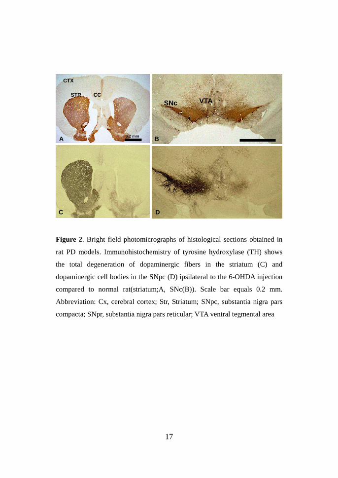
The extent and location of the lesions caused by the 6-OHDA were
confirmed by assessing the loss of TH-immunoreactive cells and fibers in the
substantia nigra pars compacta (SNpc) and striatum in the rat parkinsonian
model with 6-OHDA (Fig. 2). The STN lesions were also evaluated following
the completion of the experiments, and revealed evidence of local gliosis at
the level of the STN (Fig. 3).
17
Figure 2. Bright field photomicrographs of histological sections obtained in
rat PD models. Immunohistochemistry of tyrosine hydroxylase (TH) shows
the total degeneration of dopaminergic fibers in the striatum (C) and
dopaminergic cell bodies in the SNpc (D) ipsilateral to the 6-OHDA injection
compared to normal rat(striatum;A, SNc(B)). Scale bar equals 0.2 mm.
Abbreviation: Cx, cerebral cortex; Str, Striatum; SNpc, substantia nigra pars
compacta; SNpr, substantia nigra pars reticular; VTA ventral tegmental area
STR CC
CTX
0.2 mm
SNc VTA
A B
C D

18
Figure 3. Cresyl violet-stained sections illustrating a unilateral kainic acid
lesion in the subthalamic nucleus. The arrow indicates the location of the
lesion (left: normal side, right: lesion side). Scale bar equals 0.2 mm
50 um
0.2 mm

19
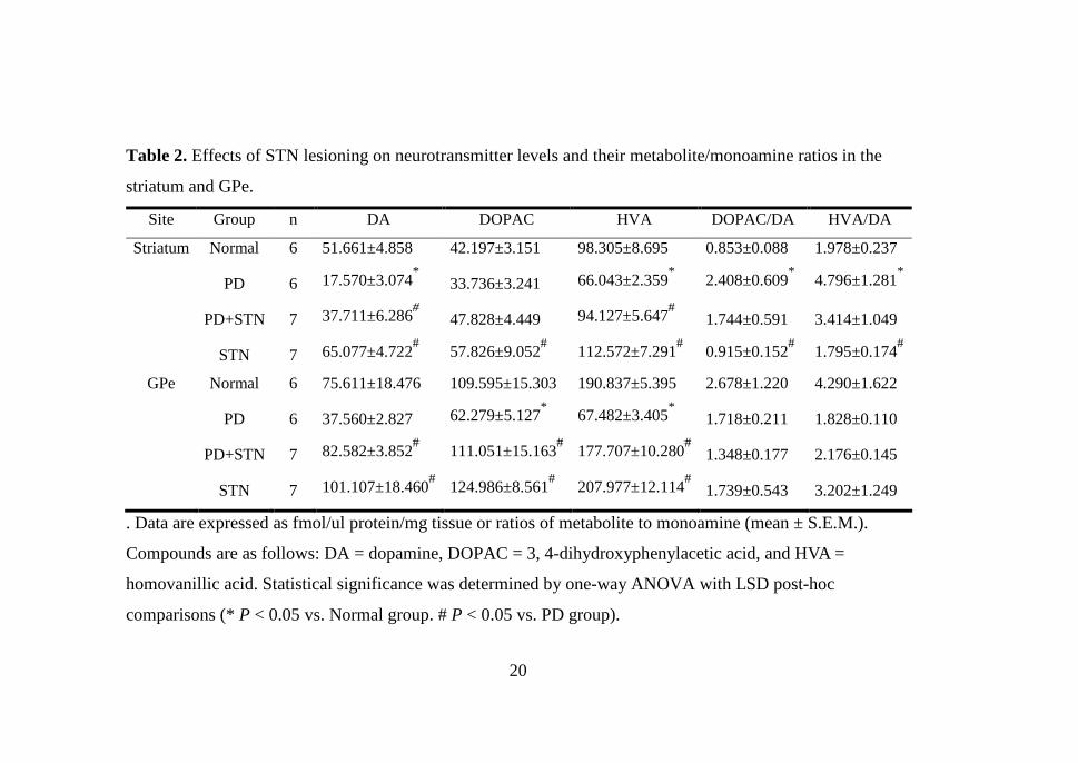
The levels of DA and its metabolites were measured in the striatum and
GPe for each group of rats. Table 2 shows the change of neurotransmitter
level in the striatum and GPe. There was a decrease of DA and DA
metabolites (DOPAC and HVA) in these two regions in the 6-OHDA induced
rat PD model, while the STN lesion group demonstrated a significant increase
in these levels. After STN lesion induction the amount of DA in the striatum
and GPe was significantly increased up to 20.14 ± 3.21 fmol/µl/mg and 45.02
± 1.03 fmol/µl/mg, respectively (n=7, mean ± SEM, P < 0.05). The PD group
followed by the STN lesion group demonstrated a significant increase in GPe
DOPAC levels (62.279 ± 5.127 to 111.051 ± 15.163 fmol/µl/mg, P < 0.05),
but did not show any statistical significance in the striatum. After STN
lesioning in the rat PD model, HVA levels in the striatum and GPe increased
significantly, 28.08 ± 3.29 fmol/µl/mg and 50.23 ± 6.88 fmol/µl/mg,
respectively.
20
Table 2. Effects of STN lesioning on neurotransmitter levels and their metabolite/monoamine ratios in the
striatum and GPe.
Site Group n DA DOPAC HVA DOPAC/DA HVA/DA
Striatum Normal 6 51.661±4.858 42.197±3.151 98.305±8.695 0.853±0.088 1.978±0.237
PD 6 17.570±3.074* 33.736±3.241 66.043±2.359
* 2.408±0.609
* 4.796±1.281
*
PD+STN 7 37.711±6.286# 47.828±4.449 94.127±5.647
# 1.744±0.591 3.414±1.049
STN 7 65.077±4.722# 57.826±9.052
# 112.572±7.291
# 0.915±0.152
# 1.795±0.174
#
GPe Normal 6 75.611±18.476 109.595±15.303 190.837±5.395 2.678±1.220 4.290±1.622
PD 6 37.560±2.827 62.279±5.127* 67.482±3.405
* 1.718±0.211 1.828±0.110
PD+STN 7 82.582±3.852# 111.051±15.163
# 177.707±10.280
# 1.348±0.177 2.176±0.145
STN 7 101.107±18.460# 124.986±8.561
# 207.977±12.114
# 1.739±0.543 3.202±1.249
. Data are expressed as fmol/ul protein/mg tissue or ratios of metabolite to monoamine (mean ± S.E.M.).
Compounds are as follows: DA = dopamine, DOPAC = 3, 4-dihydroxyphenylacetic acid, and HVA =
homovanillic acid. Statistical significance was determined by one-way ANOVA with LSD post-hoc
comparisons (* P < 0.05 vs. Normal group. # P < 0.05 vs. PD group).

21
In 6-OHDA and STN lesioned rats, both striatal DOPAC/DA and HVA/DA
ratios tended to increase but did not show any significant difference from the
PD group.
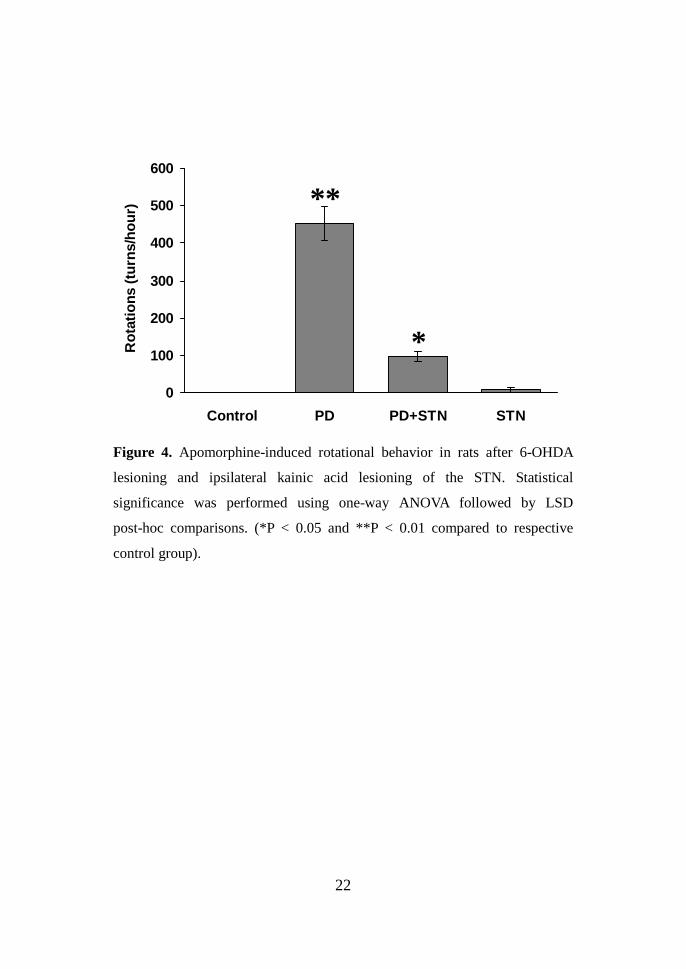
The mean number of rotations in the rat PD model (n=6) for a period of
one hour was 452.8 ± 45.82 (P < 0.01 compared with values control group,
Fig. 4). After STN lesioning was performed in the rat PD models, the mean
number of rotations for a period of one hour was significantly decreased, from
452.8 ± 45.82 to 97 ± 13.5 (P < 0.05). We also evaluated the effect of STN
lesioning on forepaw adjusting steps. The mean number of forepaw-adjusting
steps in each group is given in Fig. 5. After STN lesion induction,
contralateral forepaw adjusting increased 5.0 ± 0.91, which was significantly
higher than that of the 6-OHDA lesion group (2.6 ± 0.5, P < 0.05).
22
0
100
200
300
400
500
600
Control PD PD+STN STN
Ro
tati
ons
(tu
rns/
ho
ur) **
*
Figure 4. Apomorphine-induced rotational behavior in rats after 6-OHDA
lesioning and ipsilateral kainic acid lesioning of the STN. Statistical
significance was performed using one-way ANOVA followed by LSD
post-hoc comparisons. (*P < 0.05 and **P < 0.01 compared to respective
control group).
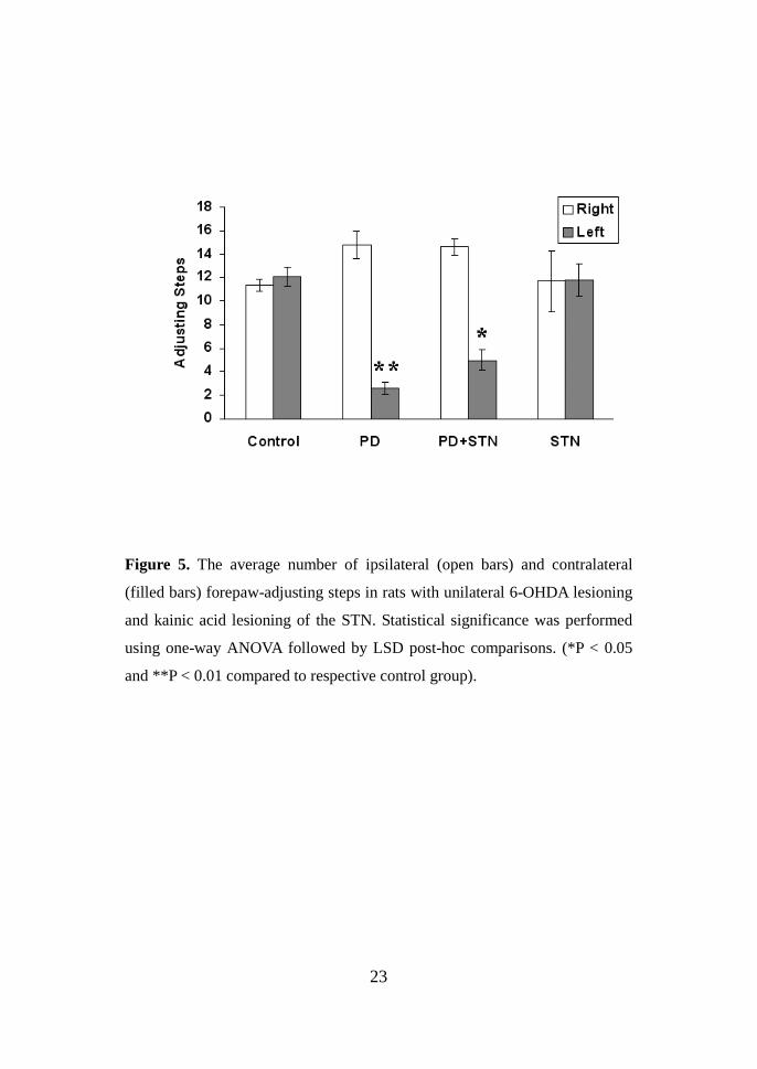
23
Figure 5. The average number of ipsilateral (open bars) and contralateral
(filled bars) forepaw-adjusting steps in rats with unilateral 6-OHDA lesioning
and kainic acid lesioning of the STN. Statistical significance was performed
using one-way ANOVA followed by LSD post-hoc comparisons. (*P < 0.05
and **P < 0.01 compared to respective control group).
24
2. STN-DBS gives rise to spontaneous and drug-induced behavioral
improvement in rat parkinsonian model.
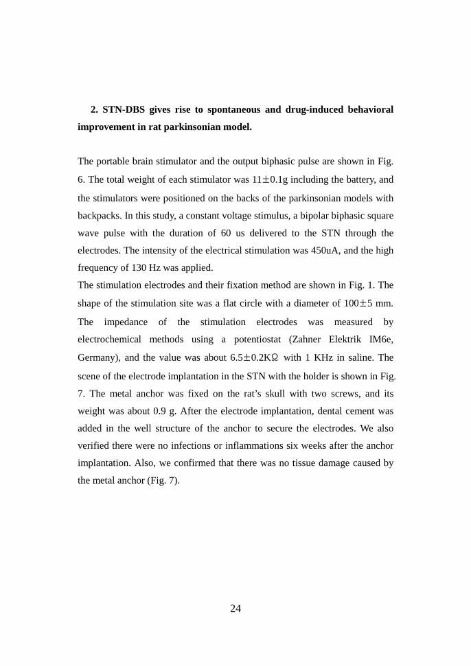
The portable brain stimulator and the output biphasic pulse are shown in Fig.
6. The total weight of each stimulator was 11±0.1g including the battery, and
the stimulators were positioned on the backs of the parkinsonian models with
backpacks. In this study, a constant voltage stimulus, a bipolar biphasic square
wave pulse with the duration of 60 us delivered to the STN through the
electrodes. The intensity of the electrical stimulation was 450uA, and the high
frequency of 130 Hz was applied.
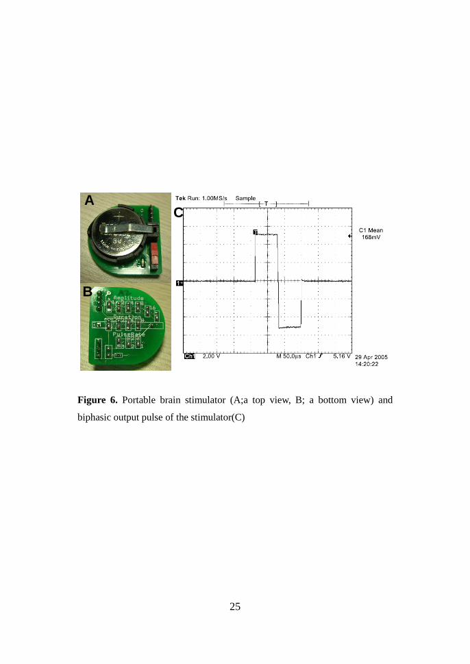
The stimulation electrodes and their fixation method are shown in Fig. 1. The
shape of the stimulation site was a flat circle with a diameter of 100±5 mm.
The impedance of the stimulation electrodes was measured by
electrochemical methods using a potentiostat (Zahner Elektrik IM6e,
Germany), and the value was about 6.5±0.2KΩ with 1 KHz in saline. The
scene of the electrode implantation in the STN with the holder is shown in Fig.
7. The metal anchor was fixed on the rat’s skull with two screws, and its
weight was about 0.9 g. After the electrode implantation, dental cement was
added in the well structure of the anchor to secure the electrodes. We also
verified there were no infections or inflammations six weeks after the anchor
implantation. Also, we confirmed that there was no tissue damage caused by
the metal anchor (Fig. 7).
25
Figure 6. Portable brain stimulator (A;a top view, B; a bottom view) and
biphasic output pulse of the stimulator(C)
A
B
C
26
Figure 7. We verified there were no infections or inflammations six weeks
after the anchor implantation. Also, we confirmed that there was no tissue
damage caused by the metal anchor.
Screw
Anchor

27
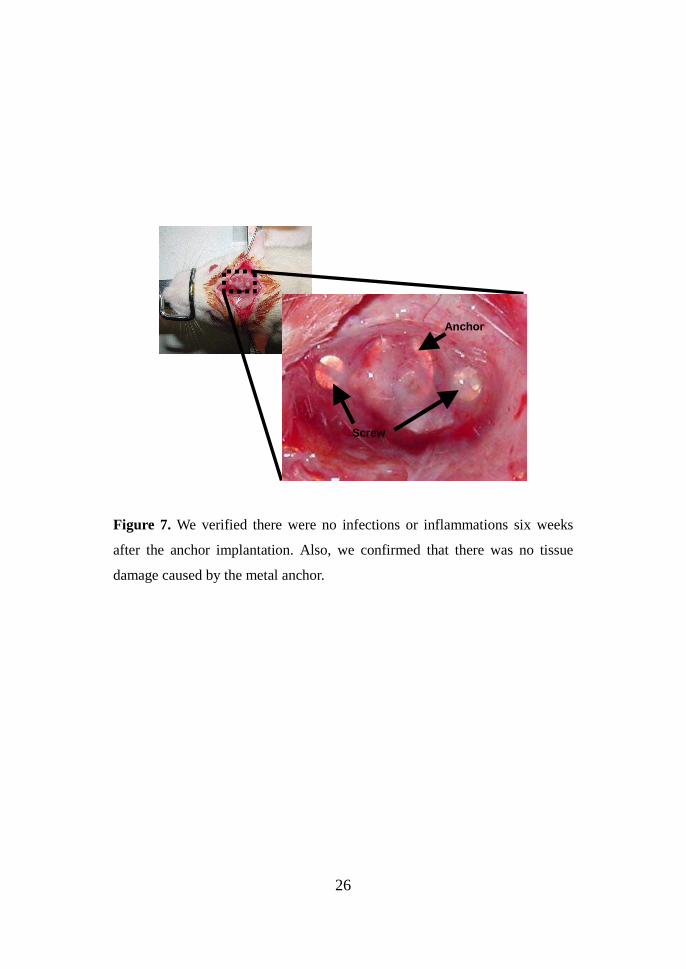
Forepaw –adjusting steps Consistent with previous reports 29, 30, the rat
hemiparkinsonian 6-OHDA model produced a deficit in adjusting steps
measured by the contralateral forelimb. The contralateral forepaw-adjusting
steps appeared to have an all-or-none relationship to the conformity of the rats
with the rat PD models (fig. 8). During STN-DBS, DBS group show
increment of contralateral forepaw adjusting steps. Significant difference in
the number of adjusting steps could be detected between PD and DBS group.
After STN-DBS, contralateral forepaw adjusting increased 1.97 ± 0.23, which
was significantly higher than that of the 6-OHDA lesion group (0.33 ± 0.11, P
< 0.05).
28
0
2
4
6
8
10
12
14
16
Normal PD PD-DBS
Ad
just
ing
ste
ps
LeftRight
Figure 8. The average number of ipsilateral (open bars) and contralateral
(filled bars) forepaw-adjusting steps in rats with unilateral 6-OHDA lesioning
and DBS of the STN. Statistical significance was performed using one-way
ANOVA followed by LSD post-hoc comparisons. (*p < 0.05 and ** p < 0.01
compared to respective control group, # p < 0.05 and ## p < 0.01 compared to
respective PD group).
********
#

29
Elevated body swing test The effect of STN-DBS in 6-OHDA induced rat PD
model was investigated on drug-free EBST after the 6-OHDA lesion and 2
weeks after the DBS electrode implant. PD group exhibited a 33.72 ± 2.97%
significantly more right-biased swing behavior in the comparison with the
control group on the test session (P<0.05), but during STN-DBS there was no
significant improvement between the PD group and DBS group (results not
shown).
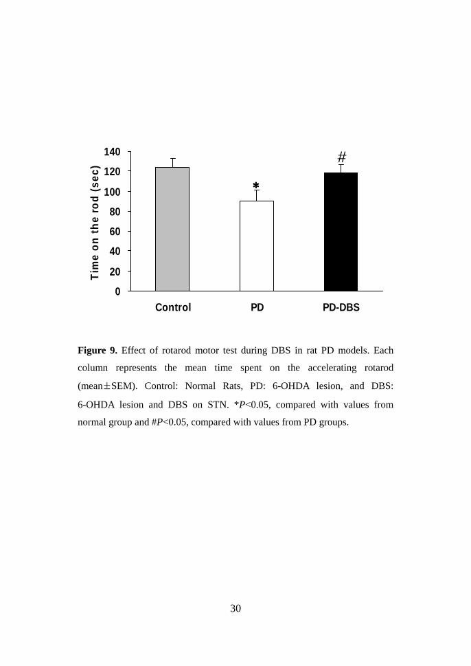
Rotarod motor test The rotarod test was performed at 2 weeks after the
6-OHDA lesion and 2 weeks after the DBS electrode implant (Fig. 8). Prior to
6-OHDA lesion, The mean time spent on the accelerating rotarod in control
group was 124.40 ± 8.36 sec (mean ± SEM). After lesion, the riding time was
significantly decreased in the PD group (90.33 ± 10.73 sec, P<0.05). During
STN-DBS, this impaired performance showed significant amelioration in the
DBS group(118.25 ± 8.08 sec, P<0.05).
30
Figure 9. Effect of rotarod motor test during DBS in rat PD models. Each
column represents the mean time spent on the accelerating rotarod
(mean±SEM). Control: Normal Rats, PD: 6-OHDA lesion, and DBS:
6-OHDA lesion and DBS on STN. *P<0.05, compared with values from
normal group and #P<0.05, compared with values from PD groups.
0
20
40
60
80
100
120
140
Control PD PD-DBS
Tim
e o
n t
he
rod
(se
c)
****
#
31
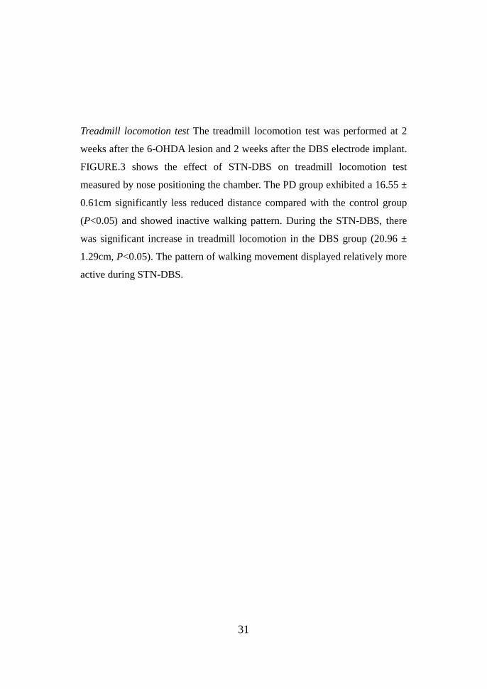
Treadmill locomotion test The treadmill locomotion test was performed at 2
weeks after the 6-OHDA lesion and 2 weeks after the DBS electrode implant.
FIGURE.3 shows the effect of STN-DBS on treadmill locomotion test
measured by nose positioning the chamber. The PD group exhibited a 16.55 ±
0.61cm significantly less reduced distance compared with the control group
(P<0.05) and showed inactive walking pattern. During the STN-DBS, there
was significant increase in treadmill locomotion in the DBS group (20.96 ±
1.29cm, P<0.05). The pattern of walking movement displayed relatively more
active during STN-DBS.

32
0
5
10
15
20
25
30
35
40
Normal PD PD-DBS
No
se p
osi
tion
(cm
)
Figure 10. Effect of Treadmill locomotion test during DBS in rat PD models.
Each bar represents the average distance between the rat’s noses and posterior
wall of chamber. Mean±S.E.M. *P<0.05, compared with values from normal
group and #P<0.05, compared with values from PD groups.
****
#
33
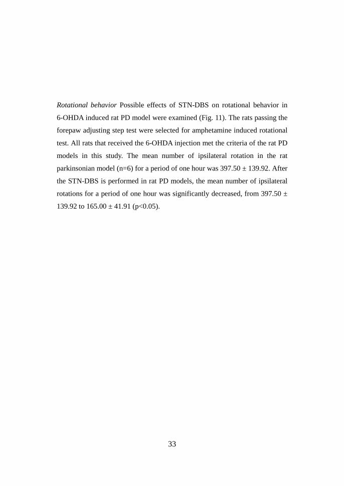
Rotational behavior Possible effects of STN-DBS on rotational behavior in
6-OHDA induced rat PD model were examined (Fig. 11). The rats passing the
forepaw adjusting step test were selected for amphetamine induced rotational
test. All rats that received the 6-OHDA injection met the criteria of the rat PD
models in this study. The mean number of ipsilateral rotation in the rat
parkinsonian model (n=6) for a period of one hour was 397.50 ± 139.92. After
the STN-DBS is performed in rat PD models, the mean number of ipsilateral
rotations for a period of one hour was significantly decreased, from 397.50 ±
139.92 to 165.00 ± 41.91 (p<0.05).

34
0
100
200
300
400
500
600
Normal PD PD-DBS
Ro
tatio
ns
per
ho
ur
contralateralipsilateral
Figure 11. Effect of amphetamine-induced rotation test during DBS in rat PD
models. A. Mean rotation showing amphetamine-induced ipsilateral rotational
behavior. The sum of 360° rotations towards the ipsilateral sides recorded for
60 min are represented for each groups. Mean±SEM. *P<0.05, compared
with values from normal group and #P<0.05, compared with values from PD
groups.
#
****
35
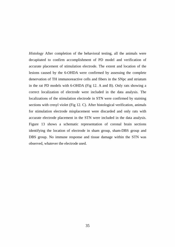
Histology After completion of the behavioral testing, all the animals were
decapitated to confirm accomplishment of PD model and verification of
accurate placement of stimulation electrode. The extent and location of the
lesions caused by the 6-OHDA were confirmed by assessing the complete
denervation of TH immunoreactive cells and fibers in the SNpc and striatum
in the rat PD models with 6-OHDA (Fig 12. A and B). Only rats showing a
correct localization of electrode were included in the data analysis. The
localizations of the stimulation electrode in STN were confirmed by staining
sections with cresyl violet (Fig 12. C). After histological verification, animals
for stimulation electrode misplacement were discarded and only rats with
accurate electrode placement in the STN were included in the data analysis.
Figure 13 shows a schematic representation of coronal brain sections
identifying the location of electrode in sham group, sham-DBS group and
DBS group. No immune response and tissue damage within the STN was
observed, whatever the electrode used.
36
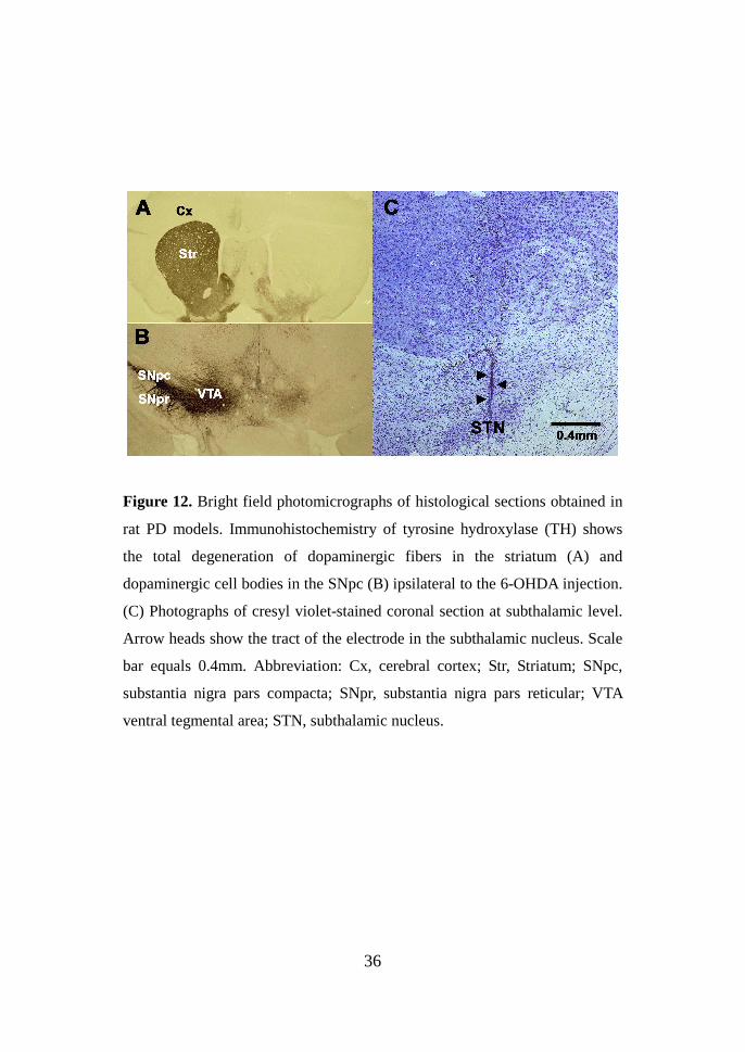
Figure 12. Bright field photomicrographs of histological sections obtained in
rat PD models. Immunohistochemistry of tyrosine hydroxylase (TH) shows
the total degeneration of dopaminergic fibers in the striatum (A) and
dopaminergic cell bodies in the SNpc (B) ipsilateral to the 6-OHDA injection.
(C) Photographs of cresyl violet-stained coronal section at subthalamic level.
Arrow heads show the tract of the electrode in the subthalamic nucleus. Scale
bar equals 0.4mm. Abbreviation: Cx, cerebral cortex; Str, Striatum; SNpc,
substantia nigra pars compacta; SNpr, substantia nigra pars reticular; VTA
ventral tegmental area; STN, subthalamic nucleus.
37
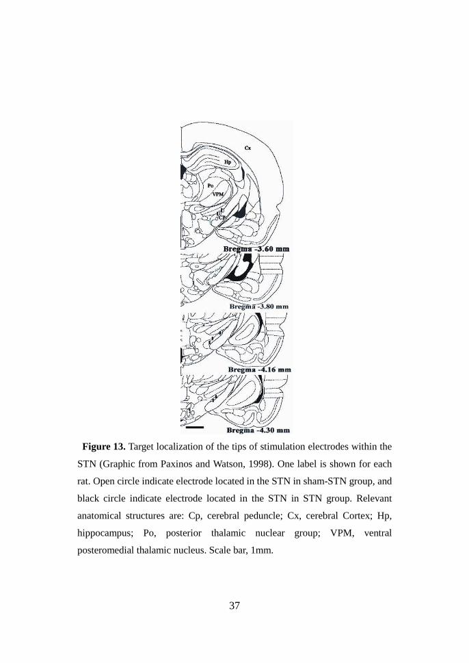
Figure 13. Target localization of the tips of stimulation electrodes within the
STN (Graphic from Paxinos and Watson, 1998). One label is shown for each
rat. Open circle indicate electrode located in the STN in sham-STN group, and
black circle indicate electrode located in the STN in STN group. Relevant
anatomical structures are: Cp, cerebral peduncle; Cx, cerebral Cortex; Hp,
hippocampus; Po, posterior thalamic nuclear group; VPM, ventral
posteromedial thalamic nucleus. Scale bar, 1mm.
38
3. STN-DBS does induce striatal dopamine release of rat parkinsonian
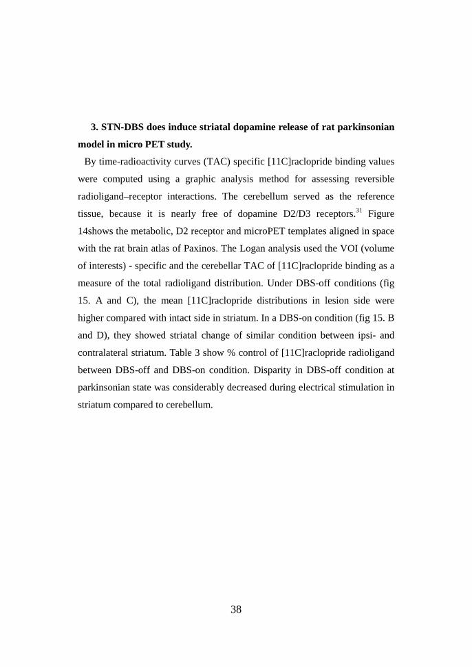
model in micro PET study.
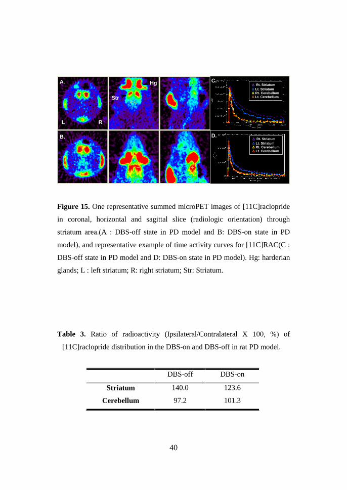
By time-radioactivity curves (TAC) specific [11C]raclopride binding values
were computed using a graphic analysis method for assessing reversible
radioligand–receptor interactions. The cerebellum served as the reference
tissue, because it is nearly free of dopamine D2/D3 receptors.31 Figure
14shows the metabolic, D2 receptor and microPET templates aligned in space
with the rat brain atlas of Paxinos. The Logan analysis used the VOI (volume
of interests) - specific and the cerebellar TAC of [11C]raclopride binding as a
measure of the total radioligand distribution. Under DBS-off conditions (fig
15. A and C), the mean [11C]raclopride distributions in lesion side were
higher compared with intact side in striatum. In a DBS-on condition (fig 15. B
and D), they showed striatal change of similar condition between ipsi- and
contralateral striatum. Table 3 show % control of [11C]raclopride radioligand
between DBS-off and DBS-on condition. Disparity in DBS-off condition at
parkinsonian state was considerably decreased during electrical stimulation in
striatum compared to cerebellum.
39
Figure 14. One representative coronal, horizontal and sagittal rat MR
template through striatum area(top) and reconstruction image using
3D-OSEM algorithm(middle : DBS-off state in PD model and bottom:
DBS-on state in PD model)
Left Right
40
Figure 15. One representative summed microPET images of [11C]raclopride
in coronal, horizontal and sagittal slice (radiologic orientation) through
striatum area.(A : DBS-off state in PD model and B: DBS-on state in PD
model), and representative example of time activity curves for [11C]RAC(C :
DBS-off state in PD model and D: DBS-on state in PD model). Hg: harderian
glands; L : left striatum; R: right striatum; Str: Striatum.
Table 3. Ratio of radioactivity (Ipsilateral/Contralateral X 100, %) of
[11C]raclopride distribution in the DBS-on and DBS-off in rat PD model.
DBS-off DBS-on
Striatum 140.0 123.6
Cerebellum 97.2 101.3
ΔΔΔΔ Rt. Striatum ∆ Lt. Striatum ∆ Rt. Cerebellum ∆ Lt. Cerebellum
Hg
L R
Str
A.
B.
C.
D. ΔΔΔΔ Rt. Striatum ∆ Lt. Striatum ∆ Rt. Cerebellum ∆ Lt. Cerebellum
41
IV. DISCUSSION
Output neurons from the STN are excitatory and use glutamate as a
neurotransmitter. The degeneration of nigral dopamine neurons in PD leads to
increased activity of glutamatergic neurons in the STN.3 Therefore, inhibition
of the STN plays a key role in the pathophysiological study of Parkinson's
disease. We have previously demonstrated that ipsilateral STN lesioning by
kainic acid in the rat hemi-parkinsonian 6-OHDA model induced behavioral
amelioration of motor deficits and normalized overactivity of GABAergic
SNpr neurons which amplified by increased excitatory input from the STN
and the hypoactive firing rate of GP neurons, resulting in the depletion of
dopaminergic neurons in the SNc.6 Reductions in the circling induced by
apomorphine and other DA agonists in the rat PD model following STN lesion
induction have also been reported by others.32 Recent studies have shown that
STN lesions ameliorate parkinsonian motor symptoms in nonhuman
primates.4, 33
In the present study, the obtained results provide the first evidence that STN
lesions induced by kainic acid in the rat hemiparkinsonian 6-OHDA model
produced increased dopamine neurotransmission in the striatum. A previous
study indicated that stimulation of the STN produced an increased level of
extracellular DA metabolites without an increase of extracellular DA in
intrastriatal 6-OHDA lesioned rats.22 Therefore, alteration in dopamine
neurotransmission may reflect changes in nigrostriatal dopaminergic neuronal
firing or dopamine release. Moreover, we observed increased levels of DA
and DA metabolites after STN lesion induction in the GPe, suggesting that
output neurons from the STN project to the GPe, GPi, and SNpr. Disinhibition
of the STN neurons might increase thereby restoring the dopamine deficit.3
Interestingly, STN lesions in the rat PD model produced decreased
DOPAC/DA and HVA/DA ratios in the striatum. These findings prompt the
42
speculation that increased DA turnover and DA receptor sensitization (a
compensatory mechanism in surviving neurons at parkinsonian condition)
were normalized.34
Our finding that DA levels increased in the striatum after STN lesion
production might explain the disinhibition of the thalamo-cortical projection.
The striatum is known to be influenced along the
nigro-thalamo-cortico-striatal connections. The decreased basal ganglia output
would lead to disinhibition of the thalamo-cortical projection, which could
result in an increased glutamatergic input from the cortical efferent projection
to the striatum.4, 35 The resulting increment of cortical afferent activation
would account for some mediation of Parkinsonian symptoms. Alternatively,
such a strong improvement in the DA level might be the consequence of a
reduced availability of STN output.
Our findings might prompt speculation that a kainic acid lesion of the
subthalamic nucleus alleviates disinhibition of nigral DA neurons. SNpc
neurons are activated as a result of STN ablation in the rat hemiparkinsonian
6-OHDA model. This is supported by deep brain stimulation studies in which
high frequency stimulation of the subthalamic nucleus increases the activity of
nigral dopaminergic neurons.23, 36
A battery of behavioral tests susceptible to changing degrees of dopaminergic
cell loss in nigrostriatal dopaminergic pathway was used in this study to
evaluate motor function. The present study was carried out to see the effect of
deep brain stimulation of 6-OHDA induced parkinsonian rat model. It was
observed that 6-OHDA lesioning caused a decrease in motor function as
evidenced by a decrease in forepaw-adjusting steps, rotarod motor test, and
treadmill locomotor activity and increase in right-biased body swing,
apomorphine-induced rotation. These parameters point toward neuronal
deficit in the territory of SNc and striatum that regulate motor coordination.
43
This was also confirmed histologically as ipsilateral SNc and striatum were
depleted after 6-OHDA injection as assessed by TH immunostaining.
We have previously demonstrated that ipsilateral STN lesioning by kainic acid
in the rat hemi-parkinsonian 6-OHDA model induced behavioral amelioration
of motor deficits.6 Reductions in the circling induced by apomorphine and
other DA agonist in rat PD model following STN lesions have also been
reported by others.32, 37, 38 We now show that STN-DBS has a similar effect in
the rat PD models in apomorphine induced rotational test. The rat PD models
with 6-OHDA showing an intense contralateral circling behavior were
significantly reducing rotation by STN-DBS. This is similar to other
observation.39 When STN-DBS was applied, the beneficial improvements
were assessed by decrease of contralateral rotational behavior induced by
amphetamine in rat PD models. However, other reports showed controversial
result. Chang et al.40, 41 reported that there is no significant effect reducing
rotation by STN-DBS. There is some contrary experimental procedure
comparing with our experiment. We turned on the STN-DBS and continued
for one hour during the rotation test, while in Chang’s study,40, 41 STN-DBS
maintained for only 2 min after 20 minute apomorphine injection and
measured the rotation immediately before and after stimulation. It could
partially account for this discrepancy that extended period of time was
different with the effect of DBS on rotation. Amphetamine-induced rotation is
a useful parameter for evaluating imbalance of DA in asymmetrical
hemiparkinsonian model. However, it is an artificial pharmacologically
induced behavior may confound the interpretation while other important
lesion-induced deficits remain basically unaffected.
Forepaw adjusting steps is an established test used for the simplified
assessment of akinesia in unilaterally 6-OHDA lesioned rats. We found that
forepaw adjusting steps showed the stepping deficits in the contralateral paw,
44
while having significant improvement on STN-DBS. We think the reason why
restoration of motor deficit was showed in the STN-DBS is stimulation
method attain to therapeutic threshold. We suggest that the adjusting steps
measure a different behavioral deficit.42
We think that the EBST, first described by Borlongan and Sanberg43 is not
sensitive enough to discern asymmetrical motor behavior at STN-DBS of
6-OHDA induced hemiparkinsonian rat model. EBST has been reported
applicable teat for the analysis of motor asymmetry in the 6-OHDA induced
hemiparkinsonian rat model.44 However, biased swing behavior by handling
the animal by its tailing may lead to stressful stimuli and give rise to
conflicting results. Some reports have been even described an ipsilateral
instead of contralateral bias.45, 46 In this respect, a similar condition may have
occurred for no change or relative reduction in steps and swing in STN-DBS
group. We conclude that this test is no useful in detecting amelioration of
motor deficit on STN-DBS.
Rotarod test has showed to be useful for evaluating assessment of motor
deficit. This test has usually been used as a drug-free for unilaterally
6-OHDA-lesioned rat to estimate for akinesia and postural instability.46, 47 The
present study shows that STN-DBS increase running time on the rod in
6-OHDA induced hemiparkinsonian rat model.
In the present study, using s treadmill locomotion test, we elucidated motor
deficit after unilateral 6-OHDA lesion and then measured these restoration
after STN-DBS. The treadmill locomotion test has previously been described
as a appropriate tool of evaluating locomotion impairment following
neurotoxin-induced motor deficit.48 We didn’t use the electrical stimulation
because the stress by electrical shock could affect movement during
examination.48, 49
Interestingly, we observed abnormal behavior such as contralateral circling

45
and dystonia. Motor initiation deficit of contralateral circling by STN-DBS in
a unilateral rat model of parkinsonism has been reported by Meissner et al.50
And analogous tendency that subthalmic lesion induced the contralateral bias
head position and body axis(‘curling’)has been reported by Henderson et al51.
In differences with STN-DBS and STN lesioning, contralateral circling has
also been described in cats after receiving local injection of gamma
amminobutyric acid(GABA) receptor agonist muscimol into STN 52 and in rat
PD model after administrating 5-hydroxytryptamine(5-HT; serotonin) into
STN.53 The contralateral circling may be explained by a decrease in the
excitatory input from STN-SNpr indirect pathway. And we also observed
dystonia symptom during STN-DBS. In previous study, we found that
disabling of STN whether by lesion or stimulation in non-human primate
showed side effect chorea, dyskinesia and hemiballism.54, 55
Using [11C]raclopride and microPET, We find that considerable effect of
STN-DBS on striatal dopamine release. Previous PET studies in clinical
humans have shown that did not provide evidence for an increased striatal
dopamine using [11C]raclopride PET radiotracer.24-26 But, other studies
demonstrated that STN-DBS induce dopamine release with microdialysis
data21 and increase firing rate of SNc dopaminergic neuron.36 These suggest
that STN-DBS might have some effects on the striatal dopamine metabolism,
such effects must be too small to be detected by PET in humans.
We suggest that DBS might have beneficial effects, mediating that STN-DBS
function could lead to downstream changes in ipsilateral GPi, thalamus and
cortical areas including supplementary motor area (SMA). The SMA projects
bilaterally to STN and may mediate a bilateral effect from unilateral
STN-DBS.56, 57
46
V. CONCLUSION
We conclude that STN lesions significantly influence the striatal DA system.
The results indicate that STN ablation can mediate the pathophysiology of
Parkinson's disease. However, the entire PD mechanism cannot be determined
by the present experimental study. The present study investigated the effect of
STN-DBS in a rat parkinsonian model produces an improvement in drug
induced rotational behavior. But spontaneous behavior shows little
enhancement. This hypothesis may assess the therapeutic strategy of STN
stimulation on the PD patient. Further study is necessary to elucidate a crucial
role for the stimulation’s mechanisms of action during STN-DBS.
REFERENCES
1. Youdim MB, Riederer P. Understanding Parkinson's disease. Sci Am
1997;276:52-9.
2. Betarbet R, Sherer TB, Di Monte DA, Greenamyre JT. Mechanistic
approaches to Parkinson's disease pathogenesis. Brain Pathol
2002;12:499-510.
3. Rodriguez MC, Obeso JA, Olanow CW. Subthalamic nucleus-mediated
excitotoxicity in Parkinson's disease: a target for neuroprotection. Ann
Neurol 1998;44:S175-88.
4. Bergman H, Wichmann T, DeLong MR. Reversal of experimental
parkinsonism by lesions of the subthalamic nucleus. Science
1990;249:1436-8.
5. Benazzouz A, Gross C, Feger J, Boraud T, Bioulac B. Reversal of rigidity
and improvement in motor performance by subthalamic high-frequency
47
stimulation in MPTP-treated monkeys. Eur J Neurosci 1993;5:382-9.
6. Jeon MF, Ha Y, Cho YH, Lee BH, Park YG, Chang JW. Effect of
ipsilateral subthalamic nucleus lesioning in a rat parkinsonian model: study
of behavior correlated with neuronal activity in the pedunculopontine
nucleus. J Neurosurg 2003;99:762-7.
7. Chang JW, Yang JS, Jeon MF, Lee BH, Chung SS. Effect of subthalamic
lesion with kainic acid on the neuronal activities of the basal ganglia of rat
parkinsonian models with 6-hydroxydopamine. Acta Neurochir Suppl
2003;87:163-8.
8. Carvalho GA, Nikkhah G. Subthalamic nucleus lesions are neuroprotective
against terminal 6-OHDA-induced striatal lesions and restore postural
balancing reactions. Exp Neurol 2001;171:405-17.
9. Krack P, Pollak P, Limousin P, Benazzouz A, Benabid AL. Stimulation of
subthalamic nucleus alleviates tremor in Parkinson's disease. Lancet
1997;350:1675.
10. Benabid AL, Koudsie A, Benazzouz A, Vercueil L, Fraix V, Chabardes S,
et al. Deep brain stimulation of the corpus luysi (subthalamic nucleus) and
other targets in Parkinson's disease. Extension to new indications such as
dystonia and epilepsy. J Neurol 2001;248 Suppl 3:III37-47.
11.Benabid AL, Koudsie A, Benazzouz A, Fraix V, Ashraf A, Le Bas JF, et al.
Subthalamic stimulation for Parkinson's disease. Arch Med Res
2000;31:282-9.
12. Pollak P, Benabid AL, Gross C, Gao DM, Laurent A, Benazzouz A, et al.
[Effects of the stimulation of the subthalamic nucleus in Parkinson
disease]. Rev Neurol (Paris) 1993;149:175-6.
13. Limousin P, Pollak P, Benazzouz A, Hoffmann D, Broussolle E, Perret JE,
et al. Bilateral subthalamic nucleus stimulation for severe Parkinson's
disease. Mov Disord 1995;10:672-4.
48
14.Krause M, Fogel W, Heck A, Hacke W, Bonsanto M, Trenkwalder C, et al.
Deep brain stimulation for the treatment of Parkinson's disease:
subthalamic nucleus versus globus pallidus internus. J Neurol Neurosurg
Psychiatry 2001;70:464-70.
15. Gao DM, Benazzouz A, Piallat B, Bressand K, Ilinsky IA, Kultas-Ilinsky
K, et al. High-frequency stimulation of the subthalamic nucleus suppresses
experimental resting tremor in the monkey. Neuroscience 1999;88:201-12.
16. Ashkan K, Wallace B, Bell BA, Benabid AL. Deep brain stimulation of
the subthalamic nucleus in Parkinson's disease 1993-2003: where are we
10 years on? Br J Neurosurg 2004;18:19-34.
17. Benabid AL, Benazzouz A, Hoffmann D, Limousin P, Krack P, Pollak P.
Long-term electrical inhibition of deep brain targets in movement
disorders. Mov Disord 1998;13 Suppl 3:119-25.
18. Krack P, Hamel W, Mehdorn HM, Deuschl G. Surgical treatment of
Parkinson's disease. Curr Opin Neurol 1999;12:417-25.
19. Schuurman PR, Bosch DA, Bossuyt PM, Bonsel GJ, van Someren EJ, de
Bie RM, et al. A comparison of continuous thalamic stimulation and
thalamotomy for suppression of severe tremor. N Engl J Med
2000;342:461-8.
20. Volkmann J, Allert N, Voges J, Weiss PH, Freund HJ, Sturm V. Safety
and efficacy of pallidal or subthalamic nucleus stimulation in advanced PD.
Neurology 2001;56:548-51.
21. Bruet N, Windels F, Bertrand A, Feuerstein C, Poupard A, Savasta M.
High frequency stimulation of the subthalamic nucleus increases the
extracellular contents of striatal dopamine in normal and partially
dopaminergic denervated rats. J Neuropathol Exp Neurol 2001;60:15-24.
22. Meissner W, Reum T, Paul G, Harnack D, Sohr R, Morgenstern R, et al.
Striatal dopaminergic metabolism is increased by deep brain stimulation of
49
the subthalamic nucleus in 6-hydroxydopamine lesioned rats. Neurosci
Lett 2001;303:165-8.
23. Maesawa S, Kaneoke Y, Kajita Y, Usui N, Misawa N, Nakayama A, et al.
Long-term stimulation of the subthalamic nucleus in hemiparkinsonian
rats: neuroprotection of dopaminergic neurons. J Neurosurg
2004;100:679-87.
24. Thobois S, Fraix V, Savasta M, Costes N, Pollak P, Mertens P, et al.
Chronic subthalamic nucleus stimulation and striatal D2 dopamine
receptors in Parkinson's disease--A [(11)C]-raclopride PET study. J Neurol
2003;250:1219-23.
25. Hilker R, Voges J, Ghaemi M, Lehrke R, Rudolf J, Koulousakis A, et al.
Deep brain stimulation of the subthalamic nucleus does not increase the
striatal dopamine concentration in parkinsonian humans. Mov Disord
2003;18:41-8.
26. Strafella AP, Sadikot AF, Dagher A. Subthalamic deep brain stimulation
does not induce striatal dopamine release in Parkinson's disease.
Neuroreport 2003;14:1287-9.
27. Chang JW, Lee BH, Lee MS, Chang JH, Park YG, Chung SS.
Microelectrode Recording-Guided Deep Brain Stimulation in Patients with
Movement Disorders (The First Trial in Korea). In: Lultas-Ilinsk K,
Ilinsky IA, editors. Basal ganglia and thalamus in health and movement
disorder. New York: Kluwer Academic/ Plenum Publishers; 2001.
p.341-7.
28. Paxinos G, Watson C. The Rat Brain in Stereotaxic Coordinates. 4th ed.
San Diego: Academic Press; 1998.
29. Chang JW, Wachtel SR, Young D, Kang UJ. Biochemical and anatomical
characterization of forepaw adjusting steps in rat models of Parkinson's
disease: studies on medial forebrain bundle and striatal lesions.

50
Neuroscience 1999;88:617-28.
30. Olsson M, Nikkhah G, Bentlage C, Bjorklund A. Forelimb akinesia in the
rat Parkinson model: differential effects of dopamine agonists and nigral
transplants as assessed by a new stepping test. J Neurosci
1995;15:3863-75.
31. Hall H, Sedvall G, Magnusson O, Kopp J, Halldin C, Farde L. Distribution
of D1- and D2-dopamine receptors, and dopamine and its metabolites in
the human brain. Neuropsychopharmacology 1994;11:245-56.
32. Burbaud P, Gross C, Benazzouz A, Coussemacq M, Bioulac B. Reduction
of apomorphine-induced rotational behaviour by subthalamic lesion in
6-OHDA lesioned rats is associated with a normalization of firing rate and
discharge pattern of pars reticulata neurons. Exp Brain Res
1995;105:48-58.
33. Aziz TZ, Peggs D, Agarwal E, Sambrook MA, Crossman AR. Subthalamic
nucleotomy alleviates parkinsonism in the
1-methyl-4-phenyl-1,2,3,6-tetrahydropyridine (MPTP)-exposed primate.
Br J Neurosurg 1992;6:575-82.
34. Hefti F, Enz A, Melamed E. Partial lesions of the nigrostriatal pathway in
the rat. Acceleration of transmitter synthesis and release of surviving
dopaminergic neurones by drugs. Neuropharmacology 1985;24:19-23.
35. Paul G, Reum T, Meissner W, Marburger A, Sohr R, Morgenstern R, et al.
High frequency stimulation of the subthalamic nucleus influences striatal
dopaminergic metabolism in the naive rat. Neuroreport 2000;11:441-4.
36. Benazzouz A, Gao D, Ni Z, Benabid AL. High frequency stimulation of
the STN influences the activity of dopamine neurons in the rat.
Neuroreport 2000;11:1593-6.
37. Anderson JJ, Chase TN, Engber TM. Differential effect of subthalamic
nucleus ablation on dopamine D1 and D2 agonist-induced rotation in
51
6-hydroxydopamine-lesioned rats. Brain Res 1992;588:307-10.
38. Blandini F, Garcia-Osuna M, Greenamyre JT. Subthalamic ablation
reverses changes in basal ganglia oxidative metabolism and motor
response to apomorphine induced by nigrostriatal lesion in rats. Eur J
Neurosci 1997;9:1407-13.
39. Darbaky Y, Forni C, Amalric M, Baunez C. High frequency stimulation of
the subthalamic nucleus has beneficial antiparkinsonian effects on motor
functions in rats, but less efficiency in a choice reaction time task. Eur J
Neurosci 2003;18:951-6.
40. Chang JY, Shi LH, Luo F, Woodward DJ. High frequency stimulation of
the subthalamic nucleus improves treadmill locomotion in unilateral
6-hydroxydopamine lesioned rats. Brain Res 2003;983:174-84.
41. Shi LH, Woodward DJ, Luo F, Anstrom K, Schallert T, Chang JY.
High-frequency stimulation of the subthalamic nucleus reverses limb-use
asymmetry in rats with unilateral 6-hydroxydopamine lesions. Brain Res
2004;1013:98-106.
42. Mehta A, Chesselet MF. Effect of GABA(A) receptor stimulation in the
subthalamic nucleus on motor deficits induced by nigrostriatal lesions in
the rat. Exp Neurol 2005;193:110-7.
43. Borlongan CV, Sanberg PR. Elevated body swing test: a new behavioral
parameter for rats with 6-hydroxydopamine-induced hemiparkinsonism. J
Neurosci 1995;15:5372-8.
44. Roghani M, Behzadi G, Baluchnejadmojarad T. Efficacy of elevated body
swing test in the early model of Parkinson's disease in rat. Physiol Behav
2002;76:507-10.
45. Henderson JM, Watson S, Halliday GM, Heinemann T, Gerlach M.
Relationships between various behavioural abnormalities and nigrostriatal
dopamine depletion in the unilateral 6-OHDA-lesioned rat. Behav Brain
52
Res 2003;139:105-13.
46. Rozas G, Labandeira Garcia JL. Drug-free evaluation of rat models of
parkinsonism and nigral grafts using a new automated rotarod test. Brain
Res 1997;749:188-99.
47. Iancu R, Mohapel P, Brundin P, Paul G. Behavioral characterization of a
unilateral 6-OHDA-lesion model of Parkinson's disease in mice. Behav
Brain Res 2005;162:1-10.
48. Hattori S, Li Q, Matsui N, Nishino H. Treadmill running test for
evaluating locomotor activity after 6-OHDA lesions and dopaminergic cell
grafts in the rat. Brain Res Bull 1993;31:433-5.
49. Hattori S, Naoi M, Nishino H. Striatal dopamine turnover during treadmill
running in the rat: relation to the speed of running. Brain Res Bull
1994;35:41-9.
50. Meissner W, Harnack D, Paul G, Reum T, Sohr R, Morgenstern R, et al.
Deep brain stimulation of subthalamic neurons increases striatal dopamine
metabolism and induces contralateral circling in freely moving
6-hydroxydopamine-lesioned rats. Neurosci Lett 2002;328:105-8.
51. Henderson JM, Annett LE, Ryan LJ, Chiang W, Hidaka S, Torres EM, et
al. Subthalamic nucleus lesions induce deficits as well as benefits in the
hemiparkinsonian rat. Eur J Neurosci 1999;11:2749-57.
52. Murer MG, Pazo JH. Circling behaviour induced by activation of GABAA
receptors in the subthalamic nucleus. Neuroreport 1993;4:1219-22.
53. Belforte JE, Pazo JH. Turning behaviour induced by stimulation of the
5-HT receptors in the subthalamic nucleus. Eur J Neurosci
2004;19:346-55.
54. Benabid AL, Koudsie A, Pollak P, Kahane P, Chabardes S, Hirsch E, et al.
Future prospects of brain stimulation. Neurol Res 2000;22:237-46.
55. Guridi J, Herrero MT, Luquin MR, Guillen J, Ruberg M, Laguna J, et al.
53
Subthalamotomy in parkinsonian monkeys. Behavioural and biochemical
analysis. Brain 1996;119 ( Pt 5):1717-27.
56. Tabbal SD, Ushe M, Mink JW, Revilla FJ, Wernle AR, Hong M, et al.
Unilateral subthalamic nucleus stimulation has a measurable ipsilateral
effect on rigidity and bradykinesia in Parkinson disease. Exp Neurol
2008;211:234-42.
57. Alberts JL, Okun MS, Vitek JL. The persistent effects of unilateral pallidal
and subthalamic deep brain stimulation on force control in advanced
Parkinson's patients. Parkinsonism Relat Disord 2008;14:481-8.
54
< ABSTRACT(IN KOREAN)>
6-hydroxydopamine에 의해 유발된 파킨슨 모형 백서의
시상하핵 손상과 전기 자극의 효과
<지도교수 장 진 우>
연세대학교 대학원 의과학과
황 용 섭
파킨슨병은 중뇌흑질 치밀부의 도파민성의 점진적인 손실과 그에 따른
선조체의 도파민 감소를 야기하는 퇴행성 운동 질환이다. 시상하핵의
비정상적 활성은 중뇌흑질 그물부와 담창구내핵에 과활성된 글루타민성
신경전달을 일으키고 그 결과 파킨슨 증상의 주된 원인으로 여겨진다.
본 실험은 6-hydroxydopamine에 의해 손상된 백서에서 시상하핵을
손상시키거나 전기자극을 주었을 때의 행동학적 전기생리학적 기능의
회복을 탐구하였다. 6-hydroxydopamine에 의해 손상된 백서에서 kainic acid를
사용하여 시상하핵을 손상시킨 후에 행동검사와 조직절개를 통하여
도파민과 대사물질의 양을 측정하였다. 그 시상하핵을 파괴시킨 결과
파킨슨 병의 운동손상 징후의 행동학적 향상과 선조체와 담창구 외핵의
도파민 증가를 보였다.
파킨슨 동물모형을 위한 휴대용 자극기를 만들었으며, 파킨슨 동물
모형에서 시상하핵의 심부뇌자극 시 행동학적 향상과 도파민 D2 수용체의
방사성리간드인 [11C]raclopride의 분포의 변화를 유도하였다.
55
본 연구에서 파킨슨 모형에서 kainic acid 손상을 통해 운동결핍의 향상을
가져왔다. 휴대형 심부뇌자극 장치와 전극의 고정방법은 자유롭게
움직이는 파킨슨 백서 모형에서의 심부뇌자극 후 행동검사를 하는데
유용하게 쓰일 수 있다. 본 결과에서 6-hydroxydopamine으로 유도된
파킨슨모형의 시상하핵 손상은 선조체와 담창구외핵의 신경화학적 변화를
중화시키면 선조체의 도파민 대사과정에 영향을 준다.
핵심되는 말 : 파킨슨 병, 시상하핵, 심부뇌자극, 액체고속 크로마토그래
피, 마이크로 양전자 방사 단선촬영
56
PUBLICATION LIST
Hwang YS, Shim I, Lee BB, Chang JW. Effect of subthalamic
nucleus lesions in a 6-hydroxydopamineinduced rat parkinsonian
model: behavioral and biochemical studies, J Neurosurg 2006;
105(2): 284-7