Embed Size (px)
Citation preview
Vol. 32, No. 5ANTIMICROBIAL AGENTS AND CHEMOTHERAPY, May 1988, p. 678-6830066-4804/88/050678-06$02.00/0Copyright © 1988, American Society for Microbiology
Efficacy of S26308 against Guinea Pig Cytomegalovirus InfectiontMAN CHEN,"12t BRIGITTE P. GRIFFITH,12* HELEN L. LUCIA,3 AND G. D. HSIUNG"12
Department of Laboratory Medicine, Yale University School of Medicine, New Haven, Connecticut 065101; VirologyLaboratory, Veterans Administration Medical Center, West Haven, Connecticut, 065162*; and Department of Pathology,
University of Texas Medical Branch, Galveston, Texas 775503
Received 17 November 1987/Accepted 5 February 1988
Prophylactic use of antiviral agents against cytomegalovirus (CMV) is particularly indicated for theimmunocompromised host because morbidity and mortality due to CMV occur most frequently followingimmunosuppression. We have evaluated the new Riker compound S26308 for its therapeutic and prophylacticantiviral activity against CMV in guinea pigs. The efficacy of the compound was assessed in vitro in guinea pigembryo cells and in vivo in both immunocompetent and immunocompromised guinea pigs. Guinea pig CMVplaque formation was reduced only in cells treated with S26308 prior to virus infection. The antiviral activityremained even when the compound was removed after virus absorption and was due to neither virusdestruction nor inhibition of cell growth. The frequency of viremia was reduced in guinea pigs for which S26308therapy was initiated 24 h prior to virus inoculation compared with sham-treated animals. This reduction inthe frequency of viremia did not prevent virus spread to target tissues but did result in a reduction of theseverity of CMV-induced disease in immunocompromised guinea pigs. Low levels of interferon were detectedin supernatants of S26308-treated cells, and interferon was detected in the serum of guinea pigs given S26308.These results indicate that S26308 can induce interferon and reduce CMV infectivity in vivo and in vitro whenused prophylactically. This antiviral activity, although modest, was accompanied by beneficial effects onCMV-induced morbidity and mortality. Prophylactic use of S26308 in combination with other therapeuticagents may be a useful strategy against CMV infections.
A broad spectrum of morbidity and mortality is associatedwith infections of humans with cytomegalovirus (CMV).Few antiviral agents have shown efficacy against CMVinfections, and there is a clear need for new potent agentsthat can be used for the prevention and treatment of CMVinfections. The antiviral compound 9-(1,3-dihydroxy-2-pro-poxymethyl)guanine (DHPG) is the most potent agentagainst CMV described to date (5, 7, 16, 18). However, itsusefulness remains limited to specific sites of CMV infec-tions, such as the eye and gastrointestinal tract (5, 6, 14, 19).In addition, mutants of human CMV resistant to DHPG havebeen isolated (2), and development of CMV retinitis in apatient treated with DHPG for CMV colitis has been re-ported (17). Evaluation of the efficacy of candidate antiviralcompounds requires not only testing in vitro, but also in vivostudies in a well-defined experimental model. The guinea pigmodel of CMV infection is well suited for testing candidateantiviral compounds because the pathogenesis of the viralinfection in this animal model closely approximates that inthe human host (1).The new Riker drug S26308, 1-isobutyl-lH-imidazo(4,5-
c)quinolin-4-amine (Fig. 1), has shown efficacy against her-pes simplex virus infections in an experimental model (C. J.Harrison, L. Jenski, R. Miller, T. Voychehovski, and D. I.Bernstein, Program Abstr. 26th Intersci. Conf. Antimicrob.Agents Chemother., abstr. no. 383, p. 167, 1986; R. L.Miller, L. M. Imbertson, M. J. Reiter, D. S. Schwartzmiller,S. E. Pecore, and J. F. Gerster, 25th ICAAC, abstr. no. 775,p. 235, 1985). The compound apparently induces significant
* Corresponding author.t Publication no. 94 from the Cooperative Antiviral Testing
Group, Antiviral Substance Program, National Institute of Allergyand Infectious Diseases, Bethesda, Md.
t Present address: Virus Research Institute, Hubei Medical Col-lege, Wuhan, People's Republic of China.
levels of interferon in guinea pigs (R. L. Miller, L. M.Imbertson, M. J. Reiter, S. E. Pecore, and J. F. Gerster,26th ICAAC, abstr. no. 385, p. 168, 1986) and reduces theseverity of infections with herpes simplex virus types 1 and2 in intravaginally infected guinea pigs. In the present report,we evaluated the antiviral activity of S26308 against guineapig CMV infection in cultured cells. In addition, we assessedthe ability of S26308 to limit CMV infection and disease inimmunocompetent and immunocompromised guinea pigs.
MATERIALS AND METHODSViruses. The prototype strain of guinea pig CMV (CL
22122; American Type Culture Collection, Rockville, Md.)was used. For in vitro studies, a virus stock passaged 12times in guinea pig embryo (GPE) cells and with a virusinfectivity titer of 7 x 10⁶ to 9 x 10⁶ PFU/ml was used. Forin vivo studies, virus stocks maintained by serial passage inHartley guinea pigs were prepared as described previously(10). The virus used in these experiments was at passagenumber 28 to 31. As needed, samples of virus suspensionsstored at -70°C were thawed and diluted to the desiredinfectivity titer with Hanks buffered saline solution. Vesic-ular stomatitis virus (VSV) was originally obtained from theAmerican Type Culture Collection (wild type, strain Indi-ana; ATCC VR158). The virus stock used in the interferonassays had an infectivity titer of 7 x 10⁶ PFU/ml.
Chemical compounds. Compound S26308 was supplied asa free-base powder by Riker Laboratories, St. Paul, Minn.Thomas Matthews (Syntex Research, Mountain View,Calif.) kindly provided DHPG. Compound 2'-fluoro-5-methyl-arabinosyluracil (FMAU) was obtained from Jack J.Fox (The Sloan-Kettering Institute for Cancer Research,Rye, N.Y.). Because of solubility limitations, S26308 wasused as a suspension in animal experiments.
Plaque reduction assays. Confluent GPE cell monolayercultures were grown as described previously (10) in six-well
678
on August 16, 2018 by guest
http://aac.asm.org/
Dow
nloaded from
EFFICACY OF S26308 AGAINST CMV INFECTION 679
NH2
_Na
,CH \CH3 CH3
FIG. 1. Chemical structure of S26308.
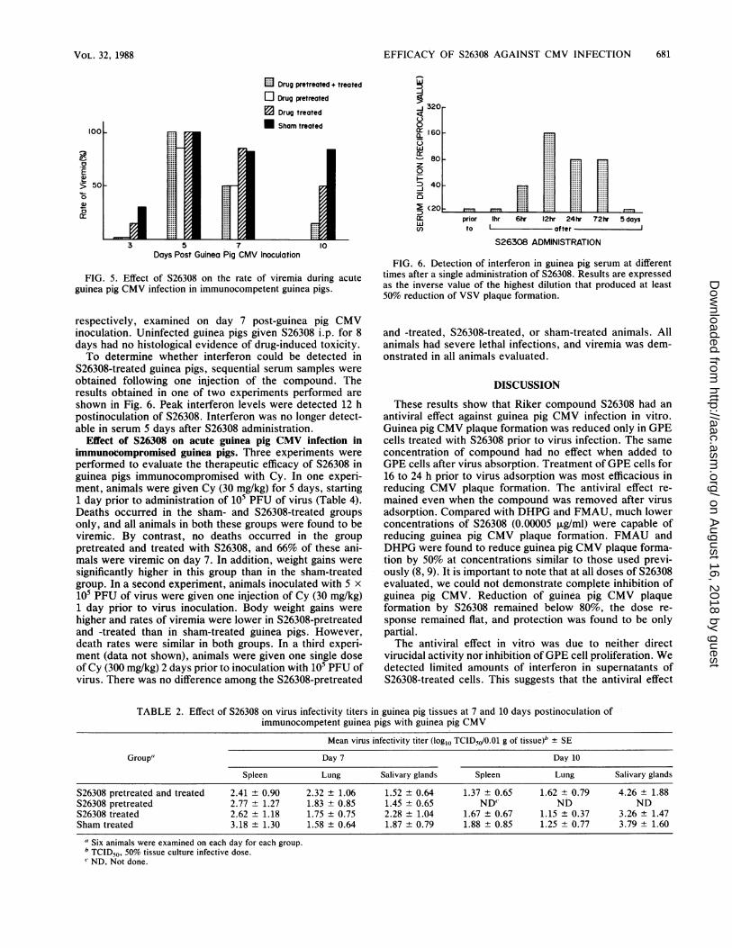
panels (Costar). Cells were inoculated with approximately100 PFU of guinea pig CMV per well and incubated at 37°Cfor 1 h. The cells were then overlaid with Eagle minimalessential medium containing Earle balanced salt solution-and5% heat-inactivated newborn calf serum (MEME-5% DCS)containing 0.5% methylcellulose and appropriate concentra-tions of the chemical compounds. In each experiment, twoto four wells were evaluated for each concentration ofantiviral agent and for each set of controls. Cell cultureswere incubated at 37°C for 8 days and were fixed and stainedwith a solution of 10% Pormalin containing 1.3% crystalviolet. Plaques were enumerated with an inverted micro-scope. In sonme experiments, the cell monolayer cultureswere pretreated with various concentrations of S26308 for 8to 72 h prior to virus inoculation.
Cell toxicity assays. Cytotoxicity of S26308 was examinedin confluent GPE cell monolayers grown in plastic petridishes (60 mm diameter). Cells were incubated withMEME-5% DCS containing various concentrations ofS26308 for 3 days. Cell counts were performed in duplicateat 24-h intervals by the trypan blue exclusion method. Priorto cell counts, control and drug-treated cells were exposed tophosphate-buffered saline (PBS) containing 0.25% trypsinand 0.02% EDTA for 6 to 8 min and resuspended inMEME-5% DCS.The effect of S26308 on GPE cell growth was also as-
sessed. GPE cells were seeded into each well of 24-wellculture plates and incubated for 24 h at 37°C. Medium wasthen replaced with MEME-5% DCS supplemented withappropriate concentrations of S26308. At 24-h intervals for 4days, the number of live cells was determined by the trypanblue exclusion method.
Interferon assay. Serum or cell culture supernatant sam-ples were tested for the presence of interferon as describedbefore (23, 24) with GPE cell monolayers grown in six-wellpanels and challenged with 50 PFU of VSV. The interferontiter was expressed as the inverse of the highest dilution ofserum or supernatant that reduced VSV plaque formation by50% or more. Supernatants from S26308-treated cultureswere obtained as follows. Monolayer cultures of GPE cellswere treated with S26308 for 12, 16, or 24 h. The cells werethen rinsed three times to remove the compound, freshmedium was added, and the supernatant was collected 4 hlater. Serial twofold dilutions of each supernatant samplewere assayed in duplicate. In each assay, supernatants ofGPE cell cultures not treated with S26308 were included asa negative control. Human leukocyte interferon (20 IU) wasalso included as a positive control; this concentration re-sulted in 95% or more reduction of VSV plaque formation.Animal inoculation and evaluation. Hartley female guinea
pigs (250 to 300 g) were purchased from Camm ResearchInstitute (Wayne, N.J.). Prior to virus inoculation, serum
samples were collected by cardiac puncture and evaluatedfor the presence of CMV-neutralizing antibodies as de-scribed before (11). Animals without preexisting CMV anti-bodies were used in this study. The guinea pigs were
inoculated subcutaneously in the right axilla with 1 ml ofsalivary-gland-passaged guinea pig CMV containing 105 to106 PFU of virus.For studies in immunocompetent guinea pigs, four exper-
imental groups were evaluated: (i) the S26308-pretreated and-treated group was given S26308 (3 mg/kg) intraperitoneally(i.p.) daily until the day of sacrifice, starting 1 day prior tovirus inoculation; (ii) the S26308-pretreated group was givenone single dose of S26308 (3 mg/kg) i.p. 1 day prior to virusinocullation; (iii) the S26308-treated group was injected withS26308 (3 mg/kg) i.p. daily until the day of sacrifice, starting1 day after virus inoculation; and (iv) the sham-treated groupwas given PBS i.p. daily until the day of sacrifice, starting 1day after virUs inoculation. Animals were sacrificed on day 7or 10 after CMV inoculation. To evaluate the toxicity ofS26308, some uninfected animals were given S26308 (3mg/kg) i.p. daily for 8 days.For studies in immunocompromised guinea pigs, animals
were given cyclophosphamide (Cy) i.p. Depending on theexperiment, animals received Cy at 30 mg/kg 1 day aftervitus inoculation, at 30 mg/kg daily for 5 days starting 1 dayprior to virus inoculation, or at 300 mg/kg 2 days prior tovirus inoculation. In each experiment, guinea pigs receivedone of the following treatments: (i) the S26308-pretreatedand -treated group was given S26308 (3 mg/kg) i.p. daily from1 day prior to 10 days after virus inoculation; (ii) theS26308-treated group was injected with S26308 (3 mg/kg)daily from 1 to 10 days after virus inoculation; and (iii) thesham-treated group was given PBS daily until 10 days aftervirus inoculation.At selected time points, S26308- and sham-treated animals
were evaluated for frequency of viremia, hematocrit values,leukocyte counts, and body weights as described before (10,12, 15). At the time of sacrifice, spleen weights were re-corded and tissues were collected for virus isolation andhistology as described previously (12, 15). The spleen, lung,and salivary glands were removed aseptically at sacrifice,and virus titrations were carried out by cocultivation on GPEfibroblast monolayers in 24-well panels (10). Cultures wereobserved for 4 weeks for characteristic CMV-induced cyto-pathic effect, and viral isolates were identified by specificneutralizing antibodies (12).
RESULTS
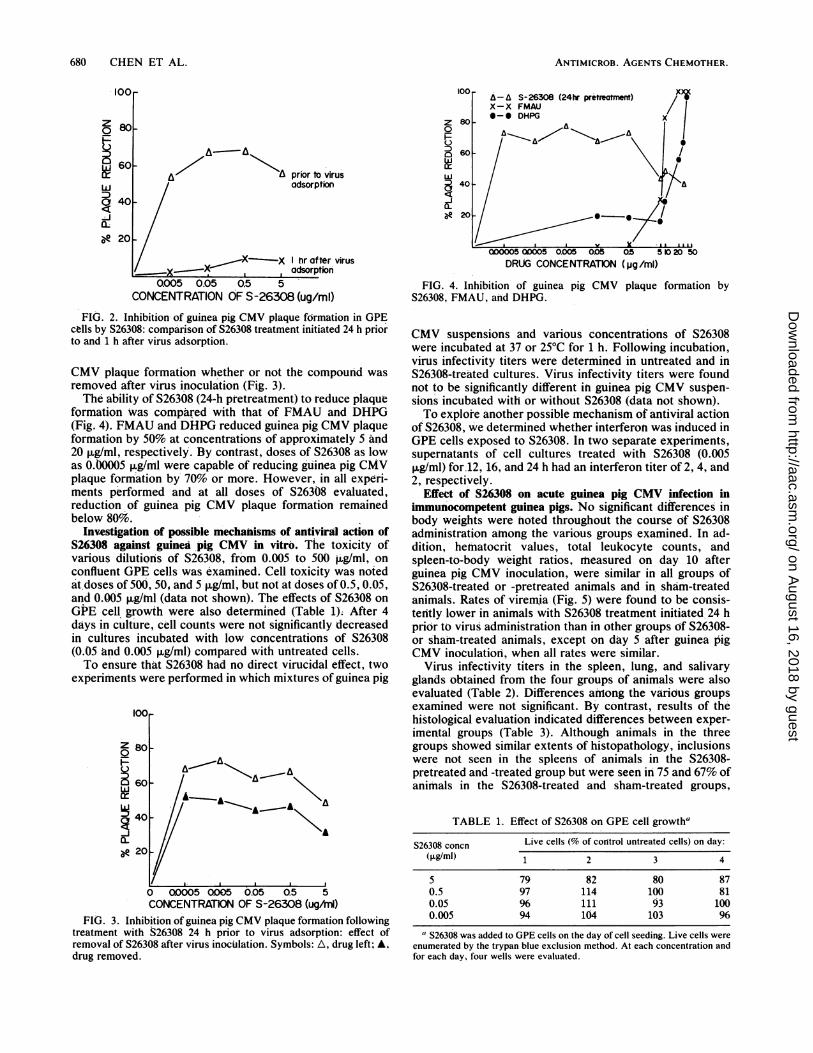
Antiviral activity of S26308 against guinea pig CMV infec-tion in vitro. In the first group of experiments, inhibition ofguinea pig CMV plaque formation was compared in GPE cellmonolayers with drug treatment initiated either 24 h beforeor 1 h after virus adsorption. S26308 reduced guinea pigCMV plaque formation only when treatment was initiated 24h prior to virus inoculation (Fig. 2). To determine whetherthe antiviral activity of S26308 could be enhanced by reduc-ing or prolonging the duration of S26308 pretreatment; weinitiated four separate experiments in which cells weretreated with S26308 for 8, 16, 20, 24, 48, and 72 h beforevirus inoculation. Optimal antiviral activity was obtainedwhen cells were treated with S26308 for 16 or 24 h beforevirus inoculation (data not shown).
Since in the experiments described above, S26308 was leftin contact with the cells for several days after virus inocu-lation, we next determined whether the antiviral activity ofS26308 remained present if the compound was removed afterthe pretreatment period. Pretreatment of GPE cells for 24 hprior to virus inoculation resulted in reduction of guinea pig
VOL. 32, 1988
on August 16, 2018 by guest
http://aac.asm.org/
Dow
nloaded from
ANTIMICROB. AGENTS CHEMOTHER.
0 80-
i 60 / A^ priorto virus
DJ /adsorption
40/
--jo 20 -
/XX-X X hr after virusadsorption
0.005 0.05 0.5 5CONCENTRATION OF S-26308 (ug/ml)
FIG. 2. Inhibition of guinea pig CMV plaque formation in GPEctlls by S26308: comparison of S26308 treatment initiated 24 h priorto and 1 h after virus adsorption.
CMV plaque formation whether or not the compound wasremoved after virus inoculation (Fig. 3).The ability of S26308 (24-h pretreatment) to reduce plaque
fQrmation was compared with that of FMAU and DHPG(Fig. 4). FMAU and DHPG reduced guinea pig CMV plaqueformation by 50% at concentrations of approximately 5 and20 ,ug/ml, respectively. By contrast, doses of S26308 as lowas O.1O005 ,ug/ml were capable of reducing guinea pig CMVplaque formation by 70% or more. However, in all experi-ments performed and at all doses of S26308 evaluated,reduction of guinea pig CMV plaque formation remainedbelow 80%.
Investigation of possible mechanisms of antiviral action ofS26308 against guinea pig CMV in vitro. The toxicity ofvarious dilutions of S26308, from 0.005 to 500 - g/ml, onconfluent GPE cells was examined. Cell toxicity was notedat.doses of 500, 50, and 5 pug/ml, but not at doses of 0.5, 0.05,and 0.005 ug/tnl (data not shown). The effects of S26308 onGPE cell growth were also determined (Table 1); After 4days in culture, cell counts were not significantly decreasedin cultures incubated with low concentrations of S26308(0.05 and 0.005 jig/ml) conmpared with untreated cells.To ensure that S26308 had no direct virucidal effect, two
experiments were performed in which mixtures of guinea pig
100
Z 800
60 X
A
8 20
0 00005 0005 0.05 0.5 5CONCENTRATION OF S-26308 (ug/mln)
FIG. 3. Inhibition of guinea pig CMV plaque formation followingtreatment with S26308 24 h prior to virus adsorption: effect ofremoval of S26308 after virus inoculation. Symbols: A, drug left; A,drug removed.
0
40-
FIG. 05Do050.005 ooS OS 510 50
DRUG CONCENTRATION (pg/mi)
FIG. 4. Inhibition of guinea pig CMV plaque formation byS26308, FMAU, and DHPG.
CMV suspensions and various concentrations of S26308were incubated at 37 or 25°C for 1 h. Following incubation,virus infectivity titers were determined in untreated and inS26308-treated cultures. Virus infectivity titers were foundnot to be significantly different in guinea pig CMV suspen-sions incubated with or without S26308 (data not shown).To explore another possible mechanism of antiviral action
of S26308, we determined whether interferon was induced inGPE cells exposed to S26308. In two separate experiments,supernatants of cell cultures treated with S26308 (0.005,ug/ml) for.12, 16, and 24 h had an interferon titer of 2, 4, and2, respectively.
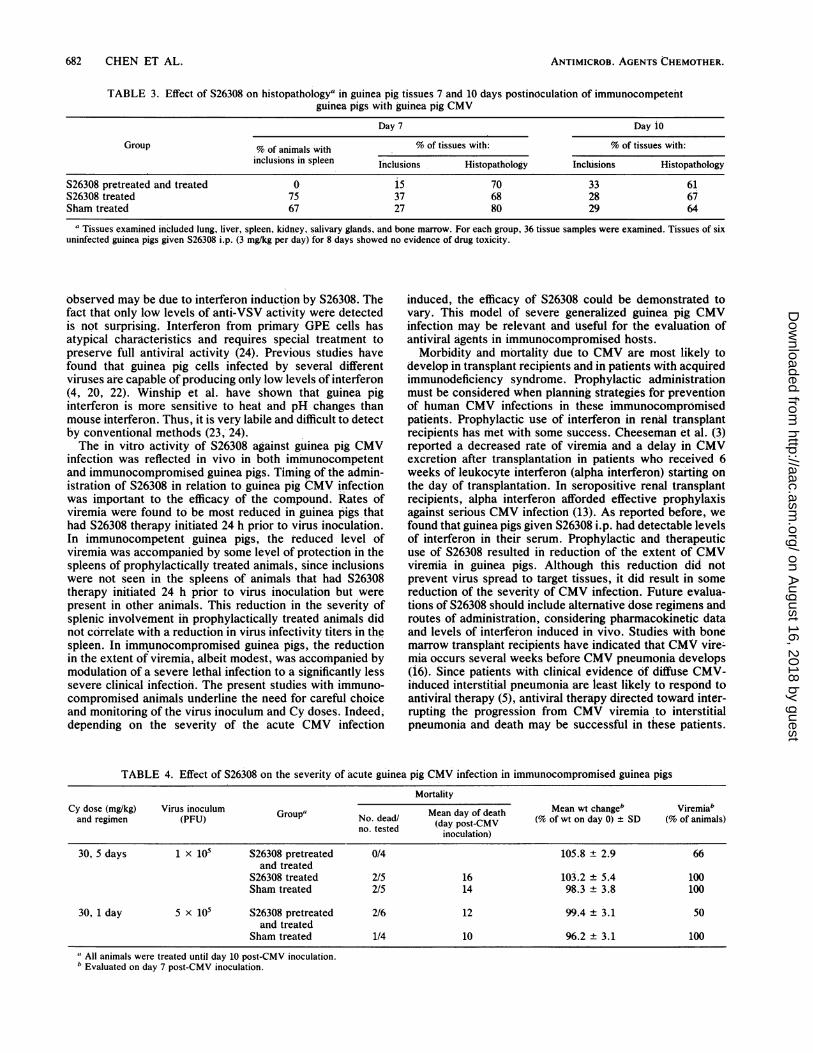
Effect of S26308 on acute guinea pig CMV infection inimmunocompetent guinea pigs. No significant differences inbody weights were hoted throughout the course of S26308administration atnong the various groups examined. In ad-dition, hematocrit values, total leukocyte counts, andspleen-to-body weight ratios, measured on day 10 afterguinea pig CMV inoculation, were similar in all groups ofS26308-treated or -pretreated animals and in sham-treatedanimals. Rates of viremia (Fig. 5) were found to be consis-tently lower in animals with S26308 treatment initiated 24 hprior to virus administration than in other groups of S26308-or sham-treated animals, except on day 5 after guinea pigCMV inoculation, when all rates were similar.
Virus infectivity titers in the spleen, lung, and salivaryglands obtained from the four groups of animals were alsoevaluated (Table 2). Differences among the various groupsexamined were not significant. By contrast, results of thehistological evaluation indicated differences between exper-imental groups (Table 3). Although animals in the threegroups showed similar extents of histopathology, inclusionswere not seen in the spleens of animals in the S26308-pretreated and -treated group but were seen in 75 and 67% ofanimals in the S26308-treated and sham-treated groups,
TABLE 1. Effect of S26308 on GPE cell growtha
S26308 concn Live cells (% of control untreated cells) on day:(pLg/ml) 1 2 3 4
5 79 82 80 870.5 97 114 100 810.05 % 111 93 1000.005 94 104 103 96
"S26308 was added to GPE cells on the day of cell seeding. Live cells wereenumerated by the trypan blue exclusion method. At each concentration andfor each day, four wells were evaluated.
680 CHEN ET AL.
on August 16, 2018 by guest
http://aac.asm.org/
Dow
nloaded from
EFFICACY OF S26308 AGAINST CMV INFECTION 681
E Drug pretreated + treated
Drug pretreated
Drug treated
Sham treated100 L
0..
3 5 7 10
Days Post Guinea Pig CMV Inoculation
FIG. 5. Effect of S26308 on the rate of viremia during acuteguinea pig CMV infection in immunocompetent guinea pigs.
respectively, examined on day 7 post-guinea pig CMVinoculation. Uninfected guinea pigs given S26308 i.p. for 8days had no histological evidence of drug-induced toxicity.To determine whether interferon could be detected in
S26308-treated guinea pigs, sequential serum samples were
obtained following one injection of the compound. Theresults obtained in one of two experiments performed areshown in Fig. 6. Peak interferon levels were detected 12 hpostinoculation of S26308. Interferon was no longer detect-able in serum 5 days after S26308 administration.
Effect of S26308 on acute guinea pig CMV infection inimmunocompromised guinea pigs. Three experiments wereperformed to evaluate the therapeutic efficacy of S26308 inguinea pigs immunocompromised with Cy. In one experi-ment, animals were given Cy (30 mg/kg) for 5 days, starting1 day prior to administration of 105 PFU of virus (Table 4).Deaths occurred in the sham- and S26308-treated groupsonly, and all animals in both these groups were found to beviremic. By contrast, no deaths occurred in the grouppretreated and treated with S26308, and 66% of these ani-mals were viremic on day 7. In addition, weight gains weresignificantly higher in this group than in the sham-treatedgroup. In a second experiment, animals inoculated with 5 x
105 PFU of virus were given one injection of Cy (30 mg/kg)1 day prior to virus inoculation. Body weight gains werehigher and rates of viremia were lower in S26308-pretreatedand -treated than in sham-treated guinea pigs. However,death rates were similar in both groups. In a third experi-ment (data not shown), animals were given one single doseof Cy (300 mg/kg) 2 days prior to inoculation with 105 PFU ofvirus. There was no difference among the S26308-pretreated
320
160 _
80
z
0
D 40-
~(20,O M M
prior Ihr 6hr 12hr 24hr 72hr 5 dayswIento ofater
S26308 ADMiNISTRATION
FIG. 6. Detection of interferon in guinea pig serum at differenttimes after a single administration of S26308. Results are expressedas the inverse value of the highest dilution that produced at least50% reduction of VSV plaque formation.
and -treated, S26308-treated, or sham-treated animals. Allanimals had severe lethal infections, and viremia was dem-onstrated in all animals evaluated.
DISCUSSION
These results show that Riker compound S26308 had an
antiviral effect against guinea pig CMV infection in vitro.Guinea pig CMV plaque formation was reduced only in GPEcells treated with S26308 prior to virus infection. The same
concentration of compound had no effect when added toGPE cells after virus absorption. Treatment of GPE cells for16 to 24 h prior to virus adsorption was most efficacious inreducing CMV plaque formation. The antiviral effect re-
mained even when the compound was removed after virusadsorption. Compared with DHPG and FMAU, much lowerconcentrations of S26308 (0.00005 jig/ml) were capable ofreducing guinea pig CMV plaque formation. FMAU andDHPG were found to reduce guinea pig CMV plaque forma-tion by 50% at concentrations similar to those used previ-ously (8, 9). It is important to note that at all doses of S26308evaluated, we could not demonstrate complete inhibition ofguinea pig CMV. Reduction of guinea pig CMV plaqueformation by S26308 remained below 80%, the dose re-
sponse remained flat, and protection was found to be onlypartial.The antiviral effect in vitro was due to neither direct
virucidal activity nor inhibition of GPE cell proliferation. Wedetected limited amounts of interferon in supernatants ofS26308-treated cells. This suggests that the antiviral effect
TABLE 2. Effect of S26308 on virus infectivity titers in guinea pig tissues at 7 and 10 days postinoculation ofimmunocompetent guinea pigs with guinea pig CMV
Mean virus infectivity titer (log1o TCID50/0.01 g of tissue)b ± SE
Group" Day 7 Day 10
Spleen Lung Salivary glands Spleen Lung Salivary glands
S26308 pretreated and treated 2.41 ± 0.90 2.32 ± 1.06 1.52 ± 0.64 1.37 ± 0.65 1.62 ± 0.79 4.26 ± 1.88S26308 pretreated 2.77 ± 1.27 1.83 ± 0.85 1.45 ± 0.65 ND" ND NDS26308 treated 2.62 ± 1.18 1.75 ± 0.75 2.28 ± 1.04 1.67 ± 0.67 1.15 + 0.37 3.26 ± 1.47Sham treated 3.18 ± 1.30 1.58 ± 0.64 1.87 ± 0.79 1.88 ± 0.85 1.25 ± 0.77 3.79 ± 1.60
' Six animals were examined on each day for each group.bTCID50, 50% tissue culture infective dose.' ND, Not done.
VOL. 32, 1988
on August 16, 2018 by guest
http://aac.asm.org/
Dow
nloaded from
ANTIMICROB. AGENTS CHEMOTHER.
TABLE 3. Effect of S26308 on histopathologya in guinea pig tissues 7 and 10 days postinoculation of immunocompetehtguinea pigs with guinea pig CMV
Day 7 Day ioGroup % of animals with % of tissues with: % of tissues with:
inclusions in spleen Inclusions Histopathology Inclusions Histopathology
S26308 pretreated and treated 0 i5 70 33 61S26308 treated 75 37 68 28 67Sham treated 67 27 80 29 64
a Tissues examined included lung, liver, spleen, kidney, salivary glands, and bone marrow. For each group, 36 tissue samples were examined. Tissues of sixuninfected guinea pigs given S26308 i.p. (3 mg/kg per day) for 8 days showed no evidence of drug toxicity.
observed may be due to interferon induction by S26308. Thefact that only low levels of anti-VSV activity were detectedis not surprising. Interferon from primary GPE cells hasatypical characteristics and requires special treatment topreserve full antiviral activity (24). Previous studies havefound that guinea pig cells infected by several differentviruses are capable of producing only low levels of interferon(4, 20, 22). Winship et al. have shown that guinea piginterferon is more sensitive to heat and pH changes thanmouse interferon. Thus, it is very labile and difficult to detectby conventional methods (23, 24).The in vitro activity of S26308 against guinea pig CMV
infection was reflected in vivo in both immunocompetentand immunocompromised guinea pigs. Timing of the admin-istration of S26308 in relation to guinea pig CMV infectionwas important to the efficacy of the compound. Rates ofviremia were found to be most reduced in guinea pigs thathad S26308 therapy initiated 24 h prior to virus inoculation.In immunocompetent guinea pigs, the reduced level ofviremia was accompanied by some level of protection in thespleens of prophylactically treated animals, since inclusionswere not seen in the spleens of animals that had S26308therapy initiated 24 h prior to virus inoculation but werepresent in other animals. This reduction in the severity ofsplenic involvement in prophylactically treated animals didnot correlate with a reduction in virus infectivity titers in thespleen. In immunocompromised guinea pigs, the reductionin the extent of viremia, albeit modest, was accompanied bymodulation of a severe lethal infection to a significantly lesssevere clinical infection. The present studies with immuno-compromised animals underline the need for careful choiceand monitoring of the virus inoculum and Cy doses. Indeed,depending on the severity of the acute CMV infection
induced, the efficacy of S26308 could be demonstrated tovary. This model of severe generalized guinea pig CMVinfection may be relevant and useful for the evaluation ofantiviral agents in immunocompromised hosts.
Morbidity and mortality due to CMV are most likely todevelop in transplant recipients and in patients with acquiredimmunodeficiency syndrome. Prophylactic administrationmust be considered when planning strategies for preventionof human CMV infections in these immunocompromisedpatients. Prophylactic use of interferon in renal transplantrecipients has met with some success. Cheeseman et al. (3)
reported a decreased rate of viremia and a delay in CMVexcretion after transplantation in patients who received 6weeks of leukocyte interferon (alpha interferon) stafting onthe day of transplantation. In seropositive renal transplantrecipients, alpha interferon afforded effective prophylaxisagainst serious CMV infection (13). As reported before, wefound that guinea pigs given S26308 i.p. had detectable levelsof interferon in their serum. Prophylactic and therapeuticuse of S26308 resulted in reduction of the extent of CMVviremia in guinea pigs. Although this reduction did notprevent virus spread to target tissues, it did result in somereduction of the severity of CMV infection. Future evalua-tions of S26308 should include alternative dose regimens androutes of administration, considering pharmacokinetic dataand levels of interferon induced in vivo. Studies with bonemarrow transplant recipients have indicated that CMV vire-mia occurs several weeks before CMV pneumonia develops(i6). Since patients with clinical evidence of diffuse CMV-induced interstitial pneumonia are least likely to respond toantiviral therapy (5), antiviral therapy directed toward inter-rupting the progression from CMV viremia to interstitialpneumonia and death may be successful in these patients.
TABLE 4. Effect of S26308 on the severity of acute guinea pig CMV infection in immunocompromised guinea pigsMortality
Cy dose (mg/kg) Virus inoculum Gr" N ded/ Mean day of death Mean wt changeb Viremiaband regimen (PFU) (roupno. ded (day post-CMV (% of wt on day 0) + SD (% of animals)
inoculation)30, 5 days 1 x 10⁵ S26308 pretreated 0/4 105.8 + 2.9 66
and treatedS26308 treated 2/5 16 103.2 ± 5.4 100Sham treated 2/5 14 98.3 ± 3.8 100
30, 1 day 5 x 10⁵ S26308 pretreated 2/6 12 99.4 ± 3.1 50and treated
Sham treated 1/4 10 96.2 ± 3.1 100a All animals were treated until day 10 post-CMV inoculation.b Evaluated on day 7 post-CMV inoculation.
682 CHEN ET AL.
on August 16, 2018 by guest
http://aac.asm.org/
Dow
nloaded from
EFFICACY OF S26308 AGAINST CMV INFECTION 683
The efficacy of S26308 as a prophylactic compound that canreduce the extent of CMV viremia and prevent severe CMVdisease might be improved if S26308 is used in combinationwith other therapeutic antiviral agents.
ACKNOWLEDGMENTS
This work was supported by Public Health Service contractA162519 from the National Institutes of Health.We thank Jacquelyn T. Lavallee for her excellent assistance.
LITERATURE CITED1. Bia, F. J., B. P. Griffith, C. K. Y. Fong, and G. D. Hsiung. 1983.
Cytomegaloviral infections in the guinea pig: experimental mod-els for human disease. Rev. Infect. Dis. 5:177-195.
2. Biron, K. K., J. A. Fyfe, S. C. Stanat, L. K. Leslie, J. B. Sorrell,C. U. Lambe, and D. M. Coen. 1986. A human cytomegalovirusmutant resistant to the nucleoside analog 9-([2-hydroxyl-1-(hydroxymethyl)ethoxy]methyl)guanine (BW B759U) inducesreduced levels of BW B759U triphosphate. Proc. Natl. Acad.Sci. USA 83:8769-8773.
3. Cheeseman, S. H., R. H. Rubin, J. A. Stewart, N. E. Tolkoff-Rubin, A. B. Cosimi, K. Cantell, J. Gilbert, S. Winkle, J. T.Herrin, P. H. Black, P. S. Russell, and M. S. Hirsch. 1979.Controlled clinical trial of prophylactic human-leukocyte inter-feron in renal transplantation: effects on cytomegalovirus andherpes simplex virus infection. N. Engl. J. Med. 300:1345-1349.
4. Christofinis, G. J. 1980. Heterospecific antiviral activity ofhuman interferon on guinea pig endothelial cells. Dev. Biol.Stand. 46:193-196.
5. Collaborative DHPG Treatment Study Group. 1986. Treatmentof serious cytomegalovirus infection with 9-(1,3-dihydroxy-2-propoxymethyl)guanine in patients with AIDS and other immu-nodeficiencies. N. Engl. J. Med. 314:801-805.
6. D'Amico, D. J., J. H. Talamo, D. Felsenstein, M. S. Hirsch,D. M. Albert, and R. T. Schooley. 1986. Ophthalmoscopic andhistologic findings in cytomegalovirus retinitis treated withBW-B759U. Arch. Ophthalmol. 104:1788-1793.
7. Field, A. K., M. E. Davies, C. DeWitt, R. Liou, J. Germershau-sen, J. D. Karkas, W. T. Ashton, D. B. R. Johnston, and R. L.Tolman. 1983. 9-[(2-Hydroxy-1-(hydroxymethyl)ethoxy)]meth-ylguanine: a selective inhibitor of herpes group virus replica-tion. Proc. Natl. Acad. Sci. USA 80:4139-4143.
8. Fong, C. K. Y., S. Cohen, S. McCormick, and G. D. Hsiung.1984. Effect of 2'-fluoroarabinosides (FIAC, FIAU and FMAU)on CMV infection in cell cultures and in guinea pigs, p. 400-403.In S. A. Plotkin, S. Michelson, J. S. Pagana, and F. Rapp (ed.),Birth defects: original articles series. Alan R. Liss, Inc., NewYork.
9. Fong, C. K. Y., S. D. Cohen, S. McCormick, and G. D. Hsiung.1987. Antiviral effect of 9-(1,3-dihydroxy-2-propoxymethyl)gua-nine against cytomegalovirus infection in a guinea pig model.Antiviral Res. 7:11-13.
10. Goff, E., B. P. Griffith, and J. Booss. 1987. Delayed amplifica-tion of cytomegalovirus infection in the placenta and maternaltissues during late gestation. Am. J. Obstet. Gynecol. 156:1265-
1270.11. Griffith, B. P., J. T. Lavallee, T. A. Jennings, and G. D. Hsiung.
1985. Transmission of maternal cytomegalovirus specific immu-nity in the guinea pig. Clin. Immunol. Immunopathol. 35:169-181.
12. Griffith, B. P., H. L. Lucia, F. J. Bia, and G. D. Hsiung. 1981.Cytomegalovirus-induced mononucleosis in guinea pigs. Infect.Immun. 32:857-863.
13. Hirsch, M. S., R. T. Schooley, A. B. Cosimi, P. S. Russell, F. L.Delmonico, N. E. Tolkoff-Rubin, J. T. Herrin, K. Cantell, M. L.Farrell, T. R. Rota, and R. H. Rubin. 1983. Effects of interferon-alpha on cytomegalovirus reactivation syndromes in renal trans-plant recipients. N. Engl. J. Med. 308:1489-1493.
14. Holland, G. N., M. J. Sakamoto, D. Hardy, Y. Sidikaro, A. E.Krieger, and L. M. Frenkel. 1986. Treatment of cytomegalovirusretinopathy in patients with acquired immunodeficiency syn-drome. Use of the experimental drug 9-[2-hydroxy-1-(hydroxy-methyl)ethoxymethyl]quanine. Arch. Ophthalmol. 104:1794-1800.
15. Lucia, H. L., B. P. Griffith, and G. D. Hsiung. 1984. Effect ofacyclovir and phosphonoformate on cytomegalovirus infectionin guinea pigs. Intervirology 21:141-149.
16. Masur, H., H. C. Lane, A. Palestine, P. D. Smith, J. Manische-witz, G. Stevens, L. Fujikawa, A. M. Macher, R. Nussenblatt, B.Baird, M. Megill, A. Wittek, G. V. Quinnan, J. E. Parrillo, A. H.Rook, L. J. Eron, D. M. Poretz, R. I. Goldenberg, A. S. Fauci,and E. P. Gelmann. 1986. Effect of 9-(1,3-dihydroxy-2-propoxy-methyl)guanine on serious cytomegalovirus disease in eightimmunosuppressed homosexual men. Ann. Intern. Med. 104:41-44.
17. O'Donnell, J. J., M. A. Jacobson, and J. Mills. 1987. Develop-ment of cytomegalovirus (CMV) retinitis in a patient with AIDSduring ganciclovir therapy for CMV colitis. N. Engl. J. Med.316:1607-1608.
18. Plotkin, S. A., L. W. Drew, D. Felsenstein, and M. S. Hirsch.1985. Sensitivity of clinical isolates of human cytomegalovirusto 9-(1,3-dihydroxy-2-propoxymethyl)guanine. J. Infect. Dis.152:833-834.
19. Rosencan, L. R., C. M. Stahl-Bayliss, C. M., Kalman, and 0. L.Laskin. 1986. Antiviral therapy for cytomegalovirus retinitis inAIDS with dihydroxy propoxymethyl guanine. Am. J. Ophthal-mol. 101:405-418.
20. Sonnenfeld, G. 1981. Induction and production of guinea piginterferon. Methods Enzymol. 78:162-165.
21. Spector, S. A., J. A. Rua, D. H. Spector, and R. McMillan. 1984.Detection of human cytomegalovirus in clinical specimens byDNA-DNA hybridization. J. Infect. DIs. 150:121-126.
22. Warfel, A. H., and W. E. Stewart II. 1980. Production andcharacterization of guinea pig interferon. J. Interferon Res. 1:19-22.
23. Winship, T. R., C. K. Y. Fong, and G. D. Hsiung. 1983.Improved conditions for the production and detection of inter-feron from guinea pig embryo cells. J. Interferon Res. 3:71-74.
24. Winship, T. R., C. K. Y. Fong, and G. D. Hsiung. 1984.Distinctive characteristics of crude interferon from virus-in-fected guinea pig embryo fibroblasts. J. Gen. Virol. 65:843-847.
VOL. 32, 1988
on August 16, 2018 by guest
http://aac.asm.org/
Dow
nloaded from