Embed Size (px)
Citation preview

©FUNPEC-RP www.funpecrp.com.brGenetics and Molecular Research 13 (3): 5036-5047 (2014)
Efficient method of protein extraction from Theobroma cacao L. roots for two-dimensional gel electrophoresis and mass spectrometry analyses
F.Z. Bertolde1,2, A.-A.F. Almeida1, F.A.C. Silva1, T.M. Oliveira1 and C.P. Pirovani3
1Departamento de Ciências Biológicas, Universidade Estadual de Santa Cruz, Ilhéus, BA, Brasil2Instituto Federal de Educação Ciência e Tecnologia da Bahia, Campus Eunápolis, Eunápolis, BA, Brasil3Laboratório de Proteômica, Centro de Biotecnologia e Genética, Universidade Estadual de Santa Cruz, Ilhéus, BA, Brasil
Corresponding author: F.Z. BertoldeE-mail: [email protected] / [email protected]
Genet. Mol. Res. 13 (3): 5036-5047 (2014)Received July 1, 2013Accepted December 5, 2013Published July 4, 2014DOI http://dx.doi.org/10.4238/2014.July.4.19
ABSTRACT. Theobroma cacao is a woody and recalcitrant plant with a very high level of interfering compounds. Standard protocols for protein extraction were proposed for various types of samples, but the presence of interfering compounds in many samples prevented the isolation of proteins suitable for two-dimensional gel electrophoresis (2-DE). An efficient method to extract root proteins for 2-DE was established to overcome these problems. The main features of this protocol are: i) precipitation with trichloroacetic acid/acetone overnight to prepare the acetone dry powder (ADP), ii) several additional steps of sonication in the ADP preparation and extractions with dense sodium dodecyl sulfate and phenol, and iii) adding two stages of phenol extractions.

5037
©FUNPEC-RP www.funpecrp.com.brGenetics and Molecular Research 13 (3): 5036-5047 (2014)
Efficient method of protein extraction from cocoa roots
Proteins were extracted from roots using this new protocol (Method B) and a protocol described in the literature for T. cacao leaves and meristems (Method A). Using these methods, we obtained a protein yield of about 0.7 and 2.5 mg per 1.0 g lyophilized root, and a total of 60 and 400 spots could be separated, respectively. Through Method B, it was possible to isolate high-quality protein and a high yield of roots from T. cacao for high-quality 2-DE gels. To demonstrate the quality of the extracted proteins from roots of T. cacao using Method B, several protein spots were cut from the 2-DE gels, analyzed by tandem mass spectrometry, and identified. Method B was further tested on Citrus roots, with a protein yield of about 2.7 mg per 1.0 g lyophilized root and 800 detected spots.
Key words: Cocoa; Plant protein extraction; Root; Two-dimensional gel electrophoresis
INTRODUCTION
Theobroma cacao L. (Malvaceae) is a preferentially allogamous perennial woody spe-cies with geographical origin in South America and several series in the Amazon and Guyana regions (Almeida and Valle, 2007). The species is cultivated in the American, African, and Asian continents, and many countries worldwide are involved in cocoa production, market-ing, and consumption (Almeida and Valle, 2007, 2009). The cacao tree is commercially ex-plored for seed production because seeds are fermented, dried, and ground to produce liquor and fat. These two products are mixed with sugar, milk, and other ingredients to produce the most popular derivative of cacao: chocolate. Cacao is an important commodity: more than 20 million people depend directly on cocoa for their livelihood, and approximately 90% of the production is exported in the form of beans or semi-manufactured cocoa products to Europe and the USA (Food and Agriculture Organization, http://www.fao.org).
Biotic and abiotic stresses are a major problem for cocoa production, causing heavy crop losses. Diseases caused by pathogens, such Phytophthora spp and Moniliophthora spp, destroy cacao tree plantations and cause great economic losses and social changes in affected areas (Purdy and Schmidt, 1996; Andebrhan et al., 1999). Furthermore, water stress, flooding of the soil, and excessive solar radiation can affect T. cacao in the different areas that have been cultivated; these events can also cause plant death and reduced production (Sena Gomes and Kozlowski, 1986; Almeida and Valle, 2007). The economic losses and social impacts of different stresses that affect cocoa pushed forward the advancement of genomic studies to understand the genomic program of cacao during infection by pathogens (Jones et al., 2002; Verica et al., 2004; Gesteira et al., 2007; Leal et al., 2007; Argout et al., 2008, 2011), provid-ing information in databases that can be used to identify proteins by mass spectrometry (MS). However, cacao has received little attention with respect to proteomic research. Until now, few studies have been carried out to analyze the protein pattern of cacao during its response to different stresses (Pirovani et al., 2008, 2010; Rehem et al., 2011). In particular, proteomics offers the possibility of identifying post-translational modifications of proteins, which are largely not predictable from genome sequences. Proteomics can also test whether proteins are

5038
©FUNPEC-RP www.funpecrp.com.brGenetics and Molecular Research 13 (3): 5036-5047 (2014)
F.Z. Bertolde et al.
translated and accumulated after the expression of their encoding genes (Gallardo et al., 2001; Hoa le et al., 2004).
Sample preparation is one of the most crucial steps in obtaining high-quality resolu-tion of proteins in proteomic analysis, yet it can be problematic (Görg et al., 2004). Proteins isolated from plant tissues are often difficult to resolve by two-dimensional gel electrophoresis (2-DE) because of the abundance of secondary metabolites. In particular, plant tissues such as roots contain lower concentrations of proteins and often contain high levels of materials that strongly interfere with 2-DE, resulting in horizontal and vertical streaking, smearing, and reduced numbers of distinctly resolved protein spots (Wang et al., 2003; Saravanan and Rose, 2004). These interfering compounds, which are present in plant materials such as T. cacao, disturb protein separation and proteome analysis by 2-DE (Görg et al., 2004; Saravanan and Rose, 2004). To our knowledge, there are no reports on successful protein extraction from the roots of T. cacao for 2-DE.
To overcome these problems, we developed a new protocol for T. cacao root protein extraction that is based on a protocol for protein extraction from T. cacao leaves and meri-stems (Pirovani et al., 2008). This protocol attempts to minimize the presence of interfering compounds and has a higher yield of obtained protein. In our study, we also compared the protocol that we developed with the protocol of protein extraction from T. cacao leaves and meristems, and we evaluated their suitability for MS analysis. Our protocol was tested further on Citrus roots, which are known for their difficult protein extraction.
MATERIAL AND METHODS
Plant materials
The roots were obtained from plants of T. cacao and Citrus. The T. cacao plants were grown in a greenhouse at the Universidade Estadual de Santa Cruz (UESC), Ilhéus, BA, Brazil (14° 47' S, 39° 16' W, 55 m above sea level). The plants were grown in plastic pots with a 25-L capacity that were filled with organic substrate (peat and shredded Pinus cortex + shredded coconut fiber in a 1:1 ratio) and enriched with mineral macronutrients and micronutrients ac-cording to the nutritional needs of the species (Souza, 2007). The Citrus plants were grown in a greenhouse at the Embrapa Mandioca e Fruticultura, Cruz das Almas, Ba, Brazil (12° 40' 39'' S, 39° 6' 23'' W, 226 m above sea level). The plants were grown in plastic pots with a 15-L capacity that were filled with the substrate Plantmax (Eucatex Agro, Brazil), washed sand, and clay in a 2:1:1 ratio and fertilized via the shoot. After being washed with water, the root samples were stored at -80°C after fixation in liquid nitrogen and subsequently lyophilized and stored at -20°C.
Protein extraction
The preparation of the acetone dry powder (ADP) from the plant total extract was based on the protocol described by Pirovani et al. (2008) (Method A) with modifications (Method B) that are described below (Figure 1). Initially, 1.0 g plant tissue (lyophilized roots) was finely pounded into a powder in liquid nitrogen using a pestle and mortar and intermixed with 7% polyvinylpolypyrrolidone (w/w). The fine powder was resuspended in 10 mL 10%

5039
©FUNPEC-RP www.funpecrp.com.brGenetics and Molecular Research 13 (3): 5036-5047 (2014)
Efficient method of protein extraction from cocoa roots
trichloroacetic acid (TCA) in acetone (cold) and 0.07% 2-mercaptoethanol and sonicated on ice (3 pulses of 5 s each, 70% output, 10-s intervals) on an Ultrasonic processor (Gex 130, 130 W, USA). This process was repeated four times at intervals of 10 min. The mixture was incubated overnight at -20°C for complete precipitation of proteins. The mixture was then centrifuged at 10,000 g and 4°C for 10 min. The supernatant was discarded, and the pellet was washed three times with the same volume of cold acetone and 0.07% 2-mercaptoethanol. The pellet was completely resuspended by sonication on ice (3 pulses of 5 s each, 70% output, 10-s intervals) and centrifuged at 10,000 g for 10 min at 4°C. The final pellet was dried at room temperature and used for protein extraction or stored at -20°C for future use.
Protein extraction from ADP was based on the protocol described by Pirovani et al. (2008) (Method A) with modifications (Method B). The ADP was resuspended in 10 mL dense sodium dodecyl sulfate (SDS) extracting buffer (30% sucrose, 2% SDS, 0.1 M Tris-HCl, pH
Figure 1. Scheme showing the Methods A and B of protein extraction of the roots.

5040
©FUNPEC-RP www.funpecrp.com.brGenetics and Molecular Research 13 (3): 5036-5047 (2014)
F.Z. Bertolde et al.
8.0, 5% 2-mercaptoethanol). Samples were sonicated on ice (4 pulses of 10 s each, 70% out-put, 10-s intervals), and then 5 mL saturated phenol, pH 8.0, was added. The sample was then centrifuged at 10,000 g and 4°C for 15 min. The supernatant was then transferred to a new tube and stored on ice. The proteins from the lower phase were extracted two times with one vol-ume of dense SDS buffer and phenol. Phenol phases from the three extractions were grouped and incubated overnight at -20°C with five volumes of 0.1 M ammonium acetate in methanol. The pellet was generated through centrifugation at 10,000 g and 4°C for 20 min. Proteins were washed twice with 1 M ammonium acetate in methanol, twice with acetone, and once with 80% ethanol, and then the pellets were finally dried at room temperature. In each wash, the sample was centrifuged at 10,000 g and 4°C for 5 min.
The pellets were treated with the 2-D Clean-Up kit according to manufacturer rec-ommendations (GE Healthcare, UK) and resuspended in the appropriate rehydration buffer. Proteins were quantified using the 2-D Quant Kit according to manufacturer instructions (GE Healthcare).
2-DE
For 2-DE, the first dimension of separation was performed on an Ettan IPGphor sys-tem (GE Healthcare). Protein samples were applied in 250 µL 2-DE rehydration solution by reswelling 13 cm Immobiline DryStrip (pH 4-7, GE Healthcare) for 12 h. Afterwards, focus-ing was performed on the same apparatus under the following conditions: step and hold at 500 V for 1 h, gradient 1000 V for 1 h, gradient 8000 V for 2.5 h, and step and hold 8000 V for 55 min. After isoelectric focusing, the strips were stored at -80°C until the second-dimension analysis. Before the SDS-polyacrylamide gel electrophoresis (PAGE), the strips were incu-bated for 15 min in equilibration buffer (6 M urea, 7.5 mM Tris-HCl, pH 8.8, 29.3% glycerol, 2% SDS, 0.002% bromophenol blue) with 1% dithiothreitol (w/v) and for another 15 min in equilibration buffer with 2.5% iodoacetamide (w/v). The strips were transferred to verti-cal 12.5% SDS-PAGE gels. The second dimension (SDS-PAGE) was performed on a Ruby SE600 system (GE Healthcare) under the following conditions: 15 mA for 45 min, 40 mA for 30 min, and 50 mA per gel for 3 h for every strip at a constant temperature of 11°C. The High-Range Rainbow Molecular Weight Marker was used (GE Healthcare). All 2-D gel sepa-rations were repeated three times using Methods A and B. After electrophoresis, proteins were visualized with 0.08% colloidal Coomassie G-250 (w/v) (Neuhoff et al., 1988). Gels were scanned using ImageScanner II (Amersham, GE Healthcare) and analyzed using ImageMaster 2D Platinum (GE Healthcare).
Protein identification
Selected protein spots were excised from the 2-DE gel, equilibrated with 50% aceto-nitrile containing 25 mM ammonium bicarbonate to remove Coomassie blue stain, and rinsed with distilled water. The gel plugs were dehydrated with 100% acetonitrile, vacuum dried, digested with 4 µL 25 ng/µL Trypsin Gold, Mass Spectrometry Grade (Promega, USA) in 25 mM ammonium bicarbonate, and incubated overnight at 37°C. The tryptic fragments were eluted from the gel with 50% acetonitrile and 5% formic acid (Yin et al., 2005). The extracts were dried under vacuum until a volume of 15 µL was reached.

5041
©FUNPEC-RP www.funpecrp.com.brGenetics and Molecular Research 13 (3): 5036-5047 (2014)
Efficient method of protein extraction from cocoa roots
The solution containing the proteolytic digests was then fractionated in an ionic ex-change column. Subsequently, the digests were fractionated on a reversed-phase C18 col-umn using two mobile phases, phase A, containing H2O and 0.1% formic acid, and phase B, containing acetonitrile and 0.1% formic acid. For peptide separation, a linear gradient of 5-95% acetonitrile was used. The eluted peptides were directly introduced to the quadrupole mass spectrometer Q-TOFmicro (Waters, Manchester, UK) by its electrospray probe. The most abundant ions that were observed in the MS spectrum were automatically selected for collision-induced dissociation using the Masslynx software, generating their MS/MS spectra. Argon gas was used for peptide collision.
The resulting spectra were processed by the algorithm MaxEnt3 of the Masslynx Pro-teinLynx software to generate a list of masses, which correspond to peaks obtained in the spectra analyzed. The list of peaks generated by Proteinlynx 2.4 was searched against the T. cacao genome and National Center for Biotechnology Information databases. For this search, we used MASCOT version 2.1.0 (Matrix Science). The identification was made by the peptide mass fingerprint and sequenced by MS/MS.
RESULTS
The quantitative comparisons of protein extracted using these two protocols and the number of spots are listed in Table 1. Method B gave significant yields that were greater than those of Method A. With Method B, 1.0 g lyophilized roots of T. cacao typically yielded approximately 2.5 mg protein, which was more than the 0.7 mg yield that was obtained by Method A.
Method Protein yield (mg/g dry weight) Spot number
A 0.65 60B 2.45 400
Table 1. Yield proteins of Theobroma cacao root (mg/g dry weight) and total number of spots using two extraction methods (A and B).
The quality and amount of protein extracted using the two methods (A and B) were monitored by one-dimensional SDS-PAGE and 2-DE. Both experiments were repeated at least three times to confirm the reproducibility. Our main modifications in Method B compared with the original (Method A) were increasing the number of extractions with phenol and sonication of the samples at the end of the process to maximize the solubilization of the extracted proteins.
Method B proved to be more efficient for the extraction of root proteins than Method A (Figure 2, columns 1 and 2). Method A did not eliminate interferents that may affect the protein quantification of the samples. This demonstrates that our new method of protein ex-traction improved the quality and the quantity of proteins extracted from T. cacao roots.
In the 2-DE analysis, protein extracts that were obtained by the two methods were tested despite the low quality of the protein extract from Method A. Lower resolution and fewer spots were observed on 2-DE gels with protein obtained by Method A than with protein obtained by Method B (Figure 3). Approximately 60 spots were observed on protein gels obtained by Method A, while 400 spots were observed from Method B as estimated by the ImageMaster 2D Platinum software (GE Healthcare). Thirteen spots were excised from the gel

5042
©FUNPEC-RP www.funpecrp.com.brGenetics and Molecular Research 13 (3): 5036-5047 (2014)
F.Z. Bertolde et al.
and identified by MS (Table 2). From these 13 spots, 14 proteins were identified and listed in Table 2 with their properties. It was possible to identify proteins from different cellular com-partments by Method B, such as mitochondrion, nucleus, plasma membrane, cytosol, apoplast, ribosome, endoplasmic reticulum lumen, vacuole, and peroxisome.
Figure 2. SDS-PAGE root proteins. Method B (column 1) and Method A (column 2) of protein extraction. Twenty micrograms of proteins was loaded on 12.5% acrylamide gel.
Figure 3. 2-DE proteins from Theobroma cacao root. A. Method A of protein extraction. B. Method B of protein extraction. Each gel was loaded with 350 mg of total protein and stained with 0.08% colloidal Coomassie G-250 (w/v).

5043
©FUNPEC-RP www.funpecrp.com.brGenetics and Molecular Research 13 (3): 5036-5047 (2014)
Efficient method of protein extraction from cocoa roots
Spot
No.
a Pr
otei
n na
me/
acce
ssio
n nu
mbe
r E-
valu
e Th
eore
tical
MW
(kD
a)/p
I B
iolo
gica
l pro
cess
Su
bcel
lula
r loc
atio
n
1
Rub
isco
subu
nit b
indi
ng-p
rote
in
0.0
63.8
/4.9
Pr
otei
n re
fold
ing
Aux
in b
iosy
nthe
tic p
roce
ss
Mito
chon
drio
n
alph
a su
buni
t/BA
E713
11.1
U
nfol
ded
prot
ein
bind
ing
Cyt
osol
ic ri
boso
me
M
embr
ane/
Apo
plas
t 2
B
isph
osph
ogly
cera
te-in
depe
nden
t 0.
0 61
.1/5
.5
Res
pons
e to
col
d R
espo
nse
to c
adm
ium
ion
Plas
ma
mem
bran
e
phos
phog
lyce
rate
mut
ase/
XP_
0025
1997
5.1
M
itoch
ondr
ial e
nvel
ope
Gly
coly
sis
Cyt
osol
/Apo
plas
t 3
M
ethy
lmal
onat
e-se
mia
ldeh
yde
1,8
0E-1
39
57.8
/8.0
R
espo
nse
to o
xida
tive
stre
ss
Mito
chon
drio
n
dehy
drog
enas
e/X
P_00
2518
343.
1
O
xida
tion
redu
ctio
n
Va
line
met
abol
ic p
roce
ss 4
AT
P sy
ntha
se b
eta
chai
n/A
CS8
3602
.1
0.0
59.7
/6.0
Pl
asm
a m
embr
ane A
TP sy
nthe
sis
Mito
chon
dria
l pro
ton-
trans
porti
ng
co
uple
d pr
oton
tran
spor
t AT
P sy
ntha
se, c
atal
ytic
cor
e 5
En
olas
e/A
BW
2168
8.1
0.0
47.9
/5.8
R
espo
nse
to sa
lt st
ress
C
ell s
urfa
ce
R
espo
nse
to li
ght s
timul
us
Phos
phop
yruv
ate
hydr
atas
e co
mpl
ex
R
espo
nse
to c
old
6
Alc
ohol
deh
ydro
gena
se a
/AB
O41
830.
1 0.
0 41
.6/6
.6
Oxi
datio
n re
duct
ion
Cyt
osol
Alc
ohol
deh
ydro
gena
se (N
AD
) act
ivity
Py
ruva
te d
ehyd
roge
nase
e1
alph
a 0.
0 43
.5/7
.2
Oxi
datio
n re
duct
ion
Nuc
leus
su
buni
t/AC
J117
43.1
G
lyco
lysi
s C
ytos
ol
R
espo
nse
to sa
lt st
ress
7
Caf
feic
aci
d o-
met
hyltr
ansf
eras
e/A
CT3
2029
.1
0.0
40.0
/5.2
Li
gnin
bio
synt
hetic
pro
cess
C
ytos
ol 8
Pr
otei
n di
sulfi
de/A
EK80
406.
1 0.
0 39
.5/5
.5
Polle
n tu
be d
evel
opm
ent
Plan
t-typ
e ce
ll w
all
Elec
tron
trans
port
chai
n Cy
topl
asm
ic m
embr
ane-
boun
ded
vesic
le
R
espo
nse
to e
ndop
lasm
ic re
ticul
um st
ress
En
dopl
asm
ic re
ticul
um lu
men
Cel
l red
ox h
omeo
stas
is
Em
bryo
sac
deve
lopm
ent
9
Late
em
bryo
gene
sis a
bund
ant
9,8
6E-1
36
30.3
/4.5
R
espo
nse
to c
adm
ium
ion
Plas
ma
mem
bran
e
grou
p 2/
XP_
0025
3334
5.1
Res
pons
e to
des
icca
tion
Embr
yoni
c de
velo
pmen
t end
ing
in
seed
dor
man
cy10
C
yste
ine
synt
hase
/XP_
0025
1225
3.1
0.0
34.3
/5.0
C
yste
ine
bios
ynth
etic
pro
cess
from
serin
e C
ytop
lasm
11
Cla
ss I
chiti
nase
/Q41
596.
1 0.
0 34
.8/5
.1
Chi
tin c
atab
olic
pro
cess
Va
cuol
e
D
efen
se re
spon
se
C
ell w
all m
acro
mol
ecul
e ca
tabo
lic p
roce
ss12
M
lp-li
ke p
rote
in 2
8/gi
|215
9259
2|A
AM
6454
1.1
4,66
E-73
17
.6/5
.2
Res
pons
e to
bio
tic st
imul
us
Cyt
osol
Def
ense
resp
onse
13
Nuc
leos
ide
diph
osph
ate
kina
se 1
/AD
B85
102.
1 1,
53E-
93
16.4
/6.4
A
uxin
bio
synt
hetic
pro
cess
Pe
roxi
som
e
R
espo
nse
to sa
lt st
ress
Va
cuol
e
R
espo
nse
to c
adm
ium
ion
Plas
ma
mem
bran
e
C
TP b
iosy
nthe
tic p
roce
ss
Apo
plas
ta S
pot n
umbe
rs c
orre
spon
d to
the
num
bers
on
the
2-D
gel
imag
es in
Fig
ure
3B.
Tabl
e 2.
Pro
tein
s ide
ntifi
ed b
y m
ass s
pect
rom
etry
.
5044
©FUNPEC-RP www.funpecrp.com.brGenetics and Molecular Research 13 (3): 5036-5047 (2014)
F.Z. Bertolde et al.
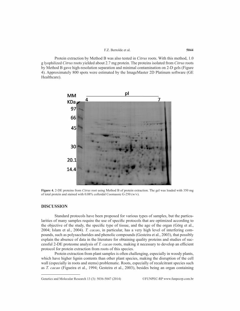
Protein extraction by Method B was also tested in Citrus roots. With this method, 1.0 g lyophilized Citrus roots yielded about 2.7 mg protein. The proteins isolated from Citrus roots by Method B gave high-resolution separation and minimal contamination on 2-D gels (Figure 4). Approximately 800 spots were estimated by the ImageMaster 2D Platinum software (GE Healthcare).
Figure 4. 2-DE proteins from Citrus root using Method B of protein extraction. The gel was loaded with 350 mg of total protein and stained with 0.08% colloidal Coomassie G-250 (w/v).
DISCUSSION
Standard protocols have been proposed for various types of samples, but the particu-larities of many samples require the use of specific protocols that are optimized according to the objective of the study, the specific type of tissue, and the age of the organ (Görg et al., 2004; Islam et al., 2004). T. cacao, in particular, has a very high level of interfering com-pounds, such as polysaccharides and phenolic compounds (Gesteira et al., 2003), that possibly explain the absence of data in the literature for obtaining quality proteins and studies of suc-cessful 2-DE proteome analysis of T. cacao roots, making it necessary to develop an efficient protocol for protein extraction from roots of this species.
Protein extraction from plant samples is often challenging, especially in woody plants, which have higher lignin contents than other plant species, making the disruption of the cell wall (especially in roots and stems) problematic. Roots, especially of recalcitrant species such as T. cacao (Figueira et al., 1994; Gesteira et al., 2003), besides being an organ containing

5045
©FUNPEC-RP www.funpecrp.com.brGenetics and Molecular Research 13 (3): 5036-5047 (2014)
Efficient method of protein extraction from cocoa roots
tissues that are difficult of macerate, have low protein content (Isaacson et al., 2006; Xie et al., 2007). Roots have many non-protein contaminants that affect 2-DE, including polysaccha-rides, polyphenols, nucleic acids, terpenes, and organic acids; these contaminants accumulate mainly in the vacuole in various soluble forms (Tsugita and Kamo, 1999; Pan, 2000). The contaminants can be coextracted with protein and affect protein migration in 2-DE, resulting in streaking (Görg et al., 2000).
According to Vâlcu and Schlink (2006), the grinding of the roots in liquid nitrogen, fol-lowed by precipitation by TCA/acetone and sonication, is one of the most effective approaches for plant samples that are rich in lignin. In this study, we found that compared to leaf samples (Pirovani et al., 2008), preparing the ADP of the T. cacao roots required precipitation with TCA/acetone overnight with several stages of sonication to obtain a high yield of protein (Table 1).
The extraction of proteins from ADP was performed using a mixture of phenol and dense SDS buffer (Wang et al., 2003; Pirovani et al., 2008). SDS is a good solubilizing agent, and phenol minimizes the protein degradation that often occurs during sample preparation because of the action of proteolytic enzymes that are found in the sample itself (Schuster and Davies, 1983); phenol has been reported to remove interfering compounds, such as polyphe-nols, efficiently prior to electrophoresis (Wang et al., 2003; Saravanan and Rose, 2004). The difference between the two methods that were compared in this study was that two extrac-tions with phenol were added in Method B. Additional extractions with phenol, which were associated with changes that were made in the preparation of ADP, allowed the acquisition of proteins that were free of degradation and high resolution (Figures 2 and 4), indicating the ef-ficiency of Method B in obtaining high-quality protein in a high quantity.
When separated on 2-DE gels, the protein extract that was obtained by Method B showed high resolution and clear separation of more spots than were described in the literature for other methodologies. Caetano et al. (2007), using the methodology of De Mot and Vanderleyden (1989) for extraction of proteins from roots of Vigna unguiculata, obtained 2-DE gels with approximately 250 spots at pI 4-7, while Bona et al. (2007), using the method Bestel-Corre et al. (2002) for the extraction of proteins from roots of Cannabis sativa, obtained 300 spots on 2-DE gels at pI 4-7.
The clear separation of a large number of protein spots with minimal streaking on 2-DE gels and the successful identification of protein spots by mass-spectrum comparisons to protein mass databases for T. cacao demonstrates that extraction Method B was suitable to obtain the quantity and quality of proteins that are required for proteomic studies of roots.
The efficiency of Method B for protein extraction from recalcitrant roots of other species was demonstrated by its use in Citrus. Citrus, similar to T. cacao, is a genus with recalcitrant species (Moore et al., 1992); its roots are a difficult material to extract proteins because they have low protein content and a large amount of interfering contaminants. To our knowledge, no reports of protein extraction from roots of Citrus have been successful for the 2-DE method. The proteins isolated by Method B showed high yield and quality and 2-DE gels with well-resolved spots that were free of stains and streaks.
In conclusion, through Method B, we succeeded in isolating a high yield of high-qual-ity proteins from T. cacao roots. The 2-DE gels that were obtained were of high quality, free of smearing and streaking, and could be used for protein identification by MS. It is expected that our protocol could also be applied for other recalcitrant plant roots, despite plants varying considerably in the amounts and types of interfering compounds they produce.

5046
©FUNPEC-RP www.funpecrp.com.brGenetics and Molecular Research 13 (3): 5036-5047 (2014)
F.Z. Bertolde et al.
ACKNOWLEDGMENTS
Research supported by Financiadora de Estudos e Projetos (FINEP), Conselho Nacio-nal de Desenvolvimento Científico e Tecnológico (CNPq), Coordenação de Aperfeiçoamento de Pessoal de Nível Superior (CAPES), and Fundação de Amparo à Pesquisa do Estado da Bahia (FAPESB). We thank Dr. Fabienne Michele (CIRAD, France) for critical reading of the manuscript.
REFERENCES
Almeida A-AF and Valle RR (2007). Ecophysiology of the cacao tree. Braz. J. Plant Physiol. 19: 425-448.Almeida A-AF and Valle RR (2009). Cacao: Ecophysiology of Growth and Production. In: Ecophysiology of Tropical
Tree Crops (DaMatta F, ed.). Nova Science Publishers, Inc., Hauppauge, 37-70.Andebrhan T, Figueira A, Yamada MM, Cascardo J, et al. (1999). Molecular fingerprinting suggests two primary outbreaks
of witches’ broom disease (Crinipellis perniciosa) of Theobroma cacao in Bahia, Brazil. Eur. J. Plant Pathol. 105: 167-175.
Argout X, Fouet O, Wincker P, Gramacho K, et al. (2008). Towards the understanding of the cocoa transcriptome: Production and analysis of an exhaustive dataset of ESTs of Theobroma cacao L. generated from various tissues and under various conditions. BMC Genomics 9: 512.
Argout X, Salse J, Aury JM, Guiltinan MJ, et al. (2011). The genome of Theobroma cacao. Nat. Genet. 43: 101-108.Bestel-Corre G, Dumas-Gaudot E, Poinsot V, Dieu M, et al. (2002). Proteome analysis and identification of symbiosis-
related proteins from Medicago truncatula Gaertn. by two-dimensional electrophoresis and mass spectrometry. Electrophoresis 23: 122-137.
Bona E, Marsano F, Cavaletto M and Berta G (2007). Proteomic characterization of copper stress response in Cannabis sativa roots. Proteomics 7: 1121-1130.
Caetano P, Santana CG, Carneiro RMD, Grossi de Sá MF, et al (2007). Otimização de Protocolos para Extração de Proteínas em Raízes de Feijão-de-Corda (Vigna unguiculata) Infectadas com o Nematóide Meloidogyne incognita. Boletim de Pesquisa e Desenvolvimento, Embrapa Recursos Genéticos e Biotecnologia, Brasília.
De Mot R and Vanderleyden J (1989). Application of two-dimensional protein analysis for strain fingerprinting and mutant analysis of Azospirillum species. Can. J. Microbiol. 35: 960-967.
Figueira A, Janick J and BeMiller JN (1994). Partial characterization of cacao pod and stem gums. Carbohydr. Polymer. 24: 133-138.
Gallardo K, Job C, Groot SP, Puype M, et al. (2001). Proteomic analysis of Arabidopsis seed germination and priming. Plant Physiol. 126: 835-848.
Gesteira AS, Micheli F, Ferreira CF and Cascardo JC (2003). Isolation and purification of functional total RNA from different organs of cacao tree during its interaction with the pathogen Crinipellis perniciosa. Biotechniques 35: 494-500.
Gesteira AS, Micheli F, Carels N, Da Silva AC, et al. (2007). Comparative analysis of expressed genes from cacao meristems infected by Moniliophthora perniciosa. Ann. Bot. 100: 129-140.
Görg A, Obermaier C, Boguth G, Harder A, et al. (2000). The current state of two-dimensional electrophoresis with immobilized pH gradients. Electrophoresis 21: 1037-1053.
Görg A, Weiss W and Dunn MJ (2004). Current two-dimensional electrophoresis technology for proteomics. Proteomics 4: 3665-3685.
Hoa le TP, Nomura M, Kajiwara H, Day DA, et al. (2004). Proteomic analysis on symbiotic differentiation of mitochondria in soybean nodules. Plant Cell Physiol. 45: 300-308.
Isaacson T, Damasceno CM, Saravanan RS, He Y, et al. (2006). Sample extraction techniques for enhanced proteomic analysis of plant tissues. Nat. Protoc. 1: 769-774.
Islam N, Lonsdale M, Upadhyaya NM, Higgins TJ, et al. (2004). Protein extraction from mature rice leaves for two-dimensional gel electrophoresis and its application in proteome analysis. Proteomics 4: 1903-1908.
Jones PG, Allaway D, Gilmour DM, Harris C, et al. (2002). Gene discovery and microarray analysis of cacao (Theobroma cacao L.) varieties. Planta 216: 255-264.
Leal GA Jr, Albuquerque PS and Figueira A (2007). Genes differentially expressed in Theobroma cacao associated with resistance to witches’ broom disease caused by Crinipellis perniciosa. Mol. Plant Pathol. 8: 279-292.

5047
©FUNPEC-RP www.funpecrp.com.brGenetics and Molecular Research 13 (3): 5036-5047 (2014)
Efficient method of protein extraction from cocoa roots
Moore GA, Jacono CC, Neidigh JL, Lawrence SD, et al. (1992). Agrobacterium-mediated transformation of Citrus stem segments and regeneration of transgenic plants. Plant Cell Rep. 11: 238-242.
Neuhoff V, Arold N, Taube D and Ehrhardt W (1988). Improved staining of proteins in polyacrylamide gels including isoelectric focusing gels with clear background at nanogram sensitivity using Coomassie Brilliant Blue G-250 and R-250. Electrophoresis 9: 255-262.
Pan SL (2000). Bupleurum Species: Scientific Evaluation and Clinical Applications. CRC Press, Boca Raton.Pirovani CP, Carvalho HA, Machado RC, Gomes DS, et al. (2008). Protein extraction for proteome analysis from cacao
leaves and meristems, organs infected by Moniliophthora perniciosa, the causal agent of the witches’ broom disease. Electrophoresis 29: 2391-2401.
Pirovani CP, da Silva SA, dos Santos LS, Micheli F, et al. (2010). Theobroma cacao cystatins impair Moniliophthora perniciosa mycelial growth and are involved in postponing cell death symptoms. Planta 232: 1485-1497.
Purdy LH and Schmidt RA (1996). Status of cacao witches’ broom: biology, epidemiology, and management. Annu. Rev. Phytopathol. 34: 573-594.
Rehem BC, Almeida A-AF, Santos IC, Gomes FP, et al. (2011). Photosynthesis, chloroplast ultrastructure, chemical composition and oxidative stress in Theobroma cacao hybrids with the lethal gene Luteus-Pa mutant. Photosynthetica 49: 127-139.
Saravanan RS and Rose JK (2004). A critical evaluation of sample extraction techniques for enhanced proteomic analysis of recalcitrant plant tissues. Proteomics 4: 2522-2532.
Schuster AM and Davies E (1983). Ribonucleic acid and protein metabolism in pea epicotyls: I. The aging process. Plant Physiol. 73: 809-816.
Sena Gomes AR and Kozlowski TT (1986). The effects of flooding on water relations and growth of Theobroma cacao var. catongo seedlings. J. Hort. Sci. 61: 265-276.
Souza JOJ (2007). Substratos e Adubação para Mudas Clonais de Cacaueiro. Doctoral thesis, Universidade de São Paulo, São Paulo.
Tsugita A and Kamo M (1999). 2-D electrophoresis of plant proteins. Methods Mol. Biol. 112: 95-97.Vâlcu CM and Schlink K (2006). Efficient extraction of proteins from woody plant samples for two-dimensional
electrophoresis. Proteomics 6: 4166-4175.Verica JA, Maximova SN, Strem MD, Carlson JE, et al. (2004). Isolation of ESTs from cacao (Theobroma cacao L.)
leaves treated with inducers of the defense response. Plant Cell Rep. 23: 404-413.Wang W, Scali M, Vignani R, Spadafora A, et al. (2003). Protein extraction for two-dimensional electrophoresis from
olive leaf, a plant tissue containing high levels of interfering compounds. Electrophoresis 24: 2369-2375.Xie H, Pan S, Liu S, Ye K, et al. (2007). A novel method of protein extraction from perennial Bupleurum root for 2-DE.
Electrophoresis 28: 871-875.Yin QY, de Groot PW, Dekker HL, de Jong L, et al. (2005). Comprehensive proteomic analysis of Saccharomyces
cerevisiae cell walls: identification of proteins covalently attached via glycosylphosphatidylinositol remnants or mild alkali-sensitive linkages. J. Biol. Chem. 280: 20894-20901.