Embed Size (px)
Citation preview
Ehlers, S; Schaible, UE (2012) The granuloma in tuberculosis: Dy-namics of a host-pathogen collusion. Frontiers in Immunology, 3(JAN). ISSN 1664-3224 DOI: 10.3389/fimmu.2012.00411
Downloaded from: http://researchonline.lshtm.ac.uk/2064802/
DOI: 10.3389/fimmu.2012.00411
Usage Guidelines
Please refer to usage guidelines at http://researchonline.lshtm.ac.uk/policies.html or alterna-tively contact [email protected].
Available under license: http://creativecommons.org/licenses/by/2.5/
REVIEW ARTICLEpublished: 07 January 2013
doi: 10.3389/fimmu.2012.00411
The granuloma in tuberculosis: dynamics of ahost–pathogen collusionStefan Ehlers1,2* and Ulrich E. Schaible1,3
1 Priority Research Area “Infections”, Research Center Borstel, Borstel, Germany2 Molecular Inflammation Medicine, Institute for Experimental Medicine, Christian-Albrechts-University, Kiel, Germany3 Department of Immunology, Faculty of Infectious and Tropical Medicine, London School of Hygiene and Tropical Medicine, London, UK
Edited by:Dov Lewis Boros, Wayne StateUniversity School of Medicine, USA
Reviewed by:Masato Kubo, Tokyo University ofScience, JapanShinjiro Hamano, Nagasaki University,Japan
*Correspondence:Stefan Ehlers, Research CenterBorstel, Parkallee 1, D-23845 Borstel,Germany.e-mail: [email protected]
A granuloma is defined as an inflammatory mononuclear cell infiltrate that, while capable oflimiting growth of Mycobacterium tuberculosis, also provides a survival niche from whichthe bacteria may disseminate. The tuberculosis lesion is highly dynamic and shaped byboth, immune response elements and the pathogen. In the granuloma, M. tuberculosisswitches to a non-replicating but energy-generating life style whose detailed molecularcharacterization can identify novel targets for chemotherapy. To secure transmission to anew host, M. tuberculosis has evolved to drive T cell immunity to the point that necro-tizing granulomas leak into bronchial cavities to facilitate expectoration of bacilli. From anevolutionary perspective it is therefore questionable whether vaccination and immunityenhancing strategies that merely mimic the natural immune response directed against M.tuberculosis infection can overcome pulmonary tuberculosis in the adult population. Juxta-position of molecular pathology and immunology with microbial physiology and the use ofnovel imaging approaches afford an integrative view of the granuloma’s contribution to thelife cycle of M. tuberculosis. This review revisits the different input of innate and adaptiveimmunity in granuloma biogenesis, with a focus on the co-evolutionary forces that redirectimmune responses also to the benefit of the pathogen, i.e., its survival, propagation, andtransmission.
Keywords: granuloma, tuberculosis, pulmonary, life cycle stages, immunopathology, evolution
INTRODUCTIONThe pathogenesis of tuberculosis used to be the investigativedomain of two relatively separate, albeit complementary disci-plines. On the one hand, molecular microbiologists decoded thesurvival strategies of Mycobacterium tuberculosis (M. tuberculosis),i.e., physiological and metabolic adaptation to the host environ-ment, dynamics of replication, and synthesis and structures ofthe cell wall in order to define microbial factors important forvirulence and persistence. On the other hand, infection immunol-ogists analyzed innate and adaptive immune responses requiredto contain M. tuberculosis growth and dissemination, often wel-coming the pathologist’s view on granuloma initiation, main-tenance, and disintegration. In recent years, a more integratedview of tuberculosis pathogenesis has prevailed: the granulomais not only recognized as a tissue reaction to limit bacillarygrowth and sequester infection but also as part of the success-ful life cycle of M. tuberculosis, thus representing the dynamiccombat zone between both, the pathogen and host defense ele-ments. This co-evolutionary perspective emphasizes the mutualshaping of the tissue microenvironment, which, at the sametime, allows propagation and transmission of M. tuberculosis,yet restricts tissue damage to safeguard survival of the host.This review will highlight recent findings that have shifted thelong-held paradigm that in TB the granuloma is primarily oruniquely relevant for protection of the host. This integrative modelof the granuloma’s function in TB pathogenesis also challenges
the concept that granulomas exclusively serve the interest of thepathogen.
INCIPIENT GRANULOMAS: FERTILE SOIL FORMYCOBACTERIAL REPLICATIONAfter aerosol inhalation, the first host cell M. tuberculosis encoun-ters is the alveolar macrophage, which phagocytoses but fails to killthe mycobacterial invaders, but does produce chemoattractants.Chemokines produced by alveolar macrophage and pneumocytesattract the first round of inflammatory cells, i.e., neutrophils,monocyte derived macrophages, NK cells, and γδ-T cells, whichfurther promote inflammation and tissue remodeling (Feng et al.,2006; Lockhart et al., 2006; Eum et al., 2010). In mouse models ofaerosol infection with mycobacteria, granuloma formation pro-ceeds in the complete absence of specific immunity (North andIzzo, 1993; Hänsch et al., 1996; Smith et al., 1997). While TNF-αand IFN-γ accelerate inflammatory cell infiltration, they are notessential for granuloma initiation (Flynn et al., 1995; Smith et al.,1997). Non-activated macrophages, however, serve as feeder cellswithin the nascent granuloma (Davis and Ramakrishnan, 2009;Ehlers, 2010). Indeed, in the transparent zebrafish embryo modelof M. marinum infection, a single virulence factor, which is presentalso in M. tuberculosis, ESAT6, is sufficient to induce matrix met-alloprotease 9 production in epithelial cells (Volkman et al., 2010).This results in the recruitment of resting macrophages in whichM. marinum replicates, and which even function as vectors that
www.frontiersin.org January 2013 | Volume 3 | Article 411 | 1
Ehlers and Schaible TB granulomas: a host–pathogen collusion
spread infection to other body tissues (Davis and Ramakrishnan,2009). In sum, early innate responses to M. tuberculosis infectiondo little to restrict and much to promote M. tuberculosis replica-tion. Consequently, a focal accumulation of mononuclear cells invarious states of differentiation, i.e., the initial stage of a granu-loma, is not per se protective. Therefore it is no big surprise thatthe lack of the innate Toll-like or NOD-like receptors in mice,though involved in recognition of mycobacteria and subsequentinduction of inflammation, has no major impact on the courseof aerosol M. tuberculosis infection (Gandotra et al., 2007; Reilinget al., 2007; Walter et al., 2010). Similarly, C-type lectins includingmannose receptor, CD38, DC-SIGN, or MINCLE, all recognizingvarious mycobacterial cell wall glycolipids, do not contribute muchto protection, most probably due to a high degree of redundancybetween those receptors (Court et al., 2010; Behler et al., 2012;Heitmann et al., 2012). It should however be mentioned that themycobacterial glycolipid ligand of Mincle, trehalose dimycolate,alone is sufficient to induce inflammation and granuloma-likestructures upon injection into mice, which is diminished in Min-cle KO mice (Geisel et al., 2005; Ishikawa et al., 2009; Schoenenet al., 2010; Lee et al., 2012). Trehalose dimycolate, a mycobacterialvirulence factor on its own right, may therefore represent a drivingforce in granuloma formation.
MATURE GRANULOMAS: DYNAMIC HETEROGENEITYFollowing migration from the initial inflammatory focus to theregional lymph nodes, dendritic cells prime T cells for differen-tiation into predominantly TH1 and TH17 as well as cytotoxicT effector cells (Cooper, 2009). Recirculating primed pathogenspecific T cells are critical for activating macrophages and curtailmycobacterial growth within the nascent granulomatous lesion(North and Jung, 2004). In the presence of activated T cells, thegranuloma becomes fully organized, with mycobacteria-harboringmacrophages at the center surrounded by a rim of lymphocytes.The ensuing stale-mate between host and pathogen, however, ismuch more dynamic than previously thought, and involves con-tinuous loss of cells by cell death and replenishment by cellularrecruitment, as well as vascular and tissue remodeling (Figure 1).Apart from inflammatory immune cells also mesenchymal stemcells are recruited, which seem to promote infection (Raghuvanshiet al., 2010). Pathologists have long been enamored with describingtuberculosis lesions as “exsudative, suppurative, miliary, caseous,circumscribed, fibro-calcified,”and so on. A relatively recent analy-sis of lung lesions removed by surgery focused on two highlydisparate structures, cavitary lesions and tuberculomas (Ulrichsand Kaufmann, 2006). Cavities were surrounded by few vesselsand a diffuse pattern of proliferating cells, while tuberculomasexhibited a highly organized framework of follicle-like structuresand high vascularization.
By describing variety and heterogeneity, pathologists have oftentried to place different types of lesions in a seemingly logicaltemporal sequence to one another, insinuating developmentalrelationships (Dannenberg and Rook, 1994). Using sophisticatedin vivo imaging reporter technology with molecular probes detect-ing host tissue metabolism as a measure for inflammatory activity,scientists are currently re-discovering this enormous heterogeneityof tuberculosis lesions, with the pioneers’ advantage of following
the same granuloma over time (Barry et al., 2009). Thus, “centro-acinary, perifocal, tree-in-bud, metabolically active” have becomeuseful phenotypic labels identifying several distinct types of lesionsco-existing within a single individual. More surprisingly, theselesions may independently regress, and even vanish over time,while others flourish and exacerbate even under treatment (Barryet al., 2009; Lin and Flynn, 2010). There does not appear to be aclear linear relationship between these lesions, but rather a contin-uous spectrum. Macrophages within infiltrates form a scaffold todirect the movement of lymphocytes in search of antigenic stimu-lation (Egen et al., 2008, 2011), but both, a delay in T cell arrival aswell as inhibition of T cell responses within the lesion account for aheterogeneous antimicrobial response and persistence of tuberclebacilli.
One theory contends that, even during latency, macrophagesfrom time to time egress from the lesion and spread the infec-tion to other parts of the lung (Cardona and Ruiz-Manzano,2004; Cardona, 2009). Although this defies the very definitionof a latent infection since migration of infective macrophagesthrough the airspaces would effectively make this latent infec-tion a patent one, it may reflect the flaring-up of quiescentlesions during apparent “latency.” A consensus is emerging thatnot only active, but also latent disease shows a spectrum oflesional activity (Lin et al., 2009). In this respect, in vivo imagingmay for the first time deliver functional classifications of diverse“latency” stages, superseding the current rather static descriptionof lasting immune responsiveness to an infectious stimulus thatoccurred in the past (Mack et al., 2009). It would be interest-ing to apply the imaging approach to screen clinically healthyindividuals in high-transmission populations for incipient activelesions. Therapeutic consequences are evident, in that the iden-tification of functional biomarkers indicating latent infectionswould permit specific preventive chemotherapy of those individ-uals at greatest risk for reactivating tuberculosis (Russell et al.,2010). Interferon-α and neutrophil driven transcriptome signa-tures have recently been described as markers of susceptibility toactive human tuberculosis (Berry et al., 2010). Experimentally, theabsence of IFN-αβ signaling in mice improved outcome after infec-tion with highly virulent but not with attenuated (Cooper et al.,2000; Manca et al., 2001), strains of M. tuberculosis, and geneticallysusceptible mouse strains survived longer when neutrophils weredepleted (Keller et al., 2006). Whether these signatures can serveas prospective susceptibility markers for preventive treatment oflatent tuberculosis patients has to be proven in longitudinal stud-ies in high-transmission populations (Berry et al., 2010). Onecaveat is that some of these “biomarkers” may not be entirelyspecific for tuberculosis, but rather indicative of other chronicgranulomatous conditions such as sarcoidosis (Maertzdorf et al.,2012).
A number of animal model systems, ranging from mice, guineapigs, rabbits, minipigs, to marmoset, cynomolgous, and macaquenon-human primates, are in use to depict various aspects of gran-uloma immunopathology. No single model system is perfect, butthey all reflect individual aspects of human tuberculosis and, iftaken with a grain of salt, contribute to stratifying the complexityof pathology in humans, which cannot yet be examined directly inmolecular detail at the lesional site in humans. What has become
Frontiers in Immunology | Inflammation January 2013 | Volume 3 | Article 411 | 2
Ehlers and Schaible TB granulomas: a host–pathogen collusion
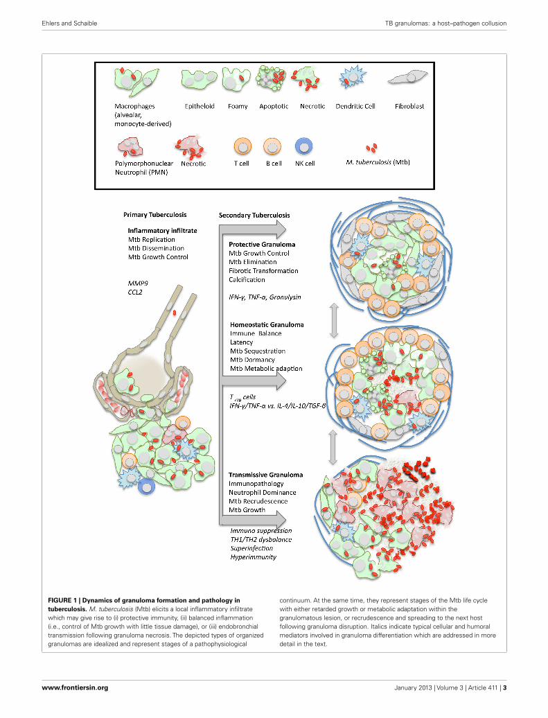
FIGURE 1 | Dynamics of granuloma formation and pathology intuberculosis. M. tuberculosis (Mtb) elicits a local inflammatory infiltratewhich may give rise to (i) protective immunity, (ii) balanced inflammation(i.e., control of Mtb growth with little tissue damage), or (iii) endobronchialtransmission following granuloma necrosis. The depicted types of organizedgranulomas are idealized and represent stages of a pathophysiological
continuum. At the same time, they represent stages of the Mtb life cyclewith either retarded growth or metabolic adaptation within thegranulomatous lesion, or recrudescence and spreading to the next hostfollowing granuloma disruption. Italics indicate typical cellular and humoralmediators involved in granuloma differentiation which are addressed in moredetail in the text.
www.frontiersin.org January 2013 | Volume 3 | Article 411 | 3
Ehlers and Schaible TB granulomas: a host–pathogen collusion
most evident from analyzing the diversity of granulomas is that nocross-sectional or even systemic marker can adequately representthe succinctly local microenvironments of pulmonary granulo-mas. Therefore, biomarker development is facing a formidablechallenge!
VERSATILE METABOLISM WITHIN GRANULOMAS:NON-REPLICATING PERSISTENCE OF M. TUBERCULOSISGluconeogenesis is necessary for M. tuberculosis throughout theinfection, i.e., not only in the phase of persistence, and phospho-enolpyruvate kinase is the critical gate enzyme (Marrero et al.,2010). Within the granuloma, it is assumed that M. tuberculosisreplicates only very little, but remains fully capable of generatingenergy (see Figure 1). Genome-wide expression profiling of M.tuberculosis RNA isolated from chronically infected mouse lungsor sputum of tuberculosis patients revealed transcriptional sig-natures reflective of environmental conditions such as low pH,oxygen depletion, iron limitation, nitrosative stress, and nutrientstarvation (Timm et al., 2003; Voskuil et al., 2004; Talaat et al.,2007; Garton et al., 2008). This explains the attenuated phenotypeof mutant M. tuberculosis defective in the leucin, lysine, purine, orpantothenate biosynthetic pathways, or which are deficient in ironacquisition or the stringent response (Boshoff and Barry, 2005).
The DosR regulon is a genetic program that critically governssurvival in the absence of respiration (Leistikow et al., 2010). Theconserved presence of long sequences of the DosR regulon in envi-ronmental mycobacteria suggests that it did not evolve specificallyfor survival within the mammalian host. Instead, the regulon mayhave evolved to cope with conditions within the environmentsuch as very low oxygen tension. Thus, in a hypothetical ances-tral M. tuberculosis strain, DosR gene products may have allowedthe bacteria to survive in a specific niche in a non-replicating state,ensuring positive selection (Bartek et al., 2009).
Using a dual staining approach, three M. tuberculosis subpop-ulations were found in hypoxic culture and in lung sections ofmice and guinea pigs. Bacteria were either exclusively acid-fastpositive, exclusively immunofluorescent positive or acid-fast andimmunofluorescent positive (Ryan et al., 2010). These results sug-gest that M. tuberculosis exists as multiple populations with distinctcell wall properties even within a seemingly single microenviron-ment, advocating the development of analytical tools at the singlecell level. Indeed, intracellular mycobacteria have altered cell walllipid pattern as those grown in broth (Fischer et al., 2001). Evi-dence from guinea pigs treated with antibiotics indicates that M.tuberculosis may persist extracellularly within biofilm-like struc-tures that consist of DNA and neutrophil debris, in a hypoxicand iron-rich environment with incomplete dystrophic calcifica-tion (Lenaerts et al., 2007). These biofilms appear similar to thoseshown for biofilms of another lung pathogen, Pseudomonas aerug-inosa (Parks et al., 2009). These niches where access of antibioticsis likely compromised may serve as the primary sites of diseasereactivation, and mimicking these tissue conditions in axenic cul-ture will be essential for successful in vitro compound screeningfor next generation anti-mycobacterials.
Even when the drugs reach the bacteria, the actual mechanismof killing M. tuberculosis remains unclear for most anti-tuberculosis drugs. A case in point is isoniazid. It is generally
assumed that INH is only effective on actively replicating M.tuberculosis. The fact that INH preventive chemotherapy canreduce the level of tuberculosis manifestation in individuals diag-nosed with latent infection has been a major argument for thecontention that quiescent tuberculosis lesions contain activelydividing M. tuberculosis (Diel et al., 2008). It is clear that INH,following its activation by the catalase peroxidase KatG, inhibitsmycolic acid synthesis. More specifically, an INH-NAD adductinhibits the fatty acid synthase II enoyl-ACP reductase InhA, lead-ing to the accumulation of long-chain fatty acids (Vilcheze andJacobs, 2007). How exactly this precipitates M. tuberculosis death,however, is the subject of ongoing investigations at the single celllevel within calibrated fermentation chambers, using fluorescentreporter probes (Golchin et al., 2012).
Detailing the mechanisms and kinetics of how drugs kill M.tuberculosis in vitro will be a crucial initial step for improvinganti-tuberculosis drug efficacy. An even more challenging task willbe to define and refine how drugs penetrate and work effectivelywithin tuberculosis lesions. This requires full knowledge of the tis-sue microenvironment influenced by both, mycobacteria as wellas host responses, and includes micronutrient availability for both“partners,” defense cells and microbe. Innovative imaging tech-niques paired with fluorescent or luminescent reporter strains ofM. tuberculosis are important tools for monitoring pathogenesisin situ and drug efficacy testing (Andreu et al., 2010; Carroll et al.,2010; Kong et al., 2010; Zelmer et al., 2012). Of even greater interestwill be M. tuberculosis sensor strains, which can report physico-chemical conditions in situ such as low iron concentration or drugexposure.
How does M. tuberculosis exit the state of seeming dormancyand resume growth? It may take one cue from the production ofresuscitation promoting factors (Rpf), RpfB being the most rele-vant isoform out of five encoded in the M. tuberculosis genomebased on persistence assays with targeted deletion mutants (Kanaet al., 2008; see Figure 1). The structure of RpfB contains pecu-liar features that are also shared by G5 domains involved inbiofilm formation (Ruggiero et al., 2009), providing a furtherlink to evolutionarily conserved pathways of adaptation to adverseenvironmental conditions by changing growth patterns.
Foamy macrophages are key constituents of tuberculosislesions, representing macrophages packed with lipid bodies fol-lowing activation via Toll-like receptors and proinflammatorysignals (D’Avila et al., 2006). These lipid bodies are the conse-quence of an imbalance between the influx and efflux of low-density lipoprotein particles from the serum. The phenotype canbe induced in vitro, in peripheral blood cells incubated with dis-tinct mycobacteria-derived oxygenated ketomycolic and hydroxyl-mycolic acids (Peyron et al., 2008). Lipid bodies may provide idealnutrition for a bacterium that is changing its metabolism to digestfatty acids, as evidenced by upregulation of the gating enzymeof the glyoxylate shunt, isocitrate lyase, and of the methylcitratepathway, methylcitrate dehydratase, in the intracellular life stage(McKinney et al., 2000; Gould et al., 2006; Mattow et al., 2006;Munoz-Elias et al., 2006).
Mycobacterium tuberculosis has also been shown to metabo-lize cholesterol (Pandey and Sassetti, 2008), and several genessuch as Mce4, HsaC, and Icl1, which were previously linked
Frontiers in Immunology | Inflammation January 2013 | Volume 3 | Article 411 | 4

Ehlers and Schaible TB granulomas: a host–pathogen collusion
to propionate metabolism, may be functionally linked throughcholesterol degradation. Cholesterol metabolism requires alter-ing of transcription and metabolic profiles of M. tuberculosis toaccess propionyl-CoA and pyruvate pools through the methyl-citrate cycle. Consequently, gene deletion mutants therein arehandicapped for intracellular growth (Griffin et al., 2012). Analy-sis of lipids from the caseum of human tuberculosis granulo-mas revealed that the main lipid species are cholesterol, cho-lesteryl ester, triacylglycerides, and lactosylceramide, implicatinglow-density lipoprotein-derived lipids as the most likely source(Kim et al., 2010). Granuloma necrosis, at least in mouse M.tuberculosis or M. avium infection, is often associated with highbacterial burden, and virulent mycobacteria can inhibit mem-brane repair, which causes necrosis in infected macrophages(Divangahi et al., 2009). A conceivable scenario, therefore, isthat the death of foamy macrophages results in the accumula-tion of lipid debris making up the caseum at the center of thegranuloma (Russell et al., 2009; Behar et al., 2010). Other cellsmay also contribute to cellular debris as neutrophils representa major cell population in TB lesions, which has an exquisitelyhigh death toll due to their short half-life and M. tuberculo-sis driven necrosis (Eum et al., 2010; Corleis et al., 2012; seeFigure 1).
The initial necrotic nidus may be induced in the absence ofany adaptive immune response, as guinea pigs exhibit focal necro-sis within granulomas as early as 10 days after infection (Turneret al., 2003). Overwhelming experimental and clinical evidence,however, suggests that promotion of full-fledged central caseationin mycobacterial granulomas requires a hypersensitivity reaction,precipitated by CD4+ T cells faced with a high antigenic load(Dannenberg, 1991; Ehlers et al., 2001; Sanghi et al., 2011).
PREPARING FOR THE EXIT: GRANULOMA NECROSIS AS ATRANSMISSION STRATEGYThere is much debate about whether tissue necrosis beginswith central necrotization of preformed granulomas, or whethercaseation and cavitation develop from so-called lipid pneumoniaaccompanying regrowth of M. tuberculosis persisting outside ofgranulomas (e.g., in epithelial cells, adipocytes, etc.; Hernandez-Pando et al., 2000; Neyrolles et al., 2006). Evidence from the formercomes from studies in rabbits, guinea pigs, and designer mousemodels, while evidence for the latter is derived from histopatho-logical studies in men and mice. For example, mice bearingthe sst-susceptible allele develop centrally necrotizing pulmonarygranulomas in which M. tuberculosis growth is rampant. Thehost resistance gene, Ipr1, encoded within the sst1 locus, regu-lates the infected macrophages’ mechanism of death (Kramnik,2008). In dermally infected NOS2-deficient mice treated with aneutralizing anti-IFN-γ antibody, established granulomas cen-trally necrotize, showing signs of hypoxia and abundant cathepsinG activity (Reece et al., 2010). In guinea pigs, necrosis withingranulomatous lesions can occur even before a robust T cellresponse has developed (Turner et al., 2003). In rabbits, cavita-tion has generally been attributed to a strong DTH response toM. bovis infection precipitating caseation, cavitation, and lique-faction of caseum within circumscript granulomas (Dannenbergand Sugimoto, 1976; Dannenberg and Collins, 2001).
By examining a histological archive of tuberculosis lesions inhumans and revisiting the Cornell model of reactivation tuber-culosis in mice, Hunter et al. (2007) came to the conclusionthat reactivation of M. tuberculosis infection begins as a lipidpneumonia with bronchial obstruction and does not start fromdisintegrating granulomas. A very potent component of M. tuber-culosis to which this necrosis-inducing activity is attributed is cordfactor, or trehalose dimycolate which can induce inflammatoryinfiltrates including foam cell formation but is also a virulencefactor promoting intracellular survival (Hunter et al., 2006; Kimet al., 2010). Taken together, caseation appears to correlate withpathogen-mediated dysregulation of host lipid metabolism.
As neutrophils represent the predominant cell population inbroncho-alveolar lavages of active tuberculosis patients they maybe more relevant in transmission of M. tuberculosis than in con-trolling infection (Eum et al., 2010). Considerable controversyexists over whether neutrophils are able to kill mycobacteria and arecent review on the issue concluded that these otherwise potentanti-bacterial effector cells fail to eliminate M. tuberculosis (Korbelet al., 2008). Indeed, virulent M. tuberculosis quickly cause necroticcell death of human neutrophils upon infection in vitro by induc-ing reactive oxygen intermediates (ROI) allowing the mycobacteriato escape neutrophil-mediated killing. In contrast, an attenu-ated M. tuberculosis mutant lacking the virulence associated RD1region fails to induce necrosis and falls victim to neutrophils’armamentarium, i.e., ROI (Corleis et al., 2012). More impor-tantly, invading neutrophils with their payload of cytotoxic mol-ecules may cause substantial tissue destruction and remodeling.It can be envisaged that misguided but anti-microbially ineffec-tive neutrophils invade existing (latent?) tuberculosis lesions andprompt the immunopathological cascade toward active lesions,providing further host cell lipid substrate for M. tuberculosisgrowth and biofilm formation to secure subsequent transmission.Here, super-infection with a novel M. tuberculosis strain or evenanother unrelated co-infecting lung pathogen may represent anas yet under-appreciated cause of reactivation (see Figure 1). Inaddition, systemic co-infections of M. tuberculosis and unrelatedpathogens beyond HIV may also modulate lung immunity in thelatently infected. This was recently demonstrated in the murinemodel of malaria-associated acute respiratory distress syndrome,in which the mycobacterial burden was exacerbated in malaria – M.tuberculosis co-infected mice (Mueller et al., 2012).
Sequencing the genomes of 21 M. tuberculosis strains repre-sentative of the global diversity identified very little sequencevariation in 491 experimentally confirmed human T cell epi-topes, indicating purifying selection (Comas et al., 2010). Thisfinding led to the hypothesis that M. tuberculosis benefits fromrecognition by human T cells. In view of its confinement to thegranuloma, M. tuberculosis may have devised an exit strategy thatexploits T cell activation. TH1 responses (IFN-γ, IRF-1) wereshown to drive granuloma necrosis in a model of mycobacteria-induced pulmonary immunopathology (Ehlers et al., 2001; Alyet al., 2009), and TH2 responses have also been associated withincreased cavity formation and tissue destruction in humans andmice (Rook, 2007); Hölscher, C., personal communication). Cav-ity formation in rabbits, monkeys, and man are clearly resultsof a long-lasting T cell-driven immune activation, low numbers
www.frontiersin.org January 2013 | Volume 3 | Article 411 | 5
Ehlers and Schaible TB granulomas: a host–pathogen collusion
of CD4+ cells virtually precluding granuloma necrosis (Chais-son et al., 1987; Capuano et al., 2003). Moreover, AIDS patientswith tuberculosis have a different pathology and often, their spu-tum is negative for acid-fast bacteria, further suggesting thattransmission-facilitating granulomas are immune driven (Aaronet al., 2004, see Figure 1).
An integrated view of current findings would hold that individ-ual components of M. tuberculosis, such as trehalose dimycolate,allow for foam cell formation and macrophage necrosis, but full-blown central necrosis of an established granuloma requires T cellparticipation, either in the form of a hyperactive IFN-γ produc-tion or a superimposed IL-4/IL-13 response. The latter inducesalternative macrophage activation (Gordon and Martinez, 2010),and arginase 1 is a potential biomarker for reactivation tubercu-losis and granulomas prone to necrotizing. Neutrophils, whichare also potent producers of arginase 1, may even amplify thisimmunopathogenic cascade.
Are the “fat and lazy” persister bacilli present in the sputumof cavitary patients specifically adapted for transmission (Gartonet al., 2008)? Very little is known about the physical parameters inindividual M. tuberculosis bacteria relevant for aerosolization, sur-vival in the environment, or infectivity (i.e., inoculum“take” in thealveolar macrophage; Fennelly et al., 2004). Also, there is a growingdebate as to whether low dose aerosol infection adequately reflectsthe situation in endemic countries where exposure to putativelylarger numbers of coughed-up M. tuberculosis may be the crucialfactor overriding naturally existing or vaccine-induced immunity(Rook et al., 2009).
THE BOTTOM LINE: DOES M. TUBERCULOSIS UNIVERSALLYPROFIT FROM GRANULOMA FORMATION?Given the recent focus on the stunning discovery that M. tuber-culosis may direct granuloma induction and maturation as partof its life cycle, the fact that, in the absence of granuloma for-mation there is no protection, at least in the human host, hasbeen unduly neglected. Granulomas afford a unique juxtaposi-tion of activating T cells and mycobacteria-laden macrophages,and the temporal coincidence of T cell differentiation, granulomaconsolidation, and reduction of M. tuberculosis growth in all ani-mal models that mimic human disease suggests causality (Northand Jung, 2004). It is important to emphasize that T cell memoryaffords 10-fold lower bacterial loads in infected organs of vac-cinated animals, and that the majority of bacteria reside withingranulomas and not randomly distributed throughout the body.Therefore, while a mycobacteria-focused view on granulomas waslong overdue (Russell, 2007; Ramakrishnan, 2012), this shouldby no means neutralize the evidence in favor of the protectiveinfection-sequestrating granuloma:
– T cells transfer protective immunity and granuloma formation(Orme and Collins, 1983).
– HIV-infected individuals exhibit poor granulomatous inflam-mation and poor control of mycobacterial growth (Lawn et al.,2002).
– Macrophage accumulations (or innate granulomas) do not effi-ciently kill mycobacteria unless activated by T cells (North andIzzo, 1993; Smith et al., 1997).
– Interferon-gamma provided by NK cells alone is insufficient toprovide full protection in the absence of T cells (Feng et al.,2006).
It is certainly true that the host-centric view has prevailedfor too long, neglecting the important input of M. tuberculosisin shaping the granuloma to its advantage, but it is unwise tounderestimate the power of T cell-mediated macrophage activa-tion, which takes place at the site of granulomatous inflammation,as it currently provides the only venue for preventive vaccinationstrategies.
CONCLUSION: IS IT POSSIBLE TO BE BETTER THAN NATURE?One of the implications of the integrative view on M. tuberculo-sis’ life cycle embraced here is that, to stop M. tuberculosis frommultiplying and transmitting, simple imitation, or augmentationof the natural host response to infection is likely to fail. UnlessT cells can be trained to recognize M. tuberculosis as soon as itenters the alveolar macrophage, one of the best vaccination strate-gies might be to bypass the regulatory networks M. tuberculosisitself initiates to establish its niche for replication. If anything,vaccines would have to mitigate TH1 and TH2 responses andaltogether blunt regulatory T cell responses to allow more protec-tive immunity while avoiding damaging pathology (Rook et al.,2005). This may be impossible to achieve purely by vaccina-tion, leaving ample opportunity for adjunct immunomodulatorymeasures. Indeed, inhibition of inflammation may prove to bea superior adjunct strategy to improve the outcome of antibi-otic therapy (Churchyard et al., 2009). For example, blockade ofPDE4 together with INH shortened the duration of treatment byone month and reduced pathology in a rabbit model of tubercu-losis (Subbian et al., 2011). During the Immune ReconstitutionInflammatory Syndrome (IRIS), which occurs in M. tuberculo-sis-infected AIDS patients receiving highly active antiretroviraltherapy, corticosteroid therapy is highly effective. However, itdoes not reproducibly interfere with TH1 responses but reducesgranzyme B and perforin levels, indicating an involvement ofCD8+ T or NK cells in inflammatory exacerbation (Meintjes et al.,2009).
A potential problem with current immunization strategiesagainst tuberculosis might be that they prime an immune responseto epitopes that have been highly conserved in M. tuberculosis,because these dominant T cell targets ensure escape from thegranuloma and thus transmission. Indeed, in an alternative strat-egy that refocused the T cell response to specificities that are notnormally recognized during natural infection, a more efficientprotection against murine M. tuberculosis infection was inducedthan afforded by immunization with the immunodominant epi-tope (Aagaard et al., 2009). This increased efficacy was associatedwith elevated numbers of multifunctional T cells, producing TNF,IFN-γ, and IL-2 at high levels.
Further considerations for improving vaccines take intoaccount that M. tuberculosis responds to the host immune responseby regulating and diversifying its own gene expression; for exam-ple, during latency, stage-specific antigens are expressed that rep-resent its metabolic adaptation and can effectively be utilized todiscriminate, by immunodiagnosis of host T cell responses against
Frontiers in Immunology | Inflammation January 2013 | Volume 3 | Article 411 | 6
Ehlers and Schaible TB granulomas: a host–pathogen collusion
these antigens, between acute and latent infection (Demissie et al.,2006). Based on these findings, a novel subunit combinationvaccine was developed: H56, comprising three M. tuberculosisantigens, which are expressed either early in infection (Ag85,ESAT6) or during the latent phase (Rv2660c), not only boostsBCG-primed immunity but also induces multifunctional T cell-mediated immune protection on its own before and after exposureto M. tuberculosis (Aagaard et al., 2011). Ag85 is expressed byboth, BCG and M. tuberculosis, and is therefore responsible forthe boost effect, whereas ESAT6 is exclusive for the latter one andexpressed mostly during early, active replication stages. Rv2660chowever, is expressed during the entire course of infection in mice(even by starved and dormant M. tuberculosis) but is only a weakIFN-γ inducer by itself. It becomes mildly immunogenic onlywhen fused to Ag85-ESAT6 thereby obviating immune-mediated
exacerbation of the disease in infected individuals. In sum, theselection and combination of antigens and epitopes specific fordifferent stages of TB may help outwit M. tuberculosis and controlreactivation.
It would be unreasonable to call the TB granuloma an unsuc-cessful host defence, as it successfully contains the infectious focusin more than 90% of cases. The 10% of individuals that progresstoward TB disease suffer from a disbalanced inflammatory reac-tion, be it due to too little innate or adaptive immunity or dueto unrestrained hypersensitivity reactions. There is no balancewithout a trade-off: in the case of TB this is the Janus face of Tcell immunity (which can be detrimental when overzealous). Anyintervention thus must be regulatory in nature rather than proin-flammatory, and the rational design of therapies and vaccines musttake this into account.
REFERENCESAagaard, C. S., Hoang, T., Dietrich, J.,
Cardona, P. J., Izzo, A., Dolganov, G.,et al. (2011). A multistage tuberculo-sis vaccine that confers efficient pro-tection before and after exposure.Nat. Med. 17, 189–195.
Aagaard, C. S., Hoang, T., Vingsbo-Lundberg, C., Dietrich, J., andAndersen, P. (2009). Quality andvaccine efficacy of CD4+ T cellresponses directed to dominant andsubdominant epitopes in ESAT-6from Mycobacterium tuberculosis. J.Immunol. 183, 2659–2668.
Aaron, L., Saadoun, D., Calatroni, I.,Launay, O., Memain, N., Vincent, V.,et al. (2004). Tuberculosis in HIV-infected patients: a comprehensivereview. Clin. Microbiol. Infect. 10,388–398.
Aly, S., Mages, J., Reiling, N., Kalinke,U., Decker, T., Lang, R., et al.(2009). Mycobacteria-induced gran-uloma necrosis depends on IRF-1. J.Cell. Mol. Med. 13, 2069–2082.
Andreu, N., Zelmer, A., Fletcher, T., Elk-ington, P. T., Ward, T. H., Ripoll, J.,et al. (2010). Optimisation of bio-luminescent reporters for use withmycobacteria. PLoS ONE 5:e10777.doi:10.1371/journal.pone.0010777
Barry, C. E. III, Boshoff, H. I., Dar-tois, V., Dick, T., Ehrt, S., Flynn, J.,et al. (2009). The spectrum of latenttuberculosis: rethinking the biologyand intervention strategies. Nat. Rev.Microbiol. 7, 845–855.
Bartek, I. L., Rutherford, R., Gruppo, V.,Morton, R. A., Morris, R. P., Klein,M. R., et al. (2009). The DosR reg-ulon of M. tuberculosis and antibac-terial tolerance. Tuberculosis (Edinb.)89, 310–316.
Behar, S. M., Divangahi, M., andRemold, H. G. (2010). Evasion ofinnate immunity by Mycobacteriumtuberculosis: is death an exit strategy?Nat. Rev. Microbiol. 8, 668–674.
Behler, F. F., Steinwede, K. F., Balboa,L. F., Ueberberg, B. F., Maus, R. F.,Kirchhof, G. F., et al. (2012). Roleof Mincle in alveolar macrophage-dependent innate immunity againstmycobacterial infections in mice. J.Immunol. 189, 3121–3129.
Berry, M. P., Graham, C. M., McNab, F.W., Xu, Z., Bloch, S. A., Oni, T., etal. (2010). An interferon-inducibleneutrophil-driven blood transcrip-tional signature in human tubercu-losis. Nature 466, 973–977.
Boshoff, H. I., and Barry, C. E. III.(2005). Tuberculosis – metabolismand respiration in the absence ofgrowth. Nat. Rev. Microbiol. 3,70–80.
Capuano, S. V. III, Croix, D. A.,Pawar, S., Zinovik, A., Myers, A.,Lin, P. L., et al. (2003). Experi-mental Mycobacterium tuberculosisinfection of cynomolgus macaquesclosely resembles the various man-ifestations of human M. tubercu-losis infection. Infect. Immun. 71,5831–5844.
Cardona, P. J. (2009). A dynamic rein-fection hypothesis of latent tubercu-losis infection. Infection 37, 80–86.
Cardona, P. J., and Ruiz-Manzano, J.(2004). On the nature of Mycobac-terium tuberculosis-latent bacilli.Eur. Respir. J. 24, 1044–1051.
Carroll, P., Schreuder, L. J., Muwanguzi-Karugaba, J., Wiles, S., Robertson,B. D., Ripoll, J., et al. (2010).Sensitive detection of gene expres-sion in mycobacteria underreplicating and non-replicatingconditions using optimized far-redreporters. PLoS ONE 5:e9823.doi:10.1371/journal.pone.0009823
Chaisson, R. E., Schecter, G. F., Theuer,C. P., Rutherford, G. W., Echen-berg, D. F., and Hopewell, P. C.(1987). Tuberculosis in patients withthe acquired immunodeficiency syn-drome. Clinical features, response
to therapy, and survival. Am. Rev.Respir. Dis. 136, 570–574.
Churchyard, G. J., Kaplan, G., Fallows,D., Wallis, R. S., Onyebujoh, P.,and Rook, G. A. (2009). Advancesin immunotherapy for tuberculo-sis treatment. Clin. Chest Med. 30,769–782, ix.
Comas, I., Chakravartti, J., Small, P. M.,Galagan, J., Niemann, S., Kremer,K., et al. (2010). Human T cell epi-topes of Mycobacterium tuberculosisare evolutionarily hyperconserved.Nat. Genet. 42, 498–503.
Cooper, A. M. (2009). T cells inmycobacterial infection and disease.Curr. Opin. Immunol. 21, 378–384.
Cooper, A. M., Pearl, J. E., Brooks, J. V.,Ehlers, S., and Orme, I. M. (2000).Expression of the nitric oxide syn-thase 2 gene is not essential for earlycontrol of Mycobacterium tubercu-losis in the murine lung. Infect.Immun. 68, 6879–6882.
Corleis, B. F., Korbel, D. F., Wilson,R. F., Bylund, J. F., Chee, R. F.,and Schaible, U. E. (2012). Escapeof Mycobacterium tuberculosis fromoxidative killing by neutrophils. Cell.Microbiol. 14, 1109–1121.
Court, N. F., Vasseur, V., Vasseur, V. F.,Vacher, R. F., Fremond, C. F., She-bzukhov, Y. F., et al. (2010). Partialredundancy of the pattern recog-nition receptors, scavenger recep-tors, and C-type lectins for thelong-term control of Mycobacteriumtuberculosis infection. J. Immunol.184, 7057–7070.
Dannenberg, A. M. Jr. (1991).Delayed-type hypersensitivityand cell-mediated immunity inthe pathogenesis of tuberculosis.Immunol. Today 12, 228–233.
Dannenberg, A. M. Jr., and Collins, F.M. (2001). Progressive pulmonarytuberculosis is not due to increas-ing numbers of viable bacilli in rab-bits, mice and guinea pigs, but is
due to a continuous host response tomycobacterial products. Tuberculosis(Edinb.) 81, 229–242.
Dannenberg, A. M. Jr., and Rook, G.A. W. (1994). “Pathogenesis of pul-monary tuberculosis: an interplay oftissue-damaging and macrophage-activating immune response – dualmechanisms that control bacil-lary multiplication,” in Tuberculosis:Pathogenesis, Protection, and Control,ed. B. R. Bloom (Washington, DC:American Society for MicrobiologyPress), 459–483.
Dannenberg, A. M. Jr., and Sugimoto,M. (1976). Liquefaction of caseousfoci in tuberculosis. Am. Rev. Respir.Dis. 113, 257–259.
D’Avila, H., Melo, R. C., Parreira, G.G., Werneck-Barroso, E., Castro-Faria-Neto, H. C., and Bozza, P. T.(2006). Mycobacterium bovis bacil-lus Calmette–Guerin induces TLR2-mediated formation of lipid bodies:intracellular domains for eicosanoidsynthesis in vivo. J. Immunol. 176,3087–3097.
Davis, J. M., and Ramakrishnan, L.(2009). The role of the granulomain expansion and dissemination ofearly tuberculous infection. Cell 136,37–49.
Demissie, A., Leyten, E. M., Abebe, M.,Wassie, L., Aseffa, A., Abate, G., et al.(2006). Recognition of stage-specificmycobacterial antigens differenti-ates between acute and latent infec-tions with Mycobacterium tubercu-losis. Clin. Vaccine Immunol. 13,179–186.
Diel, R., Loddenkemper, R., Meywald-Walter, K., Niemann, S., and Nien-haus, A. (2008). Predictive value ofa whole blood IFN-gamma assayfor the development of active tuber-culosis disease after recent infec-tion with Mycobacterium tuberculo-sis. Am. J. Respir. Crit. Care Med. 177,1164–1170.
www.frontiersin.org January 2013 | Volume 3 | Article 411 | 7
Ehlers and Schaible TB granulomas: a host–pathogen collusion
Divangahi, M., Chen, M., Gan, H.,Desjardins, D., Hickman, T. T., Lee,D. M., et al. (2009). Mycobacteriumtuberculosis evades macrophagedefenses by inhibiting plasmamembrane repair. Nat. Immunol.10, 899–906.
Egen, J. G., Rothfuchs, A. G., Feng, C.G., Horwitz, M. A., Sher, A., andGermain, R. N. (2011). Intravitalimaging reveals limited antigen pre-sentation and T cell effector func-tion in mycobacterial granulomas.Immunity 34, 807–819.
Egen, J. G., Rothfuchs, A. G., Feng, C.G., Winter, N. F., Sher, A. F., andGermain, R. N. (2008). Macrophageand T cell dynamics during thedevelopment and disintegration ofmycobacterial granulomas. Immu-nity 28, 271–284.
Ehlers, S. (2010). TB or not TB? Fish-ing for molecules making permis-sive granulomas. Cell Host Microbe7, 6–8.
Ehlers, S., Benini, J., Held, H. D.,Roeck, C., Alber, G., and Uhlig, S.(2001). Alphabeta T cell receptor-positive cells and interferon-gamma,but not inducible nitric oxide syn-thase, are critical for granulomanecrosis in a mouse model ofmycobacteria-induced pulmonaryimmunopathology. J. Exp. Med. 194,1847–1859.
Eum, S. Y., Kong, J. H., Hong, M. S., Lee,Y. J., Kim, J. H., Hwang, S. H., etal. (2010). Neutrophils are the pre-dominant infected phagocytic cellsin the airways of patients with activepulmonary TB. Chest 137, 122–128.
Feng, C. G., Kaviratne, M., Rothfuchs,A. G., Cheever, A., Hieny, S., Young,H. A., et al. (2006). NK cell-derivedIFN-gamma differentially regulatesinnate resistance and neutrophilresponse in T cell-deficient hostsinfected with Mycobacterium tuber-culosis. J. Immunol. 177, 7086–7093.
Fennelly, K. P., Martyny, J. W., Fulton,K. E., Orme, I. M., Cave, D. M.,and Heifets, L. B. (2004). Cough-generated aerosols of Mycobacteriumtuberculosis: a new method to studyinfectiousness. Am. J. Respir. Crit.Care Med. 169, 604–609.
Fischer, K. F., Chatterjee, D. F., Tor-relles, J. F., Brennan, P. J., Kauf-mann, S. H., and Schaible, U.E. (2001). Mycobacterial lysocar-diolipin is exported from phago-somes upon cleavage of cardiolipinby a macrophage-derived lysosomalphospholipase A2. J. Immunol. 167,2187–2192.
Flynn, J. L., Goldstein, M. M., Chan, J.,Triebold, K. J., Pfeffer, K., Lowen-stein, C. J., et al. (1995). Tumor
necrosis factor-alpha is required inthe protective immune responseagainst Mycobacterium tuberculosisin mice. Immunity 2, 561–572.
Gandotra, S., Jang, S., Murray, P. J.,Salgame, P., and Ehrt, S. (2007).Nucleotide-binding oligomerizationdomain protein 2-deficient micecontrol infection with Mycobac-terium tuberculosis. Infect. Immun.75, 5127–5134.
Garton, N. J., Waddell, S. J., Sherratt,A. L., Lee, S. M., Smith, R. J., Sen-ner, C., et al. (2008). Cytologicaland transcript analyses reveal fat andlazy persister-like bacilli in tuber-culous sputum. PLoS Med. 5:e75.doi:10.1371/journal.pmed.0050075
Geisel, R. E., Sakamoto, K. F., Rus-sell, D. G., and Rhoades, E.R. (2005). In vivo activity ofreleased cell wall lipids of Mycobac-terium bovis bacillus Calmette–Guerin is due principally to tre-halose mycolates. J. Immunol. 174,5007–5015.
Golchin, S. A., Stratford, J., Curry,R. J., and McFadden, J. (2012). Amicrofluidic system for long-termtime-lapse microscopy studies ofmycobacteria. Tuberculosis (Edinb.)92, 489–496.
Gordon, S., and Martinez, F. O.(2010). Alternative activation ofmacrophages: mechanism and func-tions. Immunity 32, 593–604.
Gould, T. A., van de Langemheen, H.,Munoz-Elias, E. J., McKinney, J. D.,and Sacchettini, J. C. (2006). Dualrole of isocitrate lyase 1 in theglyoxylate and methylcitrate cyclesin Mycobacterium tuberculosis. Mol.Microbiol. 61, 940–947.
Griffin, J. E., Pandey, A. K., Gilmore,S. A., Mizrahi, V., McKinney, J. D.,Bertozzi, C. R., et al. (2012). Choles-terol catabolism by Mycobacteriumtuberculosis requires transcriptionaland metabolic adaptations. Chem.Biol. 19, 218–227.
Hänsch, H. C., Smith, D. A., Mielke,M. E., Hahn, H., Bancroft, G. J.,and Ehlers, S. (1996). Mechanismsof granuloma formation in murineMycobacterium avium infection: thecontribution of CD4+ T cells. Int.Immunol. 8, 1299–1310.
Heitmann, L., Schoenen, H., Ehlers, S.,Lang, R., and Hölscher, C. (2012).Mincle is not essential for con-trolling Mycobacterium tuberculo-sis infection. Immunobiology. PMID:22784441. [Epub ahead of print].
Hernandez-Pando, R., Jeyanathan, M.,Mengistu,G.,Aguilar,D.,Orozco,H.,Harboe, M., et al. (2000). Persistenceof DNA from Mycobacterium tuber-culosis in superficially normal lung
tissue during latent infection. Lancet356, 2133–2138.
Hunter, R. L., Jagannath, C., and Actor, J.K. (2007). Pathology of postprimarytuberculosis in humans and mice:contradiction of long-held beliefs.Tuberculosis (Edinb.) 87, 267–278.
Hunter, R. L., Olsen, M. R., Jagannath,C., and Actor, J. K. (2006). Multipleroles of cord factor in the pathogen-esis of primary, secondary, and cavi-tary tuberculosis, including a reviseddescription of the pathology of sec-ondary disease. Ann. Clin. Lab Sci.36, 371–386.
Ishikawa, E., Ishikawa, T., Morita, Y. S.,Toyonaga, K., Yamada, H., Takeuchi,O., et al. (2009). Direct recognitionof the mycobacterial glycolipid, tre-halose dimycolate, by C-type lectinMincle. J. Exp. Med. 206, 2879–2888.
Kana, B. D., Gordhan, B. G., Down-ing, K. J., Sung, N., Vostroktunova,G., Machowski, E. E., et al. (2008).The resuscitation-promoting factorsof Mycobacterium tuberculosis arerequired for virulence and resuscita-tion from dormancy but are collec-tively dispensable for growth in vitro.Mol. Microbiol. 67, 672–684.
Keller, C., Hoffmann, R., Lang, R.,Brandau, S., Hermann, C., andEhlers, S. (2006). Genetically deter-mined susceptibility to tuberculo-sis in mice causally involves accel-erated and enhanced recruitmentof granulocytes. Infect. Immun. 74,4295–4309.
Kim, M. J., Wainwright, H. C., Lock-etz, M., Bekker, L. G., Walther, G. B.,Dittrich, C., et al. (2010). Caseationof human tuberculosis granulomascorrelates with elevated host lipidmetabolism. EMBO Mol. Med. 2,258–274.
Kong, Y., Yao, H., Ren, H., Subbian, S.,Cirillo, S. L., Sacchettini, J. C., etal. (2010). Imaging tuberculosis withendogenous beta-lactamase reporterenzyme fluorescence in live mice.Proc. Natl. Acad. Sci. U.S.A. 107,12239–12244.
Korbel, D. S., Schneider, B. E., andSchaible, U. E. (2008). Innate immu-nity in tuberculosis: myths andtruth. Microbes Infect. 10, 995–1004.
Kramnik, I. (2008). Genetic dissec-tion of host resistance to Mycobac-terium tuberculosis: the sst1 locusand the Ipr1 gene. Curr. Top. Micro-biol. Immunol. 321, 123–148.
Lawn, S. D., Butera, S. T., andShinnick, T. M. (2002). Tuber-culosis unleashed: the impact ofhuman immunodeficiency virusinfection on the host granulomatousresponse to Mycobacterium tubercu-losis. Microbes Infect. 4, 635–646.
Lee,W. B., Kang, J. S.,Yan, J. J., Lee, M. S.,Jeon, B. Y., Cho, S. N., et al. (2012).Neutrophils promote mycobacte-rial trehalose dimycolate-inducedlung inflammation via the Minclepathway. PLoS Pathog. 8:e1002614.doi:10.1371/journal.ppat.1002614
Leistikow, R. L., Morton, R. A., Bartek,I. L., Frimpong, I., Wagner, K., andVoskuil, M. I. (2010). The Mycobac-terium tuberculosis DosR regulonassists in metabolic homeostasis andenables rapid recovery from non-respiring dormancy. J. Bacteriol. 192,1662–1670.
Lenaerts, A. J., Hoff, D., Aly, S., Ehlers,S., Andries, K., Cantarero, L., etal. (2007). Location of persistingmycobacteria in a Guinea pig modelof tuberculosis revealed by r207910.Antimicrob. Agents Chemother. 51,3338–3345.
Lin, P. L., and Flynn, J. L. (2010). Under-standing latent tuberculosis: a mov-ing target. J. Immunol. 185, 15–22.
Lin, P. L., Rodgers, M., Smith, L., Big-bee, M., Myers, A., Bigbee, C., et al.(2009). Quantitative comparison ofactive and latent tuberculosis in thecynomolgus macaque model. Infect.Immun. 77, 4631–4642.
Lockhart, E., Green, A. M., and Flynn,J. L. (2006). IL-17 production isdominated by gammadelta T cellsrather than CD4 T cells duringMycobacterium tuberculosis infec-tion. J. Immunol. 177, 4662–4669.
Mack, U., Migliori, G. B., Sester, M.,Rieder, H. L., Ehlers, S., Goletti, D.,et al. (2009). LTBI: latent tubercu-losis infection or lasting immuneresponses to M. tuberculosis? ATBNET consensus statement. Eur.Respir. J. 33, 956–973.
Maertzdorf, J. F., Weiner, J. III, Mol-lenkopf, H. J., Bauer, T., Prasse, A.,Muller-Quernheim, J., et al. (2012).Common patterns and disease-related signatures in tuberculosisand sarcoidosis. Proc. Natl. Acad. Sci.U.S.A 109, 7853–7858.
Manca, C., Tsenova, L., Bergtold, A.,Freeman, S., Tovey, M., Musser, J.M., et al. (2001). Virulence of aMycobacterium tuberculosis clinicalisolate in mice is determined by fail-ure to induce Th1 type immunityand is associated with induction ofIFN-alpha/beta. Proc. Natl. Acad. Sci.U.S.A. 98, 5752–5757.
Marrero, J., Rhee, K. Y., Schnappinger,D., Pethe, K., and Ehrt, S. (2010).Gluconeogenic carbon flow of tri-carboxylic acid cycle intermediates iscritical for Mycobacterium tubercu-losis to establish and maintain infec-tion. Proc. Natl. Acad. Sci. U.S.A. 107,9819–9824.
Frontiers in Immunology | Inflammation January 2013 | Volume 3 | Article 411 | 8
Ehlers and Schaible TB granulomas: a host–pathogen collusion
Mattow, J., Siejak, F., Hagens, K.,Becher, D., Albrecht, D., Krah, A.et al. (2006). Proteins unique tointraphagosomally grown Mycobac-terium tuberculosis. Proteomics 6,2485–2494.
McKinney, J. D., Honer zu, B. K.,Munoz-Elias, E. J., Miczak, A., Chen,B., Chan, W. T., et al. (2000). Persis-tence of Mycobacterium tuberculosisin macrophages and mice requiresthe glyoxylate shunt enzyme isoci-trate lyase. Nature 406, 735–738.
Meintjes, G., Rabie, H., Wilkinson,R. J., and Cotton, M. F. (2009).Tuberculosis-associated immunereconstitution inflammatorysyndrome and unmasking of tuber-culosis by antiretroviral therapy.Clin. Chest Med. 30, 797–810.
Mueller, A. K., Behrends, J., Hagens,K., Mahlo, J., Schaible, U. E.,and Schneider, B. E. (2012).Natural transmission of Plas-modium berghei exacerbateschronic tuberculosis in anexperimental co-infectionmodel. PLoS ONE 7:e48110.doi:10.1371/journal.pone.0048110
Munoz-Elias, E. J., Upton, A. M., Cher-ian, J., and McKinney, J. D. (2006).Role of the methylcitrate cycle inMycobacterium tuberculosis metabo-lism, intracellular growth, and viru-lence. Mol. Microbiol. 60, 1109–1122.
Neyrolles, O., Hernandez-Pando,R., Pietri-Rouxel, F., Fornes, P.,Tailleux, L., Barrios Payan, J. A.,et al. (2006). Is adipose tissue aplace for Mycobacterium tuberculosispersistence? PLoS ONE 1:e43.doi:10.1371/journal.pone.0000043
North, R. J., and Izzo, A. A. (1993).Granuloma formation in severecombined immunodeficient (SCID)mice in response to progressive BCGinfection. Tendency not to formgranulomas in the lung is associatedwith faster bacterial growth in thisorgan. Am. J. Pathol. 142,1959–1966.
North, R. J., and Jung, Y. J. (2004).Immunity to tuberculosis. Annu.Rev. Immunol. 22, 599–623.
Orme, I. M., and Collins, F. M. (1983).Protection against Mycobacteriumtuberculosis infection by adoptiveimmunotherapy. Requirement for Tcell-deficient recipients. J. Exp. Med.158, 74–83.
Pandey,A. K., and Sassetti, C. M. (2008).Mycobacterial persistence requiresthe utilization of host cholesterol.Proc. Natl. Acad. Sci. U.S.A. 105,4376–4380.
Parks, Q. M., Young, R. L., Poch, K.R., Malcolm, K. C., Vasil, M. L.,and Nick, J. A. (2009). Neutrophilenhancement of Pseudomonasaeruginosa biofilm development:human F-actin and DNA as targetsfor therapy. J. Med. Microbiol. 58,492–502.
Peyron, P., Vaubourgeix, J., Poquet, Y.,Levillain, F., Botanch, C., Bardou, F.,et al. (2008). Foamy macrophagesfrom tuberculous patients’ gran-ulomas constitute a nutrient-richreservoir for M. tuberculosis per-sistence. PLoS Pathog. 4:e1000204.doi:10.1371/journal.ppat.1000204
Raghuvanshi, S. F., Sharma, P., Singh,S., Van Kaer, L., and Das, G. (2010).Mycobacterium tuberculosis evadeshost immunity by recruiting mes-enchymal stem cells. Proc. Natl.Acad. Sci. U.S.A. 107, 21653–21658.
Ramakrishnan, L. (2012). Revisiting therole of the granuloma in tuberculo-sis. Nat. Rev. Immunol. 12, 352–366.
Reece, S. T., Loddenkemper, C., Askew,D. J., Zedler, U., Schommer-Leitner,S., Stein, M., et al. (2010). Serineprotease activity contributes to con-trol of Mycobacterium tuberculosis inhypoxic lung granulomas in mice. J.Clin. Invest. 120, 3365–3376.
Reiling, N., Ehlers, S., and Holscher, C.(2007). MyDths and un-TOLLedtruths: sensor, instructive andeffector immunity to tuberculosis.Immunol. Lett. 116, 15–23.
Rook, G. A. (2007). Th2 cytokines insusceptibility to tuberculosis. Curr.Mol. Med. 7, 327–337.
Rook, G. A., Dheda, K., and Zumla,A. (2005). Immune responses totuberculosis in developing coun-tries: implications for new vaccines.Nat. Rev. Immunol. 5, 661–667.
Rook, G. A., Hernandez-Pando, R.,and Zumla, A. (2009). Tuberculosisdue to high-dose challenge in par-tially immune individuals: a prob-lem for vaccination? J. Infect. Dis.199, 613–618.
Ruggiero, A., Tizzano, B., Pedone, E.,Pedone, C., Wilmanns, M., and Beri-sio, R. (2009). Crystal structure ofthe resuscitation-promoting factor(DeltaDUF)RpfB from M. tubercu-losis. J. Mol. Biol. 385, 153–162.
Russell, D. G. (2007). Who puts thetubercle in tuberculosis? Nat. Rev.Microbiol. 5, 39–47.
Russell, D. G., Barry, C. E. III. and Flynn,J. L. (2010). Tuberculosis: what wedon’t know can, and does, hurt us.Science 328, 852–856.
Russell, D. G., Cardona, P. J., Kim, M.J., Allain, S., and Altare, F. (2009).Foamy macrophages and the pro-gression of the human tuberculo-sis granuloma. Nat. Immunol. 10,943–948.
Ryan, G. J., Hoff, D. R., Driver, E. R.,Voskuil, M. I., Gonzalez-Juarrero,M., Basaraba, R. J., et al. (2010).Multiple M. tuberculosis phenotypesin mouse and guinea pig lungtissue revealed by a dual-stainingapproach. PLoS ONE 5:e11108.doi:10.1371/journal.pone.0011108
Sanghi, S., Grewal, R. S., Vasudevan,B., and Lodha, N. (2011). Immunereconstitution inflammatory syn-drome in leprosy. Indian J. Lepr. 83,61–70.
Schoenen, H., Bodendorfer, B.,Hitchens, K., Manzanero, S., Wern-inghaus, K., Nimmerjahn, F., etal. (2010). Cutting edge: mincleis essential for recognition andadjuvanticity of the mycobacterialcord factor and its synthetic analogtrehalose-dibehenate. J. Immunol.184, 2756–2760.
Smith, D., Hansch, H., Bancroft,G., and Ehlers, S. (1997). T-cell-independent granuloma forma-tion in response to Mycobacteriumavium: role of tumour necrosisfactor-alpha and interferon-gamma.Immunology 92, 413–421.
Subbian, S., Tsenova, L., O’Brien, P.,Yang, G., Koo, M. S., Peixoto, B.,et al. (2011). Phosphodiesterase-4inhibition alters gene expressionand improves isoniazid-mediatedclearance of Mycobacteriumtuberculosis in rabbit lungs.PLoS Pathog. 7:e1002262.doi:10.1371/journal.ppat.1002262
Talaat, A. M., Ward, S. K., Wu, C. W.,Rondon, E., Tavano, C., Bannantine,J. P., et al. (2007). Mycobacterialbacilli are metabolically active dur-ing chronic tuberculosis in murinelungs: insights from genome-widetranscriptional profiling. J. Bacteriol.189, 4265–4274.
Timm, J., Post, F. A., Bekker, L. G.,Walther, G. B., Wainwright, H. C.,Manganelli, R., et al. (2003). Differ-ential expression of iron-, carbon-,and oxygen-responsive mycobacte-rial genes in the lungs of chroni-cally infected mice and tuberculosispatients. Proc. Natl. Acad. Sci. U.S.A.100, 14321–14326.
Turner, O. C., Basaraba, R. J., andOrme, I. M. (2003). Immunopatho-genesis of pulmonary granulomas in
the guinea pig after infection withMycobacterium tuberculosis. Infect.Immun. 71, 864–871.
Ulrichs, T., and Kaufmann, S. H. (2006).New insights into the function ofgranulomas in human tuberculosis.J. Pathol. 208, 261–269.
Vilcheze, C., and Jacobs,W. R. Jr. (2007).The mechanism of isoniazid killing:clarity through the scope of genetics.Annu. Rev. Microbiol. 61, 35–50.
Volkman, H. E., Pozos, T. C., Zheng, J.,Davis, J. M., Rawls, J. F., and Ramakr-ishnan, L. (2010). Tuberculous gran-uloma induction via interaction of abacterial secreted protein with hostepithelium. Science 327, 466–469.
Voskuil, M. I., Visconti, K. C., andSchoolnik, G. K. (2004). Mycobac-terium tuberculosis gene expres-sion during adaptation to stationaryphase and low-oxygen dormancy.Tuberculosis (Edinb.) 84, 218–227.
Walter, K., Holscher, C., Tschopp, J., andEhlers, S. (2010). NALP3 is not nec-essary for early protection againstexperimental tuberculosis. Immuno-biology 215, 804–811.
Zelmer, A., Carroll, P. F., Andreu,N. F., Hagens, K. F., Mahlo, J.F., Redinger, N. F., et al. (2012).A new in vivo model to testanti-tuberculosis drugs using flu-orescence imaging. J. Antimicrob.Chemother. 67, 1948–1960.
Conflict of Interest Statement: Theauthors declare that the research wasconducted in the absence of any com-mercial or financial relationships thatcould be construed as a potential con-flict of interest.
Received: 05 October 2012; paper pendingpublished: 05 November 2012; accepted:17 December 2012; published online: 07January 2013.Citation: Ehlers S and Schaible UE(2013) The granuloma in tubercu-losis: dynamics of a host–pathogencollusion. Front. Immun. 3:411. doi:10.3389/fimmu.2012.00411This article was submitted to Frontiers inInflammation, a specialty of Frontiers inImmunology.Copyright © 2013 Ehlers and Schaible.This is an open-access article distributedunder the terms of the Creative Com-mons Attribution License, which per-mits use, distribution and reproductionin other forums, provided the originalauthors and source are credited and sub-ject to any copyright notices concerningany third-party graphics etc.
www.frontiersin.org January 2013 | Volume 3 | Article 411 | 9