Embed Size (px)
Citation preview

BIMERIA. TENELLA : COMPARATIVE PATHOLOGY ANDLESIONS OP EXPERIMENTAL INPECTIONS IN
BACTERIA-PREE, SPEGIPIG PATKOGEN-PREE AND CONVENTIONAL CHICKENS
By
Ghittur Venkitasubhan Radhakrishnan
A Dissertation Presented to the Graduate Council ofthe University of Florida in Partial Pulfillment of
the Requirements for the Degree of Doctor of Philosophy
University of Floridai971

To my parents Subhalakshray and Venkitasubhan
who inspired me to pursue a scientific career.

ACKNOWLEDGMENTS
The author expresses his appreciation and deep sense
of gratitude to Dr. Richard E. Bradley, Sr., chairman of
his Supervisory Conmittee, for helpful suggestions, counsel,
and encouragement throughout this study and during prepara-
tion of this dissertation. He is also indebted to Drs
.
G. T. Edds, E. M. Hoffmann, G. C. Smart, Jr., S. G. Zam,
and Mr. P. E. Loggins, members of his Supervisory Committee,
for advice and guidance. Special thanks for help in many
ways go to Mrs. Beth Presnell and fellow graduate stu
Mr. David J. VJeiner. Mr. Louis N. Ergle and Mr. James E.
Dickinson, Jr., provided technical help for ;;hich the
author is indebted to them. Grateful thanks are to Miss
Jean Barry for special technical help. The faculty and
graduate students in the Department of Veterinary Science
have also been helpful in many vmys.
The author wishes to gratefully acknowledge the
financial support of the Department of Veterinary Science
in providing a graduate research assistantship vjhich sup-
ported him throughout the study. The research was carried
out as a part of a major project, Hatch 14-19 (W-102),
entitled "Biological Methods of Control for Internal

Parasites of Livestock." He is greatly indebted to his
v;ife, Jaya, for her help, encouragement, and understanding
throughout the entire course of this work.- Thanks also
are extended to Mrs. Joyce Jarvis for typing the manuscript
IV

TABLE OF CONTENTS
ACKNOWLEDGMENTS iii
LIST OP TABLES vii
LIST OF FIGURES i^
ABSTRACT ^
INTRODUCTION 1
LITERATURE REVIEW «
Eimeria tenella : Life Cycle and Morphology 9
Pathogenesis of E. tenella Infection 15
Pathology of Cecal Coccidiosis 21
Microflora and Hosts •• . • 26
The Germ-Free Chick 2u
Pat'-n -enicity of Intestinal Parasites andPar. Ibism in Gnotobiotic Hosts 3^
NORMAL MICROBIAL FLORA OF CHICKENS ^0
MATERIALS AND METHODS ^^
I. Production of Gnotobiotic Chickens 4^
II , Production of SPF Chickens '1-5
III. Production of Coaventional Chickens ^6
IV. Source of Eimeria tenella ^7
V. Pathological Examinations ^7
VI. Bacteriological Procedures ^9
A. Determination of microbial flora ofthe cecum in conventional chickens 49

B. Determination of microbial flora ofthe cecum in germ-free, SPF, andconventional chickens , . . . . 51
?II . Packed Cell Volume 51
VIII . Total Serum Protein 51
IX. Serum Electrophoresis 53
X. Exposure to Bacteria and Fungi 5^
RESULTS 55
Development of Microbial Flora in the Cecum ofConventional Chickens Inoculated with E. tenellaand Uninoculated Controls 55
Bacterial Isolates in 3-Week-Old SPF and Con-ventional Chickens Inoculated with E. tenellaand Uninoculated Controls 56
DISCUSSION .' 6:
SUJIMARY AMD CONCLUSIONS 79
APPENDIX A 82
APPENDIX B ; 120
BIBLIOGRAPHY IS^P
BIOGRAPHICAL SKETCH 1^^
VJ.

LIST OP TABLES
Table Page
I Primary Isolation Media Used for theRecovery and Identification of Bacteriaand Fungi from Ceca of SPF and Con-ventional Chickens 52
II Cecal Flora of Young ConventionalChickens Exposed to Eimeria tenella 83
III Predominant Bacterial and Fungal Speciesfrom Chickens Shovfing Lesions of CecalCoccidiosis and from Nonexposed Con-ventional Chickens 8^
IV Pathology Due to Eimeria tenella inBacteria-, Fungi- and Pleuropneumonia-Like Organisms-Free Chickens Raised inPlastic Film Isolators • 86
V Pathology Due to Eimeria tenella and aSingle Species of Fungus in Bacteria-Free Chickens Raised in Plastic FilmIsolators 89
VI Pathology Due to Eimeria tenella and aSingle Species of Bacteria in ChickensRaised in Plastic Film Isolators 90
VII Pathology Due to Eimeria tenella inChickens Raised in Plastic FilmIsolators and Polycontaminated withBacteria and/or Fungi 9^
VIII Pathology Due to Eimeria tenella inSpecific Pathogen-Free Chickens 99
IX Pathology Due to Eimeria tenella inConventional Chickens 105
X Occurrence of Clostridium perfrinp:ensin Three V/eeks Old Specific Pathogen-Free and Conventional Chickens Exposedto Eimerj.a tenella 110
Vll
Table Page
XI Mean Packed Cell Volume (PCV) ofBacteria-, Fungi-, and PPLO-Free,Specific Pathogen-Free (SPP) andConventional Chickens Exposed toEimeria toiiolla . . . . 112
XII Total Serum -Proteins and Serum ProteinFractions in Infected and NoninfectedConventional, Specific Pathogen-Free(SPP) and Bacteria-, Fungi- and PPLO-Free Chickens 118
Vlll

LIST OP FIGURES
Figure Page
1 Geca of 3-week-old specific pathogenfree chicken exposed to Eimeriatenella , 7th day post-inoculation.Macroscopic grading of the lesion++++ 121
2 Photomicrograph of a transverse sec-tion of cecum of 3-v.'eek-old specificpathogen-free chicken exposed toEimeria tenell a shouing denudation ofmucosa, and large 2nd generationschizonts. Hematoxyli.n-eosin stain.XI25. Microscopic grading of para-sitism ++++ 121
3 Geca of 3-v.'eek-old bacteria-, fungi-ond FPLO-free chicken exposed toEimeria tenella . 7th day post-inoculation. Macroscopic grading ofthe lesion 122
^ Photomicrograph of a transverse sec-tion of cecum of 3-^'feek-old bacteria-,fungi- and PPLO-free chicken exposedto Eimeria tenella shovjing a singlecoccidium and no denudation of mucosaor other tissue damage. Hematoxylin-eosin stain. X400 122
3 Geca of 3-vreek-old bacteria-, fungi-and PPLO-free chicken exposed toEimeria tenella and Clostridiumperfringens . Macroscopic grading oflesion ++++. \ 123
IX
Abstract of Dissertation Presented to the Graduate Councilof the University of Florida in Partial Fulfillment
of the Requirements for the Degree of Doctor of Philosophy
EIMERIA TEMELLA : COMPARATIVE PATHOLOGY ANDLESIONS OF EXPERIMENTAL INFECTIONS IN
BACTERIA-FHEE, SPECIFIC PATHOGEN-FREE AND CONVENTIONAL CHICKENS
By
Ghittur Venkitasubhan Radhakrishnan
December, 1971
Chairman: Richard E. Bradley, Sr.Major Department; Animal Science
In order to determine the role, if any, of the
indigenous cecal microflora of chickens in influencing the
development of and disease due to Eimeria tenella, the
pathology and lesions follov;ing experimental inoculation
with a standard dose of E. tenella infective oocys'ts ii
bacteria-, fungi-, pleuropneumonia-like organisms-free
(PPLO-free), specific pathogen-free (SPP) and conventional
chickens vjere studi'ed.
The dominant microflora of apparently healthy con-
ventional chickens and changes in the indigenous flora
following infection with E. tenella xv'ere studied in chickens
aged 1, 7, 1^, 21, 28, and 35 days, using standard micro-
biological procedures. Experimental exposure was carried
out by oral inoculation with 100,000 surface-sterilized E.
tenella oocysts alone or combined with single or multiple
species of bacteria and/or fungi.
No clinical symptoms, mortality or gross lesions

were observed in a total of 32 bacteria-, fungi- and PPLO-
free chickens inoculated vjith E. tenella alone. In these
hosts a retardation of the development of the endogenous
stages of E. tenella vias evident. The presence of a pure
strain of Bacteroides sp. , Clostridium perfringens .
Escherichia coli . Lactobacillus sp. or Streptococcus
fecal is or single species of the fungi Candida albicans
or Mucor sp. resulted in mild cecal coccidiosis following
inoculation vjith E. tenella . The typical cecal coccidio-
sis syndrome developed in chickens harboring 2 or more
species of microflora, viz. , C. perfringens and S. fecalis ;
E. coli and S. fecalis ; Bacteroides sp. , C. perfrin,°:ens .
E. coli and S. fecalis . In SPF chickens, typical cecal
coccidiosis developed following experimental infection,
with a mortality rate of 3Q% and a mean gross lesion score
of 2.6. In conventional chicKens, the mortality rate wap
22.1;'^ and the mean gross lesion score was 3-1. C.^ ^-
fringen.v j.s isolated more frequently from noninfected SPP
chickens than from noninfected conventional chickens. A
stimulation of grovrth of C. perfringens and coliforms
occurred with a concomitant reduction in the grovrth of
Lactob.?oillus sp. in SPP and conventional chickens suffer-
ing from typical cecal coccidiosis.
The results indicate that certain species of the
indigenous microflora of the ceca are essential to produce
the typical cecal coccidiosis syndrome, follov/ing ingestion
of E. tenella infective oocysts.
XI

INTRODUCTION
Eimeria tenella [Sailliet and Lucet, I89I; Protozoa:
Eiraeriidae] is the most common and pathogenic of the 9
species of Eimeria described from the chicken (Callus
domesticus ) . All the 9 species of Eimeria occurring in
the chicken are intracellular parasites of the epithelial
cells of the intestinal tract producing the disease knovm
as coccidiosis.
Coccidiosis is a disease of great economic importance
to the poultry industry throughout the world. In the Un' '- "''
States alone, a total loss to the poultry industry of
$34-, 85^, 000 was estiraatvjd during the period 1951 to i960
by the United States Department of Agriculture (1965). Of
this sum, .^^15, 123, 000 were attributed to mortality and
^19,731,000 to morbidity. In 1966, the expenditure for
coccidiostatic drugs in the United States v;as estimated to
be between $i^0, 000,000-.';i;50, 000, 000 for broiler and laying
flock repla-ceraent chickens. Acute coccidiosis with a high
rate of mortality is regularly associated with infection
due to E. tenella and since the lesions are confined to
the ceca, the disease is often referred to as acute cecal
coccidiosis. Young chickens 3-8 weeks of age are highly
susceptible to this disease with a peak susceptibility at

about ^ weeks of age (Gardiner, 1955)- Several other
authors also suggest that young chickens are more sus-
ceptible than older ones (Tyzzer, 1929; Karmann and Presch,
1933)- Conversely, many others suggest that the reverse
is true (Tyzzer ejb al. , 1932; Mayhew, 193^; Jones, 1932;
Horton-Smith, 19^7). It is important to distinguish between
susceptibility of chickens to clinical disease and sus-
ceptibility to ccccidial infection as measured by oocyst
production. Based on oocyst production, older birds are
more susceptible to the parasite than the younger ones
(Rose, 1967b). This is due to the higher rate of excysta-
tion of the oocysts (Doran- and Farr, 1965; Rose, 1967b).
Older birds are also susceptible to clinical infection but
the rate of mortality is usually lov; due to an apparent
acquired immunity after previous nonfatal exposures.
Levine (I963) suggested that the acquired immunity is often
not absolute, but generally only a condition of relative
immunity. One-day-old or l-v;eek-old chickens are less
susceptible to the infection v;hen compared to chickens
3-5 weeks of age (Rose, 1967b). Thus the severity of the
infection and disease in chickens under field conditions
is related to age and breed of the chickens, previous ex-
posure, and the degree of exposure to the infective stage
of the parasite. At the present time, coccidiosis is
controlled by the routine use of various coccidiostatic
drugs. For practical and economic reasons these drugs have

to be incorporated in the feed or drinking vrater of the
chicken from the day of hatching and continued throughout
the life of the bird. This continued use of drugs has
resulted in interference vrith immunity (Davies and Kendall,
1955; Reid, i960), side effects such as reduced fertility
(Joyner, 196^), and development of drug-resistant strains
(McLoughlin and Gardiner, 196la, 196lb, 1962; Pellerdy,
I96I, 1962a, 1962b; Gardiner and McLoughlin, 1963; Vegh,
1963; Joyner, 1970; McLoughlin, 1970). Moreover, drugs
presently available do not offer effective protection
against all the species of Eimeria parasitic in chickens
and most of the current broad spectrum coccidiostats are
not suitable for prolonged periods of use in chickens
intended for human consumption. In spite of the high
efficacy of modern coccidiostats, outbreaks of the disease
may occur (Joyner, 1970) due to high levels of contami-
nation in the environment, "reduced uptake of the drug or
development of drug-resistance, and a high degree of sus-
ceptibility (Joyner, 196'+, 1970).
Under natural conditions, cecal coccidiosis occurs
through ingestion of large numbers of the infective stage
of E. tenella called sporulated oocyst. The unsporulated
oocysts which are formed in the epithelial cells of the
ceca are expelled with the feces. They develop and sporu-
late on the ground if conditions of oxygen tension, moisture,
temperature, and other environmental factors are suitable.

The sporulated oocyst, containing 8 sporosoites, is the
infective stage. The first step in the pathogenesis of
the disease is excystation of the ingested oocysts. Rose
(1967b) found rapid excystation of the majority of the
oocysts in highly susceptible chickens ^, 5, or 6 v/eeks
old, v/hile less successful excystation and lov/ oocyst pro-
duction occurred in 0-3-*^esk-old chickens vrhich are also
less susceptible to cecal coccidiosis. She ascribed the
reasons for very lovf percentage of excystation to im-
maturity of the hosts (vreak action of the gizzard wall
and sub-optimal concentration of tryptic juices). There
are a combination of factors necessary for excystation
such as bile and pancreatic juice (Levine, 19^2; Ikeda,
1956; Hibbert et al.
, 1969).
Our knowledge regarding the role of intestinal flora
in the initiation, development, or severity of cecal cocci-
diosis is limited. However", it has been reported by
Johansson and Sarles (19^8) that during E. tenella infec-
tion the grovrth of Clostridium perfrinficens is stimulated
while grov/th of Lactobacillus sp. is suppressed. The
cecal bacterial flora constitutes 90/o of the total gastro-
intestinal flora and is of great biological importance to
the health of chickens for synthesis of certain vitamins
(Coates et al. , 1968; Timms, I968).
The effect of normal bacterial flora on the biology
and immunology of the host and possible relationship between

this flora and certain diseases have been investigated by
many workers. Phillips et^ al. (1955), using germ-free
guinea pigs, proved that presence of bacterial flora is
essential for survival of Entamoeba histolytica and patho-
genesis of amoebiasis. Based on these findings, V/ittner
and Rosenbaura (1970) studied the role of bacteria in modi-
fying the virulence of E. histolytica . Bradley and Reid
(1966) demonstrated a dual etiology involving a protozoan
( Histomono-s meleap;ridis ) and a single species of bacteria
(Escherichia coli . C. perfringens . or Bacillus subtlis )
for infectious enterohepatitis in turkeys. Several other
studies have been made on the role of bacterial flora af-
fecting the course of infection in infectious entero-
hepatitis (Doll and Franker, 1963; Franker and Doll, 196-<,
Reid et al.. , 1969; Springer et. al. , 1970). Hegde et al.
(1969) studied the pathogenicity of E. brunetti in bacteria-
free chickens and shov/ed that the parasite can develop and
produce disease in bacteria-free chickens. No substantial
studies, however, have been made as to the possible role of
bacteria or other microflora in relation to pathology due
to E. tenella using gnotobiotic (bacteria-, fungi- and
pleuropneuraonia-like organisms (PPLO)-free) chickens. In
preliminary studies, Clark et_ al. (I962) found no differ-
ence in the course of E. tenella infection in bacteria-free
and conventional chickens. There v/as, however, a delay of
12 to 15 hours in the appearance of the 2nd generation

merozolte£3' in the feces of giiotobiotic chicks. Visco and
Burns (I966, quoted by Hegde ^ al.
, I969) reported a close
relationship "betvreen the host microflora and E. tenella in
the production of the cecal coccidiosis syndrome. As Hegde
et al . (1969) reported, "the effects of the bacterial flora
on the pathogenicity of this species remains unsolved."
It is not clear v/hether the bacterial flora hinders or
aids E. tenella to initiate and develop the disease entity.
The present study was undertaken to find out the influence,
if any, of the microbes normally present in the ceca of
chickens in the development of cecal coccidiosis, by
determining the ability of E. tenella to produce the
specific pathology and lesions of cecal coccidiosis in
chickens harboring no detectable microorganisms and by
comparing the pathology of experimental infection with E.
tenella in bacteria-, fungi-, and PPLO-free chickens,
specific pathogen-free chickens and conventional chickens.
To prove Koch's postulates v;ith regard to cecal coccidiosis,
bacterial isolates from conventional disease-free chickens
and chickens showing typical lesions of cecal coccidiosis
were also compared. Bacteria-, fungi-, and PPLO-free and
SPP chickens were inoculated with standard doses of E.
tenella sporulated oocysts isolated from naturally-infected
cases, either alone or combined with bacterial species.
Any organism or combination of organisms capable of pro-
ducing cecal coccidiosis in gnotobiotic chickens were then

isolated and the isolates used for further inoculation of
conventional, SPP and bacteria-free chickens. The Icnow-
ledge of the interrelationship between the normal microbial
flora of the ceca and E. tenella gained by the present and
subsequent studies may ultimately lead to better means of
control of cecal coccidiosis.

LITERATURE REVIEW
Elrnerla tenella [Railliet and Lucet, I891] is a
protozoan belonging to the Family Eimeriidae, Glass
Sporozoa. The term "coccidia" is generally used to de-
scribe species belonging to the Family Eimeriidae (Becker,
193^). During the last 10 years, studies on the fine
structure of coccidia and related groups have revealed a
great number of nevj similar structures namely the pellicle,
the polar rings, the subpellicular microtubules, the
rhopteries, the micronemes, the micropore and the conoiu..
These fine structural similarities are considered as
indication of a close relationship and Levine (I969) pro-
posed a slight modification of this classification and
Scholtyseck and Mehlhorn (1970) have discussed the problems
of taxonomy of Sporozoa. In the domestic chicken ( Gallus
domesticus ) . 9 species of the genus Eimeria . E. acervulina .
E. brunetti . E. hagani . E. maxima . E. mitis . E. mivati . E.
necatrix . E. praecox . and E, tenella, have been described
as parasites of the epithelial cells of the various regions
of the intestinal tract (Blester and Schv/arte, I965). E.
mivati is the only species which may be found in several
regions of the intestinal tract (Edgar and Seibold, 196'i).
The 9 species can be differentiated by morphological charac-
teristics, sporulation time of the oocysts, developmental

features, localization- in the host, and degree of patho-
genicity. Of the morphological cho.ractors, the structure
of the oocyst is usually used to identify the species at
least within a given host (Levine, I96I), but oocyst
characters alone have only limited value in differentiating
species of Eimeria (Horton-Smith and Long, 1963).
Among the many means of biological differentiation
for Eimeria species, the location of endogenous stages in
the specific region of the intestinal tract of the host
and species specific immunity are of major importance.
Each species shows a marked regional specificity (Tyzzer,
1929; Tyzzer et. al.
, 1932;
' Herrick, 1936) for the develop-
ment of endogenous stages. Also infection v/ith a species
results in immunity against that species but not against
others even within the same host. Hence cross-immunity
tests can be used to differentiate the various species of
coccidia (Tyzzer, 1929; Tyzzer et al. , 1932) but the
specificity of acquired resistance may not be rigid.
Rose (1967a) found cross-immunity between E. tenella and
E. necatrix v;hen using sporozoites of E. necatrix to
induce infection in the cecum. Therefore a combination
of factors is always used for species identification.
Eimeria tenella : Life Cycle and Morphology
Acute cecal coccidiosis in young chic;:ens is regu-
larly associated vfith E. tenella and this species is the
most common and most pathogenic of all coccidia found in

10
chickens (Davies et al. , I963). Tyzzer (I929) published
a detailed description of the morphology and life cycle of
E. tenella v/hich has been confirmed in all details by sub-
sequent investigation (Edgar, 19^1). Like other species
of Eimeria, asexual' and sexual generation occur in the
same host following ingestion of viable sporulated oocysts
through food and/or vmter. Through a process of excysta-
tion, sporozoites escape from the sporocysts and oocysts,
but the factors contributing to excystation have not been
definitely established. Studies by Levine (19^12) and by
Ikeda (1955a, 1955b, 1956, i960) revealed that pancreatic
juice, in particular trypsin, is one of the factors re-
sponsible for excystation. Goodrich (194^) observed o.i^
escape of sporozoites through any available fracture in
the cyst v/all 5-10 minutes after the cyst wall has been
placed in a 5f^. trypsin solution, maintained at 37° C.
Hydrogen-ion concentration,- bile and buffers were also
found to be important factors in excystation of various
species ' coccidia (Smetana, 1933; Lotze and Leek, 196O;
Doran and Parr, 1962; Nyberg and Hammond, 196^+). Hibbert
et al. (1969), studying the effects of pH, buffers, bile
and bile acids on the excystation of sporozoites of various
Eimeria species including E. tenella, found no excystation
when any of the bile acids or bovine or chicken bile was
used alone without trypsin. However, they observed excysta-
tion of E. bovis and E. ellipsoidalis in bovine bile con-
taining a heavy s^^pension of bacteria and fungi. When

11
trypsin alone was used only E. bovis oocysts excysted, the
other 9 species of Eimeria including E. tenella did not
excyst. The precise vjay in v/hich bile acts in excystation
of oocysts is not known at present. Doran and Farr (1962)
suggested that bile acids may alter the protein or lipo-
protein surface of the steidae body in such a way that it
is then readily acted upon by pancreatic enzymes and/or
may facilitate entrance of enzymes into the intact oocysts
through the altered micropyle. Lotze and Leek (I969) found
that in adult chickens about ^0-6 minutes may be required
for E. tenella oocysts to be carried from the mouth to the
large intestine. A permanent opening or micropyle was not
observed in the oocyst v^all through vjhich sporozoites might
escape. Therefore the release of large numbers of "sporo-
zoites into the digestive tract of chickens requires the
wall of the oocyst to be broken, weakened or partially di:-
solved. Lotze and Leek (I969) observed the walls of many
sporulated oocysts expelled through feces to be structurally
changed. Development of immunity does not hinder excysta-
tion and normal excystation vjill occur in immune and non-
immune chickens under optimal conditions (Horton-Smith et al.
.
1963).
Liberated sporozoites are fusiform, 10 ;u x 1.5 J^ in
diameter, transparent and motile. Each sporozoite has a
nucleus, a prominent refract ile globule at the rounded end,
and exhibits various types of movement. They rapidly invade

12
the surface epithelial cells of the cecum and then, pene-
trate the basement membrane to enter the tunica propria
through which they either pass free or within the macro-
phages, to finally reach the epithelial cells lining the
fundus of the Lieberkiihn glands vjhere asexual reproduction
by schizogony occurs (Challey and Burns, 1959; Pattlllo,
1959)- After entry into a glandular epithelial cell, the
sporozoite rounds up and becomes a trophozoite which
develops into a 1st generation schizont (2i^ )x in diameter)
within 2^-^Q hours. The nucleus of the invaded cell be-
comes hypertrophied (Levine, 1963) and the parasitized
cell bulges out into the cecal lumen. Each schizont forms
about 900 raerozoites (Tyzzer, 1929) 2-4 ;ji in length 1-1 ^,
in width. Merozoites after release from the mature 1st
generation schizonts enter a nevf host cell by direct pene-
tration and migrate into subepithelial layers of the tissue
to develop as 2nd generation schizonts. The ultrastructure
of merozoites and the fine structural changes have been
described by McLaren and Paget (1968) and McLaren (I969).
Growth of the 2nd generation schizonts is rapid and within
2k hours mature schizonts, containing numerous merozoites,
can be observed. The large 2nd stage schizonts of E.
tenella are found in the epithelial cells v;hich appear to
have moved from the epithelial layers into the subepithelial
layers, submucosa and even into the muscular layers of the
ceca. The maturation and release of large numbers of 2nd

13
generation raerozoites causes extensive destruction of the
epithelial cells and severe hemorrhage occurs into the cecal
lumen follov/ed by tissue necrosis and thickening of the
cecal ;mll by the ^Ith and 6th day. The 2nd generation
merosoites are considerably larger than the 1st, averaging
about 16 ;u in length, 2 ^i in v;idth and 200 to 350 in number,
many of vjhich enter nev/ host cells and begin the sexual
phase of life cycle by developing into either macrogameto-
cytes or microgametocytes. Microgametocytes are smaller
both in size and numbers than macrogametocytes and the 2
are found in close proximity v^ithin columnar epithelial
cells of the ceca, belov/ the host cell nuclei. From each
microgaraetocyte, microgaraetes develop. Each microgamete
has 3 flagella and is motile.
Young macrogametocytes are large irregularly shaped
cells measuring approximately 5-3 ;um x "^ .3 ;-m. Though ["he
young macrogametocyte still retains the shape of the mero-
zoite, it can be distinguished from schizont or microgameto-
cytes by the presence .of "v:all forming" or membraneous
bodies (Scholtyseck, 1962) under the electron microscope.
Later "dark bodies" develop vrhich are thought to correspond
to the "plastic granules" described from light microscope
investigations (Reich, 1913; Doflein and Reichenovj, 1953;
Cheissin, 1958). After fertilization these bodies migrate
to the periphery of zygote. The limiting membranes of the
zygote then separate from the cell to become the outermost
1^
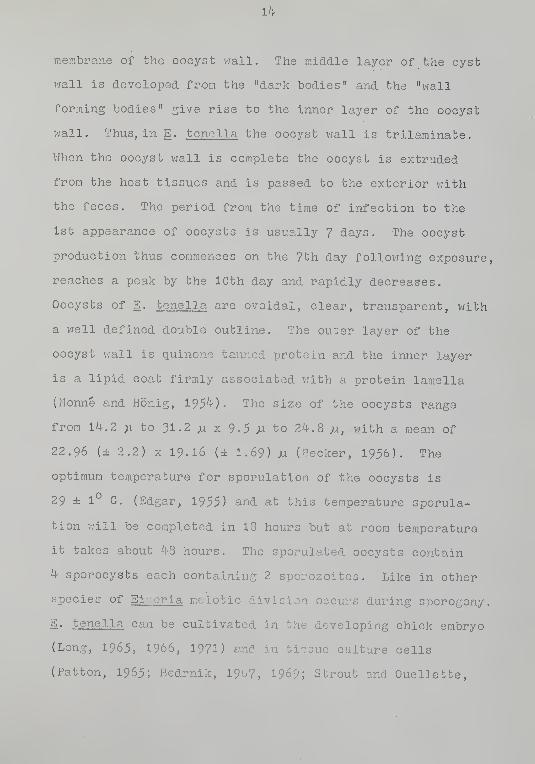
membrane of the oocyst vrall. The middle layer of the cyst
v/all is developed from the "dark bodies" and the "wall
forming bodies" give rise to the inner layer of the oocyst
wall. Thus, in E. tenella the oocyst v;all is trilarainate.
When the oocyst vjall is complete the oocyst is extruded
from the host tissues and is passed to the exterior vjith
the feces. The period from the time of infection to the
1st appearance of oocysts is usually 7 days. The oocyst
production thus commences on the 7th day following exposure,
reaches a peak by the 10th day and rapidly decreases.
Oocysts of E. tenella are ovoidal, clear, transparent, with
a V7ell defined double outline. The outer layer of the
oocyst wall is quinone tanned protein and the inner layer
is a lipid coat firmly associated with a protein lamella
(Monne and Honig, 195^). The size of the oocysts range
from 1^.2 ;i to 31.2 jLi X 9-5 Ai to 2^.8 /i, with a mean of
22.96 (± 2.2) X 19.16 (± 1.69) M (Backer, 1956). The
optimum temperature for sporulation of the oocysts is
29 ± 1° C. (Edgar, 1955) and at this temperature sporula-
tion V7ill be completed in 18 hours but at room temperature
it takes about 4-8 hours. The sporulated oocysts contain
4 sporocysts each containing 2 sporozoites. Like in other
species of Eir.eria meiotic division occurs during sporogony.
E. tenella can be cultivated in the developing chick embryo
(Long, 1965, 1966, 1971) and in tissue culture cells
(Patton, 1965; Bedrnik, I967, 1969; Strout and Ouellette,

15
1969; Matsuoka et al. , 1969; Doran, 1970), using sporozoites
obtained by in vitro excystation.
Pathogenesis of E. tenolla Infection
Factors affecting pathogenicity of E. tenella in-
clude the number of oocysts ingested', the number of host
cells destroyed per ingested oocyst, the degree of re-
infection and the state of immunity in the host. The
severity of the disease depends upon the interplay of these
known and other unknown factors and range from an imper-
ceptible reaction to death (Gardiner, 1955)- Cecal cocci-
diosis under field conditions occurs pi'incipally in young
chickens but seldom in those less than 10 or 11 days old.
The range of age of susceptibility is from 2 weeks .to 15
months. Many of the worst outbreaks occur at the age of 6
to 8 vjeeks (Blester and Schvmrte, 1965). Herrick et. al .
(1936) in a study on experimental infection found that the
heaviest mortality and greatest decrease in erythrocytes
occurred in chicks 1 month old; heavy mortality also occurred
in chicks aged 2 vjeeks and 2 months while in older birds
(3i ^, 7, 10, and 15 months of age) mortality v^fas lovi or
lacking though drop in red cell count ranged from 29;t to
^^6.8;o. Gardiner (1955) employing a dosage of 50,000,
100,000, and 200,000 sporulated oocysts infected young
chickens in age groups of 1, 2, 3, ^, 5, and 6 weeks.
Those in the ^ week group v:ere severly affected and those
in the 2 vreek group v;ere least affected. Those birds which

16
recover from infection become immune to reinfection with
E. tenella . Hov/ever, this is not an absolute immunity.
Under conditions of stress, the acquired immunity of older
birds may break down causing symptoms of the disease to
reappear (Levine, 1963)- Levine (19^' 0) in a study of sub-
clinical coccidial infection in pullets at least 8 months
old reported the presence of E. tenella in 2'yfo . In general,
however, it can be shown that chicks rigidly isolated from
infection remain fully and uniformly susceptible throughout
their lives and that age per se has no influence on resist-
ance. Under field conditions almost all chickens get early
exposure to at least light infection and so nearly all
chicks more than a few days old have some degree of resist-
ance .
Inherited resistance in some strains of chickens to
infection with E. tenella has been reported by Rosenberg
(19^1) and Rosenberg and McGibbon (19^8) but in general
there is little evidence of any significant variation in
susceptibility between different breeds or strains of
chickens (Horton-Smith and Long, I963). Jeffers and
Wagenbach (I969) reported higher susceptibility and mor-
tality of female chick embryos from widely different genetic
sources to E. tenella infection. Edgar and Herrick (19^1-^)
produced evidence to sho\^ that the presence of food in the
digestive tract of birds at the time of infection reduced
the severity of the disease. Holmes et_ al. (1937) suggested

17
that increased death rate may result in chickens having
higher amount of oyster shells in the ration. The number
of oocysts resulting from an infection is not a true
indication of the degree of infection. Tyzzer (I929)
postulated that, theoretically, infection v/ith a single
oocyst of E. tenella could give rise to approximately
1,800,000 oocysts in a period of ^ to 5 days. During the
course of an infection, hovvever, there are several factors
v/hich may cause a reduction in this potential including
loss of merozoites, over crowding, and tissue damage vfhich
results in a loss of suitable cells. Brackett and Bliznick
(1949, 1952) reported that for each oocyst of E. tenella
inoculated in light infection approximately 10,000 oocysts
are produced, and there is no direct correlation between
the size of infective dose and the final degree of ini
tion. Hov/ever, Johnson (I927) reported that the severity
of cecal coccidiosis depends on the number of sporulated
oocysts that the bird receives. Jankiev/icz and Scofield
(193^) reported that a dosage of up to I50 sporulated
oocysts produced neither symptoms nor mortality; 150 to
500 oocysts produced slight hemorrhage and no mortality;
1,000 to 3,000 oocysts a fairly heavy degree of hemorrhage
and moderate mortality and over 5,000 oocysts produced
severe hemorrhage and high mortality. The prepatent period
in E. tenella infection is 7 days but the patent period
varies viith individual infections. Fish (1931) reported

18
that oocysts were not .present in the droppings of the in-
fected birds after 1? days although Tyzzer et al. (1932)
recorded oocyst passage for as long as 19 days post-
infection. The greatest numbers of oocysts are discharged
in a very short time (Tyzzer e_t al. , 1932), the fev/ re-
maining being trapped either in the tissues, or in the
cecal contents, and irregularly released. Under natural
conditions, birds are usually infected repeatedly and thus
may pass oocysts for long periods of time. For example,
Levine (19^1-0) observed oocysts of E. tenella in the drop-
pings of 9 out of 30 birds which did not show any symptoms
of infection.
The disease symptoms in cecal coccidiosis are closely
related to the course of infection and, in general j the
degree of pathogenicity is related to the depth to which
the cecal vrall is parasitized. E. tenella penetrates
deeply and is very destructive. During the development of
the parasites there is a migration of parasitized cells
into the subepithelial region where they increase enormously
in size. Much tissue is destroyed and sloughing of mucosa
occurs at the time of maturation of 2nd generation schizonts
as early as the 96th hour after infection and profuse
hemorrhage occurs due to mechanical damage to the blood
vessels. This bleeding is the most important effect of the
parasitism. Mortality is likely to be great when profuse
and continuous bleeding occurs from the ^1-th to 7th days
post-exposure. Much of the damage may also come from

19
secondary bacterial infection of the area in v;hich the
epithelium is completely destroyed (Briggs, 1968). Hem-
orrhage is a great stress on the infected chicken and
feeding and movement are at a minimum during this period,
but consumption of v/ater is increased 2 to 3 times that
seen in uninfected birds. In a typical severe infection,
bloody droppings vjill occur 96 hours post-exposure and
passage of large quantities of blood in the droppings on
the 5th and 6th day post-exposure occur. The disease is
at its peak on the 7th day post-exposure and ^0% of the
mortality occurs vfithin 9 days follovring initial exposure
to oocysts. Chickens surviving 9 days follovjing exposure
vrill usually recover. A chronic condition, however, may
occur as the result of retention of a core of necrotic
tissue in the ceca with consequent cecal dysfunction. In
the flock as a v;hole, the disease is nearly alvfays of short
duration. It is often found that a condition of disease
arises only when infection, heavy in relation to the pre-
vious experience of the birds, is acquired during a period
of 72 hours or less. If infection is picked up more slowly,
then the birds become resistant before clinical effects
appear. When chickens are raised on deep litter as in most
parts of the xMorld, the oocysts are not necessarily destroyed
by the heat of fermentation, but due to the unfavorable
environment are predominantly unsporulated. Changes in the
environment, notably an increase in moisture and/or temperature.

20
favor a high and rapid rate of sporulation leading to a
clinical disease in the flock.
Appearance of fresh blood in the droppings and
sudden death are of diagnostic value in cecal coccidiosis.
Clotting of tlood is prevented during the acute stages by
some unknovm factor(s) (Davies e_t al.
, 1963) and deficiency
of vitamin K increases pathogenicity to E. tenella and E.
necatrix (Davies _ejt al.
, 1963). Blood drops can be ex-
pressed from the vent of dead birds picked up within a few
hours. At necropsy, blood-filled ceca and presence of
developmental stages of E. tenella can confirm the dia-MO-
sis of cecal coccidiosis. The mere presence of oocysts ir
not indicative of disease since in E. tenella infections,
oocysts are not ordinarily seen in an infection sufficiently
acute to cause disease and death (Davies et^ al.
, 1963).
For experimental infections of coccidiosis, knovm
numbers of sporulated oocysts are administered orally.
However, Davies and Joyner (1962) and Sharma and Reid (l^o2)
succeeded in producing cecal infections by introducing E.
tenella oocysts subcutaneously, intravenously, intra-
peritoneally or intramuscularly in chickens. VJhen viable
sporozoites vieve introduced by the same routes, light in-
fections were produced. The method of excystation and
transfer to the site of infection for chicken coccidia
inoculated parenterally is not yet understood. Horton-Smith
and Long (I963) confirmed these findings partially in that

21
although they could obtain oocysts in the coca aft-er
intravenous and intramuscular administration of oocysts,
they failed to recover oocysts from ceca vrhen chickens
v;ere inoculated intraperitoneally or subcutaneously . They
assumed that oocysts inoculated into the blood stream v/ould
be removed from the circulation by the liver along viith
other foreign bodies. They reported the presence of dis-
integrating oocysts in the liver of chickens inoculated
intramuscularly and suggested that the sporocysts and
sporozoites might reach the intestine and ceca via the bile
duct. Patnaik (1966) reported that when oocysts v/ere
placed in Hillipore chambers grafted within muscles, ex-
cystati.on took place inside the chamber with the help of
enzymes produced by the infiltrating leukocytes. He also
found engulfment of sporozoites by macrophages inside the
chamber and postulated that they might carry the spoi .
to various parts of the body. In all these cases much
lighter infections occurred after parenteral inoculation
compared vjith the infections occurring after oral inocula-
tion (Horton-Smith and Long, 1963).
Pathology of Cecal Coccidiosis
Involvement of the ceca rather than of the small
intestine is one characteristic of E. tenella infection.
However, if the ceca are surgically removed or, in very
heavy infections, the terminal portion of the large in-
testine will be parasitized. The lesions associated with

22
E. tenella infection in the ceca have been described by
Tyzzer (1929), Tyzzer et al. (1932) and Mayhew (1937).
The dilated part of the cecum is primarily involved and
substantial damage is due to the large numbers of rela-
tively large 2nd .generation schizonts present in the
deeper lamina propria of the mucosa. Production of
specific toxin has not yet been demonstrated although
parenteral administration of extracts of oocysts is lethal
to rabbits and not to chickens (Burns, 1959)' The patho-
logic effects of the maturation of schizonts and release
of 2nd generation merozoites are hemorrhage and sloughing
of the epithelial lining of the cecum, vjhich is sometimes
stripped dovm to the base of the subraucosa and its replace-
ment by a core composed of necrotic tissue, coagulated
blood, cecal contents, and developmental stages of the
parasite, chiefly oocysts. This core is at first adherent
to the cecal wall, but later may get detached and lie free
within the lumen. An infected bird may pass this core or
a blood clot in the droppings. The cecum of recovering
birds may regain its gross appearance but remain slightly
thickened. In lighter infections, recovery is often com-
plete and rapid, but in heavier infections, recovery is
slow and the mucosa often shovi only incomplete regeneration.
The continuous hemorrhage from the ^th to 7th day post-
exposure results in profound anemia vrhich is often the
cause of death. The exposed skin and mucous membranes

23
become pallid. Erythrocyte counts and hematocrit decrease
to about 50fo of thG normal on the 5th and 6th day after
infection vjith 50,000 E. tenella oocysts, and the values
return to normal in about 8 days (Natt and Herrick, 1955)
•
Natt (1959) observed lymphocytopenia and heterophilia on
the 5th day and an eosinophil.ia on the 10th day following
infection v/ith E. tenella . There were no significant
changes in the monocyte and basophil numbers during the
course of infection. A marked leucocytosis began on the
7th day post-infection and persisted through the recovery
phase of the disease. Pratt (19^0, 19^1) observed an
increase in blood sugar during the acute stages of the
disease v/ith a decrease in the muscle glycogen. V/axlov
(19^1) also found a rise in blood sugar on the 5th day
post-infection and a rise in blood chlorides on the 6th
and 7th days post-infection v/ith accompanying reduction
in muscle chloride. However, Freeman (1970) found no
hyperglycemia or change in hepatic glycogen, but he noted
a significant reduction in the plasma lactate concentra-
tion on the 1st and 2nd days after exposure 3Jid a rise in
cardiac glycogen on the 5th day post-infection. According
to Daugherty and Herrick (1952), during the acute stages
of the infection, a substance produced in the cecum reduced
the ability of the brain of the chicken to utilize glucose
but not hexose diphosphate. Thus, in cecal coccldiosis
a severe interference V7ith normal phosphorylative ciarbo-
hydrate utilization may occur. Challey (l';62) noted an

2if
increase in adrenal ascorbic acid and adrenal corti-
costerone concentrations during the acute hemorrhagic
phase of the infection. Bertke (1963) found renal clear-
ance of uric acid in chickens infected vfith E. tenella
greatest at 2 to ^ days after infection. This study-
suggested that death is not due to cecal tissue destruc-
tion per se nor is it entirely due to cecal bleeding, but
to the failure to recover from an initial shock resulting
from the development of large numbers of endogenous stages.
It is also reported by Johnson and Reid (1970) that in
some cases the gross lesions in the ceca in live birds
will be more severe than those in dead birds. Husajev
and Surkova (1970) studied the nitrogen metabolism of
chickens infected v/ith E. tenella and noted that the total
and protein nitrogen decrease within the categories of
all ages, on the 3rd and especially on the 5th day and
this coincides with the period of development of endogenous
stages '-^ the parasite. They assume that these disorders
are associated with many factors, such as disorders of
fermentation and suction and influence of metabolic pro-
duct of the parasite. They concluded that on the basis of
protein metabolism in the liver, deeper pathological changes
occur in young chickens than in older birds.
Early work by Levine and Herrick (195^, 1957)
showed that the voluntary muscles in infected birds are
unable to do more than 50;^ of the work done by muscles of

25
uninfected birds when stimulated via the nerves.- Hov/ever,
Freeman (1970) found that v;hen the muscles are directly
stimulated they are able to do i;ork and speculated that
an impairment of nervous conduction ab the neuro-muscular
junction occurred in coccidiosis.
As for cellular responses, Pierce _et_ aJ. (I962)
shovred that during the primary infection with E. tenella .
heterophilpolymorphonuclear cells infiltrate into the
submucosa in increasing numbers especially on the ^1-th or
5th day post-exposure when the 2nd generation schizonts
are developing and maturing. Lesion scoring has been
frequently used to compare quantitatively the extent of
gross lesions and pathology. Herrick e_t al^. (19^!'2) first
described a method of scoring E. tenella lesions using a
to k+ scoring system. This scoring system has been
followed by many vjorkers (Ripsom and Herrick, 19^5;
Gardiner and Farr, 195^; Guckler, 1957; Bankowski et al.
.
1959; Horton-Smith et al. , I96I; Lynch, I96I; Britton
et al. , 196'!; Turk and Stephens, 1967; Dunkley, I968). A
modification of this was also used by Guckler et al.
(1958), Waletzky et. al. (19^9-1950), Ball (1959), Boney
(19^8), Farr and VJehr (19-!'5), Levine and Barber (19'-l-7),
Wale 1 2 ky and Hughes (19'4-9-1950) . Horton-Smith et al.
(1961) used a system for macroscopic grading of lesions
and correlated this by microscopic grading of parasitism,
depending upon the presence of endogenous stages. Johnson
and Reid (1970) used a grading system for gross pathology

26
supplemented by examination for developmental stages in
the cecal contents. Lesion scoring is time consuming.
Should other disease conditions such as ulcerative enteri-
tis appear in the pens, more extensive microscopic studies
may be required to decide vfhether lesions are induced by
coccidiosis. Norcross and Washko (1970) examined in-
testinal tissues from 73^ cases of clinically diagnosed
or suspected cases of coccidiosis histologically and con-
firmed coccidiosis only in 53.2)'^ of the cases. No ;pecific
pathological manifestations could be found in the intesti-
nal tissues of 28.6;^ of the cases. The remaining cases
were diagnosed in descending order as ulcerative enteritis,
leukosis or Marek's disease, other enteritides, helminthia-
sis and histomoniasis. These studies stress the necesf^i+-
of both macroscopic and microscopic examination of tn.
intestinal tissues for confirming pathology due to E.
tenella infection.
Microflora and Hosts
In nature, animals live in intimate contact vjith
many microorganisms. The microorganisms are thus found
either in the immediate environment, on the superficial
tissues, or in the gastrointestinal tract of animals.
This close association has led, in many cases, to symbio-
tic relationships betvreen the host animal and the micro-
organisms. The apparently healthy laboratory and other
animals used to study many biological processes carry

27
their indigenous microflora and these animals are known as
"conventional" animals. In contrast, a "germ-free" animal
is one from vjhich it is not possible to recover any viable
organism. Many laboratories employ tests to detect bac-
teria, fungi, helminth parasites, PPLO, and certain viruses
to determine the germ-free state of the animals (Nev/ton,
1965). The terra "gnotobiote" (Reyniers e_t al . , 19'^9) is
also used in referring to the germ-free animal and also
animals carrying known species of organisms. A "specific
pathogen-free" (SPF) animal is one free of specified micro-
organisms and parasites known to cause disease (Sabourdy,
1965). The SPF animals are functionally and structurally
identical v^-ith their conventional counterparts but their
flora and fauna are, to some extent, controlled. The
production of germ-fi'^ee or SPF animals is a problem if the
particu.i...'-r' animal species have certain congenital infec-
tions. Salnonella pullorum in chicks and Toxocara canis
in dogs (Reece e_t al. , I968; Griesemeret al. , I963) are
two infections transmissible congenitally . Careful selec-
tion and isolation of breeding stock free of these infec-
tions and prompt elimination of young animals or chicks
shovring any congenital infections are necessary to insure
absence of such infections in the breeding stock (Reece
ejt al . , 1968). These elaborate procedures to establish
and maintain germ-free or specific pathogen-free animals
for research purposes vrill eliminate the necessity for
using experimental animals of unknovjn disease exposures,

28
age, breeding and most important environmental background.
The possible influence of the "macro" and "micro" environ-
ment in disease processes can be studied v/^ien a stock of
animals derived from a breeding colony is raised
under 3 different environments—conventional, SPP and
germ-free.
A number of workers have attempted to raise germ-
free animals since Pasteur's speculation in 1885 that the
host-microflora relationship is obligate. But the in-
vestigations of Schottelius (1899), Cohendy (1912),
Cohendy and V/ollman (191^), Kilster (1912), Glimstedt
(1936), Balzara (1937a, 1937b), Reyniers (19^^6, 19^9,
i960), Gustafsson (19^8) and Miyakawa (195'-^-) proved
Pasteur's original assumption v7rong. Novf the germ-free
animal has become a very useful tool for studying true
homeostasis of the gnotobiotic host, the individual actu".
and interactions of microorganisms and the response of the
host to these organisms. These interactions have not yet
been v/ell defined although a few significant microflora-
host relationships have been established.
The Germ-Free Chick
The embryo of healthy birds is maintained in a
germ-free condition within the shell until hatching. This
has enabled germ-free chicks, turkeys and other birds to
be obtained v:ith relative ease. Gerin-free chickens are
very popular as experimental animals and have been

29
successfully used to study such research problems- as the
origin of blood group B agglutinins (Springer et al.
,
1959), the growth stimulation of dietary antibiotics
(Lev and Forbes, 1959), experiments on tumorigenesis
(Reyniers and Sacksteder, 1959), development of parasitic
infections (Bradley et al. , 1967), and for a study of the
germ-free state per se compared with the conventional
animals (Reyniers ejb ad . , 19^9, i960).
The germ-free chickens are less clean than con-
ventional chicks due to the high humidity in the imits
and also by the frequent occurrence of anal blockage and
loose nature of the feces of the germ-free chickens. The
morphology and function of the gastrointesti]ial tract
are altered in the absence of viable flora in the germ-
free animals (Reyniers, 196O). The intestinal mucosa
had less lymphatic development and less connective ti ?
mass in germ-free chickens and the general picture v;as
that there were more absorptive elements but fewer and
less well developed elements of defense (Reyniers e_t al. .
i960). Gordon (i960) founo about 3 times greater numbers
of reticulo-endothelial cells in the mucosa and the sub-
mucosa of the ileum of young conventional chickens than
in germ-free chickens. This difference was also noted
for "schollen" or globule leukocytes found within the
epithelium and in the number of plasma cells and lympho-
cytes in the submucosa and lamina propria of the lower

30
ileum. Hdvfever, the -epithelial cell content vjas greater
in germ-free chicks . The amount of lamina propria in the
total area studied vias 25-6 ± 0.9fo in germ-free chicks
and 36.8 ± U\'fo in conventional chicks. Eyssen and
DeSomer (I967) reported that the v^e.ight of the small
intestine vras 105/;^ greater in conventional than in germ-
free chicks. Enlargement of the cecum v;as a notable
change in several species of germ-free animals including
the rat, mouse, rabbit and guinea pig (Wostmann and
Bruckner-Kardoss, 1959). However, cecal distention vms
not observed in germ-free chickens and turkeys (Luckey,
1963). The ceca of the germ-free chickens vrere found to
be significantly shorter in length than those of chickens
with a normal bacterial flora (Hegde e_t al. , 1969). From
gross observation, the large intestine and cloaca, of con-
ventional and germ-free chicks v;ere similar. Hov/ever,
the relative wet weight of large intestine per 100 grams
body weight vias greater in germ-free Leghorn chickens.
The feces of germ-free and conventional chicks were grossly
similar, but the germ-free chicks v:ere more susceptible to
diarrhea. The lymphatic system is poorly developed in
germ-free animals. Using the ileo-cecal tonsils of birds
as an index of the lymph node development, generally a
great difference was found (Reyniers e_b al . , 196O; Gordon,
i960) between germ-free and conventional chickens. The
ileo-cecal tonsils of conventional chickens v;ere of larger
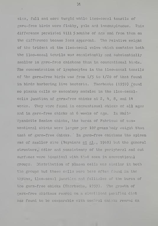
31
size, full and more turgid v/hile ileo-cecal tonsils of
germ-free birds V7ere flabby, pale and inconspicuous. This
difference persisted till 5 months of age and from then on
the difference became less apparent. The relative v;eight
of the trident at the ileo-cecal valve vrhich contains both
the ileo-cecal tonsils v/as consistently and substantially
smaller in germ-free chickens than in conventional birds.
The concentration of lymphocytes in the ileo-cecal tonsils
of the germ-free birds ;7as from 1/5 to 1/2 of that found
in birds harboring live bacteria. Thorbecke (1959) found
no plasma cells or secondary nodules in the ileo-cecal-
colic junction of germ-free chicks at 2, ^l, 8, and 14
v:eeks . They vjere found in conventional chicks of all ages
and in germ-free chicks at 6 vfeeks of age. In White
V/yandotte Bantam chicks, the bursa of Fabricus of con-
ventional chicks were larger per 100 grams 'Dody weight than
that of germ-free chicks. " In germ-free chickens the spleen
was of smaller size (Reyniers et_ al.
, I960) but the general
structure, color and consistency of the peripheral and cut
surfaces v:ere identical vjith that seen in conventional
groups. Distribution of plasma cells vras similar in both
the groups but these cells were less often found in the
thymus, ileo-cecal junction and follicles of the bursa of
the germ-free chicks (Thorbecke, 1959). The growth of
germ-free chickens reared on a sterilized purified diet
v/as found to be comparable vrith control chicks reared on

32
natural commercial diet, but the grovfth rate is reported
to be faster in gerra-free chicks (Forbes and Park, 1959;
Forbes et al.
, 1959). Reyniers et_ al. (I960) found growth
and reproduction to be normal but egg production and
hatchability poor in germ-free chicks. The red cell
morphology, hemoglobin concentration, hematocrit values, and
the expressed blood volume vrere identical in germ-free and
conventional chickens, but leukocytes were 2 to 5 times
more numerous in conventional chickens. Circulating lympho-
cytes v;ere also high enough to state that the presence of
living microorganisms and/or their products had an effect
on the numbers of lymphocytes (Luckey, 1963). The low
level of antibody containing globulin fractions is one of
the characteristics of the gern-free animal (Balish and
Phillips, 1966; Thorbecke e^ al. , 1957). The gamma-1
,
gamma-2 and beta fractions of the globulin fraction of the
blood of germ-free chicks 'vrere also low (Wostmann, 1959)
and unlike in conventional chicks, no change in serum
gamma globulin occurred in germ-free chicks as they
matured. It is not knovrn if the low gamma globulin of
the gerra-free chicks is "innate" or produced as a result
of an unknown antigenic stimulus. Boggs e_t al. (1967)
studying granulocytopoiesis in germ-free mice reported a
lower concentration of neutrophils in blood of gerra-free
mice but the presence or absence of microorganism did not
alter the overall granulocytopoiesis.
A more positive oxidation-reduction potential of

33
cecal contents appears directly related to .the absence of
gastrointestinal microflora. Balish and Phillips (I966)
and Springer (1968) found the oxidation-reduction poten-
tials of bacteria-free cecal contents of the chicken
strongly positive v;hile those of the conventional chicken
vjere strongly negative. Balish and Phillips (I966) re-
ported that the pH vms higher in all segments of the gut
in germ-free chicks when compared to that seen in conven-
tional chicks. Tlie above studies indicated that the germ-
free chicks shov; acceptable normal grovrth and reproduction.
In addition, porosis or spontaneous tumors are not common
in germ-free chicks. They survived x-irradiation better
than conventional chicks when the dosage was below 800 r
at the rate of 32 r per hour (McLaughlin et_ al.
, 1958).
No gross physiological abnormality has been reported in
germ-free chicks. One condition called "jitters" v:as re-
ported by Gordon et_ al. (1959) in germ-free chicks due to
cellular proliferation in the brain.
The serum of germ-free animals has been shovm to have
a low globulin content and very few anti-bacterial agglu-
tinins, though complement and heteroheraagglutinins are
present. This, together with paucity of leukocytes and
poor phagocytic response, may make the germ-free animals
very susceptible to pathogens, although, surprisingly,
germ-free animals were found to be very effective in clear-
ing injected particles or dead bacteria (Luckey, 1963).
The pathology of many infectious diseases has been studied
in gei'm-free animals.

3^
Pathop:eni city of Intestinal Parasites and Parasifcism
in Gnotobiotic Hosts ••'
Several investigators have utilized gnotobiotic
hosts for the study of certain aspects of the host-parasite
relationship. In contrast to the research on axenic (in
vitro ) cultivation of parasitic organisms which has been
directed tov;ards learning the biochemical and immunochemical
characteristics of the organisms, the use of gnotobiotic
hosts has been oriented towards the in vivo study of the
etiology and pathogenesis of certain diseases. A number of
studies on the development and pathogenesis of intestinal
protozoa and helminths of human and animal importance have
been done in gnotobiotic hosts. These studies have shown
the relationship between the host, the parasite and the
host's intestinal microflora (chiefly bacteria and fungi).
Phillips et_ al. (1955) showed that Entamoeba histolytica
can produce pathological lesions in guinea pigs only vjhen
species of bacteria such as Escherichia coli and Aerobacter
aerogenes are present. However, another parasitic proto-
zoan, Pentatrichomonas ( Trichomonas ) hominis . developed in
large numbers in germ-free guinea pigs (Phillips, 1962).
Pentatrichomonas ( Trichomonas ) vaginalis , iihen subcutan-
eously injected into germ-free guinea pigs, produced large
lesions, but similar administration in conventional guinea
pigs resulted in disappearance of the protozoans in a few
days (Ne^^fton et al,. , I960).
Experimental infection of gnotobiotic mice with
Nematospiroides dubius and Nippostrongylus brasiliensis

35
have been produced and studied (Nev/ton et_ al . , 1959;
Westcott, 1971). In general, more parasites developed,
infections were of longer duration, and more helminth eggs
were produced in the conventional than in germ-free hosts.
Eosinophilia was marked in germ-free mice folloviing nema-
tode infection, but no eosinophilia was seen in conventional
mice. Nodule development in the intestinal vjall was seen •
in both types of hosts, however, the nodules disappeared
from conventional hosts rapidly but persisted up to 60
days in germ-free mice. VJeinstein ejt al,. (1962) reported
that the larvae of N. clubius vrill not develop to the in-
fective stage in feces from germ-free mice as they normally
do in feces of conventional mice. At least in this car;,
the intestinal microflora contributed to the prolonged
survival and egg production of the helminth species.
Another interesting study by Newton _et al. (1959) revealed
that the mouse helminths N. dubius and Hymenolepis nana ,
which do not develop to maturity in the conventional guinea
pigs, can do so in the germ-free animal.
Johnson et al_. (196?) and Rohovsky and Griesemcr
(1967) found feline infectious enteritis in the germ-free
cat is a mild, nonfatal disease vjith symptoms of leuko-
penia, thymic atrophy and lymphoid depletion, but v?ithout
morphologic intestinal lesions and clinical signs. In SPP
cats, clinical signs, ultrastructural alteration of the
intestinal mucosa and reduced enzyme activity were noted
(Johnson et_ al . , I967; Fovjler and Rohovsky, 1970).
36
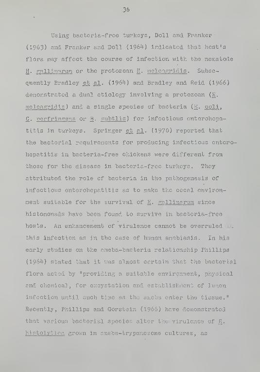
Using bacteria-free turkeys, Doll and Franker-
(1963) and Franker and Doll (1964) indicated that host's
flora may affect the course of infection with the nematode
H. p:allinarum or the protozoan H. melea,p:ridis . Subse-
quently Bradley e_t al. (1964) and Bradley and Reid (I966)
demonstrated a dual etiology involving a protozoan (H.
meleagridis ) and a single species of bacteria (E. coli .
G. perfringens or B. subtlis ) for infectious enterohepa-
titis in turkeys. Springer e_t al. (1970) reported that
the bacterial requirements for producing infectious entero-
hepatitis in bacteria-free chickens were different from
those for the disease in bacteria-free turkeys. They
attributed the role of bacteria in the pathogenesis of
infectious enterohepatitis as to make the cecal environ-
ment suitable for the survival of H. gall inarurn since
histomonads have been found to survive in bacteria-free
hosts. An enhancement of virulence cannot be overruled lu
this infection as in the case of human amebiasis. In his
early studies on the araeba-bacteria relationship Phillips
(1964) stated that it vjas almost certain that the bacterial
flora acted by "providing a suitable environment/ physical
and chemical, for excystation and establishment of lumen
infection until such time as the ameba enter the tissue."
Recently, Phillips and Gorstein (I966) have demonstrated
that various bacterial species alter the virulence of E.
histolytica grown in araeba-trypanosome cultures, as

37
measured by subsequent inoculation into animals. V/ittner
and Rosenbaum (1970) studying the role of bacteria in
modifying the virulence of E. histolytica found that the
increased virulence is associated only vjith contact of
ameba with live bacteria and speculated transfer of an
episome-like virulence factor from bacteria to the proto-
zoans.
Reid and Botero (1967) reported the growth of the
cestode, Raillietina cesticillus . in bacteria-free chickens
and concluded that no contribution to tho establishment of
the tapeworn or interference from the normal bacterial
flora of the digestive tract occurs. Johnson (1971) re-
ported tho grov.'th and development of Ascaridia galli in
gnotobiotic chickens and the data indicate an inhibit;--
of development of the nematode in the bacteria-free
chickens
.
On the other hand, -Balish and Phillips (I966) re-
ported that oral challenge v/ith Candida albicans resulted
in crop infection in all bacteria-free chicks but no in-
fection occurred in conventional chicks. Layton and
Simkins (I971), in their studies with f''ycoplasma ^alli -
septicum . found higher mortality (Sjfo) in germ-free chicks
than in conventional chicks (38/0. In. gnotobiotic svrine,
Kohler and Gross (I969, 1971) have described diarrheagenic
effects due to heat-stable filtrates of broth cultures
and whole cell lysates of Escherichia coli and Meyer et al.
(1964, 1967, 1971) described a polyserositis-like syndrome

38
due to E. coll in gerin-free pigs. These studies -all
indicate that normal flora has either a beneficial or
antagonistic action on many of these pathogenic organ-
isms. Another intriguing role for the associated micro-
flora in the host-parasite relationship, perhaps in
determining host specificity, was the resistance of the
conventional guinea pigs to Trypanosoma cruzi . Hov^ever,
a majority of the germ-free guinea pigs harbored the
trypanasomes in their blood following intracecal inocula-
tion (Phillips and Wolfe, 1959). In contrast, the
bacterial flora had no role in the establishment and
pathogenesis of E. brunetti in the chicken intestine
(Hegde et al. , I969). Clark et_ al. (I962) also found very
little difference in the pathogenicity of E. tenella in
conventional and bacteria-free chickens although there
V7as a delay of 12 to 15 hours in the appearance of the
2nd generation merozoites in the feces of gnotobiotic
hosts. In a more recent study, Visco and Burns (I966,
quoted by Hegde et al.
, I969) reported no mortality in ^1
gnotobiotic chickens infected vjith E, tenella as compared
to 77/3 mortality in infected conventional chickens. They
concluded that a close relationship exists betvreen the
host flora and the protozoan in the production of cecal
coccidiosis syndrome. Kemp e_t al. (197I) reported a
delayed development of endogenous stages of E. tenella
in germ-free chicks, especially 2nd generation schizonts,
gametocytes and oocysts. There v/as a striking lack of

39
reticulo-endothelial cells in the lamina propria and
submucosa and substantially lov; numbers of mononuclear
inflammatory cells. Thus, the effects of the bacterial
flora on the pathogenicity of this species remain
unresolved.
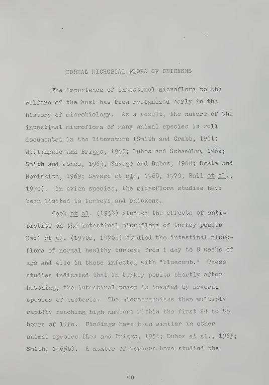
KORIiAL MICROBIAL FLORA OP CHICKENS
The importance of intestinal microflora to the
welfare of the host has been recognized early in the
history of microbiology. As a result, the nature of the
intestinal microflora of many animal species is vjell
documented in the literature (Smith and Crabb, I96I;
Willingale and Briggs, 1955; Dubos and Schaedler, 1962;
Smith and Jones, 1963; Savage and Dubos, 1968; Ogata and
Morishita, I969; Savage ejb al, , I968, 1970; Rail et al . ,
1970). In avian species, the microflora studies have
been limited to turkeys and chickens.
Cook et_ al. (195'-0 studied the effects of anti-
biotics on the intestinal microflora of turkey poults,
Naqi et. al. (1970a, 1970b) studied the intestinal micro-
flora of normal healthy turkeys from 1 day to 8 vreeks of
age and also in those infected vrith "bluecorab." These
studies indicated that in turkey poults shortly after
hatching, the intestinal tract is invaded by several
species of bacteria.- The microorganisms then multiply
rapidly reaching high numbers v:ithin the first 2^1- to ^8
hours of life. Findings have been similar in other
animal species (Lev and Briggs, 1956; Dubos et. al. , 1965;
Smith, 1965b). A number of workers have studied the
40

^1
normal bacterial flora of conventional chickens (Johansson
ejb al., 19^8; Shapiro and Sarles, 19^9; Lev and .Briggs,
1956; Lgv et al.
, 1957; Huhtanen and Pensack, 1965; Smith,
1965a; Timms, I968; Barnes and Impey, I968). Factors such
as age, alimentary tract structure and function, diet,
feeding habits and environmental factors have been shown
to influence the bacterial flora of the normal gut
(Johansson et al. , 19^8; Smith, 196I; Smith, 1965a, 1965b;
Smith and Crabb, 196I). All these studies indicated that
the numbers of bacteria of all groups were found to be
highest in the cecal contents and progressively lov;er
numbers in the posterior large and anterior small intes-
tine, respectively. The organisms constituting the major
part of the flora v;ere E. coli . enterococci ( Streptococcu.-
fecalis ) . Lactobacillus sp. , Bacteroides sp. and C. per-
fringed.". The absence of Bacteroides sp. in the small
intestine, the preponderance of Bacteroides sp. and Lacto-
bacillus sp. in the ceca, and the low levels of C. per -
fringens in all sites v:ere of particular interest to the
investigators. Shapiro and Sarles (19^9) found the count
of aerobic and anaerobic bacteria to be similar in chickens
of different ages. On the contrary, Huhtanen and Pensack
(1965) found a preponderance of anaerobes after 2 weeks of
age. Their results also indicated that in day-old unfed
chicks the flora consisted mainly of S. fecalis. These
enterococci gradually disappeared from the duodenum after
6 days of age. The cecum also showed an initially high

^2
count of enterococci and aerobic bacteria. These were
replaced by anaerobic types at around 1^ days of age.
The normal bacterial flora has been reported to
influence the host-specificity of some parasites (Newton
ejb al.. , 1959). Another interesting phenomenon is the
decrease and/or increase in the population of some members
of the flora during certain pathological conditions.
Balish and Phillips (I966) reported that in C. albicans
infection of the crop, the count of enterococci v;as in-
creased. Naqi et_ al. ( 197Cq) reported significant differ-
ences in the intestinal microflora in turkeys inoculated
vjith an infectious enteritis ("bluecomb") agent and un-
infected control turkeys. The changes were characterized
by a rise in total microbial count of the entire intes;'
a significant increase in number of conforms, lactose
nonfermenters and Clostridia. Lactobacillus sp. decreased
with severe infectious enteritis but increased when the
disease was mild. Microflora changes similar to these
findings have been observed by Smith and Jones (I963,
1967) and Ogata and Morishlta (1969) in pigs inoculated
experimentally v/ith an enteric pathogen. Johansson and,
Sarles (19^f-8) noted that in E. tenella infection, a stimu-
lation of the growth of C. perfringens occurred with con-
current decrease of Lactobacillus sp. and a rise in blood
glucose level during the 5th to 7th day post-infection.
This may be related to an iiiterf erence in glucose metabolism

^3
and a role for the flora in the pathogenesis of cecal
coccldiosis. Thus, in cecal coccidiosis the problem to
be studied is vrhether or not the bacterial flora present
in the intestine aid or hinder the initiation of the
disease and subsequent development of pathological changes.
A particular species or a combination of species may (or
may not) help excystation of oocysts, subsequent liberation
and survival of sporozoites and invasion of cecal epithe-
lium, development of schizonts and/or gametocytes and
thereby contribute to the tissue damage.

MATERIALS AND METHODS
I. Production of Gnot obi otic Chickens
Gnotobiotic (bacteria-, fungi- and PPLO-free)
chickens vjere raised in flexible plastic film isolators
follov;ing the method of Bradley et al. (1967). All
isolators, accessories and supplies were obtained from
the same source.^ The methods employed in sterilization,
maintenance, and operation of gnotobiotic environment
chambers and equipment, the sterilization of feed sup-
plies and the scheme used for determining the micro-
biological status were the same as those described by
Bradley et al. (196?)
•
Day-old or 19-day-old embryonated White Leghorn
chicken eggs vjere obtained from a commercial hatchery*^
free of Salmonella and Mycoplasma infection and incubated
at the laboratory. Prior to introduction into the isolator
chambers, all eggs were candled at least tv:ice to insure
the viability of the embryos. The surface of the egg
shells was sterilized by immersing the eggs (packed in a
tubular nylon net) for 8 minutes in a 2% solution of
'•G. F. Supply Division, ^31 North Quentin Road,Palatine, Illinois 6006?.
pFlorida State Hatcheries, Gainesville, Florida
32601.
44

^5
mercuric chloride held at 37° C. The eggs v;ere then .drawn
into the presterilized isolator by means of an egg chute
and placed in a plastic tray containing a cotton towel.
After removal of the egg chute and sealing of the entry
port, the entire isolator vjas placed in a room held at
37° C and 80-85>^ relative humidity for hatching. After
hatching, the egg shells v/ere removed and the chickens
transferred to a plastic vjire-floored basket inside the
isolator. Sterile feed and v;ater vrere provided ad libitum .
II, Production of SPF Chickens
Chickens hatching from fertile eggs obtained from
the same source as that from which eggs v;ere obtained for
production of gnotobiotic chickens v;ere immediately trans-
ferred to modified Horsfall-Bauer units. Altogether 10
such units were kept in a room adjoining those in v/hich
the plastic film isolators v;ere kept for the production
of gnotobiotic chickens. Air entering the Horsfall-Bauer
units vms sterilized by passage through sterilized fiber-
glass filter media. Sterile water was supplied in gallon-
sized bottles attached to the inlet tube of the unit and
the level controlled by gravity flow. Feed consisted of
chicken starter mash free of any antibiotics or added
chemicals and v/as of composition meeting National Research
Council (NRG) standards. The feed was pasteurized in a
hot air oven at 150° C for at least 60 minutes and vjas
supplied to the chickens in a metal self-feeder inside

46
each unit. Temperature was controlled eleefcrically and
ventilation vias fan-forced, negative pressure. Before
and after each use, the units vfere cleaned and scrubbed
vfith hot detergent solution and steara sterilized. As far
as possible, the units viere opened only 3 times during an
experiment—to enter nevjly-hatched chicks, for exposure
of the chicks to E. tenella oocysts, and to remove dead
birds. Periodically, clinical laboratory tests were
conducted to check the pathogen-free nature of the birds.
All experimental chickens raised in these units were
monitored by standard laboratory methods for the follow-
ing specific pathogens:
1. the 9 species of Eiraeria causing coccidiosic
in chickens
;
2. the common intestinal helminths of chickens
(Ascaridia, Heterakis . and Baillietina
species);
3. Salmonella and Pasteurella species; and
4. H. meleagridis .
III. Production of Conventional Chickens
Newly-hatched chickens viere transferred from the
incubator and reared in electrically-heated Lattery brood-
ers. Unsterilized chicken starter mash, with no anti-
biotics or added chemicals, satisfying NRG requirements,
and water vrere made available ad libitum . Before and
after each use the pens were cleaned, scrubbed with hot
detergent solution, and steara sterilized.

47
IV. Source of Eimerj-a tenella
The same strain of E. tenella isolated from a
natural case of cecal coccidiosis v;as used throughout
this series of experiments. Oocysts were produced
according to need in disease-free 3-v^eek-old conventional
chickens. Fecal material from donor chickens was col-
lected at 7-9 days after inoculation v:ith a sublethal
dose of oocysts and the fecal debris and other gross
particles removed by sieving through 30 and 80 mesh
sieves. The oocysts viere then sedimented by centrifuga-
tion and allov?ed to sporulate in 2fo potassium dichromate
solution at room temperature and after sporulation were
stored at 5° G. Immediately before use in experimental
trials, all oocysts viere surface-sterilized vrith a 0.5^
solution of peracetic acid (Doll et al. , 1963). Sterility
v/as tested using standard bacteriological and mycological
procedures. Approximately 100,000 sporulated oocysts were
administered orally for experimental infection of chickens,
using a small syringe with an attached cannula to insure
deposition into the crop.
V. PatholOFcical Examinations
Chickens inoculated with oocysts v;ere kept under
close observation. Bleeding or any other clinical signs
were noted. Blood samples for packed cell volume deter-
mination and serum, analysis were taken both before

k8
inoculation and on the 7th day after exposure. Bacterial
isolates vrere made on both the control and experimental
chickens at necropsy, according to the bacteriological
procedure described belov;. Chickens dying after exposure
to oocysts and all those surviving on the 7th day after
exposure vrere necropsied, examined for gross lesions of
cecal coccidiosis and the cecal contents examined for
various developmental stages of E. tenella . The infection
produced in each inoculated group was compared and graded
for macroscopic lesions. For histopathological examina-
tion, cecal tissues showing gross lesions and cecal
tissues from comparable sites from chickens showing no
gross lesions vjere fixed in neutral 10/^ formalin and
sectioned at 5 or 10 ;ji thickness and stained v/ith'
hematoxylin-eosin. The system of macroscopic grading
of lesions and microscopic grading of parasitism was
modeled after Horton-Smith et al. (I96I) as follows;
Scheme of Grading of Lesions and Parasitism
Extent of Macroscopic MicroscopicLesions and Grading of Grading ofParasitism Lesions Parasitism
No detectable No coccidial stageslesions found even on care-
ful search
Small number Small number ofof lesions gametocytes found
by careful search(5 or less)

^9
Extent ofLesions' andParasitism
++
+++
++++
MacroscopicGrading ofLesions
Moderate number oflesions vjith somehemorrhage
Numerous lesionsand hemorrhage
Numerous lesionswith severe hemor-rhage and cecalenlargement
MicroscopicGrading ofParasitism
Small number of 2ndgeneration schizontsand/or gametocytes inscattered groups withsome associated tissuedamage
Numerous vjidely dis-tributed gametocytesin localized foci withappreciable tissuedamage
Numerous schizontsand/or gametocytesv/ith widespread tissuedamage
VI. Bacteriological Procedures
A. Determination of microbial flora of the cecum in
conventional chickens
The development of cecal microbial flora in disease-
free young conventional chicks and those inoculated with
100,000 sporulated oocysts was studied as follows. Newly-
hatched White Leghorn chicks were raised on standard
electrically-heated battery brooders and fed unsterilized
feed and water ad libitum . Four uninfected control and ^
infected chickens were necropsied at various age intervals:
day-old (control only), 6-day-old, 1, 2, 3, ^ and 5 weeks
of age. Inoculation with oocysts was adjusted so that the
day of necropsy v;ould be 7 days after exposure. At
necropsy, the viscera vms exposed and the cecal pouches
together vfith 2 inches of anterior small intestine and

50
posterior large intestine were transferred to a sterile
petri dish. Using aseptic procedures, the cecal pouches
were opened, the sterile tip (0.0025 crn^) of an inoculat-
ing needle was inserted into the cecal contents, withdrawn,
and the material on the loop streaked on the surface of
each of the various solid media used in a standard petri
dish or inoculated into a tube of broth or semisolid medi
In case of dry cecal contents or a formed cecal core, a
drop of sterile pH 7-0, 0.06? M phosphate buffer was used
to soften the material prior to insertion of the sampling
loop. Aerobes v/ere enumerated on Difco^^ brain heart
infusion agar; enterococci in Difco^ azide-dextrose broth
supplemented with 10 p. p.m. methylene blue and 1. 5fo agar;
and conforms on MacGonkey agar. The medium used 'for
determining the anaerobic populations was Difco^ brain
heart infusion agar supplemented with O.lfo cynteine-
hydrochloride and 0.1/^ bovine serum. C. Dsrfringens xvas
enumera'ed on Difco^ sulphite polymyxin sulphadiazine agar.
For fungus isolation, Difco^ Sabouraud dextrose agar andRDifco Pagano-Levine agar with antibiotics were used; for
protozoan isolation,Difco^ Balarauth medium was used. Time
of incubation vjas 18 hours for coliforms, 2^ hours for
Clostridia and ^8 hours for all other organisms. After
incubation, the colonies from each of the 4 uninfected
controls and the ^ infected ceca were counted and mean
numbers calculated.
^Difco Laboratories, Detroit, Michigan 48232
51
B. Determination of microbial flora of the cecumin germ-free. SPF. and conventional chickens
At necropsy, the primary isolation media used for
the recovery and identification of bacteria, fungi and
PPLO from the ceca of the germ-freo, SPF and conventional
chickens are listed in Table I. The criteria listed by
Breed e_t al. (1957) were employed for characterization,
identification, and verification of bacterial species.
Anaero^ ic plates vfere incubated in a Gaspack Anaerobic
Jar #60^10 using Gaspack gas generator envelopes for
generating hydrogen and carbon dioxide gas. Methylene
blue was used as the indicator.
VII. Packed Cell Volume
Hematocrit values vieve determined by drav;ing the
blood into a heparinized microcapillary tube and centri-
fuging the blood in an International Microcapillary
Centrifuge for 5 minutes at 15,000 r.p.m. The packed
cell volume (PCV) was directly read using an Internatic.ial
oCapillary Reader.
VIII. Total Serum Protein
Total serum protein was determined by the method of
Weichselbaum (19^6) with slight modifications. A standard
^Baltimore Biological Laboratory, Baltimore,Maryland.
2 International Equipment Company, Needham Heights,Massachusetts.

52
Table I
Primary Isolation Media Used for the. Recoveryand Identification of Bacteria and Fungi from
Ceca of SPF and Conventional Chickens
Difco azide-dextrose broth vfith 10 p. p.m. methylene blueand 1
.
5% agar
Difco^ blood agar ^^/ith 0.5/^ defibrinated bovine blood
DifccP brain heart infusion agar with O.l^o cysteine-Hcland ^.1% bovine serum
Difco^ Brewer's anaerobic agar
Difco^ blood agar with 0.5/o blood agar and neomycin
Difco^-Columbia broth
Difco enterococci presumptive broth
pDifco MacConkey agar
PDifco Pagano-Levine base v;ith triphenyl tetrazoliumchloride and antibiotics
PDifco PPLO broth with antibiotics
pDifco Sabouraud dextrose agar with antibiotics
Difco^ sulphite, polymyxin sulfadiazine agar
Difco^ Salmonella-Shigella agar
PDifco thioglycollate medium v/ithout dextrose
PDifco tomato juice agar special
PDifco trypticase soy broth

53
curve vjas plotted using various dilutions of a 10/"^ bovine
serum albumin solution and corresponding optical density
in a Turner Spectrophotometer at a wave length of 5^0 nm.
In the procedure, 0.1 ml of unknovm serum and 8.0 ml of
stable biuret reagent^ v/ere mixed and incubated at 37° C
for A-5 minutes. The optical density was then read at
5^0 nm. The total serum protein was directly calculated
from the standard curve and expressed as grams per 100 ml
of serum.
IX . Serum Electrophoresis
Electrophoresis of serum proteins v;as done by
Microzone^ Electrophoresis-^ on cellulose-acetate membranes
using pH 0.6, 0.075 ionic strength barbital buffer. Using
the applicator, 0.50 )xl of the serum sample was applied
onto the membrane and electrophoresised at 300 volts for
35 minutes. The membrane then was stained in Ponceau-5
fixative dye solution for « minutes and cleared in 33^
cyclohexanone in absolute alcohol for 1 minute. After
drying, the membrane was scanned in a Microzone^ Densito-
meter and the chart tracings evaluated to obtain the
component percentages.
^G. K. Turner Associates, Palo Alto, California.
^Hycel, Inc., Houston, Texas.
^Beclcman Instruments, Inc., Fullerton, California.
^Ibid.
5^
X. Exposure to Bacteria and. Fuiija:!
Single species or combinations of bacteria or fungi
to be used along with E. tenella for experimental exposure
vjere selected on the basis of microbial isolates from ceca
of conventional chickens having very severe coccidiosis.
Approximately 0.5 ml of 2^ to -48 hour broth culture of
the respective organism v/as administered orally using a
cannula. In case of fungi other than Candida sp. , 0.5 ml
of a heavy spore suspension was administered. C. albicans
was grc n in Difco^ Sabouraud dextrose broth at 37*^ G for
^8 hours and 0.5 ml of the broth was administered orally.
Bacterial species were administered 2^ hours before E.
tenella oocysts were given; fungal suspensions were given
^8 hours prior to oocyst administration.

RESULTS
Development of Microbial Flora in the Cecum of ConventionalChickens Inoculated vrith E. tenella and Uninoculated
Controls
The predominant aerobic and anaerobic bacterial
species in the cecum at different ages of control chicks
and of those exposed to E. tenella oocysts are listed in
Table II and Table III.
In day-old and 2 -day-old conventional chicks, cecal
organisms vrere predominantly enterococci (S. focalis ) v;ith
E. cpli making up most of the remainder. In noninfected
chicks at 7 days of age, enterococci were not found to be
the predominajit organisms in the ceca, these being re,,
by E. coli and Lactoba.cillus sp. In E. tenella-infected
chickens, the enterococci vjere greatly outnumbered by
other species, especially E. coli and Bacteroides sp., but
numbers of Lactobacillus sp. vrere greatly reduced as com-
pared to noninfected controls.
On the l^l-th day of age in noninfected conventional
chicks, E. coli and Lactobacillus sp. vrere predominant
with fevr enterococci, and Bacteroides sp. v/ere rare. In
the infected chicks, E. coli vras the most predominant
species, and Lactobacillus sp. were not detected.
55

56
At :?A and 28 days of age, E. coll and Lactobacillus
sp. predominated in the noninfected group vihlle in the
infected group, E. coli , Bacteroides sp. , enterococci and
Lactobacillus sp. viere detected. A stimulation of growth
of E. coli . Bacteroides sp. and S. fecalis was observed
during E. tenella infection v;hile the growth of Lacto-
bacillus sp. vjas suppressed. At 35 days of age, the same
pattern was observed except that in chickens infected v/ith
E. tenella , C. perfringens and anaerobic fecal streptococci
were also detected.
Fungal isolates vjere rare. In 6-day-old non-
infected chickens, Cajidida and Mucor species v/ere detected,
but in the infected ceca, only Mucor species were seen.
At 1 , 3 and 5 weeks of age, Candida species were detected
in noninfected groups- and in 2-vjcek-old infected chickens
Mucor species vjere detected. In ^-vjeek-old chickens, no
fungi vTere detected in either noninfected and infected
groups
.
Bacterial Isolates in 3-''^eek-01d SPP and ConventionalChickens Inoculated vjith E. tenella and Uninoculated
Controls
Bacterial isolates in 3-week-old SPF and conventional
chickens inoculated with E. tenella were more or less
similar and are shovm in Tables VIII and IX. C. per-
fringens ivas isolated from infected and noninfected SPF
chickens and from infected conventional chickens more fre-
quently than from noninfected conventional chickens. A

51
stimulation of grov/tH of C. perfrin.g:ens was noted both
in infected SPP and conventional chickens (Table- X). In
noninfected conventional chickens, C. oerfringens v/as
isolated in only 2 trials vrhile in infected chickens this
species was isolated in all the trials. The number of
colonies of C. T3erfrin,q:ens developing from a standard
inoculum was higher for SPF chickens than for conventional
chickens. Occurrence of fungal species v^ras also frequent
in SPP chickens, especially in the noninfected ones.
Candida species were isolated from noninfected controls in
h of the 8 trials, while Candida species v;ere isolated
from only 1 of the 8 trials in conventional chickens
(Tables VIII and IX). Ki^cor species were infrequent and
were isolated only in 1 trial from a noninfected conven-
tional chicken inoculated with E. coli . _C. perfringens
and S. fecalis.
Pathology due to E. tenella in bacteria-, fungi-
and PPLO-free chickens is reported in Table IV. Clinical
symptoms like bleeding, anorexia, vreakness, and drooping
were not noted in any of these chickens. No mortality
occurred in 32 of these chickens raised bacteria-, fungi-
and PPLO-free. The macroscopic grading of lesions at
necropsy in all these cases was negative since there was
no visible thickening, hemorrhage, core formation, or
sloughing of the mucosa. Cecal enlargement was also not
noted. The appearance of ceca, liver, kidneys, small and
large intestine, bursa of Pabricus, spleen, heart, muscles,

50
and lungs were normal as compared to the viscera from
nonlnfected controls raised bacteria-, fungi-, PPLO-free
and also those raised specific pathogen-free and con-
ventionally. Histopathologically, there v/as no tissue
damage, hemorrhage, sloughing or thickening of the mucosa
evident on microscopic examination. However, E. tenella
appeared to survive and undergo some development since
endogenous stages were seen in the epithelial cells of
the mucosa, especially of the gland fundi. These endoge-
nous stages were identical to immature schizonts and
early gametocytes. No large 2nd generation schizonts
containing mature raerozoites were seen. Cecal coccidiosis
as described by Tyzzer (I929) and Tyzzer et al. (1932) was
not seen in these chickens when exposed experimentally to
a standard inoculum of E. tenella oocysts.
There v^as no decrease in hematocrit values in
bacteria-, fungi- and PPLO-free chickens exposed to E.
tenella (Table XI). On the contrary, hematocrit values
increased from a mean pre-infection volume of 27 to 28 on
the 7th day post-infection.
In chickens harboring only C. albicans , exposure
with E. tenella caused mild lesions of thickening but no
profound bleeding or sloughing (Table V). Histologically,
the tissue damage was negligible but endogenous stages
were seen, especially immature schizonts. Denudation of
mucosa was minimal. Hematocrit values showed an increase
of 1% in these chickens on the 7th day post-infection.

59
In chickens harboring i'ucor species, moderate
numbers of lesions vjith some hemorrhage and thickening
v/as noted macroscopically . Histologically, there v/as
moderate tissue damage and large numbers of endogenous
stages including oocysts were seen.
In chickens monocontaminated vjith anaerobic fecal
streptococci and exposed to E. tenella there vrere no
visible lesions or hemorrhage, but histologically, endoge-
nous stages viere evident. Tissue damage, sloughing of
mucosa, hemorrhage, and large 2nd generation schizonts
were also absent. Hematocrit values increased from a pre-
exposure volume of 17. to a post-exposure volume of 21.5.
In chickens monocontaminated v;ith anaerobic fecal strepto-
cocci and a suspension of killed E. coli . C. perfringens .
S. fecalis . infection v^ith E. tenella did not produce any
clinical symptoms, death, visible lesions, hemorrhage or
thickening of cecal mucosa. Histologically, no tissue
damage, large 2nd generation schizonts, or bleeding was
demonstrated, but immature schizonts vrere seen in the
epithelial cells of the mucosa. The hematocrit values v/ere
27 at day of inoculation and 31 on the 7th day post-infection.
In chickens monocontaminated v;ith either Bacteroides
sp. , C. perfrin.crens , E. col 1 . or other colif orms like A.
aerogenes . moderate numbers of lesions with hemorrhage v;ere
noticed (Table VI). Death due to cecal coccidiosis occurred
only in 2 out of 12 chickens monocontaminated v/ith S.
fecalis , and no mortality occurred in those monocontaminated
60
vfith other species of bacteria. In chickens monocontami-
nated v/ith S. fecal is there ^^^as a tendency for the blood
to coagulate more rapidly. Partially coagulated blood in
the ceca was characteristically present in these chickens.
Hematocrit values also shov/ed a reduction in chickens
raonocontaminated v;ith bacteria and infected vjith E. tenella
(Table XI). Comparatively, chickens raonocontaminated
either with Bacteroides sp. , C. perfrinp:ens or S. fecalis
developed moderate to severe pathology when exposed to E.
tenella . Chickens monocontarainated X'/ith E. coli or Lacto-
bacillus sp. developed only mild to moderate lesions, and
those raonocontaminated with anaerobic fecal streptococci
showed little pathology xvhen exposed to E. tenella .
Chickens infected with E. tenella and polycontami-
nated with 2 or more species of bacteria or fungi showed
lesions intermediate between those of raonocontaminated and
SPP or conventional chickens (Table VII). Heavy mortality
(80/J) and typical macroscopic and microscopic lesions were
noted in chickens polycontaminated with C. perfringens and
S. fecalis . Death and typical lesions developed even when
day-old chickens polycontaminated with Bacteroides sp.
,
C. perfringens . E. coli and S. fecalis were exposed to
E. tenella . Comparatively, pathological manifestations
were more severe in chickens polycontaminated either with
G. perfringens and S. fecalis . Bacteroides sp. and S.
fecalis than in those polycontaminated with C. perfringens
and E. coli . Lactobacillus sp. and S. fecalis. Association

61
of bacteria and fungi favored development of moderate
lesions, but did not favor development of greater pathol-
ogy. Hovrever, the pathology was more severe than that
seen either v/ith the bacterial species alone or fungus
species alone.
Pathological manifestations in E. tenella-infected
SPP chickens vjere typical of cecal coccidiosis (Table VIII)
Clinical symptoms like bleeding, anorexia, droopiness and
mortality v^Jere noted. Thirty-three of the total 88
chickens inoculated died, establishing a mortality rate
of 38fo. Macroscopic as vfell as microscopic lesions were
observed in all infected chickens establishing an infec-
tion rate of 100j2. The mean gross lesion score vjas 2.6
and the mean hematocrit value dropped from a pre-exposure
volume of 28.3 to 23.7 on 7th day post-exposure.
In conventional chickens, clinical symptoms were
identical with those seen in SPP chickens (Table IX).
Infection rate v;as 100^'^ and mean gross lesion score was 3-1
Twenty-three out of 10^ inoculated chickens died due to
cecal coccidiosis registering a mortality rate of 22.1>o.
The mean hematocrit value dropped from a pre-exposure
volume of 30.3 to 2 0.1 on the 7th day post-exposure. The
clinical symptoms, gross lesions in the cecum and the
presence of large numbers of 2nd generation schizonts,
extensive denudation of the cecal mucosa and hemorrhage
on histopathologic examination confirmed the presence of
cecal coccidiosis as described by Tyzzer (I929) and Tyzzer

62
et al. (1932). In cases having a gross lesion score of ++++
or+++, endogenous stages vjere seen extending deep in the
muscle layers of the cecal vrall and bacteria could often
be seen among the damaged mucosal layers. The total serum
protein concentration and the serum protein fractions in
infected and noninfected conventional, SPF and bacteria-,
fungi- and PPLO-free chickens are reported in Table XII.
In conventional and SPF chickens infected with E. tenella
there vjas a consistent reduction in total serum protein
concentration while such reduction vras not seen in bacteria-,
fungi- and PPLO-free chickens exposed to E. tenella , when
compared to the total ser-um protein concentration of non-
infected controls.

DISCUSSION
The cecal microflora v/as found to change in chickens
1 to 35 days of age. Also, changes in certain taxonomic
groups of bacteria occurred when the chickens were exposed
to standard doses of sporulated oocysts of E. tenella .
Earlier workers reported changes in the indigenous micro-
flora depending upon the age, feed, and environment of the
chickens. Changes in certain groups of bacteria in the
ceca have been reported in cases of coccidiosis by
Johansson and Sarles (19^8) and in cases of "bluecomb" by
Naqi et. al. (1970a). Enterococci (S, fecalis ) was the
dominant organism of the ceca of newly-hatched chickens.
E. coli was present in small numbers and became dominant
only in v;eek-old or older chickens. These results, as well
as the occurrence of large numbers of Lactobacillus sp. and
E. coli in chickens aged 1^ to 35 days old, and the con-
stant isolation of Bacteroides sp. agree iirith the results
obtained by earlier workers in chickens (Huhtanen and
Pensack, 1965; Timras, I968), and in turkeys (Naqi et al.
.
1970b).
Rail et al. (1970) studied the distribution of bac-
teria in feces of sviine and identified E. coli . other
lactose ferraenters, lactose nonfermenters , Staphylococcus
63

6k
sp., Clostridium sp. , enterococci and Lactobacillus sp.
They concluded that distribution of bacterial cells is
nonrandom in feces and organisms might occur as discrete
microcolonies rather than as individual cells in samples.
Jones and Griffiths (196^) reported that in soil, bacteria
occur as colonies and Savage et al, (I968) also reported
the occurrence of colonies of bacteria on the walls of thn
gastrointestinal tract in rodents. Kolacz e_t al. (1970)
using conventional dilution techniques noted more varia-
tion among samples than among animals for several groups
or organisms. Thus, the concept of bacteria occurring in
colonies in fecal samples and in the gastrointestinal .tract
makes meaningful ecological interpretation of data derived
from conventional dilution techniques difficult because
such information may constitute absolute density data and
not indicate the actual number of colonies present. Gr^^'^^pv
£t al. (1966) reported successful isolation of Bacteroide.
sp. from SPF sviine after vigorous sv;abbing of the mucosa to
elute the entrapped bacteria. Since the dilution technique
has such definite limitations, the technique of taking
samples using a standard loop (0.0025 cm^ ) vfas used in
this study. This technique has been successfully employed
by Rail et al. (1970).
The very rare occurrence of Clostridium sp. in
apparently healthy conventional chickens in this study
agrees vjith the findings of Huhtanen and Pensack (1965).
This rare occurrence of species like G. perfringens vjhich

65
though ubiquitous in .nature, has been variously explained.
Lev ejt al. (1957) reported that _C. welchii (C. r3rf rin.p:ens )
was present in day-old chicks in "infected" quarters but
not in "uninfected" quarters. They established a correla-
tion betvreen the effects of antibiotics on the young bird
in the presence of this organism. Huhtanen and Pensack
(1965) failed to substantiate this report. Naqi et al
.
(1970b) found Clostridia lactose nonfermenters and Bac-
teroides sp. present in turkeys early in life, but the
organisms decreased in numbers or even were absent in older
turkeys. They believed that such organisms, on account of
their pathogenic and invasive properties, are eliminated
by the defense mechanisms of the host, as reported by Dubos
et^ al. (1965)* This contention seems unlikely since Timms
(1968) reported regular isolation of Bacteroides sp. and
C. vfelchii (_C. perfringens ) from ceca of chickens 18 days,
7 weeks, and 5 months old. These species v/ere, hovjever,
absent in the small intestine.
In the present study, C. perfringens v/as isola.ted
from SPP chickens frequently. These SPF chickens were
healthy and no "infection" was noticed. They were raised
on pasteurized feed and v/ater. Also, Bacteroides sp. vjere
isolated from these SPF chickens as well as from conven-
tional chickens. Moreover, in agreement v/ith the report
by Johansson and Sarles (19^-8), in chickens exposed to E.
tenella . a stimulation of growth of G. perfringens occurred

66
both in SPF and conventional chickens after exposure to E.
tenella. In conventional chickens with cecal coccidiosis,
C. perfring:ens was regularly isolated, in contrast to non-
exposed controls in which this organism was' isolated in
only 2 out of 8 trials. In 1 of those trials, the other
chicks of the experimental group were deliberately inocu-
lated v;ith this organism. The mechanisms by which the
intestinal bacterial population is caused to fluctuate
were not ascertained in this study, but factors such as
antagonism betvreen bacteria, competition for common
nutrients, and changes in pH and oxidation-reduction
potential may play an important part (Hentges, I967, I969;
Lev et al., 1957; Meynell, 1963; Schaedler et al. , 1965).
The present study confirms the observation of Johansson
and Sarles (19^8) that growth of C. perfrin/rens in the
cecal flora of chicks shows an increase during infection
with E. tenella . and growth of Lactobacillus sp. show a
reduction. In 7-day-, 1^-day-, 21-day-, 28-day- and 35-
day-old chickens with cecal coccidiosis, a suppression
of growth of Lactobacillus sp. and a stimulation of growth
of Bacteroides sp. , C. perfring:ens . and E. coli was noted.
The factor(s) responsible for suppression of an anaerobic
species during cecal coccidiosis v/hich occurs abundantly
in normal, healthy chickens and stimulation of a species
which occurs only sparsely in normal chickens is not
yet known. It may be that during the prepatent and/or
patent period of cecal coccidiosis, factor(s) favoring

67
grov7th and multiplication of C. perf rin.g^ens may be readily-
available v/ith a concomitant reduction of those favoring
grovrth and multiplication of Lactobacillus sp. A differ-
ence in severity of the cecal coccidiosis syndrome v/as
noted betvjeen chickens monocontaminated with C. perfring:ens
and chickens monocontaminated vjith Lactobacillus sp. In
the former, the pathological manifestations were more
severe than the latter. Also, more severe pathology
developed in chickens monocontaminated either vjith S.
fecalis . E. coli or Bacteroides sp. than in those mono-
contaminated V7ith Lactobacillus sp. when exposed to standard
doses of E. tenella oocysts. This indicates that the in-
fluence of microbial flora in the pathology of cecal
coccidiosis depends upon the species involved. This is
much more clearly evident vfhen the pathological manifesta-
tion in chickens monocontaminated with anaerobic fecal
streptococci and also those monocontaminated vrith C.
albicans is compared v^ith that seen in chickens mono-
contaminated v/ith other species of microorganisms. Little
pathology v/as noted in chickens harboring only anaerobic
fecal streptococci and Inoculation of heat-killed E. coli ,
C. perfringens and S. fecalis did not influence the de-
velopment of symptoms or pathology.
Pathology and clinical symptoms vjere negligible in
chickens monocontaminated with C. albicans and only mild
to moderate lesions developed in chickens monocontaminated
with Nucor species. Under natural conditions, fungi seem

68
to have little influence in the development of cecal cocci-
diosis. In healthy conventional chickens, fungi occurred
only infrequently. This may be due to the antagonism
betv/een the fungi and bacteria vjhich is well knoxm in
microbiology. Balish and Phillips (1966) found that in
the presence of bacteria, C. albicans was able to produce
no lesions in the crop of chickens, but in the absence of
other microflora, C. albicans produced crop lesions. In
noninoculated SPP chickens, G. albicans was more frequent
than in noninoculated conventional chickens. Infection
with E. tenella did not influence the growth of this fungus
and an inhibition of growth was evident since isolation of
fungi in chickens having cecal coccidiosis was infrequent,
both in SPP and conventional environments.
A comparison of the pathology of cecal coccidiosis
in SPP, conventional, and polycontaminated chickens raise"
in plastic film isolators clearly shows the influence of
the environment and microflora in cecal coccidiosis syn-
drome. The mortality rate from coccidiosis was higher in
infected SPP chickens than in infected conventional chickens,
The occurrence and the number of colonies of C. perfringens
was higher in SPP chickens than in conventional chickens.
This was the only substantial difference in the microflora
of these two groups. In polycontaminated chickens raised
in plastic film isolators (these are then designated as
"conventionalized" chickens), the mortality rate was high
^^'^^ ^' Perfrin.Tens and S. fecalis were members • of the

69
microflora. A possil:)le role for these organisms in causing
death in chickens cannot be ruled out. Balish and Phillips
(1966) reported vascular and renal invasion by enterococci
in chickens and Domermuth and Gross (I969) reported acute
septicemia of young and bacterial endocarditis of older
birds due to S. fecalis . Diarrhea, lesions of hemorrhagic
necrotic enteritis and death have been reported in nevrborn •
piglets due to C. perfringens type C infection (Field and
Gibson, 1955; Barnes and Moon, 196^; Bergeland, I965).
In conventional chickens the lesions and reduction
in hematocrit values as a result of E. tenella infection
v/ere more noticeable than' in SPP chickens, despite a lower
rate of mortality. Johnson and Reid (1970) reported that,
in certain cases, lesions will be more severe in live birds
than in dead birds during an outbreak of cecal coccidiosis.
The f.-'-lure of chicks to recover from the shock due to the
large number of developmental stages within the mucosal
layers sufficiently early before cecal bleeding occurs
may be influencing the death rate (Bertke, I963). The
role of bacterial species like C. perfringens and S.
fecalis in the initiation of this suspected shock or in
the failure of chicks to recover from this shock, if any,
is not knovm.
Another feature of cecal coccidiosis, unrestricted
hemorrhage with no coagulation, vjas seldom seen in chickens
harboring only S_. fecalis. Bacteria may have a definite

70
role in keeping the blood noncoagulated since poly-
contamination v;ith _C. perfrinp:ens and S. fecal ip or C.
perfrinpcens , E. coli . Bpcteroides sp. and S. fecalis in
chickens resulted in classical cecal coccidiosis syndrome
with hemorrhage beginning about 96 hours post-exposure,
and presence of fresh blood and blood-filled cecal contents
on the 7th day post-exposure. Mortality also occurred in
these groups.
The influence of bacteria as a group and of the
various taxonomic groups is more evident vj-hen 1 day-old
chickens are exposed to E. tenella . Bacteria-, fungi-,
and PPLO-free chickens vjhen exposed on the d.ay of hatch
did not develop the pathognomonic lesions of coccidiosis
due to E. tenella . Mo mortality occurred in these groups.
However, when the inoculation v/as preceded by inoculation
with single or multiple species of bacteria, lesions de-
veloped Mortality and bloody cecal contents were noted
only in the group polycontaminated with Bacteroides sp.
,
E. coli, C. perfrlngens and S, fecalis . Groups mono-
contaminated with S. fecalis developed mild lesions, but
did not show mortality or blood-filled ceca. The ceca
characteristically contained only partially clotted blood.
Day-old chickens are generally regarded as less
susceptible to E. tenella than chickens 2, 3, or ^ weeks
old. Rose (1967b), studying the excystation of E. tenella
oocysts, observed rapid and greater excystation in chicks

71
aged 4, 5- and 6 v/eeks and less successful excystatlon in
1 day- or 1 week-old chickens. The greater suscepti-
bility of the 4, 5 and 6 v;eek-old chickens was thus
ascribed to the successful rapid excystation of the
oocysts and liberation of large numbers of sporozoites
in the cecal lumen. The lovj susceptibility of day-old
or week-old chickens vjas likev/ise ascribed to the less
succe sful excystation of oocysts as a consequence of the
less povrerful grinding action of the immature gizzard and
dearth of sufficient trypsin in the newly-hatched or young
chickens. Trypsin, bile, and hydrogen-ion concentration
are some of the factors contributing to the excystation
(Levine, 19^2; Ikeda, 1955, 1956, I960; Hibbert et al.
,
1969). Hibbert et al. (1969) found that oocysts of many
species of Ejjmeria, including E. tenella, failed to excyst
if bile alone or trypsin alone is present. They also
found that oocysts of E. bovis and E. ellipsoidalis readily
excyst in bovine bile containing a heavy suspension of
bacteria and fungi. The specific action of the bile or
the bacteria and fungi in the excystation is not known.
A structural alteration of the v;alls of the sporu-
lated oocysts expelled in the feces of exposed chickens
was observed by Lotse and Leek (1969). It is possible
that enzymes like trypsin can enter readily into the
oocysts whose wall is structurally changed. The present
study indicated that development of cecal coccidiosis as

72
a result of rapid and greater rate of excyctation seems to
occur in day-old chickens harboring certain bacterial
species v/hich are also regularly isolated from ceca of
chickens shovring typical lesions of cecal coccidiosis
vjhile excystation and development of the disease seems to
be less successful in day-old chickens monocontaminated
with S. fecalis . In chickens 3 weeks old, less successful
and slow excystation seemed to have occurred when only S.
fecalis or E. coli or both vjas harbored in the ceca, if
the rate and degree of excystation is correlated with the
development of the disease. In the nevjly-hatched conven-
tional chicken the only microorganisms predominant v/ere
S. fecalis and E. coli . It was also found that the patho-
logical manifestations of coccidiosis vfere greater if C.
perfringens was associated with either S. fecalis or E.
coli . Whether C. perfringens aids in the excystation of
the oocysts is not knovm and has not been studied.
Normal excystation and subsequent liberation of
sporozoites may not be the sole process which will in-
fluence the development of pathological manifestations
in E. tenella infection. Horton-Smith e_t al. (1963)
reported normal excystation of oocysts and penetration
of liberated sporozoites in immune chickens. It is also
kno;^nl that much of the pathology in cecal coccidiosis is
due to the maturation of enormous numbers of the large 2nd
generation schizonts vihich destroy the epithelial cells.

73
causing sloughing of the mucosa and hemorrhage. The role
of bacteria and/or fungi in favoring the rapid development
of the endogenous stages, if any, is not known.
The present study shows that typical cecal cocci-
diosis originally described by Tyzzer (I929) and Tyzzer
et al. (1932) failed to develop in chickens raised
bacteria-, fungi- and PPLO-free. No clinical symptoms,
mortality or decrease in hematocrit values following ex-
posure V7ith standard doses of sporulated oocysts were
observed. Likev/ise, no visible lesions vrere noted in
these chickens. Chickens raised simultaneously under SPF
and conventional environments and inoculated with the same
standard dose of sporulated oocysts exhibited the classi-
cal cecal coccidiosis syndrome.
There V7as also a marked difference betvjeen bacteria-
free and SPF or conventional chickens based on histopatho-
logic findings. Ceca of the SPF and conventional chickexis
inoculated with E. tenella oocysts and necropsied on the
7th day post-exposure showed extensive denudation of the
mucosa, hemorrhage and large numbers of 2nd generation
schizonts. In severe cases, the hemorrhage and tissue
damage even reached the muscular layers, and the entire
mucosal layer vras seen to be stripped dc-ra. to the base of
the submucosa. Large 2nd generation schizonts lying mostly
free and a few within their host cells v/ere the predominant
endogenous stage seen in such cases. But in the ceca of

7^
bacteria-, fungi- and PPLO-free chickens exposed to E.
tenella oocysts and necropsied on 7th day post-exposure,
no extensive denudation of mucosa, hemorrhage or large
2nd generation schizonts were seen. The mucosal layer
v;as more or less intact and other tissues viere undamaged;
no large 2nd generation schizonts were seen. A few en-
dogenous stages identified as immature schizonts were
noticed in the epithelial cells of the mucosa, especially
those lining the gland fundi. This location of the stages
suggests that they may be the 1st generation schizonts
developing very slowly. This suggests that in bacteria-,
fungi- and PPLO-free chickens the degree and rate of
development of endogenous stages are markedly inhibited.
This inhibition of the development of endogenous -stages,
especially the pathogenic and immunogenic 2nd generation
schizont may. afford protection to the host and the patho-
logical manifestations are not developed. In fact, the
bacteria-, fungi- and PPLO-free chickens seem to behave
as an immune host. Excystation has occurred in these
hosts since endogenous stages are seen in the host cells
post-exposure to E. tenella . but their subsequent develop-
ment seems to have been retarded. Clark e_t al. (I962),
in their studies with bacteria-free chickens, observed a
12-15 hour delay in the appearance of 2nd generation mero-
zoites in the feces of bacterla-frec chickens. However,
they found no difference in the course of infection in

75
bacteria-free and conventional chickens. The exact
experimental procedures and results are not completely
knovm to compare their results and those from the present
study.
Wagenbach ejb al. (I966), describing a method for
sterilizing coccidian oocysts employing Glorox and sul-
furic acid-dichromate solution, indicated that they could
produce cecal coccidiosis in "gnotobiotic chickens."
Since the exact nature of their "gnotobiotic chickens"
has not been indicated nor has the description of the
experimental procedure and results of their v/ork been
given, it is not possible to compare their results with
the present work. Their vjork was designed to obtain
sterile oocysts for cultivation of the parasite in tissue
culture.
Nyberg and Knapp (1970a, 1970b), examining the
oocysts of E. tenella by means of a scanning electron
microscope, reported certain structural alterations of
oocysts. Monne and Honig (195^) reported that v/hen
oocysts are treated v;ith sulfuric acid prior to sodium
hypochlorite, the outer layer of the oocyst wall became
wrinkled and apparently elevated away from the inner
layer. Employment of such structurally altered oocysts
might influence pathogenesis and development of disease
processes of coccidiosis. In contrast, the use of 0.5/^
peracetic acid for surface sterilization of invertebrate

76
eggs and oocysts has been found to be very effective- (Doll
et al., 1963; Bradley et al. , 196^1-; Hegde et al . , 1969;
Springer et al.,
I970). Under the light microscope, no
damage or changes to the oocyst wall v/ere seen v;hen pera-
cetic acid was used for surface sterilization and thereforeit is assumed that peracetic acid is not injurious to the
oocyst wall when used according to the method of Doll
et al. (1963) and this method was used during the present
study. The data obtained in the present study can be
compared to those obtained by Visco and Burns (I966,
quoted by Hegde et al. , 1969) who observed no mortality
in bacteria-free chicks ^,hen exposed to E. tenella oocysts
and by Kemp et al. (1971) who reported a delaying of the
development of the endogenous stages in germ-free 'chicks.
In the present study, there was also a delay in the de-velopment of endogenous stages observed and this delay maybe beneficial, allowing the host to develop sufficient
resistance and thereby inhibit the development of clinicalsymptoms and other manifestations of the disease. It is
also possible that the development of the large 2nd
generation schizonts, if delayed or inhibited by the
absence of microflora, will lead to minimal developmentof pathology. Inhibition of development and developmentof fewer numbers of nematode parasites have been found in
germ-free hosts including the chicken (Newton st al., I959Johnson, 1971). Springer et al. (1970) reported survival

11
of H. melea,?ridis v/ithout disease production, in gnotobio-
tic hosts. Confirming the synergistic role of certain
bacterial species (especially C. perfringens ) and H.
meleagridis in the proiuction of infectious enterohepati-
tis in turkeys reported by Bradley e_t al. (196^), Springer
et al. (1970) established that the role of bacteria is
more vital for the survival of H. gallinarum larvae than
H. meleagridis in the pathogenesis of infectious entero-
hepatitis
.
In E. tenclla infections, the exact role of the
bacterial species is not knovm, but the present study
indicates that for the disease syndrome to develop in
full, certain bacterial species are essential since no
mortality or gross lesions were seen in hosts free' of
any detectable organisms. Johnson ejt al. (196?) and
Rohovsky and Griesemer (196?) found feline Infectious
enteritis in the germ-free cat to be a mild, nonfatal
disease consisting of leukopenia, thymic atrophy and
lymphoid depletion but vrithout morphologic intestinal
lesions. On the contrary, SPF cats developed clinical
signs of typical intestinal involvement as seen in frank
infectious enteritis. Since the ceca of the chickens con-
tain the major portion of the gastrointestinal flora
(Timms, I968), E. tenella may have developed an apparent
obligate relationship vrith certain members of this micro-
flora for pathogenesis to commence and this relationship
would have influenced the overall host-parasite relationship.
78
The contribution of indigenous bacterria of. the
cecal pouches in producing cecal coccidiosis syndrome
vjas first suspected by Ott (1937). Mann (19^7) reported
the bacteriology of cecal coccidiosis to be similar to
that of "six-day disease" incited by G. perfrinp:ens .
enterococci and coliforms, and characterized by occlu-
sion of ceca V7ith a core and hemorrhage. Johansson and
Sarles (19^8) also reported a possible involvement of
E. coll in the etiology of cecal coccidiosis. Their
study and the present study shov;ed that coliforms are
present in large numbers throughout the course of cecal
coccidiosis. They assumed that the reduced severity .
in older chickens is due to low coliform count in the
cecum. The reduced severity of cecal coccidiosis" se
in day-old chickens may also be due to the same reason.

SUHMARY AND CONCLUSIONS
Typical cecal coccidlosis did not develop in
bacteria-, fungi- and PPLO-free chickens inoculated v/ith
a standard dose of infective oocysts of E. tenella .
However, small numbers of E. tenella were able to develop
at a slovfer rate in bacteria-, fungi- and PPLO-free
chickens as compared with the rate seen in conventional
chickens. In chickens monocontaminated vjith C. perfrin-
gens , S. fecalis . E. coll . Bacteroides sp. , Lactobacillus
sp. , Huoor sp. or C. albicans ^ exposure to E. tenolla
oocysts resulted in raild cecal coccidiosis. Cecal cocci-
dlosis in chickens polycontaminated with bacteria was
comparable vfith that seen in conventional or SPP chickens.
Higher mortality but lovrer mean gross lesions due to
coccidiosis were seen in SPP chickens exposed to E. tenella .
compared to that in conventional chickens.
Indigenous flora of the ceca of chickens of various
ages was not identical. In very young chickens (up to 1
week of age), entsrococci predominated- in the ceca v/hile
in older chickens (^ to 5 v;eeks of age), Lactobacillus sp.
predominated. Bacteroides sp. , E. coli , Lactobacillus sp.
and S. f eca!l.is vjere regularly isolated from conventional
chickens at 1, 2, 3, ^ and 5 weeks of age. Isolation of
79

80
C. perfrln,e;ens v/as infrequent in conventional chickens,
but frequent in SPP chickens. Inoculation of chickens
vjith E. tenclla resulted in stimulation of growth of
C. perfrinQ;ens both in conventional and SPP birds. Also,
large numbers of E. coli and Bacteroides sp. , but low
numbers of Lactobacillus sp. were seen in the ceca of
infected chickens. The data indicate that the indigenous
bacteria aid in rapid development of endogenous stages of
E. tenella and production of typical cecal coccidiosis.

APPENDICES

APPENDIX A

83

8^
o
m mCD ^i^ CD
b.0 OS-H•H ^S OoS: rH
W O
-P
O (D
•H >
o ooso -d?H 0)
OKl ft0) X•H a)
o rla) o
wE
1-1 O
-dS wn3 -Hw
iH O
•H "d!m -HO
4J OO OCO OPQ
+5

85
CO
•H
Oo
CO
EH
Osupoo-d
CO
o
CD
g
r-H
CD
Pi
CD
-P
o\
•Hwl
op-d(D
CQ
oftW
p.f^H O(U oPhi-:1
Cm

86
e
&•H
OB
<D
S m
o
3 050) HiH OCL, wM
-ds:^ ECd rH
•H1 lx<
•Hboo

87
0)
§•H
Oo
E-i
o

88
o

89
EH
ri to
P^ o-p
Cm 03
O iHO
K! CO
<D M•HO E(D iH
CO fo
<D OiH .HfciO-P
S w•H Cti
CO r-i
cti -d
CO
r-\
iH(D
-P
O<D
O U-P fe
1
(1) «3
O ?H
(D
tooO «i
iH CQO
-pai
Ph
o

90
tt-t

91
<D
g•H-P
oo
>
iH
aEM
O•H (i-i BP< O WO •H'—o ho-p ri
O -H Kl Q)
Jh rd nj So 03 y^—
'
•H 5h CO
omH Ohft O W
O boo fn
cQ ?:! -H a,'
O .H W (D
J-i 'd 0) ^:o oj t-q^-cC fnSO
>5-p
1 -H
o aj
S -P
05
O CO
•H rt
•H -HrH cn
C«
CO -H(U ^•H <D
O <l-<-PQ) O Oft CC
to PQ
CO iHO 4J ^ CI
ft+3 4J <M
X nj -H CW PM «: -P
CO
Q) (D
BOO13 .Hs Sio
CO

92
o
a o wO •H'—O M4^ SW C .H CO
O -H 01 CDWd ci ^O CO SL,—•H ?H CO
S C^ CM
o•H Cm
O CO-^o fafl r; sCQ s o cO
O ^H -HJm fd CO KO CO CD—'CO !h -!
oCM
w

93
Oo
>
iH
EH
O•H Chh £3
P( O UO •H—^
CO s:! -H ct5
O mH W (!)
O cC ?:,--'
•H ?H CO
S C!j (-M
O
ft oo wO tiO rt—w j:^ O ?:)
O -H -H C[5
O n3 Q) rr^:
SO
I -H
o ns
O K)
•H fj
'rt Go
rH toO
k5
•H a)
0) o o
CO CQ
0)
W <D rHo -p ,c: O
« cd -H a)
O CD
tQ <M JxJ
E O O;3 .H
o
EH
I
O ra
I 4J -P•H (D C;J
Si S r-i
O Oj OCQ tiD O
Or^ (D -.
• !m W CQ
CM 3 40 (U rt-p C -P c)
CO O >j Q)
;i; N o w
(U-p01
r-4
• O M(SI o ca
r-i oCQ o
c^
o
I
•H
O (D
O P CQ CU
!^ O >3 U-d s CO :3
id r CO
fCiJ CQ 4^0rH CQvO CQ p.•H (D I O X
col
o
Wl
O
ooo
'd<D
CD
OCO
CO
o•H
++
CO
>s o
CU
> -p
iH5:^ -O rd
>j orH -HrH -PCO Oo rt•Hft 0)
O JL4
O CD
CQ ^O^ CQ
O -P•H rt
EJ ON
rd -H(U ^o o^ CQ4-5
O Q)
Pi
CQ 4-5
CO CO& EE
S Mo•HCQ •
CD ^rH CD
CD CQ
r-i
,Q G)
•H fn
CQ CD
(D CD
O toS CO CQ
E^ CX5 CD
O >d in
•H CD
,C (D £:
CQ CQ
Si CQ 43•H .H Si
4J OS NCD fd -H^ Si ^O CO o-•H CQ
s:O bO si
CO o
S fH43O Sh CO
oS EMQ ^ si
rdrH -d•H Si
Ecv)
42o4JCO
^CmO
CO
d
Sipsi
o
dCQ
oftX
Sioo
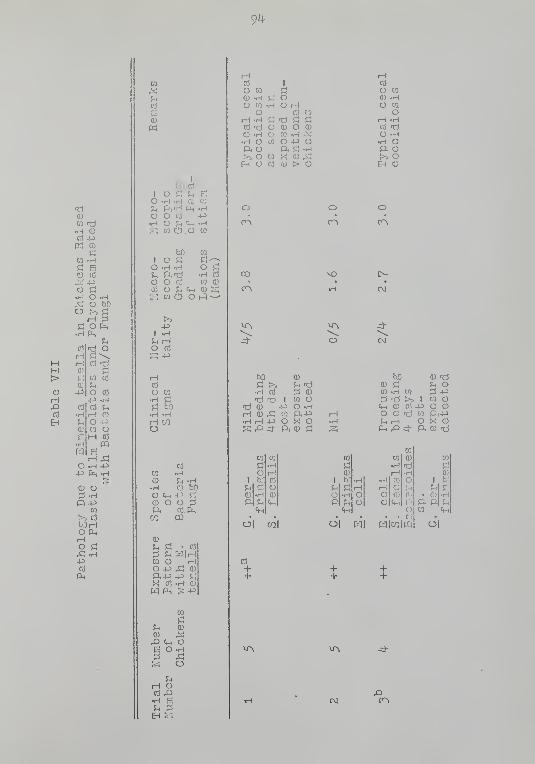
9^
r-\
(D
m "d
m -H
<D to
o i:^
t-i o^ oO >j
bO
rd O
03 -d
CO CO
cti
i-l oO 4^CQ OM CO
PQs
O .H -P+:> fe .H
O O;3.H
CQ
CO
mJOP4H2 ^
CO
cd
E
I
fiOcOI O Pi fH EO -H -H CVJ W
O O CO -P•H O ;h <<-( -Ha CO C!3.0 CQ
g-5•H CO
CO Q)
hf)
I O ^,
O -H .H!h P-fdo o oj
CO O ?M «M (D Ss: CQ c5 o t^^--
-p
5^ rHO CO
S -P
rHCO
O CQ
W
CQ

95
•H
oo
Eh
in
I
ho 03
\ o a ^ eO -H -H Oj CO
f^ ft rcJ PL, .HO O C(5 4JhH O ^^ <*-. -HS m O O W
to
O C•H R5to Q)
hoI o SI!
O -H -H
o o nJ
CO O J^ «M <D SS CO ci> Of-:)--'
-p! -HJh r-i
O rt
S 4J
o to
iH COO
Q)

96
B
I
O S Jh SH .H K5 W
O Cti -PO f-1 <^ -Hw O O m
H .H
CO
•H CD
W (Do o nJ
C3 O Jh «« Q) SS CQ O O t^—
-p
5h pHO k5
rH
o w
r-l COo
03to .H
O c„-P rtQ) O O 3
W (D iH
ft4-> -P fi
m$:!
,Q «M OE O .H
^ U
•H E
rH >^ Q)

97
g-P
o
MH>OrH,QCO
EH
CO
e(D
hD CO

98

99
EH
O•H
o(D
Q)
ShPh
I
QJ
hOO
ce
oCD
ft
n3
rH
0)-p
<D
E
O
CD
o>5hoOrHO4̂-5
03PL,
w

100
m
•H ctf
o u(D o rdftrH Q)
in ^ A-^
O cS
sd O OCO -H W
ft
E Cmo oQ
CO

101
J
•H-P
oo
>(D
rH
EH
WQ)H K)
O in
Q) O -dP<rH (U
m ^ -^O rj
+iS^. rH
Si O OCd -H CO
•H
o oQ
O•H <i-. GD, O wO .H-—O M4J Sm f^ .H cC
O -H W CD
Sh fd OS ;^O CO u—S O PM
o•H Ch&, OO WO to j:::;
—m fi O j:1
o -H -H a
O (ti (D SS C5
-pI -HJ^ rHO CO
S -P
CO
O CO
CO
Sh s::^ . CO
;3 Jh WliHCQ CD iHO 4J ^ CD
ft4:> -P S« CO -H a''
CQ
u s
-Q Cm ^E O O:3 .H
o
iHCO ,Q•H E
EH £2;
ft

102
CO
0)
•H ao uQ) O nd
CO «i-i -PO ce
-P ?H rHSH O OCO .H CO
fi S M•HE <iH
O OQ
O•H <V-i Sft O to
o ha+j sCO <::; .H CD
O .H CO
O 03 u-^•H ^ cd
S C) ^
o•H Cmft oo CO
o Jin 5:^—
CO fi O SO -H .H CV5
y^ fC5 CO
O CO sCO su (-:i--
I .H
O CO
S-P
CO
O CO
CO r
O -P ^ft-P -P ^« CO -HW Pm S: -P
u

103
•H-P
oo
03
EH
w
O Jh
Q) O nd
C/2 «i-i •JO K)
4^ SU rHfl O O
E Cho o
o•H fM Rp. O UO -H-^o bo-p s:!
O -H W CD
O CO ?H—
'
SOPM
o•H c„P. OO CQ
O fc£) ^^^CQ j:; o «O -H .H CC
U Ti V} CD
O CO (D SCO Jh t^—S C5
>-5
O CO
S 4^
CO
O C/3
•H .HrH CO
?^ i^! -CO
CO 0) iHO +3 ^ 0.1
p.+i -p r;
X CO -H CD
?-.

10^

105
0)
o•H
o
o•H-P
>
OO
><!

106
r.c>
(D
•H c\3
O UCD O -dPhiH OJ
CO <<-i -1^
O CO
•P ^ rHS O OC\3 -H W
•H
o oo
O CO
!h CO
O g:
Ph E CO
S E0) <D CO
CVl i-H
oO CO
Cu CD
^ Cfl
o a
o

107
PS
ou
X
iH
E-i
w
•H o3
O Sh
Q) O fd
CO tM 4Jo a
-P ^ rHPi O OCd -H CO
Pi S M•HE «wO OQ
O•H Cm eP< O WO -H—O hO-P flCO iZj -H Ki
O .H CO Q)
O Cd ?H^-'•H J-i CS
o•H Ci-,
ft OO CO
O W) S'--CO S O SO -H .H CO
Pi -d CO 0)
o c\5 o j;:
ct> p, t^--^
Pt MO ct3
a -P
CI?
O CO
p; bo•H .HiH CO
(D
p< p! • ct;
rS Pi m|mCO <D (HO +:> ^ CD
ft+3 +i j;:
X ce .H CD
W Ph ^ -P
CO
P4 Pi
^ Cm ^BOO1^ -H^ Sio
p<
iH (D
CO rO•H ep. ^E-I a
.

108
ra
CD
•H CO
o u0) O TiftiH 0)
W tM -PO CO
U rHO O
cC
•H
o oQ
CQ rH OO CD
C|Cm

109
•H-P
oo
X
EH
CO
(D
•H CO
O !:h
0) o fdP,rH 0)
CO Cj_, +3O td
P ^ Hrj o O
•H ra
E Cho cQ
o•H <4H BP, O CO
O -H—O W)j3 r!
CO ri -H Cd
O -H CO 0)
^ nc) cC >So ce Sh—•H i:.! cti
S C5 Ph
O•H q-i
a. oO CO
o fao S'--CO s:; o sO -H -H a5
Jh re) CO CD
o ce 0) ;a
so
f-1 H
ft ft ft
cti

110
-d

Ill
§•H
oo
X
B

112
Mx:
(D

113
1
-p
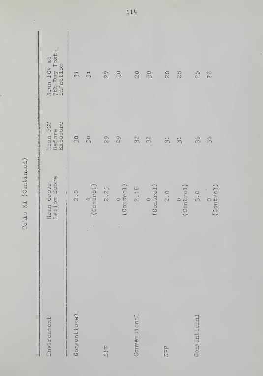
114-

115
OO
03
EH
1

116
I
-p
a o
> oo >>.Hf^ CO -P
^(^ ^^, o
-p rt
> <DO UPh (D ;3

117
I
-p
03 O
> o
Ph 03 -P^O O
S£>-M
> 0)

118
C<M

119
•H
oo
MM
-Q

APPENDIX B

121
Figure 1. Ceca of 3 vjeek-old specific pathogen-free chicken exposed to Eimeria tenella, 7th day post-inoculation. Macroscopic grading of the lesion ++-I-+.
Figure 2. Photomicrograph of a transverse sectionof cecum of 3 vjeek-old specific pathogen-free chickenexposed to Eineria tenella shovring denudation of mucosa,and large 2nd generation schizonts. Hematoxylin-eosinstain. XI25. Microscopic grading of parasitism ++++.

122
Figure 3. Ceca of 3 vieek-old bacteria-, fungi- andPPLO-free chicken exposed to Eimeria tenella , 7th day post-inoculation. Macroscopic grading of the lesion 0.
Figure ^. Photomicrograph of a transverse sectionof cecum of 3 vjeek-oid bacteria-, fungi and PPLO-freechicken exposed to Eimeria tenella shovjing a singlecoccidiura and no denudation of mucosa or other tissuedamage. Hematoxylin-eosin stain. X400.

123
Figure 5. Ceca of 3 vjeck-old bacteria-, fungi-and PPLO-free chicken exposed to Eimeria tenella andClostridium perfringens . Ilacroscopic grading of lesion

BIBLIOGRAPHY
Anon. 1965. ARS-USDA. Losses in agriculture. Agric.Handbook No. 291 . U.S. Gov't Printing Office,V/ashington, D.C., p. 80.
Balish, E. and A. W. Phillips. I966. Growth, morpho-genesis and virulence of Candida albicans after oralinoculation in the germ-free and conventional chick.J. Bact. 91:1736-17^2.
Ball, S. J. 1959. Antibiotics in chemotherapy of caecalcoccidiosis in chickens. J. Gomp. Path, and Therap.69:327-333.
Balzam, N. 1937a. Aseptisc'he zucht von tiern.^ I.Appareilla'ge et method'e. II. Aseptisclie zucht vonhaken vjorm rait vitamin haltigen und vitamin fi'
futter. Acta. Biol. Exptal. (V/arsaw) 11:^3-56.Ber. Biol. -4-8:5^5.
Balzam, N. 1937b. Elevage ^aseptique des animaux. I.Appareilla'ge et methode. II.
^Elevage aseptique
des poules maintenues aux regimes complets etdeficie'nts. Ann. Physiol. Physicochim. Biol.13:370-385. •
Bankowski, R. A., D. E. Stover, and S. L. Jamison. 1959.Coccidiosis immunization at a poultry testing pro-ject. Ann. Proc. U.S. Livestock Sanitary As. Dec.p. 5.
Barnes, D. M. and H. W. Moon. 196^. Enterotoxemia in pigsdue to Clontridium perfrin.gens type C. J. Am. Vet.Med. As. li{-4: 1391-139^.
Barnes, E. M. and C. S. Impey. I968. Anaerobic gramnegative, non-sporing bacteria from the ceca ofpoultry. J. Appl. Bact. 31:530-5^1.
Becker, E. R. 193^- Goccidia and Coccidiosis of Domesti-cated Game, and Laboratory Animals and of Man.lovia State College Press, Ames, Iov;a.
Becker, E. R. 1956. AproDOS oocyst measurements. J.Parasit. ^2 (Suppl.):24.
\2h

125
Bedrnik, P. 1967. Development of sexual stages andoocysts from second generation Eineria tenellamerozoites in tissue cultures. Folia Parasit.
Bedrnik, P. 1969- Some results and problems of cultiva-tion of Eineria tenella in tissue cultures. Acta.Vet. 30:31-35.
Bergeland, H. S. 1965. Studies of a porcine enterotoxemiacaused by Clostridiuni perf rinp:ens type C. Doctoraldissertation, University of Minnesota.
Bertke, E. M. 19^3. Renal function during the course ofEineria tenella infection. J. Parasit. ^1-9:937-
Blester, H. E. and L. H. Schvmrte (eds.). 1965. Diseasesof Poultry. Iowa State University Press, Ames,Iowa, p. 828-858.
Boggs, D. R., P. A. Chervenick, J. C. Marsh, H. I. Pilgrim,G. E. Cartwright, and M. M. Wintrobe. I967.Granulocytopoiesis in germ-free mice. Proc. Soc.Exp. Biol. Med. 125:325-330.
Boney, F. A. 19^w. The efficacy of methyl bromide as afumigant for the prevention of cecal coccidiosisin chickens. Am. J. Vet. Res. 9:210-21^.
Brackett, S. and A. Bliznick. 19^9- The effect of smalldoses of drugs on oocyst production of infectionsvrith Biraeria " tenellri . Ann. N. Y. Acad. Sci. 52:595-6T0:
-
Brackett, S. and A. Bliznick. 1952. The reproductivepotential of five species of coccidia of the chickenas demonstrated by oocyst production. J. Parasit.38:133-139.
Bradley, R. E. , H. Botero, J. Johnson, and W. 11. Reid.1967. Gnotobiotic poultry in plastic film isolatorsand some applications to parasit ologlcal research.Exp. Parasit. 21 :^!-03-^13.
Bradley, R. E., J. Johnson, and W. H. Reid. 196^1'.
Apparent obligate relationship between Histomonasmeieagridis and Escherichia col i in producing dis-ease. J. Parasit. 50:51*

126
Bradley, R. E. Gnd V/. H. Re id. I966. Hlsto'^on?.s ..mele-pp-rldis and several bacteria as agents of infectiousenterohepatitis in gnotobiotic turkeys. Exp.Parasit. 19:91-101.
Breed, R. S., E. G. D. Murray, and N. R. Smith. 1957-Bergey's Manual of Determinative Bacteriology.7th ed. Williams and Wilkins Co., Baltimore,Maryland, p. 109^.
Briggs, B. D. (ed.). 19^8. Coccidiosis of Chickens.Norwich Animal Industry, Inc., Norvjich, New York,p. 1-32.
Britton, \-J . 11., C. H. Hill, and C. \1 . Barber. 196^. Amechanism of interaction betvjeen dietary proteinlevels and coccidiosis in chicks. J. Nutrit. 82:306-310.
Burns, W. C. 1959. The lethal effects of Eimeria tenellaextracts on rabbits. J. Parasit. ^5: 38-^6.
Challey, J. R. I962. The role of bursa of Fabricius inadrenal response and mortality due to Eimeriatenella infection in the chicken. J. Parasit.^3:352-357.
Challey, J. R. and W. C. Burns. 1959. The invasion ofthe cecal mucosa by Eimeria tenella sporozoites andtheir transport by macrophages. J. Prot. 6;238-2iH.
Cheissir, E. M. 1958. Cytologische untersuchungenvorschiedener stadiem des lebens-zyklus der kaninchencoccidien. I. Eimeria intestinal is . Arch.Protistenk. 102:265-290.
Clark, D. T., C. K. Smith, and R. B. Dardas . I962.Pathological and immunological changes in gnotobio-tic chickens due to Eimeria tenella . Poultry Sci.^H:1635-1636.
Coates, M. E., J. E. Ford, and G. F. Harrison. I968.Intestinal synthesis of some vitamins of the Bcomplex in chickens. In K. Miyakavra and T. D.Luckey (eds.). Advances in Germ-Free Research andGnotobiology . The Chemical Rubber Company Press,Cleveland, Ohio, p. 110-111.
Cohendy, M. 1912. Experiences sur la vie sans microbes.Ann. Inst. Pasteur. 26:106-137.

127
Cohendy, M. and E.^ V/ollman. 191^. Presentee par.H. Roux.Bacteriologie. ExpSriences sur la vie sans micrbbes.Elevage aseptl'que de cobayes. Compt. Rend. Acad.Sci. 158:1283-128^.
Cook, F. D., R. M. Blakely, H. I. McGregor', and R. \1
.
Anderson. 195^- The effect of antibiotics on theintestinal microflora of turkey poults. PoultrySci. 33:38-^1-0.
Cuckler, A. C. 1957- The effect of nicarbazin on thedevelopment of immunity to reinfection with coccidiain poultry. Merck Poultry Nutrition and HealthSymposium, St. Louis, Missouri, p. 22.
Cuckler, A. C, L. R. Chapin, C. M. Malanga, E. F. Rogers,H. J. Becker, R. L. Clark, N. J. Leanza, A. A.Pessolano, T. Y. Shen, and L. H. Sarett. 1958.Antiparasitic drugs. II. Anticoccidial activityof 4,5-imidazoldicar boxaraide and related compounds.Proc. Soc. Exp. Biol, and Med. 98:167-170.
Daugherty, J. W. and C. A. Herrick. 1952. Cecal cocci-diosis and carbohydrate metabolism in chickens.J. Parasit. 38:298-304.
Davies, S. F. M. and L. P. Joyner. 1962. Infection ofthe fowl by the parenteral inoculation of ooc.y
'
of Eimeria . Nature 19^:996-997.
Davies, S. F. M., L. P. Joyner, and S. B. Kendall. 1963
.
Coccidiosis. Oliver and Boyd, Edinburgh.
Davies, S. F. M. and S. B. Kendall. 1955- An experimentalassessment of the value of nitrofurazone used con-tinuously as a coccidiostatic drug. Vet. Rec. 67:867-870.
Doflein, P. and E. Reichenow. 1953- Lehrbuch der pro-tozoenkunde, Jena: Fischer Verlag. p. 1213.
Doll, J. P. and C. K. Franker. 1963- Experimental histo-moniasis in gnotobiotic turkeys. I. Infection andhistopathology of the bacteria-free host. J.Parasit. 49:411-414.
Doll, J. P., P. C. Trexler, L. I. Reynolds, and G. R.Bernard. 1963^ The use of peracetic acid to obtaingerm-free invertebrate eggs for gnotobiotic studies.Am. Midi. Nat. 69:231-239.

128
Doraerrauth, C. H. and W. B. Gross. 1969- A medium forisolation and tentative identification of fecalstreptococci, and their role as avian pathogens.Avian Dis. 13:39^-399-
Doran, D. J. 1970. Eimeria tenella :
oocysts in cell culture.37:8^-91
Proc,from sporozoites toHelm'. Soc. VJash.
Doran, D. J. and M. M. Parr. 1962. Excystatlon of thepoultry coccidiura, Eimeria acervulina . J. Prot.9:15^-161.
Doran, D. J. and M. M. Farr. 1965. Susceptibility of1- and 3-day-old chicks to infection v;ith the cocci-dium, Eimeria acervulina . J. Prot. 12;l60-166.
Dubos, R. J. and R. W. Schaedler. 1962. The effect ofdiet on the fecal bacterial flora of mice and ontheir resistance to infection. J. Exp. Med. 115:1161-1172.
Dubos, R. J., R. W. Schaedler, R. Gostello, and P. Hoet.1965. Indigenous and autochthonous flora of thegastrointestinal tract. J. Exp. Med. 122:67-75-
Dunkley, M. J. W. 1968. Laboratory trials with buquino-late—a new broad-spectrum coccidiostat for poultry.Vet. Rec. 83:30-3^.
Edgar, S. A. .1941. The development of the protozoanparasite Eimeria tenella Railliet and Lucet I89Iin domestic fov;l. Doctoral dissertation, Universityof Wisconsin Memorial Library, Madison.
Edgar, S. A. 1955 • Sporulation of oocysts at specifictemperatures and notes on the prepatent period ofseveral species of avian coccidia. J. Parasit.^l:21^~216.
Edgar, S. A. and C. A. Herrick. 194-4. Feeding habits inrelation to the severity of cecal coccidiosis,Eimeria tenella . Poultry Sci. 23:30-35-
Edgar, S. A. and C. T. Seibold. 1964. A new coccidium ofchickens Eimeria mivati sp. n. (Protozoa: Eimeriidae)V7ith details of its life-history. J. Parasit. 50:193-204.
Eyssen, H. and P. DeSomer. I967. Effects of Streptococcusfecalis and a filterable agent on the growth andnutrient absorption in gnotobiotic chicks. PoultrySci. 46:323-333.

129
Farr, M. M .• and E. E. Wehr. 19^5- Sulfamer?.zine therapyin experimental cecal coccidiosis of chickens. J.Parasit. 31:353-358.
Field, H. I. and E. A. Gibson. 1955- Studies on pigletmortality. II. Clostridium vielchii infection.Vet. Rec. 67:31-35.
Fish, F. F. 1931. Quantitative and statistical analysisof infections viith Eineria tenolla in the chicken.Am. J. Hyg. 1^:560-576.
Forbes, M. and J. T. Park. 1959- Growth of germ free andconventional chicks: effect of diet, dietary peni-cillin, and bacterial environment. J. Nutrit. 67:69-84.
Forbes, M. , J. T. Park, and M. Lev. 1959. Role of theintestinal flora in the growth response of chicksto dietary penicillin. Ann. N. Y. Acad. Sci. 78:321-327.
Fowler, E. H. and M. W. Rohovsky. I970. Enzyme histo-chemistry of the small intestine in germ free andspecific pathogen free cats inoculated with felinei^'ectious enteritis (panleukopenia) virus. Am. J.. :.. Res. 31:2055-2060.
Franker, C. K. and J.. P. Doll. 1964. Experimental histo-moniasis in turkeys. II. Effects of some cecalbacteria on pathogenesis. J. Parasit. 30:636-640.
Freeman, B. M. I970. Carbohydrate stores in chickensinfected with Eimeria tenella . Parasitology 61:245-251.
Gardiner, J. L. 1955. The severity of cecal coccidiosisinfection in chickens as related to the age of thehost and the number of oocysts ingested. Poultry.Sci. 34:415-420.
Gardiner, J. L. and M. M. Parr. 1954. Nitrofurazone forthe prevention of experimentally induced Eimeriatenella infections in chickens. J. Parasit. 40:42-49.
Gardiner, J. L. and D. K. McLoughlin. 1963- Drug resist-ance in Eimeria tenella . III. Stability of re-sistance to glycarbylamide. J. Parasit. 49:657-659.
Glimstedt, G. 1936. Der stoffwechsel bakterienfrier tiere,I. Allgemeine methodik. Skand. Arch. Physiol. 73:48-62.

130
Goodrich, H. P. 19^^. Coccidian oocysts. Parasitology36:72-79.
Gordon, H. A. I960. The germ free animal. Its use inthe study of "physiologic" effects of the normalmicrobial flora on the animal host. . Am. J. DigestDis. 5:8^1-867.
Gordon, H. A., M. Wagner, T. D. Luckey, and J. A. Reyniers,1959. An encephalo-raeningeal syndrome selectivelyaffecting newly hatched germfree and monocontarainatedchickens. J. Inf. Dis. 105:31-37.
Graber, C. D., R. W. Moore, M. Suzuki, W. E. Redmond, R. M.O'Neil, and B. M. Lockhart. I966. Autochthonousintestinal bacterial flora and cholesterol levelsin specific pathogen-free sv;ine fed high lipid er-id
high sucrose diets. J. Bact. 92:1290-1297.
Griesemer, R. A., J.. P. Gibson, and D. S. Elasser. I963.Congenital ascariasis in gnotobiotic dogs. J. Am.Vet. Med. As. 1^3:962-96^.
Gustafsson, B. E. 19i^8. Germ free rearing of rats. Acta.Path. Microbiol. Scand. (Suppl.) 73:1-130.
Hegde, K. S., W. M. Reid, J. Johnson, and H. E. Womack.1969. Pathogenicity of Eiraeria brunetti in bacteriafree and conventional chickens. J. Parasit. 55:^02-405.
Hentges, D. J. I967. Influence of pH on the inhibitoryactivity of formic and acetic acids for Shigella '.
J. Bact. 93:2029-2030.
Hentges, D. J. 1969- Inhibition of Shigella flexneri bythe normal intestinal flora. II, Mechanism ofinhibition by coliform organisms. J. Bact. 97:513-517.
Herrick, C. A. 1936. Organ specificity of the parasiteEimerla tenella . J. Parasit. 22:226-227.
Herrick, G. A., G. E. Holmes, and D. L. Degiusti. 19^1-2.The experimental use of organic sulfur compoundsfor the prevention of cecal coccidiosis in chickens.Am. J. Vet. Res. 3:117-127.
Herrick, C. A., G. L. Ott, and C. E. Holmes. I936. Ageas a factor in the development of resistance of thechicken to the effect of the protozoan parasiteEimeria tenella . J. Parasit. 22:262-272.

131
Hibbert, L. E., D. M. Hammond, and J. R. Simmons. • 1969.The effects of pH, buffers, bile, and bile acidson excystation of sporozoites of various Eimeriaspecies. J. Prot. 16:^^1-^^^.
Holmes, C. E., C. A. Herrick, and G. L. Ott. 1937.Studies in coccidiosis: calcium carbonate addi-tions and coccidia. Poultry Sci. 16:335-339.
Horton-Smith, C. 19^1-7. Coccidiosis. Some factors in-fluencing its epidemiology. Vet. Rec. 59:6^5-6^6.
Horton-Smith, C, J. Seattle, and P. L. Long. 1961.Resistance to Eimeria tenella and its transfer-iio..from one cecum to the other in individual fowls.Immunology ^: 111-121.
Horton-Smith, C. and P. L. Long. 1963. Coccidia andcoccidiosis in domestic fov/1 and turkey. Adv.Parasit. 1:68-104.
Horton-Smith, C, P. L. Long, and A. E. Pierce. I963.Behavior of invasive stages of Eimeria tenella inthe immune fowl ( Gallus domesticus ). ^Exp. Parasit.1^1-: 66-7^1-.
Huhtanen, C. N. and J. M. Pensack. I965. The developmentof the intestinal flora of the young chick. PoultrySci. ^^:825-830.
Ikeda, M. 1955a. Factors necessary for Eimeria tenellainfection of the chicken. II. Influence of thepancreatic juice on infection. Jap. J. Vet. Sci.17:225-230.
Ikeda, M. 1955b. Factors necessary for Eimeria tenellainfection of chickens. Jap. J. Vet. Sci. 18:25-30.
Ikeda, M. 1956. Factors necessary for Eimeria tenellainfection of the chicken. IV. Investigations ofthe site of action of the pancreatic juice on in-fection. Jap. J. Vet. Sci. 18:^5-53.
Ikeda, M. i960. Factors necessary for E. tenella in-fection of the chicken. VI. Excystation of oocystin vitro. Jap. J. Vet. Sci. 22:27-^1.
Jankiewicz, H. A. and R. H. Scofield. 193^. The adminis-tration of heated oocysts of Eimeria tenella as ameans of estaulishing resistance and immunity tocecal coccidiosis. J. Am. Vet. Med. As. 84:507-526.

132
Jeffers, T. K. and G. -E. Wagenbach. 1969. Sex differencesin embryonic response to Eimeria tenella infection.J. Parasit. 55:9^9-951.
Johansson, K. R. and 17. B. Sarles. 19^8. Bacterialpopulation changes in the ceca of young chickensinfected vrith Eimeria tenella . J. Bact. 56:635-6^7.
Johansson, K. R.,.W. B. Sarles, and S. K. Shapiro. 19^8.The intestinal microflora of hens as influenced byvarious carbohydrates in a biotin-def icient diet.J. Bact. 56:619-63^.
Johnson, G. R. , A. Koestner, and M. W. Rohovsky. I967.Experimental feline infectious enteritis in thegerm free cat. An electron microscopic study.Path. Vet. ^:275-288.
Johnson, J. 1971- The development of Ascaridia galli ingerm-free chickens. J. Parasit. 56:17^.
Johnson, J. and W. M. Reid. 1970. Anticoccidial drugs:lesion scoring techniques in battery and floor-penexperiments with chickens. Exp. Parasit. 28:30-36.
Johnson, 17. T. 1927- Immunity or resistance of thechickens to coccidial infection. Oregon Agric.Exp. Sta. Bull. 230.
Jones, D. and E. Griffiths. 196^. The use of thin soilsections for the study of soil microorganisms.Plant Soil 20:232-2^1-0.
Jones, E. E. 1932. Size as a species characteristic incoccidia. Variations under diverse conditions ofinfection. Arch. Protistenk. 76:130.
Joyner, L. P. 196^. Coccidiosis in the domestic fowl.A review of the disease in Britain and its chemo-therapeutic control during the past decade. Vet.Bull. 3^:311-315.
Joyner, L. P. 1970. Coccidiosis: problems arising fromthe development of anticoccidial drug resistance.Expt. Parasit. 28:122-128.
Karmann,^P. and I. Presch. 1933. Wird die kokzidiose derkilken durch eine oder^^durch mehrere Eimeria artenherrvorgerufen. Tierarzt Rdsdr. 39:529-531.
133
Kemp, R. L., V/, H. Reid, and J. Johnson. 1971. Delayeddevelopment of endogenous stages of Eimeria teriellain germ-free chicks. The American Society ofParasitologists. August 23-2?, University ofCalifornia, Los Angeles, California. Programs andAbstracts, p. ^2.
Kohler, E. M. and R. P. Cross. 1969- Studies of Escherichiacoli in gnotobiotic pigs. VI. Effects of feedingbacteria-free filtrates of broth cultures. Canad.J. Corap. Med. 33:173-177.
Kohler, E. M. and R. F. Cross, 1971. Feeding bacteria-freevjhole cell lysates of Escherichia coli to gnotobio-tic pigs and the effects of giving antiserums. Am.J. Vet. Res. 32:739-7^8.
Kolacz, J. V/., R. B. VJescott, and A. R. Dommert. 1970.Microflora of Hormel miniature sv/ine. Am. J. Vet.Res. 31:1173-1178.
Kiister, E. 1912. Die keim^^freie zuchtung von saugetierenund ihre bedeutung fiir die er for schung der kori^er-funktionen. Zentr. Baketeriol. Parasitenk. 5^:55-58.
Layton, H. W. and K. L. Simkins. 1971- Growth and sur-vival of germ free and conventional chicks inoculatedwith Mycoplasma gallisepticum . Am. J. Vet. Res.32:1269-1274.
Lev, M. and C. A. E. Briggs. 1956. Gut flora of thechick. II..
' The establishment of flora. J. Apol.Bact. 19:224-230.
Lev, M., C. A. E. Briggs, and M. E. Coates. 1957- The gutflora of the chick. III. Differences in cecalflora between "infected," "uninfected," and penicil-lin fed chicks. Brit. J. Nutrit. 11:364-372.
Lev, M. and M. Forbes. 1959. Growth response to dietarypenicillin of germ-free chicks and of chicks withdefined intestinal flora. Brit. J. Nutrit. 13:78-84.
Levine, L. and C. A. Herrick. 195^. The effect of theprotozoan Eimeria benella on the ability of thechicken to do muscular work when its muscles arestimulated directly or indirectly. J. Parasit.^0:525-531.

13^
Levine, L. and C. A. Herrick. 1957. Effects of cecalcoccidia on the ability of Plymouth Rocks to domuscular work. Poultry Sci, 36:2M—29.
Levine, N. D. I96I. Protozoan Parasites of DomesticAnimals and Man. Burgess Publishing Company,Minneapolis, Minnesota, p. 158-253-
Levine, N. D. 1963- Coccidiosis. Ann. Rev. Microbiol.17:179-198.
Levine, N. D. 19^9 • Uniform terminology for sporozoanprotozoa. Prog. Prot. 3rd Int. Cong. Prot
.
,
Leningrad, p. 3^0.
Levine, N. D. 1970. Taxonomy of the sporozoa. J. Parasit.56:208-209.
Levine, P. P. 19^0. Subclinical coccidial infection inchickens. Cornell Vet. 30:127-132.
Levine, P. P. 19^2. Excystation of coccidial oocysts ofthe chicken. J. Parasit. 28: '+26-4-28.
Levine, P. P. and C. W. Barber. 19^7. The comparativeefficacy of some coccidiostatic agents againstexperimental infection with Eimeria tenella .
Cornell Vet. 37:155-159.
Long, P. L. 1965. Development of Eimeria tenella in avianembryos. Nature 208:509-510.
Long, P. L. 1966. The growth of some species of Eimeriain avian embryos. Parasitology 56:575-581.
Long, P. L. 1971. Schizogony and gametogony of Eimeriatenella in the liver of chick embryos. J. Prot.18:17-20.
Lotze, J. C. and R. G. Leek. i960. Some factors involvedin excystation of the sporozoites of three speciesof sheep coccidia. J. Parasit. 46 (Suppl. ) :4-6.
Lotze, J. C. and R. G. Leek. I969. Observations onEimeria tenella in feces of inoculated chickens.J. Prot. 16:4-96-4-93.
Luckey, T. D. I963. Germfrec Life and Gnotobiology
.
Academic Press, New York.

135
Lynch, J. E. 1961. A nev; method for the primary evalua-tion of anticoccidial activity. Am. J. Vet. Hes.22:32^-326.
Wann, T. B. 19^1-7. Chick rearing. X. Roughage and pro-tein as dietary factors influencing coccidlosisin chicks, v;ith notes on the limitation of sulpha-methazine in the control of coccidiosis. J. Agri.Sc. 37:1^5-151.
Matsuoka, T., M. E. Callender, and R. P. Shumard. I969.Embryonic bovine trachea cell line for in vitrocultivation of Eimeria tenella . Am. J. Vet. Res.30:1119-1122.
Mayhew, R. L. 193^- Studies on coccidiosis. VI. Effectof early attack on egg production. Poultry Sci.13:1^8-15^.
Mayhew, R. L. 1937- Studies on coccidiosis. IX. Histo-pathology of the cecal type in the chicken. Tr ns.Am. Micros. Soc. 56:431-4'4-6.
McLaren, D. J. 1969- Observations on the fine structuralchanges associated with schizogony and gametogonyin Eimeria tenella . Parasitology 59:563-574.
McLaren, D. J. and G. E. Paget. I968. A fine structuralstudy on the merozoite of Eimeria tenella withspecial reference to the conoid apparatus.Parasitology 58:561-571.
McLaughlin, M. M., II. P. Dacquisto, D. P. Jacobus, M.Forbes, and P. E. Parks. 1958. The effect of thegerm-free state on survival of the ten day old chickafter X-radiation. Radiation Res. 9:1^7.
McLoughlin, D. K. 1970. Coccidiosis: experimentalanalysis of drug resistance. Expt. Parasit. 28:129-136.
McLoughlin, D. K. and J. L. Gardiner. 1961a. Drug-resistance in Eimeria tenella . I. The experimentaldevelopment of a glycarbylamide-resistant strain.J. Parasit. ^7: 1001-1006.
McLoughlin, D. K. and J. L. Gardiner. 1961b. Zoalenetolerance by Eimeria tenella . J. Parasit. ^7(Suppl. ):46.

136
McLoughlin, D. K. and J. L. Gardiner. 1962. Drug- •
resistance in Eimeria tenella . II. The experi-mental development of a zoalene-resistant strain.J. Parasit. 53:930-932.
Meyer, R. G.,
E. H. Bohl, and E. M. Kohler. 196-4-. Pro-curement and maintenance of germ-free swine formicrobiological investigations. Appl. Microbiol.12:295-300.
Meyer, R. C. , H. E. Rhoades, and S. P. Saxena. I967. Theeffect of 5 E, coli serotypes on gnotobiotic swine.Proc. U.S. Livestock San. As., p. 3-^5-348.
Meyer, R. C., S. P. Saxena, and H. E. Rhoades. I97I.Polyserositis induced by Escherichia coli in gnoto-biotic swine. Infection and Immunity 3:41-'44.
Meynell, G. G. 1963- Antibacterial mechanisms of themouse gut. II. The role of Eh and volatile fattyacids in the normal gut. Brit. J. Exp. Path. ^^:209-219.
Miyakawa, M. 195^. Long term germ free rearing of guineapigs. Trans. Soc. Path. Japon ^3:^50-^1-52.
Monne, L. and G. Honig. 195^. On the properties of theshells of the coccidian oocysts. Ark. Zool.Stockholm 7:251-256.
Musajev, M. A. and A. M. Surkova. 1970. Some indices ofthe nitrogen metabolism in the liver of chickensunder experimental coccidiosis. J. Parasit. 56:451-^52.
Naqi, S. A., C. P. Hall, and D. H. Lewis. 1970a. Micro-flora of turkeys: comparison of apparently healthyand bluecomb-infected turkey poults. Avian Dis.15:1^-21.
Naqi, S. A., D. H. Lewis, and C. F. Hall. 1970b. Theintestinal microflora of turkeys. Avian Dis. 1^4-:
620-625.
Natt, H. P. 1959. The effect of cecal coccidiosis on theblood cells of the domestic fov;l. III. The changesin the leukocyte picture during the course of theinfection. Exp. Parasit. 8:182-187.
Natt, M. P. and C. A. Herrick. 1955- The effects of cecalcoccidiosis on the blood cells of the domestic fowl.I. A comparison of the changes in the erythrocyte

137
count resulting from hemorrhage in infection andmechanically bled birds. Poultry Sci. 3^:1100-1106.
Nevjton, V/. L. 1965. Methods in germ-free animal research.In 17. I. Gay (ed.), Methods of Animal Experimenta-tion, Vol. I. Academic Press, Nev7 York, p. 215-271,
Newton, 11. L. , L. V. Reardon, and A. M. DeLeva. I960.A comparative study of the subcutaneous inoculationof germ-free and conventional guinea pigs with twostrains of Trichomonas vaginalis . Am. J. Trop. Med.Hyg. 9:56-61.
Newton, VJ. L. , P. P. V/eingstein, and M. P. Jones. 1959-A comparison of the development of some rat andmouse helminths in germ-free and conventionalguinea pigs. Ann. N. Y. Acad. Sci. 78:290-39?.
Norcross, M. A. and P. V. Washko, 1970. Coccidiosis:laboratory confirmation of clinical disease. Exp.Parasit. 28:137-1^6.
Nyberg, P. A. and D. M. Hammond. 196^. Excystation ofEimeria boy is and other species of bovine coccidia.J. Prot."TlT57^-480.
Nyberg, P. A. and S. E. Knapp. 1970a, Scanning electronmicroscopy of Eimeria tenella oocysts. Proc. HelSoc. Wash. 37:29-32.
Nyberg, P. A. and S. E. Knapp. 1970b. Effect of sodiumhypochlorite on the oocyst wall of Eimeria tenellaas shovjn by electron microscopy. Proc. Helm. Soc.V/ash. 37:32-36.
Ogata, M. and Y. Morishita. 1969. Studies on the ali-mentary flora of pigs. IV. The alimentary floraof pigs infected with hog cholera. Jap. J. Vet.Sci. 31:71-82.
Ott, G. L. 1937. Studies on some relationships betweencolon bacilli and the protozoan parasite Eimeriatenella of chickens. Doctoral dissertation,University of V/isconsin, Madison, V/isconsin.
Pasteur, L. 1885. Observations relatives a la noteprecedente de M. Duclaux. Gomr)t. Rend. Acad. Sci.100:68.
Patnaik, B. I966. Experiments v/ith Eimeria tenellaRailliet and Lucet, I891 in chickens by parenteralroutes of infection. Ind. Vet. J. ^3:868-880.

13b
Pattillo, W. H. 1959. Invasion of the cecal mucosa ofthe chickens by SDorozoites of Eineria tenella .
J. Parasit. ^,^5: 253-258.
Patton, W. H. 1965. Eimeria tenella : cultivation of theasexual stages in cultured animal cells. Science150:767-769.
Pellerdy, L. I96I. On the occurrence of coccidiumstrains resistant to chemicals. Magyar AllatorvosokLapja l6:422-^^23.
Pellerdy, L. 1962a. Drug resistance in Eimeria tenella .
Vet. Bull. 32:2249.
Pell§rdy, L. 1962b. Uber das vorkommen sulfonamid-resis tenter coccidien stamme. Monatshefte FiirVeterinarraedizin 17:3^6-3^8.
Phillips, B. P. 1962. Personal communication quoted byW. L. Newton, 1965. Methods in germ-free animalresearch. In VJ. I. Gay (ed.), Methods of AnimalExperimentation, Vol. I. Academic Press, Nev; York.
Phillips, B. P. 1964. Studies on the ameba-bacteriarelationship in amebiasis. III. Induced amebiclesions in the germ-free guinea pig. Am. J. Trop.Med. Hyg. 13:391-395-
Phillips, B. P. and P. Gorstein. I966. Effects of dif-ferent species of bacteria on the pathology ofenteric amebiasis in mono-contaminated guinea pi -^
.
Am. J. Trop. Med. Hyg. 15:863-868.
Phillips, B. P. and P. A. iJolfe. 1959- The use of germ-free guinea pigs in studies on microbial inter-relationships in amoebiasis. Ann. N. Y. Acad. Sci.78:308-314.
Phillips, B. P., P. A. V7olfe, and I. L. Bartgis. 1958.Studies on the ameba-bacteria relationship inamebiasis. II. Some concepts on the etiology ofthe disease. Am. J. Trop. Med. Hyg. 7:392-399.
Phillips, B. P., P. A. Wolfe, C. W. Bees, H. H. Gordon,W. H. Wright, and J. A. Reyniers. 1955. Studieson the amoeba-bacteria relationship in amoebiasis.Comparative results of the intracecal inoculationof germ-free, mono-contaminated and conventionalguinea pigs with Entamoeba histolytica . Am. J.Trop. Med. Hyg. 4:675-692.

139
Pierce, A.E., P. L. Long, and C. Horton-Smith. 1962.Immunity to Eimeria tenella in young fowls.Immunology 5". 129-152.
Pratt, I. 19^0. The effect of Eimeria tenella (Coccidia)upon the blood sugar of the chicken. Trans. Am.Micr. Soc. 59:31-37.
Pratt, I. 1941. The effect of Eimeria tenella (Coccidia)upon the glycogen stores of the chicken. Am. J.Hyg. 3^:5^1-0-610.
Rail, G. D., A. J. Wood, R. B. Westcott, and A. R. Dommert,1970. Distribution of bacteria in feces of svjine.•\ppl. Microbiol. 20:789-792.
Reece, W. 0., H. S. Hofstad, and H. J, Swenson. I968.Establishment and maintenance of an SPP beagle dogcolony. Lab. Anim. Care 18:509-512.
Reich, P. 191 3- Das kaninchen'coccid Eimeria steiedae,nebst einen beitragezur kenntnis von Eimeriafalciformis . Arch.' Prot. 28:1-^2.
Reid, W. M. i960. The relationship between coccidiostatsand poultry flock immunity in coccidiosis controlprograms. Poultry Sci. 39:1^31-1^37.
Reid, W. M. and H. Botero. 1967. Growth of the cestodeRaillietina cesticillus in bacteria-free chickens.Exp. Parasit. 21:14-9-153.
Reid, W. M., W. T. Springer, and J. K. Johnson. I969.Requirements of avian parasites for support bymicroflora in the intestine. 8th Annual Meeting.Association for Gnotobiotics , June 10-13, OakRidge, Tennessee.
Reyniers, J. A. (ed.). 19^^6,
Lobund Reports No. 1.
Notre Dame, Indiana.
Reyniers, J. A. (ed.). 19^9
,
Lobund Reports No. 2.Notre Dame, Indiana.
Germ-free life studies.University of Notre Dame,
Germ-free life studies.University of Notre Dame,
Reyniers, J. A. (ed.). i960. Germ-free life studies.Lobund Reports No. 3* University of Notre Dame,Notre Dame, Indiana.
Reyniers, J. A. and M. R. Sacks teder. 1959. Tumor i-genesis and the germ-free chicken. A preliminaryreport. Ann. N. Y. Acad. Sci. 78:328-353.
1^0
Reyniers, J. A., P. Ci Trexler, R. P. Ervin, M. Wagner,T. D. Luckey, and H. A. Gordon. 19^9. The needfor a unified terminology in gerra-free life studies.Lobund Reports 2:1^9-162.
Reyniers, J. A., M. Wagner, T. D. Luckey, and H. A. Gordon.i960. Survey of germ-free animals. The WhiteWyandotte Bantam and White Leghorn chicken. LobundReports 3:.7-171.
Rlpsom, G. A. and C. A. ?Ierrick. 19^5- Effects of varioussulfa compounds on the protozoan parasite, Eimeriatenella . J. Parasit. 31:98-108.
Rohovsky, M. W. and R. A. Griesemer. 196?. Experimentalfeline infectious enteritis in the germfree cat.Path. Vet. J^:391-i|'10.
Rose, M. E. 1967a. Immunity to Eimeria tenella and Eimerianecatrix infection in the fov.'l. II. Influence ofthe site of infection and the stage of the parasite.II. Gross infection. Parasitology 57:5&7-583.
Rose, H. E. 1967b. The influence of age of host on in-fection vjith Eimeria tenella . J. Parasit. 53:924-929.
Rosenberg, M. M. 19^1. A study of the inheritance ofresistance to 'E, tenella in the domestic fowl.Poultry Sci. 20:^72.
Rosenberg, M. M. and W. H. McGibbon. 19^1-8. Selection forhereditary resistance and susceptibility to cecalcoccidiosis in the domestic fowl. 8th World'sPoultry Gongress 1:7^5.
Sabourdy, M. A. 1965. The need for specification inlaboratory animals: suggested terms and defini-tions. Pood Gosraet. Toxicol. 3:19.
Savage, D. G. and R. Dubos. I968. Alterations in themouse cecum and its flora produced by antibacterialdrugs. J. Exp. Med. 128:97-110.
Savage, D. G., R. Dubos, and R. W. Schaedler. 1968. Thegastro intestinal epithelium and its autochthonousbacterial flora. J. Exp. Med. 127:67-76.
Savage, D. G., J. S. MacAllister, and P. G. Davies. I970.Anaerobic bacteria on the mucous membrane epitheliumof murine large bowel. Bact. Proc. 98.

1^1
Schaedler, R. W. , R. Diibos, and R. Costello.- I965. Thedevelopment of the bacteria flora in the gastro-intestinal tract of mice. J. Exp. Med. 122:59-66.
Scholtyseck, E. 1962. Electron microscope studies onSimeria perforans (Sporozoa). J. Prot. 9:^07-^1^.
Scholtyseck, E. and H. Mehlhorn. 1970. Recent problems oftaxonomy and morphology of coccidia. J. Parasit.56:307.
Schottelius, M. 1899. Die bedeutung der darmbacterien fu.die errahrung. I. Irch. Hyg. 3^:210-2^3.
Shapiro, S. K. and W. B. Sarles. 1949. Microorganisms inthe intestinal tract of normal chickens. J. Bact.58:531-5^4.
Sharma, N. N. and V/. H. Reid. 1962. Successful infectionof chickens after parenteral inoculation of oocystsof Eimeria sp. J. Parasit. 48 (Suppl.):33.
Smetana, H. 1933. Coccidiosis of the liver in rabbits.I. Experimental study on the excystation of oocystsof Eimeria steidae . Arch. Path. 15:175-192.
Smith, H. W. I96I. The development of the bacterial floraof the faeces of animals and man: the changes thatoccur during aging. J. Appl. Bact. 24:235-241.
Smith, H. W. 1965a, Observation of the flora of the ali-mentary tract of animals and factors affecting 1:
composition. J. Path. Bact. 89:95-122.
Smith, H. W. 1965b. The development of the flora of thealimentary tract in young animals. J. Path. Bact.90:495-513.
Smith, H. W. and W. E. Crabb. I96I. The faecal bacterialflora of animals and man: its development in theyoung. J. Path. Bact. 82:53-66.
Smith, H. W. and J. E- T. Jones. 19^3 . Observations onthe alimentary- tract and its bacterial flora inhealthy and diseased pigs. J. Path. Bact. 86:387-412.
Smith, H. W. and J. E. T. Jones. 1967 . Observations onexperimental oral infection vjith Salmonella dublinin calves and Salmonella choleraesuis in pigs. J.Path. Bact. 93:141-156.

m-2
Springer, G. F., R. E. Horton, and M. Forbes. 1959
•
Origin of antihuman blood group B agglutinins ingerm-free chicks. Ann. N, Y. Acad. Sci. 78:272-275.
Springer, W. T. 1968. Histomoniasis: biological aspectsof transmission, pathogenesis, and relationships tothe indigenous bacterial flora of chickens andturkeys. Doctoral dissertation, University ofGeorgia, Athens, Georgia.
Springer, W. T., J. Johnson, and V/. M. Reid. 1970.Histomoniasis in gnotobiotic chickens and turkeys:biological aspects of the role of bacteria in theetiology. Exp. Parasit. 28:383-392.
Strout, R. G. and C. A. Ouellette. 1969- Gametogony ofEimeria t enella (Coccidia) in cell cultures.Science 163:695-696.
Thorbecke, G. J. 1959- Some histological and functionalaspects of lymphoid tissue in germfree animals.I. Morphological studies. Ann. N. Y. Acad. Sci.78:237-2^6.
Thorbecke, G. J., H. A. Gordon, B. VJostmann, M. Wagner,and J. A. Reyniers. 1957. Lymphoid tissues andserum gammaglobulin in young germ-free chickens.J. Inf. Dis. 100:237-251.
Timms, L. I968. Observations on the bacterial flora ofthe alimentary tract in three age groups of normalchickens. Brit. Vet. J. 12^:470-^77-
Turk, D. E. and J. F. Stephens. 1967- Gastrointestinaltract disease and zinc absorption: cecal cocci-diosis. Poultry Sci. 46:936-9^3.
Tyzzer, E. E. I929. Coccidiosis in gallinaceous birds.Am. J. Hyg. 10:269-383-
Tyzzer, E. E., H. Theiler, and E. E. Jones. 1932.Coccidiosis in gallinaceous birds. II. A com-parative study of species of Eimeria of thechicken. Am. J. Hyg. 15:319-393.
Vegh, E. 1963. Coccidia resistant to furazolidone. Vet.Bull. 33:117-
Wagenbach, G. E., J. R. Challey, and V/. C. Burns. 1966.A method for purifying coccidian oocysts employ-ing Glorox and sulphuric acid-dichromate solution.J. Parasit. 52:1222.

li^3
Waletzky, E. and C. 0. Hughes. 19^9-1950. . Factors in-volved in tests for acquired immunity in Eimerlatenella infections of the chicken. Ann. N. Y. Acad.Sci. 52:^78-^95.
Waletzky, E., C. 0. Hughes, and M. C. Brandt. 19^9-1950.The anticoccidial activity of nitrophenide. Ann.N. Y. Acad. 52:5^3-557-
Waxier, S. H, 19^1. Changes occurring in the blood andtissues of chickens during coccidiosis and artifi-cial hemorrhage. Am. J. Physiol. 13^:19-26.
Weichselbaum, T. E. 19^6. An accurate and rapid m.ethodfor the determination of proteins in small amountsof blood serum and plasma. Am. J. Clin. Path.16:^0-^9.
Weinstein, P. P., VJ. L. Newton, T. K. Sav;yer, and I.Sommerville. 1962. Quoted by 17. L. Neviton. InW. I. Gay (ed.). Method of Animal Experimentation,Vol. I, 1965. Academic Press, New York, p. 265.
Westcott, R. B. 1971. Experimental infections of gnoto-biotlc mice with Nematospiroides dubj.us and Nippo-strongylus brasiliensis . J. Parasit. ^6:k'?jZ.
Willingale, J. M. and C. A. E. Briggs. 1955. The normalintestinal flora of the pigs. II. Quantitativebacteriological studies. J. Appl. Bact. 18:2b^-293.
Wittner, M. and R. M. Rosenbaum. 1970. Role of bacteriain modifying virulence of Entamoeba histolytica .
Studies of amebae from axenic cultures. Am. J.Trop. Med. Hyg. 19:755-761.
Wostmann, B. S. 1959- Serum proteins in germ free verte-brates. Ann. N. Y. Acad. Sci. 78:25^1-260.
Wostmann, B. S. and E. Bruckner-Kardoss . 1959- Develop-ment of cecal distention in germ-free baby rats.Am. J. Physiol. 197:13^1-5-13^6.
BIOGRAPHICAL SKETCH
Chittur Venkitasubhan Radhakrishnan was born in
Trichur, India, June 6, 1937. He attended primary and
secondary schools in Trichur and graduated with first class
from Salvation Army English high school, Trivandrum, in
March, 1953-
In June, 1953, he enrolled at the Intermediate
College, Trivandrum, and passed the interscience examination
with distinction in physics and chemistry in March, 1955-
From August, 1955, to April, 1959, he attended the Kerala
Veterinary College and Research Institute, Trichur", India,
and received the degree of Bachelor of Veterinary Science
with first class. During his college days he was a member
of the soccer, hockey, and field and track teams, and also
represented his college in the intercollegiate debate com-
petitions. From July, 1959, to July, 1964, he served as
Junior Lecturer at the Kerala Veterinax^ College and from
July, 1966, to September, I968, he served as scientific
officer at the Antibiotics Research Laboratories, Poena,
India.
He married the former Jayalakshmy Subramaniara in
December, 1965- They have one son, Raja Venkitasubhan.
Ik-ii

145
He is a member of the American Society of Para-
sitologists, the Southeastern Society of Parasitologists,
and the honorary fraternities Alpha Zeta and Phi Sigma.
He entered the University of Florida in September,
1968, and was granted a research assistantship with the
Department of Veterinary Science. At present he is a
candidal ^ for the degree of Doctor of Philosophy.

I certify that I have read this study and that in rayopinion it conforms to acceptable standards of scholarlypresentation and is fully adequate, in scope and quality,as a dissertation for th^^egree of Doctoxy-iDf Philosophy.
aici:,:iL'r. i:;. Bradley, Sr., ChapmanAssociate ProfessorDepartment of Veterinary Science
I certify that I have read this study and that in myopinion it conforms to acceptable standards of scholarlypresentation and is fully adequate, in scope and quality,as a dissertation for the degree of Doctor of Philosophy.
l-g--<g^57^ [ tJjiGeorge T. Kfrtds '
^""^"^
Professor ^Department of Veterinary Science
I certify that I have read this study and that inopinion it conforms to acceptable standards of schol r
•
presentation, and is fully adequate, in scope and qual /,as a dissertation for the degree of Doctor of Philosopny.
CdM^sjJU^Edvrard M. HoffmarAssistant ProfessorDepartment of Microbiology
I certify that I have read this study and that in rayopinion it conforms to acceptable standards of scholarlypresentation and is fully adequate, in scope and quality,as a dissertation for the degree of Doctor of Philosophy.
liPhillip/e. LoggingAssociq^e Profess!Department of Animal Science

I certify that I have read this study and that in myopinion it conforms to acceptable standards of "scholarlypresentation and is fully adequate, in scope and quality,as a dissertation for the degree of Doctor of Philosophy.
'/t^v<-^t^
. Smart, Jr. fGrover G,
Associate ProfessorDepartment of Entomology andHematology
I certify that I have read this study and that in myopinion it conforms to acceptable standards of scholarlypresentation and is fully adequate, in scope and quality,as a dissertation for the degree of Doctor of Philosophy.
Sxepiienf/Q. ^amAssociate ProfessorDepartment of Zoology
This dissertation v;as submitted to the Dean of theCollege of Agriculture and to the Graduate Council, andwas accepted as partial fulfillment of the requirementsfor th'3 degree of Doctor of Philosophy.
December, 1971
Dean, Graduate School
![Eimeria tenella sowie deren Nutzung in der Wirkstoffforschung · gondii und Cryptosporidium parvum) [MEHLHORN, 2008]. INLEITUNG E 3 Sporozoa (Apicomplexa) Perkinsea Sporozoea Gregarinia](https://img.pdfslide.net/doc/110x75/5be5645d09d3f288458b923f/eimeria-tenella-sowie-deren-nutzung-in-der-wirkstoffforschung-gondii-und-cryptosporidium.jpg)