Embed Size (px)
Citation preview
Ej Vysis ALK Break ApartFISH Probe Kit en
RF!! 06N38-020
Vysis ALK Break Apart 30-608495/R1FISH Probe KitREF 06N38-02030-608495/R1
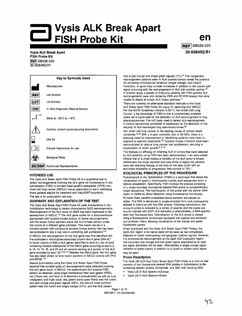
had single orange and single green signals (7%).10 The cytogeneticKey to Symbols Used rearrangement patterns seen in ALK-positive tumors reveal the potential
for activating chromosomal deletions (single orange), and fusion/Manufacturer truncation, or gene copy number increases in addition to the classic split
signal occurring with the rearrangement of ALK with another partner.10
In another study, a subset of thirty-one patients with FISH positive ALKList Number rearrangements were also tested by PCR and RT-PCR assays that were
unable to detect all known ALK fusion partners."Lot Number There are currently no alternative standard methods to the Vysis
ALK Break Apart FISH Probe Kit assay for detecting ALK NSCLC.FVD In Vitro Diagnostic Medical Device Per the NCCN Guidelines (Version 3.2011) Non-Small Cell Lung
Cancer, a big advantage of FISH is that a commercially available
Store at -30'C to -10*C. probe set is applicable for the detection of ALK-rearrangement in lungadenocarcinomas. The IHC tests used to detect ALK-rearrangementin clinical laboratories worldwide is inadequate for the detection of the
Caution, consult accompanying documents majority of ALK-rearranged lung adenocarcinomas.25
i cNon-small cell lung cancer is the leading cause of cancer deathworldwide. 12,13 With a 5-year morbidity rate of 85-95%, there is a
Use By pressing need for improvement in identifying patients most likely torespond to specific treatments.13 Tyrosine kinase inhibitors have beendemonstrated to reduce lung cancer cell proliferation, resulting in
Consult instructions for use suppression of tumor growth. 914-16
The therapeutic efficacy of inhibiting ALK in tumors that were selectedby ALK positivity using FISH has been demonstrated in an early-phase
Biological Risks clinical trial of a small molecule inhibitor of the ALK tyrosine kinase.Additionally, the study reported that sixty-three of eighty-two patients
Authorized Representative were still receiving therapy at the time of the data cutoff with anestimated probability of progression free survival of 72%."
INTENDED USE BIOLOGICAL PRINCIPLES OF THE PROCEDUREFluorescence in situ hybridization (FISH) is a technique that allows the
The Vysis ALK Break Apart FISH Probe Kit is a qualitative test to visualization of specific chromosome nucleic acid sequences within adetect rearrangements involving the ALK gene via fluorescence in situ cellular preparation. Specifically, FISH involves the precise annealinghybridization (FISH) in formalin-fixed paraffin-embedded (FFPE) non- of a single-stranded, fluorophore-labeled DNA probe to complementarysmall cell lung cancer (NSCLC) tissue specimens to aid in identifying target sequences. The hybridization of the probe with the cellular DNAthose patients eliible for treatment with XALKORIo(crizotinib). region is visible by direct detection using fluorescence microscopy.The test is for prescription use only. Formalin-fixed, paraffin-embedded tissue sections are placed onSUMMARY AND EXPLANATION OF THE TEST slides. The DNA is denatured to single-stranded form and subsequentlyThe Vysis ALK Break Apart FISH Probe Kit uses fluorescence in situ allowed to hybridize with the DNA probes. Following hybridization, thehybridization technology to detect chromosome 2p23 rearrangements. unbound probe is removed by a series of washes and the nuclei areRearrangement of the ALK locus on 2p23 has been implicated in the counter-stained with DAPI (4,6 diamidino-2-phenylindole), a DNA-specificdevelopment of NSCLC." 3 The ALK gene codes for a transmembrane stain that fluoresces blue. Hybridization of the ALK probe is viewedglycoprotein with tyrosine kinase activity. In-frame rearrangements using a fluorescence microscope equipped with appropriate excitationwith the known fusion partners place the ALK kinase domain under and emission filters, allowing visualization of the orange and greenthe control of a different gene promoter. This fusion results in a fluorescent signals.chimeric protein with constitutive tyrosine kinase activity that has been When hybridized with the Vysis ALK Break Apart FISH Probes, thedemonstrated to play a key role in controlling cell proliferation. 4-6 2p23 ALK region in its native state will be seen as two immediatelyIn NSCLC, the rearrangement of the ALK gene was first identified with adjacent or fused (overlapping) orange/green (yellow) signals. However,the echinoderm microtubule-associated protein-like 4 gene (EML4).1 if a chromosome rearrangement at the 2p23 ALK breakpoint regionIn-frame fusions of EML4-ALK genes identified to date include variants has occurred, one orange and one green signal separated by at leastcontaining multiple breakpoints of the EML4 gene occurring at exons 2, two signal diameters will be seen. Alternatively, a single orange signal6, 13, 14, 15, 18, and 20 and all variants starting at a portion of the ALK (deletion of green signal) in addition to a fused or broken apart signalgene encoded by exon 20.1-2- Besides the EML4 gene, the ALK gene may be seen.has also been shown to form fusion partners in NSCLC tumors with TFG Probe Descriptionand KIF5B. 4,Sal pbican ug tThe Vysis LSI ALK Dual Color Break Apart FISH Probe is a mixture thatSeveral publications using the Vysis ALK Break Apart FISH Probe , consists of two fluorophore-labeled DNA probes in hybridization bufferreported that multiple types of rearrangements were detected involving containing dextran sulfate, formamide, and SSC with blocking DNA:the ALK gene locus. In NSCLC, the predominant ALK-positive FISHpattern as detected using single interference filter sets [green (FITC), * Vysis LSI 3-ALK SpectrumOrangered (Texas red), and blue (4',6-diamidino-2-phenylindole) as well as dual * Vysis LSI 5'-ALK SpectrumGreen(red/green) and triple (blue, red, green) band-pass filters] was the fusionand split orange and green signals (62%), the second most commonpattern was the fusion and single orange (31%), and the final pattern
Page 1 of 10MD16917_v3 Confidential
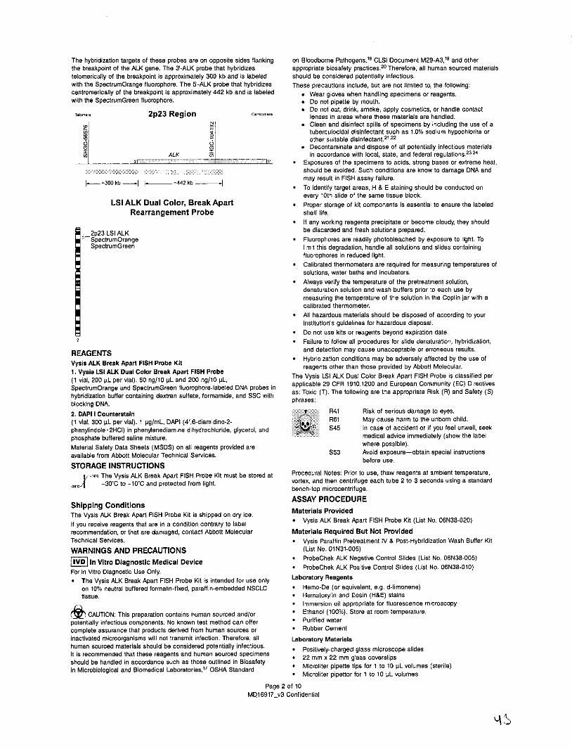
The hybridization targets of these probes are on opposite sides flanking on Bloodborne Pathogens,18 CLSI Document M29-A3,19 and otherthe breakpoint of the ALK gene. The 3-ALK probe that hybridizes appropriate biosafety practices.20 Therefore, all human sourced materialstelomerically of the breakpoint is approximately 300 kb and is labeled should be considered potentially infectious.with the SpectrumOrange fluorophore. The 5-ALK probe that hybridizes These precautions include, but are not limited to, the following:centromerically of the breakpoint is approximately 442 kb and is labeled * Wear gloves when handling specimens or reagents.with the SpectrumGreen fluorophore. * Do not pipette by mouth.
* Do not eat, drink, smoke, apply cosmetics, or handle contactToer 2p23 Region Centromem lenses in areas where these materials are handled.
* Clean and disinfect spills of specimens by including the use of atuberculocidal disinfectant such as 1.0% sodium hypochlorite orother suitable disinfectant.
21,22
0* Decontaminate and dispose of all potentially infectious materialsALK in accordance with local, state, and federal regulations.23,24
31 is * Exposures of the specimens to acids, strong bases or extreme heat,should be avoided. Such conditions are know to damage DNA and
0kb -. [ -- 442 kb may result in FISH assay failure.* To identify target areas, H & E staining should be conducted on
every 10th slide of the same tissue block.LSI ALK Dual Color, Break Apart . Proper storage of kit components is essential to ensure the labeled
Rearrangement Probe shelf life.* If any working reagents precipitate or become cloudy, they should
2p23 LSI ALK be discarded and fresh solutions prepared.SpectrumOrange * Fluorophores are readily photobleached by exposure to light. ToSpectrumGreen limit this degradation, handle all solutions and slides containing
fluorophores in reduced light.* Calibrated thermometers are required for measuring temperatures of
solutions, water baths and incubators.* Always verify the temperature of the pretreatment solution,
denaturation solution and wash buffers prior to each use bymeasuring the temperature of the solution in the Coplin jar with acalibrated thermometer.
* All hazardous materials should be disposed of according to yourinstitution's guidelines for hazardous disposal.
* Do not use kits or reagents beyond expiration date.2 * Failure to follow all procedures for slide denaturation, hybridization,
REAGENTS and detection may cause unacceptable or erroneous results.
Vysis ALK Break Apart FISH Probe Kit * Hybridization conditions may be adversely affected by the use of
1. Vysis LSI ALK Dual Color Break Apart FISH Probe reagents other than those provided by Abbott Molecular.1. 200 iL per vial). 50 ng/10 pL and 200 ng/10 PL, The Vysis LSI ALK Dual Color Break Apart FISH Probe is classified per
SpectrumOrange and SpectrumGreen fluorophore-labeled DNA probes in apial 9CR11.20adErpa omnt E)Drcie
hybridization buffer containing dextran sulfate, formamide, and SSC with as: Toxic (T). The following are the appropriate Risk (R) and Safety (S)blocking DNA. phrases:
2. DAPI I Counterstain T R41 Risk of serious damage to eyes.(1 vial, 300 pL per vial). 1 pg/mL, DAPI (4',6-diamidino-2- R61 May cause harm to the unborn child.phenylindole* 2HCI) in phenylenediamine dihydrochloride, glycerol, and S45 In case of accident or if you feel unwell, seekphosphate buffered saline mixture. medical advice immediately (show the label
Material Safety Data Sheets (MSDS) on all reagents provided are where possible).available from Abbott Molecular Technical Services. S53 Avoid exposure-obtain special instructions
before use.STORAGE INSTRUCTIONS
. The Vysis ALK Break Apart FISH Probe Kit must be stored at Procedural Notes: Prior to use, thaw reagents at ambient temperature,-30*C to -10'C and protected from light. vortex, and then centrifuge each tube 2 to 3 seconds using a standard
bench-top microcentrifuge.ASSAY PROCEDURE
Shipping ConditionsThe Vysis ALK Break Apart FISH Probe Kit is shipped on dry ice. Materials ProvidedIf you receive reagents that are in a condition contrary to label * Vysis ALK Break Apart FISH Probe Kit (List No. 06N38-020)recommendation, or that are damaged, contact Abbott Molecular Materials Required But Not ProvidedTechnical Services. * Vysis Paraffin Pretreatment IV & Post-Hybridization Wash Buffer KitWARNINGS AND PRECAUTIONS (List No. 01N31-005)
IVD In Vitro Diagnostic Medical Device * ProbeChek ALK Negative Control Slides (List No. 06N38-005)
For In Vitro Diagnostic Use Only. * ProbeChek ALK Positive Control Slides (List No. 06N38-010)
The Vysis ALK Break Apart FISH Probe Kit is intended for use only Laboratory Reagentson 10% neutral buffered formalin-fixed, paraffin-embedded NSCLC * Hemo-De (or equivalent, e.g. d-limonene)tissue. * Hematoxylin and Eosin (H&E) stains
* Immersion oil appropriate for fluorescence microscopy
CAUTION: This preparation contains human sourced and/or * Ethanol (100%). Store at room temperature.potentially infectious components. No known test method can offer * Purified watercomplete assurance that products derived from human sources or * Rubber Cementinactivated microorganisms will not transmit infection. Therefore, all Laboratory Materialshuman sourced materials should be considered potentially infectious. * Positively-charged glass microscope slidesIt is recommended that these reagents and human sourced specimens * 22 mm x 22 mm glass coverslipsshould be handled in accordance such as those outlined in Biosafety * Microliter pipette tips for 1 to 10 pL volumes (sterile)in Microbiological and Biomedical Laboratories,17 OSHA Standard
* Microliter pipettor for 1 to 10 pL volumesPage 2 of 10
MD16917 v3 Confidential
* Timer 5. Perform conventional H&E staining for one specimen slide.* Microtome Note: The specimen slide used for the assay procedure should be* Microcentrifuge within 10 serial sections of the H&E slide.* Graduated cylinders Note: Step 6 to be performed by a pathologist.* Static or circulating water baths (37'C)* Circulating water baths (74'C and 80'C) Note: Static water baths do 6. Examine and mark the largest possible area of tumor on the H&E
not provide adequate temperature control for higher temperature. slide, excluding necrotic areas, in situ carcinoma areas, and small* Purified water bath (37*C to 42'C) cell carcinoma areas using a solvent resistant marker or diamond-* Diamond-tipped scribe tipped glass scribe.* Solvent Resistant Marker (optional) 7. Using a glass scribe, transfer the mark from the H&E slide to the* Forceps corresponding areas of the unstained slide by marking the glass* Disposable syringe (5 mL) slide opposite the tissue section.* Coplin jars (12 x 50 mL) Suggested type: vertical staining jar 8. Store prepared slides at ambient temperature until ready to bake* Fluorescence microscope equipped with recommended filter(s) prior to Slide Deparaffinization Procedure.
(Refer to next section) Working Reagent Preparation* Calibrated thermometer 9. Preparation of Hemo-De - Fill three Coplin jars with 50 mL of Hemo-* Vortex mixer De. Keep covered when not in use. Store under vented conditions at* Microscope slide box with lid and/or carton slide folders ambient temperature and discard after seven days.* ThermoBrite® (List No. 7J68-020) 10. Preparation of Pretreatment Solution - Fill one Coplin jar with 50* ThermoBrite humidity cards (List No. 7J68-001) mL of Pretreatment Solution. Transfer the Coplin jar to a circulatingMicroscope Equipment and Accessories water bath at ambient temperature and bring the temperature of theMicroscoDe An epi-illumination fluorescence microscope is required for water bath to 81 ±2C (slightly higher than the desired temperatureviewing the hybridization results. The microscope should be checked inside of the Coplin jar) prior to deparaffinizing the slides. Ensureto confirm it is operating properly to ensure optimum viewing of FISH the temperature of the solution has reached 80±2*C prior to use.assay specimens. A microscope used with general DNA stains such as Discard the solution after using one (1) day.DAPI, propidium iodide, and quinacrine may not function adequately for 11. Preparation of Protease Solution - Add one vial of Vysis Protease IVFISH assays. Routine microscope cleaning and periodic maintenance by to one bottle of Vysis Protease IV Buffer. Rinse the vial with a smallthe manufacturer's technical representative, especially alignment of the volume of Vysis Protease IV Buffer and return to the bottle of Vysismercury lamp, are advisable. Protease IV Buffer. Replace the cap and gently invert several timesExcitation Light Source A 100 watt mercury lamp is the recommended to mix. Transfer the prepared solution to Coplin jar, and place theexcitation source. Record the number of hours that the bulb has been Coplin jar in a 37'C water bath. Wait a minimum of one hour afterused and replace the bulb before it exceeds the rated time. Ensure that mixing to ensure that the protease is in solution and confirm thatthe lamp is properly aligned, the temperature of the buffer is 37i± C before use. Discard solution
Oblectives Use oil immersion fluorescence objectives with numeric after one day.apertures 2 0.75 when using a microscope with a 100 watt mercury 12. Preparation of Purified Water - Fill one Coplin jar with 50 mL oflamp. A 1OX to 25X objective, in conjunction with 10X eyepieces, is purified water. Use at ambient temperature. Replace after each use.suitable for scanning the specimen to select regions for enumeration. 13. Preparation of Ethanol Solutions (70%, 85%, and 100%) - PrepareFor enumeration of FISH signals, satisfactory results can be obtained v/v dilutions of 70%, and 85% using 100% ethanol and purified water.with a 60X to 10OX oil immersion achromat type objective. Store at room temperature in tightly capped containers when not inImmersion Oil The immersion oil used with oil immersion objectives use. Solutions may be used for one week unless evaporation occursshould be one formulated for low auto fluorescence and specifically for or the solution becomes diluted or cloudy due to excessive use.use in fluorescence microscopy. Slide Deparaffinization ProcedureFilters Hybridization of the ALK probes to their target regions of the Note: Include one ProbeChek Negative Control slide and oneDNA is marked by orange and green fluorescence. All of the other DNA ProbeChek Positive Control slide starting with Step 14.present will fluoresce blue as a result of the DAPI I Counterstain. Singleand dual-bandpass fluorescence microscope filter sets optimized for use 14. Bake the unstained specimen and control slides for 2 to 24 hours atwith the FISH DNA probe kits are available from Abbott Molecular for 60'C on a ThermoBrite.most microscope models. 15. Immerse slides in the first Coplin jar containing Hemo-De for 5The recommended filters for use with the Vysis ALK Break Apart FISH minutes at ambient temperature.Probe Kit are the Vysis Dual Band (V2) - Green, Orange Filter, the Vysis 16. Repeat Step 15 twice using fresh Hemo-De each time.Single Band DAPI filter, the Vysis Single Band Orange Filter, and the 17. Dehydrate slides in 100% ethanol for 1 minute at ambientVysis Single Band Green Filter. temperature. Repeat in a second Coplin jar of 100 % ethanol.ASSAY PROTOCOL 18. Allow slides to air dry for 2 to 5 minutes (optional).Refer to the Warnings and Precautions section of this package insert Slide Pretreatmentbefore preparing samples. 19. Immerse up to eight slides in Vysis Pretreatment Solution which hasSpecimen Collection and Processing been previously warmed to 80±2'C for 12±3 minutes.The following procedure has been optimized for use on FFPE lung Note: If necessary, two slides may be placed back-to-back in eachcancer tissue specimens. Exposure of the specimens to acids, such slot of the Coplin jar, with one slide placed in each end slot. Foras decalcifying agents, strong bases and extreme heat should be slides in the end slots, the side of the slide with the tissue sectionavoided. Such conditions are know to damage DNA and may result in must face the center of the jar, for a maximum of eight slides perFISH assay failures. Coplin jar at one time.Use lung cancer tissue specimens that were fixed in formalin (10% neutralbuffered formalin) and that are well processed and produce good tissue 20. Immerse slides in purified water for 3 minutes.sections. The preferred fixation duration for tissue samples is 6 to 48 hours. Protease PretreatmentSlide Preparation of NSCLC FFPE Tissue Specimens 21. Remove slides from the purified water.
Note: Start processing specimens for which only slides rather than 22. Remove excess water by blotting the edges of the slide on a paperspecimen blocks are available at Step 5. towel.
23. Immerse slides in Protease Solution previously warmed to 37±1'C1. Cut two or more serial paraffin sections, 5±1 pm thick, using a for 20±2 minutes.
microtome. 24. Immerse slides in purified water for 3 minutes.2. Float the sections on the surface of a purified water bath set at Hybridization Procedure
40±2'C. A ThermoBrite should be used for the denaturation and hybridization3. Mount the sections on positively-charged glass slides steps. Refer to the ThermoBrite Operators Manual for instructions on4. Allow the slide to air-dry. instrument use.
Page 3 of 10MD16917_v3 Confidential
T4 L
25. Immerse the slides in 70% ethanol for one minute. * Background: the background should not contain particles that26. Immerse the slides in 85% ethanol for one minute. interfere with enumeration.27. Immerse the slides in 100% ethanol for one minute. Note: Fluorescent haze or glow may be noticeable outside of the28. Air-dry the slides for 2 to 5 minutes. nuclei, but as long as the fluorescent haze/glow does not cover the29. Moisten a humidity card with water and place in the card slots of the nuclei and make enumeration difficult, it is acceptable.
ThermoBrite. Ensure that the surface of the ThermoBrite is cleanand free of debris. * Probe signal intensity: the signals should be bright, distinct, and
30. Set the denaturation temperature (Melt Temp) to 73*C and the easily evaluable. Signals should be in bright, compact, round or oval
denaturation time (Melt Time) to three minutes. Set the hybridization shapes. Overly diffuse signals should be avoided.
temperature (Hyb Temp) to 37*C and the hybridization time * The majority of the target viewing area should meet these quality(Hyb Tlime) from 14 to 24 hours. criteria.
31. Apply 10 pL of probe mixture to a slide and immediately apply a * The target viewing area must contain at least 50 evaluable cells.
coverslip. Ensure no air bubbles are in the probe mixture prior to * If control slide hybridization adequacy met the hybridization criteriaapplying the coverslip. then repeat slide hybridization adequacy evaluation (step 44) for all
32. Seal the coverslip with rubber cement, specimen slides. If control slide hybridization adequacy did not meet. criteria refer to Quality Control, Use of Control Sides section for33. Place slides on the ThermoBrite and begin the hybridization program. additional information regarding the use of control slides.
Hybridize the slides overnight for 14 to 24 hours.At the end of the hybridization period, proceed to the Slide Washing Slide Evaluation
Procedure. 44. Locate Target Viewing Area* Use the H&E stained slide to confirm the target area prior to viewing
Note: Leave the slides on the ThermoBrite until ready to begin, the FISH slides.
Slide Washing Procedure * Use a 1oX to 25X objective and the DAPI bandpass filter to locatethe hybridized area of interest.
Note: Hybridized slides must be washed on the day hybridization * Avoid areas of necrosis and where the nuclear borders arewas completed. ambiguous. Skip nuclei with insufficient counterstain to determine
34. Pour 50 mL of Wash Buffer I into a Coplin jar. Use at ambient the nuclear border.temperature. Use one day, then discard. 45.Assess Target Area
35. Pour 50 mL of Wash Buffer 11 into a Coplin jar. Place the Coplin * Using a 60X to 10OX objective, use the prescribed filters to examinejar into a room temperature water bath prior to heating to prevent the quality of ALK signals and quality of tissue morphology. Adjustbreakage of the jar. Allow the jar to warm to 74±1C before using for the depth of the focus and become familiar with the size and shapeat least 30 minutes prior to use. Use one day, then discard. of the target signals and noise (debris). Verify that background
36. Remove the rubber cement from one slide while minimally disturbing appears dark and relatively free of strong fluorescence that canthe coverslip, and immerse the slide in ambient temperature Wash make enumeration difficult.Buffer 1. Repeat with the other slides and let stand 2 to 5 minutes to * Scan the entire scribed area(s). Observe the signal distributionallow the coverslips to float off the slides, among tumor cells during scanning in order to select a
Note: To maintain the proper temperature In Wash Buffer 11, representative area for enumeration.
wash only four slides simultaneously. If there are less than four 46. Select and Enumerate Cells Within Target Area
slides, add blank slides to bring the total number to four. Start * Select an area of good nuclear distribution (i.e., where individualtiming when the fourth slide is immersed. nuclei can be distinguished) and ensure areas chosen for
enumeration are representative of the signal distribution observed.37. Immediately immerse the slide in Wash Buffer liet 74sC. Gently * Using a 60X to 10OX objective and prescribed filters, begin analysis
agitate for 1 to 3 seconds. Repeat with the other slides, of the cells selected for enumeration and record signals in each cell.3 Move to the next representative area for enumeration.
Note: Ensure the temperature of Wash Buffer II has returned to * Repeat bullets 2 and 3 until 50 cells have been enumerated.74tl C before washing another four slides. * Stop when 50 cells selected from representative areas were
Counterstaining Procedure enumerated.
39. Air-dry the slide(s) protected from light at ambient temperature. Note: The field diaphragm may be narrowed around the cells of40.Apply 10 pL of DAPI counterstain to the target area of the slide, interest to aid in enumeration.
apply coverslip, and store protected from light for a minimum of5 minutes. 47. Signal Enumeration Rules
41. Enumerate specimens under a fluorescence microscope within 4 * Focus up and down to find all of the signals present in the nucleus.
hours or store at -20'C (±10'C). Enumerate the signals within the nuclear boundary of each selectedinterphase tumor cell according to the guidelines provided in
Archiving Procedure (optional) Figure 1.Store the hybridized slides at -20'C (±10'C) while protecting from light. Figure 1.Under these conditions, the slides can be stored for up to one week * Cells are considered negative (non-rearranged) when:after the application of DAPI I Counterstain without significant loss in * Orange and green signals are adjacent or fused (appearfluorescence signal intensity. yellow under the Orange/Green V2 filter). Orange and green
signals that are less than two signal diameters apart areNote: Allow slides to come to ambient temperature prior to considered as a single fused signal (Figure 2, Panel 1).viewing. * There is a single green signal without a corresponding orange
Slide Examination signal (Figure 2, Panel 1).42. View slides using a suitable filter set on an optimally performing * Cells are considered positive (re-arranged) when:
fluorescence microscope (Refer to Microscope Equipment and * At least one set of orange and green signals are two or moreAccessories - Filters section of this Package Insert). signal diameters apart (Figure 2, Panel 2).
INTERPRETATION AND RESULT REPORTING * There is a single orange signal without a correspondinggreen signal in addition to fused and/or broken apart signals
Quality Control (Figure 2, Panel 2).Assessing Slide Hybridization Adequacy43. Evaluate control slide hybridization adequacy using the following
criteria:* Nuclear morphology: Borders of tumor nuclei observed by DAPI
should generally be distinguishable, and nuclei should have goodintegrity.
Page 4 of 10MD16917_v3 Confidential
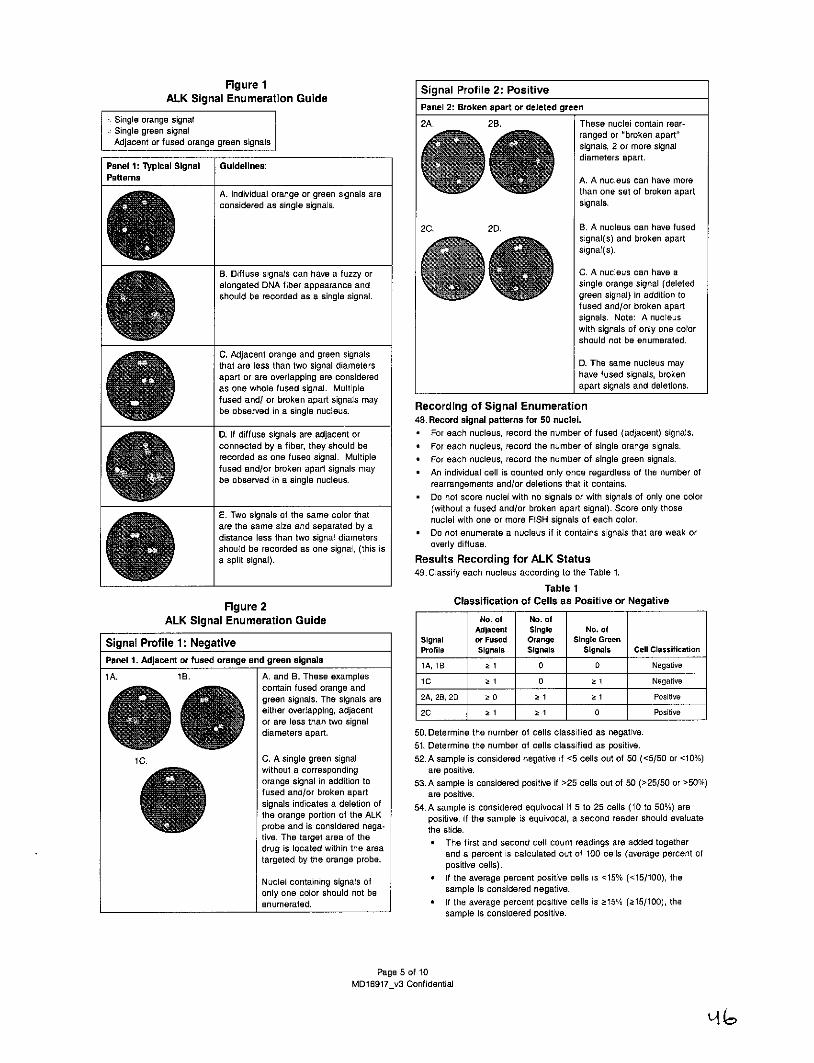
Figure 1 Signal Profile 2: PositiveALK Signal Enumeration Guide Sga rfl :Pstv
Panel 2: Broken apart or deleted greenSingle orange signal 2A. 2B. These nuclei contain rear-Single green signal ranged or "broken apart"Adjacent or fused orange green signals signals, 2 or more signal
diameters apart.Panel 1: Typical Signal Guidelines:Patterns A. A nucleus can have more
A. Individual orange or green signals are than one set of broken apartconsidered as single signals. signals.
2C. 2D. B. A nucleus can have fusedsignal(s) and broken apartsignal(s).
B. Diffuse signals can have a fuzzy or C. A nucleus can have aelongated DNA fiber appearance and single orange signal (deletedshould be recorded as a single signal. green signal) in addition to
4fused and/or broken apartsignals. Note: A nucleuswith signals of only one colorshould not be enumerated.
C. Adjacent orange and green signalsthat are less than two signal diameters D. The same nucleus mayapart or are overlapping are considered have fused signals, brokenas one whole fused signal. Multiple apart signals and deletions.fused and/ or broken apart signals maybe observed in a single nucleus. Recording of Signal Enumeration
48. Record signal patterns for 50 nuclei.D. If diffuse signals are adjacent or * For each nucleus, record the number of fused (adjacent) signals.connected by a fiber, they should be * For each nucleus, record the number of single orange signals.recorded as one fused signal. Multiple * For each nucleus, record the number of single green signals.fused and/or broken apart signals may * An individual cell is counted only once regardless of the number ofbe observed in a single nucleus. rearrangements and/or deletions that it contains.
Do not score nuclei with no signals or with signals of only one color(without a fused and/or broken apart signal). Score only those
E. Two signals of the same color that nuclei with one or more FISH signals of each color.are the same size and separated by adistance less than two signal diameters * Do not enumerate a nucleus if it contains signals that are weak or
should be recorded as one signal, (this is overly diffuse.
a split signal). Results Recording for ALK Status49. Classify each nucleus according to the Table 1.
Table 1
Figure 2 Classification of Cells as Positive or Negative
ALK Signal Enumeration Guide No. of No. ofAdjacent Single No. of
Signal Profile 1: Negative Signal or Fused Orange Single GreenProfile Signals Signals Signals Cell Classification
Panel 1. Adjacent or fused orange and green signals 1A, 1B 2 1 0 0 Negative1A. 1B. A. and B. These examples 10 2 1 0 1 Negative
contain fused orange andgreen signals. The signals are 2A, 28, 2D I 0 k 1 Z 1 Positiveeither overlapping, adjacent 20 1 5 1 0 Positiveor are less than two signaldiameters apart. 50. Determine the number of cells classified as negative.
51. Determine the number of cells classified as positive.1 C. C. A single green signal 52. A sample is considered negative if <5 cells out of 50 (<5/50 or <10%)
without a corresponding are positive.orange signal in addition to 53.A sample is considered positive if >25 cells out of 50 (>25/50 or >50%)fused and/or broken apart are positive.signals indicates a deletion of 54.A sample is considered equivocal if 5 to 25 cells (10 to 50%) arethe orange portion of the ALK positive. If the sample is equivocal, a second reader should evaluateprobe and is considered nega- the slide.tive. The target area of the * The first and second cell count readings are added togetherdrug is located within the area and a percent is calculated out of 100 cells (average percent oftargeted by the orange probe. positive cells).
Nuclei containing signals of * If the average percent positive cells is <15% (<15/100), the
only one color should not be sample is considered negative.
enumerated. * If the average percent positive cells is z15% (z15/100), thesample is considered positive.
Page 5 of 10MD16917_v3 Confidential

Uninformative Result: Problem Probable Cause Possible SolutionDesignate a specimen as Uninformative if the specimen failed Variation of Probe unevenly Repeat assay on next adjacentthe quality checks as described in the section Assessing Slide signal intensity distributed on slide section of same tissue blockHybridization Adequacy. across tissue due to air bubbles and make sure no air bubbles* If there are fewer than 50 tumor nuclei within the scribed area that section under coverslip are trapped under coverslip.
can be enumerated for a specimen, the specimen is uninformative. Apply coverslip by firstUse of Control Slides touching the surface of the* Control slides must be run concurrently with patient slides to probe mixture.
monitor assay performance and to assess the accuracy of signal Tissue loss Tissue section Verify protease digestion time.enumeration. Control slides should be processed with specimen or tissue under-fixed (poorslides, beginning at Slide Deparaffinization Procedure step 14 morphology DAPI staining)(baking at 60'C). degraded
* Control slides should be run on each day of FISH testing and with DNA loss (poor DAPI Verify fixation conditions.each new kit lot. staining)
* The established range for acceptable test performance for Inappropriate slides Use positively-charged slides.ProbeChek ALK Control Slides are specified on each lot-specific usedCertificate of Analysis included with the control slide kit.
* If a control slide fails to meet any of the acceptance criteria, theassay may not have been performed properly or the ALK BreakApart FISH Probe Kit components may have performed inadequately. Improper slide baking Verify temperature ofIn no case should FISH results be reported if either control slide ThermoBrite.fails. A repeat analysis with fresh control slides and clinicalspecimen slide(s) will be necessary. Over pretreatment Verify time and temperature
Vysis Pretreatment Solution.Over denaturation Verify Melt time.
When viewing the results of a FISH assay, ensure that the microscope is (Melt Time)properly aligned and functioning optimally. Tissue section was Allow additional time forThe following table lists some less than optimal results that may be torn when removing coverslip to soak off in washencountered using the LSI probes. Probable causes and suggestions to coverslip after buffer.improve assay performance are included. hybridization
Problem Probable Cause Possible SolutionNo signal or Inappropriate filter set Use recommended filters. LIMITATIONS OF THE PROCEDUREweak signals used to view * FOR IN VITRO DIAGNOSTIC USE ONLY.
slides * Optimal performance of this test requires appropriate specimenMicroscope not Call microscope handling, preparation, and storage as described in these instructionsfunctioning properly manufacturer's technical for use.
representative. * The Vysis ALK Break Apart FISH Probe Kit has been optimized onlyImproper lamps (i.e., Use a mercury lamp (100 watt for identifying and quantifying rearrangements of the ALK gene fromXenon or Tungsten) recommended). formalin-fixed, paraffin-embedded human NSCLC tissue specimens.Mercury lamp too old Replace with a new lamp. The assay should be performed only on 10% neutral buffered
formalin FFPE human lung cancer tissue. Other types of specimensMercury lamp Realign lamp. or fixatives should not be used.misaligned * The performance of the Vysis ALK Break Apart FISH Probe Kit was
No signal or Dirty or cracked Clean or replace lens. established using the procedures provided in this package insertweak signals collector lenses only. Modifications to these procedures may alter the performance(Continued) of the assay.
Dirty or broken mirror Clean or replace mirror. * The clinical interpretation of any test results should be evaluatedin lamp house within the context of the patient's medical history and otherInappropriate Verify hybridization time. diagnostic laboratory test results.hybridization time * FISH assay results may not be informative if the specimen qualityInappropriate post- Verify temperature of Wash and/or specimen slide preparation is inadequate.hybridization wash Buffer II. * Technologists performing the FISH signal enumeration must betemperature capable of visually distinguishing between the orange, green, andAir bubbles trapped Apply coverslip by first yellow signals.under coverslip touching the surface of theprevented probe probe mixture.accessInadequate protease Verify temperature of thedigestion Protease Solution.Section over fixed Prolonged tissue fixation(cell boundaries times may lead to progressivewill be distinct) degradation of signal intensity
and may require longerdigestion times.
Uninformative Too few nuclei Repeat assay with new slide.Result (<50) available for
enumerationNoisy Inadequate wash Verify temperature of thebackground stringency Wash Buffer II.
Page 6 of 10MD16917_v3 Confidential
uLi
EXPECTED VALUES Control Slide ReproducibilityNormal Cutoff Control slide reproducibility was evaluated using three lots of both the
The normal cutoff value is defined as the maximum amount of scoreable ProbeChek ALK Negative Control Slides and ProbeChek ALK Positive
interphase nuclei with a specific abnormal signal pattern at which a Control Slides. Each lot was run on 5 non-consecutive days over a 23-dayspecimen is considered negative for that signal pattern. The normal time period and evaluated by three readers for a total of 90 data points (3cutoff value is expressed in terms of a percentage or the actual number lots x 5 runs x 3 readers - 45 evaluations per control slide type).of nuclear FISH patterns positive for rearrangement per the standard For each specimen, the signal patterns of 50 nuclei were evaluated bynumber of nuclei tested. The normal cutoff was established as 15% counting the number of fused signals, single orange signals and singleusing NSCLC FFPE tissue specimens. green signals present for each target by each reader.
SPECIFIC PERFORMANCE CHARACTERISTICS There was no statistical difference.in FISH classification between 3readers by the Fisher-Freeman-Halton test at the significance level of
Probe Localization on Metaphase Chromosomes 0.05. (Refer to Table 4 and Table 5) Therefore, it was demonstrated thatThe location of hybridization of the Vysis ALK Break Apart FISH Probe Probe Check ALK Negative Control Slides and ProbeChek ALK Positivewas evaluated on metaphase spreads (a total of eight) from cultured Control Slides could be reproducibly classified. All slides in this studylymphocyte slide preparations in conjunction with the inverted DAPI were found to be within specifications.chromosome banding technique. Table 4The Vysis LSI 3-ALK SpectrumOrange and Vysis LSI 5'-ALK Reproducibility of ProbeChek ALK Negative Control SlidesSpectrumGreen probes, components of the Vysis LSI ALK Dual Color Number of Observations with theBreak Apart FISH Probe were shown to hybridize to the intended locus Percent ALK Rearrangement(2p23) on a total of 8 metaphase spreads and to no other locations. Within Specification Outside SpecificationAnalytical Sensitivity and Specificity Readers (<58%) (>8%) TotalAnalytical sensitivity is defined as the percentage of chromosome 1 15 0 15targets with the expected normal signal pattern. Analytical specificity is 2 15 0 15defined as the percentage of signals that hybridize to the correct locus 3 15 0 15and no other location. Fisher-Freeman-Halton p-value - 1.00The analytical sensitivity and analytical specificity of the Vysis LSI3-ALK SpectrumOrange and Vysis LSI 5-ALK SpectrumGreen FISH Table 5probes was evaluated using metaphase chromosomes prepared from Reproducibility of ProbeChek ALK Positive Control Slides6 peripheral blood cultures of karyotypically normal specimens from 5 Number of Observations with theindividual donors (6 slide lots). Percent ALK RearrangementFor the analytical sensitivity calculation, the signals for Vysis LSI 3-ALK Within Specification Outside SpecificationSO and Vysis LSI 5'-ALK SGn FISH probes were enumerated for each Readers ( 20%) (<20%) Totalmetaphase spread (normal - 2 signals). In total, 240 signals were 1 15 0 15expected for each probe (2 signals per cell x 20 metaphase spreads per 2 15 0 15lot x 6 slide lots). Refer to Table 2. 3 15 0 15For the analytical specificity calculation, the number of metaphase Fisher-Freeman-Halton p-value - 1.00spreads with the expected signal pattern was enumerated. In total, 120metaphase spreads were evaluated (20 metaphase spreads x 6 slide Tissue Reproducibilitylots). Refer to Table 3. Tissue reproducibility was evaluated using FFPE lung tumor sections.For each probe, the analytical sensitivity was calculated to be 100.0% This study was conducted using six serial sections (5 pm) prepared(240/240)(95% Cl 98.5-100.0) and the analytical specificity was from twenty NSCLC FFPE specimen blocks. The panel included threecalculated to be 100% (120/120)(95% Cl 97.0-100.0). positive specimens with >50% of the cells with ALK rearrangement, three
specimens falling within the range of 10% to 50% cells with the ALKTable 2 rearrangement and fourteen negative specimens with <10% cells with
Analytical Sensitivity the ALK rearrangement. Two slides were prepared from each specimenNo. of Metaphase and each slide was evaluated by two readers. Between-reader (Table 6)
Chromosome Signals Sensitivity and between-slide reproducibility (Table 7) were evaluated.Total True Point Estimate 95% Confidence
Probe Positive Total Expected (%) Interval The Vysis ALK Break Apart FISH Probe Kit was shown to be
Vysis LSI 3-ALK SO 240 240 100.0 (98.5, 100.0) reproducible based upon the between-reader and between-slide
vysis LSI SALK s~n 240 240 100.0 (98.5, 100.01 analyses resulting in a Fisher-Freeman-Halton p-value of 1.00.
Table 6Between-Reader Reproducibility
Table 3Analytical Specificity Number of Panel Members
Negative Positive TotalNo. of Metaphase
Chromosome Spreads Specificity Reader 1 14 6 20
Point 95% Reader 2 14 6 20Total False Total True Total Estimate Confidence Reader 3 14 6 20
Probe Positive Positive Expected (%) Interval Fisher-Freeman-Halton p-value: 1.00Vysis LSI 3-ALK SO 0 120 120 100.0 (970, 100.0)
Table 7Vysis LSI 5-ALK SGn 0 120 120 100.0 (97.0, 100.0) Between-Slide Reproducibility
Microbial Contamination Number of Panel MembersThe Vysis ALK Break Apart FISH Probe Kit met the requirements Negative Positive Totalfor a microbiologically uncontrolled product per "Guideline for the Slide 1 14 6 20Manufacture of In Vitro Diagnostic Products", 1/10/1994, as none of the Slide 2 15 520reagents would sustain growth of the selected microorganisms and in Slide 3 14 6fact killed the applied inoculum of microorganisms as referenced by the Fisher-Freeman-Halton p-value: 1.00lack of growth upon subculture. Additionally, upon testing the reagents inthe normal QC procedure, all the reagents performed satisfactorily evenafter three days of incubation with the selected organisms at 35 to 3TC.
Page 7 of 10MD16917_v3 Confidential
External Reproducibility Table 10Reproducibility of the Vysis ALK Break Apart FISH Probe Kit was Reproducibility by Siteevaluated at three external laboratories by testing a coded, randomized12-member specimen panel (6 unique specimens, 2 slides each) that Number of Slidesconsisted of four unique ALK-positives with varying levels of positivity Across Lots/(Panel Member 1, 2, 3, and 6) and two unique ALK negative NSCLC Runs/Readers Kappa AnalysisFFPE tissue specimens (Panel Member 4 and 5). Panel StandardThree lots of the Vysis ALK Break Apart FISH Probe Kit reagents were Site Member Negative Positive Kappa 95% C Error Z-Score
used in the evaluation. A run consisted of one replicate each of a 1 1 0 20 0.96 (0.83, 1.00) 0.068 14.21ProbeChek Negative Control slide, a ProbeChek Positive Control slide 2 0 20and each panel member. Each of the three clinical sites tested theReproducibility Panel using two of the three clinical lots. Each of the 3 0 20two technologists at each of the three testing sites enumerated 6 study 4 20 0specimens along with control slides once a day, for 5 non-consecutivedays, per reagent lot over a period of 20 days. Each site evaluated 120 5 20 0specimen slides for a total of 360. This resulted in 240 enumerations at 6 1 19each site for a minimum of 720 enumerations. Each site evaluated 40 2 1 0 20 0.96 (0.83,1.00) 0.068 14.21controls slides (20 positive and 20 negative slides) for a total of 120.This resulted in 80 enumerations at each site for a minimum of 240 2 0 20enumerations. For each panel member and control slides, the signal 3 0 20patterns of 50 nuclei were enumerated by two readers.
4 20 0The overall kappa coefficient was 0.92 (95% Cl 0.85 - 0.98). TheZ-Score of 27.08, which is greater than 1.96, showed the kappa 5 20 0coefficient is significantly different from zero at a 0.05 level of 6 1 19significance. These results are found in Table 8. The overall percent 3 1 1 19 0.83 (0.72, 0.94) 0.056 14.90agreement (PA) between all reader results was 97.64% (95% Cl 96.25- 98.52). The positive percent agreement (PPA) was 96.46% (95% Cl 2 0 2094.40 - 97.78) and the negative percent agreement (NPA) was 100.00% 3 2 18(95% Cl 98.42 - 100.00). The results are found in Table 9. The kappacoefficient demonstrated the reproducibility for each site, ranging from 4 20 00.83 to 0.96, and for each lot, ranging from 0.86 to 0.96. The results are 5 20 0found in Tables 10 and 11, respectively. 6 2 18
Table 8Overall Reproducibility Table 11
Number of Slides Across Sites/Lots/Runs/Readers Reproducibility by LotNegative Positive Total Number of Slides
Panel Member 1 1 59 60 Across Sites/Panel Member 2 0 60 60 Runs/Readers Kappa AnalysisPanel Member 3 2 58 60 Panel StandardPanel Member 4 60 0 60 Lot Member Negative Positive Kappa 95% Cl Error Z-ScorePanel Member 5 60 0 60 1 1 0 20 0.86 (0.75, 0.98) 0.059 14.75Panel Member 6 4 56 60
Kappa Statistic: 0.92 (0.85, 0.98) 2 0 20
Table 9 3 2 18
Percent Agreement Between All Readers with Expected Results 4 20 0Expected Results 5 20 0
Reader Results Positive Negative Total 6 2 18Positive46043Negative 17 0 257 2 1 0 20 0.96 (0.83, 1.00) 0.068 14.21
Total 480 240 720 2 0 20
PA: 97.64 (95%Cl: 96.25, 98.52) 3 0 20PPA: 96.46 (95%Cl: 94.40, 97.78) 4 20 0
NPA: 100.00 (95%Cl: 98.42, 100.00)5 20 0
6 1 19
3 1 1 19 0.93 (0.80, 1.00) 0.065 14.34
2 0 20
3 0 20
4 20 0
5 20 0
6 1 19
Page 8 of 10MD16917_v3 Confidential
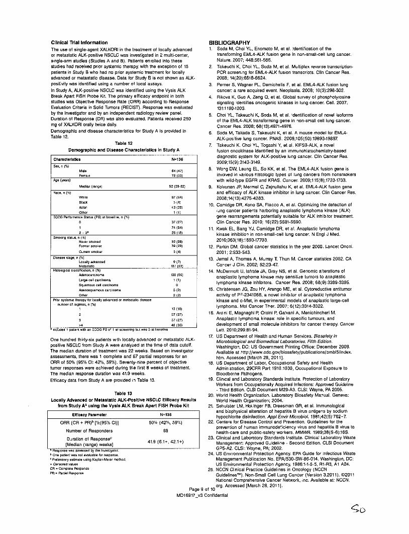
Clinical Trial Information BIBLIOGRAPHYThe use of single-agent XALKORI in the treatment of locally advanced 1. Soda M, Choi YL, Enomoto M, et al. Identification of theor metastatic ALK-positive NSCLC was investigated in 2 multi-center, transforming EML4-ALK fusion gene in non-small-cell lung cancer.single-arm studies (Studies A and B). Patients enrolled into these Nature. 2007; 448:561-566.studies had received prior systemic therapy, with the exception of 15 2. Takeuchi K, Choi YL, Soda M, et al. Multiplex reverse transcription-patients in Study B who had no prior systemic treatment for locally PCR screening for EML4-ALK fusion transcripts. Clin Cancer Res.advanced or metastatic disease. Data for Study B is not shown as ALK- 2008; 14(20):6618-6624.positivity was identified using a number of local assays. 3. Perner S, Wagner PL, Demichelis F, et al. EML4-ALK fusion lungIn Study A, ALK-positive NSCLC was identified using the Vysis ALK cancer: a rare acquired event. Neoplasia. 2008; 10(3):298-302.Break Apart FISH Probe Kit. The primary efficacy endpoint in both 4. Rikova K, Guo A, Zeng 0, et at. Global survey of phosphotyrosinestudies was Objective Response Rate (ORR) according to Response signaling identifies oncogenic kinases in lung cancer. Cell. 2007;Evaluation Criteria in Solid Tumors (RECIST). Response was evaluated 131:1190-1203.by the investigator and by an independent radiology review panel. 5. Choi YL, Takeuchi K, Soda M, et al. Identification of novel isoformsDuration of Response (DR) was also evaluated. Patients received 250 of the EML4-ALK transforming gene in non-small cell lung cancer.mg of XALKORI orally twice daily. Cancer Res. 2008; 68(13):4971-4976.Demographic and disease characteristics for Study A is provided in 6. Soda M, Takada S, Takeuchi K, et al. A mouse model for EML4-Table 12. ALK-positive lung cancer. PNAS. 2008;105(50):19893-19897.
Table 12 7. Takeuchi K. Choi YL, Togashi Y, at al. KIF5B-ALK, a novelDemographic and Disease Characteristics in Study A fusion oncokinase identified by an immunohistochemistry-based
Characteristics N=136 diagnostic system for ALK-positive lung cancer. Clin Cancer Res.2009;15(9):3143-3149.
Sex, n Male 64(47) 8. Wong DW, Leung EL, So KK, et al. The EML4-ALK fusion gene isFemale 72 (53) involved in various histologic types of lung cancers from nonsmokers
Age lyears) with wild-type EGFR and KRAS. Cancer. 2009;115(8);1723-1733.Median (range) 52 (29-82) 9. Koivunen JP, Mermel C, Zejnullahu K, at al. EML4-ALK fusion gene
Race, n (%) and efficacy of ALK kinase inhibitor in lung cancer. Clin Cancer Res.White 87 (64) 2008;14(13):4275-4283.slack 5(141Asian 43132) 10. Camidge DR, Kono SA, Flacco A, et al. Optimizing the detection ofOther 1 (1) lung cancer patients harboring anaplastic lymphoma kinase (ALK)
ECOG Performance Status (PS) at baseline, n gene rearrangements potentially suitable for ALK inhibitor treatment.0 37(27) Clin Cancer Res. 2010; 16(22):5581-5590.1 74(54) 11. Kwak EL, Bang YJ, Camidge DR, et al. Anaplastic lymphoma2- 31 25 (18) kinase inhibition in non-small-cell lung cancer. N Engl J Med.
Smoking status. n (%)Never smoked 92 (68) 2010;363(18):1693-17703.Former smoker 39 (29) 12. Parkin DM. Global cancer statistics in the year 2000. Lancet Oncol.Current smoker 5 (4) 2001; 2:533-543.
Disease stage, n (%) 13. Jemal A, Thomas A, Murray T, Thun M. Cancer statistics 2002. CAMetastatic 127 (53) Cancer J Clin. 2002; 52:23-47.
Histological classification, n 1%) 14. McDermott U, lafrate JA, Gray NS, et al. Genomic alterations ofAdenocarcinoma 130 (96) anaplastic lymphoma kinase may sensitize tumors to anaplasticLarge cell carcinoma 1 (1)squamous cell carcinoma lymphoma kinase inhibitors. Cancer Res. 2008; 68(9):3389-3395.
Adenosquamous carcinoma 3(2) 15. Christensen JG, Zou HY, Arango ME, et al. Cytoreductive antitumorOther 2(2) activity of PF-2341066, a novel inhibitor of anaplastic lymphoma
Prior systemic therapy for locally advanced or metastatic disease kinase and c-Met, in experimental models of anaplastic large-cell- number of regimes, n (%) 13 (10) lymphoma. Mol Cancer Ther. 2007; 6(12):3314-3322.
2 37(27) 16. Ardini E, Magnaghi P, Orsini P, Galvani A, Menichincheri M.
3 37(27) Anaplastic lymphoma kinase: role in specific tumours, andk4 49 (36) development of small molecule inhibitors for cancer therapy. Cancer
includes 1 patient with an ECOG PS of 1 at screening but was 3 at baseline Lett. 2010;299:81-94.
17. US Department of Health and Human Services. BiosafetyinOne hundred thirty-six patients with locally advanced or metastatic ALK- Microbiological and Biomedical Laboratories, Fifth Edition.positive NSCLC from Study A were analyzed at the time of data cutoff. Washington, DC: US Government Printing Office; December 2009.The median duration of treatment was 22 weeks. Based on investigator Available at http://www.cdc.gov/biosafety/publications/bmbl5/index.assessments, there was 1 complete and 67 partial responses for an htm. Accessed [March 28, 2011].ORR of 50% (95% Cl: 42%, 59%). Seventy-nine percent of objective 18. US Department of Labor, Occupational Safety and Healthtumor responses were achieved during the first 8 weeks of treatment. Administration, 29CFR Part 1910.1030, Occupational Exposure toThe median response duration was 41.9 weeks. Bloodborne Pathogens.Efficacy data from Study A are provided in Table 13. 19. Clinical and Laboratory Standards Institute. Protection of Laboratory
Workers from Occupationally Acquired Infections: Approved Guideline- Third Edition. CLSI Document M29-A3. CLSI: Wayne, PA 2005.
Table 13 20. World Health Organization. Laboratory Biosafety Manual. Geneva:Locally Advanced or Metastatic ALK-Positive NSCLC Efficacy Results World Health Organization; 2004.
from Study As using the Vysis ALK Break Apart FISH Probe Kit 21. Sehulster LM, Hollinger FB, Dreesman GR, at al. Immunologicaland biophysical alteration of hepatitis B virus antigens by sodium
Efficacy Parameter N=136 hypochlorite disinfection. Appl Envir Microbiol. 1981;42(5):762-7ORR (CR + PR)b [%(95% Cl)] 50% (42%, 59%) 22. Centers for Disease Control and Prevention. Guidelines for the
prevention of human immunodeficiency virus and hepatitis B virus toNumber of Responders 68 health-care and public-safety workers. MMWR. 1989;38(S-6):16S.Duration of Responsec 23. Clinical and Laboratory Standards Institute. Clinical Laboratory Waste
[Median (range) weeks] 41.9 (6.1+, 42.1+) Management: Approved Guideline - Second Edition. CLSI DocumentResponse max assessed by bhe Investigator. GP5-A2. OLSI: Wayne, PA; 2002.
bOne patient was not evaluable or response. 24. US Environmental Protection Agency. EPA Guide for Infectious Wastec Preliminary estimate using Kaplan-Meier method. Management Publication No. EPA/530-SW-86-014. Washington, DC:+ Censored values US Environmental Protection Agency, 1986:1-1-5-5, R1-R3, Al A24.CR - Complete Response 25. NCCN Clinical Practice Guidelines in Oncology (NCCNPR- Partial Response Guidelines'M). Non-Small Cell Lung Cancer (Version 3.2011). @2011
National Comprehensive Cancer Network, Inc. Available at: NCCN.
Page 9 of 10org. Accessed [March 28, 2011].
MD16917_v3 Confidential
IL
Technical Assistance:For technical assistance, call Abbott Molecular Technical Services +1-800-533-7042 in the US and from outside the US +49-6122-580 or visitthe Abbott Molecular website at http://www.abbottmolecular.com.CEP, LSI, WCP, Vysis, SpectrumGreen, SpectrumOrange, SpectrumAqua,SpectrumBlue, and SpectrumGold are trademarks of the Abbott Groupof companies in various jurisdictions.All other trademarks are property of their respective owners.The Vysis ALK Break Apart FISH Probe Kit and other multiple directlabel DNA FISH probe products are covered by U.S. Patents 5,663,319and 5,491,224 assigned to Abbott Molecular. Vysis LSI direct labelfluorescence probes are covered by U.S. Patents RE40,494, 6,596,479,7,115,709, 5,756,696 and 6,607,877, 6,280,929 exclusively licensed toAbbott Molecular Inc. by The Regents of the University of California.Methods of detecting multiple hybridization signals simultaneouslyis covered by U.S. Patent 6,203,977, exclusively licensed to AbbottMolecular Inc. by Yale University.Manufacturer's Address Authorized Representative's
Address (AR)Abbott Molecular Inc. EC REP ABBOTT GmbH&1300 East Touhy Avenue Co. KGDes Plaines, IL 60018 USA Max-Planck-Ring 2Within the US +1-800 553-7042 65205 Wiesbaden
Fax: +1-224-361-7522 GermanyEmail: [email protected]
C E@ 2011 Abbott Laboratorieswww.abbottmolecular.comAugust 201130-608495/R1
Page 10 of 10 AbbotMD16917_v3 Confidential

ProbeChek ALK Negative enControl Slides 06N38-005
30-608496/RiProbeChek ALK Negative Control SlidesREF 06N38-005
30-608496/R1Consult instructions for use
Key to Symbols Used S t o
ListNumbr m Store at 15 to 30'CREF] List Number
Shipping ConditionsFHD In Vitro Diagnostic Medical Device The ProbeChek ALK Negative Control Slides are shipped at ambient
temperature.Lot Number BIBLIOGRAPHY
c 1. US Department of Labor, Occupational Safety and HealthWJX Store at 15 to 30'C Administration, 29 CFR Part 1910.1030, Occupational Exposure to
Bloodborne Pathogens.2. US Department of Health and Human Services. Biosafety in
Consult instructions for use Microbiological and Biomedical Laboratories, Fifth Edition.Washington, DC: US Government Printing Office; February 2007.
3. World Health Organization. Laboratory Biosafety Manual. Geneva:Authorized Representative World Health Organization; 2004.
4. Clinical and Laboratory Standards Institute (CLSI). Protectionof Laboratory Workers from Occupationally Acquired Infections:
Manufacturer Approved Guideline - Third Edition. CLSI Document M29-A3. CLSI(formerly NCCLC): Wayne, PA 2005.
5. Sehulster LM, Hollinger FB, Dreesman GR, et al. ImmunologicalBiological Risk and biophysical alteration of hepatitis B virus antigens by sodium
hypochlorite disinfection. Appl Envir Microbiol. 1981;42(5):762-76. Centers for Disease Control and Prevention. Guidelines for the
Intended Use prevention of human immunodeficiency virus and hepatitis B virus toThe ProbeChek ALK Negative Control Slides are intended for use as an health-care and public-safety workers. MMWR. 1989;38(S-6):1-37assay control for appropriate hybridization conditions during routine use 7. Clinical and Laboratory Standards Institute (CLSI). Clinicalof the Vysis ALK Break Apart FISH Probe Kit (List No. 06N38-020). The Laboratory Waste Management: Approved Guideline - Third Edition.ProbeChek ALK Negative Control Slides should be assayed in conjunction CLSI Document GP5-A3. CLSI (formally NCCLS): Wayne, PA; 2011.with the user's specimen slides according the package insert for the 8. US Environmental Protection Agency. EPA Guide for Infectious WasteVysis ALK Break Apart FISH Probe Kit (List No. 06N38-020). Management Publication No. EPA/530-SW-86-014. Washington, DC:
US Environmental Protection Agency, 1986:1-1-5-5, R1-R3, Al A24.Materials Provided Technical Assistance:* ProbeChek ALK Negative Control Slides (List No. 06N38-005) For technical assistance, call Abbott Molecular Technical Services +1-
(5 slides). Formalin-fixed paraffin-embedded (FFPE) cultured cell 800-533-7042 in the US and from outside the US +49-6122-580 or visitlines applied to microscope slides the Abbott Molecular website at http://www.abbottmolecular.com.
Materials Required but not Provided* Vysis ALK Break Apart FISH Probe Kit (List No. 06N38-020) Vysis is a trademark of the Abbott Group of companies in various* Vysis Paraffin Pretreatment IV & Post-Hybridization Wash Buffer Kit jurisdictions.
(List No. 01N31-005) All other trademarks are property of their respective owners.* ProbeChek ALK Positive Control Slides (List No. 06N38-010) Manufacturer's AddressWarnings and Precautions Abbott Molecular Inc.* FWD In Vitro Diagnostic Medical Device 1300 East Touhy Avenue
* For In Vitro Diagnostic Use Only Des Plaines, IL 60018 USA+1-800 553-7042* Do not use beyond expiration date Fax: +1-224 361-7522E-maii: [email protected]
* CAUTION: This preparation contains human sourced components.No known test method can offer complete assurance that products Authorized Representative's Address (AR)derived from human sources will not transmit infection. Therefore, all _______ Abbott GmbH & Co. KGhuman sourced materials should be considered potentially infectious. It Max-Planck-Ring 2is recommended that these reagents and human specimens be handled 65205 Wiesbadenin accordance such as those outlined in OSHA Standard on Bloodborne GermanyPathogens.I Biosafety Level2 or other appropriate biosafety practiceS3,4
should be used for materials that contain or are suspected of containinginfectious agents. C EThese precautions include, but are not limited to, the following:
* Wear gloves when handling specimens or reagents. @ 2011 Abbott Laboratories-Do not pipette by mouth.wwabotleurcm
-Do not eat, drink, smoke, apply cosmetics, or handle contact www.abbottmolecular.comlenses in areas where these materials are handled. July 2011
- Clean and disinfect spills of specimens by including the use of atuberculocidal disinfectant such as 1.0% sodium hypochlorite orother suitable disinfectant.'6s
- Decontaminate and dispose of all potentially infectious materials inaccordance with local, state, and federal regulations. 78 1 Abbott
,:;2
ProbeChek ALK Positive enControl Slides F!!k06N38-010
30-608487/RiProbeChek ALK Positive Control SlidesREF 06N38-01030-608487/Ri
Consult instructions for use
Key to Symbols Used St-c
LisNuberor Store at 15 to 30'CR EF List Number
Shipping ConditionsIYD In Vitro Diagnostic Medical Device The ProbeChek ALK Positive Control Slides are shipped at ambient
temperature.Lot Number BIBLIOGRAPHY
CT 1. US Department of Labor, Occupational Safety and HealthStore at 15 to 300C Administration, 29 CFR Part 1910.1030, Occupational Exposure to
Bloodborne Pathogens.2. US Department of Health and Human Services. Biosafety in
Consult instructions for use Microbiological and Biomedical Laboratories, Fifth Edition.Washington, DC: US Government Printing Office; February 2007.
3. World Health Organization. Laboratory Biosafety Manual. Geneva:Authorized Representative World Health Organization; 2004.
4. Clinical and Laboratory Standards Institute (CLSI). Protectionof Laboratory Workers from Occupationally Acquired Infections:
Manufacturer Approved Guideline - Third Edition. CLSI Document M29-A3. CLSI(formerly NCCLC): Wayne, PA 2005.
5. Sehulster LM, Hollinger FB, Dreesman GR, et al. ImmunologicalBiological Risk and biophysical alteration of hepatitis B virus antigens by sodium
hypochlorite disinfection. Appl Envir Microbiol. 1981;42(5):762-7.6. Centers for Disease Control and Prevention. Guidelines for the
Intended Use prevention of human immunodeficiency virus and hepatitis B virus toThe ProbeChek ALK Positive Control Slides are intended for use as an health-care and public-safety workers. MMWR. 1989;38(S-6):1-37.assay control for appropriate hybridization conditions during routine use 7. Clinical and Laboratory Standards Institute (CLSI). Clinicalof the Vysis ALK Break Apart FISH Probe Kit (List No. 06N38-020). The Laboratory Waste Management: Approved Guideline - Third Edition.ProbeChek ALK Positive Control Slides should be assayed in conjunction CLSI Document GP5-A3. CLSI (formally NCCLS): Wayne, PA; 2011.with the user's specimen slides according the package insert for the 8. US Environmental Protection Agency. EPA Guide for Infectious WasteVysis ALK Break Apart FISH Probe Kit (List No. 06N38-020). Management Publication No. EPA/530-SW-86-014. Washington, DC:
US Environmental Protection Agency, 1986:1-1-5-5, R1-R3, Al A24.Materials Provided Technical Assistance:* ProbeChek ALK Positive Control Slides (List No. 06N38-010) For technical assistance, call Abbott Molecular Technical Services +1-
(5 slides). Formalin-fixed paraffin-embedded (FFPE) cultured cell 800-533-7042 in the US and from outside the US +49-6122-580 or visitlines applied to microscope slides. the Abbott Molecular website at http://www.abbottmolecular.com.
Materials Required but not Provided* Vysis ALK Break Apart FISH Probe Kit (List No. 06N38-020) Vysis is a trademark of the Abbott Group of companies in various* Vysis Paraffin Pretreatment IV & Post-Hybridization Wash Buffer Kit jurisdictions.
(List No. 01 N31-005) All other trademarks are property of their respective owners.* ProbeChek ALK Negative Control Slides (List No. 06N38-005) Manufacturer's AddressWarnings and Precautions Abbott Molecular Inc.* IVo In Vitro Diagnostic Medical Device 1300 East Touhy Avenue* For In Vitro Diagnostic Use Only Des Plaines, IL 60018 USA
+1-800 553-7042* Do not use beyond expiration date Fax: +1-224 361-7522E-mail: [email protected]
* CAUTION: This preparation contains human sourced components.No known test method can offer complete assurance that products Authorized Representative's Address (AR)derived from human sources will not transmit infection. Therefore, all _ Abbott GmbH & Co. KGhuman sourced materials should be considered potentially infectious. It ECTREP Max-Planck-Ring 2is recommended that these reagents and human specimens be handled 65205 Wiesbadenin accordance such as those outlined in OSHA Standard on Bloodborne GermanyPathogens.' Biosafety Level2 or other appropriate biosafety practices34
should be used for materials that contain or are suspected of containinginfectious agents. C EThese precautions include, but are not limited to, the following: @ 2011 Abbott Laboratories
- Wear gloves when handling specimens or reagents. www.abbottmolecular.com- Do not pipette by mouth.- Do not eat, drink, smoke, apply cosmetics, or handle contact July 2011
lenses in areas where these materials are handled.* Clean and disinfect spills of specimens by including the use of a
tuberculocidal disinfectant such as 1.0% sodium hypochlorite orother suitable disinfectant.5 8
- Decontaminate and dispose of all potentially infectious materialsin accordance with local, state, and federal regulations.76
1 Abbott
Vysis Paraffin Pretreatment IV &Post-Hybridization Wash Buffer Kit en
__ R 01 N31-005Vysis Paraffin Pretreatment IV & 30-608210/R5Post-Hybridization Wash Buffer KitREF 01N31-00530-608210/R5
Key to Symbols Used * Vysis Protease IV(5 bottles, 75 mg per bottle)Pepsin, 2500 - 4000 units/mg
Manufacturer* Vysis Wash Buffer I
(1 bottle, 250 mL per bottle)FR-F List Number 0.3% NP-40 / 0.7x Sodium chloride, Sodium citrate (SSC), pH 7
VD In Vitro Diagnostic Medical Device 1 ott Was2h5 Buffer bottle)
Lot Number 0.1% NP-40 I 2x Sodium chloride, Sodium citrate (SSC), pH 7Limitations of the Procedure
-lo tc a ,0 This procedure was optimized using FFPE lung tissue. Other probes
-30T Store at 2 (+ 1C) and/or tissue types may require adjusted pretreatment, hybridization,and/or wash conditions.
Stc aWarnings and Precautionsjp8~cStore at 2 to 8'C
2"c ' Safety Precautions* All biological specimens should be treated as if capable of
Consult instructions for use transmitting infectious agents. Guidelines for specimen handling areavailable from the U.S. Centers for Disease Control and Prevention.2
* All hazardous materials should be disposed of according to theUse by institution's guidelines for hazardous disposal.
* Refer to the ThermoBrite Operator's Manual, Hazards Section, forAuthorized Representative instructions on safety precautions.
The Vysis Paraffin Pretreatment IV & Post-Hybridization Wash Buffer Kitis classified per applicable 29 CFR 1910.1200 and European Community
Intended Use (EC) Directives as: Corrosive (C) and Harmful (Xn). The following areTo prepare paraffin-embedded lung cancer tissue sections fixed on the appropriate Risk (R) and Safety (S) phrases:positively charged slides for use in fluorescence in situ hybridization(FISH) with Vysis DNA FISH probes. R32 Contact with acids liberates very toxic gas.Summary and Principles R34 Causes burns.Solid tumors are generally fixed and embedded for cell and tissue R36/37/38 Irritating to eyes, respiratory system and skin.
morphology preservation. Conventional staining methodologies have x" R42 May cause sensitization by inhalation.been optimized for use on such preparations. Consequently, FFPE S22 Do not breathe dust.tissues are often the only samples routinely available for analysis S24 Avoid contact with skin.by in situ hybridization. FISH involves the precise annealing of a S26 In case of contact with eyes, rinse immediatelysingle stranded DNA probe to complementary target sequences. The with plenty of water and seek medical advice.hybridization of the probe with the cellular DNA site is visualized S35 This material and its container must be disposedby fluorescence microscopy using a probe directly labeled with a of in a safe way.fluorophore (e.g., a SpectrumOrange labeled probe). DNA FISH Probes S36/37/39 Wear suitable protective clothing, gloves and eye/are hybridized to cells within FFPE samples. The accessibility of the face protection.target DNA determines how successful FISH will be on a given sample. S45 In case of accident or if you feel unwell, seekTo prepare FFPE samples for FISH, samples are deparaffinized and medicalpretreated to maximize tissue permeability and hybridization.' The advice immediately (show the label wheresample DNA and the probe are co-denatured on the slide and then possible).hybridized. After hybridization, unbound probe is removed via a rapid S46 If swallowed, seek medical advice immediately andwash procedure followed by application of counterstain to detect the show this container or label.cell nucleus. Analysis is performed through enumeration of probe signals S60 This material and its contents must be disposed ofwithin the cell nuclei. Occasionally a more intense pretreatment for as hazardous waste.some samples may be appropriate. The procedure that follows has been * Proper storage of kit components is essential to ensure thedesigned to maximize tissue permeability when using DNA FISH probes labeled shelf life. Assay results may be adversely affected by kitwith FFPE lung tissue sections. components stored under other conditions.Vysis Paraffin Pretreatment IV & Post-Hybridization * Calibrated thermometers are required for measuring temperatures ofWash Buffer Kit solutions, water baths, and incubators.
* Always verify the temperature of the pretreatment solution and* Vysis Pretreatment Solution wash buffers prior to each use by measuring the temperature of the
(5 bottles, 50 mL per bottle) solution in the Coplin jar with a calibrated thermometer.1 N Sodium thiocyanate (NaSCN) Material Safety Data Sheets (MSDS) on all reagents are available from
* Vysis Protease Buffer IV Abbott Molecular Technical Service.(5 bottles, 50 mL per bottle) Reagent Storage and Handling Instructions0.1 N hydrochloric acid (HCI) The Vysis Paraffin Pretreatment IV and Post-Hybridization Wash Buffer
Kit are stable to their stated expiration date when stored at 2 to 8'C.
Note: Upon receipt the protease must be taken out of the kit andstored at -20C (*10'C).
1S
Shipping Conditions 3. Dehydrate the slides in 100% Ethanol for 1 minute at ambientThe Vysis Paraffin Pretreatment IV & Post-Hybridization Wash Buffer Kit temperature. Repeat this step 1 time.is shipped on cold packs. 4. Air dry the slides for 2 to 5 minutes (optional).Materials Required But Not Provided Slide Pretreatment* Hemo-De (or equivalent, e.g. d-limonene) or xylene If over-digestion, as judged by DAPI staining, of the sample occurs, a* Hematoxalin and eosin milder pretreatment procedure may be employed. If under-digestion* Ethanol (100%). Store at room temperature. of the sample occurs, a harsher pretreatment procedure may be* Purified water employed or the same sample may undergo additional processing.* Rubber cement Refer to the Troubleshooting Section of the FISH probe kit package* 22 mm x 22 mm glass coverslips insert.* Microliter pipettor (1 to 10 pL) and sterile tips 5. Immerse slides in Pretreatment Solution at 80 ± 2'C for* Timer 12 ± 3 minutes.* Microtome If necessary, two slides may be placed back-to-back in each slot in* Vortex mixer the Coplin jar, with one slide placed in each end slot. For the end* Microcentrifuge slides, the side of the slide with the tissue section must face the* Static or circulating water baths (37'C) center of the jar.* Circulating water baths (75'C and 81'C)* A maximum of 8 slides can be processed per Coplin jar at one time* Purified water bath (40 to 45'C) 6. Immerse the slides in purified water for 3 minutes.* ThermoBrite Denaturation/Hybridization System/ Air incubator/
oven/ hot plate (54 to 68*C) Protease Pretreatment* Forceps 7 Remove the slides from the jar of purified water.* Disposable syringe (5 mL) 8. Remove excess water by blotting the edges of the slides on a paper* Coplin jars (12 x 50 mL) Suggested type: vertical staining jar towel.* pH meter and pH paper 9. Immerse the slides in Protease solution at 37 ± VC for* Calibrated thermometer 20 ± 2 minutes.* Microscope slide box with lid and/or carton slide folders If necessary, 2 slides may be placed back-to-back in each slot in
Note: Static water baths do not provide adequate temperature control for the Coplin jar, with 1 slide placed in each end slot. For the endhigher temperature baths. slides, the side of the slide with the tissue section must face the
Preparing the Reagents center of the jar.Clearing Agent 10. Immerse slides in purified water for 3 minutes.
Fill 3 Coplin jars with 50 mL of Hemo-De or xylene. Keep covered when Hybridization Procedurenot in use. Discard after using 1 week. Refer to FISH probe kit package insert for hybridization procedure.Pretreatment Solution Post-Hybridization ProcedurePour one bottle (50 mL) of Pretreatment Solution into a Coplin jar. Place Washing the Slidesthe jar in circulating water bath at ambient temperature. Heat circulatingwater bath to 81 C. Ensure that the temperature of the solution is 80 ± Note: Hybridized slides must be washed on the day hybridization is2'C before deparaffinizing the slides. Discard solution after 1 day. completed.Protease Solution Pour 50 mL of Wash Buffer 1 (0.3% NP-40/0.7X SSC) into a Coplin jar.Add one tube of protease to one bottle of protease buffer. Rinse the Place the jar in an ambient temperature water bath. Heat water bath totube with a small volume of protease buffer and add back to the bottle 75'C for at least 30 minutes prior to use. Ensure that the temperature ofof protease buffer. Cover bottle and gently invert several times to mix. the wash solution is 74 ± 1C before washing slides. The solution mayPlace the protease solution into a Coplin jar, and place the Coplin jar in be used for 1 day, then discard it.a 37'C water bath. Wait a minimum of 1 hour after mixing to ensure that Pour 50 mL of Wash Buffer 11 (0.1% NP-40/2X SSC) into an additionalthe protease is in solution and confirm that the temperature of the buffer Coplin jar and use it at ambient temperature for Steps 1 and 4 below.is 37 ± 1C before use. Discard solution after 1 day. The solution may be used for 1 day, then discard it.Purified Water To maintain the proper temperature of Wash Buffer I, wash only 4 slides
Fill one Coplin jar with 50 mL of purified water. Use at ambient simultaneously. If you have less than 4 slides, add blank slides to bringtemperature. the total number to 4. Start timing when the 4th slide is immersed.
Ethanol Solutions Note: Leave slides on the ThermoBrite System until ready to begin.Prepare v/v dilutions of 70%, 85%, and 100% ethanol using 100% 1. Remove rubber cement from one slide while minimally disturbing theethanol and purified water. Dilutions may be used for one week unless coverslip, and immerse the slide in ambient temperature Washevaporation occurs or the solution becomes diluted or cloudy due to Buffer II. Minimally disturbing the coverslip while removing the rubberexcessive use. Store at room temperature in tightly capped containers cement is necessary to minimize tissue loss. Repeat with otherwhen not in use. slides and let stand 2 to 5 minutes to allow coverslips to float off the
Note: When using the Vysis ALK Break Apart FISH Probe Kit, refer slides.to the product specific package insert assay procedure. 2. Immerse the slide in the 74 ± 1C Wash Buffer I. Gently agitate for 1
to 3 seconds. Repeat with the other slides.Sample Procedure 3. Remove the slides after 2 minutes.
Note: If over-digestion, as judged by DAPI staining, of the sample 4. Immerse slides into the ambient temperature Wash Buffer II.
occurs, a milder pretreatment procedure may be employed. 5. Gently agitate slides for 1 to 3 seconds. Remove slides after 5If under-digestion of the sample occurs, a more vigorous seconds to 1 minute.pretreatment procedure may be employed or the same sample Note: Ensure the temperature of the Wash Buffer I is 74 ± 1'Cmay undergo additional processing. Refer to the troubleshooting before washing another four slides.section of the FISH Probe package insert. Visualizing Hybridized Slide
Use careful laboratory technique so as not to allow cross-contamination Refer to FISH probe kit package insert for visualization procedure.from one case to another in preparing the slides. ReferencesQuantities are based on preparing four slides with one or two 22 x 22mm sample areas. 1. Hopman A, Clsessen 5, Speel E. Multi-colour brightfield in situ
hybridisation on tissue sections. Histochem Cell Biol 1997;108:291-Pre-Hybridization Procedure 298.Deparaffinizing Slides 2. Centers for Disease Control. Recommendations for prevention of1. Immerse the slides in the first jar with Hemo-De or an equivalent HIV transmission in healthcare settings. MMWR 1987;36:(suppl no.
(e.g. d-limonene) or xylene for 5 minutes at ambient temperature. 2S):1-18.2. Repeat Step 1 twice using fresh Hemo-De (or an equivalent) or
xylene each time.
2
Abbott Molecular Inc. is the legal manufacturer of the Vysis ParaffinPretreatment IV and Post-Hybridization Wash Buffer Kit.
Vysis is a trademark of the Abbott Group of companies in variousjurisdictions.
All other trademarks are the property of their respective owners.
Technical Assistance:For technical assistance, call Abbott Molecular Technical Services +1-800-533-7042 in the US and from outside the US +49-6122-580 or visitthe Abbott Molecular website at http://www.abbottmolecular.com.Manufacturer's Address
Abbott Molecular Inc.1300 East Touhy AvenueDes Plaines, IL 60018 USA+1-800-553-7042Fax: +1-224 361-7522E-mail: [email protected]
Authorized Representative's Address (AR)EC REP Abbott GmbH & Co. KG
Max-Planck-Ring 265205 WiesbadenGermany
C E@ 2011 Abbott Laboratorieswww.abbottmolecular.com
August 201130-608210/R5
E Abbott3S