Embed Size (px)
Citation preview

FACULTY OF SCIENCES
Department of Chemistry
Professor A.-S. Duwez
Elaboration of green bioactive surfaces by
combining functionalized plasma layer
deposited at atmospheric pressure using a
Dielectric Barrier Discharge and covalent
immobilization of biomolecules
Academic year 2015-2016
Dissertation presented by
Rodolphe Mauchauffé
to obtain the grade of
Doctor of Sciences

2

3
Jury members
Prof. Bénédicte Vertruyen, Université de Liège (President)
Dr. Lydie Ploux, Institut de Science des Matériaux de Mulhouse, Université de Haute-
Alsace
Dr. Maryline Moreno, Luxembourg Institute of Science and Technology
Prof. Jean-Jacques Pireaux, Université de Namur
Dr. Christophe Detrembleur, Université de Liège
Prof. Anne-Sophie Duwez, Université de Liège (Supervisor)
Dr. Patrick Choquet, Luxembourg Institute of Science and Technology (Co-supervisor)

4

5
Elaboration of green bioactive surfaces by combining functionalized plasma layer
deposited at atmospheric pressure using a Dielectric Barrier Discharge and covalent
immobilization of biomolecules
The functionalization of non-reactive surfaces, allowing the immobilization of active
compounds for the impartment of new properties, attracted a strong interest from scientific
community and industrial R&D. To date, surface modification methods are mainly based on
wet chemical processes, involving numerous steps and leading to a high amount of produced
wastes. The emergence of dry processes and notably, the deposition of organic coating
through plasma methods is offering a new perspective for surface treatments thanks to their
environmental aspect, such as the production of few/no wastes and their ability to ensure a
fine control of the morphology and chemistry of the produced surfaces. Among the available
active molecules, peptides and proteins, renewable resources, show a plethora of potential
applications, ranging from antibacterial abilities to catalytic degradation properties of
molecules such as antibiotics. Atmospheric pressure plasma techniques allow thin films
deposition, adequate for the immobilization of biomolecules. With high deposition rate and
operating at low temperature, those processes are easily implementable in production lines
for the treatment of different types of materials, showing a real interest for industrial
applications. However, such methods still remain poorly studied and reported in literature.
This thesis aims at developing and characterizing different layers obtained by Atmospheric
Pressure Dielectric Barrier Discharge (AP-DBD) plasma deposition for the immobilization of
active biomolecules. Hence, efficient antibacterial coatings were obtained thanks to the
immobilization of nisin, an antimicrobial peptide, onto carboxylic-functionalized plasma
copolymerized layers deposited onto stainless steel, through a three steps protocol. Despite
extremely promising results, in order to reduce the number of the protocol steps, highly
reactive epoxy-functionalized layers were developed using pulsed plasma polymerization
with Duty Cycles (D.C. = tplasma on/(tplasma on+tplasma off)) down to 11%. This strategy allowed
achieving two different green bioactive layers through the immobilization of microorganisms
antiadhesive enzymes and antibiotics degrading enzymes. Further investigations were led for
the elaboration of epoxy functionalized layers, using a new approach, involving the use of
ultra short plasma pulses, with extremely low D.C. (0.003%), drastically limiting the
influence of plasma during layer growth and initiating the free radical polymerization of the
precursor, allowing obtaining oligomer chains containing coatings. Multifunctional surfaces
properties were additionally obtained combining antibiotics degrading enzymes immobilized
on the latter layer and surface saturation with surfactant to lead to protection against
microorganisms‟ adhesion possibly occurring in real use conditions. As plasma techniques
are considered as eco-friendly processes, in the last part of the thesis, an effort has been made
to reinforce the green dimension of the method. The development of sustainable innovative
and robust plasma polymer coatings functionalized with catechol and reactive quinone
groups, inspired by the outstanding adhesion properties of mussel feet due to those groups,
was achieved for antibiotics degradation purpose through the immobilization of a newly
discovered effective enzyme. The formation and growth mechanism understanding of these
bio-based coatings show the capacity to immobilize biomolecules thanks to reactive quinone
groups and to exploit red-ox properties of present catechol groups, e.g. for metal
nanoparticles formation. Atmospheric pressure plasma deposition appears as a promising
sustainable strategy combined with the immobilization of biomolecules for the development
of bioactive surfaces able to face real current industrial, environmental and health issues.

6

7
Elaboration de surfaces bioactives durables en combinant le dépôt de couches
fonctionnalisées par plasma à la pression atmosphérique utilisant une Décharge à
Barrière Diélectrique et l’immobilisation covalente de biomolécules
La fonctionnalisation de surfaces non-réactives, permettant l‟immobilisation de composés
actifs menant à de nouvelles propriétés de surfaces, n‟a eu de cesse ces dernières années d‟attirer
l‟attention de la communauté scientifique et des industriels. Les méthodes, à l‟heure actuelle,
restent essentiellement basées sur des traitements en plusieurs étapes par voie liquide et générant
des déchets à retraiter. L‟émergence de traitements par voies sèches et notamment de dépôt de
couches polymères par plasma a offert une nouvelle perspective au monde du traitement de
surface de par leur aspect environnemental, i.e. produisant très peu voire aucun déchets et de par
leur contrôle précis des propriétés de surface. Parmi les nombreuses molécules actives
disponibles, les peptides et protéines, une ressource renouvelable, présentent tout un éventail
d‟applications potentielles, allant de propriétés antibactériennes jusqu‟à des propriétés
catalytiques de dégradation de molécules tels que les antibiotiques. Les traitements par plasma à
pression atmosphérique permettent le dépôt de couches minces fonctionnalisées adéquates pour
l‟immobilisation de ces biomolécules. Ayant des vitesses de dépôt élevées et pouvant opérer à
basse température, ces procédés sont implantables dans des lignes de production pour le
traitement de différents types de matériaux, présentant un réel intérêt industriel mais restant
encore très peu étudiées. Cette thèse a pour but le développement et la caractérisation de couches
plasma obtenues à pression atmosphérique avec une décharge à barrière diélectrique (AP-DBD)
pour l‟immobilisation de biomolécules. Des revêtements antibactériens efficaces sont obtenus sur
acier, en immobilisant de la nisine, un peptide antibactérien, par un procédé en 3 étapes, sur des
couches plasma co-polymérisées fonctionnalisées par des groupements carboxyliques activés par
un linker. En dépit de leur efficacité, afin de réduire le nombre d‟étapes pour une implémentation
industrielle viable, des couches fonctionnalisées avec des groupements epoxy sont obtenues par
polymérisation plasma en mode pulsé, utilisant des duty cycles (D.C. = tplasma on/(tplasma on+tplasma
off)) allant jusqu‟à 11% minimum. Cette stratégie permet ainsi le greffage direct d‟enzymes pour
former d‟une part des couches antiadhésives vis-à-vis de microorganismes et d‟autre part des
couches dégradant des antibiotiques. Par la suite, une nouvelle approche est abordée afin
d‟élaborer des couches contenant des groupes epoxy, impliquant l‟utilisation de pulses de plasma
très courts, menant à des D.C. de 0.003%, limitant ainsi grandement l‟effet du plasma lors de la
croissance de la couche et initiant la polymérisation radicalaire du précurseur, formant des
couches composées de courtes chaines de polymères. Des propriétés multifonctionnelles ont été
obtenues en combinant sur ces couches le greffage d‟enzymes dégradant les antibiotiques et la
saturation de la surface avec un surfactant pour protéger la couche de l‟adhésion de
microorganismes pouvant intervenir dans des conditions réelles d‟application. Les techniques
plasmas s‟inscrivant dans une démarche de développement durable, la dernière partie de la thèse
a pour but de renforcer cet aspect par l‟utilisation d‟un précurseur bio-inspiré par les formidables
propriétés d‟adhésion des moules. Une méthode robuste, innovante et « verte » a été élaborée,
combinant le développement de couches fonctionalisées avec des groupements catéchols et
quinones ainsi que l‟immobilisation d‟une enzyme récemment découverte pour la dégradation
d‟antibiotiques. Ces couches, au travers de l‟étude de la formation et de la compréhension des
mécanismes de croissance, montrent leur capacité à pouvoir immobiliser des biomolécules grâce
aux groupements quinones et à pouvoir exploiter les propriétés d‟oxydo-réduction des
groupements catechols présents pour par exemple former des nanoparticules à partir de sels
métalliques. Les procédés de dépôt plasma à la pression atmosphérique combinés à
l‟immobilisation de biomolécules pour le développement de surfaces bioactives semblent fournir
des solutions durables à des problématiques environnementales, sanitaires et industrielles et
présentent un réel potentiel applicatif.

8

9
Remerciements
Je voudrais remercier le Fonds National de la Recherche Luxembourg (FNR) ainsi
que la région wallone-DG06, sans qui, le projet européen M-ERA.NET « BioADBD »
(INTER/MAT/11-01), rédigé par les docteurs Patrick Choquet et David Duday, impliquant la
collaboration entre le Luxembourg Institute of Science and Technology (LIST) à
Esch/Alzette et le Groupe Interdisciplinaire de Génoprotéomique Appliquée (GIGA) de
Liège, véritable point de départ de ma thèse, n‟aurait pu voir le jour.
Je souhaite remercier le Professeur Bénédicte Vertruyen, du Laboratoire de Chimie
Inorganique Structurale (LCIS) de l‟Université de Liège, pour m‟avoir fait l‟honneur d‟être la
présidente du jury de doctorat.
Je remercie aussi le Docteur Lydie Ploux, de l‟Institut de Science des Matériaux de
Mulhouse (IS2M) ainsi que le Professeur Jean-Jacques Pireaux, de l‟Université de Namur,
pour leur participation au jury de thèse en tant que rapporteurs. Je souhaite exprimer mes
profonds remerciements pour l‟intérêt porté à mes travaux.
Je tiens à remercier ma directrice de thèse, le professeur Anne-Sophie Duwez,
directrice et fondatrice du laboratoire de NanoChimie et Systèmes Moléculaires à
l‟Université de Liège, qui m‟a permis de réaliser cette thèse. Bien que faisant mes travaux de
recherche dans un contexte tout particulier, car passant la plupart de mon temps dans un
laboratoire éloigné, au Luxembourg, elle a su prendre son temps pour me suivre tout au long
de mon évolution au cours de ces 4 années.
Mes prochains remerciements vont tout droit au co-encadrant de cette thèse au LIST,
le Dr. Patrick Choquet, qui m‟a accueilli dans son équipe de recherche, pour réaliser ma thèse
dans le cadre du projet qu‟il a rédigé. Son expertise dans le domaine des dépôts de couches
minces par dépôt chimique en phase vapeur assisté par plasma (PE-CVD) a permis, au travers
de nos nombreux échanges enrichissants tout au long de ces années, la mise en place et le
développement des différentes stratégies présentées dans ce manuscrit ainsi que la rédaction
de différentes publications scientifiques. Son expérience lui a permis de toujours manager son
équipe pour en tirer le meilleur tout en mettant en place une atmosphère propice pour mener à
bien mes travaux de thèse.
Bien évidemment, je ne pourrais jamais assez remercier le Dr. Maryline Moreno-
Couranjou, qui m‟a encadré et suivi au jour le jour pendant toutes ces années et qui a su me
former au travail de chercheur. Sa passion, son dévouement dans son travail, ses
connaissances de la littérature scientifique, son esprit de synthèse et sa rigueur scientifique
resteront pour moi un exemple. Son aide et son soutien, dans les bons moments comme dans
les moins bons qui ont rythmé le déroulement de cette thèse, sont inestimables.
Un remerciement particulier au Dr. Nicolas Boscher, pour qui, science rhyme aussi
avec endurance et performance. Il m‟aura appris à me dépasser et que la persévérance finit
toujours par payer, pas seulement sur une paillasse mais aussi dehors, sur la route, dans la
boue ou dans l‟eau.
Un grand merci au Dr Sébastien Bonot, avec qui j‟ai pris grand plaisir à travailler et à
échanger sur nos deux domaines bien distincts mais tellement complémentaires, me
permettant ainsi d‟élargir le spectre de mes compétences en partageant avec moi son savoir
sans faille sur la protéomique et surtout sur la microbiologie.

10
Je souhaite aussi remercier le professeur Christophe Detrembleur, dont l‟expertise
dans le domaine de la chimie des quinones et catechols a été d‟une grande aide tout au long
de ma thèse. Merci aussi pour l‟ensemble des discussions enrichissantes et les bons conseils
toujours donnés dans la bonne humeur, avec enthousiasme.
Je remercie toute l‟équipe du GIGA de Liège, dirigée par le Dr. Cécile Van de
Weerdt, pour leurs conseils, discussions et les nombreuses campagnes de tests antibactériens
et antibiofilms menées ensemble.
Je voudrais aussi remercier tous les autres membres de notre équipe de traitement de
surface, anciennement l‟équipe UTS, et tous les membres du laboratoire d‟analyse pour tous
les bons moments passés au labo ou en dehors.
Je tiens à remercier le Dr. Giuseppe Camporeale, avec qui il fut bon travailler pour
mener à bien l‟élaboration de couches fonctionnelles mais aussi avec qui il fut plaisant de
partager quelques moments lors de son séjour au laboratoire.
Je ne peux pas ne pas remercier tous mes collègues du bureau F1.14, ce lieu rempli
d‟histoire, qui ont partagé la même aventure que moi, partis depuis bien longtemps ou que je
laisse derrière moi. Sans eux toutes ces années auraient eu un goût bien différent.
Encore un grand merci à vous tous et aussi aux personnes que je n‟ai pas
mentionnées, dont le soutient, pour certains, aura su traverser les frontières et parfois même
les continents et aura eu une importance toute particulière tout au long de ces années.
Pour finir, un immense merci à mes parents et à ma sœur pour leur soutien de tous les
instants qui m‟a permis de me concentrer sur mon travail.

11
“A journey of a thousand miles begins with a single step.”
Laozi

12

13
Table of contents
Aim of the thesis p. 17
Chapter 1: Recent advances in plasma surface modification of materials for
the immobilization of biomolecules p. 25
Chapter 2: Robust Bio-Inspired Antibacterial Surfaces Based on the Covalent
Binding of Peptides on Functional Atmospheric Plasma Thin Films p. 91
Chapter 3: Atmospheric-Pressure Plasma Deposited Epoxy-Rich Thin Films
as Platforms for Biomolecule Immobilization - Application for Anti-Biofouling
and Xenobiotic-Degrading Surfaces
p. 119
Chapter 4: Self-defensive Coating for Antibiotics Degradation – Atmospheric
Pressure Chemical Vapor Deposition of Functional and Conformal Coatings
for the Immobilization of Enzymes
p.145
Chapter 5: Fast Atmospheric Plasma Deposition of Bio-Inspired
Catechol/Quinone-Rich Nanolayers to immobilize NDM-1 Enzymes for Water
Treatment
p. 177
Chapter 6: Liquid-Assisted Plasma-Enhanced Chemical Vapor Deposition of
Catechol and Quinone Functionalized Coatings: Insights into the Surface
Chemistry and Morphology
p. 195
General conclusions p. 231

14

15
List of abbreviations
AB Antibacterial AGE Allyl glycidyl ether AFM Atomic Force Microscopy AP Atmospheric Pressure APTES (3-Aminopropyl)triethoxysilane APTMS (3-Aminopropyl)trimethoxysilane BSA Bovine Serum Albumine COP Cyclo olefin polymer CPA Cyclopropylamine CW Continuous Wave DBD Dielectric Barrier Discharge D.C. Duty Cycle DOA Dopamine Acrylamide DspB Dispersin B EDC 1-Ethyl-3-(3-dimethylaminopropyl)carbodiimide EGDMA Ethyleneglycoldimethacrylate FTIR Fourier Transform InfraRed Spectroscopy GMA Glycidyl Methacrylate HDPE High Density Polyethylene HEPES 4-(2-hydroxyethyl)-1-piperazineethanesulfonic acid LDPE Low Density Polyethylene LP Low Pressure MA Maleic Anhydride MAA methacrylic acid NDM-1 New Delhi metallo-β-lactamase 1 NHS N-hydroxysuccinimide PBS Phosphate Buffered Saline PCL Polycaprolactone PE Polyethylene PET Polyethylene Terephthalate PFM Pentafluorophenyl methacrylate PHA Polyhydroxyalkanoate PLGA poly(lactic-co-glycolic acid) PLLA Polylactic acid PP Polypropylene PW Pulsed Wave SEM Scanning Electron Microscopy SS Stainless Steel TMDSO Tetramethyldisiloxane ToF-SIMS Time of Flight – Secondary Ion Mass Spectrometry VTMOS Vinyltrimethoxysilane WTP Water Treatment Plant XPS X-ray Photoelectron Spectroscopy

16

17
AIM OF THE THESIS
Aim of the thesis

18
AIM OF THE THESIS

19
AIM OF THE THESIS
Since decades, a strong will to impart new surface properties to widen the field of
application of unreactive materials commonly used for their good physical or chemical
properties is rising. To this end, several strategies are developed. Among others, the chemical
modification of the topmost surface of the materials is widely reported in literature in order to
favor interactions allowing an efficient immobilization of active compounds.
Different ways of modifying surfaces to allow biomolecule immobilization were
investigated. In literature, wet chemical methods are extensively reported. However, these
techniques, characterized by time-consuming multi-steps procedures and wastes production,
present poor industrial up-scaling perspectives. Therefore, dry processes, in particular
plasma-based techniques, are investigated for their environmentally friendly aspects (i.e. low
consumption of chemical compounds and generation of few wastes), their easy up-scaling to
industrial-size surfaces and their ability to operate various surface modifications on any type
of materials at low temperature. The surface properties of materials such as surface energy,
roughness as well as the introduction of a wide range of functional groups can be finely tuned
through plasma treatment or fast thin films deposition, providing a versatile way for active
compound immobilization.
Recently, research groups and industry have become growingly concerned with the
substitution of toxic, non-renewable or pollutant active compounds with more
environmentally friendly alternatives. The use of biomolecules appears to be an efficient bio-
inspired sustainable solution to impart new functionalities to the surface. Indeed, living
organisms like bacteria or plants, offer a large renewable feedstock of biomolecules. The
complexity of these organisms, constantly evolving, involves numerous reactions with
enzymes and proteins/peptides, each presenting very specific functions. Thus, the production
and use of those biomolecules allow a wide range of possible applications, such as
antibacterial, antibiofouling or xenobiotic degradation properties.
In addition to lead to biofunctional surfaces, the immobilization of proteins
substantially increases their lifetime and activity compared to their free form in solution and
facilitates their use by avoiding filtration steps required for medium and biomolecules
separation. Thanks to this synergy, the combination of biomolecules immobilization and
surface modification by plasma techniques, notably the deposition of plasma polymerized
thin films, shows a real interest for the sustainable development of viable new applications.
However, still a large part of the reported work in literature are performing plasma surface

20
AIM OF THE THESIS
modification at low pressure, hence, involving the use of pumps and making its
implementation more difficult in view of a potential upscaling in a roll-to-roll process for
example. Despite an increased amount of articles these last years, relatively few works
remain published on atmospheric pressure plasma functionalized surfaces.
The aim of the thesis is to elaborate green bioactive surfaces through the covalent
immobilization of active biomolecules over substrates previously coated with highly
chemically reactive functional plasma deposited thin films at atmospheric pressure (Scheme
1).
The main challenges here are both the control and optimization of the synthesis at
atmospheric pressure of plasma polymerized layers using an atmospheric pressure dielectric
barrier discharge (AP-DBD) setup and the determination of the optimal parameters for the
right immobilization of biomolecules to conserve their bioactivity. Indeed, the deposited
layers should provide suitable surface properties for the covalent immobilization of
biomolecules, i.e. available and sufficient functional groups, favorable surface morphology
and good resistance to provide high stability in the targeted applications. Several parameters
such as plasma power, continuous/pulsed signal, precursor injection method and flow rate can
be controlled in order to finely tune both morphology and chemistry of the layers. The choice
of the chemical precursor also allows obtaining a wide range of functional groups with
different reactivity. The parameters of the biomolecule immobilization step, such as
biomolecule solution buffer type, pH, temperature and reaction duration can influence the
structure of the biomolecules, their orientation on the surface and the reactivity of the
functional groups, hence having an effect on the efficiency of the resulting bioactive layer.

21
AIM OF THE THESIS
• Various precursors
OOOH
HO OO
Unreactive
surface
AP-DBD
process
Functionalized
surface
Biomolecule
immobilization
Several
applications
• Process parameters:o Plasma power
o Signal (CW/PW)
o Precursor
o Precursor f low rate
Antibacterial
Antibiofouling
Xenobiotics
degradationPlasma
Electrode
Glycidyl Methacrylate (GMA)
Dopamine Acrylamide (DOA) +
Vinyltrimethoxysilane (VTMOS)
Maleic Anhydride (MA) +
Vinyltrimethoxysilane (VTMOS)
Carboxylic groups
Epoxide groups
Catechol/quinone groups
Atmospheric pressure method
Scheme 1: Summary of the different developed atmospheric pressure methods comprising an
atmospheric pressure plasma deposition step to form functionalized coatings subsequently exploited
for biomolecule immobilization in a second step, imparting various properties to the initially non-
reactive surface.
In this work, we report on different methods to produce bioactive surfaces through the
immobilization of various biomolecules on carboxylic, epoxide and quinone functionalized
plasma deposited interlayers. To avoid their denaturation the covalent immobilization has to
be performed in mild conditions either in a single step (on epoxide and quinone groups) or
two steps (carboxylic groups) for surfaces needing an activation step. For example, Nisine
and Dispersin B were respectively used to provide antibacterial and antibiofouling properties
to a stainless steel surface. Laccase, β-lactamase from Pseudomonas aeruginosa and New
Dehli Metallo β-lactamase 1 (NDM-1) were grafted on polymeric grid for the degradation of
antibiotics in solution.
This manuscript is divided into six chapters. The first one is a state-of-the-art of the
recent advances in plasma surface modifications for the immobilization of biomolecules
while the five following chapters focus on the elaboration and study of three different types
of functionalized layers. All chapters are written as articles already published or to be
published in peer-reviewed journals.
In the first chapter, the last advances in the field of surface modification by plasma
techniques for the immobilization of biomolecules are reviewed. This chapter shows the

22
AIM OF THE THESIS
versatility of the plasma methods thanks to the tuning of process parameters. Numerous
plasma based immobilization strategies are reported, ranging from reversible/irreversible
surface immobilization over materials via surface plasma treatment or plasma-polymerized
functionalized thin film deposition, to the formation of plasma deposited biocomposites for
the entrapment of biomolecules into a layer.
The second chapter reports the synthesis of carboxylic functionalized plasma thin
films deposited by an atmospheric pressure dielectric barrier discharge process for the
covalent grafting of Nisin, a natural antimicrobial peptide, to confer antibacterial properties to
stainless steel. The three different steps of the developed procedure, namely the deposition of
a carboxyl rich thin layer, its surface activation by using a zero-length crosslinking agent and
the nisin immobilization, are reported and thoroughly characterized. The influence of the
resulting plasma polymer properties on the antibacterial properties of the layers after
immobilization is also assessed.
The third chapter aims at improving the biomolecule immobilization procedure by
exploiting a highly chemically reactive surface allowing a one-step biomolecule grafting in
mild conditions. To this end, an atmospheric-pressure dielectric barrier discharge process is
exploited for the fast deposition of adherent epoxy-rich layers from glycidyl methacrylate
(GMA), a vinyl based monomer. The influence of the plasma discharge pulse duration,
leading to duty cycles (D.C. = tplasma on/(tplasma on+tplasma off)) ranging from 11% to 100%, on the
chemical and morphological properties of the deposited layers and on their subsequent
possible exploitation for chemical interfacial reactions, is investigated. In order to prove the
versatility of the developed surfaces, two enzymes with drastically different biological
properties, namely dispersin B, an antibiofilm protein, and a laccase, able to degrade
antibiotics, were immobilized onto the functionalized surfaces leading to bioactive materials.
In the fourth chapter, an innovative layer deposition approach is described based on
the formation of epoxy functionalized surfaces using Plasma initiated Chemical Vapor
Deposition (PiCVD), a newly developed process at atmospheric pressure. Indeed,
nanosecond square plasma pulses coupled with long plasma off times (several ms), hence
leading to small duty cycles (D.C.~0.003%), allow limiting the effect of the plasma discharge
on the growing layer and initiating the free radical polymerization of GMA, forming coatings
composed of poly(GMA) oligomers. These layers are used as a platform for the
immobilization of enzymes degrading antibiotics (laccase and β-lactamase). In view to

23
AIM OF THE THESIS
upscale the method to waste water treatment plant containing many microorganisms likely to
adhere and reduce the efficiency of the layers, multifunctional coatings are developed,
imparting self-defensive properties to the surface through surface saturation with Tween 20.
Feeling concerned about green approaches, the aim of the 5th
chapter is to synthesize a
bio-inspired functionalized coating. A catechol-bearing monomer, i.e. Dopamine Acrylamide
(DOA), is plasma copolymerized with Vinyltrimethoxysilane (VTMOS) to form reactive
layers containing both catechol groups and their oxidized form quinone groups; known as
responsibles of the remarkable mussel feet adhesion and cohesion properties. This new type
of surface offers an universal platform able to enable both metal nanoparticles or biomolecule
immobilizations. These surfaces are then used to develop an environmentally friendly robust
and sustainable alternative method to degrade antibiotics present in waste water. Indeed, this
chapter describes pioneering results, on how it is possible to give to a surface the ability to
degrade xenobiotics, mimicking an emerging multidrug-resistant bacteria, i.e. the newly
discovered New Delhi Metallo-β-lactamase-1 (NDM-1)-producing bacteria and its aptitude to
hydrolyze and inactivate almost all β-lactam-based antibiotics. To do so, the NDM-1 enzyme
is covalently immobilized via the quinone groups of the novel mussel-inspired plasma
deposited layer and the efficiency and robustness in water flow of the developed coatings for
antibiotics degradation are studied.
The last chapter deals with the understanding of the growth mechanisms of this
plasma deposited DOA-VTMOS copolymerized layer. A parametric study is led in order to
highlight the influence of the different operating parameters, such as the injected precursor
amount and the plasma power, on the chemistry and morphology of the formed layers.


25
CHAPTER 1
Chapter 1
Recent advances in plasma surface modification of
materials for the immobilization of biomolecules
R. Mauchauffé, M. Moreno-Couranjou, P. Choquet, in preparation.

26
CHAPTER 1

27
CHAPTER 1
Recent advances in plasma surface modification of materials for the
immobilization of biomolecules.
Contents
1. INTRODUCTION ....................................................................................................................... 28
2. BIOMOLECULES AND IMMOBILIZATION TECHNIQUES ........................................... 30
2.1 BIOMOLECULES APPLICATIONS ............................................................................................. 30
2.2 TECHNIQUES FOR THE SURFACE IMMOBILIZATION OF BIOMOLECULES ................................ 32
3. VERSATILITY OF PLASMA TECHNIQUES FOR SURFACE MODIFICATION .......... 35
3.1 PLASMA PROCESSES .............................................................................................................. 35
3.2 PLASMA GAS-BASED SURFACE TREATMENT ......................................................................... 39
3.2.1 Radical formation ............................................................................................................. 39
3.2.2 Graft polymerization ......................................................................................................... 40
3.2.3 Plasma gas treatment ........................................................................................................ 42
3.3 PLASMA POLYMER DEPOSITION ............................................................................................ 44
3.3.1 Plasma polymer for interfacial reactions ......................................................................... 45
3.3.1.1 Radical-containing coatings ...................................................................................... 45
3.3.1.2 Functionalized plasma polymer coatings .................................................................. 45
3.3.2 Plasma polymer for biocomposites formation .................................................................. 50
4. BIOMOLECULE IMMOBILIZATION STRATEGIES ON PLASMA MODIFIED
SURFACES ......................................................................................................................................... 52
4.1 BIOMOLECULE SURFACE IMMOBILIZATION TECHNIQUES ..................................................... 53
4.1.1 Irreversible immobilization ............................................................................................... 53
4.1.1.1 Direct covalent binding ............................................................................................. 53
4.1.1.2 Multi step binding ..................................................................................................... 58
4.1.2 Reversible immobilization ................................................................................................. 66
4.1.2.1 Adsorption ................................................................................................................. 66
4.1.2.2 Disulfide bonds ......................................................................................................... 70
4.1.2.3 Metal ions affinity binding ........................................................................................ 71
4.2 BIOCOMPOSITE FORMATION .................................................................................................. 73
4.2.1 Entrapment of biomolecules .............................................................................................. 73
4.2.1.1 Irreversibly entrapped ............................................................................................... 73
4.2.1.2 Reversibly entrapped................................................................................................. 76
4.2.2 Cross-linked biomolecules coatings .................................................................................. 77
5. CONCLUSION ........................................................................................................................... 79

28
CHAPTER 1
1. Introduction
Metals, polymers or ceramics are surrounding us in our everyday life. Even though
their intrinsic properties make them materials of choice for many applications, their
environment of use is sometimes harsh and the natural non-reactive surfaces of the majority
of those materials can lead to serious issues such as microorganism spreading, prematurated
ageing, loss of properties through corrosion etc... To overcome these issues, external
maintenance or the setting up of additional protection technologies have to be performed. It is
the case, for example, of the materials used to design industrial water pipes, air conditioning
or even catheter, lacking or showing few antibacterial and antibiofilm properties.
Antibacterial solutions, detergents or filters are used in these cases to limit the propagation of
disease, leading to the production of wastes, sometimes toxic or to costly heavy solution to
put in place to simply allow those materials to keep their functions.
In the era of smart technologies, the development of advanced materials with reactive
surface properties appears as a possible solution to overcome those problems by confering
totally new functionalities to a material. With the emergence of bio-inspired materials and the
current position of the industries to adopt a sustainable approach, a strong will of combining
bio-inspired methods with new surface treatment is rising. Since decades, wet chemistry
treatments allowed modifying surfaces to immobilize biomolecules. However these
techniques, often time-consuming multi-steps procedures and producing wastes, present poor
industrial up-scaling perspectives. Alternative methods, such as dry processes were largely
investigated and more especially plasma surface modification methods for their
environmentally friendly aspects, their easy up-scaling to industrial-size surfaces and their
ability to operate various surface modification on any type of material.
This chapter provides a state-of-the-art related to the current strategies reported for
chemical surface functionalization using plasma techniques in order to immobilize
biomolecules. Several interesting reviews are already reported in litterature. However, they
only focus on some functional groups obtainable by plasma for the covalent immobilization
of biomolecules and mainly on low-pressure surface modification, thus offering a poor
overview of all the wonderful possibilities offered by the versatility of the plasma techniques.
Indeed, plasma methods allow far more possibilities ranging from simple biomolecule
adsorption to the complexe elaboration of biocomposite coatings, including the covalent
immobilization.

29
CHAPTER 1
Taking into account the extensive litterature in this field, the present state-of-the-art
is mainly focused on the recent developments in research over the last decade.
The aim of this chapter is to give an overview, through the latest innovative findings,
of the suitability of these methods to be combined with nature-inspired approach, through the
immobilization of biomolecules.
Firstly, the current use of biomolecules, the importance of their immobilization as
well as the current grafting methods will be discussed. Secondly, plasma methods exploited
to create plasma modified surfaces presenting adequate properties for bio-immobilization will
be studied. Finally, the different latest strategies developed to immobilize biomolecules on
the plasma modified surfaces for various applications are reviewed.

30
CHAPTER 1
2. Biomolecules and immobilization techniques
2.1 Biomolecules applications
Bio-inspired and bio-sourced materials are nowadays of main interest and attract a
large attention from the research community. Indeed, the study of the living organism
structures and survival mechanisms offers a wide source of inspiration to develop bio-
inspired processes or materials having a strong potential to overcome emerging economic,
ecologic and health challenges.[1–5]
Biomolecules and their various properties, e.g. hydrophobicity/hydrophilicity, electric
charge, reactive chemical groups, surface energy, enable them to be bioactive and to
accomplish a large range of functions. They are produced by living organisms and are present
under several forms, from small biomolecules, e.g. lipids, saccharides, antibiotics or single
amino acids,… to complex macromolecules such as biopolymers like polynucleotides,
polysaccharides, polyamino acids (peptides and proteins) or polyterpenes.[6–9]
Beyond the fact that the biomolecules offer a large renewable feedstock,
biomolecules are generally considered as an alternative to existing solutions presenting
limitations such as non-renewable compound use, toxic chemicals and at a deeper extent may
replace solution with long-term issues. This is the case looking at the field of antibacterial
properties for example, a large choice of antibacterial agents is available to kill bacteria or
inhibit their growth. Biomolecules could substitute the introduction of metallic nanoparticles
and quaternary ammonium, which, in spite of their large antibacterial spectra efficiency, are
mainly toxic.[10,11]
In medicine, for decades, synthetic antibiotics were used to treat bacterial
infections but their overuse led to the emergence of bacterial resistance. [12]
Indeed,
multiresistant drug bacteria strains are nowadays emerging and resist to a large variety of
antibiotics and some bacterial infections, e.g. Staphylococcus Aureus, are hardly treated, and
threaten human lives. Hence, there is a real need to find alternatives such as antimicrobial
peptides and proteins, not toxic and with no or with low rate bacterial resistance
induction.[13,14]
Among the numerous biomolecules available and potentially interesting for various
applications, some are already widely studied. One can cite heparin a natural anticoagulant
polysaccharide enhancing hemocompatibility; bone morphogenetic proteins (e.g. rhBMP-2)
enhancing osteoblast growth; enzymes such as laccases or beta-lactamases catalyzing the
degradation of pollutants such as antibiotics; antimicrobial peptides and proteins able to kill

31
CHAPTER 1
gram + and – bacteria or RGD containing peptides/proteins for enhanced cell growth and
biocompatibility.[15–18]
All these different properties which can be achieved, explain why
nowadays biomolecules are particularly interesting for industrial and economic sectors.
Indeed, for example, heparin is produced and injected in patient to avoid blood coagulation
and thrombosis.[15]
Nisin, an antimicrobial peptide, produced by Lactococcus lactis, is largely
introduced as a preservative agent in food processing.[19]
Nature is a real source of
inspiration, which one can try to mimic to overcome problems in an environmentally friendly
way.
The exploitation of these biomolecules at an industrial scale remains a challenging
topic. Their production involves many prior steps. Parameters such as toxicity, stability,
efficiency, etc have to be assessed according to the targeted application and current
regulations; and the production protocols, sometimes requiring several time taking steps (i.e.
for biomolecule extraction from bacteria involving growth and extraction) have to be
optimized to get the best yield for mass production.
Biomolecules, despite their eco-friendly aspect and efficiency, are however not
devoid of drawbacks. Though the majority of small biomolecules are stable, proteins such as
enzymes can be relatively unstable. Indeed, to keep their functional properties, they are
dependent of their conformation. Parameters related to their operating environment such as
temperature, pH, chemical agents, proteases, ionic strength and mechanical stress can have a
strong impact on them. To overcome this drawback, different strategies are possible:
- the production of stable enzymes regarding the targeted environment through the
production of proteins mutants,
- the direct chemical modification of the peptides/proteins amino acids using
coupling agents,
- the addition of additives such as cofactors, providing that they are compatible with
the reactive media.[20]
However, these stabilization methods need a prior phase of development to lead to
applicative results and this is why, another strategy is used and preferred to stabilize
biomolecules: their immobilization on a surface.[21]

32
CHAPTER 1
2.2 Techniques for the surface immobilization of biomolecules
Several advantages are obtained through simple surface immobilization. In addition to
enhance the activity and the durability of biomolecules (especially in the case of proteins
compared to their free form), immobilization also helps isolating the biomolecules from the
reaction mixture after reaction, leading to an easier industrial handling optimization and to a
potential re-usability of the biomolecules.[22]
New surface properties are obtained from the
molecules immobilization; from an inert surface we can obtain a bioactive surface. The key
factor is however to immobilize biomolecules in a way that their structure and their
function/activity are not or just slightly altered.
Immobilization can be just performed thanks to the intrinsic surface properties of the
materials such as hydrophobicity, metal affinities (thiol on gold) or via reaction with specific
functional groups. However, the majority of materials are usually inert and non-reactive,
hence surface modification has to be performed. Surfaces can be modified through direct
physical or chemical surface treatment of the material or through introduction of a reactive
interlayer. Once modified, surfaces may lead to new surface interactions and may allow of
reversible or irreversible immobilization of biomolecules (Table 1).
It is worth noticing that immobilization of biomolecules is known to be driven both by
the surface properties of the substrate (charges, morphology, functional groups, …) and by
the grafting environment of the biomolecule (pH, T, …). Biomolecules generally present
functional groups in their structure able to lead to interactions with the surfaces. For example,
in the case of proteins, composed of a combination of 21 amino acids, several functional
groups may offer a wide range of possible interactions with surfaces. Indeed, proteins are
always composed of one terminal free amino group and one free carboxylic group and
additional side chain groups, these groups can be either electrically charged, uncharged or
hydrophobic (e.g. amine/protonated amine, carboxylic/carboxylate, thiol,
imidazole/imidazolium, hydroxyl, phenyl, etc…) illustrating the wonderful reactive potential
of biomolecules. It is worth noticing that the functional groups charges, either present on the
biomolecules or on the surface, might change according to the surrounding medium
properties during immobilization step. The pH of the solution, for example, can lead to more
or less charged biomolecule or modified surface due to protonation and deprotonation of
functional groups leading to a large range of possible interactions and different reactivities.
Reversible immobilization can be obtained through adsorption of biomolecules thanks

33
CHAPTER 1
to weak interactions such as ionic, electrostatic, hydrogen bonds or hydrophobic interactions.
Weak metals affinities interactions or formation of cleavable disulphide bonds can also be
exploited. In the contrary, some surface modifications can lead to an irreversible covalent
immobilization of biomolecules, possible in proteins, for example, thanks to terminal COOH
and NH2 groups and several side chain groups such as COOH, NH2, SH and imidazole,
allowing obtaining long lasting properties in a large range of conditions but hardly allowing
the regeneration of the biomolecules on the surface. Biomolecules can also be immobilized in
a deposited interlayer, through physical entrapment or through its cross-linking.
Table 1: Biomolecules immobilization interactions.
Advantages Drawbacks References
Rev
ersi
ble
Adsorption
Reversible upon
conditions changes,
conservation
biomolecules
conformation,
regenerable
Short term application:
sensible to change of
environment (ionic
strength, pH, temperature,
detergent), random
orientation
[23–27]
Metal ion
affinity Reversible
Need presence of specific
groups on the biomolecule
(e.g. imidazole)
[28,29]
Disulfide
bond
Cleavable, high pH
stability range Weak against reductant
[30]
Irre
ver
sib
le
Surface
covalent
bond
formation
Stabilisation of
biomolecules,
Durable activity
Loss of activity due to loss
of conformation in some
case, irreversible (bond
cleavage can happen at
certain pH)
[20,30–33]
Cross
linking Durable activity
Part of entrapped/cross
linked biomolecules in the
bulk material are not
available for surface
reaction
[34]
Entrapment
Biomolecule
properties
conservation,
controlled leaching
possible
[25,35]
Surface treatment or interlayer formation for enhanced surface interactions or
entrapment/cross-linking of biomolecules are currently performed by several ways as
summarized in Table 2. Wet chemical methods allow both to activate surfaces by hydrolysis
of polymeric surfaces or through the deposition of a layer by techniques such as layer by
layer, sol-gel or dopamine autopolymerisation, … Even though those methods are easily set

34
CHAPTER 1
up they are mainly based on batches production processes and involve many steps, they are
time taking, and generate chemical wastes, hence making them not the most suitable methods
nowadays for up-scaling to industry where the current trend is to favor environmentally
friendly processes.
Table 2: Summary of different surface modification methods for biomolecule
immobilization.
Modification methods Modification Advantages Drawbacks References
Wet
-ch
emic
al m
eth
od
s
Surface hydrolysis Surface
activation
One step
activation
Compatible with
few materials (such
as polymers), waste
production
[18]
Layer by layer
Reactive layer
deposition
-Easy to apply
Suitable for a
large range of
materials (metals,
polymers or
ceramics)
Batch processes,
chemical wastes,
slow deposition
rate, numerous and
time taking
procedure
[36]
Dopamine
autopolymerization [37]
Sol-gel [35]
Dry
phy
sica
l/ch
emic
al m
eth
ods
Chemical Vapor Deposition
(CVD) Layer formation
No/few chemical
wastes, high
coating purity
Low pressure
process, operating
at high temperature
[38]
Plasma
processes
Plasma-
Enhanced-
CVD
(PE-CVD)
Surface
activation /
Layer
deposition
Large availability
of precursors,
deposition on
large range of
materials
(polymers, metals,
ceramics), poor
chemical amount
required, fast
deposition rate on
large surfaces,
low temperature
plasma possible
Low pressure,
limited shape of the
sample to treat due
to the reactor
geometry
[39,40]
Plasma-
Immersion Ion
Implantation
(PIII)
Highly reactive
groups formation
(radicals), linker
free
Mainly used on
polymer substrates [41]
Plasma
treatment
Large range of
obtainable
functional groups
and surface
radicals
Mainly used on
polymer substrates [31]

35
CHAPTER 1
3. Versatility of plasma techniques for surface modification
Due to the numerous drawbacks of wet-chemical methods for industrial up-scaling,
research groups developed dry processes for surface modification, considered as
environmental-friendly processes and industrially interesting thanks to their potential use as
continuous processes and low production of wastes, subscribing to a sustainable development
approach. Some works show the successful functionalization of surfaces through chemical
vapor deposition (CVD) (Table 2) by for example pyrolysis of functionalized
paracyclophanes, however presenting limited possibilities due to a two-step nature, involving
a high temperature step and allowing only the deposition of layers on a surface.[42–44]
Another
type of CVD, called Initiated CVD (iCVD) overcome some drawbacks, allowing the
deposition of functional coatings at low temperature but is involving the use of highly
reactive chemical initiators and is still running at low pressure making it still problematic for
a potential industrial upscaling.[38,45]
3.1 Plasma processes
Thanks to the reactive energetic species present in plasma, it is possible to broaden the
field of application of dry processes. Plasma assisted surface modification techniques, with
their environmentally friendly aspects and advantages (Table 2), are of main interest for the
immobilization of biomolecules on a material surface. Thanks to the versatility of plasma
techniques it is possible with an all-in-one one-step process to clean the substrate and to
modify the surface through two main different strategies (Figure 1):
plasma treatment: the substrate, typically a polymeric materials, is exposed to a plasma
discharge, highly energetic species may allow the formation of reactive radicals which
can be further exploited to immobilize either a functional monomer, offering the
possibility to have a large choice of surface functional groups, or any other molecules, i.e.
biomolecules. Some reactive plasma gases may also allow the direct introduction of
functional groups on the surface of polymeric surfaces. The morphology, i.e. roughness,
of the surface can also be tuned. [31,46]

36
CHAPTER 1
plasma layer deposition: a plasma polymer layer can be formed thanks to plasma species
initiating the polymerization of a precursor on a large range of materials.[40]
Firstly,
coatings containing surface functional groups can be synthesized using coating-forming
precursors bearing the desired moiety. Secondly, the exposition to ions and other highly
reactive species of the growing layer during plasma deposition leads to the formation of
radicals in the layer, which can be directly exploited right after plasma polymerization for
further immobilization of molecules.[41]
Thirdly, the deposition of a plasma polymerized
matrix for the entrapment of molecules can also be developed.[47]
As immobilization of biomolecules is reported to be influenced both by the surface
properties of the substrate (charges, morphology, functional groups, …) and by the grafting
environment of the biomolecule (pH, T, …), plasma techniques appear as a versatile way to
efficiently tune the different parameters of the surface for the successful immobilization of
biomolecules.
Plasma surface modification
Plasma polymerizationPlasma treatment
N2, NH3, N2/H2, CO,
CO2, …
Various polymerizable
precursorsO2, N2, Ar, He…
. .. . .. .. .. . .. .Reactive radicals
formation
Monomer grafting
on radicals
Biocomposite formation:
- Plasma polymerized matrix for
biomolecule entrapment
- Cross-linked biomolecules with
monomers
- Coatings from pure
biomolecules
Mainly on polymeric substrates Possible on a large range of materials
Surface radicals and groups formation
Surface groups formation Biocomposite formation
Film-forming
precursor+
High energetic
species collision
Reactive radicals
formation. . . . .functional groups
Radicals formation
Figure 1: Scheme of the different plasma surface modification pathways. Adapted from [39–41]

37
CHAPTER 1
Different types of reactor are exploited for the surface modification. Cold plasmas are of
main interest thanks to their versatility. They allow the treatment of heat sensitive substrates.
Mainly two types of plasma reactor are exploited in the literature for the modification of
surfaces for biomolecules immobilization purpose (Figure 2):
- Low pressure (LP) reactors, operating at pressure ranging from 0.01 mbar to few
mbar and exploiting cold plasma glow discharges generated from DC or AC voltages
excitations (most commonly in the Radio Frequency RF range at 13.56 MHz) can be
used to treat surfaces with inert gases and deposit coatings from layer-forming
precursors. A DC bias voltage can additionally be applied to the substrate to increase
the energy of the species entering in contact as in Plasma-immersion ion implantation.
These processes are interesting because low vapor pressure precursors can easily be
introduced and they offer a confined environment allowing avoiding contamination by
the surrounding atmosphere. However due to pumps use to produce vacuum, those
low pressure processes are energy costly and time taking hence presenting several
drawbacks for an industrial upscaling.
- Atmospheric pressure (AP) reactors are also interesting due to their easy up-scalable
properties. Atmospheric pressure plasma processes are already currently used in the
industry to clean surfaces of materials or to enhance adhesion. Among these various
atmospheric plasma processes, Dielectric Barrier Discharges (DBD) reactors are
largely reported in literature to treat/activate surfaces and deposit polymers.
Generally, DBD discharge consists in two electrodes which at least one of those
electrodes is covered by a dielectric material (quartz, alumina, etc.) and applying an
AC excitation to allow the formation of cold discharges at atmospheric pressure
between electrodes. Separated of few mm to cms, the dielectric material avoids the
formation of plasma arcs even at high voltage. DBD reactors allow either treatment
directly in the discharge between the electrodes in presence of all the highly energetic
species or in post-discharge afterglow, with a blown discharge, free of the highest
energetic species. Atmospheric pressure methods offers several advantages, such as a
high deposition rate, higher than at low pressure and has the potential to be
implemented in an existing in-line process where the other steps are also performed at
atmospheric pressure, hence avoiding investment and substantially saving time.

38
CHAPTER 1
Pump
(ex: RF 13.56 MHz)AC/DC
Discharge
Electrodes
Substrate
Gas/
Precursor
Low pressure
AC
Discharge
ElectrodeSubstrate
Dielectric material
Small gap (~mm)
Low pressure
glow discharge
Atmospheric Pressure
Dielectric Barrier Discharge
Gas/
Precursor
Figure 2: Schematic drawings illustrating the principles of RF/DC glow discharge and DBD
direct discharge.
At low pressure, in addition to be able to treat mere planar samples, glow discharge
allows the treatment of complex pieces thanks to the high mean free path of species. At
higher pressure, species will have lower mean path and tend to recombine before reaching the
surface but the reactor geometry can be adapted to treat complex forms, e.g. plasma jet for
3D pieces or tube discharge to treat wires.[48]
Due to the wide natures of available precursors,
several injections systems are developed. At low pressure, low vapor pressure monomers can
be easily injected, while at atmospheric pressure the introduction of such precursors become
more challenging and imply the use of heated-bubblers or nebulization systems. To enhance
the homogeneity of the coatings over the whole surface, either electrodes can be mobile or
samples can move under the electrode. Plasma parameters such as power, discharge signal
(pulsed or continuous discharge) as well as additional voltage bias can be tuned and may
have a strong influence on the coating reactivity and are discussed in the next two sections.

39
CHAPTER 1
3.2 Plasma gas-based surface treatment
Plasma treatment may lead to the formation of various surface reactive compounds
among which radicals and functional groups are of main interest and largely exploited.
3.2.1 Radical formation
The simplest way to use plasma to modify surface is to merely expose material to a
plasma discharge. The generation of surface reactive radicals can be, by this mean, easily
achieved on surfaces, generally polymers (Figure 3). The collision of high energetic species
generated in the plasma leads to the breaking of chemical bonds and to the excitation of
atoms on the surface and in the bulk material near the interface, part of these generated
compounds recombines to further cross link the polymeric surface but some radical groups
remain available on the surface or can migrate to the surface from the bulk. The lifetime of
the radicals depend on the energy of species in the plasma and is proportional to the depth of
implantation of ions.[49]
Indeed, the radicals formation can occur by simple exposition to a
simple plasma generated using a DBD discharge, RF discharge or DC discharge. However
additional energy can be provided to the bombarding ions to increase their energy using a
negative DC voltage bias (1-20 keV) applied to the polymer substrate. The latter process
involving a plasma and voltage bias is called plasma immersion ion implantation (PIII). PIII
treatments were shown to lead to long lasting radicals formation compared to plasma
treatment without bias. To avoid the polymer heating during the ion implantation a pulsed
bias might be applied. The versatility of this surface modification technique is extensively
reviewed in the recent works of Bilek et al. [41,49]

40
CHAPTER 1
Figure 3: Scheme of the plasma-immersion ion implantation principle on a polymer
substrate. Adapted from [49]
3.2.2 Graft polymerization
The production of surface free radicals in polymers by plasma exposure to a discharge
is commonly exploited for the grafting of small monomers to introduce various functional
groups.[50]
Common plasma gases (Ar, O2, Air, etc.) are used to activate the surface, the
remaining free radicals on the surface can be directly exploited after plasma exposure for
graft polymerization of monomers and various functional groups can be obtained on the
surface (Table 3).
To do so, activated radicals containing surfaces are commonly immersed out of the
reactor in functional groups-bearing monomer solutions but some works also report the
exposition to a monomer vapor in-situ allowed by low pressure systems.
Plasma parameters such as treatment time and power are known to influence the
surface reactive species formation.[51]
It was shown that under argon plasma treatment, an
increase of oxygen-containing species is observed together with the increase of treatment
time and the same tendency is observed for the plasma power, more radicals are formed with
increasing power however a plateau is reached due to higher recombination of radicals at
higher power.[51]
Plasma surface modification for graft-polymerization appears in the
literature to be mainly performed at low pressure (LP) with radio frequency discharge and
only few works are reported on the use of atmospheric pressure plasma processes like
Dielectric Barrier Discharges (AP DBD).[52,53]
41
CHAPTER 1
Graft polymerization is mostly reported to be performed using monomer solutions.
The immersion of the treated samples right after treatment for the direct exploitation of
radicals is performed in order to form functional groups. Acrylic acid solution is largely used
to form carboxylic groups on the surface.[52,54,55]
Maleic anhydride is also reported to form
carboxylic groups.[56]
Grafting parameters in solution such as monomer concentration, reaction temperature
and reaction time are shown to have an influence on the grafting. The monomer concentration
is a key parameter because firstly allowing the control of the availability of monomer at the
grafting sites and secondly allowing the control of the homopolymerization reaction of the
monomer and the solution viscosity change occurring in the medium during grafting. Indeed,
at high concentration, high homopolymerization of the monomer is likely to occur, forming
long polymer chains that do not attach to the surface, increasing the viscosity of the medium
and decreasing the mobility of the monomer and the monomer concentration hence leading to
reduced monomer availability and less grafted amount.[51,57,58]
To optimize the degree of
grafting of monomer on the surface low concentrations of monomers are used (less than 20%
in the case of acrylic acid) sometimes combined with the use of salts such as sodium
metabisulfite or potassium persulfate as redox agents as observed in the case of acrylic acid
grafting to inhibit homopolymerization.[52,59,60]
Reaction time study shows that the kinetic of
the monomer grafting is slow and reach a plateau after several hours of reaction and explain
the observed contact duration from the literature, generally of the order of few hours. The
increase of reaction temperature can also be tuned to favor the grafting of monomers.[53]
However the formed radicals during plasma treatment have a short lifetime and if they
are not exposed to a monomer directly after formation they will tend to form peroxide groups
after few minutes at air. Hopefully a work of Gupta et al.[51]
shows that those groups are also
likely to allow graft polymerization of monomers and several works report the successful
immobilization of monomers such as acrylic acid to form carboxylic groups.[53,57,59–62]
UV
irradiation of samples immersed in grafting solution can also be performed to assist and favor
the graft polymerization. Acrylic acid, Glycidyl Methacrylate (GMA) and 2-aminoethyl-
methacrylate (AEMA) were grafted to form respectively carboxylic, epoxy and amine
functionalized surfaces under UV exposure.[63–66]
To avoid air exposition and to avoid the use of liquid solution showing some
drawbacks, vapor phase grafting is employed, introducing monomer vapors directly in the

42
CHAPTER 1
plasma low pressure reactor after plasma treatment. Vapor of ethylene/propylene diamine,
allylamine and pentafluorophenyl methacrylate allowed forming amine and active ester
functionalized layers respectively.[58,67,68]
Table 3: Graft polymerization of various monomers for surface functionalization.
Grafted
functional
group
Substrate Gas Grafted monomer Plasma discharge References
Active
ester PS Ar
Pentafluorophenyl
methacrylate
LP RF Discharge 13,56
MHz [68]
COOH PET Ar Acrylic acid LP DC Glow discharge [54]
COOH PE/PP Ar Maleic Anhydride LP RF Discharge 13,56
MHz [56]
COOH LDPE Air Acrylic acid AP DBD [52]
COOH PP O2 Acrylic acid LP RF Discharge 13,56
MHz [55]
COOH PE O2 Acrylic acid LP glow discharge [63]
COOH PCL Ar Acrylic acid LP RF discharge 13,56
MHz [64]
COOH LDPE Ar/O2
(95/5 %) Acrylic acid RF Discharge 13,56 MHz
[61,62]
COOH PLLA Ar Acrylic acid LP RF discharge 13,56
MHz [59]
COOH Linen fabric Air Acrylic acid AP DBD [57]
COOH PET Ar Acrylic acid LP glow discharge [53]
COOH PET/PHA O2 Acrylic acid LP RF discharge 13,56
MHz [60]
Epoxy PP O2 Glycidyl methacrylate LP glow discharge [65]
NH2 PCL Ar 2-aminoethyl-
methacrylate (AEMA) LP glow discharge
[66]
NH2 PP Ar or O2 Ethylene/propylene
diamine
LP RF discharge 13,56
MHz [67]
NH2 LDPE Air Allylamine LP RF discharge [58]
3.2.3 Plasma gas treatment
While inert gases generate mainly radicals, reactive gases can lead to the introduction
of functional groups on the surface upon exposure to generated plasmas. Depending on the
used plasma gas, different groups were reported to be formed in the literature (Table 4)
mainly on polymers (polypropylene (PP), polyethylene (PE), polycaprolactone (PCL),
polyethylene terephthalate (PET), etc.) but also on materials like graphite, carbon nanotubes
or even ceramics like Gallium Nitride (GaN).

43
CHAPTER 1
Oxidation of polymer surfaces with oxygen plasma treatment was shown to lead to
oxygen-containing groups such as carboxylic, aldehyde and hydroxyl groups.[69–72]
The
combination of H2O and O2 allowed the introduction of hydroxyl groups.
[73,74] Thanks to
ammonia, ammonia/nitrogen or even air plasma treatment, amine-functionalized surfaces can
be formed.[72,75–79]
Halogenated surfaces were also obtained using bromine (Br2), bromoform
(CHBr3), krypton (Kr) and tetrafluoromethane (CF4) plasma treatments.[70,80]
Thiol groups
were successfully incorporated on carbon nanotubes by H2S/He plasma exposure.[81]
Air
plasma treatment, was shown to produce oxygen containing species on polyethylene (PE)
surface, the addition of H2O in the air discharge could lead to amine groups formation on
gallium nitride (GaN) surfaces, providing enough hydrogen atoms to form amine groups.[77,82]
Tuning of parameters such as plasma treatment time shows an influence on the
functional groups concentration. Via derivatization of amine-functionalized GaN achieved
through humidified air plasma the formation of amine groups shows an increase with
treatment time until reaching an optimum followed by a decrease of group concentration,
likely to be due to surface oxidation after long time plasma exposure.[77]
Same behavior was
observed during thiol groups formation on carbon nanotubes; a maximum amount of thiol
groups is observed after 4 minutes of exposition while a decreased amount is obtained for
longer plasma treatment.[81]
However contamination due to residual gases in processes can lead to the
incorporation of other groups. Indeed, the presence of residual oxygen might lead, as it is the
case for surface modification of ultrananocrystalline diamond, to a competition between the
introduction of the desired functional group, i.e. here, amine groups formation and non-
desired oxygen-containing groups such as OH groups on the surface.[79]
Plasma gas treatment processes are versatile. A simple gas switching, with the same
process, allows to change the surface properties of materials, for example, as reported by
Recek et al. using CF4 or O2 to modify PET surfaces, introducing fluorine or oxygen species
(COOH, etc.) and making the surface respectively hydrophobic or hydrophilic.[70]
Shen et al.
studied ammonia, carbon dioxide and oxygen plasma using the same process reactor to
determine the most suitable gas treatment to optimize the production of functional groups on
poly(lactide-co-glycolide) for the best biomolecule immobilization properties showing
versatility of the plasma technique.[72]

44
CHAPTER 1
Although the large majority of the plasma treatments are performed at low pressure,
atmospheric pressure processes are also used to form functional groups. Dielectric Barrier
Discharge process is exploited to treat PP with oxygen plasma to form oxygen-containing
species, i.e. COOH and hydroxyl groups.[71]
As generated groups either at low pressure and
even more during atmospheric pressure treatment may experience hydrolysis and oxidation,
the exploitation of those functional groups has to be done promptly after plasma treatment.[69]
Table 4: Summary of plasma treatments, based on various gases, allowing the formation of
different functional groups on a wide range of materials.
Functional group Plasma
gas(es) Substrate
Plasma
reactor References
Br, Kr Br2/CHBr3/Kr Highly-Ordered Pyrolitic
Graphite (HOPG) LP RF
[80]
F CF4 Polyethylene Terephthalate
(PET)
LP RF 27.12
MHz [70]
COOH/C=O O2 Cyclo Olefin Polymer LP RF 13.56
MHz [69]
COOH/C=O/OH Air Polyethylene (PE) LP DBD [82]
COOH/C=O/OH O2 Polyethylene Terephthalate
(PET)
LP RF 27.12
MHz [70]
COOH/C=O/OH CO2 Poly(lactide-co-glycolide) LP RF 13.56
MHz [72]
COOH/C=O/OH O2 Poly(lactide-co-glycolide) LP RF 13.56
MHz [72]
COOH/OH O2 Polypropylene (PP) AP DBD [71]
OH H2O/O2 Polycaprolactone (PCL) LP 40 KHz glow
discharge [74]
OH H2O/O2 Polyethylene Terephthalate
(PET) LP 40 KHz
[73]
NH2 NH3 Polyethylene (PE) LP 70 kHz glow
discharge [75]
NH2 NH3/N2 Ultrananocrystalline diamond LP RF 13.56
MHz discharge [79]
NH2
Air (75%
relative
humidity)
Gallium Nitride (GaN) LP 35 kHz glow
discharge [77]
NH2 NH3 Graphite LP RF 13.56
MHz [76]
NH2 NH3 Poly(lactide-co-glycolide) LP RF 13.56
MHz [72]
NH2 NH3 Polystyrene (PS) LP 13.56 MHz [78]
SH H2S/He Carbon nanotube AP DBD [81]
3.3 Plasma polymer deposition

45
CHAPTER 1
The plasma deposition of coatings is also possible to provide 1) reactive species and
functional groups on surfaces for biomolecule immobilization 2) polymerized matrix for the
formation of biocomposites.
3.3.1 Plasma polymer for interfacial reactions
3.3.1.1 Radical-containing coatings
The plasma polymerization of a coating-forming precursor, under bombardment of
ions and excited species, may lead to the entrapment of reactive radicals in the bulk layer and
offer a reactive surface for further biomolecules immobilization. Indeed, Gogoi et al. show
the possibility to form free radicals containing layers polymerizing aniline under silver
sputtering in a magnetron.[83]
Plasma-immersion ion implantation processes are also widely
used, using a low voltage bias (up to 1 keV) during plasma polymerization.[49]
Extensive
works on radicals containing layers are reported in the works of Bilek et al. highlighting the
versatility of this method.[41]
This technique is substrate independent compared to ion
implantation on a polymeric surface. Plasma polymer coating containing radicals can be
deposited over metals, ceramics and polymers and do not alter the underlying material
properties. The deposition time and then the layer thickness have an influence on the lifetime
of radicals in the layer, as radicals are formed all along the plasma polymer deposition.[49]
3.3.1.2 Functionalized plasma polymer coatings
The deposition of plasma polymerized coatings from precursors bearing functional
groups are of main interest, they can be deposited on a large range of materials and the wide
choice of available precursors allows forming plasma polymerized coatings with various
functional groups. The used precursors generally contains a cross-linking, matrix-forming
polymerizable part, such as double carbon bonds, i.e. vinyl or allyl, or any easily breakable
bonds, i.e. Si-O-CH3, …, and another other part, bearing specific moieties, offering the
introduction of various types of functional groups (Table 5).[40]
Table 5: Example of used precursors for functionalized plasma polymer deposition.
Functional Precursors

46
CHAPTER 1
groups
Amine
(3-Aminopropyl)triethoxysilane (APTES)[84,85]
; (3-Aminopropyl)trimethoxysilane
(APTMS)[86]
; Allylamine[87–100]
; Pyrrole[101]
; Cyclopropylamine (CPA)[102,103]
;
propargylamine[104]
Carboxyl Acrylic acid (AA)[105–113]
; Maleic anhydride/Vinyltrimethoxysilane (MA/VTMOS)[114,115]
Thiol Allyl mercaptan[116]
Aldehyde Propionaldehyde[117,118]
; Propionaldehyde/Ethanol[119]
; 3-Vinylbenzaldehyde[120]
Halogens 2-Bromoethylacrylate[121]
; 1-Bromopropane[122,123]
Epoxy Glycidylmethacrylate (GMA)[124–128]
; Allyl glycidyl ether (AGE)[129–132]
Active ester Pentafluorophenyl methacrylate (PFM)[68,133]
Quinone Dopamine Acrylamide/Vinyltrimethoxysilane (DOA/VTMOS)
The main used plasma processes are cold plasmas, allowing the deposition on heat
sensitive substrates. They are generated either at low pressure commonly using RF (13.56
MHz) discharge or atmospheric pressure (AP), using Dielectric Barrier Discharge, although
relatively very few works exploit this latter promising process. While low-pressure plasma
reactors allow easy injection of both high vapor pressure precursors and low vapor pressure
ones (necessitating in some cases to heat the precursor hence limited to non-heat-sensitive
precursors). At atmospheric pressure, the injection is more challenging. Although high vapor
pressure precursors can easily be injected through the use of bubblers or heating systems
(Figure 4A), low vapor pressure ones are more demanding and versatile injection methods
were developed, now allowing the introduction of many precursors (Figure 4B). The main
method referenced in the literature is an aerosol-assisted method consisting in the injection of
a spray/mist composed of small liquid droplets (nanometer to micrometer range) in the AP
discharge.[47]
Other introduction methods report the plasma exposure of previously droplets
spray-coated surfaces.[134]
As plasma is a highly reactive medium, due to the presence of
many energetic species, plasma parameters during plasma deposition such as plasma power
or electrical signal (pulsed or continuous) have to be finely tuned to find a compromise
between generation of activated species, allowing the cross-linking of the plasma polymer,
ensuring the stability of the layer, and the retention of the monomer functional groups,
providing surface chemical reactivity.

47
CHAPTER 1
A)
B)
Figure 4: Schematic representation of atmospheric pressure DBD reactors for A) carboxylic
functionalized MA-VTMOS plasma co-polymer formation using bubblers system B) aerosol
liquid-assisted deposition of quinone/catechol functionalized layer.
Plasma power is shown to have an influence on the amount of functional groups. An
increased power will favor the breaking of the monomer bonds, due to a high density of
energy. Indeed, the study of aldehyde containing coatings, obtained from propionaldehyde
plasma polymerization at low pressure with a RF discharge, show that, at low power (5W),
functional groups are retained, while increasing power to 40 W is likely to lead to the
destruction of the groups.[118]
Low power (10W) was also shown to favor the deposition of
primary amine and secondary amine rich coatings using a low pressure reactor.[89]
The same
behavior is observed at atmospheric pressure for the deposition of a MA-VTMOS copolymer
layer using a DBD, where a continuous 5W deposition leads to a 13% COOH content while
at 30 W only 5.5% groups are remaining.[114]
However, not only the functional groups concentration is influenced by plasma
power, but also the surface morphology. Indeed, the continuous plasma exposure leads at the

48
CHAPTER 1
same time to the polymerization of the monomer and to the etching of the surface. MA-
VTMOS copolymerized layers deposited in continuous mode from 5W to 30W clearly show
the influence of the increasing power on the roughness of the layer.[114]
Roughness and
carboxylic content can then be modified tuning only power parameter.
In comparison to the continuous mode where layer deposition, monomer
fragmentation and surface etching are occurring simultaneously, the pulsing of the discharge
allows favoring the polymerization of monomers, and more specifically for double bonds
bearing monomers, favoring free radical polymerization. Indeed, when the plasma is on (ton),
the excited species are generated, among which fragmented monomers, surface radicals and
metastables are able, during the time off (toff), to initiate the polymerization of the newly fed
monomer.[40]
Commonly the percentage of the “time on” duration of the plasma is called the
duty cycle: D.C. (D.C. = ton/(ton+toff)).
Extensive works mainly carried out at low pressure report on the interest of pulsing
the discharge to achieve a higher monomer structure retention in the deposited film. The
polymerization of 3-vinylbenzaldehyde, using a RF discharge, to form surface aldehyde
groups clearly shows that, derivatizing the functional group with fluorescent DNA probe, a
short ton coupled with a 4 ms toff (D.C. = 1%)) lead to high fluorescence intensity, hence
higher functional groups density.[120]
Similarly, highest densities of fluorine containing
groups were obtained depositing plasma polymerized pentafluorophenyl methacrylate (PFM)
using a pulsed plasma at a duty cycle of about 4%.[133]
Plasma polymerization of 2-
bromoethylacrylate also shows that pulsed plasma leads to superior structural retention of the
monomer compared to continuous mode and interestingly a slight difference of ton from 30 μs
to 20 μs with a fixed toff (10 ms) is highlighted to also have an influence on the layer
chemistry.[121]
Epoxy and amine functionalized layers were also preferably deposited by
pulsed plasma polymerization.[87,129]
Thierry et al. show that allyl glycidyl ether pulsed
polymerization leads to good epoxy retention (highlighted by a conservation of the oxygen
content compared to the monomer content) using a 25W pulsed discharge (D.C. = 5%)
compared to a continuous treatment displaying a significant loss of oxygen of about 44%.
However a 50% duty cycle is shown in this work to lead to the same poor functional group
retention as continuous wave deposition, highlighting the interest to decrease the duty cycle
value and having ton << toff.[129]
While a large majority of the plasma coatings are obtained at low pressure,
atmospheric pressure deposition, using dielectric barrier discharge reactors (AP-DBD), are
also reported for the formation of functional layers. The influence of the discharge pulse

49
CHAPTER 1
previously exposed is supported by analogous results obtained with these atmospheric
pressure plasma processes. Carboxylic groups containing MA-VTMOS copolymer layer
deposition, using a time off of 40 ms with a time on discharge of 10 ms (D.C. = 20%) at 20
W allows obtaining 13.5 % COOH while 20W in continuous mode leads to 6.4% of COOH
groups.[114]
The deposition rate is however slower. Extensive works were performed on the
influence of the pulse duration on the epoxy functionalized plasma polymerized Glycidyl
methacrylate (GMA). Klages et al. first showed that a 1 ms ton/49 ms toff AP DBD treatment
(D.C. = 2%) leads to highest retention of the monomer structure, as observed by conventional
low-pressure glow-discharge activated plasma deposition.[124]
Those results are further
supported by Camporeale et al. showing that the reduction of the duty cycle leads to high
epoxy content and smooth layers.[125]
Further investigations were then performed in order to
produce layers with ultra short plasma pulses to reach very low duty cycle values to further
minimize influence of plasma on the polymer. Boscher et al. thanks to a fast voltage rise of
about 70V/ns are able to generate nano second range homogeneous discharges with high
energy electrons able to produce many free radicals favoring the initiation of a free radical
polymerization reaction during a long time off.[126]
Glycidyl methacrylate free radical
polymerization was performed with duty cycles as low as 0.003% with plasma pulses of 100
ns and time off of 33 ms. This method allows obtaining polyGMA polymer layers with
molecular weight up to 30000 g/mol. [127]
To summarize, ton << toff appears to be the key
parameter to form layers with a high retention of functional groups, generating highly
reactive species during a short ton, and allowing the polymerization during a long toff, limiting
the influence of plasma during the deposition.
However, Manakhov et al. show, polymerizing cyclopropylamine, that, interestingly,
pulsing the discharge, in their case, leads to undesirable side functional groups, such as
nitriles and imines, while only lowering the power in continuous mode do not show
significant amount of those groups.[102]
This shows the versatility of plasma and the large
possibilities of parameters tuning to reach the desired surface properties.
It was shown at atmospheric pressure that the injected amount of precursor might also
have an effect on the final layer chemical properties (cf. Chapter 6). Indeed, for a given
power, the increase or decrease of precursor amount will vary the ratio energy per monomer.
Hence, as presented in chapter 6, in an aerosol-assisted plasma reactor, low injected amount
of precursor can lead to a high fragmentation of the monomer. In our case, the silane
compounds tend to be fragmented into inorganic silica-like network at low injected precursor
whereas at high injected amount of precursor the network is likely to be more organic.

50
CHAPTER 1
The plasma polymerization in presence of a selected co-monomer is also an
alternative solution to tune the layer properties, allowing to increase the layer cross-linking
rate, thus its stability in different solvent, and allowing the retention of the co-monomer
structure and functional groups. As an example, plasma polymerized acrylic acid-ethylene
layers present higher stability during H2O immersion test than acrylic acid plasma
polymerized layer, together with high carboxylic group density.[110]
Compared to low pressure deposition, atmospheric pressure plasma processes are still
poorly reported in the literature for the synthesis of plasma polymer coatings for biomolecule
immobilization purpose. Although rare, other functional surface moieties introduction is
reported using atmospheric pressure processes. Among those works, carboxylic group
functionalized surfaces are obtained through acrylic acid polymerization through an
atmospheric pressure glow discharge plasma jet [113]
and also through deposition of a MA-
VTMOS copolymer with a DBD reactor.[114]
Amine functionalized coatings were also
synthesized at atmospheric pressure comparing influence of two different DBD discharges on
the layers properties, showing the formation of stable coating in aqueous media using a post
discharge configuration compared to poorly stable coating with direct discharge.[86]
Quinone
functionalized layers are obtained through an AP aerosol assisted system (cf. Chapter 5 & 6).
3.3.2 Plasma polymer for biocomposites formation
Plasma techniques are not only used to form reactive surfaces for the immobilization
of biomolecules but are also of main interest to form biomolecules-containing coatings.
Indeed, the low temperature of generated plasma, the possibility to perform deposition at
atmospheric pressure and the large range of precursors for the formation of polymer coating
with different properties is a good source of inspiration for the formation of biocomposites.
The formation of biodegradable coatings, such as poly(lactic acid) polymerized layers,
interesting for embedded biomolecules control leaching [135]
or the synthesis of water stable
polymeric networks, obtained from precursors such as ethylene,
ethyleneglycoldimethacrylate (EGDMA), methacrylic acid - ethyleneglycoldimethacrylate
(MAA-EGDMA), tetramethyldisiloxane (TMDSO) or acetylene, able to durably entrap
biomolecules are possible.[47,136–140]
Those biomolecules can be entrapped during the
formation of a plasma polymeric network under plasma discharge introducing the
biomolecule with a spray system.[47]
A compromise on the deposition conditions may be

51
CHAPTER 1
found to allow both polymerization of the matrix and to retain properties of biomolecule and
structure.[136,140]
The swollen properties of some layers, such as MAA-EGDMA layers, can
also be exploited to trap biomolecules through immersion of those layers in biomolecule
solutions.[138]
Plasma barrier coatings can also be deposited on previously adsorbed or
immobilized biomolecules in order to entrap them. For this purpose, plasma versatility allows
controlling the deposition time and then the thickness of the layer, a key parameter to control
the porosity and permeability of the barrier layer.[137,139]
Thanks to the soft conditions
possibly reached in plasma reactors, some works show that plasma techniques are promising
to directly deposit coatings from the cross-linking of biomolecules themselves or through
biomolecules co-polymerization with various other molecules. Amino acids, such as L-
tyrosine are exposed to a RF plasma at low pressure to form layers in a custom-built flowing
afterglow plasma chamber. In order to show the ability to copolymerize a biomolecule with
different co-monomers for a fast, direct and facile biomaterial formation purpose, L-tyrosine
is co-polymerized with monomers such as acrylonitrile, 2-hydroxyethyl methacrylate and
also titanium tetraisopropoxide.[141]
Other types of coatings formed by plasma polymerized
peptides were also investigated. Solutions containing small peptides, such as RGDS, KRSR
and IKVAV, are injected into a plasma reactor and ionized thanks to a corona plasma
discharge under low pressure to successfully form coatings.[142]
All these latest developed
strategies reported in the literature are presented with further details in the part 4.2 of this
chapter.

52
CHAPTER 1
4. Biomolecule immobilization strategies on plasma modified
surfaces
Plasma techniques and their versatility provide a powerful tool to develop original
strategies for the immobilization of biomolecules. As referenced in the literature, the
immobilization can be performed either through irreversible or reversible interactions with a
plasma modified surface (Table 6) or through the formation of a biocomposite coating with
biomolecules either entrapped or cross-linked in it.
Table 6: Functional groups obtainable by plasma surface modification and possible
interactions for biomolecule immobilization.
Surface
groups
Biomolecule surface
interactions Biomolecule
reactive groups References
Reversible Irreversible
Carboxyl
Amine
Adsorption
Multistep
covalent bond
formation using
linkers
(EDC/NHS,
Glutaraldehyde,
…)
- Non-specific for
adsorption
- Linker dependent for
covalent multistep
formation
[23,143]
Thiol
Disulfide
bond
formation:
cleavable in
specific
solution
- Thiol [81]
Aldehyde
Imine
formation
with amine
groups :
reversible in
acidic
conditions
Covalent bond
formation:
reductive
amination of
imine bonds
Amine [31]
Epoxy -
Direct covalent
bond formation
Thiol/Amine [125]
Halogen - Thiol/Amine [121]
Active
ester - Amine
[133]
Quinone - Thiol/Amine/Imidazole [144]
Radicals - Non-specific [41]

53
CHAPTER 1
4.1 Biomolecule surface immobilization techniques
4.1.1 Irreversible immobilization
A large variety of functional groups are obtainable by plasma surface modification of
materials to form irreversible covalent bonds with groups present in biomolecules. Among
those, some are poorly reactive and necessitate additional steps through a multi-step reaction,
using for example linkers, to form a covalent bond with the biomolecules while some other
groups have direct affinities with moieties present on biomolecules and can form covalent
bonds (Table 6 and Figure 5).
One step reaction
Multi-step reaction with functional groups
Activation with a linkerFunctionalization Biomolecule grafting
Functionalization Biomolecule grafting
Direct solution reaction
Material Material Material
Material
Layer
Material
Layer
Biomolecule
OK
Biomolecule Biomolecule BiomoleculeBiomolecule
Biomolecule
BiomoleculeBiomolecule
Biomolecule BiomoleculeBiomolecule
Layer Layer Layer
Biomolecule
Figure 5: Scheme of one step and multi-step reaction immobilization of biomolecule over a
plasma modified surface.
4.1.1.1 Direct covalent binding
The easiest method to immobilize biomolecules on a surface is the direct reaction
with groups present on the modified surfaces.
Firstly, the simple ions impacts over surfaces during plasma treatment or coating
deposition leads to the entrapment of radicals species in the material surface which can be
exploited for the biomolecule direct binding.[145]
The radicals diffuse to the surface and
unpaired electrons reaching the surface are able to lead to the formation of a covalent bond

54
CHAPTER 1
with a biomolecule reaching the surface (Figure 6).[146]
Gogoi et al. exploit the plasma
generated free radicals present on the surface of a polyaniline layer polymerized under silver
sputtering to immobilize Trypsin.[83]
Bilek et al. successfully immobilized various type of
enzymes on PIII treated surfaces.[146–149]
The covalent binding properties of PIII treated
surfaces are however likely to be affected by the plasma species, while argon and nitrogen
plasma lead to good immobilization properties, oxygen and hydrogen addition may lead to a
reduction of electron mobility in the material and are suspected to lead to the formation of
less reactive radicals toward biomolecules functional groups.[41]
Reaction kinetic of radicals
with biomolecules is really fast and within the first minute of the reaction grafting is
performed. Radical lifetime can be tuned changing the bias modifying the implantation depth
of the radicals but their lifetime is relatively short, thus requiring exposing the treated
surfaces to the biomolecules solution right after treatment.[150]
The surfaces containing
radicals exposed to air leads nevertheless to the formation of reactive peroxide groups still
exploitable for biomolecules grafting. Enzymes are however grafted non-specifically on
surface radicals, no specific functional group present on the biomolecule is targeted to control
the orientation, sometimes at the expense of the enzymatic activity.
Figure 6: Biomolecules immobilization on plasma treated through covalent bond formation
on the surface containing free electrons. Adapted from [49]
Extensive research work has been performed and several types of functional groups
are reported to be obtained by plasma techniques. Those groups, with well-known chemical
reactions possibilities, allow a better control and understanding on the interactions between
functional groups present on biomolecules and on the modified surface. The optimization of

55
CHAPTER 1
the biomolecules immobilization and a higher probability to conserve the conformation of
some sensitive biomolecules such as proteins and peptides and to avoid blocking their active
site during immobilization can be achieved.
Among the numerous obtainable groups with plasma techniques, the aldehyde groups
allow the formation of Schiff bases, i.e. imine bond, with amine groups present on
biomolecules.[46]
Reductive amination can be performed in an additional step to reduce the
imine bond to an amide bond using a reducing agent such as sodium cyanoborohydride to
prevent detachment of biomolecule from the surface. However some authors prefer to skip
this step, considering that grafting is mainly irreversible even without reduction of imine
bonds due to the numerous imine bond formed with certain biomolecules like proteins.[31]
Immobilization of biomolecules onto aldehyde plasma polymer is reviewed by Coad et al.
and shows numerous works assessing the interest for those type of coatings.[31]
Among the
literature, such coatings were successfully exploited for the immobilization of poly(L-lysine)-
g-poly(ethylene glycol) to form protein-repellent surface [117]
and of streptavidin, to form
bonds with biotin.[118]
A method to obtain protein gradient was developed by forming a
reactive layer with a gradient of reactive aldehyde groups together with non-reactive
hydroxyl groups thank to copolymerisation of propionaldehyde and ethanol.[119]
NH2
terminated DNA was immobilized on both plasma polymerized 3-vinylbenzaldehyde and on
an aldehyde-functionalized cyclo-olefin polymer surface activated by an oxygen
plasma.[69,120]
The use of a reducing agent is mandatory in these two latter cases to form a
stable covalent bond to avoid hydrolysis and provide durability to the DNA immobilization
as only one imine bond is formed between DNA and the surface.
In order to form strong direct covalent bonds with biomolecules to impart long lasting
immobilization and properties to surfaces in a wide range of conditions, some specific groups
can be exploited. Numerous works were reported to form epoxy containing plasma polymer
by plasma deposition method. Epoxide groups can form covalent bond with nucleophilic
groups such as amine or thiol groups according to the biomolecule solution used during
immobilization step (T, pH). Some works were led and epoxide functionalized coatings were
shown to be a platform for biomolecules immobilization. Sugar-based surfactants are
immobilized (70°C during 72h) on a glycidyl methacrylate (GMA) plasma grafted over a PP
surface to form surfaces suppressing protein adsorption and platelet adhesion.[65]
Pulsed
plasma polymer coatings are also widely studied, mainly formed using glycidyl methacrylate
and allyl glycidyl ether, allowing the covalent immobilization of biomolecules for several

56
CHAPTER 1
applications. For biosensing application, an amine-modified DNA probe was successfully
immobilized in mild basic conditions (0.1 M NaHCO3). [128]
In this work, Chu et al. highlight
that the surrounding environment parameters are really important for the successful
immobilization of biomolecules. Indeed, they show that even though previous works led by
other groups suggests immobilization of DNA probes in a stronger basic environment (0.1 M
KOH), in their case, immobilization was successfully achieved only in mild conditions.
Several other peptides/proteins for different applications were then successfully immobilized
on epoxy groups using commonly used phosphate buffered saline or phosphate buffer in mild
conditions at pH ranging from 6 to 8.5. Lysozyme as well as several antimicrobial proteins,
i.e. Nisin, Trp11, 4K-C16 and Dispersin B were immobilized for antibacterial-antibiofilm
application.[125,129,131]
Herceptin was also immobilized in order to capture cancer cells.[130]
Anti-EpCAM monoclonal antibodies were immobilized in order to capture tumor cells.[132]
Antibiotics degrading surfaces were developed thanks to immobilizing laccase and β-
lactamase on surfaces and showed long lasting properties during weeks and under high speed
water flux.[126]
The immobilization of laccase on epoxy containing coating, and then the
degradation duration, was shown to be higher for an immobilization operated at pH 8.5 than
pH 7, while Dispersin B for those two different pH showed the same activity. Those
differences are then likely to be due to a rearrangement specific to the structure of the
enzyme, once again highlighting the importance in some cases to optimize both surface
deposition conditions and grafting conditions.[125]
Despite halogen functionalized surfaces also appear as a promising route for the
immobilization of biomolecules, few works are reported. Chlorine and bromine are well-
known and are reactive leaving groups for nucleophilic substitution reaction allowing grafting
biomolecules containing NH2 and SH groups.[80]
McGettrick et al. through pulsed
polymerization of 2-bromoethylacrylate were able to functionalize surfaces with Br groups
for the immobilization of thiol terminated DNA probes. As nucleophilic substitution
mechanism is either acid or base catalyzed, optimal immobilization were obtained at ph 4 and
pH 9, with fewer grafting in neutral pH.[121]
Such layers were found to be stable in the whole
studied range, from 3 to 10, used for biomolecules immobilization. Marchesan et al. formed
microchannels and microwells presenting reactive and non-reactive areas thanks to UV
nanolithography of plasma polymerized 1-bromopropane coating and were able to
immobilize different peptides and amino acids.[123]

57
CHAPTER 1
The formation of plasma functionalized coatings bearing active esters presenting good
leaving groups suitable for nucleophilic substitution are poorly reported efficient strategies
for the immobilization of biomolecules. Duque et al. and Cifuentes et al. provided interesting
surface active esters bearing pentafluorophenyl groups for covalent immobilization, thanks to
plasma polymerization of pentafluorophenyl methacrylate (PFM).[68,133]
Such coatings were
able to immobilize peptides such as IKVAV and proteins, e.g. BSA, fibrinogen in PBS.
Cifuentes et al. in their work compared the immobilization of BSA on plasma polymerized
PFM and on coating formed by PFM grafting on argon plasma treated surface. Higher
hydrophobic surfaces were obtained through PFM deposition, although higher number of
exposed groups seems available in this case for BSA immobilization, hydrophobic surface
leads to high conformal change in the BSA structure.
Recently, a new highly reactive functionalized plasma deposited layer containing
quinone groups has just been reported. These groups are originally involved in the
remarkable adhesion properties of mussel feet to surfaces, and reported to lead spontaneously
in mild conditions to the formation of covalent bonds with biomolecules containing amino,
thiols or imidazole groups through Schiff base formation and Michael addition. The work of
Mauchauffé et al. (cf. Chapter 5 and 6) appears as a pioneer work in the deposition, at
atmospheric pressure, of plasma polymerized quinone functionalized surfaces for the grafting
of biomolecules. The covalent immobilization of a New Delhi metallo-β-lactamase 1 (NDM-
1) enzyme were performed on a plasma polymerized coating for antibiotic degradation
application (cf. chapter 5). The enzyme was engineered adding a Histidine-tag, containing 6
imidazole groups at the extremity of the enzyme to favor the orientation. Unspecific oriented
immobilization of a β-lactamase from Pseudomonas aeruginosa on these surfaces was also
shown to lead to successful amoxicillin degradation (cf. chapter 6). The combination of the
plasma quinone functionalized surface deposition process, providing a green, fast, versatile
method to deposit such coatings and the covalent immobilization of NDM-1 led to robust
surfaces conserving both their enzymatic properties and mechanical stability up to 2 weeks
under a flux of 30 km/h in presence of BSA proteins (cf. chapter 5).
The one step direct binding of biomolecules was also reported in rare papers on
carboxylic functionalized surfaces. Indeed, carboxylic, amino or thiol groups do not lead to a
one step covalent bond formation in aqueous mild environment (ambient temperature and pH

58
CHAPTER 1
~7-8) but are known to possibly undergo reaction in presence of catalyst or at elevated
temperature. To impart antibacterial properties to polymeric surfaces against gram- and
gram+ bacteria such as E. Coli and S. Aureus, chitosan was covalently immobilized on
COOH functionalized polymeric surface generated via air plasma treatment.[71,82]
The
reaction between COOH from the surface and hydroxyl group from chitosan is catalyzed
according to the Fischer method in acidic conditions in acetic acid (2%). The main drawback
of this method, explaining why it is not widespread in the literature is the weakness of the
majority of biomolecules such as proteins, which may lose their conformation in such acidic
conditions. Rivolo et al., however suggest, grafting fluorescent protein on a COOH
functionalized surface, that covalent grafting through nucleophilic attack can be performed
without catalyst in PBS mild conditions (pH 7.4) thanks to the increase in the polarization of
the C=O bond due to hydrogen bonds between adjacent COOH groups, hence improving the
nucleophilic character of the carbon atom. However, as reported by the author, the covalent
bond formation proof through this method is not unambiguously supported by experimental
data.[112]
Carboxylic groups and amine groups (respectively present on the surface and on the
biomolecule and vice versa) can form amide bonds at elevated temperature (greater than
100°C), in presence of a dehydrating agent or in specific solvents (e.g. toluene), in total
disagreement with the range of use, i.e. stability, of the majority of biomolecules.[20,151]
That
is why to provide a controlled and easy way to durably immobilize biomolecules on such
layers, conserving their properties, multistep strategies are studied, involving intermediate
compounds to activate the unreactive surface to allow the reaction in mild condition (ambient
temperature and pH 5-8).
4.1.1.2 Multi step binding
A large part of the literature reports on the formation of COOH and NH2
functionalized surfaces by plasma techniques. Indeed, those surfaces attracted a large interest
because of their interesting properties notably to provide biocompatibility to materials.[92,152]
The combination of these remarkable properties with the diversified properties of
immobilized biomolecules is then an interesting solution to either enhance or give new
applications perspectives to the surfaces. The numerous investigations led this last decade
allow now to be able to finely tune both layer surface group density and stability in aqueous
environment to fulfill the desired criteria for durable biomolecule immobilization.[103]

59
CHAPTER 1
However as carboxylic groups and amine groups do not react spontaneously with functional
groups present in biomolecules an additional step is needed, requiring the use of intermediate
reactive compounds called linkers (Table 7).
Table 7: Example of used linkers for the immobilization of biomolecules on plasma
functionalized surfaces.
Linker Covalent bond formation
between : Ref.
N,N'-Diisopropylcarbodiimide (DIC) COOH NH2 [143]
1-Ethyl-3-(3-
dimethylaminopropyl)carbodiimide (EDC) COOH NH2
[143]
1-Ethyl-3-(3-
dimethylaminopropyl)carbodiimide / N-
hydroxysuccinimide (EDC/NHS)
COOH NH2 [114]
Trifluoroacetic anhydride (TFAA) COOH NH2 [153]
Glutaraldehyde (GA) NH2 NH2 [102,145]
Gallic acid activated with EDC/NHS NH2 NH2/SH/Imidazole [97]
Glycidyl methacrylate - poly(ethylene
glycol) methacrylate copolymer (GMA-
PEGMA)
NH2 NH2 [94]
Phenyldiisothiocyanate (PDITC) NH2 NH2 [85]
Sulfosuccinimidyl 4-(N-
maleimidomethyl)cyclohexane-1-
carboxylate (SSMCC)
NH2 SH [79]
Carbodiimide compounds, such as N,N'-Diisopropylcarbodiimide (DIC) or the widely
used 1-Ethyl-3-(3-dimethylaminopropyl)carbodiimide (EDC), are the main reported linkers
for biomolecule immobilization on COOH functionalized plasma modified surfaces.[143]
They
allow the activation in aqueous or organic solvent of the surface carboxylic group to form
amide bond with biomolecules. Reaction of carboxylic acid with the carbodiimide leads to
the formation of an O-acyl isourea ester intermediate compound bearing a good leaving
group compared to the OH bad leaving group. This linker hence directly allows the primary
amino groups present on the biomolecule to perform a nucleophilic attack and form an amide
bond with the activated surface without introduction of other compounds in between, it is
then called a zero-length linker.
This well-known reaction allowed Gubala et al. to efficiently immobilize NH2
terminated DNA on both plasma treated cyclo olefin polymer (COP) and COOH plasma
functionalized coating deposited on COP.[69]
Quintieri et al. immobilized antibacterial bovine
lactoferrin and lactoferricin B on acrylic acid based coating to form an active packaging to

60
CHAPTER 1
control Mozzarella cheese spoilage by Pseudomonas.[110]
Titania surface was functionalized
and RGD peptides were immobilized for enhanced cell growth.[111]
Insulin and heparin were
also immobilized on modified PET surface to enhance respectively biocompatibility and
hemocompatibility.[60]
As biomolecule accessibility and orientation are key parameters to have available
active site of molecule and allow the development of efficient active surfaces. Multi-step
processes are interesting to introduce spacer molecules between biomolecules and
functionalized surfaces, allowing the right orientation, through specific groups reaction, and
better mobility of the biomolecules. Their interest is reported throughout the literature, for
example, through silanization of a surface, where the direct grafting of biomolecules on the
surface did not allow the formation of active surfaces, while, introducing a spacer in a multi-
step process, led to successfully active surfaces.[154]
In order to further enhance the
antithrombotic properties of insulin/heparin based strategies, poly(ethylene glycol) (PEG) and
poly(ethylene oxide) (PEO) spacers was introduced between a carboxylic functionalized
surface and the biomolecules through a 2 steps procedure, then allowing a better mobility of
the proteins and conferring antifouling properties.[73]
Those bis-NH2 terminated spacers are
immobilized on the carboxylic EDC activated surfaces, the NH2 terminated resulting surface
is then immersed in an EDC/biomolecule mixture allowing the activation of the COOH
groups from the biomolecules. [54,74]
Same procedure was exploited to successfully produce
enhanced biocompatible surfaces through immobilization of RGD peptides with a PEG
spacer molecule.[155]
However the reaction formation of the amide bond on EDC activated surfaces has a
slow kinetic and the really unstable active esters can undergo several other reactions. Even
though one of these reactions is the formation of a reactive anhydride groups in presence of
another COOH groups, the hydrolysis of the active ester, the regeneration of carboxylic
groups as well as the rearrangement of the O-acyl urea forming a stable unreactive N-acyl
urea are likely to occur in aqueous solution, reducing the yield of the reaction.[156]
To increase
the stability of the intermediates reactive compounds and ensure a higher efficiency of the
grafting method, N-hydroxysuccinimide (NHS) is added to the reaction to form a NHS ester
through reaction of its hydroxyl groups on the EDC activated ester (Figure 7), providing a
more stable leaving group and increasing the amide formation yield.[143]

61
CHAPTER 1
Figure 7: Carboxylic plasma functionalized surface activated with EDC/NHS zero-length
linker for amine-bearing biomolecule immobilization.[114]
In the literature, the coupling of NHS with carbodiimide compounds like EDC,
commonly noted EDC/NHS, is the main chemical route for surface coupling between amine
and carboxylic groups, in mild conditions (pH 6-8), considering its efficiency and is widely
used with plasma modified surfaces. Jafari et al. highlight that EDC/NHS provides an
efficient way for specific immobilization of amino modified DNA.[106]
Several other
biomolecules were immobilized. Plasma deposited poly (acrylic acid) coatings (ppAA) are
largely exploited and allow obtaining various new surface properties: intercellular signaling
molecule delta-like-1 (Dll1) capture through mono-clonal antimyc-tag (9E10) antibody
tethering[157]
, enhanced biocompatibility thanks to collagen, bovine serum albumin and
fibronectin immobilization[105,153]
, antimicrobial properties through peptide LL-37 and nisin
immobilization [113,114]
and bone regeneration improvement thanks to rhBMP-2
immobilization [108]
. Acrylic acid graft polymerized surfaces also allow the immobilization of
chitosan and gelatin for enhanced endothelialization [59]
, collagen or silk fibroins for better
biocompatibility [53,158]
and chitosan-rose Bengal for antibacterial properties.[159]
However, EDC/NHS, being a zero-length linker, leads to the direct bonding of the
biomolecule to the surface and this later may lack mobility to react in some applications.
Mauchauffé et al. show that, for antibacterial purpose, nisin grafted on plasma co-
polymerized coatings of maleic anhydride/vinyltrimethoxysilane presenting high roughness,
is too closely grafted to the surface through EDC/NHS activation. The nisin immobilized in
the valleys of the rough surface and lacking mobility is not able to ensure contact with
bacteria to lead to antibacterial properties. Smoother layers, with easy accessibility of the

62
CHAPTER 1
tethered biomolecules are shown to be totally bactericidal.[114]
Introduction of spacer would
then be an interesting solution to ensure surface activity. Those spacers are introduced either
immobilized first on the surface through EDC/NHS strategies or directly immobilized on the
biomolecules.[107,109]
Other types of COOH activation are reported. TFAA (Trifluoroacetic anhydride)
activation, forming reactive anhydride reactive groups, was compared to EDC/NHS
activation on ppAA coatings. EDC/NHS allowed higher activation of the surface for BSA
immobilization and was the only activation allowing coupling with collagen, however the
reasons for this behavior remain unknown to the authors.[153]
Amine functionalized surfaces are also widely reported in literature through plasma
surface modification to provide a large variety of substrate materials new properties.
Nowadays, amine functionalized layer properties can be tuned in a way to be resistant toward
hydrolysis and to provide high densities of amine groups for biomolecule immobilization,
hence being suitable for aqueous environment applications. As amino groups are not
spontaneously reactive, either the surface activation or biomolecule groups activation is
needed.
A 3 steps immobilization method developed by Nardulli et al. consisted in converting
surface amine groups, generated by an ammonia plasma on polystyrene, to carboxylic groups
through reaction with succinic anhydride for further activation with EDC/NHS to immobilize
galactosamine to study hepatocyte cells response on these surfaces.[78]
Koegler et al. covalently grafted a polymerization macroinitiator on plasma
polymerized polyallylamine surface through EDC reaction in order to initiate the free radical
polymerization of modified polymerizable acrylamide terminated RGD peptides on the
surface.[95]
Another 3 steps method is largely used, based on glutaraldehyde, a homobifunctional
linker, terminated with two aldehyde terminal groups reactive toward amine groups through
nucleophilic attack. This linker may first react with the amino surface and subsequently
undergo imine formation with amine-bearing biomolecule (Figure 8). An additional
reductive amination step is however needed to form a covalent amide bond. This method
allowed immobilization of several biomolecules in mild conditions: NeutrAvidin (in de-
ionized water) on Gallium nitride treated with air plasma [77]
, Trypsin was immobilized in
PBS pH 6.8 on poly(allylamine) layer on silicon substrate and at pH 7.6 on ammonia plasma

63
CHAPTER 1
treated PET followed by an imino groups reduction treatment.[75,90]
Some works report using
glutaraldehyde without reductive amination and without stability problem for certain
applications. Indeed, cyclopropylamine plasma polymer was deposited onto quartz crystal
microbalance to immobilize anti-HSA antibody in PBS at ph 7 to develop an efficient sensing
application (Figure 8).[102]
Figure 8: Scheme of antibody immobilization on amine functionalized surface through
cyclopropylamine plasma polymerization using a glutaraldehyde linker.[102]
Vartiainen et al. also do not perform reduction step to immobilize glucose oxidase.
They also go as far as proposing a one pot immobilization in a protein/glutaraldehyde mixture
and this method interestingly leads to higher enzymatic activity compared to the 2 steps
method but the risk to inhibit the active part of enzymes through this method is really high
due to the side reactions and then the lack of control. Immobilization was performed in
phosphate buffer at pH 7.2 and in acetate buffer at pH 5.6, showing a slightly higher activity
at pH 5.6.[145]
Various other 2 step strategies are developed with the aim of reducing the number of
steps. Yang et al. (Figure 9) activated a plasma polymerized polyallylamine surface through
one pot grafting of gallic acid (3,4,5-trihydroxybenzoic acid) assisted by EDC/NHS
providing surface quinone groups reactive in phosphate buffered saline for the
immobilization of vascular endothelial growth factor (VEGF) for enhanced human umbilical
vein endothelial cell growth.[97]

64
CHAPTER 1
Figure 9: Scheme of vascular endothelial growth factor (VEGF) immobilization on plasma
polymerized allylamine through surface activation with gallic acid assisted by EDC/NHS.
Adapted from [97]
.
Kurkuri et al., also working with plasma polymerized allylamine coating, are using a
glycidyl methacrylate (GMA) - poly(ethylene glycol) methacrylate (PEGMA) (GMA-
PEGMA) copolymer as reactive intermediate linker. This copolymer is either firstly
synthesized and then deposited by dip coating or spin coating, or directly synthesized on the
plasma layer through radical polymerization of both monomers with AIBN initiator. Indeed,
GMA provide reactive epoxy groups toward amines. Hence, those groups allow the
copolymer to be covalently immobilized on the amine functionalized surface and
subsequently enable covalent anchorage of biomolecules.[94]
Another two step in situ process,
allowed immobilizing α-chymotrypsin in mild conditions on an ethylene diamine/propylene
diamine graft polymerized surface thanks to the formation of a chlorine containing reactive
intermediate through oxalyl chloride gaseous exposition.[67]
Plasma co-deposited diamine and
3-aminopropyltriethoxysilane (APTES) layers on cycloolefin polymer are activated with
phenyldiisothiocyanate, bearing two isothiocyanate groups readily reacting with amine
groups, allowing the immobilization of amino terminated DNA.[85]
This linker is also used on
APTES plasma modified surface to immobilize capture antibody to develop
immunoassays.[84]
Heterobifunctional linkers, such as sulfosuccinimidyl 4-(N-
maleimidomethyl)cyclohexane-1-carboxylate (SSMCC), bearing on one side an amino
reactive succinimide group and on the other side a thiol reactive maleimide group, are

65
CHAPTER 1
exploited by Koch et al.. Amination of diamond surface through ammonia plasma allowed
the grafting of a thiol bearing fluorescent compound thanks to SSMCC, hence showing the
potential of such method to graft biomolecules.[79]
Instead of activating the surface, the activation of the biomolecule to make it amine-
reactive can also be done in a prior step for a 2 steps immobilization. Dextran, a high
molecular mass antithrombotic polysaccharide, is oxidized with sodium periodate to form
aldehyde groups to allow the amine nucleophilic attack in water to form numerous imine
bonds onto ammonia plasma treated graphite-encapsulated magnetic nanoparticles.[76]
EDC/NHS activation of biomolecules is however widely used and remains one of the most
famous methods for immobilization on NH2 surface in 2 steps. This method is versatile and
allowed obtaining hemocompatible surfaces through heparin activation/immobilization[87]
,
antibacterial surfaces thanks to protein/peptide immobilization such as Nisin on APTMS
layer [86]
, antithrombotic stent surfaces through grafting of hirudin derivative on
polyallylamine coating [88]
and biosensors application thanks to laccase grafting over a
polyallylamine covered carbon-graphite electrode.[89]
However using this method the latter
work does not exclude the fact that intramolecular crosslinking can occur between
biomolecules containing additional free groups during the EDC/NHS activation showing a
lack of control.
Multi-step grafting is also a good way to overcome unspecific binding of
biomolecules. Indeed, surfaces functionalized with reactive groups such as radicals or epoxy
groups for example can interact independently with different functional groups from the
biomolecules, whereas EDC/NHS activated COOH will react specifically with an amino
group, allowing without biomolecule engineering to avoid grafting on active site.[143]
It is also
possible through multistep reactions to introduce different type of reactive functional groups
on a surface originally composed of a single type of group to co-immobilize two types of
biomolecules. Polyfunctional surfaces with carboxylic and benzenetriol groups were obtained
by Yang et al. thanks to a multistep grafting of gallic acid. Carboxylic surface groups can
bind specifically amine groups from the biomolecule through EDC/NHS activation while
imidazole, thiol or amine groups of the biomolecule can only react on the oxidized
benzenetriol group from gallic acid, involving the grafting of the second biomolecule to be
performed in a separate step at a slightly alkaline pH. Co-immobilization of two different
biomolecules, namely anti-CD34 antibody and vascular endothelial growth factor (VEGF) is
then performed.[96]

66
CHAPTER 1
4.1.2 Reversible immobilization
Either formed through direct or multistep reaction routes, covalent bonds formation
offers stable long lasting immobilization, however the surfaces are hardly regenerable. That is
why some research groups investigated reversible immobilization methods through weaker
interactions. Despite the fact that some comparison between covalent and weak interactions
show lower amount of immobilized biomolecules for adsorbed biomolecules [69]
,
biomolecules are successfully immobilized on the surface and active. Indeed, Ardhaoui et al.
show that physically adsorbed laccases present only about twice less activity than covalently
immobilized ones.[89]
However in some cases, such as presented by Liang et al., adsorption
can be a better option.[53]
Biocompatibility comparison of silk fibroin immobilized on a graft
polymerized acrylic acid film on PET through the use of a linker or on a plasma treated PET
surface shows that after 5 days higher cell number is present on the adsorbed sample. While
the covalent immobilization method appeared to be toxic for cells due to acrylic acid
presence, long lasting properties are obtained with simple adsorption. Immobilization is
likely due to the tuning of the surface properties occurring under plasma exposure such as
roughness or hydrophilicity. Reversible methods are then potentially interesting for
biomolecule immobilization and several plasma-based methods are reported in the literature.
Depending on the targeted application, e.g. antithrombotic application, sensing application,
cell endothelialization, etc; direct or multisteps methods, comprising adsorption, disulfide
bond formation or metal affinities are reviewed here.
4.1.2.1 Adsorption
Reversible immobilization of biomolecules can be achieved through a mere
adsorption on surfaces as a soft way to immobilize them. Biomolecules may have different
interactive regions in their structure; indeed for example biomolecules such as proteins
present complex structures, able to contain hydrophilic/hydrophobic, charged/neutral,
polar/non-polar areas.[70]
The material surface properties on which biomolecules have to be
immobilized will then play an important role on the nature of the interactions between the
surface and the biomolecule and on the biomolecule conformation, orientation or
immobilized amount.[23]
Indeed, as an example, if a surface is positively charged, then the
biomolecule will tend to expose its negatively charged side toward the surface. Oppositely,
hydrophilic surface will have affinities to hydrophilic domains of biomolecules. The overall

67
CHAPTER 1
activity of the immobilized biomolecules can then be affected by the different orientations.[62]
The surface functional groups density and roughness are parameters which can also influence
the surface activity, modifying the amount of immobilized biomolecules. Structure
conformation of these biomolecules is also strongly dependent on the strength of the
interactions and on the number of reactive areas or groups present on the surface. It is worth
noticing that biomolecules can present several areas with different properties and it is also the
case for surfaces which can combine several chemical and physical properties, hence
adsorption is more likely to be due to a combination of interactions and may be a complex
phenomenon.
Adsorption is a really challenging reaction, as the successful adsorption of
biomolecules and conservation of their properties involve finding a compromise between the
properties of the biomolecules and the surface to keep the targeted activity. Using non-polar,
high surface tension, charged surfaces or hydrophobic are known to lead to strong
interactions to retain proteins on the surface, however this may result in a loss of
conformation because of those too strong interactions and then to a loss of activities.
However, as it is not covalently bonded to the surface, adsorption and desorption
phenomenon may occur, either due to temperature, pH, ionic strength change or in presence
of biomolecules with higher affinities with the surface, e.g. Vroman effect, hence depending
on the environment of use the surface properties have to be chosen carefully regarding the
targeted application.[160]
The biomolecule/surface properties can be tuned to fulfill the desired
criteria either modifying the surrounding environment (solution pH, ionic forces and
temperature) of the biomolecules to tune their properties or/and modifying directly the
surface properties. Plasma treatment and plasma deposition are really interesting methods
because they allow obtaining easily a wide range of surfaces properties for reversible
immobilization.
o Plasma treatment
The effect of roughness modification by direct surface plasma treatment were
highlighted by Yeo et al., studying the effect of an oxygen plasma discharge treatment over
the properties of a HMDSO polymerized layer for protein adsorption.[161]
Some areas of a
hydrophobic HMDSO layer were etched by an O2 discharge and led to enhanced proteins
adsorption only on O2 treated areas while the untreated areas did not lead to immobilization.
No significant chemical changes could be observed by FTIR before and after plasma

68
CHAPTER 1
treatment, however the increase of protein adsorption amount is shown to be due to a 5 times
higher surface roughness after oxygen plasma treatment.
Shen et al. also used gas plasma treatment in order to modify the surfaces of
poly(lactic-co-glycolic acid) (PLGA) surface to enhance the immobilization of recombinant
human bone morphogenetic protein (rhBMP-2).[72]
Surfaces were treated with ammonia,
carbon dioxide and oxygen discharges. It was shown that the larger amount of rhBMP-2 was
immobilized on oxygen treated surfaces and a fewer amount but still larger than the untreated
PLGA surface was obtained with CO2. In the case of ammonia treatment, less biomolecules
were adsorbed than on the bare PLGA. As carbon dioxide and oxygen treatment lead to the
formation of functional groups such as C=O, COOH, CO, increasing the negative surface
charge in mild conditions hence leading to higher grafted amount of the positively charged
rhBMP-2 protein in PBS solution at pH 7.4. Ammonia leads to the formation of positively
charged species hence leading to fewer amount of adsorbed proteins because of electrostatic
repulsion. The surface roughness is however increased due to plasma treatment and can have
an influence. A difference of oxygen containing functional groups coupled to differences in
the morphology of the surface might be an explanation to the observed differences between
O2 and CO2 treatments. Adsorption of biomolecules is then here suggested to be dependent
both of chemical composition and surface morphology.
O2 and CF4 plasma treatment were used to modify the surface of polyethylene
terephthalate (PET) in order to make respectively the surface hydrophilic or hydrophobic.[70]
Both treatment leaded to an enhancement of the amount of immobilized proteins (albumin
and cell-culture medium proteins) compared to bare PET however a higher immobilized
amount and then a higher efficiency was observed for the surfaces treated with oxygen
plasma. In term of adsorption rate, fastest adsorption occurred on O2 treated surface. Even
though hydrophobic surfaces are reported to lead to higher adsorption, this rule cannot be
generalized and depend on the biomolecule. The immobilization of chitosan for antimicrobial
purpose were also favoured on hydrophilic surfaces, thanks to the formation of oxygen
containing functional groups such as COOH and OH on a plasma treated surface. In this work
the authors show that the release of the biomolecules are influenced by an increase of both
pH and temperature, suggesting that at least a part of the biomolecules are linked to the
surface through hydrogen bonds.[71]
These works illustrates the complexity of
biomolecules/surfaces adsorption interactions occurring and the utility of the versatility of

69
CHAPTER 1
plasma treatment to finely tune the desired interaction.
o Plasma deposition
Polymerized layers are also exploited for the immobilization of biomolecules. Some
groups try to introduce amino groups on materials through the formation of plasma
polymerized layers to exploit the protonated amino groups NH3+ charge properties to
immobilize biomolecules. Yang et al., willing to immobilize heparin for anti-thrombotic
application show that the pH of incubation (tuned from pH 5 to 7) of a plasma polymerized
allylamine layer during the immobilization step strongly influences the amount of bound
heparin. The adsorption amount increases with the decrease of pH. At pH 5, due to an
increased protonation degree of amino groups on the surface, higher amount of heparin
bearing COO- (pKa ~3.3) and SO3
- (pKa ~1-1.5) groups at this pH are adsorbed (Figure
10).[7,98]
Following the same protocol, DNA aptamers were immobilized on plasma
polymerized allylamine layers to enhance cell endothelialization.[99]
However those studies
do not focus on the direct influence of plasma parameters to tune the surface groups density.
It was shown by Zhang et al. and He et al. that amino groups densities can be optimized to
reach the desired properties, modifying the applied power during the deposition of
respectively allylamine coatings for the elaboration of aptamers immobilized based sensors
for thrombin detection and propargylamine based coatings for DNA sensing.[100,104]
Figure 10: Scheme of heparin (A) and subsequent adsorption on amine functionalized
surface through interactions between NH3+ and COO
-/SO3
- charged groups (B).
[98]

70
CHAPTER 1
Alternatively, charge surfaces of polypyrrole conductive coatings deposited by
plasma-polymerization at low pressure with a RF discharge were exploited in order to
enhance the immobilization of DNA for sensing application.[101]
The low plasma power
polymerization allows keeping the overall properties of the polymer through the pyrrole
structure conservation and was found to allow an optimal amount of immobilized DNA.
Indeed, the high amount of positively charged N+, after immersion in PBS at 7.4 allows high
number of electrostatic interactions between DNA and pyrrole matrix.[162]
However in
another work, Zhang et al. show that electrostatic interactions, even if responsible in this case
of the DNA adsorption enhancement, might have little influence on the immobilization of
other biomolecules.[163]
Indeed, a higher amount of bovine serum albumin proteins (BSA)
were observed to adsorb on coatings deposited at high power, presenting few N+ species but
showing higher hydrophobicity properties and higher roughnesses, both known to also have
an effect on biomolecules immobilization. Even if the immobilization on these coatings is
mainly driven by hydrophobic and roughness effect, the optimization of the pH during the
immobilization step is also shown to have a slight influence, a higher amount of BSA is
adsorbed at lower pH due to increased protonation of nitrogen compounds and negative
charge of BSA. [164]
Zhang et al. then clearly show that charges, surface hydrophobicity and
roughness can be altogether involved in the biomolecules immobilization mechanisms. The
versatility of plasma allows the fine tuning of plasma parameters to find the right compromise
between input power, gas flow rate and polymerization time, together with the tuning of the
biomolecule immobilization medium parameters, to control the adsorption mechanisms.[165]
4.1.2.2 Disulfide bonds
A work by Schofield et al. exploit the reversibility of the disulfide bond to immobilize
a thiol-terminated DNA over a plasma polymerized polyallylmercaptan coating in sodium
chloride/sodium citrate (SSC) buffer at pH 4.5. The formation of disulfide bonds is
interesting due to its cleavable capacity. Indeed, the S-S bond can be cleaved in solution
(using a boiling solution of TrisCl, SSC and sodium dodecyl sulfate (SDS)) to regenerate the
SH groups and the coatings were shown to be reutilized up to 5 times for immobilization of
DNA without loss of immobilization efficiency.[116]

71
CHAPTER 1
4.1.2.3 Metal ions affinity binding
Based on the same principle of a technique employed for protein separation:
Immobilized metal affinity chromatography (IMAC), exploiting the affinity of
phosphoproteins for certain metal ions, Fowler et al. developed a plasma based method to
reversibly immobilize phosphoproteins, for sensing application in microfluidic devices, to
isolate or pre-concentrate proteins.[28]
Surfaces were micropatterned by plasma
polymerization of acrylic acid and allylamine in order to have functionalized area with
carboxylic groups and amine groups respectively, groups present in the commonly used
nitrilotriacetic acid (NTA), iminodiacetic acid (IDA) or tris-(carboxymethyl)-
ethylenediamine (TED), chelators present in chromatography affinity columns. An additional
step of immersion of those layers in a 20 mM gallium nitrate solution in 0.1M sodium
acetate/ 1M NaCl at pH 3.8 is performed to immobilize gallium ions on the deprotonated
surface carboxylic groups. Those plasma polymerized thin films of acrylic acid (ppAAc) are
then able to coordinate gallium to subsequently selectively immobilize phosphoproteins
solubilized in solution of 33.3/33.3/33.3/ 0.1 vol% ddH2O/acetonitrile/methanol/acetic acid
(Figure 11). Due to the weak interactions between protein and metals, it is possible, using a
washing step at high pH (100mM ammonium chloride, pH 10) to remove proteins from the
surface and regenerate the plasma polymer surface.
Figure 11: Plasma polymerized poly(acrylic acid) coating coordinates gallium and allows
selective immobilization of phosphoproteins. ToF-SIMS allows the visualisation of the
chemical selectivity of the developed metal affinity-based method.[28]
Another study, by Salim et al., demonstrates the use of plasma polymerized acrylic
acid coating (ppAA) as a simple and direct method to immobilize metal ions to screen
phosphometabolites and develop a metabolite array for detecting phosphometabolites coupled

72
CHAPTER 1
with ToF-SIMS analysis (Figure 12).[166]
Immobilization of gallium, zirconium, cobalt,
copper, zinc, nickel, iron, and chromium were performed on the plasma polymer coatings.
Maximal adsorption of gallium, zirconium, copper, chromium, cobalt, nickel and iron was
obtained at pH 4 on the negatively charged ppAA surface while zinc optimal adsorption
occurred at pH 6. Those coatings are subsequently exposed to metabolites (phospho- and
non-phosphometabolites) and compared. High adsorption of phosphometabolites was
observed for gallium and zirconium. Some non-specific binding of metabolites to zirconium
is observed, however less than in the case of gallium and for both in a lesser extent than for
iron, copper, nickel, chromium, cobalt and zinc. This study hence shows the possibility to use
a plasma polymerized acrylic acid layer, with a wide range of metals to tune the specific and
non-specific interactions for further biomolecules immobilization.
Figure 12: Metal ions affinity binding of different phosphometabolites on various metal
coordinated surfaces for sensing purpose coupled with ToF-SIMS analysis.[166]
Those reversible immobilization methods used for protein sensing could then easily
be applied to selectively reversibly immobilize enzymes to impart properties to surfaces such
is the case in the work of Vesali-Naseh et al.. They develop a glucose biosensor through the
functionalization of carbon nanotubes with thiol groups thanks to a H2S/He plasma for the
immobilization of gold nanoparticles subsequently exploited to immobilize glucose oxidase,
able to form complex with the gold surface thanks to the six cysteine residues in its
structure.[81]

73
CHAPTER 1
4.2 Biocomposite formation
Compared to the richness of literature in the field of biomolecules immobilization on
plasma coated or treated surfaces, few works are reported on the formation by plasma
techniques of biomolecule-containing layers. Entrapment of biomolecules, formation of
coating from the biomolecule itself and cross-linking of the bio-compounds with monomers
are developed and reported in the following sections.
4.2.1 Entrapment of biomolecules
The entrapment of biomolecules in plasma formed layers attracted attention. They can
be trapped using several strategies leading to reversibly and irreversibly immobilized
compounds.
4.2.1.1 Irreversibly entrapped
To reduce the number of steps to immobilize biomolecules on a material, in a
pioneering work, Heyse et al.[47]
developed an atmospheric pressure one step plasma process
aiming at injecting simultaneously an aerosol composed of biomolecule solubilized in PBS
with a matrix forming precursor (in the gas phase) into a plasma DBD discharge to entrap the
biomolecule into a plasma polymerized matrix. A compromise between power and frequency
were found to allow the immobilization and the structural retention of BSA-FITC and
allophycocyanin into a matrix formed from polymerized acetylene. Thanks to the
fluorescence emission properties of those entrapped proteins, the structure conservation is
assessed as well as the homogeneous distribution in the layer, assessing the potentiality of
this method for the one step immobilization of a large variety of biomolecules.
In another work, the author shows the possibility to use this strategy to immobilize
enzymes.[140]
Glucose oxidase and lipase were immobilized in a polyacetylene matrix and
alkaline phosphatase was immobilized in a pyrrole matrix. As the elevation of temperature is
only few degrees Celsius during the plasma deposition, all type of enzymes can be entrapped
without or with little activity loss. The author‟s hypothesis is the formation of solvent
droplets shuttles protecting the proteins from the plasma heat and species. The
immobilization method allows increasing the enzyme stability, indeed, the alkaline

74
CHAPTER 1
phosphatase, already stable at elevated temperature, increases its temperature stability
through entrapment in the pyrrole matrix. Only 50% of its original activity is lost after 2h at
150°C in a dry environment compared to the free form enzyme losing half of its activity after
only 30 minutes at 90°C.
Among the entrapment methods, some groups developed multi steps methods.
Charged polyelectrolyte gels obtained by multilayer wet chemical deposition are a good
environment for the immobilization of enzymes.[137]
They allow conserving enzymes
activities and thermal stabilization through interactions with the gel. Even though enzyme
controlled release by pH induced layer decomposition, hydrolysis or charge changes are
really interesting properties; the use of such gel for applications as sensing, where time
stability is needed, is challenging. To avoid desorption of an alkaline phosphatase (AP) from
a polyelectrolyte gel layer of Hyaluronic Acid (HA) and poly-L-lysine (PLL), Amorosi et al.
produce a plasma polymerized barrier layer from an atmospheric dielectric barrier discharge,
made of plasma polymerized ethylene glycol dimethacrylate (ppEGDMA). The tuning of the
plasma parameters allows controlling both the enzyme leaching and the diffusion of
paranitrophenyl phosphate (PNP), the targeted chemical compound to react with the enzyme.
A thin deposited layer (30 nm), do not avoid the leaching of the enzymes (Figure 13) while
increasing of layer thickness tend to allow the retention of the enzyme and a too thick layer
(300 nm) reduces the diffusion rate of PNP into the gel for enzymatic reaction. Layers of
intermediate thickness allow then the successful entrapment and diffusion of PNP for
enzymatic catalysis.
Figure 13: Scheme showing the ppEGDMA barrier coating thickness tuning for control
leaching of alkaline phosphatase from polyelectrolyte gel and controlled diffusion rate of
paranitrophenyl phosphate (PNP) to be reacted with the enzymes. [137]

75
CHAPTER 1
In a recent work, Elagli et al. [139]
compared the activity levels of an entrapped β-
galactosidase following three different routes of entrapment using a low pressure plasma
reactor operating with a RF plasma remote afterglow discharge (Figure 14) : i)
tetramethyldisiloxane (TMDSO)/ethanol solution containing the enzymes is vaporized and
plasma polymerized in a one-step procedure ii) enzymes are solubilized either in ethanol or in
0.02 M acetate buffer and adsorbed on silicon and subsequently overcoated in a second step
by a plasma polymerized TMDSO coating to protect the enzymes from leaching and allowing
the migration of the target molecule to degrade iii) enzymes, solubilized either in ethanol or
in 0.02 M acetate buffer are adsorbed on a 200 nm precoated TMDSO film on the silicon
substrate and overcoated with a second plasma polymerized TMDSO coating, leading to a
“sandwich” entrapment of the enzyme. All the employed methods show an activity through
the successful hydrolysis of ortho-nitrophenyl- β -D-galactopyranoside (o-NPG) and
resorufin- β -D-galactopyranoside (RBG). However the one step process shows a reduced
enzymatic activity compared to the others methods due to an uncontrolled amount and loss of
injected enzyme with the spray process. The best activity was obtained depositing enzymes
on the bare silicon in ethanol with then the deposition of an over coating of TMDSO, clearly
showing an influence of the material surface interactions on the enzyme activity. The
thickness of the layer once again is the key parameter to limit the amount of leached
enzymes: thicker layers leading to fewer enzymes leaching.
Figure 14: Scheme of the three different ways for the immobilization of biomolecules
developed by Elagli et al.[139]

76
CHAPTER 1
4.2.1.2 Reversibly entrapped
For the development of a biosensor, Amorosi et al. exploited the properties of
swelling of a plasma deposited polymer to immobilize enzyme by migration in the layer
(Figure 15).[138]
A copolymer of methacrylic acid (MAA) with ethylene glycol
dimethacrylate (EGDMA) (ratio 80/20%) was formed by plasma polymerization at
atmospheric pressure, this polymer present a porous structure and carboxylic groups able to
lead to weak interactions with enzymes and a high water stability. An enzyme, i.e. alkaline
phosphatase, was immobilized and its enzymatic activity was followed monitoring the
production of paranitrophenol due to the enzymatic hydrolysis of paranitrophenyl phosphate
(PNP). The enzyme diffusion across the film was confirmed via depth-profile ToF-SIMS
analysis and laser scanning confocal microscopy, to diffuse across the layer. The enzyme
remains active after immobilization. Due to a higher COOH content on the top surface more
enzymes are found to be present on the surface. The mechanism of binding is discussed and
supposed to not be only due to electrostatic interactions of enzyme with COOH/COO- groups
and can be also attributed to the mere entrapment of enzymes in a nanoporous network
formed in the layer, as suggested by the observed migration of enzyme across the layer.
Moreover the desorption of enzymes occurring during and after hydrolysis assay, suggests
that the enzymes are encapsulated in the pores or by weak interactions like hydrogen bonds.
Figure 15: MAA-EGDMA plasma polymer coatings present swelling properties for the
entrapment of biomolecules such as Alkaline Phosphatase. [138]
Single step methods were also investigated for the reversible immobilization of
biomolecules. Indeed, in order to form surfaces presenting a good response for cell growth,
Da Ponte et al., synthesized coatings based on an atmospheric pressure one-step aerosol
method (with the same reactor configuration as Heyse et al.[47]
) through the injection of an
aerosol of a mixture of precursor/biomolecule directly in a dielectric barrier discharge to
entrap biomolecules in a matrix.[135]
In this article a biocompatible and biodegradable plasma
polymerized polylactic acid coating entrapping elastin, a protein present in the extracellular
77
CHAPTER 1
matrix of mammals known to mediate cells interactions, was synthesized. Parameters settings
were tuned in a way to retain lactic acid structure as well as elastin structure. The main
parameter to retain the structure and more specifically carboxylic and ester groups, involved
in the biodegradation of the polymer, was found to be the aerosol amount in the feed. In
addition, the performed analyses clearly enlighten the presence of elastin throughout the
layer, hence presenting an interesting method to form biodegradable coating with controlled
biomolecule leaching for further applications.
One work highlights the possible modifications of the biomolecules due to plasma
interaction during the biocomposite formation. Palumbo et al. developed lysozyme-
embedded polyethylene coatings able to release the immobilized protein and show interest
about the protein damages.[136]
MALDI-TOF MS analysis on enzymatically digested
Lysozyme-embedded samples with trypsin shows that most of the proteins remain unaltered
except some amino acid residues such as methionine and tryptophan that are likely to be
oxidized by oxidant species generated such as oxygen radicals.
4.2.2 Cross-linked biomolecules coatings
The development of quick and straightforward methods to reduce the number of steps
by directly depositing coatings formed mainly by biomolecules attracted some attention and
few papers are available in the literature. Plasma assisted deposition of cross-linked
biomolecules coatings are mainly developed at low pressure.
Anderson et al., after showing the successful deposition of polymerized amino acids
layers, showed the potential of plasma deposition at low pressure for the copolymerization of
amino acids with other monomers to form cross-linked layers.[141]
PECVD copolymerization
of sublimated L-tyrosine amino acids was performed with different co-monomers, i.e.
acrylonitrile, 2-hydroxyethyl methacrylate and titanium tetraisopropoxide in order to develop
a fast, direct and facile way for biomaterial formation. The obtained stable layers demonstrate
the feasibility of the copolymerization of amino acids with synthetic material through a one-
step and solvent free method.

78
CHAPTER 1
RGDS (arginine-glycine-aspartic acid-serine), KRSR (lysine-arginine-serine-
arginine), and IKVAV (isoleucine-lysine-valine-alanine-valine), peptides cell adhesion
promoter for tissue engineering purpose, were separately deposited by Balasundaram et al. on
anodized nanotubular titanium using molecular plasma deposition (MPD) (Figure 16).[142]
A
corona discharge ionizes a sprayed solution containing the peptide in a low pressure reactor.
A voltage bias, between injection needle and surface to coat, induces the deposition of the
peptides. Due to the stability of short peptides compared to proteins, the main biomolecules
properties was conserved during deposition. After contact with osteoblast cells, the coated
layers with RGDS and KRSR show an increased osteoblast densities compared with uncoated
titanium substrate and a non-cell adhesive peptide (RGES) coated on anodized nanotubular
titanium substrates, hence showing the potential of this method to coat rapidly in a single step
any material.
Figure 16: Scheme of the molecular plasma deposition (MPD) reactor used for the
deposition of peptide-based coatings.[142]
A sublimation method is also used by Vasudev et al. for the deposition of
diphenylalanine, to form vertically aligned nanotubes over a surface in a low pressure reactor
assisted by a RF discharge.[167]
Tunable morphologies and aspect ratio of the nanotubes are
obtainable by tailoring the PECVD deposition parameters.

79
CHAPTER 1
At atmospheric pressure, however, it is worth noticing that Heyse et al. do not
exclude the possibilities of covalent bond formation between biomolecules and matrix
through their developed aerosol assisted one-step method originally employed for simple
biomolecules entrapment.[47,140]
5. Conclusion
Since the earliest works on plasma surface modification in the 1960s, marvelous
progresses have been achieved in the field of plasma treatment and plasma layer deposition.
Extensive works have been done and currently surface chemistry and morphology of
numerous types of materials can be finely tuned for interfacial reactions enhancement.
Nowadays, the bio inspired technologies attract both scientific community and companies
attention. Biomolecules immobilization is of great interest to give new properties to surfaces
for a wide range of applications, e.g. in medical field, water treatment, food processing…
Plasma techniques provide sustainable, versatile and easily up scalable ways to replace non-
environmentally friendly processes now employed for surface modification. Numerous
plasma methods are reported in the literature and provide a plethora of possibilities for the
irreversible or reversible biomolecules surface immobilization. Recent works show strategies
ranging from direct immobilization by a single or multi-step method on functionalized
surfaces to more elaborated immobilization strategies leading to the formation of
biocomposites via entrapment in a plasma formed matrix or through plasma polymerization
and cross-linking of biomolecules. However, when it comes to the industrial upscaling of
such methods, challenges remain. Indeed, the mass production of numerous different
biomolecules is still a limiting point at the moment. At the level of the biomolecule
immobilization also, stability and durability in real conditions are still to be assessed. One of
the major challenges however is the optimization of the production process of such layers.
Indeed, despite all the interesting and original immobilization strategies reviewed in this
chapter obtained thanks to the versatility of the deposition processes, it is worth noticing that
a large majority are performed at low pressure, hence clearly showing a huge lack in the
atmospheric plasma techniques field for biomolecule immobilization. This method, well
known to show higher potential for upscaling purpose because easily implemented in
continuous processes, needs deeper investigations. The following five chapters of this
manuscript will then report on our works performed at atmospheric pressure, using a

80
CHAPTER 1
Dielectric Barrier Discharge set-up for the modification of surfaces for biomolecules
immobilization.

81
CHAPTER 1
References
[1] K. S. Toohey, N. R. Sottos, J. a Lewis, J. S. Moore, S. R. White, Nat. Mater. 2007, 6,
581.
[2] E. Munch, M. Launey, D. Alsem, E. Saiz, Science (80-. ). 2008, 1516.
[3] B. Ji, H. Gao, J. Mech. Phys. Solids 2004, 52, 1963.
[4] Z. Guo, W. Liu, B.-L. Su, J. Colloid Interface Sci. 2011, 353, 335.
[5] H. Lee, B. P. Lee, P. B. Messersmith, Nature 2007, 448, 338.
[6] P. S. Nigam, Biomolecules 2013, 3, 597.
[7] S. Murugesan, J. Xie, R. J. Linhardt, 2008, 000, 80.
[8] K.-M. Song, S. Lee, C. Ban, Sensors (Basel). 2012, 12, 612.
[9] M. Rinaudo, Prog. Polym. Sci. 2006, 31, 603.
[10] S. Prabhu, E. K. Poulose, Int. Nano Lett. 2012, 2, 32.
[11] A. J. Domb, I. Yudovin-Farber, J. Golenser, N. Beyth, E. I. Weiss, J. Nanomater.
2010, 2010, DOI 10.1155/2010/826343.
[12] J. Davies, D. Davies, Microbiol. Mol. Biol. Rev. 2010, 74, 417.
[13] Y. J. Gordon, E. G. Romanowski, A. M. McDermott, Curr. Eye Res. 2005, 30, 505.
[14] G. Batoni, G. Maisetta, F. Lisa Brancatisano, S. Esin, M. Campa, Curr. Med. Chem.
2011, 18, 256.
[15] W. H. Geerts, D. Bergqvist, G. F. Pineo, J. a Heit, C. M. Samama, M. R. Lassen, C. W.
Colwell, Chest 2008, 133, 381S.
[16] A. Prieto, M. Möder, R. Rodil, L. Adrian, E. Marco-Urrea, Bioresour. Technol. 2011,
102, 10987.
[17] K. V. R. Reddy, R. D. Yedery, C. Aranha, Int. J. Antimicrob. Agents 2004, 24, 536.
[18] X. Punet, R. Mauchauffé, M. I. Giannotti, J. C. Rodríguez-Cabello, F. Sanz, E. Engel,
M. A. Mateos-Timoneda, J. A. Planell, Biomacromolecules 2013, 14, 2690.
[19] P. Taylor, J. N. Hansen, W. E. Sandine, 2009, 37.
[20] P. V. Iyer, L. Ananthanarayan, Process Biochem. 2008, 43, 1019.

82
CHAPTER 1
[21] C. Mateo, J. M. Palomo, G. Fernandez-Lorente, J. M. Guisan, R. Fernandez-Lafuente,
Enzyme Microb. Technol. 2007, 40, 1451.
[22] O. Kirk, T. V. Borchert, C. C. Fuglsang, Curr. Opin. Biotechnol. 2002, 13, 345.
[23] F. Poncin-Epaillard, T. Vrlinic, D. Debarnot, M. Mozetic, A. Coudreuse, G. Legeay,
B. El Moualij, W. Zorzi, J. Funct. Biomater. 2012, 3, 528.
[24] J. N. Talbert, J. M. Goddard, Colloids Surf. B. Biointerfaces 2012, 93, 8.
[25] S. Datta, L. R. Christena, Y. R. S. Rajaram, 3 Biotech 2012, 3, 1.
[26] D. Alves, M. Olívia Pereira, Biofouling 2014, 30, 483.
[27] D. C. Kim, D. J. Kang, Sensors 2008, 8, 6605.
[28] G. J. S. Fowler, G. Mishra, C. D. Easton, S. L. McArthur, Polymer (Guildf). 2009, 50,
5076.
[29] B. Brena, P. González-Pombo, F. Batista-Viera, Methods Mol. Biol. 2013, 1051, 15.
[30] L. Cao, Curr. Opin. Chem. Biol. 2005, 9, 217.
[31] B. R. Coad, M. Jasieniak, S. S. Griesser, H. J. Griesser, Surf. Coatings Technol. 2013,
233, 169.
[32] F. Costa, I. F. Carvalho, R. C. Montelaro, P. Gomes, M. C. L. Martins, Acta Biomater.
2011, 7, 1431.
[33] J. M. Goddard, J. H. Hotchkiss, Prog. Polym. Sci. 2007, 32, 698.
[34] S. K. Sharma, N. Sehgal, A. Kumar, Curr. Appl. Phys. 2003, 3, 307.
[35] V. Kandimalla, V. Tripathi, H. Ju, Crit. Rev. Anal. Chem. 2006, 36, 73.
[36] V. Smuleac, D. Butterfield, D. Bhattacharyya, Langmuir 2006, 40506, 100.
[37] E. Faure, C. Falentin-Daudré, C. Jérôme, J. Lyskawa, D. Fournier, P. Woisel, C.
Detrembleur, Prog. Polym. Sci. 2013, 38, 236.
[38] W. E. Tenhaeff, K. K. Gleason, Adv. Funct. Mater. 2008, 18, 979.
[39] K. Vasilev, S. S. Griesser, H. J. Griesser, Plasma Process. Polym. 2011, 8, 1010.
[40] J. Friedrich, Plasma Process. Polym. 2011, 8, 783.
[41] M. M. Bilek, D. R. McKenzie, Biophys. Rev. 2010, 2, 55.
[42] F. Bally, K. Cheng, H. Nandivada, X. Deng, A. M. Ross, A. Panades, J. Lahann, ACS
Appl. Mater. Interfaces 2013, 5, 9262.

83
CHAPTER 1
[43] Y. Elkasabi, M. Yoshida, H. Nandivada, H.-Y. Chen, J. Lahann, Macromol. Rapid
Commun. 2008, 29, 855.
[44] X. Deng, J. Lahann, Macromol. Rapid Commun. 2012, 33, 1459.
[45] M.-J. Kim, B. Lee, K. Yang, J. Park, S. Jeon, S. H. Um, D.-I. Kim, S. G. Im, S.-W.
Cho, Biomaterials 2013, 34, 7236.
[46] K. S. Siow, L. Britcher, S. Kumar, H. J. Griesser, Plasma Process. Polym. 2006, 3,
392.
[47] P. Heyse, M. B. J. Roeffaers, S. Paulussen, J. Hofkens, P. a. Jacobs, B. F. Sels, Plasma
Process. Polym. 2008, 5, 186.
[48] C. Vandenabeele, S. Bulou, R. Maurau, F. Siffer, T. Belmonte, P. Choquet, ACS Appl.
Mater. Interfaces 2015, 7, 14317.
[49] M. M. M. Bilek, Appl. Surf. Sci. 2014, 310, 3.
[50] S. Ershov, F. Khelifa, P. Dubois, R. Snyders, ACS Appl. Mater. Interfaces 2013,
130508131405002.
[51] B. Gupta, J. G. Hilborn, I. Bisson, P. Frey, J. Appl. Polym. Sci. 2001, 81, 2993.
[52] A. Popelka, I. Novák, M. Lehocký, I. Junkar, M. Mozetič, A. Kleinová, I. Janigová, M.
Slouf, F. Bílek, I. Chodák, Carbohydr. Polym. 2012, 90, 1501.
[53] M. Liang, J. Yao, X. Chen, L. Huang, Z. Shao, Mater. Sci. Eng. C. Mater. Biol. Appl.
2013, 33, 1409.
[54] K. N. Pandiyaraj, V. Selvarajan, Y. H. Rhee, H. W. Kim, S. I. Shah, Mater. Sci. Eng. C
2009, 29, 796.
[55] S. Saxena, A. R. Ray, B. Gupta, Carbohydr. Polym. 2010, 82, 1315.
[56] H. a. Pearson, M. W. Urban, J. Mater. Chem. B 2014, 2, 2084.
[57] C. S. Ren, D. Z. Wang, Y. N. Wang, Surf. Coatings Technol. 2006, 201, 2867.
[58] F. Bílek, T. Křížová, M. Lehocký, Colloids Surf. B. Biointerfaces 2011, 88, 440.
[59] Y. Xia, F. Boey, S. S. Venkatraman, Biointerphases 2010, 5, FA32.
[60] K. M. Kamruzzaman Selim, Z. Xing, H. Bae, I. Kang, Biomater. Res. 2007, 11, 139.
[61] L. Karam, C. Jama, A.-S. Mamede, S. Boukla, P. Dhulster, N.-E. Chihib, Appl.
Microbiol. Biotechnol. 2013, 97, 10321.
[62] L. Karam, C. Jama, N. Nuns, A.-S. Mamede, P. Dhulster, N.-E. Chihib, J. Pept. Sci.
2013, 19, 377.

84
CHAPTER 1
[63] Z. Xin, J. Hou, J. Ding, Z. Yang, S. Yan, C. Liu, Appl. Surf. Sci. 2013, 279, 424.
[64] I. Sousa, A. Mendes, R. F. Pereira, P. J. Bártolo, Mater. Lett. 2014, 134, 263.
[65] Z. Xin, S. Yan, J. Ding, Z. Yang, B. Du, S. Du, Appl. Surf. Sci. 2014, 300, 8.
[66] T. Desmet, C. Poleunis, a. Delcorte, P. Dubruel, J. Mater. Sci. Mater. Med. 2012, 23,
293.
[67] A. de Jesús Martínez-Gómez, S. O. Manolache, R. a. Young, F. S. Denes, Polym. Bull.
2010, 65, 293.
[68] A. Cifuentes, S. Borro, Langmuir 2013.
[69] V. Gubala, N. C. H. Le, R. P. Gandhiraman, C. Coyle, S. Daniels, D. E. Williams,
Colloids Surfaces B Biointerfaces 2010, 81, 544.
[70] N. Recek, M. Jaganjac, M. Kolar, L. Milkovic, M. Mozetič, K. Stana-Kleinschek, A.
Vesel, Molecules 2013, 18, 12441.
[71] J. Lei, L. Yang, Y. Zhan, Y. Wang, T. Ye, Y. Li, H. Deng, B. Li, Colloids Surf. B.
Biointerfaces 2014, 114, 60.
[72] H. Shen, X. Hu, F. Yang, J. Bei, S. Wang, Biomaterials 2009, 30, 3150.
[73] H. T. Şaşmazel, S. Manolache, M. Gümüderelioǧlu, Plasma Process. Polym. 2010, 7,
588.
[74] H. T. Saşmazel, S. Manolache, M. Gümüşderelioğlu, J. Biomater. Sci. Polym. Ed.
2009, 20, 1137.
[75] M. Ghasemi, M. J. G. Minier, M. Tatoulian, M. M. Chehimi, F. Arefi-Khonsari, J.
Phys. Chem. B 2011, 115, 10228.
[76] T. E. Saraswati, A. Ogino, M. Nagatsu, Carbon N. Y. 2012, 50, 1253.
[77] R. Stine, B. S. Simpkins, S. P. Mulvaney, L. J. Whitman, C. R. Tamanaha, Appl. Surf.
Sci. 2010, 256, 4171.
[78] M. Nardulli, M. Belviso, P. Favia, R. D‟Agostino, R. Gristina, J. Biomed. Mater. Res. -
Part B Appl. Biomater. 2010, 94, 97.
[79] H. Koch, W. Kulisch, C. Popov, R. Merz, B. Merz, J. P. Reithmaier, Diam. Relat.
Mater. 2011, 20, 254.
[80] J. F. Friedrich, G. Hidde, A. Lippitz, W. E. S. Unger, Plasma Chem. Plasma Process.
2013, 34, 621.
[81] M. Vesali-Naseh, Y. Mortazavi, A. A. Khodadadi, P. Parsaeian, A. A. Moosavi-
Movahedi, Sensors Actuators B Chem. 2013, 188, 488.

85
CHAPTER 1
[82] S. Theapsak, A. Watthanaphanit, R. Rujiravanit, ACS Appl. Mater. Interfaces 2012, 4,
2474.
[83] D. Gogoi, T. Barman, B. Choudhury, M. Khan, Y. Chaudhari, M. Dehingia, A. R. Pal,
H. Bailung, J. Chutia, Mater. Sci. Eng. C. Mater. Biol. Appl. 2014, 43, 237.
[84] C. Volcke, R. P. Gandhiraman, V. Gubala, J. Raj, T. Cummins, G. Fonder, R. I.
Nooney, Z. Mekhalif, G. Herzog, S. Daniels, D. W. M. Arrigan, a a Cafolla, D. E.
Williams, Biosens. Bioelectron. 2010, 25, 1875.
[85] R. P. Gandhiraman, V. Gubala, N. C. H. Le, L. C. H. Nam, C. Volcke, C. Doyle, B.
James, S. Daniels, D. E. Williams, Colloids Surf. B. Biointerfaces 2010, 79, 270.
[86] D. Duday, C. Vreuls, M. Moreno, G. Frache, N. D. Boscher, G. Zocchi, C.
Archambeau, C. Van De Weerdt, J. Martial, P. Choquet, Surf. Coatings Technol. 2013,
218, 152.
[87] Z. Yang, J. Wang, R. Luo, M. F. Maitz, F. Jing, H. Sun, N. Huang, Biomaterials 2010,
31, 2072.
[88] Z. Yang, Q. Tu, M. F. Maitz, S. Zhou, J. Wang, N. Huang, Biomaterials 2012, 33,
7959.
[89] M. Ardhaoui, S. Bhatt, M. Zheng, D. Dowling, C. Jolivalt, F. A. Khonsari, Mater. Sci.
Eng. C. Mater. Biol. Appl. 2013, 33, 3197.
[90] A. Abbas, D. Vercaigne-Marko, P. Supiot, B. Bocquet, C. Vivien, D. Guillochon,
Colloids Surf. B. Biointerfaces 2009, 73, 315.
[91] E. Gallino, S. Massey, M. Tatoulian, D. Mantovani, Surf. Coatings Technol. 2010,
205, 2461.
[92] M. Crespin, N. Moreau, B. Masereel, O. Feron, B. Gallez, T. Vander Borght, C.
Michiels, S. Lucas, J. Mater. Sci. Mater. Med. 2011, 22, 671.
[93] D. Mangindaan, W.-H. Kuo, H. Kurniawan, M.-J. Wang, J. Polym. Sci. Part B Polym.
Phys. 2013, 51, 1361.
[94] M. D. Kurkuri, C. Driever, G. Johnson, G. McFarland, H. Thissen, N. H. Voelcker,
Biomacromolecules 2009, 10, 1163.
[95] P. Koegler, P. Pasic, J. Gardiner, V. Glattauer, P. Kingshott, H. Thissen,
Biomacromolecules 2014, 15, 2265.
[96] Z. Yang, Y. Yang, W. Yan, Q. Tu, J. Wang, N. Huang, ACS Appl. Mater. Interfaces
2013, 5, 10495.
[97] Z. Yang, J. Wu, X. Wang, J. Wang, N. Huang, Plasma Process. Polym. 2012, 9, 718.

86
CHAPTER 1
[98] Z.-L. Yang, S. Zhou, L. Lu, X. Wang, J. Wang, N. Huang, J. Biomed. Mater. Res. A
2012, 100, 3124.
[99] P. Qi, W. Yan, Y. Yang, Y. Li, Y. Fan, Z. Yang, Q. Tu, N. Huang, Colloids Surfaces B
Biointerfaces 2014, 126, 70.
[100] Z. Zhang, S. Liu, Y. Shi, Y. Zhang, D. Peacock, F. Yan, P. Wang, L. He, X. Feng, S.
Fang, J. Mater. Chem. B 2014, 2, 1530.
[101] Z. Zhang, P. Liang, X. Zheng, D. Peng, F. Yan, R. Zhao, C.-L. Feng,
Biomacromolecules 2008, 9, 1613.
[102] A. Manakhov, P. Skládal, D. Nečas, J. Čechal, J. Polčák, M. Eliáš, L. Zajíčková, Phys.
Status Solidi 2014, 211, 2801.
[103] A. Manakhov, L. Zajíčková, M. Eliáš, J. Čechal, J. Polčák, J. Hnilica, Š. Bittnerová, D.
Nečas, Plasma Process. Polym. 2014, 11, 532.
[104] L. He, Y. Zhang, S. Liu, S. Fang, Z. Zhang, Microchim. Acta 2014, 181, 1981.
[105] T. He, Z. Yang, R. Chen, J. Wang, Y. Leng, H. Sun, N. Huang, Mater. Sci. Eng. C
2012, 32, 1025.
[106] R. Jafari, F. Arefi-Khonsari, M. Tatoulian, D. Le Clerre, L. Talini, F. Richard, Thin
Solid Films 2009, 517, 5763.
[107] M. Kastellorizios, G. P. a K. Michanetzis, B. R. Pistillo, S. Mourtas, P. Klepetsanis, P.
Favia, E. Sardella, R. d‟Agostino, Y. F. Missirlis, S. G. Antimisiaris, Int. J. Pharm.
2012, 432, 91.
[108] J.-S. Lim, M.-S. Kook, S. Jung, H.-J. Park, S.-H. Ohk, H.-K. Oh, J. Nanomater. 2014,
2014, 1.
[109] S. Mourtas, M. Kastellorizios, P. Klepetsanis, E. Farsari, E. Amanatides, D. Mataras,
B. R. Pistillo, P. Favia, E. Sardella, R. d‟Agostino, S. G. Antimisiaris, Colloids Surf. B.
Biointerfaces 2011, 84, 214.
[110] L. Quintieri, B. R. Pistillo, L. Caputo, P. Favia, F. Baruzzi, Innov. Food Sci. Emerg.
Technol. 2013, 20, 215.
[111] H. S. Seo, Y. M. Ko, J. W. Shim, Y. K. Lim, J.-K. Kook, D.-L. Cho, B. H. Kim, Appl.
Surf. Sci. 2010, 257, 596.
[112] P. Rivolo, S. M. Severino, S. Ricciardi, F. Frascella, F. Geobaldo, J. Colloid Interface
Sci. 2014, 416, 73.
[113] G. Chen, M. Zhou, Z. Zhang, G. Lv, S. Massey, W. Smith, M. Tatoulian, Plasma
Process. Polym. 2011, 8, 701.

87
CHAPTER 1
[114] R. Mauchauffé, M. Moreno-Couranjou, N. D. Boscher, C. Van De Weerdt, A.-S.
Duwez, P. Choquet, J. Mater. Chem. B 2014, 2, 5168.
[115] A. Manakhov, M. Moreno-Couranjou, N. D. Boscher, V. Rogé, P. Choquet, J.-J.
Pireaux, Plasma Process. Polym. 2012, 9, 435.
[116] W. C. E. Schofield, J. McGettrick, T. J. Bradley, J. P. S. Badyal, S. Przyborski, J. Am.
Chem. Soc. 2006, 128, 2280.
[117] T. M. Blättler, S. Pasche, M. Textor, H. J. Griesser, Langmuir 2006, 22, 5760.
[118] B. R. Coad, T. Scholz, K. Vasilev, J. D. Hayball, R. D. Short, H. J. Griesser, ACS
Appl. Mater. Interfaces 2012, 4, 2455.
[119] B. R. Coad, K. Vasilev, K. R. Diener, J. D. Hayball, R. D. Short, H. J. Griesser,
Langmuir 2012, 28, 2710.
[120] J. D. McGettrick, W. C. E. Schofield, R. P. Garrod, J. P. S. Badyal, Chem. Vap. Depos.
2009, 15, 122.
[121] J. D. McGettrick, T. Crockford, W. C. E. Schofield, J. P. S. Badyal, Appl. Surf. Sci.
2009, 256, S30.
[122] C. Wu, G. Zhang, T. Xia, Z. Li, K. Zhao, Z. Deng, D. Guo, B. Peng, Mater. Sci. Eng.
C 2015, 55, 155.
[123] S. Marchesan, C. D. Easton, K. E. Styan, P. Leech, T. R. Gengenbach, J. S. Forsythe,
P. G. Hartley, Colloids Surf. B. Biointerfaces 2013, 108, 313.
[124] C. Klages, K. Höpfner, N. Kläke, R. Thyen, Plasmas Polym. 2000, 5, 79.
[125] G. Camporeale, M. Moreno-Couranjou, S. Bonot, R. Mauchauffé, N. D. Boscher, C.
Bebrone, C. Van de Weerdt, H.-M. Cauchie, P. Favia, P. Choquet, Plasma Process.
Polym. 2015, n/a.
[126] S. Bonot, R. Mauchauffé, N. D. Boscher, M. Moreno-Couranjou, H.-M. Cauchie, P.
Choquet, Adv. Mater. Interfaces 2015, n/a.
[127] N. D. Boscher, F. Hilt, D. Duday, G. Frache, T. Fouquet, P. Choquet, Plasma Process.
Polym. 2015, 12, 66.
[128] L.-Q. Chu, W. Knoll, R. Förch, Biosens. Bioelectron. 2008, 24, 118.
[129] B. Thierry, M. Jasieniak, L. C. P. M. de Smet, K. Vasilev, H. J. Griesser, Langmuir
2008, 24, 10187.
[130] B. Thierry, M. Kurkuri, J. Y. Shi, L. E. M. P. Lwin, D. Palms, Biomicrofluidics 2010,
4, 32205.

88
CHAPTER 1
[131] C. Vreuls, G. Zocchi, B. Thierry, G. Garitte, S. S. Griesser, C. Archambeau, C. Van de
Weerdt, J. Martial, H. Griesser, J. Mater. Chem. 2010, 20, 8092.
[132] M. D. Kurkuri, F. Al-Ejeh, J. Y. Shi, D. Palms, C. Prestidge, H. J. Griesser, M. P.
Brown, B. Thierry, J. Mater. Chem. 2011, 21, 8841.
[133] L. Duque, B. Menges, S. Borros, R. Förch, Biomacromolecules 2010, 11, 2818.
[134] B. Twomey, M. Rahman, G. Byrne, A. Hynes, L.-A. O‟Hare, L. O‟Neill, D. Dowling,
Plasma Process. Polym. 2008, 5, 737.
[135] G. Da Ponte, E. Sardella, F. Fanelli, S. Paulussen, P. Favia, Plasma Process. Polym.
2014, 11, 345.
[136] F. Palumbo, G. Camporeale, Y.-W. Yang, J.-S. Wu, E. Sardella, G. Dilecce, C. D.
Calvano, L. Quintieri, L. Caputo, F. Baruzzi, P. Favia, Plasma Process. Polym. 2015,
n/a.
[137] C. Amorosi, M. Michel, L. Avérous, V. Toniazzo, D. Ruch, V. Ball, Colloids Surf. B.
Biointerfaces 2012, 97, 124.
[138] C. Amorosi, C. Mustin, G. Frache, P. Bertani, A. Fahs, G. Francius, V. Toniazzo, D.
Ruch, V. Ball, L. Averous, M. Michel, J. Phys. Chem. C 2012, 116, 21356.
[139] A. Elagli, K. Belhacene, C. Vivien, P. Dhulster, R. Froidevaux, P. Supiot, J. Mol.
Catal. B Enzym. 2014, 110, 77.
[140] P. Heyse, A. Van Hoeck, M. B. J. Roeffaers, J.-P. Raffin, A. Steinbüchel, T. Stöveken,
J. Lammertyn, P. Verboven, P. a. Jacobs, J. Hofkens, S. Paulussen, B. F. Sels, Plasma
Process. Polym. 2011, 8, 965.
[141] K. D. Anderson, S. L. Young, H. Jiang, R. Jakubiak, T. J. Bunning, R. R. Naik, V. V
Tsukruk, Langmuir 2012, 28, 1833.
[142] G. Balasundaram, T. M. Shimpi, W. R. Sanow, D. M. Storey, B. S. Kitchell, T. J.
Webster, J. Biomed. Mater. Res. A 2011, 98, 192.
[143] G. T. Hermanson, Elsevier 2008, 2nd ed.
[144] H. Lee, S. M. Dellatore, W. M. Miller, P. B. Messersmith, Science 2007, 318, 426.
[145] J. Vartiainen, M. Rättö, S. Paulussen, Packag. Technol. Sci. 2005, 18, 243.
[146] S. L. Hirsh, M. M. M. Bilek, D. V. Bax, A. Kondyurin, E. Kosobrodova, K. Tsoutas,
C. T. H. Tran, A. Waterhouse, Y. Yin, N. J. Nosworthy, D. R. McKenzie, C. G. dos
Remedios, M. K. C. Ng, A. S. Weiss, 2013, 024126, 364.
[147] B. K. Gan, a. Kondyurin, M. M. M. Bilek, Langmuir 2007, 23, 2741.

89
CHAPTER 1
[148] S. L. Hirsh, N. J. Nosworthy, a. Kondyurin, C. G. dos Remedios, D. R. McKenzie, M.
M. M. Bilek, J. Mater. Chem. 2011, 21, 17832.
[149] C. MacDonald, R. Morrow, A. S. Weiss, M. M. M. Bilek, J. R. Soc. Interface 2008, 5,
663.
[150] M. M. M. Bilek, D. V Bax, A. Kondyurin, Y. Yin, N. J. Nosworthy, K. Fisher, A.
Waterhouse, A. S. Weiss, C. G. dos Remedios, D. R. McKenzie, Proc. Natl. Acad. Sci.
U. S. A. 2011, 108, 14405.
[151] H. S. Stoker, Cengage Learn. 2012.
[152] L. Detomaso, R. Gristina, G. S. Senesi, R. d‟Agostino, P. Favia, Biomaterials 2005,
26, 3831.
[153] S. Strola, G. Ceccone, D. Gilliand, a. Valsesia, P. Lisboa, F. Rossi, Surf. Interface
Anal. 2010, 42, 1311.
[154] M. Gabriel, K. Nazmi, E. C. Veerman, A. V. Nieuw Amerongen, A. Zentner,
Bioconjug. Chem. 2006, 17, 548.
[155] L. De Bartolo, S. Morelli, L. C. Lopez, L. Giorno, C. Campana, S. Salerno, M. Rende,
P. Favia, L. Detomaso, R. Gristina, R. d‟Agostino, E. Drioli, Biomaterials 2005, 26,
4432.
[156] J.-J. Young, K.-M. Cheng, T.-L. Tsou, H.-W. Liu, H.-J. Wang, J. Biomater. Sci.
Polym. Ed. 2004, 15, 767.
[157] R. a. Walker, V. T. Cunliffe, J. D. Whittle, D. a. Steele, R. D. Short, Langmuir 2009,
25, 4243.
[158] R. J. González-Paz, A. M. Ferreira, C. Mattu, F. Boccafoschi, G. Lligadas, J. C.
Ronda, M. Galià, V. Cádiz, G. Ciardelli, React. Funct. Polym. 2013, 73, 690.
[159] A. M. Ferreira, I. Carmagnola, V. Chiono, P. Gentile, L. Fracchia, C. Ceresa, G.
Georgiev, G. Ciardelli, Surf. Coatings Technol. 2013, 223, 92.
[160] B. Lassen, M. Malmsten, J. Colloid Interface Sci. 1997, 186, 9.
[161] S. Yeo, C. Choi, D. Jung, H. Park, … KOREAN Phys. Soc. 2006, 48, 1325.
[162] Z. Zhang, S. Liu, Y. Shi, J. Dou, S. Fang, Biopolymers 2014, 101, 496.
[163] Z. Zhang, J. Dou, F. Yan, X. Zheng, X. Li, S. Fang, Plasma Process. Polym. 2011, 8,
923.
[164] Z. Zhang, G. Li, F. Yan, X. Zheng, X. Li, Cent. Eur. J. Chem. 2012, 10, 1157.
[165] Z. Zhang, S. Liu, M. Kang, G. Yang, Y. Li, F. Yan, L. He, X. Feng, P. Wang, S. Fang,
Microchim. Acta 2014, 181, 1059.

90
CHAPTER 1
[166] M. Salim, G. J. S. Fowler, P. C. Wright, S. Vaidyanathan, Anal. Chim. Acta 2012, 724,
119.
[167] M. C. Vasudev, H. Koerner, K. M. Singh, B. P. Partlow, D. L. Kaplan, E. Gazit, T. J.
Bunning, R. R. Naik, Biomacromolecules 2014, 15, 533.
[168] E. Faure, C. Falentin-Daudré, T. S. Lanero, C. Vreuls, G. Zocchi, C. Van De Weerdt,
J. Martial, C. Jérôme, A.-S. Duwez, C. Detrembleur, Adv. Funct. Mater. 2012, 22,
5271.

91
CHAPTER 2
Chapter 2
Robust Bio-Inspired Antibacterial Surfaces Based on the
Covalent Binding of Peptides on Functional Atmospheric
Plasma Thin Films
Rodolphe Mauchauffé, Maryline Moreno-Couranjou, Nicolas D. Boscher,
Cécile Van De Weerdt, Anne-Sophie Duwez and Patrick Choquet, Journal of
Materials Chemistry B, 2014, 2, 5168.

92
CHAPTER 2

93
CHAPTER 2
1. Introduction
Iron alloys like stainless steel are currently widely used for a large range of furniture
for facilities (kitchen, operating room, cold room, ventilation system …) due to their good
properties of forming, welding, corrosion resistance, toughness, and easy cleaning. However,
their uses can be limited or associated to very constraining rule of cleaning and disinfecting
when hygiene is of main importance because of their lack of antibacterial property. Although
detergent cleaning or steaming sterilization are really effective techniques used in the
industry to treat and kill bacteria, limitations exist. Indeed,: such techniques might lead to a
premature degradation of the stainless-steel[1,2]
or to remaining unkilled bacteria.[3]
This latter
could colonize the surfaces, further leading to important drawbacks such as biologically
induced corrosion4 or proliferation of infections and diseases through the creation of resistant
biofilms[5,6,7]
. Hence, it is relevant to study other routes to eliminate or substantially reduce
bacterial attachment and biofilm formation on surfaces. One promising solution relies on the
coating of the surfaces with bactericidal or bacteriostatic substances.
A large variety of antibacterial compounds are available to give these properties.
Hence, inorganic biocides such as metal nanoparticles [8,9,10]
(like silver, copper, …) have
been widely used for their broad antimicrobial activity against both gram- and + bacteria.
However, recent studies tend to highlight the potential cytotoxicity of these particles.[11]
As
an alternative, organic biocides have been exploited. Concerning antibiotics, the observation
of antibacterial resistance [12,13,14]
tend to limit their exploitation despite their broad spectra
activity. In literature, the efficiency as well as the cytotoxicity of quaternary ammonium or
other cationic compounds has also been reported.[15]
Finally, a new generation of bio-inspired
antibacterial surfaces has emerged, based on the exploitation of antimicrobial peptides.
Indeed, these molecules, which can be found in animals, plants and bacteria, exhibit excellent
antibacterial properties against both gram+ and gram- bacteria.[16]
Moreover, low propensity
to resistance has been observed and their efficiency is reported even at low
concentration.[17,18]
The immobilization of antimicrobial peptides on stainless steel surface is not trivial
due to the absence of functional groups. Hence, the deposition of thin films presenting
different functionalities (such as carboxylic, primary amino and protonated amino groups)
has been exploited for electrostatic, hydrophobic or covalent binding based
immobilizations[19]
. Only the covalent bonding immobilization route might ensure a long-
term activity based on both the high bond strength of the peptide onto the surface and film

94
CHAPTER 2
stability, this latter being resistant against rinsing with solutions presenting different pH, ionic
strength or containing detergents. However, it is worth noting that this strategy remains
challenging. Indeed, on the one hand, the peptide is generally immobilized through a random
configuration, which might induce a loss of the peptide bioactivity and, in the other hand; the
final antibacterial property is related to the functional surface group density. Nevertheless,
this approach has already been successfully exploited by using mainly wet chemical
techniques[20,21,22]
which implies time-consuming multi-step procedure combined with the use
of solvent and potential toxic compounds. As a sustainable technique for substrate-
independent deposition of adherent thin films carrying highly reactive groups such as amine,
epoxy or anhydride groups, the plasma technology has also been exploited by using mainly
low pressure processes.[23,24,25]
However, it is worth considering the potential of the
atmospheric ones as these latters present the advantages to be easily integrated on existing in-
line process and allow the fast deposition on large substrates of functional thin films.
Recently, Duday et al. have reported the use of a remote atmospheric pressure Dielectric
Barrier Discharge (AP-DBD) plasma process for the elaboration of antibacterial surfaces.[26]
However, the peptide immobilization route was based on non-covalent interactions. Here, we
report the elaboration and characterization of robust bio-inspired antibacterial metallic
surfaces. Our approach relies on the deposition of carboxylic functionalised thin films by
using a direct AP-DBD plasma process and the covalent binding of nisin, a natural
antibacterial peptide. Nisin was selected as a model peptide because of its high activity
against a broad range of Gram-positive bacteria, its wide use as food preservative, its lack of
toxicity for humans and its stability when involved in coatings.[20]
In the first part of the chapter, we present the general method for the nisin covalent
immobilization on any metallic surfaces. Then, we show how, by adjusting the plasma
process parameters, thin interlayers with different morphologies and on top various
carboxylic group surface densities, have been deposited. We also report more in details the
role of the surface activation and the importance of the peptide immobilization conditions to
ensure the nisin covalent binding onto the functionalised thin films, thus leading to robust
antibacterial surfaces. Finally, correlations between the chemistry and morphology of the
functionalised surfaces with their antibacterial properties, evaluated according to the
normalized ISO 22196 tests, are presented and discussed.

95
CHAPTER 2
2. Experimental details
Supplies
Mirror polished stainless steel disks (304-8ND, 2 cm diameter and 1 mm thickness)
were cleaned by successive ultrasonic washings in butanone (5 min.), acetone (1 min.) and
absolute ethanol (1min) and further dried under a nitrogen flux. Before plasma deposition,
disk surfaces were plasma cleaned and activated through an Ar/O2 plasma treatment (19 slm/
1 slm (standard litre per minute)) at 30 W in continuous (CW) discharge mode during 30 sec.
Vinyltrimethoxysilane (VTMOS, 98%, Aldrich), Maleic Anhydride (MA, synthesis grade,
Merck), N-Hydroxysuccinimide (NHS, 98%, Aldrich), 1-(3-dimethylaminopropyl)-N‟-
ethylcarbodiimide hydrochloride (EDC, commercial grade, Aldrich), Sodium dihydrogen
phosphate monohydrate (NaH2PO4, Aldrich), Disodium hydrogen phosphate heptahydrate
(Na2HPO4, Aldrich), 0.01 M phosphate buffered saline (NaCl 0.138 M; KCl - 0.0027 M) pH
7.4 (PBS, Aldrich) were used as received. Before use, nisin (Danisco) was purified up to 95%
thanks to a dialysis step and a separation on an ion exchange column.
Reactor design
Atmospheric pressure plasma-enhanced chemical vapour deposition was performed
by using an atmospheric pressure dielectric barrier discharge (AP-DBD) process composed of
two flat high voltage copper electrodes covered by alumina and a moving table as ground
electrode (Figure 1). The plasma discharge surface was 19 cm2. The samples were placed on
the moving table and the gap between the substrate and the electrodes was 1 mm. The
precursors were introduced in the plasma discharge thanks to two bubbler systems. Argon
fluxes of 14.8 L min-1
and 0.25 L min-1
were introduced through the VTMOS and MA
bubblers, respectively. This corresponds to a 6.3 10-6
mol L-1
concentration for both
monomers. The plasma was generated using a Corona generator 7010R (SOFTAL electronic
GmbH) delivering a continuous or pulsed sinusoidal voltage signal, whose frequency was
fixed at 10 kHz. A 10 mm s-1
table speed was used, while the number of runs was adjusted to
deposit 100 nm thick layers, according to the deposition rates estimated for each deposition
condition. After plasma deposition, the coated substrates were annealed in an oven at 70°C
during 1 hour.

96
CHAPTER 2
Figure 1: Scheme of the AP-DBD plan-plan plasma process.
Thin films with desired carboxyl group content were prepared by adjusting the peak
power values as well as the discharge mode, i.e. continuous (CW) or pulsed (PW) discharge.
Hence, continuous plasma treatments peak powers were set in range of 5 to 30 W leading to
average power density (Pav= Ppeak [ton / (ton + toff)] / (Surface discharge), with Ppeak
corresponding to the peak power) varying from 0.27 W cm-2
to 1.60 W cm-2
. Pulsed mode
was performed with fixed 10 ms time on (ton) and 40 ms time off (toff) with peak power
ranging from 10 to 60 W, corresponding to average power density varying from 0.11 W cm-2
to 0.64 W cm-2
.
Surface activation
The COOH functionalised layers were activated using a 10 mM phosphate buffer
(Na2HPO4, NaH2PO4) solution containing a 4mM EDC/10 mM NHS mixture at pH = 5
during 15 min.[27,28]
After that, the surfaces were washed once with a pH = 6 phosphate
buffer solution during 1 min.
Antibacterial peptide immobilization
The activated layers were immersed in a 1 mL solution containing 5 mg of nisin and
left to react under constant agitation during 1 hour at ambient temperature. Nisin
immobilization was performed in a 10 mM phosphate buffer at pH= 6. The surfaces were
97
CHAPTER 2
then rinsed with deionized water 4 times during 5 minutes under 500 rpm stirring to remove
unreacted peptides.
Layer characterization
Scanning electron microscopy (SEM) observations were carried out using a field-
emission Hitachi SEM-SU-70. Prior to SEM observations, the non-conductive samples were
sputter coated with 5 nm of platinum to prevent charging and distortion. Atomic Force
Microscopy (AFM) analyses were done in ambient atmosphere using a PicoSPM LE in
intermittent-contact mode (Agilent Technologies). The arithmetic mean surface roughness
(Ra), the surface roughness depth (Rz) and the mean width of the roughness profile elements
(RSm) parameters were calculated from Equation 1, 2 and 3, respectively, thanks to SPIP
program and considering 5x5 μm surface areas.
Equation 1
where n is the number of points in the profile, and Yi the height of the profile at the point i.
Equation 2
where Rz is the average distance between the highest local maximum and lowest local
minimum obtained on 5 distinct areas of the profile. Zmi and Zli are the local maximum and
the local minimum of the area i, respectively, i varying from 1 to 5.
Equation 3
where RSm is the mean distance between two asperities and Xi the distance in μm between
two asperities.

98
CHAPTER 2
FT-IR analyses were performed on a Brüker Hyperion 2000 microscope equipped
with a grazing angle objective and a mercury−cadmium−telluride (MCT) detector cooled
with liquid nitrogen. Spectra were acquired with 200 scans in the 4000-500 cm−1
range. FT-
IR cartographies of the carboxylic (COOH) and amide I (C=O) repartition on the treated
surfaces were obtained by integration of the 1766-1677 cm-1
and 1687-1609 cm-1
zones,
respectively, using OPUS software. X-ray photon spectroscopy (XPS) analyses were
performed on samples after the annealing step by using a Kratos Axis-Ultra DLD instrument
using a monochromatic Al Kα X-ray source (hυ=1486.6 eV) at pass energy of 20 eV.
CasaXPS program was used to process the XPS spectra. C 1s core level was fitted with 5
main components: hydrocarbon and carbon bonded to Si (~285.0 eV), carbon bonded to
anhydride or carboxylic group (~285.7 eV), carbon bonded to oxygen (~286.5 eV), carbon
doubly bonded to oxygen (~287.9 eV) and carboxylic or anhydride groups (~289.3 eV). The
full width at half maximum (fwhm) was fixed at 1.4 ± 0.1 eV. The Gaussian/Lorentzian ratio
was fixed at 30%.
Antibacterial tests
The antibacterial properties of the films against Bacillus subtilis, a gram + bacteria,
were investigated by using the ISO 22196 antibacterial test. This latter is largely exploited in
the industry and derived from the JISZ2801 protocol.[26]
Sample surfaces were covered with a
200 μL drop of a known bacteria concentration (about 108 cells mL
-1). The drop was then
covered with a piece of polyethylene film cut from a sterile Stomacher bag (Fisher Scientific)
to spread the inoculum onto the substrate and avoid evaporation of the solution. The samples
were then placed for incubation at 37°C during 24 hours. The bacteria incubated onto the
surface were recollected thanks to the soaking of the disks in a glass jar containing 20 mL of
500 fold diluted Luria-Bertani broth and some 4 mm glass spheres. The jar were agitated for
10 minutes and then sonicated for 2 minutes. 3 drops of 10 μl of the dilution 0 to 10-4
from
the 20 ml solution were then plated on Luria-Bertani agar and left to grow overnight at 37°C,
in order to enumerate the surviving bacteria. A surface is considered as antibacterial if the
logarithmic reduction of the survival bacteria is superior than 2. The antibacterial results
obtained resulted from at least 2 different series of experiments done in triplicate.

99
CHAPTER 2
3. Results and Discussion
3.1 Elaboration of bio-inspired antibacterial surfaces
The developed pathway towards the covalent immobilization of nisin peptides on
metallic surfaces, depicted in Figure 2, is based on a three-step procedure. The first step,
aiming at depositing carboxyl-rich thin films (step 1), implies the MA-VTMOS
copolymerization from an atmospheric pressure plasma-enhanced chemical vapor deposition
(PE-CVD) method. In a second step, the resulting COOH functionalised surfaces are
activated by using an EDC/NHS mixture (step 2).[29]
Finally, nisin peptides are covalently
immobilized onto the surface through the nucleophilic reaction occurring between the
terminal NH2 group of the peptide and the activated carboxylic functions of the surface (step
3). Hereafter, the three steps leading to the elaboration of bio-inspired antibacterial surfaces
are fully described and characterised.
Figure 2: Scheme of the proposed three-step protocol for peptide immobilization.

100
CHAPTER 2
3.1.1. Step 1. Plasma-enhanced CVD of COOH functionalised layer
The MA-VTMOS plasma deposition step, aiming at forming an interlayer with a
strong adhesion to the metallic substrate and providing a functional carboxyl groups rich
surface, has already been described elsewhere.[30,31,32]
In the present study, both continuous
and pulsed plasma conditions (Table 1) with average power density ranging from 0.11 to
1.60 W cm-2
have been investigated in order to optimize the surface functional groups
concentration, highly related to the targeted antibacterial efficiency.[33]
Table 1. Roughness parameters and COOH surface quantification obtained from XPS C1s
curve fitting analysis from MA-VTMOS layers deposited in continuous and pulsed mode.
Ppeak W Pulse Paverage
W cm-2
COOH
at% Ra nm Rz nm
RSm
μm
Rz/RSm
x10-3
a 10 10:40ms 0.11 13.5 1.1 4.5 1 4.5
b 20 10:40ms 0.21 13.5 2.8 10.5 1.1 9.9
c 60 10:40ms 0.64 5.6 2.1 8.6 2.6 3.3
d 5 CW 0.27 12.9 1.1 4.9 1 5
e 10 CW 0.53 9.8 3.2 15.2 1.3 11.5
f 20 CW 1.07 6.4 4.9 20.4 0.8 27.1
g 30 CW 1.60 5.5 7.7 33.2 0.6 58.6
FT-IR analysis of the layers shows that the increase of the plasma peak power, either
in the continuous or pulsed mode, generally leads to a decrease of the maleic anhydride
groups retention. Indeed, when compared to the intensity of the Si-O-Si bonds (around 1100–
1200 cm-1
), the intensity of the peaks corresponding to the vibration of the C=O from the
maleic anhydride groups (1780-1850 cm-1
) and to the formation of by-products such as
carboxyl (C=O vibration at 1730 cm-1
) and hydroxyl groups (around 3000-3600 cm-1
) are
shown to become smaller with increasing power (Figure 3a). The organic functional groups
are likely to be degraded due to high energy and high concentration of active species leading
to high fragmentation of maleic anhydride. Nevertheless, for a same plasma peak power, the
thin films deposited from the plasma pulsed mode show higher maleic anhydride retention
with smaller ratios between the peaks corresponding to vibrations of C=O from maleic
anhydride/C=O from COOH and the Si-O-Si peak. This higher conservation of the monomer
structure into the deposited film is inherent to the use of a plasma pulsed mode. In pulsed
plasma, active species are generated during the ton and the deposited layer is composed of
cross-linked fragmented monomer, during toff the injected monomers can adsorb on the

101
CHAPTER 2
surface and perform a conventional polymerisation on the surface with available radicals thus
leading to the conservation of the monomer structure.[34]
As in the presence of residual
humidity, single maleic anhydride group tends to convert into two carboxyl groups,
anhydride rich layers should lead to films carrying high carboxyl groups content. To
accurately control the carboxyl group formation while ensuring the Si-O network
crosslinking, a step of annealing has been performed. The efficiency of this step has been
confirmed by FT-IR analysis. Indeed, comparison of the layer chemistry prior and after
annealing (Figure 3b & c), revealed that annealing induced a decrease of the anhydride
groups peak (1780-1850 cm-1
) and the formation of carboxyl groups (1720-1730 cm-1
). A
slight broadening of the Si-O-Si band (1100-1200 cm-1
) is also observed. Finally, by carrying
out several FT-IR COOH mappings on the elaborated surfaces (as reported in Figure 3.d), it
was possible to conclude that, whatever the deposition conditions, all the surfaces are
homogeneously covered by COOH groups.

102
CHAPTER 2
Figure 3: IR spectra of MA-VTMOS deposited at: (a) 1.6 W cm-2
in CW mode, (b) 0.11
W cm-2
in 10:40ms pulsed discharge and (c) of layer “b” after the annealing step. The FT-IR
COOH mapping of layer “c” is shown in (d).
The COOH surface quantification was carried out by XPS C 1s curve fitting (Table
1). In both continuous and pulsed mode, the increase of the average power density leads to a
decreasing amount of COOH on the surface. COOH surface concentrations ranging from

103
CHAPTER 2
5.5% to 13.5% are obtained.
Tuning of plasma treatment parameters, which has been shown to influence the thin
film chemistry, is also known to influence surface morphologies. Irrespective of the plasma
deposition conditions studied in this chapter, SEM analysis (Figure 4) revealed that the thin
films covered homogenously the surface and were pinhole free. Obvious roughness
differences were noticed, with a tendency for rougher surfaces when using continuous mode
conditions.
Figure 4: SEM pictures of the MA-VTMOS deposits “a” to “g” from Table 1.
AFM measurements were performed on all the thin films in order to quantify the
observed morphology differences. As reported in Table 1, while working in continuous
mode, increasing the average power density leads to an increase of both Ra and Rz

104
CHAPTER 2
parameters and a decrease of the RSm parameter (Figures 5d to 5g). In this mode, the
formation of a high streamer density would probably lead to a rough morphology, as a
competition between etching and deposition occurs during the thin film polymerisation.[35,36]
Pulsed deposition allowed us to be able to keep a smooth morphology on the surface; the
increase of the power density has only a slight impact on the surface morphology (Figure 5a
& c). Etching phenomenon might be reduced compared to the continuous mode because of
chain growth polymerisation occurring during time off leading to the growth of the layer
without the etching contribution of plasma thus leading to smooth morphology.
Figure 5: AFM images (5x5 μm) of MA-VTMOS deposits “a” to “g” from Table 1.

105
CHAPTER 2
3.1.2. Step 2: EDC/NHS surface activation
The second step towards the immobilization of antibacterial nisin peptides involves
the EDC/NHS activation of the available carboxyl groups. According to FT-IR analyses, it
appears that the EDC/NHS surface activation was successfully performed on all the tested
COOH functionalised layers. Indeed, as it is reported in Figure 6 with a 0.11 W cm-2
10:40
ms pulsed deposited, one can see the slight shift of the C=O peak from 1730 cm-1
to 1740 cm-
1, that is likely to be related to the conversion of C=O of the carboxylic acid groups available
at the surface into the C=O bond of the NHS ester formed.[37]
Additionally, contributions at
1780 cm-1
and 1805 cm-1
might also be assigned to the C=O bond of the NHS ester. Finally,
the apparition of a peak around 1590 cm-1
can be related to an
ionized carboxylate group
resulting from the water molecule diffusion into the layer.[38]
Figure 6: FT-IR spectra in the 1900-1350 cm-1
range of a 0.11 W cm-2
10:40 ms pulsed
discharge deposited layer before (dotted line) and after EDC/NHS activation (solid line).
3.1.3. Step 3: Nisine covalent immobilization
During the peptide immobilization step, it was observed that the solution remained
clear and transparent over the 1 hour stirring. In contrast, a noticeable color change of the

106
CHAPTER 2
substrate from dark (Figure 7a) to blue (Figure 7b) was observed after the rinsing of the
surface with water.
Figure 7: Pictures of an activated layer (0.11W cm-2
in pulsed mode) (a) and layer (a) after
nisin-immobilization (b).
Prior to the FT-IR and XPS analysis of those layers, all the sample were meticulously
rinsed with water. The effect of such washings for the removal of adsorbed peptide was first
investigated by FT-IR analysis. However, as no particular chemical surface modification was
noticed according to this technique, complementary ElectroSpray Ionization Mass
Spectrometry (ESI-MS) analyses were carried out. For that, after each rinsing, the residue
solutions were collected and analysed. It was shown that after three successive washings, no
residual peptide was found in the solution (according to the sensitivity of the analyser in full-
scan MS) while it was possible to still confirm the nisin presence on the surface according to
FT-IR and XPS analysis.
The chemistry surface modification induced by the peptide immobilization step was
investigated by FT-IR analyses. Comparison of the FT-IR spectra of activated layers (Figure
8a) and nisin-immobilized layer (Figure 8b) shows obvious chemical differences. After the
immobilization step (Figure 8b), in spite of the presence of residual C=O peak around 1730
cm-1
, probably related to the carboxyl groups present in the bulk material, the layer contains
the characteristic peptide amide peaks, i.e. NH stretching at 3200 cm-1
, C=O stretching
around 1650 cm-1
(amide I) and NH bending at 1550 cm-1
(amide II). FT-IR mappings of the
amide I peak on the surface (as reported in Figure 8c) allowed concluding that nisin was
homogenously covering all the surfaces.

107
CHAPTER 2
Figure 8: FT-IR spectra of an EDC/NHS activated COOH functionalised layer deposited at
0.11 W cm-2
in pulsed mode (a); layer (a) after nisin immobilization (b); and FT-IR amide I
mapping of layer “b” (c)

108
CHAPTER 2
To gain knowledge on the grafted nisin secondary structure, the IR amide I peak was
curve-fitted as it is now well-established that its position is related to the particular peptide
secondary structure.[39]
Inspired by the work of El-Jastimi et al.[40]
and Surewicz et al.[41]
, the
peak reported in Figure 9 was fitted with four different component bands: i) β-turns at 1680
cm-1
, ii) free amide and unordered conformation at 1660 cm-1
, iii) β-turns/ β-sheets at 1640
cm-1
and iv) side-chains vibration of histidine residues and antisymmetric mode of the
terminal COOH group at 1620 cm-1
. By comparing with the amide I spectra of free nisin in
solution reported in literature[40]
, it can be observed that binding of the peptide induced a
change in its secondary structure. Indeed, while the free nisin structure is mainly disordered
with the presence of a major wide and intense peak in the 1660 cm-1
region and β-turns, the
developed immobilization route reported here tends to promote the formation of β-turns at the
expense of the disordered component, which is in accordance with other surface-immobilized
nisin reported in the literature.[40]
Figure 9: Deconvolved FT-IR spectra of the amide I band of nisin immobilized onto
EDC/NHS activated MA-VTMOS surface.
According to XPS atomic percentages of the surfaces, the peptide immobilization can
proceed on both smooth samples with high COOH surface concentration and on rough
surfaces with low COOH surface content (Table 2 samples b & d). After the peptide
immobilization step (Table 2 samples c & e), the layers present an increased amount of
nitrogen and sulphur, two peptide characteristic trace elements, compared to their
corresponding as-deposited COOH functionalised layers. Additionally, one can notice that

109
CHAPTER 2
the nisin-immobilized layer elemental composition is getting close to the theoretical nisin
composition. The detection of silicon element originating from the activated COOH
functionalised layer correlated to the XPS analysis maximum depth (ca. 10 nm) and to the
peptide size (ca. 2 nm x 5 nm[42]
) indicates the immobilization of a thin peptide layer.
Table 2: XPS atomic percentages in different MAA-VTMOS layers before and after nisin
immobilization. (a) Theoretical Nisin (b) 0.11 W cm-2
pulsed mode layer (c) layer „b‟ after
nisin immobilization (d) 1.60 W cm-2
continuous mode layer (e) layer „d‟ after nisin
immobilization
Sample C
[at. %]
O
[at. %]
N
[at. %]
Si
[at. %]
S
[at. %]
a 61.0 18.0 18.0 0.0 3.0
b 50.1 42.4 0.0 7.5 0.0
c 63.1 21.5 12.3 1.6 1.5
d 47.8 41.6 0.0 10.6 0.0
e 55.1 31.2 6.6 6.9 0.2
3.2 Correlations between layers chemistry, morphology and antibacterial properties
Nisin was successfully immobilized on the activated layer. In a second part of this
work, the correlation between layer chemistry and morphology onto the antibacterial activity
of the nisin-immobilized layer was investigated. In the COOH surface concentration range
investigated, i.e. 5.5 to 13.5 at.%, the antibacterial properties did not seem to be related to the
COOH content and both antibacterial and non-antibacterial deposits were obtained (Figure
10). In contrast, the Ra roughness parameter was shown to influence the antibacterial
properties of the final layer, which was only achieved for Ra values lower than 3.2 nm. In an
attempt to elucidate the behavior beyond such observation, the surface roughness depth Rz
and the mean width of the roughness profile elements RSm parameters were examined. It is
interesting to note that the films leading antibacterial surfaces have shallower and wider
surface asperities. The plot of the COOH concentration against the Rz/RSm ratio (Figure 10)
allows highlighting the influence of the roughness on the antibacterial properties. Hence,
irrespective of the COOH surface concentration, ranging from 5.5 to 13.5 at.%, smooth

110
CHAPTER 2
deposits, with Rz/RSm varying from 3.3 10-3
to 11.5 10-3
, presented antibacterial properties.
In contrast, no antibacterial properties were achieved for Rz/RSm greater than 27.1 10-3
.
Finally, layers presenting a same COOH surface concentration, e.g. 5.5 %, but different
surface morphologies (respectively Rz/RSm 3.3 10-3
and 58.6 10-3
) showed opposite
antibacterial activities.
Figure 10: Plot of COOH surface concentration (at.%) against the Rz/RSm ratio. The
antibacterial tests results were here represented simply as an “all-or-nothing” round and cross
signs for respectively antibacterial and not antibacterial deposits. Herein, the positive
antibacterial results obtained through the ISO 22196 test show a log reduction comprised
between 3 and 8.3 with a median value of about 8.
This effect could come from the fact that the small size of nisin (2 nm x 5 nm)
compared to the high roughness depth Rz allows it to be grafted in the valley floor of all the
samples described in present work (Figure 11). In the case of a surface with narrow
roughness elements, the length of the bacillus subtilis bacteria, ca. 2-6 μm,[43]
largely
superior to the RSm, does not permit to the nisin immobilized in the valleys to participate to
the degradation of the bacteria membrane (Figure 11). In contrast, smoother surfaces (Rz <
16 nm) with a RSm in the same range of the bacteria size or greater (i.e. small Rz/RSm ratio),
allow the bacteria to come in the large valleys and interact with nisin, as depicted in Figure
11.

111
CHAPTER 2
Figure 11: AFM 3D pictures of a rough (Rz = 33.2 nm, RSm = 0.6 µm) and smooth (Rz =
8.6 nm, RSm = 2.6 µm) surface carrying a similar 5.5 at.% COOH density with on the top a
representation to scale of a bacteria.
3.3 Relevance of the activation step (Step 2) on the peptide immobilization (Step 3)
To deeper investigate the role of the EDC/NHS surface activation, the peptide
immobilization was carried out directly on COOH functionalised layers without the use of
coupling agents. In contrast with the procedure previously described and followed along Step
3, it was possible to observe the formation of insoluble peptide aggregates during the peptide
immobilization step. After the immobilization step, the surface presented a blue color with a
whitish film (Figure 12b) persistent even after several water rinsing of the surface.
Comparison of the FT-IR spectra of immobilized peptide layers achieved with
(Figure 8b) and without surface activation (Figure 12), revealed a slight difference in the
1720-1600 cm-1
zone with the broadening and shift of the amide I peak toward the lower
wavenumbers around 1650 cm-1
for the peptide immobilized layer achieved without surface
activation (Step 2). The amide I peak shape observation (Figure 13A) as well as its
deconvolution (Figure 13B) shows that the proportion of β-turn/β-sheets and histidine
vibrations/terminal COOH is higher. As it has been reported the β-turn/β-sheets contribution
around 1640 cm-1
might come from intermolecular interactions due to aggregation of peptide
and leading to a new β structure, this result is perfectly corroborating with the observation of

112
CHAPTER 2
solid aggregates on our films (i.e. whitish film).[44,45,46]
It can then be concluded that the final
peptide conformation is affected by the way the immobilization was performed. The activated
layers allow the peptide to be immobilized without the formation of such aggregates.
Figure 12: a) FT-IR spectra of a MA-VTMOS layer deposited at 0.11W cm-2
in pulsed
conditions after nisin immobilization achieved without EDC/NHS activation (i), layer (i) after
immersion 1h in PBS (ii) delaminated area of layer (i) (iii); Pictures of MA-VTMOS
0.11W cm-2
pulsed 10:40 ms layer recovered with nisin without EDC/NHS activation (b);
layer (b) after immersion 1h in PBS (c); grafted nisin on activated MA-VTMOS 0.11W cm-2
pulsed 10:40 ms deposit after immersion 1h in PBS (d).
In order to assess the influence of the nisin immobilization route on the layer stability,
the peptide immobilized layers, with and without the EDC/NHS activation Step 2, was
immersed in a PBS solution during 1 h. Such immersion has been reported as an efficient
way for both removing silanized coating[47]
and detaching peptide immobilized through weak
bonds (such as electrostatic or hydrophobic interactions). Activated layers with nisin
immobilized on the surface did only undergo a slight color change after immersion (Figure
12d). No visually noticeable crack or delamination was observed on the surface. The
characteristic peaks of the nisin peptide as well as peaks of the COOH functionalised layer
were observed by FT-IR, confirming the presence of both. In contrast, the immersion of an
activated layer without nisin immobilized on the surface led to the localized delamination and
dissolution of this one. Hence, it can be concluded that the homogeneous repartition of the

113
CHAPTER 2
peptide onto the surface acted as a protective layer for the silanized films and that the peptide
immobilization is performed through a strong chemical covalent bond. The same stabilising
effect was reported in a recent article where a thin layer of protein (IgG and Fg) on the
surface prevented the dissolution of the plasma polymerized layer.[48]
Interestingly, in the
case of a non-activated COOH functionalised layer with nisin immobilized on the surface, the
film behaviour was drastically different. Even if a small protecting effect was noticed, as it
can be seen from the FT-IR spectra confirming the presence of both the layer and the peptide,
the surface is not homogenous. Indeed, the partial delamination of the coating was observed
(Figure 12c) and further confirmed by FT-IR measurements in different areas (Figure 12a).
It is reasonable to assume that the peptide was mainly immobilized by electrostatic and
adsorption route.
In the case of non-activated surface, the surface, recovered by carboxylic groups is
mainly negatively charged when in immersion in buffer pH 6 for nisin grafting, due to the
pKa of carboxylic deprotonated groups (pKa~4-5). As Nisin isoelectric point is about pH 8.5,
it is mainly charged positively at pH 6. The peptide, present in high concentration (5 mg/mL),
is then fastly attracted and adsorbed on the entire surface. When the deposit is immersed in
PBS the weak electrostatic interaction are reduced and nisin can be desorbed from the
surface. Hence, the naked part of plasma layer directly exposed to PBS, delaminated.
However, as some other areas are not dissolved; one should also consider the presence of
nisin immobilized through additional weak interactions or even, through covalent bonds
issued from condensation reactions between the peptide amine and the plasma layer
aldehydes groups (i.e, secondary chemical groups detected by XPS C1s deconvolution). The
combination of these effects could then explain why when we are performing the grafting of
the nisin, the entire delamination of the layer does not occur.

114
CHAPTER 2
1720 1700 1680 1660 1640 1620 1600
Abso
rban
ce
(a
rb. u
nits)
Wavenumber (cm-1)
A)
1720 1700 1680 1660 1640 1620 1600
Absorb
ance (
arb
. units)
Wavenumber (cm-1)
B)
Histidine residues/
COOH antisym.
-turns/
-sheets
Unordered
conformation
-turns
Figure 13: A) FT-IR spectra of amide I of nisin directly immobilized onto a COOH
functionalised layer (red dashed line) or onto an activated layer (blue solid line). B)
Deconvolution of nisin immobilized on a non-activated COOH functionalized surface.
Uncontrolled and non-covalently peptide immobilization is achieved on non-activated
layers, thus resulting in unstable coatings when immersed in PBS solution. The successful
grafting of nisin and the stability of the layer clearly show that the covalent immobilization
lead to stable layer. All our layers, with different COOH content, lead to the same shape of
FT-IR peak when using EDC/NHS. In conclusion, the relevance of the EDC/NHS surface

115
CHAPTER 2
activation and covalent immobilization has been proved.
Among the numerous cited methods towards the deposition of functional coatings,
AP-DBD is particularly suitable for industrial processes.[49]
COOH functionalised layers are
obtained in a few tens of seconds of treatment, due to high growth rates, compared to the
already well documented wet methods involving several and/or long steps. In addition, tuning
of the plasma deposition parameters allows simultaneously controlling both the chemistry
and the morphology of the layer. A next step of this work will aim at up-scaling the
developed method on an industrial roll-to-roll process prototype for the treatment of stainless
steel coils. Although the current method involves 3 steps, including an additional activation
of natives COOH groups towards peptides functional groups, some advantages exist. Indeed,
due to their stability, COOH functionalised layers can be stored for days in a place prior to
any peptide grafting. The activation step being performed right before the grafting of the
peptide.
4. Conclusion
A three-step procedure has been developed to confer antibacterial properties to
metallic substrates. MA-VTMOS plasma copolymerization was performed to deposit COOH
containing layers, further activated by using EDC/NHS coupling reagents. Nisin was
covalently grafted onto the functionalised stainless steel. FT-IR method is a fast and easy to
set method for the analysis of each step of the surface functionalization. The activation of the
carboxyl rich surface with EDC/NHS coupling reagent was successfully validated thanks to
the appearance of characteristic peaks of NHS ester C=O bond by FT-IR. The apparition of
amide bonds in FT-IR analysis as well as the increase of carbon, nitrogen and sulphur content
observed by XPS, confirm the nisin immobilization on the layer. The deconvolved FT-IR
amide I band post immobilization shows that EDC/NHS allows preventing the aggregation of
peptides. The immobilization through covalent bond of nisin confers a protective effect to the
final multilayer towards delamination and dissolution in solution such as PBS. Antibacterial
tests show that the morphology of the layer is a key parameter to control the peptide
efficiency; as the surface roughness depth Rz and the mean width of the roughness profile
elements RSm parameters are likely to have an influence on the accessibility of the nisin to
interact with bacteria. Finally, logarithmic reduction of the survival bacteria up to 8 were
achieved for layers with a COOH surface density greater than 5.5 at.% and a Rz/RSm value
lower than 27.1 10-3
.
116
CHAPTER 2
References
[1] C.C. Shih, Y.Y. Su, L.C. Chen, C.M. Shih and S.J. Lin, Acta Biomaterialia. 2010, 6,
2322-2328.
[2] C. Jullien, T. Benezech, C. Le Gentil, L. Boulange-Petermann, P.E. Dubois, J.P. Tissier,
M. Traisnel and C. Faille, Biofouling. 2008, 24, 3, 163-172.
[3] J. Królasik , Z. Zakowska, M. Krepska and L. Klimek, Pol. J. Microbiol. 2010, 59, 281-
287.
[4] I. B Beech and J. Sunner, Curr. Opin. Biotech. 2004, 15, 181-186.
[5] L. Boulangé-Petermann, B. Baroux and M.N. Bellon-Fontaine, Industries Alimentaires et
Agricoles, 1994, 111, 671-675.
[6] L. Hall-Stoodley, J. William Costerton and P. Stoodley, Nat. Rev. Microbiol. 2004, 2, 95-
108.
[7] M. Jimenez, H. Hamze, A. Allion, G. Ronse, G. Delaplace and M. Traisnel, Mater. Sci.
Forum. 2012, 706-709, 2523-2528.
[8] G.J Chi, S.W Yao, J Fan, W.G Zhang and H.Z Wang, , Surf. Coat. Technol. 2002, 157,
162-165.
[9] M.K. Rai, S.D. Deshmukh, A.P. Ingle and A.K. Gade, J. Appl. Microbiol. 2012, 112, 5.
[10] K.H. Liao, K.L. Ou, H.C. Cheng, C.T. Lin and P.W. Peng, Appl. Surf. Sci. 2010, 256,
3642-3646.
[11] P. Chairuangkitti, S. Lawanprasert, S. Roytrakul, S. Aueviriyavit, D. Phummiratch, K.
Kulthong, P. Chanvorachote and R. Maniratanachote, Toxicol. In Vitro. 2013, 27, 330-
338.
[12] C. Angebault and A. Andremont, Eur. J. Clin. Microbiol. 2013, 32, 581-595.
[13] S. Walia, S. W. Rana, D. Maue, J. Rana, A. Kumar and S. K. Walia, Int. J. Environ.
Heal. R. 2013, 23, 2.
[14] S. B. Levy, J. Antimicrob. Chemoth. 2002, 49, 25-30.
[15] J. C. Cooper, , Ecotox. Environ. Safe. 1988, 16, 65-71.
[16] K.V.R. Reddy , R.D. Yedery and C. Aranha, Int. J. Antimicrob. Agents. 2004, 24, 536-
547.
[17] R.D. Joerger, Poult. Sci. 2003, 82, 640-647.
[18] S.A. Onaizi, S.S.J. Leong, Biotechnol. Adv. 2011, 29, 67-74.

117
CHAPTER 2
[19] D.C. Kim and D.J. Kang, Sensors. 2008, 8, 6605-6641.
[20] E. Faure, P. Lecomte, S. Lenoir, C. Vreuls, C. Van De Weerdt, C. Archambeau, J.
Martial, C. Jérôme, A.S. Duwez and C. Detrembleur, J. Mater. Chem. 2011, 21, 7901-
7904.
[21] M. Mohorčič, I. Jerman, M. Zorko, L. Butinar, L., R. Orel, R. Jerala and J. Friedrich, J.
Mater. Sci.: Mater. Med. 2010, 21, 2775-2782.
[22] A. Héquet, V. Humblot, J-M. Berjeaud and C-M. Pradier, Colloid. Surface. B. 2011, 84,
301-309.
[23] K. Vasilev, S.S. Griesser and H. J. Griesser, Plasma. Process. Polym. 2011, 8, 1010–
1023.
[24] B. Thierry, M. Jasieniak, L.C.P.M. de Smet, K. Vasilev and H.J. Griesser, Langmuir.
2008, 24, 10187-10195.
[25] C. Vreuls, G. Zocchi, B. Thierry, G. Garitte, S.S. Griesser, C. Archambeau, C. Van de
Weerdt, J. Martial and H. Griesser, J. Mater. Chem., 2010, 20, 8092-8098.
[26] D. Duday, C. Vreuls, M. Moreno, G. Frache, N.D. Boscher, G. Zocchi, C. Archambeau,
C. Van De Weerdt, J. Martial and P. Choquet, Surf. Coat. Technol. 2013, 218, 152-161.
[27] G.T. Hermanson, Bioconjugate Techniques 2nd Edition, Elsevier, 2008.
[28] S. Sam, L. Touahir, J. Salvador Andresa, P. Allongue, J.-N. Chazalviel, A. C. Gouget-
Laemmel, C. Henry de Villeneuve, A. Moraillon, F. Ozanam, N. Gabouze, and S. Djebbar,
Langmuir. 2010, 26, 809-814.
[29] X. Punet, R. Mauchauffé, M.I. Giannotti, J.C. Rodríguez-Cabello, F. Sanz, E. Engel,
M.A. Mateos-Timoneda and J.A. Planell, Biomacromolecules. 2013, 14, 2690-2702.
[30] A. Manakhov, M. Moreno-Couranjou, N.D. Boscher, V. Rogé, P. Choquet and J.J.
Pireaux, Plasma. Process. Polym. 2012, 9, 435 – 445.
[31] A. Manakhov, M. Moreno-Couranjou, P. Choquet, N. D. Boscher and J.J. Pireaux, Surf.
Coat. Technol. 2011, 205, 466-469.
[32] M. Moreno-Couranjou, A. Manakhov, N. D. Boscher, J. J. Pireaux and P. Choquet, ACS
Appl. Mater. Interfaces. 2013, 5, 8446-8456.
[33] L. Cao, Carrier-bound Immobilised Enzymes, Wiley-VCH, 2005.
[34] J. Friedrich, Plasma. Process. Polym. 2011, 8, 783 – 802.
[35] N.D. Boscher, P. Choquet, D. Duday and S. Verdier, Plasma. Process. Polym. 2010, 7,
163 – 171.

118
CHAPTER 2
[36] N.D. Boscher, D. Duday, S. Verdier and P. Choquet, ACS Appl. Mater. and Interfaces.
2013, 5, 1053 – 1060.
[37] T. Böcking, K.A. Kilian, P.J. Reece, K. Gaus, M. Galb and J. J. Gooding , Soft Matter.
2012, 8, 360-366.
[38] W.Jiang, A. Saxena, B. Song, B.B. Ward, T.J. Beveridge and S.C.B. Myneni, Langmuir.
2004, 20, 11433–11442.
[39] M. Byler and H. Susi, Biopolymers. 1986, 25, 469-487.
[40] R. El-Jastimi and M. Lafleur, BBA-Biomembranes. 1997, 1324, 151-158.
[41] W.K. Surewicz and H.H. Mantsch, Biochim. Biophys. Acta. 1988, 952, 115-30.
[42] C. K. Bower, M. A. Daeschel and J. McGuire, J. Dairy. Sci. 1998, 81, 10, 2771-8.
[43] M.G. Sargent, J. Bacteriol. 1975, 123, 7-19.
[44] K.E. Routledge, G.G. Tartaglia, G.W. Platt, M. Vendruscolo and S.E. Radford, J. Mol.
Biol. 2009, 389, 776-786.
[45] B. Shivu, S. Seshadri, J. Li, K.A. Oberg, V.N. Uversky and A.L. Fink, Biochemistry.
2013, 52, 5176-5183.
[46] D.M. Byler, H. Susi, Biopolymers. 1986, 25, 469 – 487.
[47] C.M. Dekeyser, C.C. Buron, S.R. Derclaye, A.M. Jonas, J. Marchand-Brynaert and P.G.
Rouxhet, , J. Colloid. Interf. Sci. 2012, 378, 77-82.
[48] M. Donegan and D.P. Dowling, Plasma Process. Polym. 2013, 10, 526 – 534.
[49] F. Massines, C. Sarra-Bournet, F. Fanelli, N. Naude and N. Gherardi, Plasma Process.
Polym. 2012, 9, 1041-1073.

119
CHAPTER 3
Chapter 3
Atmospheric-Pressure Plasma Deposited Epoxy-Rich Thin
Films as Platforms for Biomolecule Immobilization -
Application for Anti-Biofouling and Xenobiotic-Degrading
Surfaces
Giuseppe Camporeale, Maryline Moreno-Couranjou, Sébastien Bonot,
Rodolphe Mauchauffé, Nicolas D. Boscher, Carine Bebrone, Cécile Van De
Weerdt, Henry-Michel Cauchie, Pietro Favia and Patrick Choquet, Plasma
Processes and Polymers, 2015, 12, 1208.

120
CHAPTER 3

121
CHAPTER 3
1. Introduction
Materials scientists are still inspired by nature for manufacturing materials presenting
advanced surface properties based on the immobilization of enzymes onto solid surfaces.
Enzymes are polyfunctional charged macromolecules composed of amino acid chains linked
with peptide bonds and a more or less rigid three-dimensional structure.[1]
Nowadays,
immobilized enzymes have various practical applications in catalysis, analytics, therapeutics
and bio-separation, due to their several advantages. Among others, it can be reported an easy
recovery of the product at the end of an enzymatically catalyzed reaction. Considering
industrial applications, the possibility to reuse the immobilized enzymes leads to a significant
cost saving. Finally, the surface immobilization might enhance the enzyme efficiency and
stability compared to their free form in solution. [1,2]
There are numerous approaches to
irreversibly immobilize enzymes, including their inclusion/encapsulation into polymeric
matrices, their crosslinking or their covalent binding onto carrier materials.[3]
This latter
method is the most widely used and it is generally achieved through a nucleophilic
substitution reaction between a modified/activated support and the enzyme amino-acid
residues.
As an environmentally friendly technique, low-pressure plasma deposition processes
have been successfully exploited for the deposition of adherent thin films functionalized with
groups such as halogenide [4]
, amino, carboxylic, hydroxyl or epoxy [4-13]
groups for the
covalent bonding of a wide variety of peptides/proteins with biological activity retention.[14]
Considering industrial issues where each manufactured step must be connected to realize an
efficient production line, the atmospheric-pressure (AP) plasma technology offers the
possibility to be easily integrated into existing production systems, along with other
advantages.[15]
However, the exploitation of this technology remains scarcely reported in
literature. Nisol et al. [16, 17]
have reported the elaboration of non-fouling coatings AP-plasma
deposited by using a RF plasma torch in two different modes. The tetraglyme precursor was
injected in the post-discharge zone either as a liquid (i.e, spray-AP plasma liquid deposition)
or as a vapor (i.e, AP plasma enhanced chemical vapor deposition). Recently, Da Ponte et al.
[18] reported the successful deposition of non-fouling coatings using aerosol-assisted
Atmospheric-Pressure Dielectric-Barrier-Discharges (AP-DBD) fed with helium. Concerning
antibacterial surfaces, Chen et al. [19]
used an atmospheric-pressure plasma jet fed with argon
for polymerizing acrylic acid on silk fibers. The resulting carboxylic-functionalized surfaces

122
CHAPTER 3
were then exposed to an antibacterial peptide after an activation step. The potential of AP-
DBD has been also demonstrated for the elaboration of such surfaces via the deposition of
carboxylic and N-containing plasma interlayers.[20, 21]
However, despite promising results,
these strategies present the drawback to rely on a multi-step procedure, justifying thus the
necessity to develop a novel one-step immobilization method.
Recently, Klages et al. reported the deposition of plasma polymer layer carrying a
high density of epoxy groups by using a glycidyl methacrylate (GMA) monomer in an AP-
DBD process.[22]
The current work has a two-fold objective: i) a deeper understanding of the
influence of AP-DBD process parameters on the chemistry and morphology of deposited
epoxy-containing layers from GMA, ii) the demonstration that these reactive interlayers can
act as an efficient universal interlayer for one-step biomolecule immobilization. Hence, in the
first part of the chapter, it will be shown how adjustments of the plasma process parameters
can allow to tune the morphology, the deposition rate and the epoxy surface density of the
deposited films. In a second part, as a proof of concept, epoxy rich layers will be exploited
for the immobilization of two biomolecules. In particular, it will be reported and discussed
how the pH of the solution used during the enzyme immobilization step can modify the
coating biological performances.
2. Experimental Section
Materials
Plasma depositions were carried out on two kinds of substrates, namely: i) mirror
polished stainless steel disks (304-8ND, AC&CS, 2 cm diameter, 1 mm thickness), used for
Grazing Angle Fourier Transform - Infrared (FT-IR) measurements and biological tests and
ii) two face-polished silicon (111) wafers (Siltronix) for FT-IR analysis in transmission mode
and SEM analysis. Conventionally polymerized poly(GMA) powder (Sigma Aldrich, Mn 20
000 g/mol) was also characterized for comparison.
Two enzymes with very different activities have been used in order to explore the
possibilities of the epoxy-containing plasma functional layer as a platform for biomolecule
immobilization. Enzymes immobilization on plasma treated samples was carried out in sterile
polystyrene non adhesive not-treated 12 well plates (Costar®).
123
CHAPTER 3
The first enzyme is dispersin B (DspB), a 42 kDa protein with anti-biofouling
activity. It is active against both Gram negative and positive bacteria.[23]
Its anti-biofouling
activity was tested against the biofilm forming bacterial strain Staphylococcus epidermidis
ATCC35984. DspB immobilization on plasma treated samples was carried out in sterile
polystyrene 12 well plates (Thermo Scientific).
The medium used for the anti-biofouling tests are liquid Luria-Bertani (LB) medium, solid
LB medium (LB agar) and liquid M63 medium. The composition of liquid LB medium for
one liter is 10 g of bactotryptone (BD Biosciences), 5 g of yeast extract (BD Biosciences) and
10 g of NaCl (VWR). Solid LB medium has a similar composition supplemented with 15 g of
agar (VWR). M63 medium is composed of 100 mM KH2PO4 (VWR), 15 mM (NH4)2SO4
(VWR), 16 mM MgSO4 (VWR), 0.004 mM FeSO4 (VWR), 0.5 % casamino acids (VWR),
0.2 % glucose (VWR) and KOH (VWR) for setting pH.
The second biomolecule chosen is a laccase (from Pleurotus ostreatus, Sigma-
Aldrich), a non-specific hydrolase known to degrade many families of xenobiotics, including
antibiotics.[24, 25]
Laccase degradation assays were carried out in sterile polystyrene non
adhesive not-treated 12 well plate (Costar®
). In the present study, the target xenobiotic is the
sulfamethoxazole, an antibiotic used in human medicine and detected in residual waste water
treatment plants. [26]
Film deposition
The AP-DBD plasma source, depicted in Figure 1, consists of two high-voltage
electrodes covered by alumina and a moving table as ground electrode with a gap distance of
about 1 mm. In order to avoid border effect, the stainless steel substrates (1 mm thickness)
are placed into a stainless steel plate with drilled hole of 1mm deep. This latter is fixed on the
table, which is moving at a 1 cm/s speed. The distance between the HV electrodes and the
stainless steel plate of 1 mm is kept constant. A 15 standard litres per minute (slm) argon
flow (99.999 %, Air Liquide), used as carrier gas, was bubbled through the reservoir of
glycidyl methacrylate (GMA) (≥ 97 %, Sigma Aldrich, Figure 2). Considering that GMA has
a vapour pressure of 0.7 mbar at 20 °C [22]
and assuming a complete saturation of the carrier
gas stream with the GMA within the bubbler, the GMA flow rate was around 10.5 ml/min.
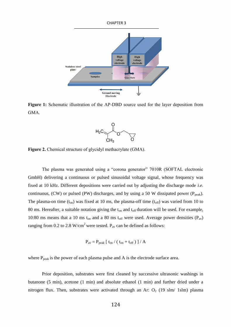
124
CHAPTER 3
Figure 1: Schematic illustration of the AP-DBD source used for the layer deposition from
GMA.
Figure 2. Chemical structure of glycidyl methacrylate (GMA).
The plasma was generated using a “corona generator” 7010R (SOFTAL electronic
GmbH) delivering a continuous or pulsed sinusoidal voltage signal, whose frequency was
fixed at 10 kHz. Different depositions were carried out by adjusting the discharge mode i.e.
continuous, (CW) or pulsed (PW) discharges, and by using a 50 W dissipated power (Ppeak).
The plasma-on time (ton) was fixed at 10 ms, the plasma-off time (toff) was varied from 10 to
80 ms. Hereafter, a suitable notation giving the ton and toff duration will be used. For example,
10:80 ms means that a 10 ms ton and a 80 ms toff were used. Average power densities (Pav)
ranging from 0.2 to 2.8 W/cm2 were tested. Pav can be defined as follows:
Pav = Ppeak [ ton / ( ton + toff ) ] / A
where Ppeak is the power of each plasma pulse and A is the electrode surface area.
Prior deposition, substrates were first cleaned by successive ultrasonic washings in
butanone (5 min), acetone (1 min) and absolute ethanol (1 min) and further dried under a
nitrogen flux. Then, substrates were activated through an Ar: O2 (19 slm/ 1slm) plasma

125
CHAPTER 3
treatment in continuous mode at 1.6 W/cm2 during 30 seconds. This Ar/O2 plasma activation
step aims to proceed to an ultimate cleaning of the surface of the substrate. [27, 28]
It is also
performed in order to grow an oxide layer subsequently exploited for the better adhesion
stability of the functional layer.
Layer characterization
Scanning electron microscopy (SEM) images were obtained with a Leica Stereoscan
430i (LEO) microscope after sputtering a 5 nm Pt thin film on the top of the plasma coating.
The thickness of the plasma-polymerized GMA (ppGMA) deposited on silicon wafers was
determined by examination of their cross sections through SEM analysis. The plasma coated
silicon wafers were broken along the cleavage plane by the help of a diamond tip cutter. It is
a very conventional procedure to obtain cross-sections of coated silicon wafers for
subsequent visualization. Before SEM investigation, however, the substrates were coated by a
thin Pt layer in order to avoid charging effect. The film deposition rate was determined from
the film thickness value and the total plasma deposition duration. The coating thickness
measurements were carried out on at least three samples for each plasma deposition
condition.
Atomic force microscopy (AFM) analysis was performed in ambient atmosphere
using a PicoSPM LE instrument in intermittent-contact mode (Agilent Technologies).
Topographic images were recorded at a 1 Hz scanning rate, 200-400 kHz resonance
frequency and 25-75 N m-1
spring constants.
Fourier-transform infrared analysis was performed with a Bruker Hyperion 2000
spectrometer in transmission and in grazing angle modes (200 scans, 4000-500 cm−1
range, 4
cm-1
resolution). The epoxy content in the coatings was estimated by integration of the 922-
892 cm-1
absorption band of normalized spectra according to the layer thickness and by using
the OPUS software.
X-ray photoelectron spectroscopy (XPS) was carried out with a Kratos Axis Ultra
DLD instrument (equipped with a monochromatic Al K X-ray source, h = 1486.6 eV
working at 50W). The samples being insulating, a charge neutralizer producing low energy
electrons of 3 eV was used. The energy calibration was done by fixing the main contribution
(adventitious carbon and hydrocarbon) at 285.0 eV. The survey and narrow scans were
acquired with 160 eV pass energy, 1 eV step size and 20 eV pass energy, 0.1 eV step size

126
CHAPTER 3
respectively. The XPS spectra were fitted with the CasaXPS software after subtraction of a
Shirley type background. The XPS C1s signal was fitted into six different carbon chemical
components [29]
, namely: i) C1: hydrocarbon (CxHy) at 285.0 eV,, ii) C2: secondary shifted
carboxyl (C-CO-O) at 285.7 eV, iii) C3: ether (C-O) at 286.7 eV, iv) C4: epoxy at 287.0 eV,
iv) C5: ketone, aldehyde (C=O) or O-C-O at 287.8eV and v) C6: ester (O-C=O) at 289.1 eV.
For all components, the full width at half maximum (FWHM) was kept between 1.1 and 1.3
eV. The peak contributions for the C-CO (C2) and O-C=O (C6) chemical groups were kept
equal. The experimental uncertainty related to the XPS surface elemental composition is
around 2 at %. For each deposition condition, at least two replicates were done and one point
per sample (localized in the middle of the coating) was analyzed.
Expression and purification of recombinant antibiofilm dispersin B (DspB)
A strain of Escherichia coli is transformed with the introduction of a pET-28a/DspB
plasmid (Life Technologies, UK). This plasmid is an expression vector for the DspB
production allowing the obtention of DspB with an hexa-histidine tag at the C-terminal
extremity of its sequence. The transformed bacteria was grown overnight at 37 °C with
shaking in 50 ml LB medium supplemented with 50 µg/ml kanamycin. The bacterial
suspension was diluted 100-fold in a total of 2 liters of LB supplemented with kanamycin (50
µg/ml), and the expression of DspB was induced with isopropyl--D-thiogalactopyranoside
(final concentration 0.5 mM). The induced culture was incubated for further 4 h (37 °C,
shaking). DspB was purified by nickel affinity chromatography as previously described in the
literature. [30]
Fractions were analyzed by sodium dodecyl sulfate-polyacrylamide gel
electrophoresis (SDS-PAGE) and by the ability to hydrolyze the chromogenic substrate 4-
nitrophenyl-N-acetyl-β-D-galactosaminide (Sigma Aldrich). Those fractions containing DspB
were pooled and dialyzed against a 10 mM phosphate buffer at pH 5.9 with 100 mM NaCl
overnight at 4 °C. Proteins were quantified using the BCA kit (Pierce).
DspB immobilization on ppGMA layers
The ppGMA coated stainless-steel coupons were immersed in 1 mg/mL of DspB-
containing 10 mM phosphate buffered solution with pH ranging from 7.0 to 8.5. The
substrates were allowed to react for 1 hour (ambient temperature, gentle agitation).

127
CHAPTER 3
Afterwards, the surfaces were rinsed with deionized water (4 times in 5 min, 250 rpm
stirring) to remove unreacted proteins.
In-vitro antiadhesion tests
A preculture of biofilm forming S. epidermidis ATCC35984 was grown overnight at
37 °C in a 3 mL Luria-Bertani (LB) media under 150 rpm agitation and used the next
morning to seed a fresh culture in LB (50 mL). The bacterial concentration of test inoculum
was adjusted to about 107 cells/mL in M63 medium. Test inoculum (200 L) was pipetted
onto each substrate. After 24 h of incubation at 37 °C, the substrates were twice rinsed with
10 mL sterile deionized water to remove non-adherent bacteria and then, they were placed
face downward in glass jars containing 500-fold-diluted LB (20 mL) and 4-mm glass beads.
The jars were shaken and then, their contents were sonicated in a water bath (50-60 kHz) for
2 min. Adherent bacteria were counted by plating 10-fold dilution on LB agar. The plates
were incubated at 37 °C overnight before the counting of the colony-forming units. The
reported results in this chapter are the averages of at least 3 replicates.
Laccase immobilization on ppGMA layers
Laccase were immobilized on ppGMA coated stainless-steel coupons through
immersion in 1 mL of a 10 mM PBS containing 1 mg/mL laccase at pH 7.0 or 8.5 (1 h, room
temperature, 150 rpm stirring). The samples were then washed (5 times during 5 min,
purified water, 250 rpm stirring).
Estimation of the immobilized laccase quantity on the coupons
The enzymatic concentrations in solution were determined with the RC DC™ Protein
Assay (Bio-Rad). The amount of immobilized laccase on the functionalized surfaces was
estimated from the difference of laccase concentrations in solution before and after the
immobilization procedure.

128
CHAPTER 3
Degradation Assays
For the immobilized enzymes, an enzymatic activity was estimated by monitoring the
sulfamethoxazole degradation over time. Enzymes were incubated in a 2 mL degradation
medium composed of sterile MilliQ water, HEPES (12.5 mM) and sulfamethoxazole (100
μg/mL). Each 24 h, the degraded sulfamethoxazole concentration was estimated by
absorbance measurement at 260 nm with a Synergy 2 Multi-Mode Plate Reader (Biotek). The
medium was removed every 24 h. Wells were washed 3 times with filtered tap water and
further filled with a fresh degradation medium.
For the free enzymes in solution, in the wells used for the first measurements (t = 24
h), 8 µg of enzymes were directly dissolved in a 2 mL degradation medium containing
sulfamethoxazole at a 100 μg/mL concentration.
For all other wells, at time t0, enzymes were dissolved in a 300 μL medium
containing 20 μg/mL of sulfamethoxazole. All wells were supplemented with the same media
every 24 h. Also, 24 h prior the enzymatic activity measurement, wells were amended with a
100 μg/mL antibiotic solution and supplemented to achieve a 2 mL final volume.
3. Results and Discussion
3.1. Influence of process deposition conditions on chemistry and morphology of ppGMA
layers
The AP-DBDs fed with Ar and GMA and operating under continuous or pulsed
discharges lead to the deposition of ppGMA films well adherent to the metallic substrates.
As shown in Figure 3, reporting the dependence of film deposition rate with toff, one
can notice that over the toff range investigated, pulsed discharges lead to higher growth film
rates than the CW one, suggesting the occurrence of different deposition mechanisms during
ton and toff, which influence the chemistry and morphology of the deposited films.

129
CHAPTER 3
Figure 3: Evolution of the ppGMA film growth rate as a function of the plasma-off time
(toff), ranging from 0 (CW mode) to 80 ms. (ton fixed at 10 ms; 50 W peak power).
SEM observations reported in Figure 4 reveal that, whatever the deposition
conditions, pinhole-free coatings covering homogeneously the entire substrate surface are
achieved. Interestingly, one can notice that the electrical discharge parameters are likely to
affect both layer deposition rate and morphology. In particular, working in CW mode lead to
rough nanostructured surfaces with an averaged roughness value (Ra) around 148 nm (Figure
4.a and Table 1) and composed of an abundant amount of aggregates presenting an average
size distribution of 94 ± 11 nm. This kind of topography is typical of coatings deposited
through an atmospheric-pressure plasma-enhanced CVD process [31, 32]
and is related to
higher plasma energy density favoring gas phase reactions and surface etching. Pulsing the
discharge leads to smoother layers with Ra value close to 24 nm at 10:80 ms conditions.
Interestingly, compared to the layers deposited in CW, those deposited in 10:10 ms
conditions have an intermediate roughness (Ra 43 nm vs 148nm), with a higher average
feature size (168 ± 25 nm). Finally, layers deposited at 10:80 ms are almost aggregate-free,

130
CHAPTER 3
which may indicate that the growth mechanism of these layers is largely controlled by
predominant surface reaction.
Figure 4: SEM (top) and AFM (bottom) pictures of ppGMA layers deposited at 50 W in CW
mode (a) and 10:10 ms (b) and 10:80 ms PW modes (c).
Table 1. Roughness values of ppGMA layers estimated by AFM measurements.
Deposition conditions Average Roughness
(Ra, nm)
Increment of surface area
(%)
50 W, CW 148 ± 13 66.00 ± 1.00
50 W, 10:10 ms 43 ± 8 0.20 ± 0.02
50 W, 10:80 ms 24 ± 11 0.10 ± 0.04
FT-IR analysis (Figure 5) was carried out in order to study the chemistry of the
deposited film. Particular attention was paid to the detection of the four epoxide characteristic
peaks, namely the epoxide ring C-H stretching at 3063 cm-1
, the ring breathing mode at 1258
cm-1
, the asymmetric and symmetric ring deformation bands at 910 cm-1
and 852 cm-1
,
respectively.[33]
For the ppGMA layers deposited at 50W in CW mode, all the above
mentioned bands were barely detectable, suggesting that most epoxide rings were opened
during the deposition. In contrast, epoxide bands were clearly noticeable for ppGMA

131
CHAPTER 3
deposited in pulsed mode. This result suggests the existence of two distinct layer growth
mechanisms when depositions are carried out in continuous or pulsed mode. In particular,
one can notice that increasing toff from 10 to 80 ms (i.e. decreasing the duty cycle) leads to
increasing the epoxide bands intensity, indicating a higher content of epoxide groups in the
films. These results are in agreement with Klages et al. [22]
, demonstrating that pulsed AP-
DBD allows the formation of coatings with high monomer structure retention due to chemical
reactions occurring during the toff between intact monomer molecules and surface radical
centers generated during the ton period.
Figure 5: FT-IR spectra of ppGMA films deposited at 50 W with different duty cycles. All
spectra were normalized to the maximum value of the C=O band (1735 cm-1
).
To estimate the epoxide content in the ppGMA layers, the area of FT-IR asymmetric
epoxide stretching band was calculated. Hence, as shown in Figure 6, it is concluded that
working in pulsed mode allows to drastically increase the epoxy content in the deposited film
up to a factor of 4.

132
CHAPTER 3
Figure 6: Area evolution of the FT-IR asymmetric epoxide ring deformation band as a
function of the plasma-off time (toff), ranging from 0 (CW mode) to 80 ms. (ton fixed at 10
ms; 50 W peak power).
In the aim of later exploiting chemical interfacial reactions between atmospheric-
pressure plasma functionalized solid surfaces and free biomolecules, XPS analysis was
carried out to get some insights concerning the ppGMA uppermost surface composition. In
particular, this analysis allows to estimate the reactive epoxy surface density, a parameter
which is reported to be strongly related to the biological performance of the functionalized
surfaces.
Considering ppGMA deposited in CW mode, the XPS atomic percentages of the
surfaces for the C:O elements were found to be equal to 77:23 (Table 2). Compared to the
commercial poly(GMA) stoichiometry of 73:27, a lesser amount of oxygen was detected in
the CW ppGMA layers, which might be correlated to the significant loss of the epoxy ring
(i.e. monomer structure degradation), already highlighted in IR analysis (Figure 5). In

133
CHAPTER 3
contrast, it can be observed that PW deposited ppGMA layers and poly(GMA) present similar
XPS compositions.
The influence of the discharge mode (CW vs PW) on the layer composition chemistry
is particularly noticeable by overlapping their XPS C1s spectra (Figure 7). Indeed, one can
clearly observe that a pulsed discharge allows a higher retention of the initial monomer ester
group (C1s contribution at 289.1eV) accompanied by the retention of epoxy groups (C1s
contribution at 287.0 eV). In particular, from the XPS C1s curve-fitting data shown in Table
2, it appears that increasing the toff leads to an increase of the epoxy surface content with
values ranging from 7 to 18 at. % for layers deposited at 10 and 80 ms toff duration,
respectively. Interestingly, despite using mild process conditions, the maximal 27% epoxy
content estimated for poly(GMA) is never reached in ppGMA layers. An explanation is that,
despite a short ton duration, there is a non-negligible contribution of the film chemistry
growth during this period associated to a high fragmentation of the monomer, and thus, a loss
of the epoxy group. This theory is fully consistent with the results obtained in CW mode.
The ageing of the richest epoxy ppGMA layer (i.e. 50 W, in 10:80 ms mode) was
investigated through a 50 days storage period at ambient temperature and humidity.
According to XPS analysis (Table 2), these storage conditions seem to have no significant
influence on the layer composition that remains comparable to the poly(GMA)‟s one, with C
and O content equal to 73 and 27 at.% respectively.
However, a careful C1s curve fitting allows highlighting a two folds decrease of the
epoxy surface density down to 9 % content, suggesting the relative chemical instability of
epoxy groups.

134
CHAPTER 3
Table 2. XPS atomic percentages and C1s curve fitting data for ppGMA layers deposited in
CW and PW modes. The results for a PW ppGMA layer after ageing 50 days at air, and for a
commercial poly(GMA) polymer are also reported.
Samples: 50 W,
CW 50 W,
10:10 ms 50 W,
10:80 ms
50 W,
10:80ms
after 50
days at
air
Poly(GMA)
XPS surface composition (at. %): C 77 75 72 73 73
O 23 25 28 27 27
XPS C1s peak fitting (%):
Contribution Binding energy (eV)
Functional group
C1 285.0 C-C 52 36 33 38 42 C2 285.7 C-CO-O 8 11 13 12 11 C3 286.7 C-O 25 25 22 25 8 C4 287.0 epoxy 0 7 18 9 27 C5 287.8 C=O/
C-O-C 5 10 2 4 -
C6 289.1 O-C=O 8 11 13 12 11

135
CHAPTER 3
Figure 7: XPS C1s curve fittings for ppGMA layers deposited at 50W, CW mode (left) and
50 W, 10:80 ms mode (right).
In conclusion, it can be reported that, among the different plasma process conditions
investigated, the pulsed atmospheric-pressure plasma polymerization of GMA carried out at
50 W in 10:80 ms allows for the fast deposition of adherent and smooth layers presenting the
highest epoxy surface density, up to 18 % (XPS data). Such freshly deposited ppGMA layers
were selected in order to maximize the efficiency of biomolecule immobilization. [34]

136
CHAPTER 3
3.2. DspB immobilization and anti-biofouling assessment
To ascertain the immobilization of DspB enzyme on the ppGMA layers, FT-IR
analysis was carried out. From the data reported in Figure 8, one can notice that,
independently of the pH of the DspB solution used during the immobilization step (i.e. pH=
7.0 or 8.5), ppGMA layers allow to efficiently immobilize the enzyme, as it is suggested by
the appearance of new peaks related to peptide bonds, namely secondary amide N-H
stretching, amide C=O stretching and amide N-H bending bands located at 3281 cm-1
, 1645
cm-1
and 1539 cm-1
, respectively.[35]
The peaks, characteristic of epoxide groups, at 1258, 910
and 852 cm-1
are probably linked to their presence into the plasma film bulk, highlighting that
the mild basic immobilization condition used does not lead to the epoxy loss through the ester
group hydrolysis.
Figure 8: FT-IR spectra of freshly deposited ppGMA layers at 50 W in 10:80 ms (a), layer
“a” after DspB immobilization at pH 7.0 (b) and pH 8.5 (c).

137
CHAPTER 3
As reported in Table 3, independently of the pH of the DspB solution used during the
immobilization steps, the DpsB-grafted coatings provide a comparable reduction of the
number of viable adherent bacteria, up to 84 %, after only 1 day of contact. This result
highlights that this enzyme has maintained its anti-biofouling property even after its covalent
immobilization on the ppGMA layers. The fact that the immobilized enzyme has kept its
activity can suggest that its biological reaction sites have not been affected during the
reaction with the ppGMA layer. The pH change during the enzyme grafting step has no effect
on the final measured enzymatic activity. This suggests that the active immobilized enzyme
amount is similar in both cases.
By reference with other works reported in literature, the reason of this success can
probably be associated to the presence of an hexahistidine tag (his-tag) added at the C-
terminal extremity of the enzyme.[23]
Kurzatkowska et al.[36]
have already reported the
exploitation of His-tag to ensure an enzyme immobilization in a proper orientation, or at
least, to initiate an attachment which could then be supplemented by other interactions with
the rest of the enzyme structure, and consequently, preventing or limiting the interaction
and/or masking of the enzyme active site. Martin et al. [37]
have also reported that his-tagged
biomolecules can be covalently immobilized on supports through the nucleophilic reaction of
the imidazole group (i.e. secondary amine) with the epoxy groups. In conclusion, if His-tag
sequence has been initially introduced at the protein chain end to ensure its purification on a
nickel affinity column during its production step, the present work highlights its usefulness
for a covalent immobilization purpose.
Table 3. Anti-biofouling activity against S. epidermidis biofilm forming of modified
stainless-steel surfaces compared to uncoated ones.
Samples Reduction of adherent population (%)
Stainless-steel 0
ppGMA layers 0
Dsp B immobilized at pH 7.0 79 ± 16
Dsp B immobilized at pH 8.5 84 ± 11

138
CHAPTER 3
3.3. Enzyme immobilization for xenobiotic degradation
Irrespective of the pH (i.e. pH= 7.0 or 8.5) used during the enzyme immobilization
step, pp-GMA layers allow the grafting of laccases, which are later active for the degradation
of sulfamethoxazole. In Table 4, the average enzyme activity duration and the total quantity
of degraded sulfamethoxazole for free and immobilized laccase are reported after different
periods of time and Figure 9 shows their degradation kinetics.
All these results show clearly that the immobilization of the enzyme increases its
efficiency and the duration of the enzymatic activity. By comparing the results between the
test where the enzymes are in solution and the other one, where they are immobilized on a
ppGMA surface at pH 7, it was observed that the total duration activity was multiplied by at
least a factor of 4 (36 h vs 144 h for free and immobilized enzymes respectively). Two
hypotheses can support this interesting result. The first one can be attributed to the advantage
of carrying out a strong enzyme immobilization through a multipoint anchorage. Indeed, this
situation is known to confer a rigidity to the three-dimensional structure of the enzyme thus,
limiting breakdown and autolysis phenomena due to physico-chemical environmental factors.
[38, 39] The second one can be associated to the combined effect of pH and stirring when the
enzymes are in solution. Indeed, these two parameters have been reported to promote free-
form protein aggregation in solution [40]
and to partly inactivate or neutralize enzymes.[3]
In
particular, the study of Shleev et al.[41]
reports that free-form laccase in phosphate buffer
solution at pH 6.5, value closed of the experimental conditions, can be aggregated and/or
inactivated.
In addition, the results show that during the enzyme immobilization, if the pH of the
solution is lightly basic (pH = 8.5), the total amount of fixed enzyme is increased by a factor
of 2. One hypothesis can be given to explain this result, it relies on the existence of different
isoenzyme forms as it is known that pH can generate new rearrangement of the laccase
periphery. Consequently, an improvement of the enzyme- plasma-treated surface interactions
might be expected at pH 8.5.
Finally, it was also observed that bioactive layers generated at pH 8.5 present an
increased xenobiotic degradation activity duration compared to the ones generated at pH 7.0
(240 h vs 144 h, respectively). More precisely, during the first 36 h of bioactivity, both
surfaces presented a similar efficiency. However, after this time lapse, the laccase activity set
at pH 7.0 sharply decreased while the enzymatic activity of active layer generated at pH 8.5

139
CHAPTER 3
slowly decreases until to reach zero after 240 h. One explanation might come from the
increased amount of fixed enzymes. Indeed, the density of immobilized proteins might have
an influence on the three-dimensional structure of the protein due to the existence of
numerous and deleterious protein-surface interactions. As reported by Tiller et al. [42]
, when
the surface of a substrate is covered with a high density of immobilized proteins, there are
few possible interactions between the protein and the substrate, thus ensuring probably the
integrity of the 3D protein structure and the preservation of its enzymatic activity. In
conclusion, these results highlight the importance of a well-adapted pH solution selection for
the enzyme immobilization step.
Table 4. Summary of the averaged enzyme activity duration and the total quantity of
degraded sulfamethoxazole for free and immobilized laccase. The quantity of immobilized
enzymes on ppGMA layers is also reported.
Assays Averaged
activity duration
(h)
Quantity of
immobilized
enzyme (µg)
Quantity of degraded
sulfamethoxazole for a
1 mL solution
(µg)
Free enzyme in solution 36 - 75 ± 5
Laccase immobilized at pH 7.0 144 5.5 ± 4.0 174 ± 15
Laccase immobilized at pH 8.5 240 12.0 ± 1.0 275 ± 14

140
CHAPTER 3
Figure 9: Activity of free and immobilized laccase.
4. Conclusion
A novel atmospheric-pressure method has been developed to efficiently immobilize
enzymes onto solid surfaces, based on the AP-DBD deposition of an epoxy-rich layer and its
subsequent interfacial one-step reaction with biomolecules in mild aqueous conditions. The
method has highlighted that it is possible: i) to modify the coating morphology by the use of
continuous or pulsed discharges, ii) to tune the epoxy surface density by adjusting the ton/ toff
ratio and iii) to optimize the amount of grafted enzymes by selecting an appropriate pH
during the immobilization step. Here, bioactive surfaces with anti-biofouling and xenobiotic
degrading properties have been successfully elaborated. In particular, the immobilized
laccase on ppGMA layers present an extended duration activity and efficiency around 4 and 7
times higher than for the free enzyme in solution, respectively. Importantly, these results tend
to show the versatility of the method as these epoxy-rich layers might offer the possibility to
immobilize different functional biomolecules paving the way to the elaboration of novel
advanced bioactive materials.
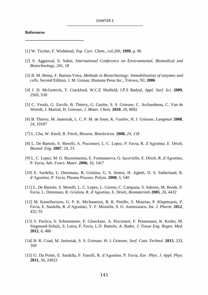
141
CHAPTER 3
References
[1] W. Tischer, F. Wedekind, Top. Curr. Chem., vol.200, 1999, p. 96
[2] S. Aggarwal, S. Sahni, International Conference on Environmental, Biomedical and
Biotechnology, 201, 18
[3] B. M. Brena, F. Batista-Viera, Methods in Biotechnology: Immobilization of enzymes and
cells, Second Edition, J. M. Guisan, Humana Press Inc., Totowa, NJ, 2006
[4] J. D. McGettrick, T. Crackford, W.C.E Shofield, J.P.S Badyal, Appl. Surf. Sci. 2009,
256S, S30
[5] C. Vreuls, G. Zocchi, B. Thierry, G. Garitte, S. S. Griesser, C. Archambeau, C. Van de
Weerdt, J. Martial, H. Griesser, J. Mater. Chem. 2010, 20, 8092
[6] B. Thierry, M. Jasieniak, L. C. P. M. de Smet, K. Vasilev, H. J. Griesser, Langmuir 2008,
24, 10187
[7] L. Chu, W. Knoll, R. Förch, Biosens. Bioelectron. 2008, 24, 118
[8] L. De Bartolo, S. Morelli, A. Piscioneri, L. C. Lopez, P. Favia, R. d‟Agostino, E. Drioli,
Biomol. Eng. 2007, 24, 23
[9] L. C. Lopez, M. G. Buonomenna, E. Fontananova, G. Iacoviello, E. Drioli, R. d‟Agostino,
P. Favia, Adv. Funct. Mater. 2006, 16, 1417
[10] E. Sardella, L. Detomaso, R. Gristina, G. S. Senesi, H. Agheli, D. S. Sutherland, R.
d‟Agostino, P. Favia, Plasma Process. Polym. 2008, 5, 540
[11] L. De Bartolo, S. Morelli, L. C. Lopez, L. Giorno, C. Campana, S. Salerno, M. Rende, P.
Favia, L. Detomaso, R. Gristina, R. d‟Agostino, E. Drioli, Biomaterials 2005, 26, 4432
[12] M. Kastellorizios, G. P. K. Michanetzis, B. R. Pistillo, S. Mourtas, P. Klepetsanis, P.
Favia, E. Sardella, R. d‟Agostino, Y. F. Missirlis, S. G. Antimisiaris, Int. J. Pharm. 2012,
432, 91
[13] S. Pavlica, S. Schmitmeier, P. Gloeckner, A. Piscioneri, F. Peinemann, K. Krohn, M.
Siegmund-Schulz, S. Laera, P. Favia, L.D. Bartolo, A. Bader, J. Tissue Eng. Regen. Med.
2012, 6, 486
[14] B. R. Coad, M. Jasieniak, S. S. Griesser, H. J. Griesser, Surf. Coat. Technol. 2013, 233,
169
[15] G. Da Ponte, E. Sardella, F. Fanelli, R. d‟Agostino, P. Favia, Eur. Phys. J. Appl. Phys.
2011, 56, 24023

142
CHAPTER 3
[16] B. Nisol, C. Poleunis, P. Bertrand, F. Reniers, Plasma Process. Polym. 2010, 7, 715
[17] B. Nisol, G. Oldenhove, N. Preyat, D. Monteyne, M. Moser, D. Perez-Morga, F.
Reniers, Surf. Coat. Technol. 2014, 252, 126
[18] G. Da Ponte, E. Sardella, F. Fanelli, R. d‟Agostino, R. Gristina, P. Favia, Plasma
Process. Polym. 2012, 9, 1176
[19] G. Chen, M. Zhou, Z. Zhang, G. Lv, S. Massey, W. Smith, M. Tatoulian, Plasma
Process. Polym. 2011, 8, 701
[20] R. Mauchauffé, M. Moreno-Couranjou, N.D. Boscher, C.Van De Weerdt, A-S. Duwez,
P. Choquet, J. Mater. Chem. B 2014, 2, 5168
[21] D. Duday, C. Vreuls, M. Moreno, G. Frache, N. D. Boscher, G. Zocchi, C. Archambeau,
C. Van De Weerdt, J. Martial, P. Choquet, Surf. Coat. Technol. 2013, 218, 152
[22] C. –P. Klages, K. Höpfner, N. Kläke, R. Thyen, Plasmas Polym. 2000, 5, 79
[23] J. B. Kaplan, C. Ragunath, N. Ramasubbu, D. H. Fine, J. Bacteriol. 2003, 185, 4693
[24] S. Rodríguez Couto, J. L. Toca Herrera, Biotechnol. Adv. 2006, 24, 500
[25] C. E. Rodríguez-Rodríguez, M. A. García-Galán, P. Blánquez, M. S. Díaz-Cruz, D.
Barceló, G. Caminal, T. Vicent, J Hazard Mater. 2012, 213-214, 347
[26] A. Göbel, A. Thomsen, C. S: McArdell, A. Joss, W. Giger, Environ. Sci. Technol. 2005,
39, 3981
[27] G. Baravian, D. Chaleix, P. Choquet, P. L. Nauche, V. Puech, M. Rozoy, Surf. Coat.
Technol. 1999, 115, 66
[28] J. M. Thiébaut, T. Belmonte, D. Chaleix, P. Choquet, G. Baravian, V. Puech, H. Michel,
Surf. Coat. Technol. 2003, 169–170, 186
[29] G. Beamson, D. Briggs, High Resolution XPS of Organic Polymers The Scienta ESCA
300 Database, Wiley-VCH, New York, 1992
[30] E. Faure, C. Falentin-Daudré, T. S. Lanero, C. Vreuls, G. Zocchi, C. Van De Weerdt, J.
Martial, C. Jérôme, A.-S. Duwez, C. Detrembleur, Adv. Funct. Mater. 2012, 22, 5271
[31] A. Manakhov, , M. Moreno-Couranjou, N. D. Boscher, V. Rogé, P. Choquet, J.-J.
Pireaux, Plasma Process. Polym. 2012, 9, 435
[32] H. Hody, P. Choquet, M. Moreno-Couranjou, R. Maurau, J.-J. Pireaux, Plasma Process.
Polym. 2010, 7, 403
[33] C. Tarducci, E. J. Kinmond, J. P. S. Badyal, S. A. Brewer, C. Willis, Chem. Mater. 2000,
12, 1884

143
CHAPTER 3
[34] L. Cao, Carrier-bound immobilized Enzymes, Wiley, Weinheim, Germany 2005.
[35] B. Smith, Infrared Spectral Interpretation. A Systematic approach, Second Edition, CRC
Press LLC, Bocar Raton, FL, 1999
[36] K. Kurzątkowska, M. Mielecki, K. Grzelak, P. Verwilst, W. Dehaen, J. Radecki, H.
Radecka, Talanta 2014, 130, 336
[37] M. T. Martin, F. J. Plou, M. Alcalde, A. Ballesteros, J. Mol. Catal. B: Enzym. 2003, 21,
299
[38] H. –C. Mahler, W. Friess, U. Grauschopf, S. Kiese, J. Pharm. Sci. 2009, 98, 2909
[39] C. Garcia-Galan, A. Berenguer-Murcia, R. Fernandez-Lafuente, R-C. Rodrigues, Adv.
Synth. Catal. 2011, 353, 2885
[40] C. Mateo, V. Grazù, B. C. Pessela, T. Montes, J. M. Palomo, R. torres, F. López-
Gallego, R. Fernández-Lafuente, J. M. Guisán, Biochem. Soc. Trans. 2007, 35, 1593
[41] S. Shleev, C. T. Reimann, V. Serezhenkov, D. Burbaev, A. I. Yaropolov, L. Gorton, T.
Ruzgas, Biochimie 2006, 88, 1275
[42] J. C. Tiller, R. Rieseler, P. Berlin, D. Klemm, Biomacromolecules 2002, 3, 1021

144
CHAPTER 4

145
CHAPTER 4
Chapter 4
Self-defensive Coating for Antibiotics Degradation –
Atmospheric Pressure Chemical Vapor Deposition of
Functional and Conformal Coatings for the Immobilization
of Enzymes
Sébastien Bonot, Rodolphe Mauchauffé, Nicolas D. Boscher, Maryline
Moreno-Couranjou, Henry-Michel Cauchie and Patrick Choquet,
Advanced Materials Interfaces, 2015, 2, 1500253.

146
CHAPTER 4

147
CHAPTER 4
1. Introduction
A wide variety of xenobiotics are released into wastewater, including antibiotics
which are extensively used in human and veterinary medicine. They are found in the effluents
of wastewater treatment plants but also in tap water itself and even in drinking water
sources.[1]
Present in sub-inhibitory concentrations, antibiotics can have many effects
profoundly altering the physiology of the bacteria with various consequences such as the
increase of virulence, the induction of biofilm formation, some changes in the balance of
populations and finally the risk of the emergence of new resistant bacteria through the
induction of gene transfers.[2,3,4,5,6]
More precisely, in water treatment plants (WTP), a
fraction of the xenobiotics is adsorbed to suspended solids and removed by settling, while
another part is dissolved in the water and partially degraded through physico-chemical and
biological reactions.[7]
However, an important quantity still persists after these primary and
secondary treatments. Therefore tertiary treatments, such as UV or ozonation, are
investigated for the ultimate degradation of these molecules. Nevertheless, despite their
efficiency, they are also known to generate undesirable and sometimes toxic secondary
metabolites molecules, which are usually more pernicious than the parental antibiotics
compounds.[8,9]
The use of specific enzymes might provide a new alternative or a
complement to existing treatments commonly used in WTP. Numerous enzymes which
degrade and inactivate antibiotics are known and are encoded by the antibiotics resistance
genes of bacteria.[10]
However it has been shown that under certain conditions antibiotics
inactivated by these enzymes could be reactivated by a reverse chemical or enzymatic
reaction.[11,12]
This is why it is necessary to degrade the target molecules into more ultimate
products through the combination of several enzymes. In this case the use of less specific
hydrolase such as laccases, which are known to degrade a wide variety of xenobiotics, is
essential.[13]
Free enzymes can quickly lose their activity due to autolysis, physico-chemical and
biological degradation in the medium and/or aggregation.[14,15]
An efficient way to overcome
these drawbacks is to immobilize the enzymes on a surface. As highlighted by a large number
of studies, the immobilization of enzymes brings most of the time multiple benefits e.g. (1)
limiting autolysis and ensuring protection against physico-chemical and biological
degradation, (2) increasing the lifetime of the enzyme, (3) increasing the enzyme activity and

148
CHAPTER 4
the degradation rate.[16]
In addition, multi-anchoring may give more rigidity to the protein
which increases its resistance. In the case of a surface immobilization via weak interactions
the release of the enzyme can easily occur by simple parameter changes such as pH,
temperature or surfactant presence, making the immobilized surfaces only usable in a limited
range of conditions. In order to have long lasting properties under a wide range of conditions,
a strong and irreversible immobilization is preferred. Among the various methods developed,
the covalent binding of enzymes over a surface remains the most employed and studied
method.[17]
Surfaces with functional groups, e.g. amine, carboxylic, propargyl, aldehyde or
epoxy groups, are able to form covalent bonds using a linker or through direct reaction, when
possible, with the functional groups present in the enzymes, i.e. amine, carboxylic, thiol or
imidazole groups, in aqueous buffer at room temperature.[18,19,20,21,22]
Surface functionalization can be achieved through many different methods. Among
them, Chemical Vapor Deposition (CVD) processes, which are solvent-free environmentally
friendly methods, provide several convenient routes for the conformal deposition of
functional polymer thin films. Notably, initiated CVD (iCVD), which implies the use of a
thermally labile initiator to form radicals that further initiate the free-radical polymerization
reaction, has proven to be a suitable method for the conformal deposition of a large number
of functional polymers.[23,24,25]
If iCVD has already been successfully up-scaled for the
coating of various materials, the required low-pressure is likely to induce some limitations
(e.g. coating of very large sized objects). Recently, an atmospheric pressure CVD approach
employing ultra-short plasma discharges was investigated for the deposition of polymer thin
films. The method, called Plasma initiated CVD (PiCVD), combines the advantages of iCVD
and atmospheric pressure Plasma Enhanced CVD (PECVD) while avoiding their respective
drawbacks. Indeed, the ultra-short homogeneous discharge generated at atmospheric pressure
thanks to a fast voltage rise (i.e. tens of nanoseconds), conveniently replaced the initiator and
heating filaments used in iCVD by producing a range of reactive species with some of them
that will efficiently initiate the free-radical polymerization reaction.[26]
The well-reported
negative impact of plasma on the monomer was minimized by pulsing the ultra-short plasma
discharges (ca. tON = 100 ns) at very low frequencies (ca. 100 to 1000 Hz). As a consequence,
plasma only affects the chemistry of the film for a very small portion of the time (ca. 0.003
%), whereas the free-radical polymerization pathway reaction occurs solely during most of

149
CHAPTER 4
the plasma-off time (tOFF = 33 ms) and polymer coatings with maximum functional groups
retention are formed.[27]
In this work, epoxy functionalized coatings are synthesized by plasma polymerization
of glycidyl methacrylate (GMA) via PiCVD at atmospheric pressure.[26]
The coatings,
deposited on various materials, are used for the grafting of two enzymes able to degrade
antibiotics, i.e. Laccase and β-lactamase. The influence of their covalent immobilization on
sulfamethoxazole and amoxicillin degradation performances is studied. The integrity of
immobilized enzymes structure and the related enzymatic activity are studied under high
velocity water flow inducing a mechanical shear stress in order to assess the potential
applicability of covalently immobilized enzymes in real harsh conditions. In order to
overcome the potential threat of microorganism adhesion, able to deteriorate immobilized
enzymes and reduce degradation performances, self-defensive properties are developed.
Surfaces are saturated with Tween 20, a non-toxic polysorbate surfactant commonly used to
remove weakly bonded proteins, to saturate non-bonded sites for adhesion prevention and, in
microbiology, to avoid microorganism aggregation and adsorption.[28]
The overall efficiency
of the newly developed advanced coatings is then herein investigated.
2. Experimental Section
Materials and Reagents
The glycidyl methacrylate (GMA, 97%) monomer was obtained from Sigma-Aldrich
and used without further purification. Argon (99.999 %) and oxygen (99.999%) gases were
obtained from Air Liquid. Laccase from Pleurotus ostreatus and β lactamase from
Pseudomonas aeruginosa (Sigma-Aldrich) were resuspended (5 mg mL-1
) in phosphate
buffered saline (PBS: 137 mM NaCl, 2.7 mM KCl, 1.5 mM KH2PO4, 8 mM Na2HPO4,2H2O;
pH=8). The mirror polished stainless steel 304 disks, with a total surface of 3.14 cm2, were
provided by the CRM group (Belgium) and the high-density polyethylene biochips
(BiofilmChipTM M), with a total surface of 75 cm2, were purchased from Anoxkaldnes
(Lund, Sweden).

150
CHAPTER 4
Plasma initiated Chemical Vapor Deposition:
The plasma-polymerized GMA thin films were deposited in an atmospheric pressure
dielectric barrier discharge (AP-DBD) reactor, as previously described. [26]
The plasma set-up
is made of two high voltage electrodes (18.72 cm2) and a stainless steel moving table as
ground electrode. An ultra-short square pulse AP-DBD, generated using an AHTPB10F
generator from EFFITECH (Gif-sur-Yvette, France), and was used to ignite the plasma
discharge. One μs square peaks of 2 kV with a peak repetition frequency of 316 Hz were used
(i.e. discharge time: 100 ns). Such conditions correspond to 0.003 % duty cycles (TOFF: 33
ms). The deposition time was 25 s for all the experiments. Argon, used as a process and
carrier gas, was flushed through the GMA bubbler at 15 L min-1
, i.e. leading to a monomer
flow rate of about 61 mg min-1
. Overall argon flow through the reactor was maintained to 20
L min-1
for all experiments. The stainless steel disks and HDPE biochips were cleaned by a
30 s exposure to a 95:5 argon: oxygen AP-DBD plasma (1 W cm-2
). The discharge current
and voltage signals were measured using a current probe (Lecroy, CP030) and a high voltage
probe (Lecroy, PPE 20 kV). The traces were recorded using an oscilloscope (LeCroy,
Wavesurfer 42XS, 400 MHz).
Grafting and estimation of immobilized protein quantity
Following the PiCVD step, the stainless steel disks and HDPE biochips were
immerged in enzymatic solutions during 1 hour at room temperature with gentle agitation
(160 rpm). The disks and biochips were then gently washed 5 times during 5 minutes with
purified milliQ water and dried with a compressed air spray. The quantity of immobilized
enzymes was estimated by measurement of proteic concentration in the enzymatic solution
before and after sample immersion in the solution using the RC DC™ Protein Assay kit (Bio-
rad). The difference between these two concentrations is considered as the immobilized
enzyme quantity on the surfaces.
Saturation of bioactive surface with Tween 20
The bioactive material surfaces were saturated with Tween 20 by immersing in a
Tween 20 solution (5% in PBS). Materials were incubated in 12-well plates or petri dishes for
stainless steel disks and biochips respectively, for 1 hour at room temperature with an orbital
agitation of 100 rpm. Surfaces were then washed 5 times with PBS solution and dried.
151
CHAPTER 4
Cartography of immobilized enzyme repartition on functionalized surfaces
The bioactive disks and biochips were immersed in LavaPurple (Serva) diluted to a
hundredth in acid solution (boric acid 31.5 g L-1
; NaOH 19.25 g L-1
) in 12-well plates or petri
dishes for stainless steel disks and biochips respectively, under slow rocking motion (GFL
Rocking Shaker model 3013) in the dark for 15 minutes. The disks and biochips were then
washed 3 times with an acidic solution (citric acid 5%; 75% pure ethanol) to activate the
fluorochrome and stored in the same solution. After drying, the surfaces were scanned with a
FLA 9500 laser scanner typhoon (GE Healthcare Life Sciences) to the wavelength 450 nm.
The images obtained were analyzed using the ImageJ software
(http://imagej.nih.gov/ij/index.html). The image analysis was based on the processing of
different grey levels indicating the presence of more or less important protein concentrations
according to the grey intensity obtained. It is, in this case, a qualitative analysis to estimate
the protein presence and distribution on the material surface. The significative difference
between the samples is estimated by statistical non parametric Mann-Whitney tests
comparing two by two the samples.
Surface Characterization
The SEM observations and film thickness measurements were performed on a Hitachi
SU-70 FE-SEM. Prior to SEM observations, the non-conductive samples were sputter coated
with 5 nm of platinum to prevent charging and distortion. FTIR analyses were performed on a
Bruker Hyperion 2000 spectrometer equipped with an ATR objective.
Antibiotics Degradation Activity
Immobilized-enzymes: Enzymatic activity was estimated by the measurement of the
amoxicillin (Sigma-Aldrich) and sulfamethoxazole (Sigma-Aldrich) degradation at different
time. The immobilized enzymes were continuously incubated in successive baths containing
a degradation medium composed of tap water filtered through a polyvinylidene syringe filter
(0.22 μm), HEPES (12.5 mM) and antibiotic (100 μg mL-1
). The volumes used were 1 mL for
stainless steel disks and 7 mL for the biochips. At an interval of 24h, the medium was
renewed every 24 hours, wells were washed 3 times with filtered tap water and a new volume
of the degradation medium was put in the wells. During weekends, the media was
supplemented with a three-fold daily antibiotic concentration in order that the antibiotic

152
CHAPTER 4
concentration was not limiting and the enzymes remain active throughout this period. At the
different monitoring times the degraded antibiotic concentration was estimated by measuring
appropriated absorbance (210 for amoxicillin or 260 nm for sulfamethoxazole) with a 2
Synergy microplate reader (Biotek).
Free-enzymes: In the wells used for the first measures (t = 24 h), enzymes were
directly dissolved in a degradation medium (2 mL) containing 100 μg mL-1
of antibiotic. For
all other wells, at time t0, enzymes were dissolved in 500 μL of the degradation medium
containing 50 μg mL-1
of antibiotic in order that the enzymes were active. All wells were fed
by this same volume every 24 hours. 24 hours before measurement wells are amended with
100 μg mL-1
of antibiotic and supplemented to achieve a final volume of 2 mL.
Erosion flow tests
The resistance of functionalized surface / enzyme bonds was evaluated by erosion
tests in the water flow. The samples were fixed to the rotor of a reactor of Biofilm Reactor
Annular LJ 1320 (Biosurface Technologies Corporation). The disks were subjected to
laminar water flows equivalent to a flow rate of 30 km h-1
over various times. At times of 48
hours and 6 days the enzymatic activity was measured as described in the “Antibiotics
Degradation Activity” section and compared to disks used for degradation assays. The
erosion of enzymes was estimated using LavaPurple. Bioactive materials were immersed in a
solution of LavaPurple for 15 min, at 100 rpm. They were then rinsed 5 fold with an acid
solution and then analyzed with a laser scanner as described in the “Cartography of
immobilized enzyme repartition on functionalized surfaces” section.
Bacteria and Fungi Adhesion
A culture of Pseudomonas aeruginosa PAO1 grown on Luria Bertani media were
concentrated by centrifugation at 5000 g for 3 min, and washed three times with PBS. The
final bacterial concentration was 8 107 colony forming unit mL
-1. The spores of Aspergillus
nidulans grown on potato dextrose agar media were dissolved in PBS to final concentrations
of 2 105 spores mL
-1. The disks or biochips were immersed in these solutions for 4 days at
room temperature with gentle agitation (100 rpm). The samples were then rinsed 5 times with
MilliQ water and analyzed by SEM.

153
CHAPTER 4
Storage of biochips
The biochips were dried and stored at 4 ° C in three different forms: (1)
functionalized, (2) functionalized + enzymes and (3) functionalized + enzymes + Tween 20.
At various times, 7, 15, 20, 30, 60, 90, 120 and 160 days, the biochips were used to estimate
the integrity of their activity. For functionalized biochips, the enzyme immobilization and the
surface saturation with Tween 20 were performed as described in the previous sections. The
biochips were then immerged in the appropriate degradation media and degradation tests
were performed as described in “Antibiotics Degradation Activity” section as the duration
when the system keeps an activity above 90% of the activity of bioactive biochips prepared
extemporaneously.

154
CHAPTER 4
3. Results
3.1. Atmospheric Pressure Plasma Initiated Chemical Vapour Deposition of Epoxy-rich
Coatings
Stainless steel disks and high-density polyethylene (HDPE) biochips are coated using
an atmospheric pressure PiCVD method as described elsewhere (Figure 1).[26]
Prior to the
CVD step, samples are cleaned with solvents (butanone/acetone/ethanol) and just before
deposition are exposed to a continuous 1 W cm-2
argon/oxygen plasma discharge (generated
by a 10 kHz sinusoidal signal with a Softal generator) to remove remaining impurities and
form surface hydroxyl, carbonyl and carboxylic acid groups.[29,30]
PiCVD
Enzymes direct immobilization
Tween 20 saturationGMA+Ar
Epoxy functionalized coating
HV Electrode
Plasma pre-treatment: OH formation
Continuous
dischargeOH
Sample
OH OHOH OH
Moving table
poly(GMA)
OO OO OO O
R. R
.e-
R.R
.R.
. . . . . . .e-
e-R.e-
e- e-
GMA+Ar
OO OO OO O R.
R.
R.
Moving table
Moving table
. . . .R.
R.
R.
R. . . .
Moving table
Plasma off
Free-radical polymerization
Laccase or
β-Lactamase
Ultra-short pulsed discharge:
Radicals initiation
Ar/O2
Figure 1: Schematic description of the method: 1) Surface Argon/Oxygen plasma pre-
treatment generates hydroxyl groups which can provide radical surface groups during
subsequent PiCVD, allowing good adhesion of the coating 2) PiCVD of a poly(glycidyl
methacrylate) coating 3) Enzyme covalent immobilization 4) Tween 20 surface saturation.
Those groups provide during the subsequent deposition step reactive radicals groups
to ensure the covalent anchoring of the formed polymer coating to the surface material.
During the PiCVD step, thanks to periodic and well controlled ultra-short square pulses with

155
CHAPTER 4
a fast voltage rise (30 ns) (Figure 2), plasma discharges with a large proportion of high
energy electrons are generated.
Figure 2: Measured gas applied external voltage and current density of the short square pulse
plasma discharge.
Owing to the use of very short plasma discharges (i.e. tON = 100 ns per pulse) and
several orders of magnitude longer plasma-off times (i.e. 33 ms), the negative impact of the
energetic species created by plasma on the chemistry of the growing coating is minimized
and the free-radical polymerization of GMA is greatly favored.[27]
Irrespective of the
substrate material, the PiCVD of poly(GMA) leads to the deposition of macroscopically
smooth and adherent thin films over the entire surface of the samples. SEM observations
confirmed that the formed coatings, covering the whole surface, are smooth and particle-free
(Figure 3).

156
CHAPTER 4
Figure 3: poly(GMA) coating on stainless steel disk observed by scanning electron
microscopy.
The film thickness, measured from the SEM cross-section observation of PiCVD
coated silicon wafer, is 65 nm for a 25 seconds deposition time. The conformal coverage of
the HDPE biochip surface, for which the highest aspect ratio value is 2:1 (1 mm width and 2
mm depth), is likely to occur as suggested by the cross-section SEM observation of a covered
5 µm deep and 600 nm wide trench on a silicon wafer (Figure 4), with a higher aspect ratio
value of 8:1, showing that conformal coverage is successfully achieved by PiCVD.
Figure 4: Cross sectional scanning electron microscopy (SEM) image of a 88 nm thick
poly(GMA) coating conformally deposited on a 600 nm x 5 mm trench on an etched silicon
wafer.

157
CHAPTER 4
FTIR analysis of different zones of the samples, confirm the uniform coverage of the
poly(GMA) coating, notably on the more complex geometry of the biochips. Irrespective of
the material and surface morphology, the FTIR spectrum of the plasma-polymerized coating
matches the spectrum obtained for conventionally polymerized poly(GMA) (Figure 5).
Interestingly, an excellent retention of the pendant epoxy groups with the presence of well-
defined FTIR absorption bands at 843 cm-1
, 905 cm-1
, 1253 cm-1
and 3063 cm-1
is
observed.[31,32]
It is also interesting to note that the C=C stretching band from the
methacrylate group of the GMA monomer (1637 cm-1
), from which the polymerization is
supposed to occur, is barely observed on the FTIR spectra of the deposited films.
Figure 5: FTIR spectra of a) GMA monomer b) the poly(GMA) coating and c)
conventionally polymerized poly(GMA) powder.

158
CHAPTER 4
3.2. Enzymes Grafting for Water Treatment
The poly(GMA) coated samples are further investigated for the immobilization of β-
lactamase and laccase enzymes such as described in the experimental section. The
immobilized enzymes are labelled thanks to a fluorescent LavaPurple dye further observed by
laser scanning analysis (Figure 6). The analysis of greyscale images, indicating the
distribution and concentration of the immobilized enzymes, shows significant grey levels
differences between the control disks and those immersed in the enzymatic solutions (Mann-
Whitney test P = <0.001). This observation, made on both the poly(GMA)-coated stainless
steel disk and the poly(GMA)-coated HDPE biochips, indicates that the enzyme
immobilization is the result of an interaction with the functional coating on the surfaces. The
immobilization of proteins by interactions with epoxy groups has already been demonstrated
in numerous studies.[33]
It results from multiple covalent bonds between the epoxy coating
and unused peripheral reactive groups of enzymes.[34,35]
However, denser areas are observed
indicating that the covalently immobilized enzymes concentrations are higher in these areas
and/or may correspond to protein/protein aggregation.
Figure 6: Surface enzyme distribution before and after grafting of Laccase and β-lactamase
on functionalized stainless steel disks and HDPE biochips. Enzymes are labelled thanks to a
fluorescent LavaPurple dye and observed by laser scanning analysis; their distribution is
represented by grey levels according to local enzyme concentration where darker surfaces
show higher enzyme concentrations.
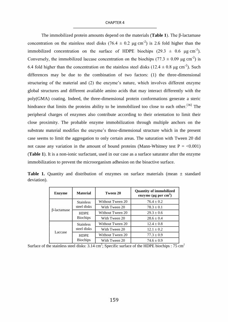
159
CHAPTER 4
The immobilized protein amounts depend on the materials (Table 1). The β-lactamase
concentration on the stainless steel disks (76.4 ± 0.2 μg cm-2
) is 2.6 fold higher than the
immobilized concentration on the surface of HDPE biochips (29.3 ± 0.6 μg cm-2
).
Conversely, the immobilized laccase concentration on the biochips (77.3 ± 0.09 μg cm-2
) is
6.4 fold higher than the concentration on the stainless steel disks (12.4 ± 0.8 μg cm-2
). Such
differences may be due to the combination of two factors: (1) the three-dimensional
structuring of the material and (2) the enzyme‟s nature, which involves different enzyme
global structures and different available amino acids that may interact differently with the
poly(GMA) coating. Indeed, the three-dimensional protein conformations generate a steric
hindrance that limits the proteins ability to be immobilized too close to each other.[36]
The
peripheral charges of enzymes also contribute according to their orientation to limit their
close proximity. The probable enzyme immobilization through multiple anchors on the
substrate material modifies the enzyme‟s three-dimensional structure which in the present
case seems to limit the aggregation to only certain areas. The saturation with Tween 20 did
not cause any variation in the amount of bound proteins (Mann-Whitney test P = <0.001)
(Table 1). It is a non-ionic surfactant, used in our case as a surface saturator after the enzyme
immobilization to prevent the microorganism adhesion on the bioactive surface.
Table 1. Quantity and distribution of enzymes on surface materials (mean ± standard
deviation).
Enzyme Material Tween 20 Quantity of immobilized
enzyme (μg per cm2)
β-lactamase
Stainless
steel disks
Without Tween 20 76.4 ± 0.2
With Tween 20 78.3 ± 0.1
HDPE
Biochips
Without Tween 20 29.3 ± 0.6
With Tween 20 28.6 ± 0.4
Laccase
Stainless
steel disks
Without Tween 20 12.4 ± 0.8
With Tween 20 12.1 ± 0.2
HDPE
Biochips
Without Tween 20 77.3 ± 0.9
With Tween 20 74.6 ± 0.9
Surface of the stainless steel disks: 3.14 cm2; Specific surface of the HDPE biochips : 75 cm
2

160
CHAPTER 4
3.3. Antibiotic Degradation Activities
3.3.1. Laccase grafted poly(GMA) coatings performances
To evaluate the antibiotic degradation potential of the prepared surfaces, tests were
conducted using sulfamethoxazole and amoxicillin as target molecules. The activity of the
immobilized enzymes, measured by following the antibiotic disappearance in solution over
time by UV spectrometry, is compared to the free enzyme activity (Table 2).
Table 2. Performances of immobilized enzymatic systems
Enzyme free form Immobilized enzyme
Average
duration
of
activity
(h)
Total
degraded
antibiotic
quantity
(µg )
Material Tween 20
Average
duration of
activity (h)
Total
degraded
antibiotic
quantity (µg)
β- Lactamase 48
50 ± 9 Stainless
steel disks
Without Tween 20 288 72 ± 9
With Tween 20 384 117 ± 2
79 ± 2 HDPE
Biochips
Without Tween 20 264 955 ± 63
With Tween 20 576 1240 ± 50
Laccase 48
120 ± 5 Stainless
steel disks
Without Tween 20 288 244 ± 18
With Tween 20 648 761 ± 88
183 ± 10 HDPE
Biochips
Without Tween 20 240 2376 ± 100
With Tween 20 648 6562 ± 363
When free laccases are immediately dissolved in media containing sulfamethoxazole,
there is a degradation rate of 21 µg mL-1
h-1
of the antibiotic after just one hour of incubation.
When the antibiotic is added in the solution where the enzymes have been stirred for 1 hour,
only 12 µg mL-1
d-1
of sulfamethoxazole are degraded after 24 hours. The degradation rate
continues to decrease until it drops to zero after 48 hours. This rapid loss of enzyme activity
can be explained by the combination of different factors which are (1) the aggregation of
these enzymes in solution favored by the combined effect of the pH and stirring, (2) the
enzymes autolysis at room temperature and (3) the possible inhibitory effect of the secondary
metabolites produced by the degradation.[37]
To the best of our knowledge no study has
shown the possible inhibitory effect of the sulfamethoxazole by-products on laccases.

161
CHAPTER 4
For the immobilized laccases on HDPE biochips, the degraded sulfamethoxazole amount is
equal to 79 µg mL-1
h-1
. This concentration is 3.8 times higher than the sulfamethoxazole
concentration degraded by a similar concentration of free enzymes during the first hour. After
24h and 48h (2 cycles of regeneration medium) the activity remains important, and
moderately decreases at 96 h to finally drop to zero after 240 h (Figure 7a).
Figure 7: a) Effect of immobilization on the degradation performances of laccase and β-
lactamase on HDPE biochips. The values of histograms correspond to the average value of
three independent assays each composed of triplicates. b) Effect of Tween-20 saturation on
HDPE biochips where β-lactamase and laccase are immobilized. The values of histograms
correspond to the average value of two independent assays each composed of triplicates.
The maximum degraded sulfamethoxazole quantity represents 95 % of the initial
antibiotic amount in the system after 24 h. The incomplete antibiotic degradation can mean
that this residual concentration is the limiting target molecule concentration in the medium
for the available enzyme amount and/or the generated secondary metabolites are inhibitors of
the activity. When antibiotic is added in the degradation medium without renewing the media

162
CHAPTER 4
and wherein the by-products accumulate, no inhibition effect is observed on the enzymatic
activity (Figure 8).
Figure 8: a) Effect of the accumulation of the sulfamethoxazole degradation products in the
medium on the performance of the laccase immobilized on the HDPE biochips and b) effect
of the accumulation of the amoxicillin degradation products in the medium on the
performance of the β-lactamase immobilized on HDPE biochips. The values of histograms
correspond to the average value of triplicates.
So this is the enzyme/target molecule ratio which is limiting. The enzyme
immobilization allows increasing by 5 fold the enzyme activity over time and finally the
degraded sulfamethoxazole quantity is 13 times higher than the degraded quantity by free
form laccase (Figure 7a and Table 2). The assays performed with stainless steel disks show

163
CHAPTER 4
both an increase of the enzyme activity in the system compared to the free-enzymes with an
enzymatic activity that increases from 48 hours to 288 hours. The degraded sulfamethoxazole
quantity is multiplied 2 fold for an equivalent enzyme concentration (Table 2). These results
are important data for the perspective of using this kind of system in water treatment. Indeed,
the water residence time in a tertiary treatment of WTP do not exceed a few hours in general.
Here we show that most of the degradation can occur in the first hour. Finally, the high
antibiotic concentrations that immobilized enzymes may degrade are also interesting data.
The sulfamethoxazole concentrations in WTP basins and in its effluent range from 0.3 to 2
μg L-1
which is 3 103 to 4.7 10
4 times lower than what can be degraded per hour by our
system.[38,39]
3.3.2. β-lactamase grafted poly(GMA) coatings performances
Similarly to the observations made from laccases, the amoxicillin degradation rate by
the free form β-lactamase during the first hour is equal to 5 µg mL-1
h-1
. The degradation rate
decreases and drops to zero after 96 hours, indicating a full loss of activity of free enzymes.
For the immobilized β-lactamase on HDPE biochips, an activity is observed until 264 hours.
The amoxicillin degradation rate varies between 2.7 and 35 µg mL-1
per 24 hours (Figure 7).
Here again, the immobilization is beneficial for the enzyme. The enzymes are active on both
functionalized materials and the total activity time is multiplied by 6 to 5.5 times with an
amoxicillin degradation rate 1.44 to 12 times higher for HDPE biochips and stainless steel
disks respectively than for free form enzymes.
3.3.3. Effect of Tween 20 on the β-lactamase and laccase grafted poly(GMA) coatings
performances
In order to provide the developed surfaces with a self-defensive behavior, which
would be of great interest for potential use in water treatments, the surfaces were saturated
with Tween 20 to prevent microorganism adhesion. The activity of those new surfaces was
investigated as the polysorbates may alter the degradation performances of bioactive surfaces.

164
CHAPTER 4
For both functionalized laccase and β-lactamases biochips and irrespective of the used
material, the Tween 20 saturation has a positive effect on the activity duration (Figure 7b).
Indeed, comparing bioactive surfaces Tween 20-saturated or not, the enzymatic activity
duration is multiplied by 2.7 in the case of the laccases and by 2 for the β-lactamases grafted
ones. Compared to free-enzyme, the immobilization and Tween 20-saturation surface allows
in both cases a multiplication of about 13.5 fold of the activity duration. Concerning the
quantities of degraded antibiotics, both saturated and non-saturated present similar efficiency
for a duration below 120 h, but along with the time while non-saturated surfaces are clearly
losing activity, in the case of saturated surfaces, the degraded quantities follow a “wave
phenomena” with important resurgences of enzymatic activity as shown in Figure 7b.
Interestingly, as shown in Figure 9, the presence of enzymes in the liquid degradation media
is detected just before activity resurgence is observed, concentrating the solution by 200-fold
during the first step of the RC DC™ Protein Assay (a total precipitated volume of 5 mL
instead of recommended volume of 25 μL).
Figure 9: Quantification of released β-lactamase and laccase in the liquid media along
degradation assays.

165
CHAPTER 4
The majority of the time, a low release of enzymes is observed in the liquid medium
and this release becomes from time to time more important with peaks ranging from 87 to
149 μg for the laccase and from 49 to 87 μg for the β-lactamase before an observed
resurgence of antibiotic-degradation activity. The observed enzyme release from the active
layer over time suggests that this “wave phenomenon” mechanism may occur as depicted in
Figure 10.
Figure 10: Effect of Tween 20 on the desorption of adsorbed laccases and β-lactamases.
Over time the adsorbed proteins by weak and few bonds on the other proteins may
lose their three-dimensional structure. The Tween 20 used for saturation helps to detach
adsorbed proteins and bare new enzymes located in the layer below, that are both structurally
intact and active, to become accessible to degrade antibiotics. Indeed, the Tween 20 allows
the total or partial release of proteins that are adsorbed to other enzymes or to a support as it
was found in the works of Feng et al. and Seo et al..[40,41]
Then there is an activity resurgence
of the bioactive surface and the phenomenon is repeated. Hence Tween 20 saturation clearly
shows a duration increase and the observed wave phenomenon allows maintaining a

166
CHAPTER 4
relatively regular high activity, compared to non-saturated surfaces, leading to higher
amounts of total degraded antibiotics (Table 2).
3.4. Surface Performance Durability
3.4.1. Erosion test
In order to highlight the durability of the enzyme-functionalized surfaces, the
prepared samples were tested under several conditions that can be met in WTP. The enzymes
are immobilized at least partly by covalent bonds. The resistance of bioactive surfaces over
time is dependent in part on (1) the coating adhesion to the material and its integrity (2) the
surface/enzymes bonds resistance to shear stresses (3) and the conservation of the enzyme
three-dimensional structure integrity to ensure an activity. The annular reactor assays
mimicking the mechanical stresses generated by a water flow rate of 30 km h-1
show that
thanks to enhanced surface adhesion brought by the plasma treatment, functionalized coating
are not delaminated and the enzymes remain anchored to the support. For the sake of
comparison it is worth adding that the flow velocities in water networks (distribution and
wastewater) vary from 10-3
to 15 km h-1
.[42]
LavaPurple analyzes show that the enzyme
repartition on the surfaces before and after the tests remains unchanged. Enzymes are
partially immobilized to the support by covalent bonds and it is precisely this multi-anchor
that provides good resistance enzymes to erosion. The measured enzymatic activity does not
show a significant difference before and after the disks were subjected to erosion testing
(Table 3). The enzyme multi-anchorage seems therefore to increase the rigidity of the
enzyme structure allowing the enzyme conformation conservation and consequently the
active site spatial conformation guaranteeing the enzymatic activity.[15]

167
CHAPTER 4
Table 3. Compared performance of immobilized β-lactamase and laccase on stainless steel
disks and HDPE biochips submitted or not to erosion.
Enzyme Material
Without erosion After erosion
Average
duration
of activity
(h)
Total
degraded
antibiotics
quantity
Average
duration of
activity (h)
Total
degraded
antibiotics
quantity
(µg) (µg)
β- Lactamase
Stainless steel
disks Tween20 384 117 ± 2 368 ± 14 100 ± 20
HDPE biochips
Tween 20 576 1240 ± 50 552 ± 24 1180 ± 32
Laccase
Stainless steel
Disks Tween20 648 761 ± 88 616 ± 24 624 ± 127
HDPE biochips
Tween 20 648 6562 ± 363 632 ± 14 6289 ± 450
3.4.2. Bacteria and Fungi Adhesion
In addition to the water flow, the bacteria and fungi encountered in WTP may also
hinder the degradation potential of the prepared coatings. Adhesion tests were performed
using Pseudomonas aeruginosa and Aspergillus nidulans, which are known to have a large
ability to adhere on supports. Based on SEM observations, Pseudomonas aeruginosa does not
adhere to the bioactive surfaces without Tween 20 saturation after 72 hours of contact. No
similar results were reported in the literature concerning potential inhibitor effects of laccase
and β- Lactamase on bacteria adhesion but some work has reported this kind of effect on
different proteins without precise explanations of the mechanisms.[43,44,45,46]
In contrast,
Aspergillus nidulans spores are present in large numbers on the surfaces and successive
surface washings do not allow to eliminate the spores (Figure 11a-b). This interaction can be
explained by the specific adhesive spore surface properties.[47]
If these spores adhere it will
cause the neutralization of the enzyme layer that will be covered by the fungus and or
metabolized by the developing fungi as a carbon source. The saturation with Tween 20 of
bioactive surfaces completely prevents fungal spores‟ adhesion (Figure 11c-d) on the surface
even in the presence of high concentrations of spores and under conditions favoring adhesion
(not stirred medium to allow spores and bacteria sedimentation on treated surfaces) after a
long contact time of 168 hours. The polysorbates antifouling properties are well known and

168
CHAPTER 4
Tween 20 is commonly used in the fungal spores resuspension to avoid their aggregation and
adsorption on the surfaces.[28]
Figure 11: Scanning electron micrographs showing adhesion of Aspergillus nidulans after
168 h of contact on poly(GMA)-functionalized HDPE biochips with (a) immobilized β-
lactamases without Tween-20 saturation; (b) with immobilized β-lactamases and Tween-20
saturation; (c) with immobilized laccases without Tween-20 saturation; (d) with immobilized
laccases with Tween-20 saturation; and Adhesion of Aspergillus nidulans after 168 h of
contact on poly(GMA)-functionalized stainless steel disks with (e) immobilized β-lactamases
without Tween-20 saturation; (f) with immobilized β-lactamases with Tween-20 saturation;
(g) with immobilized laccases and without Tween-20 saturation; (h) with immobilized
laccases with Tween-20 saturation.

169
CHAPTER 4
3.4.3. Long-term storage of bioactive surfaces
The use of bioactive biochips in industrial processes requires that they can be stored
for a long time, at least between their production and delivery to treatment plants where they
will be used. The storage of different conditionings has been tested: functionalized biochips,
functionalized biochips + enzymes and functionalized biochips + enzymes saturated with
Tween 20 (Figure 12).
Figure 12: Different conditionings tested for long term biochips storage
We defined the maximal storage duration as the duration when the system keeps an
activity above 90 % of the activity of bioactive biochips prepared extemporaneously. These
tests (Table 4) show that after 30 days of storage, the functionalized biochips show an
activity decrease when the enzymes are immobilized extemporaneously.
Table 4. Maximum storage durations for which biochips maintain their degradation activity.
Functionalized
biochips
Functionalized
biochips +
enzymes
Functionalized
biochips +
enzymes +
Tween20
β-lactamase 30 days 90 days 15 days
Laccase 30 days 120 days 30 days
This means that the coating has aged and a lower enzyme concentration is
immobilized on the surface and/or the surface coating oxidation generate new enzyme
orientation when they are immobilized on surfaces which impedes the accessibility of the

170
CHAPTER 4
active site. Whatever the conservation mode, when enzymes are immobilized before storage,
there is a better activity retention of laccases compared to the β-lactamases. The explanation
probably lies in the number of interactions between functional coating and enzyme ensuring
better temporal stability of laccases. As mentioned earlier it also may be that there are
enzymes adsorbed on top of the others. Denaturation of β-lactamases adsorbed could be faster
than that of laccases. Surprisingly, when biochips are saturated with Tween 20, overall
activity declines to 15 and 30 days for the β-lactamase and laccase respectively. This
deleterious effect on the activity retention could be caused by the Tween 20 oxidation. The
generated by products from polysorbates oxidation have also been characterized (formic
acids resulting from the cleavage of long carbon chains, resulting linoleic acids oxidation of
these chains and autoxidation generating hydroperoxides) and their negative oxidative effect
on the stability of proteins (structure and activity) has also been demonstrated.[48,49]
From a
general point of view, it is the oxidation that contributes to the protein denaturation. Storage
at 4 °C slows down this phenomenon. The storage duration could probably be increased by
limiting the presence of oxygen by a partial vacuum or saturation of the environment with
nitrogen.
The long term durability and activity of the developed surfaces, even when exposed to
significant water flow rates and high bacteria or fungi concentrations, associated to the
combination of easily up-scalable steps makes the approach described herein particularly
suitable for industrial use, and in this case suitable for water treatment. Indeed, the proposed
approach can be easily up-scaled for the functionalization of various materials and notably
HDPE biochips that are already widely used in water treatment processes as moving beds
biofilm reactors in which water has a residence time ranging from a minimum of 1 h to 5
days. Indeed, tests carried out with one liter tap water reactors containing each 50 biochips
allow to confirm the degradation performances of the systems for larger volumes in batch
reactors, with results similar to those observed in smaller volumes after one hour of contact
(Figure 13).

171
CHAPTER 4
Figure 13: Degradation performances, in one liter tap water, of laccase and β-lactamase
immobilized on HDPE biochips and saturated with Tween-20. Each batch contains 50 grafted
biochips in one liter of filtered tap water supplemented with 100 μg mL-1
of either
sulfamethoxazole or amoxicillin. The values of histograms correspond to two independent
assays.
It is worth mentioning that the PiCVD technique, in addition to leading to conformal
coatings and to be independent of the material nature and shape as shown by Boscher et al.
and Hilt et al., it may be used to functionalize surfaces with a large variety of functional
groups just by tuning the precursors used. Indeed, the PiCVD polymerization of any
precursor presenting polymerizable bonds, e.g. vinyl or allyl bonds, may be performed to
form functional coatings. Among the numerous different functional groups, amine or
carboxylic groups containing coatings could be synthesized using, for example, respectively
allylamine and acrylic acid. [50,51]
The developed coatings may be grafted with numerous
types of proteins and are not specific to the immobilization of laccases and β-lactamases. The
transposition for the grafting of other enzymes can lead to new durable surface
functionalities, including antibacterial or antibiofilm for example through the grafting of
antimicrobial peptides and proteins. The saturation step of the surface by tween 20 described

172
CHAPTER 4
herein and leading to an outstanding increase of enzymatic efficiency might be potentially
used to enhance surface properties of grafted enzymes on any type of functionalized coating.
4. Conclusion
A conformal and adherent poly(GMA) coating with high epoxy group retention is
prepared from an up-scalable atmospheric pressure plasma initiated chemical vapor
deposition method. The developed coating is a solid platform for immobilizing enzymes to
obtain a powerful bioactive material for antibiotic degradation in water. The amounts of
immobilized enzymes are very high and explain the high degradation performance observed.
Moreover, the robustness of multiple enzymes/material interactions allow that enzymes
remain fixed on the surface and the generated rigidity in the protein structure allows a
resistance to mechanical stress under water flow such that the enzymes remain active with an
unchanged degradation yield. This is a first argument to show the possible use of this process
in water treatment. No studies before this one had considered the possible alteration of
immobilized enzymes by microorganism‟s adhesion which would degrade these enzymes.
The surface saturation with Tween 20 prevents the microorganism‟s adhesion and improves
the duration and intensity of the degradation potential of the active surfaces. This method is
then promising for the development of alternative water treatment processes.

173
CHAPTER 4
References
[1] C. J. Houtman, J. Kroesbergen, K. Lekkerkerker-Teunissen, J. P. van der Hoek, Sci. Total.
Environ. 2014, 496, 54.
[2] S. Herold, J. Siebert, A. Huber, H. Schmidt, Antimicrob. Agents. Ch. 2005, 49, 931.
[3] Y. Morita, J. Tomida, Y. Kawamura, Front. Microbiol. 2014, 4, 422.
[4] D. Chang, H. Jianzhong He, Appl. Microbiol. Biotechnol. 2010, 87, 925.
[5] A. Rodríguez-Rojas, J. Rodríguez-Beltrán, A. Couce, J. Blázquez, Int. J. Med. Microbiol.
2013, 303, 293.
[6] M. Prudhomme, L. Attaiech, G. Sanchez, B. Martin, J. P. Claverys, Science 2006,313, 89.
[7] A. Joss, E. Keller, A.C. Alder, A. Göbel, C.S. McArdell, T. Ternes, H. Siegrist, Water Res.
2005, 39, 3139.
[8] V. Homem, L. Santos, J. Environ. Manage. 2011, 92, 2304.
[9] Mdel M. Gómez-Ramos, M. Mezcua, A. Agüera, A. R. Fernández-Alba, S. Gonzalo, A.
Rodríguez, R. Rosal, J. Hazard Mater. 2011, 192, 18.
[10] G. D. Wright, Adv. Drug. Deliv. Rev. 2005, 57, 1451.
[11] J. Jeong, W. Song, W. J. Cooper, J. Jung, J. Greaves, Chemosphere 2010, 78, 533.
[12] F. Kudo, T. Kawashima, K. Yokoyama, T. Eguchi, J. Antibiot. 2009, 62, 643.
[13] B. Viswanath, B. Rajesh, A. Janardhan, A. P. Kumar, G. Narasimha, Enzyme Res. 2014,
163242. DOI:10.1155/2014/163242
[14] A. A. Khan, S. Akhtar, Q. Husain, J. Mol. Catal. B-Enzym. 2006, 40, 58.
[15] R. C. Rodrigues, C. Ortiz, A. Berenguer-Murcia, R. Torres, R. Fernández-Lafuente, Chem.
Soc. Rev. 2013, 42, 6290.
[16] R. K. Singh, M. K. Tiwari, R. Singh, J. K. Lee, Int. J. Mol. Sci. 2013, 14, 1232.
[17] S. Datta, L. R. Christena, Y. R. S. Rajaram, Biotech. 2012, 3, 1.
[18] R. Mauchauffé, M. Moreno-Couranjou, N. D. Boscher, C. Van De Weerdt, A. S. Duwez, P.
Choquet, J. Mater. Chem. B 2014, 2, 5168.
[19] A. Manakhov, P. Skládal, D. Nečas, J. Čechal, J. Polčák, M. Eliáš, L. Zajíčková, Phys.
Status Solidi A 2014, 211, 2801.

174
CHAPTER 4
[20] S. G. Im, K. W. Bong, B. S. Kim, S. H. Baxamusa, P. T. Hammond, P. S. Doyle, K. K.
Gleason, J. Am. Chem. Soc. 2008, 130, 14424.
[21] B. Brena, P. González-Pombo, F. Batista-Viera, Methods Mol. Biol. 2013, 1051, 15.
[22] G. Camporeale, M. Moreno-Couranjou, S. Bonot, R. Mauchauffé, N. D. Boscher, C.
Bebrone, C. Van de Weerdt, H.-M. Cauchie, P. Favia, P. Choquet, Plasma Process. Polym.
2015, DOI: 10.1002/ppap.201400206.
[23] S. G. Im, K. W. Bong, C. H. Lee, P. S. Doyle, K. K. Gleason, Lab Chip. 2009, 9, 411.
[24] J. L. Yagüe, A. M. Coclite, C. Petruczok, K. K. Gleason, Macromol. Chem. Physic. 2013,
214, 302.
[25] W. E. Tenhaeff, K. K. Gleason, Adv. Funct. Mater. 2008, 18, 979.
[26] N. D. Boscher, F. Hilt, D. Duday, G. Frache, T. Fouquet, P. Choquet, Plasma Process.
Polym. 2015, 12, 66.
[27] F. Hilt, N. D. Boscher, D. Duday, N. Desbenoit, J. Levalois-Gru tzmacher, P. Choquet, ACS
Appl. Mater. Interfaces 2014, 6, 18418.
[28] J. F. Leslie, B. A. Summerell, S. Bullock, The Fusarium Laboratory Manual, Wiley-
Blackwell, Hoboken, NJ, 2006.
[29] K. S. Siow, L. Britcher, S. Kumar, H. J. Griesser, Plasma Process. Polym. 2006, 3, 392.
[30] N. Inagaki, Plasma Surface Modification and Plasma Polymerization, Technomic,
Lancaster, Basel 1996.
[31] C. Tarducci, E. J. Kinmond, J. P. S. Badyal, S. A. Brewer, C. Willis, Chem. Mater. 2000,
12, 1884.
[32] C. P. Klages, K. Höpfner, N. Kläke, R. Thyen, Plasmas Polym. 2000, 5, 79.
[33] C. Mateo, J. M. Palomo, G. Fernandez-Lorente, J. M. Guisan, R. Fernandez-Lafuente,
Enzyme Microb. Tech. 2007, 40, 1451.
[34] V. M. Balcão, M. M. D. C. Vila, Adv. Drug Deliv. Rev. 2014, DOI:
10.1016/j.addr.2014.10.005.
[35] O. Barbosa, R. Torres, C. Ortiz, A. Berenguer-Murcia, R. C. Rodrigues, R. Fernandez-
Lafuente, Biomacromolecules 2013, 14, 2433.
[36] D. Kim, A. E. Herr, Biomicrofluidics 2013, 7, 041501.
[37] H. C. Mahler, W. Friess, U. Grauschopf, S. Kiese, J. Pharm. Sci. 2009, 98, 2909.

175
CHAPTER 4
[38] R. Hirsch, T. Ternes, K. Haberer, K. L. Kratz, Sci. Total Environ. 1999, 225, 109.
[39] A. Göbel, A. Thomsen, C. McArdell, A. C. Alder, W. Giger, N. Theib, D. Löffler, T. A.
Ternes, J. Chromatogr. A 2005, 1085, 179.
[40] M. Feng, A. Berdugo Morales, A. Poot, T. Beugeling, A. Bantjes, J. Biomat. Sci-Polym. E.
1995, 7,415.
[41] D. J. Seo, H. Fujita, A. Sakoda, Adsorption, 2011, 17, 813.
[42] A. Hogetsu, Y. Ogino, T. Takemika, Technology Transfer Manual of Industrial Wastewater
Treatment, Ministry of Environment, Government of Japan, Tokyo, 2002.
[43] R. B. Dickinson, J. A. Nagel, R. A. Proctor, S. L. Cooper, J Biomed. Mater. Res. 1997, 36,
152.
[44] M. Fletcher, K. C. Marshall, Appl. Environ. Microbiol. 1982, 44, 184.
[45] I. K. Kang, B. K. Kwon, J. H. Lee, H. B. Lee, Biomaterials. 1993, 14, 787.
[46] Y. H. An, R. J. Friedman, R. A. Draughn, E. A. Smith, J. H. Nicholson, J. F. John, J.
Microbiol. Methods. 1995, 24, 29.
[47] R. J. Howard, N. A. R. Gow, The Mycota: A comprehensive treatise on fungi as
experimental systems for basic and applied research. VIII Biology of the Fungal Cell,
Springer-Verlag, Berlin and Heidelberg, 2001.
[48] V. M. Knepp, J. L. Whatley, A. Muchnik, T. S. Calderwood, J. Pharm. Sci. Technol. 1996,
50,163.
[49] B. A. Kerwin, J. Pharm. Sci. 2008, 97, 2924.
[50] Z. Zhang, S. Liu, Y. Shi, Y. Zhang, D. Peacock, F. Yan, P. Wang, L. He, X. Feng, S. Fang,
J. Mater. Chem. B 2014, 2, 1530.
[51] S. Mourtas, M. Kastellorizios, P. Klepetsanis, E. Farsari, E. Amanatides, D. Mataras, B. R.
Pistillo, P. Favia, E. Sardella, R. d‟Agostino, S. G. Antimisiaris, Colloids Surf. B.
Biointerfaces 2011, 84, 214.

176
CHAPTER 5

177
CHAPTER 5
Chapter 5
Fast Atmospheric Plasma Deposition of Bio-Inspired
Catechol/Quinone-Rich Nanolayers to immobilize NDM-1
Enzymes for Water Treatment
Rodolphe Mauchauffé, Sébastien Bonot, Maryline Moreno-Couranjou,
Christophe Detrembleur, Nicolas D. Boscher, Cécile Van De Weerdt, Anne-
Sophie Duwez and Patrick Choquet, Advanced Materials Interfaces, 2016,
DOI: 10.1002/admi.201500520

178
CHAPTER 5

179
CHAPTER 5
The widespread use of antibiotics in urban and hospital medicine is inducing their
important releases in wastewaters. In spite of the existing water treatment processes,
antibiotics can still be found in the effluents of wastewater treatment plants.[1]
In addition to
their relative inefficiency, advanced oxidation processes (i.e. UV or ozonation) currently in
use can also lead to the generation of secondary metabolites which are even more toxic than
their parental compounds.[2,3]
As a consequence, antibiotic levels detected in environmental
water are becoming of greater concern. As an example, amoxicillin, one of the most widely
used antibiotic and considered as a priority micropollutant, was detected at concentrations
ranging from 10 to 200 ng L-1
in environmental waters.[4,5]
This subinhibitory concentrations
can promote various disturbances such as antibiotic resistance gene transfer and the
emergence of new antibiotic-resistant bacteria.[6,7]
A recent example was the discovery in
2009 of the gene encoding the New Delhi Metallo-β-lactamase-1 (NDM-1) enzyme, which
possesses the ability to inactivate β-lactams.[8]
Since, bacteria carrying the gene encoding
NDM-1 have spread globally and pose a significant public health threat due to their very high
resistance to these antibiotics. Mimicking those multi-drug resistant bacteria, enzymes
encoded by their antibiotic resistance genes can be produced and used for the elaboration of
an effective method for the degradation of antibiotics, limiting considerably the generation of
potential toxic metabolites.[9]
Taking inspiration from Nature, the development of new surface properties through
enzyme immobilization currently attracts strong interest.[10]
Appropriate enzyme
immobilization most often enhances their activity, stability and lifetime compared to their
free form in solution.[11,12]
Numerous types of functional coatings bearing amine, carboxylic
or epoxy groups, obtained through various deposition routes have been investigated for the
covalent tethering of proteins.[12,13,14,15]
Inspired by the remarkable adhesion properties of
mussel under tough conditions, a new kind of functional coatings have been recently reported
based on the reactivity of catechol and quinone groups.[16]
The presence of catechol groups,
responsible for the mussel feet surface adhesion, ensures a strong anchoring onto virtually
any kind of substrate, while the quinone groups (i.e. catechol oxidized form) responsible for
the mussel feet cohesion, allows the formation of strong covalent bonds with proteins via
reaction with thiol, amine or imidazole groups.[17]
Currently, catechol/quinone containing
layers are exclusively issued from wet chemistry and among the various strategies reported,
two main routes emerged.[17]
The first one relies on the auto-polymerization of dopamine in
alkaline solution, while the second consists in the deposition of polymers bearing

180
CHAPTER 5
catechol/quinone from aqueous or organic solutions.[16,18,19]
Despite promising applications in
the fields of biotechnology and biomedicine, these wet chemical methods present several
limitations, such as slow deposition rates often associated to the generation of huge amount
of waste (e.g. polydopamine technique) or a multi-step procedure when considering the
upstream catechol/quinone-bearing polymer preparation before their deposition.[20]
A promising arising technology for the fast deposition of functional coatings is the
up-scalable atmospheric pressure plasma-enhanced chemical vapor deposition (AP-PECVD)
method.[15,21]
In addition, when assisted by an aerosol, such approach can even allow the use
of non-volatile monomers.[22]
Therefore, we pioneered the preparation of a hybrid
catechol/quinone-bearing coating from a dopamine-based monomer. The resulting novel
coating is further employed to covalently graft for the first time the NDM-1 enzyme to
efficiently degrade amoxicillin from polluted water.
The proposed strategy relies on the AP-PECVD copolymerization of a catechol-
containing monomer (i.e. dopamine acrylamide, DOA) and an organosiloxane monomer (i.e.
vinyltrimethoxysilane, VTMOS), both bearing a vinyl bond. Such as depicted in Scheme 1, a
solution of DOA in VTMOS (0.5 mg mL-1
) is first sprayed onto the surface of mirror
polished stainless steel substrate by means of an ultrasonic nebulizer. The thin liquid layer
formed is then promptly exposed (ca. 1 second) to a pulsed argon plasma ignited in a
dielectric barrier discharge configuration (Scheme 1.a and complete description in
Experimental Section).[23]
The reactive species (i.e. metastable and radicals species) provided
by the argon plasma interact with the VTMOS molecules present in the gas phase, due to its
high vapor pressure, and with the surface of the thin liquid layer deposited to induce a range
of reactions. Among these reactions, including fragmentation, dissociation and crosslinking, a
conventional free-radical polymerization pathway is also initiated, ensuring the efficient
curing of the co-monomers.

181
CHAPTER 5
Scheme 1: Schematic description of the innovative two-steps deposition method for the fast
preparation of bio-active surfaces. a) Step 1: aerosol liquid-assisted atmospheric-pressure
plasma deposition of a catechol-quinone-containing layer from a dopamine
acrylamide/vinyltrimethoxysilane solution b) Step 2: covalent bonding of NDM-1 onto the
coated surface through different pathways.
The formed coating, macroscopically smooth and adherent across the whole length of
the substrate, is shown to be non-soluble in common organic solvent such as
dimethylsulfoxide, ethanol or acetone and in aqueous solutions in mild conditions (i.e. pH
comprised between 5 to 8). Scanning Electron Microscopy (SEM) confirmed that the
deposited layer is smooth, powder-free and covers homogeneously the whole surface of the
substrate (Figure 1). Estimated from contact profilometry measurement and deposition
duration, the deposition rate is 10 nm s-1
, which is three orders of magnitude higher than the
one observed for current wet methods.[18]
In addition, the deposition process being performed
in a dynamic mode, the thickness of the deposited layer can easily be tuned from several
nanometers to hundreds of nanometers by varying the number of passages in the deposition
zone (i.e. spray and plasma).

182
CHAPTER 5
Figure 1: Scanning Electron Microscope micrograph of a DOA-VTMOS plasma deposited
coating on stainless-steel substrates.
Fourier Transform InfraRed spectroscopy (FTIR) analyses (Figure 2A) confirms that
the structure of the hybrid coating is composed of a silica-based network with organic
functional groups. The different modes of vibration of the organosilicon network are present,
Si-O-Si stretching (~1200 cm-1
, 1160 cm-1
, 1100 cm-1
), Si-O-CH3 (~1100 cm-1
) and Si-OH
(~940 cm-1
).[24,25]
In addition, some characteristic bonds of DOA monomer are observed such
as C=O bending (~1650 cm-1
) of amide I bond and quinone groups, NH bending from amide
II band (~1550 cm-1
), aromatic ring vibration (~1520 cm-1
) and CN/NH vibrations from
amide III (~1260 cm-1
).[26,27,28]
The strong C=C peak at 1600 cm-1
, characteristic of the
monomer vinyl bond, is not observed in the deposited layer spectra (Figure 2.A.b and c),
corroborating their consumption during the polymerization process. The UV absorption
spectra of a DOA-VTMOS plasma deposited layer (Figure 2.B.b), shows the presence of a
peak, centered at about 280 nm attributed to catechol groups and also observed in the
monomer mixture (Figure 2.B.a).[29]
The presence of a large peak at higher wavenumber and
centered at about 425 nm suggests the formation of polymerization derivatives and quinone
groups formed upon exposure to plasma generated oxidant species.[30,31]

183
CHAPTER 5
Figure 2: A) FTIR spectra of: a) DOA monomer in powder form b) plasma deposited hybrid
coating from a sprayed DOA-VTMOS solution c) plasma deposited coating from a sprayed
pure VTMOS solution B) UV-Visible spectra of a) a 0.5 mg mL-1
solution containing DOA
in VTMOS b) plasma deposited coating from solution “a” C) XPS chemical surface
quantification of VTMOS and DOA-VTMOS coatings and theoretical composition of DOA.
To investigate the upmost surface chemical composition of plasma deposited layers
X-ray Photoelectron Spectroscopy (XPS) analyses were carried out on the DOA-VTMOS
layer and a pure VTMOS layer (Figure 2.C). Non-surprisingly, the DOA-VTMOS hybrid
layer did contain nitrogen and silicon, originating from the DOA and VTMOS monomers,
respectively. It should be noted that nitrogen is not detected in the pure VTMOS layer,
highlighting the possibility to operate under atmospheric conditions without introducing a
significant amount of atmospheric contaminants. The N1s spectra curve fitting, composed of
a main component at 400.1 eV (Figure 3), indicates that the nitrogen atoms are mainly
engaged in amide bonds (ca. 90 %).[32]

184
CHAPTER 5
N 1s
NH3+
CONHName
NH3+
CONH
Pos.
401.7442
400.1185
%Area
13.81
86.19
x 102
14
16
18
20
22
CP
S
412 408 404 400 396 392 388
Binding Energy (eV)
Figure 3: XPS N1s core-level deconvolution of a DOA-VTMOS layer.
This result, together with the previously shown FTIR and UV analyses, suggests that
the DOA monomer structure is preserved through the plasma process while the
polymerization of the monomers vinyl bonds is initiated. The nitrogen content detected in the
hybrid film, 2 at.%, suggests a relatively high loading of the DOA, which roughly represent
30 mol.% of the hybrid layer. Such concentration, significantly higher than the starting DOA-
VTMOS solution where the nitrogen content is only 0.002 at.%, is related to the high vapor
pressure of VTMOS, which evaporates along the deposition process, leading to hybrid films
with a high DOA content. In order to undoubtedly confirm the availability of functional
catechol group at the surface of the hybrid coating, their redox property is then exploited to
form and immobilize silver nanoparticles. In that sense, the hybrid-coated substrate is dipped
in a silver nitrate aqueous solution. Catechols can reduce Ag+ into Ag
0 while the
subsequently formed quinone groups are expected to stabilize the metallic nanoparticles.[19,33]
SEM analyses (Figure 4.A) show the presence of nanoparticles onto the surface while
Energy-Dispersive X-ray spectroscopy (EDX) analyses (Figure 4.B) confirmed their
composition with the Ag0 characteristic peaks detection at about 3 and 3.2 keV. This

185
CHAPTER 5
experiment clearly demonstrates that catechol groups from DOA are preserved during the
plasma deposition.
Figure 4: A) SEM micrograph of a DOA-VTMOS plasma coated surface after immersion
24h in AgNO3 solution (1g L-1
) and rinsed with H2O B) EDX analysis of the coating with
immobilized nanoparticles.
In the second step of the method, the deposited hybrid layer is exploited for the
covalent immobilization of NDM-1 (Scheme 1.b). Finally, the efficiency of the developed
surfaces for the degradation of amoxicillin is reported. Quinone groups, formed during the
deposition process under the oxidative plasma atmosphere can react with the amine, thiol
and/or imidazole groups of the NDM-1 enzyme through Michael type addition or with a
Schiff base formation (for amine groups) in mild pH condition. [17]
Interestingly, the NDM-1
enzyme also presents a hexahistidine tag (his-tag) added at the N-terminal extremity. Initially
exploited in the enzyme purification step, the his-tag also provides a supplementary reactive
site (see Experimental Section). Moreover, this tag is expected to promote the appropriate
enzyme orientation during binding and lead to a better conservation of the enzymatic activity
keeping an easy access to the active site of the biomolecule.[34]
The NDM-1 immobilization is
performed at room temperature, in a PBS solution (pH 7.4) where enzyme is diluted (1
mg mL-1
), during 1 hour under gentle stirring. Samples are then washed with PBS to remove
unbound enzymes. The surface density of immobilized NDM-1 is estimated to 1.1 ± 0.2
µg cm-2
, corresponding to approximately 2.4 1013
proteins cm-2
, using a colorimetric protein
assay (see Experimental Section) to determine the enzyme concentration in the grafting
solutions, hence also giving an indirect approximate underestimated value of the surface
groups density. The performances of the biofunctional surfaces for antibiotic degradation are

186
CHAPTER 5
then assessed against amoxicillin. The activity of the immobilized enzymes (ca. 3 µg per
disk) is compared to the one of free enzymes in solution (3 µg mL-1
) (Figure 5.A & B). As a
consequence, the same amount of free form enzymes and immobilized enzymes are
introduced in the degradation assays, i.e. 3 µg (see Experimental Section). Both series are
incubated in degradation medium (HEPES 12.5 mM, BSA 10 μg mL-1
, amoxicillin 100
μg mL-1
in filtered tap water (0.22 μm) with final volume of 1 mL). Enzymatic activity is
estimated by the amoxicillin degradation monitoring over time by measuring absorbance at
210 nm (complete protocol in Experimental Section). Immobilized NDM-1 shows an activity
until 320 hours, slowly decreasing from 25 μg mL-1
of degraded amoxicillin during the first
24 hours to about 10 μg mL-1
after 320 hours. No significant activity is observed later on.
Figure 5: A) Degradation assay of amoxicillin by free and immobilized NDM-1 onto coated
stainless steel B) Average duration of NDM-1 activity under different forms and degraded
amoxicillin quantity obtained for coated stainless steel substrate for 3 independent series and
after flow test. C) Industrial scale PE-CVD setup for the coating of polyamide membranes
commonly employed in spiral membrane filters used for water purification.

187
CHAPTER 5
The reproducibility of these results were assessed for 3 different batches of NDM-1
grafted on 3 independent deposited series, each composed of 4 samples. Importantly, free
NDM-1 remains only active during 96 hours, degrading 1 to 10 μg mL-1
of amoxicillin each
24h. After this period of time, no activity is observed, clearly highlighting the beneficial
effect of the enzyme grafting to the plasma coated substrate. It is worth noting that the test is
performed using a large excess of amoxicillin compared to waste water concentrations of
about 0.001-0.2 μg L-1
.[4,5]
The immobilized enzyme is therefore able to degrade in a short
period of time a high content of antibiotics that is present in high concentrations. These
results are in agreement with previous works reporting that immobilizing enzymes enables to
increase both their activity lifetime and performance due to the reduction of enzymes
aggregation, autolysis and denaturation occurring for free form enzymes in presence of
chemical and biological compounds.[12,15,35]
In a water treatment process, water flows over the bioactive surfaces and generates
strong shear stresses that may affect the activity of the purification system. The developed
functional hybrid coating is therefore tested under extreme flow conditions, by immersing the
substrate in an annular reactor under a high laminar water flow (30 km h-1
) during 144 hours.
In comparison, the flow velocities in distribution and wastewater networks vary from 10-3
to
15 km h-1
.[36]
The measured activities remain the same than those observed for references that
are not exposed to this extreme flow (Figure 5.B). It reflects a strong anchoring of enzymes
to the surface and an enhanced rigidity through multipoint anchoring on different reactive
areas of the NDM-1 conferring integrity to the three-dimensional enzyme conformation
necessary for its activity.[35]
Moreover no delamination of the functional coating is noted
under these extreme conditions, highlighting its strong adhesion to stainless steel. To
demonstrate the up-scalability of the proposed approach this coating was also performed on
polyamide fabric commonly involved in the fabrication of spiral membrane filter for water
purification used in water treatment plant and showed successful immobilization of NDM-1
and degradation properties up to 240 h. The mussel-inspired method could then be easily up
scaled to large industrial-scale PE-CVD setups to develop spiral membrane filters (Figure
5.C).

188
CHAPTER 5
In summary, catechol and quinone functionalized hybrid layers are readily deposited
onto stainless steel by atmospheric-pressure plasma-assisted polymerization of a mixture of
dopamine acrylamide and vinyltrimethoxysilane. In particular, the quinone groups at the
surface of the coating are exploited for the grafting of an enzyme (NDM-1) that is able to
degrade a commonly used antibiotic, amoxicillin. The performance of this biofunctional
substrate for the depollution of water contaminated by this antibiotic is assessed. Thanks to
the successful immobilization of the enzyme, the antibiotics degradation activity remains
high during a long period of time, and largely surpasses that of the free enzyme. The system
is highly active, even under extreme flow conditions and no coating delamination is
observed. The grafting of virtually any biomolecules can be considered, diversifying the
range of applications for this adherent catechol and quinone-bearing hybrid coating.
Moreover, the environmentally friendly plasma process described in this chapter is easy to
implement under atmospheric pressure with no need for waste treatment, and provides high
deposition rates on any type of substrate, comprising heat sensitive materials. An industrial
up-scaling is possible with roll-to-roll technology for the treatment of plastic and metallic
foils and it is also expected to extend this process to manufactured products with complex
geometries thanks to a plasma torch configuration. The ability to deposit these materials as
robust thin films with a well-controlled thickness could have significant impact in the field of
photocatalysis, self-healing materials and energy storage.[37,38]
Experimental Section
Synthesis of N-(3,4-Dihydroxyphenethyl)acrylamide (DOA)
A two-neck round-bottom flask was charged with 12.1 g (31.6 mmol) of
Na2B4O7.10H2O and 5.0 g of Na2CO3, and 475 mL of milli-Q water (18.2 MΩ.cm,
Millipore). This basic aqueous solution was degassed in sonicator bath (Branson 2510, 100
W, 42 KHz) for 1 h, applying light vacuum followed by, bubbling with argon for another 2 h.
3 g (15.8 mmol) of Dopamine hydrochloride (Sigma-Aldrich) was added under argon
atmosphere and continued stirring for 30 minutes. The flask was then cooled at 0°C before
drop-wise addition of 5.1 mL (63.2 mmol) of acryloyl chloride with stirring. Another 9.0 g
Na2CO3 was added to maintain the pH of the solution above 9 during the reaction. After

189
CHAPTER 5
stirring for 12 h at room temperature, the solution was acidified to pH 1–2 with 6N HCl and
continued stirring for 1 h. The mixture was extracted five times with ethyl acetate, washed
with 0.1 M HCl and dried over MgSO4. The solvent was removed in vacuum to yield crude
greyish paste, which was further purified by flash silica gel column chromatography eluting
with dichloromethane/methanol (9:1) mixture (80% yield).
Atmospheric-pressure plasma-assisted polymerization
A solution of DOA in vinyltrimethoxysilane (VTMOS) (0.5 mg mL-1
) was sprayed by
a 48 kHz ultrasonic atomising nozzle (Sono-Tek Corporation). The created mist was
composed of droplets of median diameter of 40 microns and the range of size was 5 to 200
microns. 0.5 mL min-1
of solution was injected in the nozzle by using a syringe driver. The
Sonotek generator was set up to 2W to generate the mist, while at the output of the nozzle, a
nitrogen flow was used in order to shape the mist, and entrain it on the substrate. Plasma
polymerization in argon was then performed with a dielectric barrier discharge reactor
composed of two flat alumina covered electrodes connected to high voltage and ensuring an
efficient plasma surface zone of 18.72 cm2. The samples were placed on the moving table
(i.e. grounded electrode) ensuring a dynamic deposition mode. The table speed and the gap
between the electrodes were fixed at 100 mm s-1
and 1 mm, respectively. The plasma
discharge was ignited with a sinusoidal signal at 10 kHz chopped by a 1667 Hz rectangular
signal. The power density was set up to 1.6 W cm-².
Silver nanoparticles formation protocol
Inspired by the protocol of Faure et al. [19]
, plasma coated samples were immersed in
the dark in an AgNO3 aqueous solution (1g L-1
) under stirring (300 rpm) at ambient
temperature during 24h and then subsequently rinsed 5 times during 5 minutes each in
distilled H2O under stirring (500 rpm) and then dried under nitrogen flux.
NDM-1 production
The poPINF plasmid containing the synthetic gene encoding K. pneumoniae was
provided by R.Owens from the Oxford Protein Production Facility UK. The protein
production and purification were performed according to the Green et al. protocol (2011)
with an additional freezing step carried out before cell lysis with french press. The NDM-1
(28 kDa) isolation and purification were solely performed with a HisTrap FF column (GE

190
CHAPTER 5
Healthcare). Excess of imidazole was removed via three washes in Amicon ® Ultra-4
Centrifugal Filter Units 3,000 NMWL (1000 x g for 15 min). Enzymes were then
resuspended in a phosphate buffered saline (PBS) solution.
Enzymes immobilization
The enzyme immobilization was performed in PBS solution at pH 7.4 at a final
concentration of 1 mg mL-1
during 1h at room temperature under gentle agitation (100 rpm).
Samples were then washed 5 times during 5 minutes with PBS under 500 rpm stirring to
remove unbound enzymes. Enzyme concentrations were doubly measured by 2D Quant Kit
(GE Healthcare) and RC DC protein assay (Bio rad). The measured immobilized NDM-1
concentration on the steel discs was equal to 1.1 ± 0.2µg cm-2
.
Antibiotics degradation assays
Enzymatic activity was estimated by the amoxicillin degradation monitoring over
time. Samples with immobilized enzymes were incubated in a degradation medium at
ambient temperature (4-(2-hydroxyethyl)-1-piperazineethanesulfonic acid (HEPES) 12.5
mM, Bovine Serum Albumin (BSA) 10 μg mL-1
, amoxicillin 100 μg mL-1
in filtered tap
water (0.22 μm) with final volume of 1 mL). Each 24h, the degraded amoxicillin
concentration is estimated by measuring absorbance at 210 nm with 2 Synergy microplate
reader (Biotek). The medium was removed every 24 hours, wells were washed 3 times with
filtered tap water and a new volume of the degradation medium was added in wells. The
procedure was slightly different for the free enzymes in solution. In order to compare the
activities of the free form and the immobilized enzymes, the same amounts of enzymes are
introduced in the 1 mL degradation medium. The measured immobilized amount of NDM-1
on the surface was found to be about 1.1 µg cm-2
, i.e. about 3 µg of enzymes are present on
the surface for the 3 cm2 samples. Hence, the free form NDM-1 concentration was set to 3
µg mL-1
to maintain the same amount of enzymes in the degradation assay. In the wells used
for the first measurement (t= 24h), enzymes were directly dissolved in a volume of 1 mL of
the degradation medium containing 100 μg mL-1
of amoxicillin. For all other wells, at time t0,
enzymes were dissolved in a volume of 300 μL containing 20 μg mL-1
of amoxicillin in order
that the enzymes are in activity. All wells were fed by this same volume every 24 hours.
When wells were used to measure, 24 hours before they are amended with 100 μg mL-1
of
antibiotic and supplemented to achieve a final volume of 1 ml.

191
CHAPTER 5
Flow test - Resistance to shear stress
Immobilized enzymes onto plasma functionalized samples were fixed to the rotor of
an annular reactor (Biofilm Reactor Annular LJ 1320 reactor, Biosurface Technologies
Corporation). The samples were then subjected to laminar water flows equivalent to a 30
km.h-1
flow rate. After 144 hours the enzymatic activity was measured and compared to the
disks used for degradation assays.

192
CHAPTER 5
References
[1] A. Gobel, A. Thomsen, C. McArdell, A. Joss, W. Giger, Environ. Sci. Technol. 2005,
39, 3981.
[2] J.Q. Chen, R.X. Guo, J. Hazard. Mater. 2012, 209-210, 520.
[3] O.S. Keen, K.G. Linden, Environ. Sci. Technol. 2013, 47, 13020.
[4] A. Lamm, I. Gozlan, A. Rotstein, D. Avisar, J. Environ. Sci. Heal. A. 2009, 44, 1512.
[5] J. Rossmann, S. Schubert, R. Gurke, R. Oertel, W. Kirch, J. Chromatogr. B. 2014,
969, 162.
[6] J. Davies, J. Ind. Microbiol. Biotechnol. 2006, 33, 496.
[7] C. Merlin, S. Bonot, S. Courtois, J-C. Block, Water Res. 2011, 45, 2897.
[8] D. Yong, M. A. Toleman, C. G. Giske, H. S. Cho, K. Sundman, K. Lee, T. R. Walsh,
Antimicrob. Agents Chemother. 2009, 53, 5046.
[9] M. Llorca, S. Rodríguez-Mozaz, O. Couillerot, K. Panigoni, J. de Gunzburg, S. Bayer,
R. Czaja, D. Barceló, Chemosphere 2015, 119, 90.
[10] Z. Zhou, R. N. Klupp Taylor, S. Kullmann, H. Bao, M. Hartmann, Adv. Mater. 2011,
23, 2627.
[11] C. Mateo, J. M. Palomo, G. Fernandez-Lorente, J. M. Guisan, R. Fernandez-Lafuente,
Enzyme Microb. Technol. 2007, 40, 1451.
[12] G. Camporeale, M. Moreno-Couranjou, S. Bonot, R. Mauchauffé, N. D. Boscher, C.
Bebrone, C. Van de Weerdt, H.-M. Cauchie, P. Favia, P. Choquet, Plasma Process.
Polym. 2015, DOI: 10.1002/ppap.201400206.
[13] D. Duday, C. Vreuls, M. Moreno, G. Frache, N.D. Boscher, G. Zocchi, C.
Archambeau, C. Van De Weerdt, J. Martial, P. Choquet, Surf. Coat. Technol. 2013, 218,
152.
[14] R. Mauchauffé, M. Moreno-Couranjou, N.D. Boscher, C. Van De Weerdt, A-S.
Duwez, P. Choquet, J. Mater. Chem. B 2014, 2, 5168.
[15] S. Bonot, R. Mauchauffé, N. D. Boscher, M. Moreno-Couranjou, H.-M. Cauchie, P.
Choquet, Adv. Mater. Interfaces 2015, DOI: 10.1002/admi.201500253.
[16] H. Lee, S. M. Dellatore, W. M. Miller, Phillip B. Messersmith, Science 2007, 318,
426.

193
CHAPTER 5
[17] E. Faure, C. Falentin-Daudré, C. Jérôme, J. Lyskawa, D. Fournier, P. Woisel, C.
Detrembleur, Prog. Pol. Sci. 2013, 38, 236.
[18] Q. Wei, F. Zhang, J. Li, B. Li, C. Zhao, Polym. Chem. 2010, 1, 1430.
[19] E. Faure, C. Falentin-Daudré, T. Svaldo Lanero, C. Vreuls, G. Zocchi, C. Van De
Weerdt, J. Martial, C. Jérôme, A-S. Duwez, C. Detrembleur, Adv. Funct. Mater. 2012, 22,
5271.
[20] J. Sedó, J. Saiz-Poseu, F. Busqué, D. Ruiz-Molina, Adv. Mater. 2013, 25, 653.
[21] D. Merche, N. Vandencasteele, F. Reniers, Thin Solid Films 2012, 520, 4219.
[22] P. Heier, N. D. Boscher, T. Bohn, K. Heinze, P. Choquet, J. Mater. Chem. A 2014, 2,
1560.
[23] N. D. Boscher, P. Choquet, D. Duday, N. Kerbellec, J. C. Lambrechts, R. Maurau, J.
Mater. Chem. 2011, 21, 18959.
[24] I. Montero, L. Galán, O. Najmi, J. M. Albella, Phys. Rev. B 1994, 50, 4881.
[25] Y-S. Li, P.B. Wright, R. Puritt, T. Tran, Spectrochim. Acta A. 2004, 60, 2759.
[26] J. Breton, C. Boullais, J.R. Burie, E. Nabedryk, C. Mioskowski, Biochem. 1994, 33,
14378.
[27] G. Socrates, Infrared and Raman Characteristic Group Frequencies: Tables and
Charts, 3rd Edition, Wiley, Hoboken, NJ, USA 2004.
[28] K. Kaiden, T. Matsui, and S. Tanaka, Appl. Spectrosc. 1987, 41, 180.
[29] M.-H. Ryou, J. Kim, I. Lee, S. Kim, Y. K. Jeong, S. Hong, J. H. Ryu, T.-S. Kim, J.-K.
Park, H. Lee, J. W. Choi, Adv. Mater. 2013, 25, 1571.
[30] A. Rompel, H. Fischer, D. Meiwes, K. Büldt-Karentzopoulos, A. Magrini, C. Eicken,
C. Gerdemann, B. Krebs, FEBS Letters 1999, 445, 103.
[31] B.P. Lee, J.L. Dalsin, P.B. Messersmith, Biomacromolecules 2002, 3, 1038.
[32] J. S. Stevens, S. J. Byard, C. C. Seaton, G. Sadiq, R.J. Davey, S. L. M. Schroeder,
Phys. Chem. Chem. Phys. 2014, 16, 1150.
[33] H. Lee, Y. Lee, A. R. Statz, J. Rho, T. G. Park, P. B. Messersmith, Adv. Mater. 2008,
20, 1619.
[34] H. Lee, J. Rho, P. B. Messersmith, Adv. Mater. 2009, 21, 431.
194
CHAPTER 5
[35] C. Garcia-Galan, A. Berenguer-Murcia, R. Fernandez-Lafuente, R.C. Rodrigues, Adv.
Synth. Catal. 2011, 353, 2885.
[36] A. Hogetsu, Y. Ogino, T. Takemika, Technology Transfer Manual of Industrial
Wastewater Treatment, Ministry of Environment, Government of Japan, Tokyo, 2002.
[37] M. Lee, J. U. Kim, J. S. Lee, B. Il Lee, J. Shin, C. B. Park, Adv. Mater. 2014, 26,
4463.
[38] M.-H. Ryou, Y. M. Lee, J.-K. Park, J. W. Choi, Adv. Mater. 2011, 23, 3066.

195
CHAPTER 6
Chapter 6
Liquid-Assisted Plasma-Enhanced Chemical Vapor
Deposition of Catechol and Quinone Functionalized
Coatings: Insights into the Surface Chemistry and
Morphology
Rodolphe Mauchauffé, Maryline Moreno-Couranjou, Nicolas D. Boscher,
Anne-Sophie Duwez and Patrick Choquet, In preparation.

196
CHAPTER 6

197
CHAPTER 6
1. Introduction
Atmospheric-pressure (AP) cold plasma processes are drawing an ever growing
interest for the deposition of nanofilms.[1,2]
This one-step deposition approach, easily
integrable into production lines, is recognized for its fast deposition rates and its green and
up-scalable characters (i.e. ability to treat large planar surfaces of few meter width thanks to
plane to plane configuration equipment). In addition, it can enable localized deposition,
below 100 microns width or deposition on complex shape substrates using plasma
“pen”/torch configuration. Originally involving monomer gas phase injection into the
discharge via the use of vaporizing systems, the numbers of coatings and applications that
can be reached by AP-Plasma Enhanced Chemical Vapor Deposition (PECVD) have evolved
quite spectacularly over the past twenty years, notably thanks to the exploitation of liquid
injection systems.[3,4,5]
These feeding systems allow of the use of precursors with high boiling
point, including solid compounds dissolved or dispersed in a liquid medium.[3]
In addition,
the simultaneous feeding of two phases (i.e. liquid precursor containing suspended
molecules/nanoparticles) offers new perspectives for the elaboration of functional
nanocomposite coatings at a high deposition rate. Recently, some works have shown that
thanks to the careful adjustment of the plasma parameters, the deposition of coatings
containing sensible chemical compounds such as porphyrins or proteins can be achieved with
a high retention of their chemical structure.[3,6,7]
The liquid injection of precursor is most
commonly achieved using an atomizer. The formed aerosol is subsequently directly injected
into the plasma discharge,[8,9]
such as in Aerosol Assisted-Dielectric Barrier Discharge (AA-
DBD).[5,6]
An alternative approach relies on the deposition of a fine liquid layer over a
surface just before its exposure to plasma. This approach, related to an Atmospheric-Pressure
Plasma Liquid Deposition process (AP-PLD), [10,11,12,13,14]
can be considered as a Liquid-
Assisted Plasma Enhanced Chemical Vapor Deposition (LA-PECVD).
AP-PECVD provides a convenient route towards the preparation of reactive
functionalized surfaces, including carboxylic acid,[15]
epoxy[16,17]
and amine functionalized
surfaces.[18]
Recently, the range of functionalized surfaces available from an atmospheric
plasma deposition process was extended to catechol and quinone functionalized layers thanks
198
CHAPTER 6
to the use of a solid dopamine derivative compound in a LA-PECVD approach (cf. Chapter
5).[19]
Catechol and quinone groups, i.e. oxidized catechol, are providing new adhesive and
molecules grafting options inspired by the mussels‟ feet, which have the ability to strongly
adhere to surfaces in harsh conditions.[20]
More particularly, catechols are responsible for the
adherence of mussels on surfaces such as metals thanks to the formation of coordination
bonds, while quinones confer cohesion to the feet due to its ability to form covalent bonds
through Michael addition and/or Schiff-base formation with functional groups present in
proteins constituting the feet. [21,22]
Until recently, catechol and quinone containing layers
were only deposited by wet chemical techniques. Those well-documented techniques,
considered as batch processes, often involve numerous steps, present a low deposition rate
and generally lead to the production of wastes.[23]
Considering quinone functionalized layers
deposited by the layer by layer technique (LbL), Faure et al. succeeded in considerably
improving the deposition time (i.e. 1 nm/min compared to about 0.04 nm/min for the
dopamine autopolymerization technique).[21,24,25]
However, numerous and complex process
steps are required. Alternative wet chemical methods were investigated, such as UV curing
photo polymerization ones. However, they required the use of photo-initiators and mainly
lead to the formation of gels.[26]
In addition, the nature of the starting precursor (i.e. catechol
or quinone-bearing monomers/polymers) governs the chemistry of the deposited layers, thus
leading to catechol or quinone functionalized layers. Moreover, it is worth noting that only
the dopamine autopolymerization technique enables depositing catechol and quinone
functionalized layers. The LA-PECVD of dopamine acrylamide (DOA) provides a dry,
versatile and up-scalable alternative towards the deposition of catechol and quinone groups-
containing layers. This method enables the deposition, at high deposition rate (10 nm s-1
), i.e.
three orders of magnitude higher than the one observed for current wet methods, of coatings,
enabling the grafting of biomolecules. The elaborated biofunctional surfaces are able to
sustain extreme laminar water flow (30 km h-1
) for at least 6 days while maintaining high
biological activity as reported in Chapter 5.[19]
Herein, we report on the growth mechanisms of catechol and quinone groups-
containing layers formed thanks to the copolymerization of DOA and vinyltrimethoxysilane
(VTMOS) in a LA-PECVD process involving an Atmospheric-Pressure Dielectric Barrier
Discharge (AP-DBD). In a first part of the present work, the influence of the precursor
delivery rate on the chemistry, growth rate and ageing of the deposited layer is studied. The

199
CHAPTER 6
influence of the applied power on the chemical composition of the formed cross-linked
network and on the DOA monomer functional group retention is also reported. The growth
rate and ageing of the layer are investigated thanks to mass measurements and FTIR analyses.
The layer chemistry is studied thanks to bulk chemistry analysis such as FTIR and UV-Vis
spectroscopy while surface chemistry is followed by X-Ray photoelectron spectroscopy
(XPS). In a second part of this work, a deeper investigation of the growth mechanism based
on surface morphology observations is carried out in order to understand the plasma-surface
interactions in LA-PECVD and its influence on the surface chemistry. To this end, the
surface topography is analyzed by optical microscopy and contact profilometry, while the
surface chemistry is investigated by ToF-SIMS. The surface distributions of catechol and
quinone groups are indirectly estimated from the formation and immobilization of silver
nanoparticles and the grafting of a model protein, respectively.
2. Experimental Section
2.1 Materials
A home synthesized N-(3,4-dihydroxyphenethyl)acrylamide, called dopamine
acrylamide (DOA), was produced through a modified procedure inspired by the work of Lee
et al.[26]
A two-neck round-bottom flask was charged with 12.1 g (31.6 mmol) of
Na2B4O7.10H2O and 5.0 g of Na2CO3, and 475 mL of milli-Q water (18.2 MΩ.cm,
Millipore). This basic aqueous solution was degassed in sonicator bath (Branson 2510, 100
W, 42 KHz) for 1 h, applying light vacuum followed by, bubbling with argon for another 2 h.
3 g (15.8 mmol) of dopamine hydrochloride (Sigma-Aldrich) was added under argon
atmosphere and continued stirring for 30 minutes. The flask was then cooled at 0°C before
drop-wise addition of 5.1 mL (63.2 mmol) of acryloyl chloride with stirring. Another 9.0 g
Na2CO3 was added to maintain the pH of the solution above 9 during the reaction. After
stirring for 12 h at room temperature, the solution was acidified to pH 1–2 with 6 N HCl and
continued stirring for 1 h. The mixture was extracted five times with ethyl acetate, washed
with 0.1 M HCl and dried over MgSO4. The solvent was removed in vacuum to yield crude
greyish paste, which was further purified by flash silica gel column chromatography eluting
with dichloromethane/methanol (9:1) mixture (80% yield). The DOA monomer was then
dissolved at a final 0.5 mg mL-1
concentration in vinyltrimethoxysilane (VTMOS, 98%,

200
CHAPTER 6
Sigma-Aldrich) during 10 hours to obtain the precursor solution. β-lactamase from
Pseudomonas Aeruginosa, an enzyme able to degrade amoxicillin antibiotic, was provided by
Sigma-Aldrich. Mirror polished stainless steel (SS) disks 304-8ND (2 cm diameter and 1 mm
thickness) were used as substrates. They were cleaned by successive ultrasonic washings in
butanone (5 min), acetone (1 min) and absolute ethanol (1 min) and further dried under a
nitrogen flux. Prior to the plasma deposition experiments, the disk surfaces were cleaned and
activated thanks to a 30 s Ar–O2 plasma treatment (19 L min-1
/1 L min-1
) ignited by a 10 kHz
sinusoidal electrical excitation (SOFTAL generator) and operated at 1.6 W cm-2
.
2.2 Liquid-Assisted Plasma-Enhanced Chemical Vapor Deposition
The DOA-VTMOS solution was sprayed onto the SS substrates, directly in the
plasma deposition chamber, using a 48 kHz ultrasonic atomizing nozzle (Sono-Tek
Corporation). The created mist was composed of droplets with size ranging from 5 to 200
µm. Median diameter being 40 µm and 90 % of the droplet sizes (measured for water with a
viscosity of 0.9 cST at 25°C, sensibly equal to VTMOS with a 0.6 cST viscosity at 25°C) are
inferior to 100 µm. A variable precursor solution delivery rate, ranging from 0.25 mL min-1
to 1.5 mL min-1
, was carried to the atomizing nozzle using a syringe driver (Figure 1a). The
Sono-Tek generator was set up to 2 W to form the mist, while at the output of the atomizing
nozzle, a 10 L min-1
nitrogen flow was used to shape the mist and direct it on the SS disks. As
consequence, a thin liquid layer composed of DOA and VTMOS was formed at the surface of
the substrate (Figure 1b). Immediately (~1 s) after the thin liquid layer deposition, plasma
polymerization was performed via an AP-DBD fed with a 20 L min-1
constant flow of argon
(99.999%) and ignited by a 10 kHz sinusoidal signal (Figure 1c). The power density was
tuned from 0.3 to 2.7 W cm-². The AP-DBD reactor was composed of two flat high voltage
electrodes covered with alumina, creating a plasma discharge zone of 18.72 cm2. The SS
samples, located on a moving table (i.e. grounded electrode) ensuring a dynamic deposition
mode (100 mm s-1
), were placed into a 1 mm thick pierced SS mask in order to ensure an
uniform electrode gap distance (1 mm) and prevent undesirable border effects. The number of
LA-PECVD cycles (i.e. in the aerosol zone and plasma discharge) was varied between 17 and
100, according to the precursor delivery rate, in order to keep a constant amount of injected

201
CHAPTER 6
precursor per deposition experiment (i.e. 5 mL). For the layer morphology study, the number
of cycles was tuned from 1 to 50 (using a 0.5 mL min-1
precursor delivery rate). Table 1
summarizes the different parameters investigated in this study.
Gas inlet
Moving table
Ultrasonic nozzle
Syringe
Electrodes (HV)
Dopamine
acrylamide
Vinyltrimethoxysilane
Sprayed
precursor
Plasma
polymerization
Generator
Si/O/C/N/H
Substrate
a)
b) c)
Figure 1: Schematic of the DOA-VTMOS copolymer deposition method through ultrasonic
spray deposition (a) of a precursor liquid layer (b) and AP-DBD plasma polymerization (c).

202
CHAPTER 6
Table 1. Experimental liquid-assisted plasma deposition conditions of catechol/quinone
functionalized films.
Fixed parameters
Precursor solution DOA in VTMOS, 0.5 mg mL-1
Injected precursor amount 5 mL
Plasma gas Argon
Electrical discharge mode Continuous mode (CW)
Variable parameters
Precursor delivery rate (φ) 0.25 mL min-1
-1.5 mL min-1
Plasma power density 0-2.7 W cm-2
2.3 Sample characterization
Mass measurements of deposited coatings were performed in triplicate on at least 3
independent disks using a Sartorius ME36-S scale. Optical microscopy observations were
carried out using an Olympus BX51 microscope. A KLA Tencor P-17 apparatus was used for
contact profilometer measurements and 3D mapping of 225x250 µm surfaces. FT-IR
analyses of coated surfaces were carried out on a Brüker Hyperion 2000 microscope
equipped with a grazing angle objective. Liquid VTMOS and solid DOA samples were
analyzed using an IR Bruker Vertex 70 apparatus operating in the attenuated total reflectance
(ATR) mode equipped with a diamond crystal. All IR spectra were recorded using 200 scans
in the 4000-400 cm-1
range (4 cm-1
resolution). X-ray photon spectroscopy (XPS) analyses
were carried out on samples using a Kratos Axis-Ultra DLD instrument having a
monochromatic Al Kα X-ray source (1486.6 eV) at a 20 eV pass energy. CasaXPS software
was used to process the XPS spectra. The Si 2p core level was fitted with 2 main components
noted: i) T: Si3+
(SiO3C, 102.8 ± 0.1 eV), corresponding to a similar silicon atom
environment than in VTMOS; ii) Q: Si4+
(SiO2, 103.6 ± 0.1 eV), attributed to a Si atom
bonded to 4 oxygen atoms through Si-O-Si bridging bonds or Si-OH bonds. [27]
For clarity,
single components, comprising both Si 2p3/2 and 2p1/2 peaks, were reported. The N 1s core
level was fitted with 2 components at 400.1 ± 0.1 eV and 401.8 eV, corresponding to an
amide (CONH) and a protonated amine NH3+, respectively.
[28] The full width at half
maximum (fwhm) was fixed at 1.5 ± 0.1 eV. The Gaussian/Lorentzian ratio was fixed at 30
%. PerkinElmer Lambda 950 UV–vis-NIR (InGaAs) spectrophotometer equipped with an
integrating sphere was used for UV-visible absorption spectroscopy. Raman spectra were

203
CHAPTER 6
recorded between 1800 and 400 cm-1
on a Renishaw spectrometer, using a 633 nm excitation
radiation from a HeNe laser at 4.7 mW of power with a 100 x magnification objective.
A Zeiss LSM 510 confocal laser scanning microscope using a laser excitation at a
wavelength of 405 nm allowed observing the distribution of catechol and quinone groups
coming from DOA in the layer thanks to their fluorescence properties. ToF-SIMS analysis
was carried out in triplicate using a TOF-SIMS V time-of-flight mass spectrometer (ION-
TOF GmbH). Relative abundance images of Si+ (m/z = 27.97638 u), CH4N
+ (m/z = 30.03383
u), C4H10N+ (m/z = 72.08078 u) and C5H10N
+ (m/z = 84.08078 u) fragments were calculated
over 500x500 µm surfaces using the SurfaceLab 6.3 software (ION-TOF GmbH).[29]
Catechol surface distribution was estimated via the immobilization of silver
nanoparticles formed thanks to the catechol reduction properties according to the procedure
reported in [24]
. Briefly, coated samples were immersed in the dark in an AgNO3 aqueous
solution (1 g L-1
) under stirring (300 rpm) at ambient temperature during 24 h and then
subsequently rinsed 5 times during 5 minutes each in distilled H2O under stirring (500 rpm)
and then dried under nitrogen flux. The surface distribution of quinone groups was estimated
from a chemical derivatization reaction coupled with a ToF-SIMS analysis using a β-
lactamase protein as chemical reagent. The protein was immobilized onto coated samples in a
Phosphate Buffered Saline (PBS) solution containing 1 mg mL-1
of β-lactamase during 1 h
under stirring (200 RPM). Surfaces were then rinsed 5 times 5 minutes with PBS and 2 times
5 minutes with distilled H2O to efficiently remove unreacted enzymes, and dried under
nitrogen flux. Enzymatic activity of the grafted surfaces was estimated by monitoring the
amoxicillin (Sigma-Aldrich) degradation. To this end, the coated surface was immersed in a 1
mL filtered degradation medium composed of filtered (0.22 μm) tap water, 4-(2-
hydroxyethyl)-1-piperazineethanesulfonic acid (HEPES, 12.5 mM), Bovine Serum Albumin
(BSA, 10 μg mL-1
) and amoxicillin (100 μg mL-1
). The concentration of degraded antibiotic
was estimated by absorbance measurements at 210 nm using a 2 Synergy microplate reader
(Biotek). In order to investigate the surface activity over time, wells were washed 3 times
with filtered tap water and filled with fresh degradation medium.

204
CHAPTER 6
3. Results and Discussion
3.1 Influence of LA-PECVD parameters
3.1.1 Influence of precursor delivery rate in LA-PECVD
The first part of the present work focuses on the influence of the precursor delivery
rate onto the mass deposition rate and chemical composition of the LA-PECVD thin films.
From Figure 2a, which reports on the mass deposition rate of the as-deposited coatings (mass
per surface area and plasma exposure time) for different precursor delivery rates (φ) and
plasma power densities, two different growth kinetic behaviors are readily noticeable. For φ ≤
0.5 mL min-1
, the mass deposition rate is solely dependent on the precursor delivery rate and
the values obtained for each of the investigated plasma power densities are all
superimposable. On the other hand, for φ 0.5 mL min-1
, the deposition rate is also driven by
the plasma power density. After one week of ageing at air, films deposited at φ ≤ 0.5 mL min-
1 present
no mass loss (mass loss per surface area and plasma exposure time) (Figure 2b). In
contrast, significant mass losses are observed for all the films prepared from higher
monomers delivery rates (i.e. φ 0.5 mL min-1
). These different ageing behaviors suggest
different growth mechanism along with various layer chemistry depending on the precursor
delivery rate and plasma power density used during the LA-PECVD process.

205
CHAPTER 6
Figure 2: a) Influence of the precursor delivery rate and of the plasma power density onto the
mass deposition rate for as-deposited coatings b) Layers mass losses after one week of ageing
at air.
According to Figure 3, one can notice that the FTIR spectra of the liquid VTMOS
(Figure 3b) and the layer deposited at 1.6 W cm-2
using φ = 1 mL min-1
(Figure 3d) reveal
strong similarities, notably with the presence of the peak at 1075 cm-1
attributed to the Si-O-C
bond vibration. [30]
Some differences are also observed such as an intensity decrease of the
C=C vinyl bond peaks at 1600, 1410, 1010 and 968 cm-1
that might be correlated to the
polymerization of the VTMOS monomer. [31]
After ageing, the shape of the layer spectrum
changes in the 1200-1000 cm-1
range, suggesting that post-deposition reactions are occurring
(Figure 3e). Firstly, it can be assumed that unreacted VTMOS is likely to evaporate from the
layer, as suggested by the significant layer mass-losses measured (Figure 2b). In parallel, the
relative increase of Si-O-Si peaks at 1150-1200 cm-1
related to the inorganic network
formation suggests the hydrolysis of Si-O-CH3 into Si-OH, in presence of the ambient
humidity, and its subsequent condensation into Si-O-Si bonds.

206
CHAPTER 6
1800 1600 1400 1200 1000 800
Norm
aliz
ed a
bsorb
ance (
arb
. units)
Wavenumber (cm-1)
a) DOA monomer
c) VTMOS 0.5 mL min-1
as dep.
b) VTMOS (liq.)
e) DOA-VTMOS 1 mL min-1
aged
d) DOA-VTMOS 1 mL min-1
as dep.
f) DOA-VTMOS 0.5 mL min-1
as dep.
g) DOA-VTMOS 0.5 mL min-1
aged
Si-O-Si/Si-O-C
Si-OH
Si-O-C
C=C/Si-O-CH3
Si-O-Si
O-CH3
C=C
C=C
Si-O3
C-C
ring =CH
CH2
CH2
C=C ring
CN/NH amide III
C=C ring
C-C
=CH
=CH2
C=C
CH3
C=C ring
=CH2
C=O
amide
NH amide IIC-O
Figure 3: FT-IR spectra in the 1800-650 cm-1
range of a) DOA monomer, b) liquid VTMOS,
c) as deposited VTMOS coating obtained at 1.6 W cm-2
(0.5 mL min-1
), DOA-VTMOS as-
deposited and after ageing coatings obtained at (d & e respectively) 1 mL min-1
and (f & g
respectively) 0.5 mL min-1
precursor delivery rate at 1.6 W cm-2
.
Complementary Raman analyses were carried out for a better monitoring of the
carbon-carbon double-bonds. For φ = 1 mL min-1
, the as-deposited DOA-VTMOS layer
Raman spectrum (Figure 4b), shows the presence of the VTMOS monomer vinyl bond
(Figure 4a) with peaks at 640 cm-1
(vinyl CH bend), 1270 cm-1
(vinyl CH in plane bend),

207
CHAPTER 6
1404 cm-1
(CH2 deformation), indicating its incomplete polymerization in the deposited
layers (Figure 4a&b). [32,33,34]
The presence of all above-mentioned vinyl bonds peaks in the
aged coatings exhibiting high mass losses (i.e. for φ ≥ 0.75 mL min-1
) tends to confirm that
high precursor delivery rates lead to incomplete radical polymerization reactions. In spite of
their expected fast evaporation, the persistence of unreacted monomer units in the aged thin
films suggests the entrapment of liquid VTMOS into the layer network during the LA-
PECVD process and/or during post polymerization reactions.[30,35]
400 600 800 1000 1200 1400 1600 1800
C=C
vinyl/ring
=CH2
Raman shift (cm-1)
CH3
-CH2-
CH2
Si-O-C
=CH
=CH2
c) DOA-VTMOS 0.5 mL min-1 aged
C=C vinyl=CH2
b) DOA-VTMOS 1 mL min-1 aged
Si-O-CH3
=CH
No
rma
lize
d a
bso
rban
ce (
arb
. un
its)
a) VTMOS (liq.)
Figure 4: Raman spectra of DOA-VTMOS layers deposited at 1 mL min-1
and 0.5 mL min-1
precursor delivery rate and 1.6 W cm-2
.
In contrast to the layers deposited from high precursor delivery rates, the FTIR spectra
of as-deposited coatings grown at φ ≤ 0.5 mL min-1
suggest that they are more polymerized
and cross-linked. The resulting DOA-VTMOS coatings (Figure 3f) are notably composed of
a strong inorganic part attributed to a cross-linked Si-O network from VTMOS. The broad
peak in the 1200-1000 cm-1
region, also observed on a pure VTMOS coating spectrum
(Figure 3c), is mainly assigned to the Si-O-Si and Si-O-C bonds. [31]
Si-O-C bond is giving

208
CHAPTER 6
rise to a band around 1110 cm-1
, while the Si-O-Si bonds due to the different modes of the Si-
O-Si asymmetric vibrations occurring in grazing angle mode, involving transversal and
longitudinal modes, give rise to several peaks at 1200 cm-1
, 1160 cm-1
and 1110 cm-1
. [36]
It is
worth noting that the C-C and C-O contributions from DOA monomer (Figure 3a) also
present in this area cannot be clearly evidenced due to overlaps with the strong absorption
bands of the organosiloxane network. After ageing, those coatings, which exhibited no mass
loss, only show a small decrease of the Si-O-Si/Si-O-C peak intensity at 1100 cm-1
,
suggesting the hydrolysis of Si-O-C bonds to Si-OH (950 cm-1
), commonly occurring in
organosilicon-based polymer followed by a condensation reaction to form a Si-O-Si cross-
linked network. Lower delivery rates (Figure 5), leading to a higher energy/amount of
precursor ratio, i.e. higher energy/molecule ratio, results in a higher Si-O-C bond
fragmentation and to the formation of thin films with even less intensity ratio changes after
ageing between the Si-O-Si peak at 1200 cm-1
and the Si-O-C peak present at 1100 cm-1
.
Considering the DOA-VTMOS Raman spectra (Figure 4c), the absence of the characteristic
vinyl bond peaks at 640 cm-1
(vinyl C-H bend) and 1270 cm-1
(vinyl CH in-plane bend)
suggests their major consumption in polymerization reaction occurring for low precursor
delivery rate injection. Here, for clarity, only the 1.6 W cm-2
condition is reported in Figure 4
but it is worth noting that, irrespective of the investigated powers, coatings deposited at low
delivery rates (i.e. φ ≤ 0.5 mL min-1
) do not present vinyl bond peaks in their Raman spectra.

209
CHAPTER 6
1800 1600 1400 1200 1000 800
0.25 mL min-1
aged.
No
rma
lize
d a
bso
rba
nce
(a
rb.
un
its)
Wavenumber (cm-1)
0.25 mL min-1
as dep.
Si-O-Si
Si-O-Si/Si-O-C
Figure 5: FT-IR spectra in the 1800-650 cm-1
range of as-deposited and aged DOA-VTMOS
coatings obtained at 0.25 mL min-1
at 1.6 W cm-2
.
3.1.2 Influence of plasma power density in LA-PECVD
Irrespective of the plasma power density used, high precursor delivery rate (i.e. φ
0.5 mL min-1
) did result in insufficiently polymerized layers. Therefore, in the present
section, the influence of the plasma power density on layers deposited at φ ≤ 0.5 mL min-1
will be investigated. Firstly, the influence of power on the silicon-based network is studied by
FTIR. For precursor delivery rate of 0.25 or 0.5 mL min-1
, an increase of the power from 0.3
W cm-2
to 2.7 W cm-2
is leading to more inorganic and cross-linked networks with a decrease
of the Si-O-C/Si-O-Si peak at 1110 cm-1
and an increase of the peaks assigned to Si-O-Si
network formation (Figure 6). Among the Si-O-Si contributions, the 1200 cm-1
one, at
grazing incidence angle, is sometimes observed in highly cross-linked samples containing
silica network.[36]
The lower frequencies vibrations at 1160 cm-1
and 1110 cm-1
, can be
attributed to a coupling of the Si-O-Si vibrations mode, as well as linear Si-O-Si or less cross-
linked network vibrations.[37,38,39]
Therefore, high plasma power densities are unsurprisingly

210
CHAPTER 6
more prone to lead to the Si-O-C bond breaking and to the formation of a Si-O-Si cross-
linked network.
1500 1000 1500 1000
Si-O-C/Si-O-SiSi-O-Si
= 0.5 mL min-1
Wavenumber (cm-1)
0.3 W cm-2
0.6 W cm-2
1.6 W cm-2
2.7 W cm-2
2.7 W cm-2
1.6 W cm-2
0.6 W cm-2
Si-O-C/Si-O-SiSi-O-Si
Wavenumber (cm-1)
= 0.25 mL min-1
0.3 W cm-2
Figure 6: FT-IR spectra in the 680-1500 cm-1
region for DOA-VTMOS coatings deposited at
0.25 and 0.5 mL min-1
and power densities ranging from 0.3 W cm-2
to 2.7 W cm-2
.
According to the XPS chemical composition of the layers reported in Table 2, it is
shown that increasing the plasma power leads to a decrease of the carbon concentration
coupled to an increase of the oxygen content while silicon and nitrogen concentrations
remain constant. Based on the Si 2p core level fitting, the oxidation state and chemical
environment of the silicon (Figure 7) can be investigated. It appeared that increasing the
applied power leads to an increase of the “Q” contribution, which is in agreement with the
oxygen uptake observed in the XPS layer composition and with the FT-IR analyses. [27]
This
result might be explained by the high energetic species in the plasma able to easily break Si-C
bonds (3.3 eV), thus leading to the Q oxidation state. Interestingly, it is worth noting that by
adjusting the power and the precursor delivery rate used, it is possible to form coatings

211
CHAPTER 6
presenting similar chemical compositions, and more specifically similar silicon-network, as it
is shown by the similarities of the IR shape spectra (Figure 6), XPS elementary
quantification (Table 2) and Si 2p core level spectra (Figure 7) of layers deposited at 1.6
W cm-2
and 0.5 mL min-1
precursor delivery rate and at 0.6 W cm-2
using a 0.25 mL min-1
precursor delivery rate.
Table 2. XPS chemical composition for VTMOS monomer (theory) and coatings deposited
at 0.25 ml min-1
and 0.5 mL min-1
for applied powers ranging from 0.3 W cm-2
to 2.7 W cm-2
.
XPS chemical composition (at. %)
Samples Power (W cm-2
) C O N Si
VTMOS (theory) - 56 33 0 11
VTMOS coating
φ = 0.5 mL min-1
1.6 40 44 0 16
DOA-VTMOS coating
φ = 0,25 mL min-1
0.3 45 40 2 13
0.6 41 44 2 13
1.6 36 46 3 15
2.7 33 48 3 16
DOA-VTMOS coating
φ = 0,5 mL min-1
0.3 49 37 1 13
0.6 47 39 1 13
1.6 41 43 2 14
2.7 39 44 2 15

212
CHAPTER 6
106 104 102 100
2.7 W cm-2
Q T
0.3 W cm-2
0.6 W cm-2
1.6 W cm-2
Si 2p (0.25 mL min-1)
106 104 102 100
Q: 29%
T: 71%
Q: 41%
T: 59%
Q: 50%
T: 50%
Q: 62%
T: 38%
T
Binding Energy (eV)
2.7 W cm-2
1.6 W cm-2
0.6 W cm-2
0.3 W cm-2
Q
Si 2p (0.5 mL min-1)
Q: 36%
T: 64%
Q: 53%
T: 47%
Q: 72%
T: 28%
Q: 77%
T: 23%
Binding Energy (eV)
Figure 7: XPS core level fitting of the Si 2p peak between 100-107 eV for layers deposited at
power ranging from 0.3 W cm-2
to 2.7 W cm-2
for a 0.25 and 0.5 mL min-1
precursor delivery
rate.
In this second part, the influence of the power on the organic moieties of the films is
investigated. Irrespective of the applied power, XPS analyses (Table 2) show that the amount
of nitrogen remains constant, i.e. about 2-3 at.%. As no nitrogen is detected in a pure
VTMOS coating prepared in the same atmospheric pressure PECVD reactor, it could be
assumed that nitrogen contained in the DOA-VTMOS coatings is mainly originating from the
DOA monomer. The XPS curve-fitting of N 1s core level was carried out to elucidate the
nitrogen environment. Irrespective of the applied power and precursor delivery rate used, the
XPS N1s signal fitting revealed the presence of amide bonds with a contribution up to 80%,
suggesting a rather good retention of the DOA chemical structure in the deposited films
(Table 3). Interestingly, the nitrogen content in the DOA-VTMOS coatings, around 2-3 at.%
(Table 2), is higher than the theoretical one in the starting DOA-VTMOS solution (i.e. 0.002
at.%). This suggests a rather high evaporation rate of the VTMOS monomer during the spray

213
CHAPTER 6
deposition, leading to an increased DOA concentration in the deposited liquid film prior the
plasma polymerization step.
Table 3. XPS N1s curve fitting of DOA-VTMOS coatings obtained for power ranging from
0.3 W cm-2
to 2.7 W cm-2
and using a 0.25 and 0.5 mL min-1
flow rate.
P (W)
NH3+
(%)
CONH
(%)
0,5 mL/min
6 26 74
12 30 70
30 19 82
50 28 72
0,25 mL/min
6 30 70
12 29 71
30 24 76
50 24 76
Additional investigations on the FTIR analyses are completed in order to study the
influence of the plasma power density on the organic moieties of the DOA-VTMOS films.
Whatever the applied power and considering the 1480-1800 cm-1
region (Figure 8), similar
FTIR spectra are obtained with the presence of DOA related peaks, namely C=O (amide) at
1650 cm-1
, NH (amide) at 1554 cm-1
, C=C (ring) at 1620 and 1527 cm-1
.[40]
It is however
worth noticing a small shoulder for a 0.3 W cm-2
deposit at 0.5 mL min-1
, at about 1600-1620
cm-1
and also at 1525 cm-1
, possibly coming from higher C=C ring contribution in this
area.[34]

214
CHAPTER 6
1760 1600 1760 1600
2.7 W cm-2
1.6 W cm-2
0.6 W cm-2
0.3 W cm-2
Wavenumber (cm-1)
= 0.5 mL min-1
C=C
ring
NHC=C
C=C
Wavenumber (cm-1)
2.7 W cm-2
1.6 W cm-2
0.6 W cm-2
0.3 W cm-2
= 0.25 mL min-1
C=O amideC=C ring
NHC=C C=O amide
C=C ring
NH
C=C
ring
C=C
Figure 8: FT-IR spectra in the range of 1800-1480 cm-1
of DOA-VTMOS coatings obtained
at 0.3 W cm-2
to 2.7 W cm-2
at precursor delivery rates of 0.25 and 0.5 mL min-1
.
From the UV-vis spectra reported in Figure 9a, the presence of catechol chromophore
groups in the DOA-VTMOS deposited layers was assessed thanks to the presence of its
characteristic peak at 280 nm.[41]
This result is fully consistent with the XPS and IR analyses
highlighting the retention of the DOA structure. In addition, confocal microscopy
observations (Figure 9b), based on the fluorescence properties of catechol and its oxidized
form under an excitation wavelength of 405 nm, suggest the presence of catechol groups all
over the length of the DOA-VTMOS coatings.[42]
The liquid-assisted approach seems to have
a protective effect on the DOA chemical structure, with the retention of aromatic and
hydrocarbon rings despite exposure to highly energetic plasma species. This hypothesis is in
agreement with previous observations reported in the work of Heyse et al. and more recently

215
CHAPTER 6
of Palumbo et al., with the successful elaboration of bioactive biocomposite based on
proteins entrapped in a plasma polymerized matrix.[6,7]
250 300 250 300
0.5 mL min-1
2.7 W cm-2
1.6 W cm-2
0.6 W cm-2
0.3 W cm-2
2.7 W cm-2
1.6 W cm-2
0.6 W cm-2
Wavelength (nm)
0.25 mL min-1
0.3 W cm-2
rings
Excitation wavelength405 nm
VTMOS
DOA-VTMOS
450 µm
a) b)
Figure 9: a) UV-Vis spectra in the range of 250-325 nm of DOA-VTMOS coatings obtained
varying the power at precursor delivery rates of 0.25 and 0.5 mL min-1
. b) Confocal
microscope images (excitation wavelength of 405 nm) of VTMOS and DOA-VTMOS
coatings deposited at 1.6 W cm-2
and 0.5 mL min-1
flow rate.

216
CHAPTER 6
3.2 Surface characterization
In this section, the surface morphology as well as the catechol and quinone surface
distributions of the liquid-assisted plasma-polymerized layers deposited at 0.5 mL min-1
are
reported in detail.
3.2.1 Surface morphology
Firstly, according to the Scanning Electron Microscopy (SEM) observations (Figure
10), the deposited layers are smooth, powder-free and cover homogeneously the whole
surface of the substrate.
100 µm
Figure 10: Scanning electron micrograph of DOA-VTMOS 1.6 W cm-2
at 0.5 mL min-1
.
High magnification optical microscopy (Figure 11a) shows that the reflected light
passing by the sample is slightly dispersed, revealing some heterogeneities in the deposited
films. In particular, the presence of rings, with a diameter ranging from 100 to 300 µm, is
visible. The 3D mapping of the surface enables to attribute this ring observation to the
existence of ridges with a 100 nm height at the surface of the films (Figure 11b). A first
hypothesis of this ring formation could be the well-known coffee ring effect where a solute is
likely to migrate and aggregate at the edge of an evaporating droplet due to the formation of a
preferential flow toward this area where the liquid evaporates faster. [43,44]
However, as the
optical microscopy picture of a pure VTMOS deposited layer (Figure 11c) also revealed this
droplet-like morphology, it can be concluded that the coffee ring effect is probably not the
main phenomenon leading to the rings formation.

217
CHAPTER 6
100 μm
a) b)
100 μm
c)
Figure 11: a) Optical micrograph and b) 3D contact profilometer mapping picture of a DOA-
VTMOS deposited layer at 1.6 W cm-2
and 0.5 mL min
-1 . c) Optical micrograph of a VTMOS
polymerized layer at 1.6 W cm-2
at 0.5 mL min-1
.
3.2.2 Surface chemistry study
To get insights into the layer growth mechanisms, the distribution of chemical species
was investigated through various characterization approaches. ToF-SIMS analyses were
notably carried out in order to investigate the surface chemical distribution of nitrogen
(CH4N+) and silicon-containing fragments (Si
+), which are fingerprints of the DOA and
VTMOS monomers, respectively. From Figure 12a, it can be noticed that after a single cycle
of the plasma deposition process (1.6 W cm-2
at 0.5 mL min-1
condition), i.e. two passes in
the discharge zone, the surface is partially covered by isolated droplets or few stacked
droplets. CH4N+
fragments were mainly found in the center of the droplets while Si-rich
compounds are preferentially present on the edges of the droplets, forming rings (Figure
12a). Additional ToF-SIMS investigations carried out after 2, 5, 15 and 50 cycles (Figure

218
CHAPTER 6
12), show that above 15 cycles, the whole surface is covered and the resulting coatings
appear as a pileup of polymerized droplets with Si-rich outer edges (Figure 12d & e).
a) b) c)
d) e)
500 μm
Si+CH4N+
CH4N+
Si+
Figure 12. ToF-SIMS images issued from the superimposition of Si+
(red) and CH4N+
(green)
fragments distributions images for a DOA-VTMOS layer deposited at 1.6 W cm-2
using a 0.5
mL min-1
precursor delivery rate after a) 1 b) 2 c) 5 d) 15 and e) 50 deposition cycles.
To investigate the presence of catechol groups at the layer topmost surface, coatings
were immersed in a silver nitrate (AgNO3) solution. The formation of silver nanoparticles
(NPs), resulting from the following oxydo-reduction reaction: catechol + 2 Ag+ = quinone +
Ag0 + 2H
+, was then investigated via SEM and EDX analyses. SEM analyses (Figure 13 c &
d) revealed the presence of numerous NPs, which silver nature was confirmed through EDX
analysis showing peaks at 3 and 3.2 keV. The formed NPs were then immobilized onto the
surfaces through stabilizing bonds involving both catechol and quinone functional groups. [24]
As a reference test, no NP was observed for VTMOS coating (Figure 13 a & b). SEM
images allowed also observing the surface distribution of the immobilized NPs, indirectly
providing information on the catechol groups distribution (Figure 13). Lower density of
silver NPs were observed on the edges of the droplets, suggesting a lower concentration of
groups in these regions corroborating the Tof-SIMS analyses.

219
CHAPTER 6
Figure 13: SEM images after overnight immersion in a AgNO3 solution of: a pure VTMOS
reference layer (a,b) and a DOA-VTMOS coating (c,d).
The Si+ (Figure 12) and nanoparticles distribution studies (Figure 13), combined
with the strong silicon-based network changes observed by FTIR and XPS analyses, clearly
suggests that the chemical distribution is mainly related to the VTMOS monomer properties.
Tynan et al. observed similar ring-like morphology in their work and related it to a high
surface tension of the precursor without further analyses.[12]
Indeed, in our case, the surface
tension of the VTMOS is high enough to not totally wet the surface. As reported in literature,
a droplet evaporates faster at its edge due to thinner periphery, hence forming a flux from the
center of the droplet to the edge.[45]
The volatile VTMOS is likely to locally evaporate faster
at the thin droplets edges leading to a “pure” VTMOS plasma polymerization near the edges
compared to the center where VTMOS evaporation rate is slower and DOA molecules are
still entrapped by VTMOS monomer (cf. scheme Figure 14). While exposed to plasma, a
polymerized ring mainly composed of VTMOS may form, explaining the observed
morphology both for pure VTMOS and DOA-VTMOS coatings.

220
CHAPTER 6
Droplet deposition
over the surface
Faster VTMOS
evaporation at the droplet edges
Electrode
Plasma
DOA+ VTMOS
dropletsAerosol
injection
Droplet-like polymerized layer
Si-rich ring
Figure 14: Ring-like morphology formation mechanism proposition for a single deposition
cycle of the process, in the case of an aerosol injection system, leading to a polymerized
droplets pileup.
To investigate the presence and distribution of quinone groups on the DOA-VTMOS
layer topmost surfaces after deposition, a β-lactamase enzyme was exploited as a
derivatization agent. Indeed, quinone groups are potentially able to react with enzymes
functional groups (i.e. thiol, amine and imidazole groups) to form covalent bonds through
Michael addition and Schiff base formation.[23]
Here, the presence of Valine (Val, C4H10N+)
and Lysine (Lys, C5H10N+) fragments, detected by ToF-SIMS (Figure 15) analyses, on the
derivatized DOA-VTMOS surfaces confirmed the successful β-lactamase immobilization.
For both amino-acids, the fragments were relatively homogeneously distributed over the
surface, although droplets edges are slightly observed, suggesting a fewer amount of
immobilized proteins on those areas, as previously shown. Hence, it can be concluded that
part of the catechol groups from the DOA monomer are likely to be oxidized to quinone
groups because of the formation of oxidant species generated from the fragmentation of the
oxygen containing monomers or because of the bombardment of the growing layer with
highly energetic species, possibly breaking the OH bonds, leading to H abstraction.

221
CHAPTER 6
DOA-VTMOSImmobilized
β-Lactamase
C5H10N+
(Lys)
C4H10N+
(Val)
500 µm
Figure 15: ToF-SIMS images of the distribution of Valine (C4H10N+) and Lysine (C5H10N
+)
fragments on a DOA-VTMOS coating and a DOA-VTMOS coating after immersion in a β-
lactamase solution.
Further amoxicillin degradation tests highlighted the bioactivity of the immobilized β-
lactamase surfaces (Figure 16). The protein conformation was retained after the
immobilization step and the resistance of the formed covalent bond at the interface was also
assessed as the surfaces are still actives after one week in solution under 300 RPM stirring.
The latter results highlight the potential of such DOA-VTMOS deposited layers for the
elaboration of bioactive functional surfaces through the immobilization of biomolecule of
interests.

222
CHAPTER 6
Figure 16: Degradation assay of amoxicillin by immobilized β-lactamase on a DOA-
VTMOS coating obtained at 1.6 W cm-2
at a flow rate of 0.5 mL min-1
.
3.3 Growth mechanism of the catechol/quinone functionalized layer deposited by LA-
PECVD
LA-PECVD provides a convenient and substrate independent method toward the one-
step and fast deposition of catechol and quinone-functionalized layers from a non volatile
monomer (i.e. DOA). The LA-PECVD approach present several significant assets in the
preparation of such surfaces, being notably 1000 times faster than current fastest wet
chemical deposition methods, i.e. the oxidant-induced dopamine polymerization.[46]
Moreover, the LA-PECVD approach is not specific to the preparation of catechol and
quinone-rich coatings as it was previously successfully investigated for the preparation of
nanocomposite coatings grown from luminescent NPs or sensing layers composed of
heterocyclic macrocycles.[4,47]
To prepare such coatings, a low vapor pressure monomer (e.g.
DOA) or NPs were solubilized or dispersed in a volatile matrix forming monomer (e.g.
VTMOS), which was further deposited as a very thin liquid layer (deposited from a mist in
the present work, but possibly deposited through any other suitable mean, e.g. roll coating,
dipping…) and subsequently exposed to a plasma discharge to promote the formation of a
plasma-polymerized layer enclosing the low vapor pressure molecules or particles.
223
CHAPTER 6
In the present example, the volatile compound of the liquid deposited layer (i.e.
VTMOS) is evaporating during the deposition process (Figure 17a & b). This statement is,
in that case, corroborated by XPS analyses showing a high nitrogen content (i.e. 2-3 %),
originating from the non-volatile moieties (i.e. DOA), in comparison to the original solution
concentration (i.e. 0.002 at.%). Due to this evaporation process, VTMOS is likely to be
present both under vapor and liquid form while interacting with the plasma (Figure 17c).
Under plasma exposition, the layer growth mechanism is governed both by plasma
interactions with the gas and liquid phase. In Figure 2, using low precursor delivery rates,
mass deposition rate estimations show that the deposition is likely to be independent of the
power used and solely governed by the precursor delivery rate. In contrast, using higher
precursor delivery rate values, the mass deposition rate seems dependent both of the plasma
power used and the precursor delivery rate. Indeed, at these high precursor delivery rates, an
ageing phenomenon is highlighted; layer mass losses after an ageing step at air suggest the
presence of liquid VTMOS in the layer (assessed by FTIR analyses) and its subsequent
evaporation. The saturation of the gas phase by the evaporating VTMOS is then believed to
be reached during plasma deposition, thus promoting mainly gas phase reactions during layer
growth and leading to a strong influence of the used plasma power density. This then
suggests that even though at low precursor delivery rates this effect is not clearly evidenced,
part of the deposition mechanism might also be driven by gas phase reactions.
As depicted in Figure 17c, evaporated VTMOS is fragmented and forms excited
species in the gas phase while the possibly remaining liquid fraction is participating to the
partial protection of the DOA monomer from direct exposure to the energetic plasma species.
The DOA monomer copolymerization is then expected to occur thanks to the generated
VTMOS species formed, propagating radical polymerization reactions through the vinyl
bonds (vinyl bond peaks disappearance in FTIR and Raman spectra). Thanks to this reaction
mechanism, it can be proposed that the conservation of the functional groups is then achieved
(Figure 17d). Indeed, catechol groups presence is undoubtedly assessed on the deposited
layers through formation of silver nanoparticles thanks to their reductive properties. Quinone
groups are also shown to be present thanks to successful immobilization of biomolecules,
possibly formed due to oxidative species generation during deposition or hydrogen
abstraction. The presence in the deposited layer of fragile chemical structures such as the
aromatic catechol group and the quinone hydrocarbon ring can be explained by the high

224
CHAPTER 6
plasma polymer layer deposition rate and the protective effect of the VTMOS molecules
surrounding the DOA monomer compound. Despite a slightly different setup, an analogy can
be drawn with the works of Boscher et al. where they were able to incorporate active
porphyrins even though they were exposed to the plasma.[47]
While protecting the DOA
monomer, a compromise between power and precursor delivery rate has to be found in order
to obtain well cross-linked silica based network, as shown in figure 2, 3 and 4. Tuning the
power is shown to greatly influence the organic/inorganic ratio in the Si/O/C based network
and could be potentially exploited in order to tune the chemical and mechanical stability of
the layers according to the targeted application environment. This deposition method also
offers the possibility, in addition to simply vary the initial concentration of the DOA-VTMOS
solution, to tune the DOA/VTMOS ratio, and hence the functional group density coming
from DOA, through the evaporation control of VTMOS, influenced by the delay between
liquid deposition and plasma exposure or by the adjustment of the substrate temperature.
Fragmentation of VTMOS
in gas phase and polymerization initiation
Liquid layer deposition
over the surface
Electrode
PlasmaDOA+ VTMOS
liquid layer
Evaporation of
VTMOS is occuring
R.
+
e-
R.
e-
e-
R.
R.R
.e-
+
+
+
R. +
R.
Polymerized layer
Substrate Substrate Substrate Substrate
Polymerized layer
a b c d
Figure 17: a to d) Scheme of the proposed growth mechanism for layers deposited using
Liquid-Assisted PECVD and based on a low vapor pressure monomer solubilized in a high
vapor pressure solvent.
The growth mechanism of the coating is then essentially based on VTMOS and DOA
copolymerization reactions initiated via radical species formation. Consequently, through the
tuning of the monomer delivery rate and the plasma power density, it is possible to modify

225
CHAPTER 6
the inorganic-organic character of the film and, depending on the substrate temperature and
delay between the liquid deposition and the plasma exposure, it would be possible to tune the
density of the catechol/quinone functional groups.
4. Conclusion
A plasma copolymer coating functionalized with catechol and quinone groups has
been formed from dopamine acrylamide (DOA) and vinyltrimethoxysilane (VTMOS) by
using an atmospheric-pressure liquid-assisted plasma polymerization method. The precursor
delivery rate was shown to be a key parameter for the formation of the coatings. Indeed, for a
critical delivery rate value (φ 0.5 mL min-1
) and irrespective of the applied power, the
plasma process was not able to form cross-linked coatings. An ageing mechanism has also
been observed and characterized by a mass loss of the coating with time. However, precursor
delivery rate and plasma power conditions (i.e. φ ≤ 0.5 mL min-1
for power ranging from 0.3
to 2.7 W cm-²) were found to enable the high deposition rate of cross-linked coatings. The
tuning of the applied power was shown to have an influence on the SiOx matrix formed by
VTMOS, as high power apparently led to the fragmentation of the Si-O-C bond, forming
silica like cross-linked networks. Interestingly, the integration and conservation of the
catechol functional group from the DOA monomer, whatever the conditions used in this
study, was highlighted. According to FTIR, XPS and UV analyses, a protective effect of
VTMOS upon DOA is expected and the plasma copolymerization of the two monomers
offers the possibility to have catechol but also quinone functionalized surfaces. The
observations by optical microscope and contact profilometer showed that the surface present
droplet ring morphology. A ToF-SIMS study showed that the coatings are formed by a pile-
up of polymerized droplets with high amount of fragments corresponding to a fingerprint of
DOA mainly distributed in the center of the droplets while the border were likely to be
composed of SiO containing species from the matrix forming VTMOS. The SEM observation
of these LA-PECVD layers after reduction reaction of a silver nitrate solution by catechol
groups and subsequent coordination of formed silver nanoparticles on those groups enabled
to confirm the presence of catechol groups and their surface distribution. Quinone groups are
however likely to be formed during the plasma treatment, either due to a possible direct
plasma interaction with some catechol groups and due to the presence of oxidant generated

226
CHAPTER 6
species. The successful direct grafting of β-lactamase enzymes on these plasma layers
enabled to indirectly confirm the presence of the quinone groups and to study their
distribution on the surface thanks to ToF-SIMS analyses. The conservation of both functional
groups on the surface makes this type of layer promising for numerous applications. Quinone
groups provide a real platform for the immobilization of biomolecules with various
properties, e.g. antibiotics degradation, antibacterial, while the catechol groups are interesting
for trapping radicals or metal ions for water treatment applications.
The LA-PECVD deposition method used in the present work appears as a simple and
efficient route towards the one-step deposition of a wide range of functional coatings.[3,4,19,47]
The developed approach is not specific to the planar DBD configuration and could be
implemented with other plasma configurations, including DBD plasma torches, to coat
complex 3D pieces. In addition, the thin liquid layer deposition step, achieved prior to the
plasma exposure, could be performed using a range of suitable methods, including the mist
injection approach described in this work and the roll-coating method widely used in industry
to provide homogeneously distributed liquid layers. As a consequence, the LA-PECVD
technique, easily up-scalable and operating at room temperature and atmospheric pressure,
provides an interesting route toward the easy and fast deposition of innovative
multifunctional coatings. Beyond the use of dopamine-based chemicals employed to form
adhesive layers, a plethora of low vapor pressure or solid smart compounds, such as enzymes,
cage compounds and metallic nanoparticles, could be exploited, together with the wide
choice of available matrix-forming precursors, to reach a diverse range of applications, e.g.
sensing, anti-corrosion, photocatalysis, drug delivery, antibacterial surfaces.
.

227
CHAPTER 6
References
[1] B. R. Coad, M. Jasieniak, S. S. Griesser, H. J. Griesser, Surf. Coatings Technol. 2013, 233,
169.
[2] F. Massines, C. Sarra-Bournet, F. Fanelli, N. Naudé, N. Gherardi, Plasma Process. Polym.
2012, 9, 1041.
[3] P. Heier, N. D. Boscher, T. Bohn, K. Heinze, P. Choquet, J. Mater. Chem. A 2014, 2, 1560.
[4] N. D. Boscher, P. Choquet, D. Duday, N. Kerbellec, J.-C. Lambrechts, R. R. Maurau, J.
Mater. Chem. 2011, 21, 18959
[5] Y. W. Yang, G. Camporeale, E. Sardella, G. Dilecce, J. S. Wu, F. Palumbo, P. Favia, Plasma
Process. Polym. 2014, 11, 1102.
[6] F. Palumbo, G. Camporeale, Y.-W. Yang, J.-S. Wu, E. Sardella, G. Dilecce, C. D. Calvano,
L. Quintieri, L. Caputo, F. Baruzzi, P. Favia, Plasma Process. Polym. 2015, 12, 1302.
[7] P. Heyse, A. Van Hoeck, M. B. J. Roeffaers, J. -P. Raffin, A. Steinbüchel, T. Stöveken, J.
Lammertyn, P. Verboven, P. a. Jacobs, J. Hofkens, S. Paulussen, B. F. Sels, Plasma Process.
Polym. 2011, 8, 965.
[8] G. Da Ponte, E. Sardella, F. Fanelli, R. D‟Agostino, R. Gristina, P. Favia, Plasma Process.
Polym. 2012, 9, 1176.
[9] F. Fanelli, A. M. Mastrangelo, F. Fracassi, Langmuir 2014, 30, 857.
[10] B. Nisol, C. Poleunis, P. Bertrand, F. Reniers, Plasma Process. Polym. 2010, 7, 715.
[11] B. Twomey, M. Rahman, G. Byrne, A. Hynes, L. A. O‟Hare, L. O‟Neill, D. Dowling,
Plasma Process. Polym. 2008, 5, 737.
[12] J. Tynan, P. Ward, G. Byrne, D.P. Dowling, Plasma Process.Polym. 2009, 6, S51.
[13] B. Nisol, G. Oldenhove, N. Preyat, D. Monteyne, M. Moser, D. Perez-Morga, F. Reniers,
Surf. Coatings Technol. 2014, 252, 126.
[14] L.-A. O‟Hare, L. O‟Neill, A. J. Goodwin, Surf. Interface Anal. 2006, 38, 1519.
[15] R. Mauchauffé, M. Moreno-Couranjou, N. D. Boscher, C. Van De Weerdt, A.-S. Duwez, P.
Choquet, J. Mater. Chem. B 2014, 2, 5168.

228
CHAPTER 6
[16] G. Camporeale, M. Moreno-Couranjou, S. Bonot, R. Mauchauffé, N. D. Boscher, C.
Bebrone, C. Van De Weerdt, H.-M. Cauchie, P. Favia, P. Choquet, Plasma Process. Polym.
2015, 12, 1208.
[17] S. Bonot, R. Mauchauffé, N. D. Boscher, M. Moreno-Couranjou, H.-M. Cauchie, P.
Choquet, Adv. Mater. Interfaces 2015, 2, 1500253.
[18] A. Manakhov, P. Skládal, D. Nečas, J. Čechal, J. Polčák, M. Eliáš, L. Zajíčková, Phys.
Status Solidi 2014, 211, 2801.
[19] R. Mauchauffé, S. Bonot, M. Moreno-Couranjou, C. Detrembleur, N.D. Boscher, C. Van
De Weerdt, A.S. Duwez, P. Choquet, Submitted 2016.
[20] J. –H. Waite, Nat. Mater. 2008, 7, 8.
[21] H. Lee, S. M. Dellatore, W. M. Miller, Phillip B. Messersmith, Science. 2007, 318, 426–
430.
[22] H. Lee, J. Rho, P. B. Messersmith, Adv. Mater. 2009, 21, 431-434.
[23] E. Faure, C. Falentin-Daudré, C. Jérôme, J. Lyskawa, D. Fournier, P. Woisel, C.
Detrembleur, Prog. Polym. Sci. 2013, 38, 236.
[24] E. Faure, C. Falentin-Daudré, T. S. Lanero, C. Vreuls, G. Zocchi, C. Van De Weerdt, J.
Martial, C. Jérôme, A.-S. Duwez, C. Detrembleur, Adv. Funct. Mater. 2012, 22, 5271.
[25] E. Faure, P. Lecomte, S. Lenoir, C. Vreuls, C. Van De Weerdt, C. Archambeau, J. Martial,
C. Jérôme, A.-S. Duwez, C. Detrembleur, J. Mater. Chem. 2011, 21, 7901.
[26] B. P. Lee, K. Huang, F. N. Nunalee, K. R. Shull, P. B. Messersmith, J. Biomater. Sci.
Polym. Ed. 2004, 15, 449.
[27] J. Heo, H. J. Kim, J. Han, J.-W.Shon, Thin Solid Films 2007, 515, 5035.
[28] X. Luo, C. L. Weaver, S. Tan, X. T. Cui, J. Mater. Chem. B 2013, 1, 1340.
[29] E. Kaivosoja, S. Virtanen, R. Rautemaa, R. Lappalainen, Y.T. Konttinen, Eur. Cell Mater.
2012, 24, 60.
[30] C. O. Ruud, J. F. Bussière, R. E. Green, Nondestructive Characterization of Materials IV,
Volume 4, Springer Science and Business Media, 1991.
[31] Y.-S. Li, P. B. Wright, R. Puritt, T. Tran, Spectrochim.Acta. A. Mol. Biomol. Spectrosc.
2004, 60, 2759.

229
CHAPTER 6
[32] Y.-S. Li, T. Tran, Y. Xu, N. E. Vecchio, Spectrochim.Acta. A. Mol. Biomol.
Spectrosc.2006, 65, 779.
[33] L.-H. Lee, Characterization of Metal and Polymer Surfaces V2: Polymer Surfaces,
Elsevier, 2012.
[34] N. Colthup, L. Daly, S. Wiberley, Introduction to Infrared and Raman Spectroscopy,
Elsevier, 1990.
[35] Y. Abe, K. Tuchida, Y. Nagao, J. Non-Cryst. Solids 1992, 148, 47.
[36] J. Wang, B. Zou, M. A. El-Sayed, J. Mol. Struct. 1999, 508, 87.
[37] R. M. Almeida, C. G. Pantano, J. Appl. Phys. 1990, 68, 4225.
[38] P. Lange, J. Appl. Phys. 1989, 66, 201.
[39] R. Lenza, W. Vasconcelos, Mater. Res. 2001, 4, 175.
[40] G. Socrates, Infrared and Raman Characteristic Group Frequencies: Tables and Charts,
3rd Edition, Wiley, Hoboken, NJ, USA 2004.
[41] M.-H. Ryou, J. Kim, I. Lee, S. Kim, Y. K. Jeong, S. Hong, J. H. Ryu, T.-S. Kim, J.-K. Park,
H. Lee, J. W. Choi, Adv. Mater. 2013, 25, 1571.
[42] X. Zhang, S. Wang, L. Xu, L. Feng, Y. Ji, L. Tao, S. Li, Y. Wei, Nanoscale 2012, 4, 5581.
[43] R. Deegan, O. Bakajin, T. Dupont, G. Huber, Nature 1997, 827.
[44] K. L. Maki, S. Kumar, Langmuir 2011, 27, 11347.
[45] T. Ondarçuhu, J. P. Aimé, Nanoscale Liquid Interfaces: Wetting, Patterning and Force
Microscopy at the Molecular Scale, CRC Press, 2013.
[46] Q. Wei, F. Zhang, J. Li, B. Li, C. Zhao, Polym. Chem. 2010, 1, 1430.
[47] N. D. Boscher, D. Duday, P. Heier, K. Heinze, F. Hilt, P. Choquet, Surf. Coatings Technol.
2013, 234, 48.

230
GENERAL CONCLUSIONS

231
GENERAL CONCLUSIONS
General conclusions

232
GENERAL CONCLUSIONS

233
GENERAL CONCLUSIONS
Nowadays, willing to impart new properties to non-reactive materials surfaces, the
surface modification for the immobilization of active compounds became a topic of main
interest. Several routes were investigated to allow the immobilization of those active
molecules. The emergence of dry processes few decades ago for the treatment of material
surfaces has provided a real environmentally friendly alternative to waste-generating wet
chemical methods. Among the dry processes used for surface modification, plasma assisted
methods offer a high versatility. Indeed, thanks to this method, it is possible, at low
temperature, with a low consumption of chemicals and generating few wastes, to modify any
type of surfaces. Roughness and surface energy tuning as well as functional groups
introduction can be achieved through mere plasma treatment, while plasma polymerized thin
films deposition, using a wide range of available precursors, may provide a large variety of
functional groups and the ability to form composites coatings. Till now the processes used for
surface modification are mainly ran at low pressure. Atmospheric pressure processes, of main
interest for industrial production up scaling, due to their easy implementation, are still not
very widespread as reported in chapter 1.
It is worth noticing that sustainable and green approaches are more and more being
favored. The discovery in Nature of biomolecules with marvelous properties provides an
interesting renewable resource to provide various new surface properties through their
surface immobilization.
This work aimed, through the 5 last chapters, at highlighting the versatility and the
potential of atmospheric pressure plasma polymer deposition combined to the immobilization
of various peptides and enzymes in order to successfully impart properties such as
antibacterial, antibiofouling or antibiotics degradation properties to originally non-reactive
surfaces, e.g. stainless steel.
Atmospheric Pressure Plasma Enhanced Chemical Vapor Deposition (AP-PECVD)
was performed here thanks to a Dielectric Barrier Discharge plasma reactor and allowed
obtaining carboxyl, epoxy and quinone groups. The precise tuning of the surface properties
such as functional groups density, roughness and cross-linking of the coating was controlled
through the modification of the plasma deposition parameters.
The first part of this work, presented in the second chapter, highlighted the potential
of this method to impart antibacterial properties to stainless steel. Indeed, high antibacterial

234
GENERAL CONCLUSIONS
efficiency was reached thanks to a 3 steps procedure based on the covalent immobilization of
Nisin, an antimicrobial peptide, on a carboxylic functionalized maleic anhydride –
vinyltrimethoxysilane (MA-VTMOS) plasma copolymer layer thanks to the well-known and
largely investigated EDC/NHS coupling reagents. Plasma parameters such as power and
pulse of the discharge were tuned to modify both surface chemistry and morphology. A
correlation between the carboxylic group surface concentration and the surface roughness
onto the antibacterial properties of the layers was evidenced. Antibacterial tests showed that
the morphology of the layer is a key parameter to control the surface efficiency. Smooth
layers allowed obtaining good antibacterial surface properties, due to possible interactions
between bacteria membrane and the easily accessible immobilized peptides. Highly rough
surfaces, however, reduced the accessibility of the small peptide and then the interactions
possibilities between immobilized Nisin with the bacteria. Interestingly, covalent
immobilization of the peptide showed protective effect towards delamination/dissolution of
coatings in solution. Finally, in this chapter, FT-IR analyses appeared as a fast, simple and
powerful analytical tool allowing us to validate the different steps of the method, comprising
the chemical surface modifications, activation step and grafting of the peptide for which
secondary structure investigation was also possible.
As a numerous steps method is often synonymous with time taking and fastidious
process, we were then concerned in chapter 3 to reduce the number of steps. In order to do so,
plasma polymerization of glycidyl methacrylate (GMA) was performed to obtain reactive
epoxy groups functionalized surfaces to immobilize biomolecules in mild conditions without
requiring prior surface activation. As studied in the second chapter, it has been shown that
tuning the pulsing of the discharge has a strong influence during GMA plasma
polymerization either on the roughness of the deposited layer and on the epoxy group density.
Indeed, reducing the duty cycle D.C. from 100% (continuous mode) to about 11% led to
smooth layers containing on the surface up to 18 at.% epoxy groups. Although the
poly(GMA) reference epoxy groups content, i.e. 27 at.%, was not reached, these results
showed that atmospheric pressure plasma deposition is a promising method to allow the soft
polymerization of monomer retaining their functional groups through simple tuning of the
process parameters, being an asset for biomolecule immobilization. Two enzymes with
drastically different biological properties, namely dispersin B and a laccase, with respectively
anti-biofouling and xenobitics degrading properties were immobilized onto functionalized
235
GENERAL CONCLUSIONS
metallic surfaces. The pH of immobilization was studied and appeared to be a key parameter
to control the amount of immobilized enzyme leading to bioactive surfaces with improved
stability and activity. The grafted layers, notably in the case of Laccase, showed an extended
duration activity and higher efficiency, around four and seven times higher respectively, than
for the free enzyme in solution.
An innovative approach was reported in the fourth chapter to form epoxy
functionalized surfaces using a newly developed process at atmospheric pressure, i.e. Plasma
initiated Chemical Vapor Deposition (PiCVD). As discharge pulsing was shown in the
previous chapters to lead to high functional groups retention, ultra-short pulses were here
exploited. Indeed, using this new method, as the plasma discharge was “on” only 0.003% of
the time (Time on: 100 ns; Time off: 33ms), the effect of plasma on the growing layer was
very limited. The generated species during the short time on greatly favor a free radical
polymerization of the monomer during the long time off. PolyGMA coatings with high epoxy
group retention were formed, offering surfaces similar to pure polyGMA polymer. This
substrate independent process allowed the deposition of coatings both over stainless steel and
polyethylene biochips, the latter one commonly used in water treatment and offering a high
specific surface for enzyme/antibiotics interactions. On the basis of the good antibiotics
degradation efficiency obtained in chapter 3, two antibiotics degrading enzymes, laccase and
beta-lactamase were immobilized on these newly developed surfaces. The enhancement of
the enzymes activity duration and intensity were observed as noticed in Chapter 3 on plasma
polymerized GMA. The enzymes structure rigidification and stabilization upon
immobilization was further studied here and was shown to allow them to endure mechanical
stresses generated by a laminar water flow of 30 km h-1
and this with no reduction of their
enzymatic activity. In order to upscale this method to water treatment where numerous
microorganisms can adhere to the active surfaces and possibly alter the enzymes, self-
defensive surface properties were investigated. Tween 20 was used to saturate the surfaces to
prevent microorganisms adhesion and enzyme alteration in order to improve the degradation
performances. Interestingly, particularly long lasting efficient degradation properties were
observed after surface saturation. This epoxy based method remains interesting to provide a
strong platform for the anchoring of various biomolecules.
However, as research groups and industries are more and more involved in
sustainable development and green methods; we then focused our work on synthesizing bio-

236
GENERAL CONCLUSIONS
inspired coatings. The last decade the emergence of mussel-inspired wet-chemical methods to
coat surfaces, based on the polymerization of dopamine and its derivatives, attracted a large
interest from the scientific community. Indeed, these coatings show really good adhesion
properties thanks to catechol and quinone functional groups. In this chapter, mussel-inspired
coatings were deposited for the first time with a dry process. Catechol and quinone
functionalized reactive hybrid layers, about a hundred nanometers thick, were deposited in
only few seconds onto stainless steel by atmospheric pressure liquid-assisted plasma
polymerization of an aerosol mixture of dopamine acrylamide (DOA) and
vinyltrimethoxysilane (VTMOS). This process was shown to be 1000 times faster than
current wet chemistry methods. The New Delhi Metallo-β-Lactamase-1, NDM-1, an enzyme
discovered in 2009 and responsible of outstanding bacteria resistance against β-lactam
antibiotics, was exploited and immobilized for the first time in our work to degrade
antibiotics in water. An efficient synergy resulted from the combination of those two nature-
inspired discoveries. Indeed, the grafted coatings led to high and durable enzymatic activities
and also sustained extremely high water flow, assessing the strong anchoring of the NDM-1,
the robustness of the formed layer and then the overall robustness of the nature-inspired
strategy.
A parametric study was performed in chapter 6 in order to investigate the influence of
parameters such as precursor flow rate and plasma power on the DOA-VTMOS coating
formation. It was shown that these parameters mainly influence the chemical composition of
the silicon-based network and are likely to have a poor impact on the functional groups
nature and density. Low precursor flow rate was shown to be a key parameter for the
formation of cross-linked coatings and the tuning of the plasma power was likely to influence
the chemistry of the SiOx formed matrix, i.e. high power leading to high fragmentation of the
Si-O-C bonds, forming silica-like cross-linked networks. Interestingly, the functional groups
were integrated in the layer and conserved whatever the conditions used in the study
suggesting a protective effect of VTMOS upon DOA. The presence of catechol and quinone
groups on the layer top surface was highlighted respectively through the successful formation
of silver nanoparticles through reduction of silver nitrate solution and through biomolecule
immobilization. Particular attention was paid to the special surface ring-like patterns
observed after deposition likely to be due to the aerosol droplets during polymerization. The
surface chemical distribution study, led thanks to ToF-SIMS analysis, showed that the surface

237
GENERAL CONCLUSIONS
was composed of a pile-up of polymerized droplets with higher amount of nitrogen
containing fragments distributed in the center of the rings, assigned to a fingerprint of DOA
while the border of these rings were likely to be mainly composed of Si containing species
from the matrix forming VTMOS. Further SEM observations of immobilized silver
nanoparticles on plasma coated surfaces and ToF-SIMS analyses of biomolecules
immobilized surfaces confirmed the functional groups distribution. The resulting droplets
morphology and chemical distribution are then suggested to be due to a faster evaporation of
VTMOS at the edges of the evaporating droplets and to its subsequent polymerization during
plasma exposure. The layer growth mechanism was then believed to happen both in liquid
and gas phase and shown to consist in a pileup of polymerized droplets. In spite of the slight
surface functional group heterogeneities observations, enzymes are easily immobilized and
antibiotics degrading surfaces were successfully obtained through the immobilization of beta-
lactamase.
Atmospheric pressure plasma processes, through this work, clearly shows a strong
potential for the production of functional bioactive surfaces. These processes appear as
methods of choice for industrial up-scaling notably thanks to their easy implementation in
existing production lines and their fast deposition rate. Indeed, they could be simply
implemented in roll-to-roll processes commonly employed for the treatment of plastic and
metallic foils or, thanks to plasma torch/pen configuration, implemented to treat
manufactured products with complex 3D geometries. The fine tuning of the plasma
parameters such as pulse duration or power enable the formation of layers with a wide range
of properties to fulfill the specifications of the desired targeted application. Among those
properties, mechanical stability and morphology, i.e. roughness, as well as chemical
properties such as chemical stability, surface energy and the surface reactive group density
could be tuned. The versatility of the obtainable surfaces properties hence opens great
perspectives for production of robust reactive layers for biomolecule immobilization. Indeed,
taking into account that biomolecules have structures and then reactivities differing from one
another, atmospheric plasma techniques appear as a good way to adapt the surface properties
to optimize the interactions favoring the immobilization of biomolecules, according to the
targeted application. However, although the pioneering works completed over the past few
years in this work and by the scientific community show promising applicative results, the
development of bioactive surfaces, combining atmospheric plasma coatings and biomolecule

238
GENERAL CONCLUSIONS
immobilization, fulfilling the industrial specifications, still requires further investigation and
optimization from plasma surface treatment research groups in close cooperation with
proteomic research groups. Indeed, the ongoing discovery of new biomolecules, with
appealing properties, keeps widening the possibilities of use of such molecules as alternatives
to existing industrial solutions. The determination of their range of use, e.g. working
temperature, pH, storage stability; as well as the development and optimization of production
protocols are mandatory to estimate their viability for further industrial exploitation.
Concerning the layer deposition, the synthesis of viable interlayers fulfilling the applications
specifications needs a complete study of the influence of the plasma deposition parameters on
the properties of the synthesized layers and the optimization of these properties. Indeed,
highly reactive surfaces can be obtained through quantification and optimization of the
reactive surface groups density. The layers stability, for its part, can be improved through
ageing study in various media, e.g. water and air, providing reactive robust layers for
biomolecules immobilization. Atmospheric pressure plasma processes are environmentally
friendly techniques producing few wastes, however their sustainable aspect could be
reinforced notably i) through the use of natural precursors to form green applicative
polymers, limiting the use of synthetic precursors made from non-renewable resources and ii)
through the use of non-toxic precursors to facilitate their handling, hence totally avoiding
toxic fumes and providing an healthy and safe work environment. To do so, a plethora of
natural non-toxic precursors still remains to be explored by plasma deposition. Beyond the
field of plasma layers deposition for biomolecule immobilization, promising results were
obtained in literature in various other domains, e.g. anti-corrosion, superhydrophobic, anti-
fouling, biocompatible coatings formation, thanks to the wide range of available precursors
and to the possibility to finely tune the surface roughness. It is worth noting that the steady
progresses in the atmospheric pressure plasma deposition field could bring major advances in
current industrial hot topics such as photocatalysis or energy production and storage. Indeed,
the development of new atmospheric pressure plasma reactors and precursors injection
systems as well as the better control of the deposition parameters and the understanding of
the plasma layer growth mechanisms through optimized analysis techniques are currently
ongoing and offer promising new perspectives.