Embed Size (px)
Citation preview

ORIGINAL PAPER
Electrochemical, theoretical, and morphological studiesof antioxidant fullerosteroids
Mira Bjelakovic • Tatjana Kop • Rada Baosic • Mario Zlatovic •
Andrijana Zekic • Veselin Maslak • Dragana Milic
Received: 10 April 2014 / Accepted: 22 July 2014 / Published online: 12 August 2014
� Springer-Verlag Wien 2014
Abstract Four fullerosteroidal conjugates, previously
confirmed to express two- to threefold better antioxidant
activity in vitro than C60, were subjected to additional
studies, including electrochemical, theoretical, and mor-
phological examination. All tested compounds underwent
reversible, diffusion-controlled reductions. A notable
influence of the solvent properties on the reduction
potential, the level of aggregation, and the lowest unoc-
cupied molecular orbital (LUMO) energy was observed.
Theoretical calculations indicated that the energy gain
obtained by an intermediate formation, together with
compounds’ polarizability, polarity, and lipophilicity con-
tributed to the radical quenching capacity. Very large
supramolecular aggregates of all fullerosteroidal esters
with no hierarchical arrangement were observed in pre-
cipitated samples, while solvent induced self-assembling
led to round nanoplates, which further arranged to flower-
shaped hierarchically ordered architectures or uniformly
distributed discoid particles. As in electrochemical studies,
fine tuning of the aggregation level was achieved by the
solvent.
Keywords Fullerenes � Antioxidant activity �Cyclic voltammetry � Hydrodynamic radius �Molecular modeling � Electron microscopy
Introduction
Among many benefits, the progress of material science to
the nanoscale allowed the possibility to engineer biologi-
cally active molecular devices and to design targeted drug
delivery. The observation that nanoparticles, especially
fullerenes, can act as a radical scavengers opened a broad
spectrum of investigation in their potential application in
biological systems. Their limited bioavailability, resulted
from the complete lack of solubility in water and quite low
solubility in almost all organic solvents can be prevailed by
assisted solubilization or by covalent modification. Besides
increasing of solubility, the later method could also afford
compounds with significantly changed properties, thus
expanding the fullerene application.
The protective action of C60 derivatives, particularly
superoxide radical quenching activity was extensively
examined. Widely studied tris-malonyl trimethanofullerene
expressed activity smaller than corresponding natural
enzymes, but at the range of several metal-containing
superoxide dismutase (SOD)-mimetics [1]. Also, an
enhancement of the antioxidant capacity of polyamines
was observed after the formation of corresponding
N-substituted fulleropyrrolidines [2]. Structure–function
studies on methanofullerenes containing carboxylic groups
indicated an influence of several parameters on the anti-
oxidant properties, such as the redox behavior, number of
the introduced addends, charge, size, shape, hydrophobic-
ity, dipole moment, and the molecular architecture.
According to our knowledge, similar detailed examination
in the family of fulleropyrrolidines was not performed so
far. It has been shown that high catalytic SOD activity of
mono-methanofullerene derivatives and their first reduction
potential were directly proportional. Compounds with
higher first reduction potential were more susceptible to
R. Baosic � M. Zlatovic � V. Maslak � D. Milic (&)
Faculty of Chemistry, University of Belgrade, Belgrade, Serbia
e-mail: [email protected]
M. Bjelakovic � T. Kop
Center for Chemistry, ICTM University of Belgrade,
Belgrade, Serbia
A. Zekic
Faculty of Physics, University of Belgrade, Belgrade, Serbia
123
Monatsh Chem (2014) 145:1715–1725
DOI 10.1007/s00706-014-1287-5
reduction and quenched the superoxide radical easier,
acting at lower concentration and with higher rate constant
[3]. More extensive investigation, performed on the large
series of variously functionalized methanofullerenes
revealed that an increased disruption of the fullerene p-
system caused a decrease in antioxidant activity, but also
indicated that multiple independent factors other than
redox properties could play major role. In this context, in
determining the antioxidant efficiency the shape, size,
charge and hydrophobicity of each fullerene derivative
should be considered [4], as well as its dipole moment and
lipophilicity/hydrophilicity balance [5]. Theoretical calcu-
lations of energetic changes during electron transfer from
superoxide radical to methanofullerenic acids revealed that
experimentally observed direct proportionality of SOD
activity and the first reduction potential was a consequence
of decreasing energy difference between the highest
occupied and the lowest unoccupied molecular orbital
(HOMO and LUMO) of reacting species [6].
Beside the chemical structure, the susceptibility of car-
bon nanoparticles to assemble and form aggregates
appeared to be important in determining their biological
activity. For example, the pro-oxidant activity of fullerene
was decreased in more compact aggregates due to facili-
tated triplet–triplet annihilation resulted from close
proximity of individual molecules [7]. Although the
quantum yield of fullerol is lower by an order of magnitude
in comparison to C60, the hydroxylated derivative expres-
sed higher photoinduced pro-oxidant capacity than pristine
fullerene. It was considered that the formation of smaller
and less compact aggregates in which the self-annihilating
triplet–triplet interactions were reduced also increased an
effective availability to approaching oxygen, and conse-
quently facilitated production of reactive oxygen species
(ROS) [8]. As a consequence, fabricating and controlling
molecular architectures with desired structures and prop-
erties remains a constant challenge in chemistry and
material science.
A pronounced solvophobicity of fullerene and its
derivatives as well as tuning intermolecular interactions by
sample preparation under different conditions enabled
formation diverse supramolecular self-assembled aggre-
gates. Various shapes of amphiphilic fullerene-based
functional nanostructured materials, including wires [9],
rods [10], tubes [11], sheets [12], spheres [13], fibers [14],
or vesicles [15] are formed by solvent or temperature
control. Contrary to the multitude of fullerene derivatives
that have so far been published, only a few examples of
fullerenes bearing different steroid moieties have been
reported [16–23]. Such systems have all important char-
acteristics typical for self-assembling process, primarily a
large hydrophobic surface area that can provide an efficient
platform for the formation of highly organized structures.
Nevertheless, to the best of our knowledge studies of self-
organization abilities of fullerosteroids have not been
reported so far.
In our previous work [16], we showed the synthesis of
fullerosteroidal esters that expressed higher in vitro anti-
oxidant activity than the parent fullerene. In addition, all of
them fluoresced stronger with respect to pristine C60, thus
representing good potential irradiation-assisted pro-oxi-
dants, too. Therefore, it could be expected that further
investigation of their properties, primarily the electro-
chemical and the aggregation ones, might contribute to the
better understanding of observed activity.
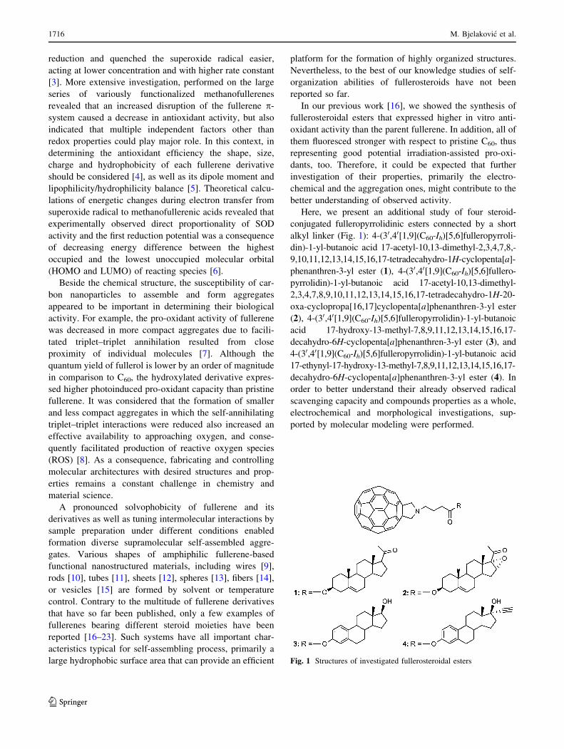
Here, we present an additional study of four steroid-
conjugated fulleropyrrolidinic esters connected by a short
alkyl linker (Fig. 1): 4-(30,40[1,9](C60-Ih)[5,6]fulleropyrroli-
din)-1-yl-butanoic acid 17-acetyl-10,13-dimethyl-2,3,4,7,8,-
9,10,11,12,13,14,15,16,17-tetradecahydro-1H-cyclopenta[a]-
phenanthren-3-yl ester (1), 4-(30,40[1,9](C60-Ih)[5,6]fullero-
pyrrolidin)-1-yl-butanoic acid 17-acetyl-10,13-dimethyl-
2,3,4,7,8,9,10,11,12,13,14,15,16,17-tetradecahydro-1H-20-
oxa-cyclopropa[16,17]cyclopenta[a]phenanthren-3-yl ester
(2), 4-(30,40[1,9](C60-Ih)[5,6]fulleropyrrolidin)-1-yl-butanoic
acid 17-hydroxy-13-methyl-7,8,9,11,12,13,14,15,16,17-
decahydro-6H-cyclopenta[a]phenanthren-3-yl ester (3), and
4-(30,40[1,9](C60-Ih)[5,6]fulleropyrrolidin)-1-yl-butanoic acid
17-ethynyl-17-hydroxy-13-methyl-7,8,9,11,12,13,14,15,16,17-
decahydro-6H-cyclopenta[a]phenanthren-3-yl ester (4). In
order to better understand their already observed radical
scavenging capacity and compounds properties as a whole,
electrochemical and morphological investigations, sup-
ported by molecular modeling were performed.
Fig. 1 Structures of investigated fullerosteroidal esters
1716 M. Bjelakovic et al.
123

Results and discussion
Electrochemistry
The electrochemical properties of fullerosteroidal esters 1–
4 were investigated by cyclic voltammetry (CV) in solution
using N,N-dimethylformamide (DMF) and dichlorometh-
ane (DCM) as the solvents. Selected CV curves of studied
compounds recorded in DMF/tetrabutylammonium per-
chlorate (TBAP) at 298 K and the scan rate of 0.3 V/s are
given in Fig. 2, while representative voltammograms of
compound 1 in different solvents at 298 K and the scan rate
of 0.1 V/s are presented in Fig. 3. Observed half-wave
reduction potentials of compounds 1–4, and corresponding
LUMO energy levels calculated from the CV data as well
as those obtained by molecular modeling are collected in
Table 1.
The studied compounds have quite similar molecular
structures, so their electronic properties, in particular
frontier orbital energy levels should also be very similar.
As expected, all four compounds gave almost identical CV
curves (Fig. 2) typical for fulleropyrrolidines. A notable
influence of the solvent on the first and the second half-
wave potentials was observed (Table 1; Fig. 3). In the
cathodic scan in a DCM solution, three regularly separated
reversible one-electron reductions were observed in the
accessible potential window, all attributable to the fuller-
opyrrolidinic subunit. Corresponding half-wave potentials
(E1/2) were located at ca. -1.1, -1.5, and -2.0 V (first to
third), respectively. The CV curves relative to a DMF
solution of fullerosteroids 1–4 display a series of four well
defined, successive reversible one-electron reductions of
fulleropyrrolidine moiety, located at ca. -1.0, -1.4, -2.0,
and -2.6 V (E1/2 values, first to fourth), respectively. All
half-wave potentials in both solvents are ca. 200 mV
negatively shifted with respect to pristine C60 due to par-
tially lost delocalization of p-electronic system over carbon
sphere upon [3 ? 2]-cycloaddition (Table 1). The first two
reductions of all four compounds proceeded easier in DMF
and corresponding half-wave potentials were ca. 100 mV
positively shifted with respect to DCM. The third reduction
potentials remained independent on the solvent used.
The reduction potentials of fullerenes strongly depend
on the solvent, mainly on its electron-accepting and
donating capacity [24, 25] described with Dimroth–Reic-
hardt parameter (ETN) and Gutmann donor number (DN),
respectively. The corresponding parameters of used sol-
vents are [24] ETN(30): 0.309 (DCM); 0.404 (DMF) and
DN: 0.0 (DCM); 26.6 (DMF). As can be seen both values
are higher for DMF indicating that facilitated reduction is a
consequence of the stabilization of generated fulleroster-
oidal anions by the solvent electron-accepting ability, as
well as the compounds reduced aggregation by the solvent
electron-donating capacity.
The effect of the scan rate was investigated by per-
forming CV experiments at 0.1, 0.3, 0.5, and 1 V/s in DMF
and 0.05, 0.1, 0.2, and 0.5 V/s in DCM. The values of the
half-wave potentials were found to be independent on the
scan rate, as was expected for a reversible system. In
addition, all anodic as well as cathodic peak currents in
both solvents varied linearly with the square root of the
scan rate (v1/2), indicating diffusion-controlled reductions.
Also, the products of all four reductions are stable on the
cyclic voltammetric time scale since neither additional
peaks during five consecutive scans, nor any change in the
NMR spectra of quantitatively recovered compounds was
not detected.
The CV measurements allow the possibility to deter-
mine the diffusion coefficient (D) of the examined
compound, and further its hydrodynamic radius. In such a
way, the potential supramolecular interactions, primarily
self-aggregation under applied conditions, can be studied
Fig. 2 Cyclic voltammograms of 1-mM solution of 1–4 in DMF
containing 0.1-M TBAP at 298 K and the scan rate of 0.3 V/s
Fig. 3 Cyclic voltammograms of 1-mM solution of 1 in DMF (solid)
and DCM (dashed) containing 0.1-M TBAP at 298 K and the scan
rate of 0.1 V/s
Studies of antioxidant fullerosteroids 1717
123
more in detail. Translation mobility of the molecules in
continuous incompressible fluids, determined by D,
depends on the medium viscosity, solvent–particle inter-
actions, as well as of the size and the shape of a molecule.
For spherical particles it can be expressed by Stokes–Ein-
stein relation:
D ¼ kBT=6grs ð1Þ
where kB represents Boltzmann constant, T temperature, gthe dynamic viscosity of the solvent (0.410 and
0.802 mPa s for DCM and DMF, respectively [24]), and
rs Stokes (hydrodynamic) radius. The diffusion coefficients
of fullerosteroids 1–4 were calculated from the slope of the
plot of the cathodic peak current vs. v1/2 using Randles–
Sevcik equation:
ip ¼ D1=2 2:69� 105� �
n3=2Ach i
v1=2 ð2Þ
where ip represents peak current, D diffusion coefficient,
n the number of electrons transferred in the half reaction
for the redox couple, A the electrode area, c concentration,
and v scan rate. Including obtained values into Stokes–
Einstein equation (1) afforded radii of solvated charged
particles, given in Table 2. Under applied conditions in
DMF fullerosteroidal anions of all tested compounds were
detected as particles of ca. 3 nm in diameter, indicating the
presence of individual molecules solvated by monomo-
lecular layer of the solvent. On the other hand, in DCM
which has no electron-donating capacity, compound’s
aggregation was not suppressed by the solvent and signif-
icantly larger particles were observed. Detected radii were
four- to fivefolds bigger than calculated radius of individ-
ual compound increased by monomolecular solvent layer.
The estrone-based fullerosteroids 1 and 2 formed particles
of ca. 8 nm in diameter, while from pregnenolone-derived
compounds 3 and 4 even larger aggregates of 10 nm in
diameter were built.
The CV is often used to calculate the energy levels of
frontier orbitals although obtained data are only fairly
comparable since the redox potentials are measured in
solution, while orbital energies are scaled in vacuum. In
this work, the LUMO energies have been additionally
determined by theoretical calculations in vacuum, and
afforded results were used only for relative comparison of
compounds. Following the IUPAC procedure [26], the
electrode potentials in non-aqueous solvents were mea-
sured against the potential of Fc/Fc? redox couple. In order
to estimate the LUMO energy level of fullerosteroids 1–4,
the value of internal standard was evaluated against refer-
ence electrode and was found to be 0.53 V and 0.32 V vs.
Ag/Ag? in DMF and DCM, respectively. Using the liter-
ature data of 0.29 V for the conversion of potentials
measured vs. Ag/Ag? electrode to SCE [27] and 0.24 V for
the potential of SCE vs. normal hydrogen electrode (NHE),
experimentally observed reduction half-potentials of Fc/
Fc? were recalculated to 1.06 and 0.85 V vs. NHE in DMF
and DCM, respectively. There are several values that cor-
relate the potential of NHE reference electrode to Fermi
energy scale (the vacuum level), varying from -4.4 to
-4.85 eV [28]. Setting the NHE vs. the vacuum level to
Table 1 Half-wave reduction potentials and the LUMO energy levels of fullerosteroids 1–4 in DMF/DCM
Compound E1/2/V LUMO/eV
I II III IV CVa MMb
1 -0.98/-1.12 -1.40/-1.52 -2.05/-2.06 -2.55/– -4.38/-4.73 -3.82
2 -0.96/-1.14 -1.40/-1.53 -2.05/-2.06 -2.58/– -4.36/-4.77 -3.90
3 -0.96/-1.14 -1.40/-1.53 -2.05/-2.05 -2.59/– -4.36/-4.77 -3.65
4 -0.96/-1.11 -1.40/-1.49 -2.05/-2.01 -2.58/– -4.36/-4.72 -3.65
C60c -0.77/-0.95 -1.25/-1.39 -1.84/-1.84 -2.38/– -4.17/-4.56 -3.49
Half-wave reduction potentials vs. Fc/Fc? (0.53/0.32 V vs. Ag/Ag? in DMF/DCM)a Calculated from the CV datab Obtained by theoretical calculationsc Half-wave reduction potentials were taken from Ref. [24] and recalculated according to the Fc/Fc? value
Table 2 Diffusion coefficients D, hydrodynamic radii rs of C60 and
fullerosteroids 1–4 in DMF/DCM containing 0.1-M TBAP and their
MM-calculated radii rMM
Compound D 9 10-10/m2/s rs/nm rMM/nm
1 1.78/1.02 1.53/5.20 0.83
2 1.68/1.00 1.62/5.32 0.83
3 1.97/1.35 1.38/3.94 0.81
4 1.87/1.32 1.45/4.02 0.82
C60 –/4.4 [24] –/1.11 0.68
Solvent radius/nm, calculated from the molecular weight and density
of the solvent: 0.31 (DMF); 0.29 (DCM)
1718 M. Bjelakovic et al.
123
-4.46 eV [29], the LUMO level in the corresponding
solvent was calculated from the formula:
ELUMO ¼ �4:46 þ E1=2Fc=Fcþvs: NHE þ EredI1=2
� �
where EredI1=2 represents the half-wave potential of the first
reduction step.
Obtained LUMO energies of all four studied fulleros-
teroids were similar within the same solvent and appeared
in a narrow range of -(4.36–4.38) eV in DMF and
-(4.72–4.77) eV in DCM. Also their LUMO levels were
ca. 200 meV more negative than the LUMO level of C60.
On the other hand, a significant variation in the LUMO
energy levels of individual compounds of ca. 400 meV was
observed in different solvents (twice higher than the dif-
ference caused by the p-conjugation disruption) with
stronger stabilization by the solvent with higher electron-
donating as well as electron-accepting capacity, i.e., DMF.
For example, the LUMO energy level of compound 2 was
located at -4.36 eV in DMF, whereas in DCM the value of
-4.77 eV was achieved. Obtained results clearly showed
the importance of the solvent selection in potential elec-
trochemical modifications of fullerene and its derivatives,
as well as the influence of an environment on redox
processes.
Electrochemical properties vs. antioxidant activity
The literature data indicate that positive shifting of the first
reduction potential of the fullerene derivatives induces the
increase of their antioxidant activity [3]. Based on that,
lower activity of fullerosteroids with respect to parent
fullerene could be expected since their first reduction
potentials were ca. 200 mV negatively shifted (Table 1).
However, our previous results [16] showed that fulleros-
teroids 1–4 expressed two- to threefold stronger radical
quenching than C60. In addition, besides the redox prop-
erties, a mutual influence of the charge, shape, size and
hydrophobicity on the antioxidant activity should be taken
into consideration as well [4]. Furthermore, such activity
was found to be related to the dipole moment too [5]. In
order to determine parameters other than redox properties
and their possible influence on the radical quenching
capacity of fullerosteroids, all compounds were subjected
to MM calculations.
Molecular modeling
In order to examine the electronic properties of four
investigated fullerosteroidal esters more in detail and to
understand better their antioxidant activity, the molecular
geometries and electron density distribution were simu-
lated by conformational search and single-point DFT
B3LYP quantum mechanical calculations, respectively.
The most stable conformations of compounds 1–4, distri-
bution of their frontier orbitals, and corresponding
electrostatic potential maps were depicted in Figs. 4, 5, and
6, respectively, while the values of the selected descriptors
and properties are given in Table 3.
The conformation with the steroidal subunit bended
towards fullerene sphere was found to be the most stable
for all compounds (Fig. 4), indicating the importance of
hydrophobic interactions in the polar environment. The
center-to-center distances between steroidal and fullerene
moieties were found to be 0.74–0.95 nm, where the mid-
point of the steroidal C(7)–C(8) bond (the B/C rings
conjunction) was chosen as the steroidal centre. Observed
distances in the aromatic steroidal fulleroesters 3 and 4
were slightly shorter than in pregnenolone-derived conju-
gates 1 and 2 suggesting an existence of the p–p stacking
interactions (Table 3). The distance of 0.36 nm between
the steroidal aromatic A-ring and the nearest fullerene ring
confirmed the presence of these interactions. Looking into
the spatial distribution of the frontier orbitals shown in
Fig. 5, it can be seen that in all cases the electron-accepting
region, responsible for free radical quenching activity and
determined by the presence of LUMOs, was localized on
the fullerene core.
The possibility of eventual electrostatic interactions
within the molecule as well as with solution/other solutes
was investigated by calculation of their molecular elec-
trostatic potentials map. The covalent modification of
fullerene sphere induced the change in the electrostatic
potential distribution (Fig. 6; Table 3). In general, small
charge separation was observed in all compounds. The
negative potential, presented by red color, was mainly
associated with the oxygen atoms belonging to carbonyl,
oxo, epoxy, and hydroxyl functions, while positive regions
marked by blue color were located at the pyrrolidine ring
and acidic hydrogen belonging to hydroxyl groups. The
main part of the steroidal subunit as well as the whole
carbon sphere remained with no charge separation (green),
therefore the favorable interactions with non-polar envi-
ronment such as the organic solvents and/or lipid
membrane could be expected. Also, total solvent accessible
(SA) volume and the corresponding surface area (SASA)
increased by the fullerene functionalization (Table 3,
entries 3 and 4). The participation of the hydrophobic
component (FOSA) was significantly increased (ca. 50 %
of the SASA) while the contribution of the hydrophilic
components (FISA) was also observed, but in lower degree
(less than 10 % of the SASA). Although fullerene-steroid
conjugation produced relatively weak charge separation
dipolar structures with dipole moment of ca. 6–8 D were
formed, and in all four investigated compounds notably
enlarged polarizability was observed (by 60–80 % with
Studies of antioxidant fullerosteroids 1719
123

respect to C60). In addition, the covalent modifications of
the fullerene sphere changed the ability of hydrogen bond
(HB) formatting. The fullero-pregnenolone derivatives 1
and 2 showed moderate HB-accepting capacity, while their
HB-donating ability remained unchanged in comparison
to C60. On the other hand, estradiol-derived fullero
compounds 3 and 4 could participate in HB formatting as
acceptors, but also as weak donors (Table 3, entries 10 and
11). Introduction of the steroidal subunit led to consider-
ably increased lipophilicity of carbon clusters augmenting
the log P values by 2–3 units (Table 3 entry 12), once more
indicating facilitated interactions with non-polar media.
Fig. 4 The most stable
conformations of fullerosteroids
1–4
Fig. 5 Molecular orbital spatial
orientation for the LUMO
energy levels of compounds 1–4
1720 M. Bjelakovic et al.
123

Fig. 6 Molecular electrostatic
potential maps for the most
stable conformations of
compounds 1–4 (conformations
determined by conformational
search, electrostatic potential is
depicted by mapping it on
electron density)
Table 3 Selected calculated properties and descriptors of C60 and fullerosteroids 1–4
Entry Property/descriptor C60 1 2 3 4
1 ED50/lMa 64 28 28 20 27
2 Center-to-center distance/nm – 0.87 0.95 0.74 0.74
3 SA volume/(A3)b 1,298 2,372 2,423 2,228 2,344
4 SASA/(A2)c 589 981 1,019 914 963
5 FOSA/(A2)d (% of SASA) 0 (0) 478 (49) 476 (47) 402 (44) 424 (44)
6 FISA/(A2)e (% of SASA) 0 (0) 83 (8) 81 (8) 60 (6) 55 (6)
7 PISA/(A2)f (% of SASA) 589 (100) 420 (43) 462 (45) 452 (50) 484 (50)
8 Dipole/Dg 0 7.22 6.06 6.96 7.60
9 QP polrz/(A3)h 51.43 88.94 91.37 83.50 87.80
10 acceptHBi 0 6 8 6.2 5.2
11 donorHBj 0 0 0 1 1.5
12 QP log P o/wk 7.79 11.24 10.66 10.40 11.54
a Experimentally obtained results of the antioxidant activity [16] are given for comparisonb Total solvent accessible volume using a probe with a 1.4 A radiusc Total solvent accessible surface area using a probe with a 1.4 A radiusd Hydrophobic component of the SASAe Hydrophilic component of the SASAf p-Component of the SASAg Computed dipole momenth Predicted polarizabilityi Estimated number of HB that would be accepted by the solute from H2O in an aq. solutionj Estimated number of HB that would be donated by the solute to H2O in an aq. solutionk Predicted octanol/water partition coefficient (lipophilicity)
Studies of antioxidant fullerosteroids 1721
123

Alterations of the solvent–solute interactions together
with dipole formation, increased affinity to HB formation
and enhanced lipophilicity could explain experimentally
observed improved solubility of fullerosteroids with
respect to pristine C60. Thus, all four investigated com-
pounds are easily soluble in chloroform (15 mg/cm3; C60
0.16 mg/cm3 [30]) and in solvent mixture CHCl3/CS2/
MeOH (1/1/1 volume ratios; 5 mg/cm3), while in more
polar CHCl3/MeOH (4/1 v/v) mixture the moderate solu-
bility of 2 mg/cm3 was reached.
Obtained results pointed out that some molecular
properties and descriptors could contribute to the previ-
ously observed in vitro free radical quenching activity [16].
Thus, calculation the Pearson correlation coefficient (PCC)
[31] showed strong positive association of the antioxidant
activity with polarizability, polarity, and the solvent
accessible surface area (Table 4). Good correlation with
lipophilicity was also observed, while very low linear
associations with the LUMO level, as well as the half-wave
reduction potentials were found.
Usually, the radical quenching activity of fullerene
derivatives decreases with lowering their first reduction
potential due to the disruption of the fullerene p-system
and consequent reduced affinity to accept an electron. On
the other hand, since an electron transfer could involve the
interaction of donating and accepting species, the energy
levels of their frontier orbitals should also be taken into
consideration. According to the equation reported by Sales
and Klopman the closer in energy the two orbitals are, the
more significant the energy gain will be achieved by their
interactions. Therefore, the comparison of the frontier
orbital energies of donating and accepting compounds
seemed reasonable.
The single occupied molecular orbital (SOMO) energy
level of peroxylinoleic radical (LOO•) used in our previous
in vitro experiments was found to be -6.22 eV [32].
Comparing the SOMO–LUMO differences, where the
LUMO values were taken from CV-derived calculations,
showed that in both investigated solvents frontier orbitals
of fullero derivatives were for ca. 200 meV (*19 kJ/mol)
closer to donating radical species with respect to non-
functionalized C60. Using the data of the LUMO energies
obtained by MM calculations afforded the SOMO–LUMO
gap of 2.73 eV for C60 and 160–410 meV (*15–40 kJ/
mol) smaller difference for fullerosteroids 1–4 (Table 5).
Consequently, it could be supposed that the energy gain
obtained by an intermediate formation played more
important role in the expression of radical quenching
activity in comparison to compound’s reducibility.
Morphology
The supramolecular self-assembly of fullerosteroidal con-
jugates 1–4, prepared under different conditions, was
studied by scanning electron microscopy (SEM). Repre-
sentative images of samples obtained by slow evaporation
of a dilute solution on silicon and brass wafers at room
temperature and solid compounds on brass substrate are
given in Fig. 7. An influence of the medium polarity on the
assembly process in solution was examined using PhMe,
CHCl3, and MeOH as well as their different combinations.
In all individual solvents low level of self-organization was
observed, while the well-organized structures were
obtained in the solvent mixtures. Therefore, the size and
morphology of the self-organized fullerosteroidal conju-
gates 1–4 formed in the PhMe/MeOH (5/1, v/v) and more
polar CHCl3/PhMe/MeOH (10/5/1, v/v) mixture were
investigated. It was also identified that the substrate exerted
no appreciable effect on the samples’ morphology although
the quality of commonly used brass and silicon pads was
quite different.
The SEM images presented in Fig. 7 revealed that all
studied compounds self-assembled into similar spherical or
circular entities with nano- and microscopic dimensions
and significant morphological uniformity in both solvent
mixtures used. The morphology of the assemblies showed
to be non-dependent on the sample concentration
(Fig. 73b, 4b). In a less-polar medium on the silicon sur-
face, all compounds formed ordered highly uniform self-
Table 4 The correlation of the antioxidant activity with molecular
descriptors/properties expressed by the PCC
Descriptor/property PCC
Polarizability 0.93
Polarity 0.90
SASA 0.89
Log P o/w 0.63
LUMO (MM) -0.24
E1/2 (I) -0.39
Table 5 SOMOLCOO•-LUMOfull energy distances and differences Dwith respect to C60
Compound SOMO–LUMO gap/eVa Db/eV
CVDMF CVDCM MM CVDMF CVDCM MM
C60 2.05 1.66 2.73 – – –
1 1.84 1.49 2.40 0.21 0.17 0.33
2 1.86 1.45 2.32 0.19 0.21 0.41
3 1.86 1.45 2.57 0.19 0.21 0.16
4 1.86 1.50 2.57 0.19 0.16 0.16
a SOMOLCOO• = -6.22 eV [32]; LUMO values taken from CV
experiments in corresponding solvent and from MM calculationsb D = (SOMO–LUMOC60) - (SOMO–LUMOFullerosteroid)
1722 M. Bjelakovic et al.
123
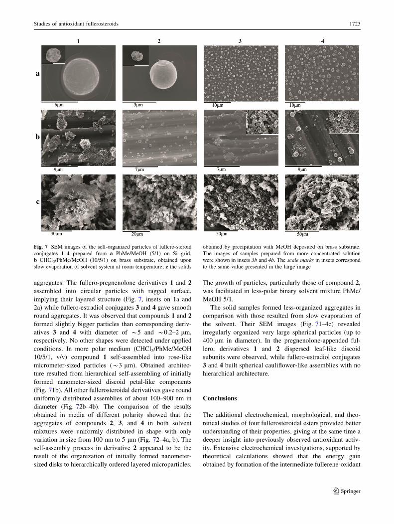
aggregates. The fullero-pregnenolone derivatives 1 and 2
assembled into circular particles with ragged surface,
implying their layered structure (Fig. 7, insets on 1a and
2a) while fullero-estradiol conjugates 3 and 4 gave smooth
round aggregates. It was observed that compounds 1 and 2
formed slightly bigger particles than corresponding deriv-
atives 3 and 4 with diameter of *5 and *0.2–2 lm,
respectively. No other shapes were detected under applied
conditions. In more polar medium (CHCl3/PhMe/MeOH
10/5/1, v/v) compound 1 self-assembled into rose-like
micrometer-sized particles (*3 lm). Obtained architec-
ture resulted from hierarchical self-assembling of initially
formed nanometer-sized discoid petal-like components
(Fig. 71b). All other fullerosteroidal derivatives gave round
uniformly distributed assemblies of about 100–900 nm in
diameter (Fig. 72b–4b). The comparison of the results
obtained in media of different polarity showed that the
aggregates of compounds 2, 3, and 4 in both solvent
mixtures were uniformly distributed in shape with only
variation in size from 100 nm to 5 lm (Fig. 72–4a, b). The
self-assembly process in derivative 2 appeared to be the
result of the organization of initially formed nanometer-
sized disks to hierarchically ordered layered microparticles.
The growth of particles, particularly those of compound 2,
was facilitated in less-polar binary solvent mixture PhMe/
MeOH 5/1.
The solid samples formed less-organized aggregates in
comparison with those resulted from slow evaporation of
the solvent. Their SEM images (Fig. 71–4c) revealed
irregularly organized very large spherical particles (up to
400 lm in diameter). In the pregnenolone-appended ful-
lero, derivatives 1 and 2 dispersed leaf-like discoid
subunits were observed, while fullero-estradiol conjugates
3 and 4 built spherical cauliflower-like assemblies with no
hierarchical architecture.
Conclusions
The additional electrochemical, morphological, and theo-
retical studies of four fullerosteroidal esters provided better
understanding of their properties, giving at the same time a
deeper insight into previously observed antioxidant activ-
ity. Extensive electrochemical investigations, supported by
theoretical calculations showed that the energy gain
obtained by formation of the intermediate fullerene-oxidant
Fig. 7 SEM images of the self-organized particles of fullero-steroid
conjugates 1–4 prepared from a PhMe/MeOH (5/1) on Si grid;
b CHCl3/PhMe/MeOH (10/5/1) on brass substrate, obtained upon
slow evaporation of solvent system at room temperature; c the solids
obtained by precipitation with MeOH deposited on brass substrate.
The images of samples prepared from more concentrated solution
were shown in insets 3b and 4b. The scale marks in insets correspond
to the same value presented in the large image
Studies of antioxidant fullerosteroids 1723
123
species affected the electron-quenching capacity in a
greater extent than the reducibility of fullerene subunit.
Also, increased polarizability, polarity, lipophilicity, and
solvent–solute interactions, achieved by fullerene func-
tionalization, could contribute to the augmentation of the
expressed activity. In addition, the tuning of solvent sup-
ported self-aggregation was attained by the medium
electron-accepting and -donating capacity. In conclusion,
presented work could open a possibility to design a broad
spectrum of targeted fullerene-based antioxidants.
Experimental
Compounds 1–4 were prepared according to the literature
procedure [16]. Their IR, UV, 1H and 13C NMR spectra
were identical to the literature data [16]. The solvents used
for the cyclic voltammetry and scanning electron micros-
copy experiments (HPLC grade) were stored over 3A
molecular sieves and degassed under vacuum prior to the
use. Ferrocene and TBAP were purchased from Sigma-
Aldrich and used as received. For the PCC calculations, the
pairwise method was employed for missing values, confi-
dence level 99 %, p \ 0.01.
Cyclic voltammetry
The electrochemical behavior of fullerosteroids was
investigated using 1-mM solution of compounds 1–4 in dry
dichloromethane or dimethylformamide containing 0.1-M
TBAP as supporting electrolyte. In order to remove oxygen
from the electrolyte, the system was bubbled with argon
prior to each experiment and the gas inlet left above the
liquid surface during the scans. The electrochemical mea-
surements were carried out on CHI760b Electrochemical
Workstation potentiostat (CH Instruments, Austin, TX,
USA) using a conventional three-electrode cell (1 cm3)
equipped with pre-polished platinum, a Ag/Ag? electrode
(a silver wire in contact with 0.01-M AgNO3 and 0.10-M
TBAP in acetonitrile) and the platinum wire as the work-
ing, reference and, auxiliary electrodes, respectively,
calibrated with ferrocene/ferrocenyl couple (Fc/Fc?) as an
internal standard. All experiments were performed at room
temperature in the potential range of -2.0 to 0.5 V vs.
saturated calomel electrode (SCE), at sweep rates between
0.01 and 1 V cm-1.
SEM
The solid samples were prepared by precipitation from a
CHCl3 solution with MeOH and subsequent drying under
vacuum. A small amount of each compound was sprinkled
on brass substrate. The samples designed for investigation
of the size and morphology of self-organized structures
formed in solution were prepared at room temperature by
‘‘drop drying’’ method [33]. Briefly, 10 or 50 mm3 of 0.1-
mM solution of fullerosteroids 1–4 in PhMe/MeOH (5/1,
v/v) or CHCl3/PhMe/MeOH (10/5/1) was deposited on the
substrate surface (10 9 10 mm Si or brass wafer) and left
overnight to slowly evaporate in a glass Petri dish under
2 cm3 of PhMe at room temperature. All samples were
gold sputtered in a JFC 1100 ion sputterer and then sub-
jected to SEM observations on a JEOL JSM-840A
microscope at an acceleration voltage of 30 kV.
Molecular modeling
All molecules were built in Maestro interface of Schro-
dinger Suite 2010 (Maestro, version 9.1, Schrodinger,
LLC, New York, NY, USA, 2010). In order to better
understand spatial properties and behavior of examined
molecules, we started Conformational Search from Mac-
roModel module in Schrodinger Suite 2010 (MacroModel,
version 9.8, Schrodinger, LLC, New York, NY, USA,
2010). OPLS_2005 force field with water as a solvent and
mixed MCMM/low-mode conformational search method
[34] were used for that purpose. Every conformation was
minimized using Polak–Ribiere conjugate gradient method
[35] with maximum of 500 iterations or 0.05 convergence
threshold. Duplicates were removed and all structures
within energy window of 21 kJ/mol were saved. The most
stable structures were used for further calculations. Cal-
culation of molecular descriptors was performed using
QikProp module from Schrodinger Suite 2010 (QikProp,
version 3.3, Schrodinger, LLC, New York, NY, USA,
2010). For calculation and visualization of electrostatic
potential, average ionization potential and HOMO/LUMO
orbitals we used single-point DFT B3LYP quantum
mechanical calculation with 6-31 ? G(d) basis set from
Jaguar (Jaguar, version 7.7, Schrodinger, LLC, New York,
NY, USA 2010).
Acknowledgments This research has been supported by Serbian
Ministry of Education, Science and Technological development,
Grant 172002 and NATO’s Public Diplomacy Division in the
framework of ‘‘Science for Peace’’ project SfP983638 (MM
calculations).
References
1. Ali SS, Hardt JI, Quick KL, Sook Kim-Han J, Erlanger BF,
Huang TT, Epstein CJ, Dugan LL (2004) Free Radical Biol Med
37:1191
2. Magoulas GE, Garnelis T, Athanassopoulos CM, Papaioannou D,
Mattheolabakis G, Avgoustakis K, Hadjipavlou-Litina D (2012)
Tetrahedron 68:7041
3. Liu GF, Filipovic M, Ivanovic-Burmazovic I, Beuerle F, Witte P,
Hirsch A (2008) Angew Chem Int Ed 47:3991
1724 M. Bjelakovic et al.
123

4. Witte P, Beuerle F, Hartnagel U, Lebovitz R, Savouchkina A,
Sali S, Guldi D, Chronakis N, Hirsch A (2007) Org Biomol Chem
5:3599
5. Ali SS, Hardt JI, Dugan LL (2008) Nanomed Nanotechnol 4:283
6. Osuna S, Swart M, Sola M (2010) Chem Eur J 16:3207
7. Hotze EM, Bottero J-Y, Wiesner MR (2010) Langmuir 26:11170
8. Hotze EM, Labille J, Alvarez P, Wiesner MR (2008) Environ Sci
Technol 42:4175
9. Geng J, Zhou W, Skelton P, Yue W, Kinloch IA, Windle AH,
Johnson BFG (2008) J Am Chem Soc 130:2527
10. Brough P, Bonifazi D, Prato M (2006) Tetrahedron 62:2110
11. Liu H, Li Y, Jiang L, Luo H, Xiao S, Fang H, Li H, Zhu D, Yu D,
Xu J, Xiang B (2002) J Am Chem Soc 124:13370
12. Sathish M, Miyazawa K (2007) J Am Chem Soc 129:13816
13. Prassides K, Keshavarz-K M, Beer E, Bellavia C, Gonzalez R,
Murata Y, Wudl F, Cheetham AK, Zhang JP (1996) Chem Mater
8:2405
14. Wang N, Li Y, He X, Gan H, Li Y, Huang C, Xu X, Xiao J, Wang
S, Liu H, Zhu D (2006) Tetrahedron 62:1216
15. Cassell AM, Asplund CL, Tour JM (1999) Angew Chem Int Ed
38:2403
16. Bjelakovic M, Godjevac D, Milic D (2007) Carbon 45:2260
17. Hummelen JC, Knight BW, LePeq F, Wudl F (1995) J Org Chem
60:532
18. Schuster DI (2000) Carbon 38:1607
19. MacMahon S, Fong R II, Baran PS, Safonov I, Wilson SR,
Schuster DI (2001) J Org Chem 66:5449
20. Li L-S, Hu Y-J, Wu Y, Wu Y-L, Yue J, Yang F (2001) J Chem
Soc Perkin Trans 1:617
21. Ishi-I T, Shinkai S (1999) Tetrahedron 55:12515
22. Chuard T, Deschenaux R (2002) J Mater Chem 12:1944
23. Maggini M, Guldi DM, Mondini S, Scorrano G, Paolucci F,
Ceroni P, Roffia S (1998) Chem Eur J 4:1992
24. Dubois D, Moninot G, Kutner W, Jones MT, Kadish KM (1992) J
Phys Chem 96:7137
25. Allemand PM, Koch A, Wudl F, Rubin Y, Diederich F, Alvarez
MM, Anz SJ, Whetten RL (1991) J Am Chem Soc 113:1050
26. Gritzner G, Kuta J (1984) Pure Appl Chem 56:461
27. Evans DH, Gilicinski AG (1992) J Phys Chem 92:2528
28. Cardona CM, Li W, Kaifer AE, Stockdale D, Bazan GC (2011)
Adv Mat 23:2367
29. Hansen WN, Hansen GJ (1987) Phys Rev A 36:1396
30. Hirsch A, Brettreich M (2005) Fullerenes: chemistry and reac-
tions. Wiley, Weinheim, p 35
31. NCSS 8 Software Package, free trial version. Available at www.
ncss.com
32. Matsubayashi K, Goto T, Togaya K, Kokubo K, Oshima T (2008)
Nanoscale Res Lett 3:237
33. Babu SS, Mohwald H, Nakanishi T (2010) Chem Soc Rev
39:4021
34. Kolossvary I, Guida WC (1999) J Comput Chem 20:1671
35. Polak E, Ribiere G (1969) Rev Fr Inform Rech O Serie Rouge
3:35
Studies of antioxidant fullerosteroids 1725
123