Embed Size (px)
Citation preview

Electron Accepting Units of the Diheme Cytochrome c TsdA, a Bifunctional Thiosulfate Dehydrogenase/Tetrathionate Reductase*
Julia M. Kurth‡1,2, José A. Brito§1,3, Jula Reuter‡4, Alexander Flegler‡, Tobias Koch‡, Thomas Franke‡, Eva-Maria Klein‡5, Sam F. RoweΣ, Julea N. ButtΣ, Kevin Denkmann‡6,
Inês A.C. Pereira§, Margarida Archer§7, Christiane Dahl‡8
From the ‡Institut für Mikrobiologie & Biotechnologie, Rheinische Friedrich-Wilhelms- Universität Bonn, 53115 Bonn, Germany, §Instituto de Tecnologia Química e Biológica António
Xavier, Universidade Nova de Lisboa (ITQB-UNL), Oeiras, Portugal, ΣCentre for Molecular and Structural Biochemistry, School of Chemistry, and School of Biological Sciences, University of
East Anglia, Norwich Research Park, Norwich NR4 7TJ, United Kingdom
Running title: Electron accepting units of TsdA
Keywords: Cytochrome c, enzyme kinetics, protein chemistry, respiratory chain, TsdA, electron acceptor, crystal structure, thiosulfate dehydrogenase
* This work was supported by the Deutsche Forschungsgemeinschaft Grant Da 351/7-2 and Fundação para a Ciência e Tecnologia through iNOVA4Health Research Unit (LISBOA-01-0145-FEDER-007344) cofunded by FCT/MCES and FEDER under the PT2020 Partnership Agreement, through R&D unit, UID/Multi/04551/2013 (GreenIT) and MostMicro unit financially supported by: Project LISBOA-01-0145-FEDER-007660 (Microbiologia Molecular, Estrutural e Celular) funded by FEDER funds through COMPETE2020 - Programa Operacional Competitividade e Internacionalização (POCI) and by national funds through FCT. SFR was supported by a studentship from the UK Biotechnology and Biological Sciences Research Council (BBSRC) and JNB by a Royal Society Leverhulme Trust Senior Research Fellowship.
The atomic coordinates and structure factors (entry 5LO9) have been deposited in the Protein Data Bank (http://www.pdb.org/)
1 Both authors contributed equally to this work 2 Recipient of scholarship 700051 funded by the Aventis Foundation and awarded by the Fonds der Chemischen
Industrie 3 Recipient of FCT Fellowship SFRH/BPD/79224/2011 4 Current affiliation: Rheinische Friedrich-Wilhelms-Universität Bonn, Institut für Pharmazeutische Mikrobiologie,
Meckenheimer Allee 168, 53115 Bonn, Germany 5 Current affiliation: Rheinische Friedrich-Wilhelms-Universität Bonn, Institut für Virologie, Siegmund-Freud-Str.
25, 53127 Bonn, Germany 6 Current affiliation: Dunn Labortechnik GmbH, Thelenberg 6, 53567 Asbach, Germany 7 Awarded FCT Investigator IF/00656/2014. To whom correspondence should be addressed: Tel. 351-214-469-
747; Fax 351-214-433-644; E-mail: [email protected]. 8 To whom correspondence should be addressed: Tel. 49-228-732119; Fax 49-228-737576; E-mail ChDahl@uni-
bonn.de.
2
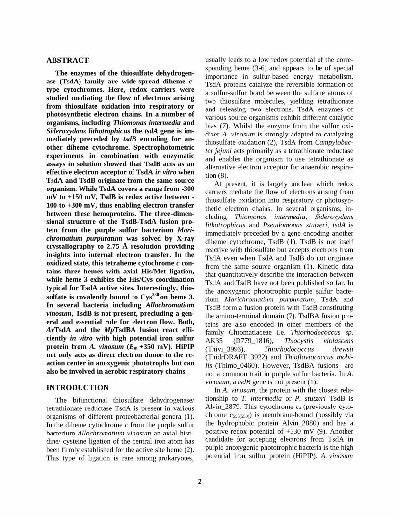
ABSTRACT
The enzymes of the thiosulfate dehydrogen- ase (TsdA) family are wide-spread diheme c- type cytochromes. Here, redox carriers were studied mediating the flow of electrons arising from thiosulfate oxidation into respiratory or photosynthetic electron chains. In a number of organisms, including Thiomonas intermedia and Sideroxydans lithotrophicus the tsdA gene is im- mediately preceded by tsdB encoding for an- other diheme cytochrome. Spectrophotometric experiments in combination with enzymatic assays in solution showed that TsdB acts as an effective electron acceptor of TsdA in vitro when TsdA and TsdB originate from the same source organism. While TsdA covers a range from -300 mV to +150 mV, TsdB is redox active between - 100 to +300 mV, thus enabling electron transfer between these hemoproteins. The three-dimen- sional structure of the TsdB-TsdA fusion pro- tein from the purple sulfur bacterium Mari- chromatium purpuratum was solved by X-ray crystallography to 2.75 Å resolution providing insights into internal electron transfer. In the oxidized state, this tetraheme cytochrome c con- tains three hemes with axial His/Met ligation, while heme 3 exhibits the His/Cys coordination typical for TsdA active sites. Interestingly, thio- sulfate is covalently bound to Cys330 on heme 3. In several bacteria including Allochromatium vinosum, TsdB is not present, precluding a gen- eral and essential role for electron flow. Both, AvTsdA and the MpTsdBA fusion react effi- ciently in vitro with high potential iron sulfur protein from A. vinosum (Em +350 mV). HiPIP not only acts as direct electron donor to the re- action center in anoxygenic phototrophs but can also be involved in aerobic respiratory chains.
INTRODUCTION
The bifunctional thiosulfate dehydrogenase/ tetrathionate reductase TsdA is present in various organisms of different proteobacterial genera (1). In the diheme cytochrome c from the purple sulfur bacterium Allochromatium vinosum an axial histi- dine/ cysteine ligation of the central iron atom has been firmly established for the active site heme (2). This type of ligation is rare among prokaryotes,
usually leads to a low redox potential of the corre- sponding heme (3-6) and appears to be of special importance in sulfur-based energy metabolism. TsdA proteins catalyze the reversible formation of a sulfur-sulfur bond between the sulfane atoms of two thiosulfate molecules, yielding tetrathionate and releasing two electrons. TsdA enzymes of various source organisms exhibit different catalytic bias (7). Whilst the enzyme from the sulfur oxi- dizer A. vinosum is strongly adapted to catalyzing thiosulfate oxidation (2), TsdA from Campylobac- ter jejuni acts primarily as a tetrathionate reductase and enables the organism to use tetrathionate as alternative electron acceptor for anaerobic respira- tion (8).
At present, it is largely unclear which redox carriers mediate the flow of electrons arising from thiosulfate oxidation into respiratory or photosyn- thetic electron chains. In several organisms, in- cluding Thiomonas intermedia, Sideroxydans lithotrophicus and Pseudomonas stutzeri, tsdA is immediately preceded by a gene encoding another diheme cytochrome, TsdB (1). TsdB is not itself reactive with thiosulfate but accepts electrons from TsdA even when TsdA and TsdB do not originate from the same source organism (1). Kinetic data that quantitatively describe the interaction between TsdA and TsdB have not been published so far. In the anoxygenic phototrophic purple sulfur bacte- rium Marichromatium purpuratum, TsdA and TsdB form a fusion protein with TsdB constituting the amino-terminal domain (7). TsdBA fusion pro- teins are also encoded in other members of the family Chromatiaceae i.e. Thiorhodococcus sp. AK35 (D779_1816), Thiocystis violascens (Thivi_3993), Thiorhodococcus drewsii (ThidrDRAFT_3922) and Thioflaviococcus mobi- lis (Thimo_0460). However, TsdBA fusions are not a common trait in purple sulfur bacteria. In A. vinosum, a tsdB gene is not present (1).
In A. vinosum, the protein with the closest rela- tionship to T. intermedia or P. stutzeri TsdB is Alvin_2879. This cytochrome c4 (previously cyto- chrome c553(550)) is membrane-bound (possibly via the hydrophobic protein Alvin_2880) and has a positive redox potential of +330 mV (9). Another candidate for accepting electrons from TsdA in purple anoxygenic phototrophic bacteria is the high potential iron sulfur protein (HiPIP). A. vinosum

3
and M. purpuratum produce HiPIP and as this protein has a quite positive reduction potential (+350 mV (10)) it would be well suited as an elec- tron acceptor for TsdA. This proposal is corrobo- rated by a previous report where a protein prepara- tion with thiosulfate dehydrogenase activity from A. vinosum reduced HiPIP in vitro (11).
Here, we study Tsd(B)A enzymes from dedi- cated sulfur oxidizers and characterize in detail the interaction of TsdA with TsdB. Additionally, we pose the question which proteins serve as immedi- ate electron acceptors for either TsdA alone or the TsdBA fusion protein (when present). It is fur- thermore intended to derive models for the electron flow involved.
RESULTS
Characterization of TsdA and TsdB–UV-vis electronic absorbance spectroscopy, X-ray diffrac- tion and activity assays have revealed a number of characteristic features of AvTsdA (1,2,12). How- ever, the electrochemical window in which the hemes are redox active remained unknown. To gain insight into this property we mapped out the redox activity of AvTsdA adsorbed as an electro- active film on optically transparent mesoporous nanocrystalline SnO2 electrodes. The spectrum of the enzyme-coated electrode equilibrated at +302 mV contained features typical of ferric c-type hemes superimposed on a small contribution from light-scattering by the electrode material (Fig. 1). The Soret maximum at 406 nm and broad lower intensity features in the αβ-region are typical of those displayed by solutions of oxidized AvTsdA (1). When the electrode potential was lowered to -648 mV in 50 mV steps with a spectrum rec- orded after a 60 s pause at each desired potential (Fig. 1), those features were replaced with peaks having maxima at 418, 523 and 553 nm, which are typical of dithionite reduced enzyme (1). Variation of the Soret intensity at 418 nm with electrode potential revealed that the hemes were reduced between approximately +150 and -350 mV (Fig. 1 inset, closed squares).
The response of reduced AvTsdA to stepwise increase of the electrode potential was assessed in a similar manner. The potential was raised in 50 mV steps and spectra recorded after a 60 second pause at each potential (Fig. 1 inset, open squares).
Between -648 and -98 mV the variation in Soret intensity with the applied potential was very simi- lar to that recorded on reduction. However, further increase of potential revealed significantly less oxidation than anticipated from the behavior seen on reduction of the enzyme. Importantly, spectra typical of the fully oxidized enzyme were meas- ured after the electrode was poised at +302 mV for approximately 30 min. It was concluded that the electrodic redox cycling of adsorbed AvTsdA was fully reversible but that full reduction occurred more quickly than complete re-oxidation. Further experiments confirmed that this behavior persisted over multiple rounds of reduction and reoxidation. The spectral changes induced by variation of po- tential between -648 and -98 mV were rapidly reversed and accounted for approximately 35% of the change in absorbance at 418 nm when spectra of the fully oxidized and fully reduced forms of the enzyme were compared. By contrast, variations of electrode potential between -98 and +302 mV showed rapid reduction and much slower reoxida- tion, and the associated changes in absorbance accounted for approximately 65% of the total seen on full redox cycling of the enzyme. It was con- cluded that the slow reoxidation associated with higher potential redox event(s) was not a conse- quence of reversible redox events that occurred at lower potentials. Detailed inspection of the spectra provided no indication for the presence of high- spin ferric- or ferrous-heme.
The hysteretic nature of the plot of absorbance versus potential prevented Nernstian analysis to define the heme reduction potentials. Nevertheless, some further conclusions regarding the redox ac- tivity of AvTsdA can be proposed in light of the crystal structures reported (2,12). The fully oxi- dized diheme cytochrome AvTsdA contains His/Cys coordination in heme 1 and His/Lys in heme 2. His/Cys ligated hemes are typically distin- guished from other low-spin c-type hemes by having much lower reduction potentials and smaller changes in extinction coefficient associated with the Fe(III)/(II) couple (4,13). As a conse- quence, we propose that reduction of His/Cys li- gated heme 1 occurs reversibly between ca. -100 and -350 mV. Reduction of His/Lys ligated heme 2 is proposed to occur between ca. +150 and -100 mV. AvTsdA X-ray structures reveal that this re-

4
duction is accompanied by a switch of Lys by Met as axial distal ligand to ferrous-heme 2 (2). Such a change of ligation would be expected to raise the reduction potential of heme 2. If the Met ligand is replaced slowly by Lys upon enzyme oxidation this would account for the hysteretic nature of the redox behavior displayed by AvTsdA.
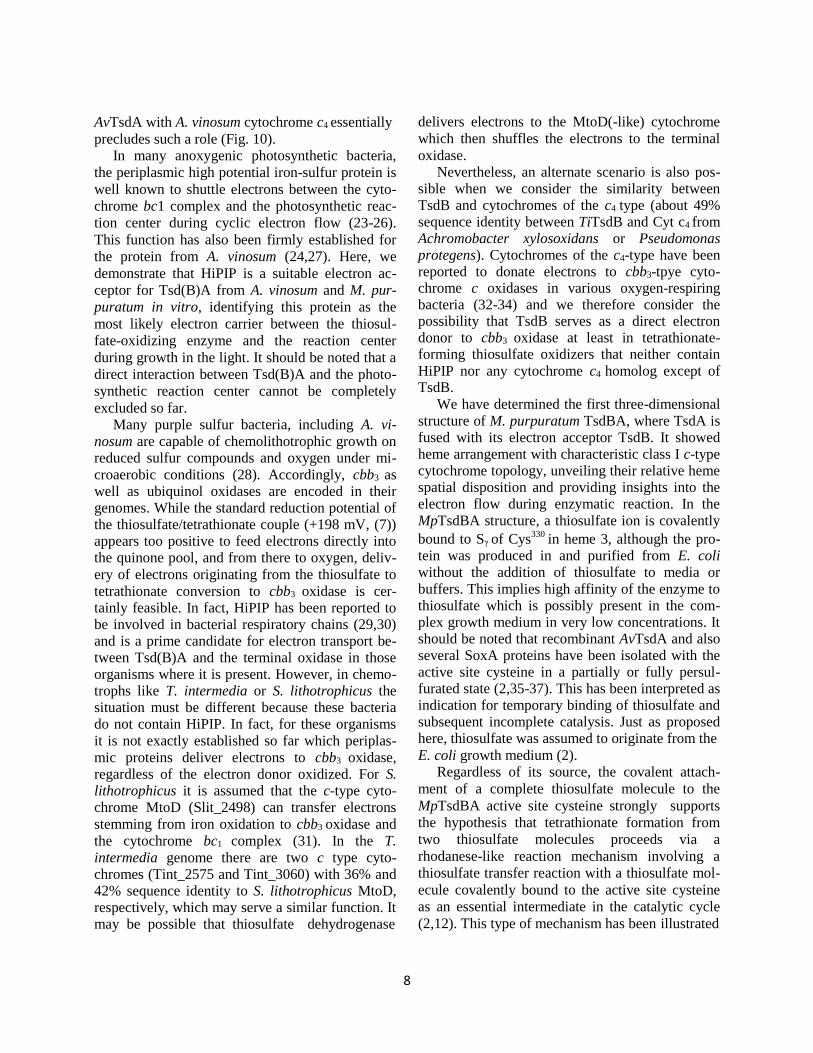
In contrast to TsdAs, TsdB proteins are not well characterized. The most closely related character- ized cytochromes on a sequence level belong to the diheme cytochromes of the c4 family (1). The re- combinant TsdB protein from T. intermedia indeed binds two heme groups of 616.5 Da as the mass of 21792.5 Da determined by MALDI-TOF mass spectrometry almost exactly matched the mass of 21783.3 Da predicted for the mature recombinant protein including Strep tag and two hemes. The same held true for recombinant S. lithotrophicus TsdB (measured mass: 22864 Da, predicted mass: 22835 Da). UV-Vis spectroscopy of pure recombi- nant TsdB from S. lithotrophicus is shown in Fig. 2. The spectrum conforms to that of TiTsdB (1).
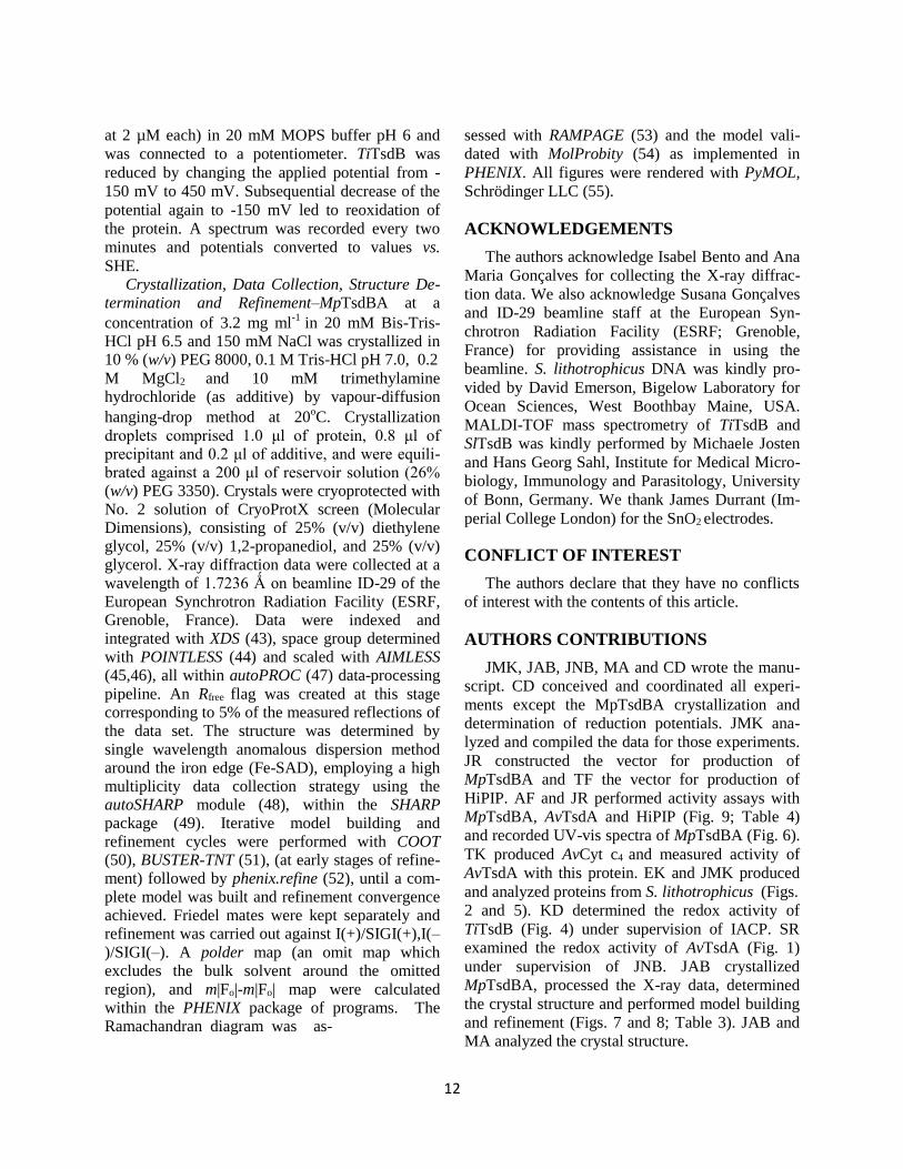
A sequence alignment of various TsdB proteins (Fig. 3) reveals two conserved methionine residues but no conserved histidines or cysteines indicating that both hemes of TsdB have axial coordination by His/Met . This is underpinned by the 700 nm peak in the spectrum, which is characteristic for methionine as the sixth axial heme iron ligand (14) and corroborated by the MpTsdBA crystal struc- ture determined herein (see below). A shift of the Soret band from 411 to 417 nm upon reduction was observed. Moreover, there is a distinct δ band
at 359 nm in the oxidized protein spectrum. In the reduced state, the band was detected at 552 nm and the β band at 523 nm. A split band charac- teristic for a number of c4-type cytochromes, in- cluding those from purple sulfur bacteria, was not observed (9,15). The UV-vis spectrum for oxidized TsdB exhibited a low intensity high spin feature at 620 nm similar to that noted for cytochrome c4
from P. stutzeri (16). Obviously, the ferric form of TsdB holds a small fraction of high-spin heme probably caused by weakening of the Fe-S bond at one of the two hemes with concomitant partial dissociation of the methionine and formation of an Fe-aquo bond just as outlined for the P. stutzeri cytochrome (16).
The reduction potential of TiTsdB was deter- mined by potentiometry with a gold electrode (Fig. 4). The potential changes determined upon reduc- tion and reoxidation of TsdB match well. The two hemes are redox active between -100 and +300 mV. Hysteresis was not observed, consistent with both hemes being His/Met ligated. Ligand changes are not apparent.
TsdB is an Electron-Transferring Unit Tightly Interacting with TsdA-The interaction of TsdA and TsdB proteins was analyzed for the recombinant proteins from S. lithotrophicus. Upon analytical gel permeation chromatography SlTsdA alone eluted at a volume corresponding to a molecular mass of 65 kDa, indicating dimerization of the protein un- der the conditions applied (monomer: 33042 Da). In contrast, SlTsdB behaved as a 22 kDa monomer (predicted molecular mass: 22835 Da). When both proteins were produced simultaneously in E. coli and purified employing the Strep tag attached to TsdA, a preparation was obtained that exhibited two heme stainable polypeptides. In size, these exactly matched SlTsdB and SlTsdA (Fig. 5). The co-purification of the two proteins is evidence for significant interaction between them. Upon size exclusion chromatography, SlTsdA and SlTsdB co- eluted again in fractions corresponding to a mass of 108 kDa, indicating formation of an α2β2 hetero- dimer.
Characterization of the TsdBA Fusion Protein from Marichromatium purpuratum–The protein encoded by tsdB-tsdA gene fusion in Marichroma- tium purpuratum provides an exceptional possibil- ity to study the complete tetraheme cytochrome for catalytic properties and internal electron transfer. A sequence alignment of MpTsdBA with com- bined TsdB and TsdA sequences from S. lithotrophicus and T. intermedia shows signficant similarity between the N-terminal region of MpTsdBA and TsdB (aa 1-199 of MpTsdBA and TiTsdB share 45% sequence identity) and between the C-terminal region of MpTsdBA and TsdA (aa 224-518 of MpTsdBA compared to AvTsdA or TiTsdA: 39% and 50 % sequence identity, respec- tively). The heme distal ligands cysteine and me- thionine of TsdA as well as the two putative heme ligating methionines are strictly conserved (Fig. 3). Therefore, we predicted MpTsdBA to contain three His/Met ligated and one His/Cys ligated heme.

5
UV-Vis spectroscopy of MpTsdBA protein is shown in Fig. 6. The presence of His/Met ligated hemes in MpTsdBA is substantiated by the 700 nm peak in the spectrum of the oxidized protein. This absorption band is characteristic for methionine as heme iron ligand (14). A shift of the Soret band from 413 to 420 nm was observed upon reduction. Moreover, there is a distinct δ band at 363 nm in
the oxidized protein spectrum. The α band is lo- cated at 553 nm, whilst the β band resides at 524
nm. The partly reduced spectrum exhibits a low intensity high spin feature at 620 nm similar to AvTsdA (2). The A413nm/A280nm for pure oxidized MpTsdBA is 3.4.
Crystal Structure Determination and Model Quality of MpTsdBA–To compare structural fea- tures of TsdA and TsdBA, to get a closer look into ligation of the four heme groups and to see how TsdA and TsdB domains are linked to each other, we have determined the X-ray structure of MpTsdBA by Fe-SAD. Crystals belong to the hex- agonal space group H32 with cell dimensions a=b=159.75 Ǻ and c= 393.09 Ǻ. There are two molecules in the asymmetric unit corresponding to a Matthews coefficient (17) of 2.8 Ǻ
3 Da-1 and a solvent content around 56%. The “as isolated” structure was refined to 2.75 Ǻ resolution with
Rcryst of 15.7% and Rfree of 19.8% using a 141o- sweep of data with overall better statistics. Data collection and refinement statistics are depicted in Table 3. The high Rmeas values observed for both sweeps are due, firstly, to the fine binning that autoPROC implements during data reduction, and secondly, to the fact that the crystal has some regions in the rotational space that are of bad quality. This is perfectly clear in the plots Rmeas vs. image number output by autoPROC (data not shown), highlighting that the high Rmeas values are due to the crystal quality and not radiation damage. No evidence for radiation damage is also observed in a m|Fo|-m|Fo| map calculated using both data sweeps (data not shown). MpTsdBA is numbered without taking into account its 23-aa signal peptide that is removed upon transport into the periplasm. The model comprises the following residues of the mature recombinant protein: Pro1-Leu191 and Arg237- Val515 (chain A) and Pro1-Ala192 and Ala240-Ala516
(chain B), eight heme molecules, two thiosulfate ions, two chloride ions, seven ethylene glycols,
two 1,2-propanediol and one triethylene glycol, and 113 water molecules. Electron density maps are of good quality except for the C-terminal His- tag and the disordered loop connecting TsdB and TsdA domains. This region (45-amino acid resi- dues long in chain A and 47 in chain B) has a pre- dicted loop-like secondary structure and is not included in the final model, since no electron den- sity was observed. Moreover, some parts of the map are somewhat “noisy” with positive and neg- ative difference map peaks in the solvent region that could not be modelled.
Overall Fold and Similar Structures of MpTsdBA–MpTsdBA is organized into two do- mains, an N-terminal TsdB domain and a C-termi- nal TsdA domain (Fig. 7A). Each domain com- prises two subdomains which are related by a pseudo-2-fold symmetry axis. Each subdomain consists of four α-helices surrounding a heme group, the typical class I c-type cytochrome topol- ogy. This has been previously reported for the AvTsdA crystal structure (2,12) and is also ob- served for the M. purpuratum N-terminal TsdB domain. The four subdomains superimpose with r.m.s.d. of 1.3-3.0 Ǻ for ~70 aligned Cα atoms cor- responding to sequence similarities between 10- 38%.
The final model coordinates were submitted to the DALI server (18), showing no similar struc- tures to the complete MpTsdBA arrangement. However, several hits structurally match each do- main of the enzyme separately. The highest matches to MpTsdB domain were cytochrome c4
from Pseudomonas stutzeri (PDB code: 1M6Z), cytochrome c552 from Acidithiobacillus ferroxidans (1H1O), the cytochrome subunit of flavocyto- chrome c sulfide dehydrogenase from A. vinosum (1FCD), and flavocytochrome c from Thermo- chromatium tepidum (3VRD), with Z-scores of 22.7-17.5, r.m.s.d.s of 2.0-2.4 Ǻ and sequence identities between 30 and 23%.
The highest hit for the MpTsdA domain was AvTsdA (4V2K, 4WQ9) with a Z-score of 30.6 (with 20% sequence identity and r.m.s.d. of 1.4 and 1.2 Ǻ, respectively), followed by SoxAX from
Rhodovulum sulfidophilum (2OZ1), the SoxD subunit of SoxCD from Paracoccus pantotrophus (2XTS), and the SoxA subunits of SoxAX from Starkeya novella (3OA8) and P. pantotrophus

6
(2C1D). Z-scores ranged from 12-7.4 and r.m.s.d.s from 2.1 to 4.7 Ǻ.
Heme Coordination in the “As Isolated”
MpTsdBA–The MpTsdBA crystal structure shows four heme groups per chain packed as a wire with closest Fe-to-Fe distances between 15-19 Ǻ and
shortest edge-to-edge distances of 3.5 to 6.6 Å (Figs. 7B and 7C). This agrees well with other multiheme cytochrome structures that show edge- to-edge distances of 4 to 8 Å (19,20).
The four hemes are covalently bound to the polypeptide chain through thioether bonds formed by cysteine residues Cys21 and Cys24 for heme 1, Cys121 and Cys124 for heme 2, Cys287and Cys290 for heme 3, and Cys402 and Cys405 for heme 4 (Fig. 8A-D). Moreover, the structure confirmed the spectroscopic evidence gathered showing that this tetraheme cytochrome c has three hemes (hemes 1, 2 and 4) with His/Met coordination (Figs. 8A,B,D). Axial ligation by histidine and cysteine is typical for the active site of TsdA proteins (2,12). Indeed, heme 3 exhibits axial ligation by His291 and the Sγ atom of Cys330 is located in close vicinity to the heme iron such that it could serve as the sixth ligand. However, the 2.9 Å distance between the sulfur and the iron atom precludes direct ligation (8C,E,F). It has been shown earlier that the Sγ atom of the corresponding active site cysteine (Cys96) in TsdA from A. vinosum can adopt two different
of thiosulfate in the MpTsdBA structure. In both structures, these ligand-bound cysteines are not coordinating the heme. Noteworthy, some conti- nuity in the electron density maps is still seen on heme 3, even though Cys330 is not ligated to the heme iron. We expect MpTsdBA Cys330 to coordi- nate the heme when no ligand/substrate is present, similar to what is observed in AvTsdA crystal structures (2,12).
The thiosulfate substrate lies in a cleft accessi- ble from the solvent to Cys330 and heme 3. This cavity is delineated by the side chains of positively charged residues Arg314, Lys316, Arg326, Arg438 and Arg480 (Fig. 8F), which have been previously pro- posed to be involved in the orientation and stabili- zation of the substrate for catalysis (2,12). Some positive electron density (m|Fo|-D|Fc|) is present nearby the substrate, although no density is ob- served in 2m|Fo|-D|Fc| maps (even at low con- tours). This electron density is observed between the Nζ atom of Lys316 and the plane formed by the three oxygen atoms of the thiosulfate ion (although independent from the density observed for the thiosulfate ion itself). Since this electron density was not amenable to refinement, nothing was included in the final 3D-structure. Furthermore, heme 3 seems to display another residual confor- mation with one of the propionates alternating between this cavity and a cleft above the heme
377 381
conformations by rotation of the cysteine C-Cβ
bond. Thereby, the sulfur atom switches between iron-ligating and iron-non-ligating states (2). The non-ligating conformation has been proposed as an essential intermediate step in the catalytic cycle, possibly involving covalent attachment of a sub- strate molecule (2,12).
Remarkably, in the “as isolated” structure of MpTsdBA a thiosulfate ion is indeed covalently bound to Cys330. A polder map supporting the modelling of the thiosulfate ion is depicted in Fig. 8E. The thiosulfate is oriented such that the S1-S2 plane points towards the heme plane, and the S2 atom lies 2.06 Ǻ away from the Sγ atom of Cys330, thus being within covalent bond distance. Thiosul- fate was refined to 66% occupancy in chain A and 72% in chain B. The Sγ of Cys330 (full occupancy) superposes well with the Sγ atom of Cys96 in AvTsdA with bisulfite (PDB code 4WQB). Here, the Sδ of persulfurated Cys96 superimposes with S2
plane delineated by Arg , Arg and the N main chain atoms of Gly378 and Tyr379. This motion is illustrated by the different conformation modelled for heme 3 in both chains, either pointing towards the active site cavity, in chain A (Fig. 8E), or to- wards the cleft above the heme plane, in chain B (Figs. 8C and 8F). However, this possible alternate conformation could not be properly refined and therefore it was not added to the crystallographic model.
Reactivity with external electron acceptors-To test different electron acceptors for TsdA and TsdBA, we performed enzyme activity assays with TsdB from T. intermedia, A. vinosum cytochrome c4 (Alvin_2879) and A. vinosum HiPIP as electron acceptors. All three potential electron acceptors were produced as recombinant proteins in E. coli.
Previously, it had been shown that TsdB from T. intermedia is not reactive with thiosulfate but that it is instantaneously reduced when TsdA is

7
added in the presence of thiosulfate (1). Here, we succeeded in obtaining quantitative kinetic data for a homologous system by analyzing electron trans- fer between TsdA and TsdB from S. lithotrophicus. Just as TiTsdB, SlTsdB alone is not reduced by addition of thiosulfate. An enzyme activity assay with SlTsdA as the catalyst and SlTsdB as the electron acceptor resulted in a specific activity of 6.3 U mg-1 for SlTsdA. S0.5 for thiosulfate was de- termined to be 0.04 mM. This unambiguously identified SlTsdB as an effective electron acceptor for TsdA from the same organism. However, a heterologous approach yielded a different result: with 0.6 U mg-1 the specific activity of AvTsdA with TsdB from T. intermedia amounted to only one tenth of that determined for the homologous system, whilst S0.5 for thiosulfate (0.03 mM) re- sided in a similar range.
In a further series of experiments, AvTsdA ac- tivity was tested with Cyt c4 originating from the same host. It should be noted that the recombinant cytochrome was electrophoretically pure and that it exhibited exactly the same spectral features as Cyt c4 purified from A. vinosum cells including the characteristic split α-band (9). The specific activity of AvTsdA with AvCyt c4 as the electron acceptor amounted to 0.6 U mg-1 and was thus not found to be higher than with TsdB from a different source organism. Therefore, we exclude those diheme cytochromes as efficient electron acceptors for AvTsdA in vitro as well as in vivo.
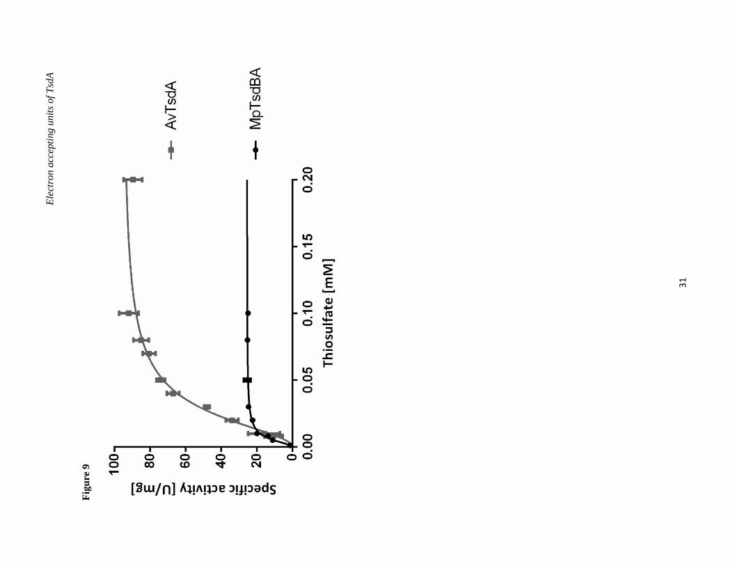
HiPIP from A. vinosum, a protein with a posi- tive reduction potential (+350 mV (10)), was tested as another potential candidate for accepting elec- trons from AvTsdA as well as from MpTsdBA. Indeed, both thiosulfate dehydrogenases reacted efficiently with A. vinosum HiPIP (Fig. 9, Table 4). AvTsdA exhibited a higher Vmax with HiPIP as electron acceptor, whilst MpTsdA featured an es- pecially low S0.5 value for thiosulfate when the reaction was measured with HiPIP as electron ac- ceptor. In both cases S0.5 for thiosulfate was much lower with HiPIP than with ferricyanide as the electron acceptor indicating cooperativity between the electron-transferring heme 2 and the active site heme 1. A. vinosum and M. purpuratum both en- code HiPIP in their genome and both thiosulfate dehydrogenases exhibit substantial specific activity with HiPIP as electron acceptor in vitro, leading us
to conclude that HiPIP also serves as an efficient in vivo electron acceptor for Tsd(B)A in both organ- isms.
DISCUSSION
In our approach to find suitable electron ac- ceptors for TsdA-type thiosulfate dehydrogenases, we first focused on TsdB, a diheme cytochrome encoded upstream of TsdA in a number of different organisms. As demonstrated here for the proteins from S. lithotrophicus and earlier for those from T. intermedia (1), TsdA and TsdB enzymes interact strongly with each other and form an α2β2 hetero- dimer. The same arrangement has been described for thiosulfate dehydrogenase from Halothio- bacillus neapolitanus (21), which consists of heme c binding subunits of 27 and 33 kDa conforming in size with TsdA and TsdB, respectively.
In this work, a redox range of -300 to +150 mV was determined for AvTsdA whilst TiTsdB is re- dox active between -100 and +300 mV. General- izing this finding, we state that the overall reduc- tion potential of TsdB is more positive than that of TsdA which should enable electron flow from TsdA to TsdB. Indeed, reduction of TiTsdB by AvTsdA had been shown previously (1) and was verified here for the proteins from S. lithotrophi- cus. Enzyme activity assays further revealed SlTsdB as an effective electron acceptor for SlTsdA but not for AvTsdA. This was not surpris- ing, as A. vinosum does not contain a gene encod- ing TsdB (Table 5).
In A. vinosum, the gene with strongest similar- ity to tsdB is Alvin_2879. The encoded high-po- tential diheme cytochrome c4 has been suggested to play a role in transferring electrons to the photo- synthetic reaction center (9). The M. purpuratum genome also encodes a protein (Marpu_15750) with high similarity to A. vinosum Cyt c4 (78 % identity on the sequence basis; Table 5). In the anoxygenic phototroph Rubrivivax gelatinosus a related cytochrome c4 indeed has an established function as an alternative electron donor to the photosynthetic reaction center (22). It was there- fore feasible to assume that electrons generated by thiosulfate oxidation could be shuttled to the reac- tion center via cytochrome c4 in purple sulfur bac- teria. However, the very low specific activity of
8
AvTsdA with A. vinosum cytochrome c4 essentially precludes such a role (Fig. 10).
In many anoxygenic photosynthetic bacteria, the periplasmic high potential iron-sulfur protein is well known to shuttle electrons between the cyto- chrome bc1 complex and the photosynthetic reac- tion center during cyclic electron flow (23-26). This function has also been firmly established for the protein from A. vinosum (24,27). Here, we demonstrate that HiPIP is a suitable electron ac- ceptor for Tsd(B)A from A. vinosum and M. pur- puratum in vitro, identifying this protein as the most likely electron carrier between the thiosul- fate-oxidizing enzyme and the reaction center during growth in the light. It should be noted that a direct interaction between Tsd(B)A and the photo- synthetic reaction center cannot be completely excluded so far.
Many purple sulfur bacteria, including A. vi- nosum are capable of chemolithotrophic growth on reduced sulfur compounds and oxygen under mi- croaerobic conditions (28). Accordingly, cbb3 as well as ubiquinol oxidases are encoded in their genomes. While the standard reduction potential of the thiosulfate/tetrathionate couple (+198 mV, (7)) appears too positive to feed electrons directly into the quinone pool, and from there to oxygen, deliv- ery of electrons originating from the thiosulfate to tetrathionate conversion to cbb3 oxidase is cer- tainly feasible. In fact, HiPIP has been reported to be involved in bacterial respiratory chains (29,30) and is a prime candidate for electron transport be- tween Tsd(B)A and the terminal oxidase in those organisms where it is present. However, in chemo- trophs like T. intermedia or S. lithotrophicus the situation must be different because these bacteria do not contain HiPIP. In fact, for these organisms it is not exactly established so far which periplas- mic proteins deliver electrons to cbb3 oxidase, regardless of the electron donor oxidized. For S. lithotrophicus it is assumed that the c-type cyto- chrome MtoD (Slit_2498) can transfer electrons stemming from iron oxidation to cbb3 oxidase and the cytochrome bc1 complex (31). In the T. intermedia genome there are two c type cyto- chromes (Tint_2575 and Tint_3060) with 36% and 42% sequence identity to S. lithotrophicus MtoD, respectively, which may serve a similar function. It may be possible that thiosulfate dehydrogenase
delivers electrons to the MtoD(-like) cytochrome which then shuffles the electrons to the terminal oxidase.
Nevertheless, an alternate scenario is also pos- sible when we consider the similarity between TsdB and cytochromes of the c4 type (about 49% sequence identity between TiTsdB and Cyt c4 from Achromobacter xylosoxidans or Pseudomonas protegens). Cytochromes of the c4-type have been reported to donate electrons to cbb3-tpye cyto- chrome c oxidases in various oxygen-respiring bacteria (32-34) and we therefore consider the possibility that TsdB serves as a direct electron donor to cbb3 oxidase at least in tetrathionate- forming thiosulfate oxidizers that neither contain HiPIP nor any cytochrome c4 homolog except of TsdB.
We have determined the first three-dimensional structure of M. purpuratum TsdBA, where TsdA is fused with its electron acceptor TsdB. It showed heme arrangement with characteristic class I c-type cytochrome topology, unveiling their relative heme spatial disposition and providing insights into the electron flow during enzymatic reaction. In the MpTsdBA structure, a thiosulfate ion is covalently bound to Sγ of Cys330 in heme 3, although the pro- tein was produced in and purified from E. coli without the addition of thiosulfate to media or buffers. This implies high affinity of the enzyme to thiosulfate which is possibly present in the com- plex growth medium in very low concentrations. It should be noted that recombinant AvTsdA and also several SoxA proteins have been isolated with the active site cysteine in a partially or fully persul- furated state (2,35-37). This has been interpreted as indication for temporary binding of thiosulfate and subsequent incomplete catalysis. Just as proposed here, thiosulfate was assumed to originate from the E. coli growth medium (2).
Regardless of its source, the covalent attach- ment of a complete thiosulfate molecule to the MpTsdBA active site cysteine strongly supports the hypothesis that tetrathionate formation from two thiosulfate molecules proceeds via a rhodanese-like reaction mechanism involving a thiosulfate transfer reaction with a thiosulfate mol- ecule covalently bound to the active site cysteine as an essential intermediate in the catalytic cycle (2,12). This type of mechanism has been illustrated

9
in detail by Grabarczyk et al. (12) for TsdA from A. vinosum. A rhodanese-like reaction cycle has also repeatedly been depicted and discussed for the closely related SoxXA protein, but could not be unambiguously proven before (4,35). The MpTsdBA structure provides conclusive evidence that the reactions catalyzed by TsdA as well as SoxXA enzymes indeed involve a cysteine S-thio- sulfonate intermediate that is formed once the first thiosulfate molecule is positioned in the substrate binding pocket by positively charged amino acid side chains (Arg314, Lys316, Arg326, and Arg438 in MpTsdBA, Fig. 8F). The latter also stabilize the cysteine S-thiosulfonate group once it has formed. Formation of the cysteine S-thiosulfonate releases two electrons which reduce the iron atoms of the two hemes in TsdA to the Fe(II) state. Heme re- oxidation by an external electron acceptor is then likely to be followed by a thiol-disulfide exchange reaction that proceeds via an attack of the sulfane atom of a second thiosulfate molecule on the thio- sulfonate group (12).
We conclude that catalysis of thiosulfate oxida- tion by Tsd(B)A enzymes and very probably also that by SoxXA proteins involves formation of a covalent adduct between the sulfane sulfur atom of thiosulfate and the Sγ of the active site cysteine. When present, TsdB is the immediate electron acceptor of TsdA. TsdB is very likely able to trans- fer electrons directly to the cbb3 terminal oxidase. In organisms containing HiPIP, this electron car- rier is likely to act as an additional shuttle not only between Tsd(B)A and the terminal oxidase during oxygen respiration but also between Tsd(B)A and the photosynthetic reaction center during photo- lithotrophic growth in the light.
EXPERIMENTAL PROCEDURES
Bacterial Strains, Plasmids and Growth Condi- tions−Table 1 lists the bacterial strains and plas- mids used for this study. Escherichia (E.) coli BL21 (DE3) was used for recombinant protein production and was grown in LB medium. E. coli DH5α was used for molecular cloning.
Recombinant DNA Techniques−All general molecular genetics techniques were described ear- lier (38). Restriction enzymes, T4 ligase and Pfu DNA polymerase were obtained from Thermo Scientific (Schwerte, Germany) and used accord-
ing to the manufacturer’s instructions. Oligonu- cleotides for cloning were obtained from Eurofins MWG (Ebersberg, Germany).
Construction of Expression Plasmids–A. vi- nosum tsdA and T. intermedia tsdB genes coding for the mature proteins without the signal peptides were amplified and cloned as described earlier (1). The tsdA gene (Slit_1878), the tsdB gene (Slit_1877) and the tsdBA gene combination (Slit_1877-Slit1878) from Sideroxydans lithotrophicus ES-1 (ATCC 700298T) were ampli- fied from genomic DNA with primers Slit1877_fw/Slit1877_rev, Slit1878_fw/ Slit1878_rev and Slit1877_fw/Slit1878_rev, re- spectively. The native signal peptide encoding sequences were included in all three cases. The tsdBA gene fusion (Marpu_02550) from Mari- chromatium purpuratum 984 (DSM 1591T) was amplified without its original signal peptide en- coding sequence using primers MarpuDR1194_for/ MarpuDR1194_rev. The gene Alvin_2879, en- coding a cytochrome c4 with similarity to TsdB was amplified with primers 2879+Sp-for and 2879- rev such that the signal peptide encoding sequence was included. The HiPIP-encoding hip (Alvin_2274) gene (39) was amplified without the signal peptide encoding sequence applying primers Alvin2274-C-strep_for/Alvin2274-C-strep_rev. Chromosomal DNA from A. vinosum DSM 180T
served as the template. For cloning of SltsdB, SltsdA, SltsdB-tsdA and Alvin_2879, primers in- cluded BsaI restriction sites and the digested PCR products were cloned into BsaI digested pASK- IBA3plus (IBA, Göttingen) resulting in vectors pASK-IBA3plus-slit1877, pASK-IBA3plus- slit1878, pASK-IBA3plus-slit1877-slit1878 and pASK-IBA3plus_Alvin_2879. For the cloning of MptsdBA into pET-22b(+) (Novagen), the re- striction enzymes NdeI and XhoI were used yield- ing plasmid pET_MarpuDRAFT_1194, Plasmid pET-Alvin2274 was constructed by cloning Avhip into a modified pET-22b(+) vector (pET-soxXAK- strep) encoding a C-terminal Strep tag. This vector had previously been constructed in the course of cloning the A. vinosum soxXAK genes. PCR pri- mers with NdeI (XAK-NdeI-for) and NcoI (XAK- NcoI-rev) sites were used and the resulting frag- ment was cloned into pET-22b(+). The XbaI/NcoI fragment of the resulting plasmid was cloned into

10
pASK-IBA3. XbaI and HindIII served for excising the soxXAK genes together with the Strep tag en- coding sequence. The XbaI/HindIII fragment was cloned between the XbaI and HindIII sites of pET22b(+) giving pET-soxXAK-strep. Replacing the soxXAK genes in this construct by Avhip yielded A. vinosum HiPIP fused to a carboxy-ter- minal Strep tag.
Overproduction, Purification and Preparation of Recombinant Proteins−AvTsdA and TiTsdB were produced as described before (1). For produc- tion of S. lithotrophicus TsdA, SlTsdB and the simultaneous production of SlTsdB and SlTdsA Escherichia coli BL21(DE3) cells containing pASK-IBA3plus-slit1877, pASK-IBA3plus- slit1878 or pASK-IBA3plus-slit1877-slit1878 and pEC86 (40) were cultured in 700 ml LB media supplemented with 100 µg ml-1 ampicillin and 25 µg ml-1 chloramphenicol at 37°C and 180 rpm after inoculation with an overnight pre-inoculum in a (1:50) dilution. At an OD600nm of 0.4 to 0.6, 200 ng ml-1 anhydrotetracycline were added and the ap- propriate culture was switched to 25°C and 90 rpm in case of TsdB production. Cells were harvested after 18 h. MpTsdBA and HiPIP were produced in Escherichia coli BL21(DE3) cells containing pET_MarpuDRAFT_1194 or pET-Alvin2274 and pEC86 (40). After 2% or 0.5% inoculation with a preculture, the cells were grown in 700 ml LB medium containing 100 µg ml-1 ampicillin and 25 µg ml-1 chloramphenicol at 37°C and 180 rpm. At an OD600nm of 0.5 to 0.6 the cultures were switched to 25°C and 120 rpm for about 18 h. For produc- tion of AvCyt c4 (Alvin_2879), E. coli BL21(DE3) cells containing pASK-IBA3plus_Alvin_2879 and pEC86 (40) were cultured in 400 ml LB medium, 100 µg ml-1 ampicillin and 25 µg ml-1 chloram- phenicol at 37°C and 180 rpm after 2% inoculation with a preculture. At OD600nm ~0.5, 200 ng ml-1
anhydrotetracycline were added and the culture was switched to 25°C and 90 rpm for 18 h. Har- vested cells were resuspended in 100 mM Tris-HCl buffer pH 8.0 containing 150 mM NaCl and lysed by sonication. After removal of insoluble cell ma- terial by centrifugation (10,000 g for 25 min at 4°C), SlTsdA, SlTsdB, SlTsdB+TsdA, A. vinosum cytochrome c4 (Alvin_2879) and AvTsdA were purified by Strep-Tactin (IBA, Göttingen, Ger- many) affinity chromatography according to the
manufacturer’s instructions. MpTsdBA was puri- fied by nickel-chelate (Qiagen, Hilden, Germany) affinity chromatography according to the manu- facturer's instructions and then subjected to a size- exclusion chromatography step performed on a Hi- Load 16/60 Superdex 75 pg column (GE Healthcare) using an ÄKTApurifier system (GE Healthcare). The column was equilibrated with 20 mM Tris-HCl buffer, pH 7.5 and 150 mM NaCl. TsdB and TsdA from S. lithtrophicus were ana- lyzed by the same procedure either separately or as a mixture of both proteins. In this case, the Super- dex 75 column was equilibrated with 100 mM Tris-HCl buffer, pH 7.5 and 150 mM NaCl. The column was calibrated with the Molecular weight marker kit MW-GF-70 (GE Healthcare). All puri- fied proteins were desalted with 5 ml HiTrap De- salting columns (GE Healthcare) and concentrated with Amicon Ultra–15 centrifugal filter units (Merck Millipore). Recombinant S. lithotrophicus proteins were stored in 100 mM sodium acetate buffer pH 5 at -70°C, A. vinosum Cyt c4 in 20 mM Tris-HCl buffer pH 7.5 at 4°C, MpTsdBA in 20 mM Tris-HCl buffer pH 7.5 with 150 mM NaCl at -70°C and HiPIP in 20 mM Tris-HCl pH 7 at 4°C. The concentration of purified proteins was determined with the BCA kit from Pierce (Rock- ford, USA). For assessment of purity, sodium do- decyl sulfate-polyacrylamide gel electrophoresis (SDS-PAGE) was performed and the proteins vis- ualized either by Coomassie or heme staining techniques.
UV-vis Spectroscopy with TsdA in Solution– UV-vis spectra were recorded between 250 and 750 nm with an Analytik Jena Specord 210 (Ana- lytik Jena, Jena, Germany).
Assay of Thiosulfate Oxidase Activity with Ferricyanide−Thiosulfate-dependent ferricyanide reduction was measured by following the decrease in absorbance at 420 nm (ε = 1.09 μM cm
-1). En- zyme activity measurements with AvTsdA at pH 4 are described in (2). Activity measurements with MpTsdBA were performed with 1 mM ferricyanide at 25°C in 100 mM ammonium acetate buffer pH 5.2 with 200 mM NaCl. Assays were started by addition of TsdA and data were recorded in a Specord 210 spectrophotometer (Analytik Jena, Jena, Germany). Activity is expressed as µmol tetrathionate produced per min and mg protein on

11
the basis of one tetrathionate formed per two ferri- cyanide reduced. In the case of enzymes that use two molecules of the same substrate (here thiosul- fate) primary v versus [S] plots provide the best way to examine the data (41). Data were fitted to the empirical Hill equation (Eq. 1) using Graph Pad Prism (version 6; Graph Pad).
n
determined in 100 mM ammonium acetate buffer pH 5.5 at 25°C.
Determination of Redox Properties of AvTsdA Adsorbed on a Mesoporous Nanocrystalline SnO2
Electrode–An optically transparent mesoporous nanocrystalline SnO2 electrode coated with AvTsdA was prepared using the previously de- scribed method (42) with adsorption from a solu-
𝑣 = 𝑉max[𝑆] 𝐾+ [𝑆]n
(Eq. 1) tion of 10 µM AvTsdA, 2 mM neomycin, 50 mM NaCl, 50 mM HEPES, pH 7. The enzyme-coated
The Hill equation resembles the classical Henri–Michaelis–Menten equation; however, the n term allows accounting for non-hyperbolic shapes. A substrate concentration [S]0.5 can be reported that yields half maximal velocity and is character- istic of the process. The constant K, which is not equivalent to Km, characterizes enzyme–substrate interaction. The relationship between K and [S]0.5 is K = [S]0.5
n. Assay of Thiosulfate Oxidase Activity with
HiPIP–For assays of electron transfer from thiosul- fate to the electron acceptor HiPIP, 10 µM HiPIP preoxidized with 40 µM ferricyanide were used. The reaction was started by addition of enzyme and followed by the absorbance decrease at 480 nm. A molar extinction coefficient at 480 nm of 10.7 mM-1 cm-1 (10) was used. Measurements with AvTsdA were performed in 100 mM ammonium acetate buffer pH 5 at 30°C and with MpTsdBA in 100 mM ammonium acetate buffer pH 5.2 with 200 mM NaCl at 25°C.
Assay of Thiosulfate Oxidase Activity with TsdB or AvCyt c4−Thiosulfate-dependent reduction of T.
electrode was rinsed with 2 mM neomycin, 50 mM NaCl, 50 mM HEPES, pH 7 to remove unbound protein, taken into a N2-filled chamber (atmos- pheric O2 < 2ppm) and immersed in an anaerobic solution of the same composition within a previ- ously described spectroelectrochemical cell (42). The cell was sealed, removed from the anaerobic chamber and inserted into a JASCO V650 UV- visible spectrophotometer thermostated at 4oC and flushed with argon to maintain anaerobicity. Spec- tral contributions from light scattering by the elec- trode were minimized by placing a bare SnO2
electrode (i.e., having no adsorbed enzyme) in the reference beam of the spectrophotometer. The electronic absorbance of the as prepared enzyme- coated electrode revealed features indicative of a mixture of ferric and ferrous hemes. After the electrode had been poised at +302 mV for 45 min the spectrum revealed that the enzyme had been converted to the fully oxidized, all ferric-state. To determine the redox activity of AvTsdA the elec- trode potential was swept from +302 to -648 mV at a scan rate of 5 mV s-1 with a pause of 150 s every
intermedia TsdB or A. vinosum Cyt c4 was meas- ured by following the increase in absorbance at 417 nm (εΔ417nm = 99 mM cm-1) for TiTsdB or SlTsdB and at 420 nm (εΔ420nm= 55 mM cm-1 (9)) for AvCyt c4. The extinction coefficient for TsdB was calculated with the help of the Beer-Lambert law using distinct concentrations of TiTsdB and the differences in absorbance at 417 nm in the reduced and oxidized spectra. A value averaged from measurements with three different protein concentrations was derived. Assays of SlTsdA activity with SlTsdB as electron acceptor were carried out in 100 mM ammonium acetate buffer pH 4 at 25°C. AvTsdA activity with TiTsdB was assayed in 100 mM ammonium acetate buffer pH 5 at 30°C, while AvTsdA activity with AvCyt c4 was
50 mV. At 60 s into each pause a spectrum was measured before the scan continued. Reoxidation of the sample was performed in a similar manner. Spectra are presented after equating absolute ab-
sorbance at 600 nm in order to account for poten- tial dependent changes in the spectral contributions that arise from scattering by the electrode material.
Redox Potentiometry with TsdB in Solution Measured with a Gold Electrode–The reduction
potential of TiTsdB was measured with help of a gold-platinum electrode system under anoxic con- ditions. The electrode extended into a cuvette con-
taining the protein solution (20 µM) and redox mediators (N,N-dimethyl-1,4-phenylenediamine,
p-benzoquinone, trimethylhydroquinone, phena- zine, 1,4-naphthoquinone and 1,2-naphthoquinone
12
at 2 µM each) in 20 mM MOPS buffer pH 6 and was connected to a potentiometer. TiTsdB was reduced by changing the applied potential from - 150 mV to 450 mV. Subsequential decrease of the potential again to -150 mV led to reoxidation of the protein. A spectrum was recorded every two minutes and potentials converted to values vs. SHE.
Crystallization, Data Collection, Structure De- termination and Refinement–MpTsdBA at a concentration of 3.2 mg ml-1 in 20 mM Bis-Tris- HCl pH 6.5 and 150 mM NaCl was crystallized in 10 % (w/v) PEG 8000, 0.1 M Tris-HCl pH 7.0, 0.2 M MgCl2 and 10 mM trimethylamine hydrochloride (as additive) by vapour-diffusion hanging-drop method at 20oC. Crystallization droplets comprised 1.0 μl of protein, 0.8 μl of precipitant and 0.2 μl of additive, and were equili- brated against a 200 μl of reservoir solution (26% (w/v) PEG 3350). Crystals were cryoprotected with No. 2 solution of CryoProtX screen (Molecular Dimensions), consisting of 25% (v/v) diethylene glycol, 25% (v/v) 1,2-propanediol, and 25% (v/v) glycerol. X-ray diffraction data were collected at a wavelength of 1.7236 Ǻ on beamline ID-29 of the European Synchrotron Radiation Facility (ESRF, Grenoble, France). Data were indexed and integrated with XDS (43), space group determined with POINTLESS (44) and scaled with AIMLESS (45,46), all within autoPROC (47) data-processing pipeline. An Rfree flag was created at this stage corresponding to 5% of the measured reflections of the data set. The structure was determined by single wavelength anomalous dispersion method around the iron edge (Fe-SAD), employing a high multiplicity data collection strategy using the autoSHARP module (48), within the SHARP package (49). Iterative model building and refinement cycles were performed with COOT (50), BUSTER-TNT (51), (at early stages of refine- ment) followed by phenix.refine (52), until a com- plete model was built and refinement convergence achieved. Friedel mates were kept separately and refinement was carried out against I(+)/SIGI(+),I(–)/SIGI(–). A polder map (an omit map which excludes the bulk solvent around the omitted region), and m|Fo|-m|Fo| map were calculated within the PHENIX package of programs. The Ramachandran diagram was as-
sessed with RAMPAGE (53) and the model vali- dated with MolProbity (54) as implemented in PHENIX. All figures were rendered with PyMOL, Schrödinger LLC (55).
ACKNOWLEDGEMENTS
The authors acknowledge Isabel Bento and Ana Maria Gonçalves for collecting the X-ray diffrac- tion data. We also acknowledge Susana Gonçalves and ID-29 beamline staff at the European Syn- chrotron Radiation Facility (ESRF; Grenoble, France) for providing assistance in using the beamline. S. lithotrophicus DNA was kindly pro- vided by David Emerson, Bigelow Laboratory for Ocean Sciences, West Boothbay Maine, USA. MALDI-TOF mass spectrometry of TiTsdB and SlTsdB was kindly performed by Michaele Josten and Hans Georg Sahl, Institute for Medical Micro- biology, Immunology and Parasitology, University of Bonn, Germany. We thank James Durrant (Im- perial College London) for the SnO2 electrodes.
CONFLICT OF INTEREST
The authors declare that they have no conflicts of interest with the contents of this article.
AUTHORS CONTRIBUTIONS
JMK, JAB, JNB, MA and CD wrote the manu- script. CD conceived and coordinated all experi- ments except the MpTsdBA crystallization and determination of reduction potentials. JMK ana- lyzed and compiled the data for those experiments. JR constructed the vector for production of MpTsdBA and TF the vector for production of HiPIP. AF and JR performed activity assays with MpTsdBA, AvTsdA and HiPIP (Fig. 9; Table 4) and recorded UV-vis spectra of MpTsdBA (Fig. 6). TK produced AvCyt c4 and measured activity of AvTsdA with this protein. EK and JMK produced and analyzed proteins from S. lithotrophicus (Figs. 2 and 5). KD determined the redox activity of TiTsdB (Fig. 4) under supervision of IACP. SR examined the redox activity of AvTsdA (Fig. 1) under supervision of JNB. JAB crystallized MpTsdBA, processed the X-ray data, determined the crystal structure and performed model building and refinement (Figs. 7 and 8; Table 3). JAB and MA analyzed the crystal structure.

13
REFERENCES
1. Denkmann, K., Grein, F., Zigann, R., Siemen, A., Bergmann, J., van Helmont, S., Nicolai, A., Pereira, I. A. C., and Dahl, C. (2012) Thiosulfate dehydrogenase: a wide-spread unusual acidophilic c-type cytochrome. Environ. Microbiol. 14, 2673-2688
2. Brito, J. A., Denkmann, K., Pereira, I. A. C., Archer, M., and Dahl, C. (2015) Thiosulfate dehydrogenase (TsdA) from Allochromatium vinosum: structural and functional insights into thiosulfate oxidation. J. Biol. Chem. 290, 9222-9238
3. Pires, R. H., Venceslau, S. S., Morais, F., Teixeira, M., Xavier, A. V., and Pereira, I. A. C. (2006) Characterization of the Desulfovibrio desulfuricans ATCC 27774 DsrMKJOP complex - a membrane-bound redox complex involved in the sulfate respiratory pathway. Biochemistry 45, 249-262
4. Bradley, J. M., Marritt, S. J., Kihlken, M. A., Haynes, K., Hemmings, A. M., Berks, B. C., Cheesman, M. R., and Butt, J. N. (2012) Redox and chemical activities of the hemes in the sulfur oxidation pathway enzyme SoxAX. J. Biol. Chem. 287, 40350-40359
5. Reijerse, E. J., Sommerhalter, M., Hellwig, P., Quentmeier, A., Rother, D., Laurich, C., Bothe, E., Lubitz, W., and Friedrich, C. G. (2007) The unusal redox centers of SoxXA, a novel c-type heme-enzyme essential for chemotrophic sulfur-oxidation of Paracoccus pantotrophus. Biochemistry 46, 7804-7810
6. Kappler, U., Bernhardt, P. V., Kilmartin, J., Riley, M. J., Teschner, J., McKenzie, K. J., and Hanson, G. R. (2008) SoxAX cytochromes, a new type of heme copper protein involved in bacterial energy generation from sulfur compounds. J. Biol. Chem. 283, 22206-22214
7. Kurth, J., Dahl, C., and Butt, J. N. (2015) Catalytic protein film electrochemistry provides a direct measure of the tetrathionate/thiosulfate reduction potential. J. Am. Chem. Soc. 137, 13232-13235
8. Liu, Y.-W., Denkmann, K., Kosciow, K., Dahl, C., and Kelly, D. J. (2013) Tetrathionate stimulated growth of Campylobacter jejuni identifies TsdA as a new type of bi-functional tetrathionate reductase that is widely distributed in bacteria. Mol. Microbiol. 88, 173-188
9. Cusanovich, M. A. and Bartsch, R. G. (1969) A high potential cytochrome c from Chromatium vinosum chromatophores. Biochim. Biophys. Acta 189, 245-255
10. Bartsch, R. G. (1978) Purification of (4Fe-4S)1-2- ferredoxins (high-potential iron-sulfur proteins) from bacteria. Methods Enzymol. 53, 329-340
11. Fukumori, Y. and Yamanaka, T. (1979) A high-potential nonheme iron protein (HiPIP)-linked, thiosulfate- oxidizing enzyme derived from Chromatium vinosum. Curr. Microbiol. 3, 117-120
12. Grabarczyk, D. B., Chappell, P. E., Eisel, B., Johnson, S., Lea, S. M., and Berks, B. C. (2015) Mechanism of thiosulfate oxidation in the SoxA family of cysteine-ligated cytochromes. J. Biol. Chem. 290, 9209-9221
13. Du, J., Sono, M., and Dawson, J. H. (2011) The H93G myoglobin cavity mutant as a versatile scaffold for modeling heme iron coordination structures in protein active sites and their characterization with magnetic circular dichroism spectroscopy. Coord. Chem. Rev. 255, 700-716
14. Miles, C. S., Manson, F. D. C., Reid, G. A., and Chapman, S. K. (1993) Substitution of a haem-iron axial ligand in flavocytochrome b2. Biochim. Biophys. Acta 1202, 82-86
15. Branca, R. M. M., Bodó, G., Várkonyi, Z., Debreczeny, M., Ösz, J., and Bagyinka, C. (2007) Oxygen and temperature-dependent structural and redox changes in a novel cytochrome c4 from the purple sulfur bacterium Thiocapsa roseopersicina. Arch. Biochem. Biophys. 467, 174-184
16. Nissum, M., Karlsson, J.-J., Ulstrup, J., Jensen, P. W., and Smulevich, G. (1997) Resonance Raman characterization of the di-heme protein cytochrome c4 from Pseudomonas stutzeri. J. Biol. Inorg. Chem. 2, 302-307
17. Matthews, B. W. (1968) Solvent content of protein crystals. J. Mol. Biol. 33, 491-497 18. Holm, L. and Rosenström, P. (2010) Dali server: conservation mapping in 3D. Nucleic Acids Res. 38, W545-
W549 19. Igarashi, N., Moriyama, H., Fujiwara, T., Fukumori, Y., and Tanaka, N. (1997) The 2.8 Å structure of
hydroxylamine oxidoreductase from a nitrifying chemoautotrophic bacterium, Nitrosomonas europaea. Nat. Struct. Biol. 4, 276-284

14
20. Taylor, P., Pealing, S. L., Reid, G. A., Chapman, S. K., and Walkinshaw, M. D. (1999) Structural and mechanistic mapping of a unique fumarate reductase. Nat. Struct. Biol. 6, 1108-1112
21. Visser, J. M., de Jong, G. A. H., Robertson, L. A., and Kuenen, J. G. (1996) Purification and characterization of a periplasmic thiosulfate dehydrogenase from the obligately autotrophic Thiobacillus sp. W5. Arch. Microbiol. 166, 372-378
22. Ohmine, M., Matsuura, K., Shimada, K., Alric, J., Verméglio, A., and Nagashima, K. V. (2009) Cytochrome c4
can be involved in the photosynthetic electron transfer system in the purple bacterium Rubrivivax gelatinosus. Biochemistry 48, 9132-9139
23. Bartsch, R. G. (1991) The distribution of soluble metallo-redox proteins in purple phototrophic bacteria. Biochim. Biophys. Acta 1058, 28-30
24. Kennel, S. J., Bartsch, R. G., and Kamen, M. D. (1972) Observations on light-induced oxidation reactions in the electron transport system of Chromatium. Biophys. J. 12, 882-896
25. Nagashima, K. V., Matsuura, K., Shimada, K., and Vermeglio, A. (2002) High-potential iron-sulfur protein (HiPIP) is the major electron donor to the reaction center complex in photosynthetically growing cells of the purple bacterium Rubrivivax gelatinosus. Biochemistry 41, 14028-14032
26. Lieutaud, C., Nitschke, W., Verméglio, A., Parot, P., and Schoepp-Cothenet, B. (2003) HiPIP in Rubrivivax gelatinosus is firmly associated to the membrane in a conformation efficient for electron transfer towards the photosynthetic reaction centre. Biochim. Biophys. Acta 1557, 83-90
27. Verméglio, A., Li, J., Schoepp-Cothenet, B., Pratt, N., and Knaff, D. B. (2002) The role of high-potential iron protein and cytochrome c8 as alternative electron donors to the reaction center of Chromatium vinosum. Biochemistry 41, 8868-8875
28. Kämpf, C. and Pfennig, N. (1980) Capacity of Chromatiaceae for chemotrophic growth. Specific respiration rates of Thiocystis violacea and Chromatium vinosum. Arch. Microbiol. 127, 125-135
29. Hochkoeppler, A., Jenney, F. E., Lang, S. E., Zannoni, D., and Daldal, F. (1995) Membrane-associated cytochrome c(y) of Rhodobacter capsulatus is an electron carrier from the cytochrome bc(1) complex to the cytochrome c oxidase during respiration. J. Bacteriol. 177, 608-613
30. Bonora, P., Principi, I., I, Monti, B., Ciurli, S., Zannoni, D., and Hochkoeppler, A. (1999) On the role of high- potential iron-sulfur proteins and cytochromes in the respiratory chain of two facultative phototrophs. Biochim. Biophys. Acta 1410, 51-60
31. Beckwith, C. R., Edwards, M. J., Lawes, M., Shi, L., Butt, J. N., Richardson, D. J., and Clarke, T. A. (2015) Characterization of MtoD from Sideroxydans lithotrophicus: a cytochrome c electron shuttle used in lithoautotrophic growth. Front Microbiol. 6, 332
32. Arai, H., Kawakami, T., Osamura, T., Hirai, T., Sakai, Y., and Ishii, M. (2014) Enzymatic characterization and in vivo function of five terminal oxidases in Pseudomonas aeruginosa. J. Bacteriol. 196, 4206-4215
33. Barco, R. A., Emerson, D., Sylvan, J. B., Orcutt, B. N., Jacobson Meyers, M. E., Ramirez, G. A., Zhong, J. D., and Edwards, K. J. (2015) New insight into microbial iron oxidation as revealed by the proteomic profile of an obligate iron-oxidizing chemolithoautotroph. Appl. Environ. Microbiol. 81, 5927-5937
34. Chang, H. Y., Ahn, Y., Pace, L. A., Lin, M. T., Lin, Y. H., and Gennis, R. B. (2010) The diheme cytochrome c4
from Vibrio cholerae is a natural electron donor to the respiratory cbb3 oxygen reductase. Biochemistry 49, 7494-7503
35. Bamford, V. A., Bruno, S., Rasmussen, T., Appia-Ayme, C., Cheesman, M. R., Berks, B. C., and Hemmings, A. M. (2002) Structural basis for the oxidation of thiosulfate by a sulfur cycle enzyme. EMBO J. 21, 5599- 5610
36. Dambe, T., Quentmeier, A., Rother, D., Friedrich, C., and Scheidig, A. J. (2005) Structure of the cytochrome complex SoxXA of Paracoccus pantotrophus, a heme enzyme initiating chemotrophic sulfur oxidation. J. Struct. Biol. 152, 229-234
37. Kilmartin, J. R., Maher, M. J., Krusong, K., Noble, C. J., Hanson, G. R., Bernhardt, P. V., Riley, M. J., and Kappler, U. (2011) Insights into structure and function of the active site of SoxAX cytochromes. J. Biol. Chem. 286, 24872-24881
38. Dahl, C., Schulte, A., Stockdreher, Y., Hong, C., Grimm, F., Sander, J., Kim, R., Kim, S.-H., and Shin, D. H. (2008) Structural and molecular genetic insight into a wide-spread bacterial sulfur oxidation pathway. J. Mol. Biol. 384, 1287-1300

15
39. Brüser, T., Trüper, H. G., and Dahl, C. (1997) Cloning and sequencing of the gene encoding the high potential iron-sulfur protein (HiPIP) from the purple sulfur bacterium Chromatium vinosum. Biochim. Biophys. Acta 1352, 18-22
40. Arslan, E., Schulz, H., Zufferey, R., Kunzler, P., and Thöny-Meyer, L. (1998) Overproduction of Bradyrhizobium japonicum c-type cytochrome subunits of the cbb3 oxidase in Escherichia coli. Biochem. Biophys. Res. Commun. 251, 744-747
41. Segel, I. H. (1993) Enzyme kinetics: behaviour and analysis of rapid equilibrium and steady-state enzyme systems, Wiley-Interscience, New York
42. Marritt, S. J., Kemp, G. L., Xiaoe, L., Durrant, J. R., Cheesman, M. R., and Butt, J. N. (2008) Spectroelectrochemical characterization of a pentaheme cytochrome in solution and as electrocatalytically active films on nanocrystalline metal-oxide electrodes. J. Am. Chem. Soc. 130, 8588-8589
43. Kabsch, W. (2010) XDS. Acta Crystallogr. D Biol. Crystallogr. 66, 125-132 44. Evans, P. R. (2011) An introduction to data reduction: space-group determination, scaling and intensity statistics.
Acta Crystallogr. D Biol. Crystallogr. 67, 282-292 45. Evans, P. (2006) Scaling and assessment of data quality. Acta Crystallogr. D Biol. Crystallogr. 62, 72-82 46. Evans, P. R. and Murshudov, G. N. (2013) How good are my data and what is the resolution? Acta Crystallogr.
D Biol. Crystallogr. 69, 1204-1214 47. Vonrhein, C., Flensburg, C., Keller, P., Sharff, A., Smart, O., Paciorek, W., Womack, T., and Bricogne, G.
(2011) Data processing and analysis with the autoPROC toolbox. Acta Crystallogr. D Biol. Crystallogr. 67, 293-302
48. Vonrhein, C., Blanc, E., Roversi, P., and Bricogne, G. (2007) Automated structure solution with autoSHARP. Methods Mol. Biol. 364, 215-230
49. de la Fortelle, E. and Bricogne, G. (1997) Maximim-likelihood heavy-atom parameter refinement for multiple isomorphous replacement and multiwavelength anomalous diffraction methods. Meth. Enzymol. 276, 472- 494
50. Emsley, P., Lohkamp, B., Scott, W. G., and Cowtan, K. (2010) Features and development of Coot. Acta Crystallogr. D Biol. Crystallogr. 66, 486-501
51. Blanc, E., Roversi, P., Vonrhein, C., Flensburg, C., Lea, S. M., and Bricogne, G. (2004) Refinement of severely incomplete structures with maximum likelihood in BUSTER-TNT. Acta Crystallogr. D Biol. Crystallogr. 60, 2210-2221
52. Afonine, P. V., Grosse-Kunstleve, R. W., Echols, N., Headd, J. J., Moriarty, N. W., Mustyakimov, M., Terwilliger, T. C., Urzhumtsev, A., Zwart, P. H., and Adams, P. D. (2012) Towards automated crystallographic structure refinement with phenix.refine. Acta Crystallogr. D Biol. Crystallogr. 68, 352-367
53. Lovell, S. C., Davis, I. W., Arendall, W. B., III, de Bakker, P. I., Word, J. M., Prisant, M. G., Richardson, J. S., and Richardson, D. C. (2003) Structure validation by Calpha geometry: phi,psi and Cbeta deviation. Proteins 50, 437-450
54. Chen, V. B., Arendall, W. B., III, Headd, J. J., Keedy, D. A., Immormino, R. M., Kapral, G. J., Murray, L. W., Richardson, J. S., and Richardson, D. C. (2010) MolProbity: all-atom structure validation for macromolecular crystallography. Acta Crystallogr. D Biol. Crystallogr. 66, 12-21
55. Delano, W. L. (2002) The PyMOL molecular graphics system, DeLano Scientific, San Carlos, California, USA 56. Hanahan, D. (1983) Studies on transformation of Escherichia coli with plasmids. J. Mol. Biol. 166, 557-580 57. Karplus, P. A. and Diederichs, K. (2012) Linking crystallographic model and data quality. Science 336, 1030-
1033

16
FIGURE LEGENDS
FIGURE 1. Redox activity of AvTsdA adsorbed on a mesoporous nanocrystalline SnO2 electrode. Electronic absorbance recorded with the electrode poised at +302 mV (black), +152 to -298 mV at 50 mV intervals (gray) and -648 mV (red). All potentials are quoted versus the standard hydrogen electrode (SHE). The arrows indicate increases in absorbance as the electrode potential was lowered. Inset shows the normalized change in absorbance at 418 nm against the applied potential as the enzyme was reduced (closed squares) and re-oxidized (open squares).
FIGURE 2. UV-vis spectra of TsdB from S. lithotrophicus. As the protein is partly reduced in the “as
isolated” state, up to 170 µM ferricyanide were added to record the oxidized spectrum (black line). For
full reduction of the protein Na-dithionite was added (grey line). 100 mM Tris buffer pH 8.0 with 150 mM NaCl and 2.5 mM desthiobiotin was used and spectra are normalized to 750 nm. The oxidized spectrum exhibits a 700 nm peak indicating methionine as heme iron ligand. Protein concentration: 21 µM in the overview and 94 µM in the blow-up.
FIGURE 3. Sequence alignment of MpTsdBA and SlTsdB+TsdA as well as SlTsdB+TsdA. Sequence comparison of TsdBA fusion protein of M. purpuratum (MARPU_02550) with the combined sequence of TsdB and TsdA from S. lithotrophicus (Slit_1877 and Slit_1878) and T. intermedia (Tint_1893 and Tint_2892). All signal peptide sequences were removed. Heme binding motifs are indicated by grey boxes, putative distal heme ligands are marked by black edging. Strictly conserved residues are marked with asterisks. TsdA sequences of S. lithotrophicus and T. intermedia start after the gap at amino acids 195 and 189, respectively.
FIGURE 4. Determination of TiTsdB reduction potential by potentiometry with a gold electrode. Potentiometric determination of redox potentials of both TsdB hemes. Applied potential according to normalized values of the α-peak (553 nm) is shown. Reduction of TsdB (black diamonds) and re-oxidation of the protein (grey squares) was measured. 10 µM of TsdB in phosphate buffer pH 5.0 was used.
FIGURE 5. Analysis of purified SlTsdA and SlTsdB+A by SDS polyacrylamide gel electrophoresis. 10-15 µg SlTsdA and SlTsdB+A obtained after Strep tag affinity chromatography were loaded per lane of a 12.5 % gel and stained for presence of heme. In case of SlTsdB+A both proteins were produced simulta- neously in E. coli and purified on the basis of a Strep tag attached to TsdA.
FIGURE 6. UV-vis spectra of MpTsdBA. As the protein is slightly reduced in the “as isolated” state,
60 µM ferricyanide were added to record the oxidized spectrum (black line). For partial (grey broken line) and full reduction (grey line) of the protein, 0.33 mM and 5 mM Na-dithionite were added, respectively. 100 mM ammonium acetate buffer pH 5 with 200 mM NaCl was used and spectra are normalized to 750 nm. The oxidized spectrum exhibits a 700 nm peak indicating methionine as heme iron ligand and the partially reduced protein exhibits a feature at 630 nm. Protein concentration: 3.3 µM.
FIGURE 7. X-ray structure of MpTsdBA and heme arrangement. A) Overall fold with TsdB N-termi- nal domain (residues 1-193) depicted in blue and TsdA C-terminal domain (residues 240-516) in red; the two shades in each domain represent the respective sub-domains (1-90 and 91-193 for TsdB, and 240-377 and 378-516 for TsdA). The heme prosthetic groups are colored by atom type (orange or yellow for car- bon, blue for nitrogen, red for oxygen and dark red for iron), B) Fe-to-Fe distances, and C) Closest edge- to-edge distances.

17
FIGURE 8. Heme coordination of “as isolated” MpTsdBA (PDB code 5LO9). A) Heme 1 is coordi- nated by His25 and Met65, B) Heme 2 is coordinated by His125 and Met167, C) Heme 3 is ligated to His291
but not to Cys330. The distance of Sγ to the heme iron is 2.9 Å and thus not close enough for direct ligation. Thiosulfate covalently bound to Sγ of Cys330 is not shown here for clarity. Presence of thiosulfate is illustrated in detail in panels E and F. D) Heme 4 is ligated by His406 and Met450
, E) Heme 3 with Sγ of
Cys330 covalently bound to thiosulfate, displayed in ball and stick, and polder map electron density contoured at 6σ level depicted as a black mesh, and F) Heme 3 in a similar view as in E) but with positively charged residues surrounding the substrate cleft depicted in sticks. Cartoon representation is shown in pale grey with heme moieties and coordinating amino acid residues shown in sticks; color code as in Fig. 7 with sulfur atoms in green.
FIGURE 9. Thiosulfate oxidation catalyzed by AvTsdA and MpTsdBA with HiPIP as electron accep- tor. Enzyme assays with AvTsdA were performed in 100 mM ammonium acetate buffer pH 5 at 30°C with 8 nM enzyme. Activity measurements with MpTsdBA were performed in 100 mM ammonium ace- tate buffer pH 5.2 with 200 mM NaCl at 25°C and with 3.9 nM enzyme. In both assays 10 µM HiPIP and 40 µM ferricyanide were used. A change in absorbance was measured at 480 nm. v versus [S] plots were fitted to the Hill equation.
FIGURE 10. Role of periplasmic electron transfer proteins in aerobic respiration or photosynthesis of A. vinosum, M. purpuratum, S. lithotrophicus and T. intermedia. All organisms contain genes for NuoA-N (Alvin_2418-2430 +Alvin_2412, Marpu_04365-04430, Slit_1070-1083, Tint_2255-2268), the cytochrome bc1 complex (Alvin_0068-0070, Marpu_01465-01475, Slit_0130-0132, Tint_2192-2194) and cbb3 oxidase (Alvin_0781-0784, Marpu_02795-02810, Slit_0411-0414, Tint_1070-1073). Moreover, A. vinosum and M. purpuratum can gain energy by photosynthetic growth. HiPIP can transfer electrons to the photosynthetic reaction center (23,24) as well as to cbb3 oxidase (29,30). Cytochrome c4 also is known to transfer electrons to the photosynthetic reaction center (22) as well as to cbb3 oxidase (32-34) in some bacteria. For S. lithotrophicus it is assumed that MtoD (Slit_2498) can transfer electrons to cbb3 oxidase and the cytochrome bc1 complex (31).

18
TABLE 1. E. coli strains and plasmids used in this study
Strains and plasmids Description Reference or
source Strains
E. coli DH5α F-
φ80d lacZΔM15 Δ(lacZYA-argF)U169 recA1 endA1hsdR17 (rk-
mk +) supE44 λ-thi-1 gyrA relA1
(56)
E. coli BL21 (DE3) - -
F- ompT hsdSB (rB mB ) gal dcm met(DE3) Novagen
Plasmids pEC86 Cmr, product from pEC66 and pACYC184 with E. coli
ccmABCDEFGH genes (40)
pET-22b (+) Apr, T7 promoter, lac operator, C-terminal His tag, pelB leader Novagen
pASK-IBA3 plus Apr, tetA promoter/operator, C-terminal Strep tag IBA (Göttingen)
pET_ MarpuDRAFT_1194 Apr, tsdA from M. purpuratum (Marpu_02550) was cloned into pET-22b (+)with Nde I and XhoI, C-terminal His tag
Kurth et al., 2015
pASK-IBATint_tsdB Apr; tsdB from T. intermedia cloned into pASK-IBA3 plus with BsaI, C-terminal Strep tag
(1)
pASK-IBA3plus-slit1877 Apr; tsdB from S. lithotrophicus (Slit_1877) cloned into pASK- IBA3 plus with BsaI, C-terminal Strep tag
This study
pASK-IBA3plus-slit1878 Apr; tsdA from S. lithotrophicus (Slit_1878) cloned into pASK- IBA3 plus with BsaI, C-terminal Strep tag
This study
pASK-IBA3plus-slit1877- slit1878
Apr; tsdBA from S. lithotrophica (Slit_1877-Slit_1878) cloned into pASK-IBA3 plus with BsaI, C-terminal Strep tag
This study
pET-soxXAK-strep Apr, soxXAK including fragment cloned into pET-22b(+) together with C terminal Strep tag by use of XbaI and HindIII
This study
pET-Alvin2274-C-strep Apr, hip gene coding for HiPIP from A. vinosum (Alvin_2274) cloned into pET-soxXAK-strep with NdeI and NcoI, C-terminal Strep tag
This study
pPR-IBAAvtsdA Apr; tsdA from A. vinosum (Alvin_0091) cloned into pASK-IBA3, C-terminal Strep tag
(1)
pASK-IBA3plus_Alvin_2879 Apr; Alvin_2879 from A. vinosum cloned into pASK-IBA3 plus with BsaI, C-terminal Strep tag
This study

19
TABLE 2. Primers used in this study
Primer Sequence 5’-3’ Reference MarpuDR1194_for CGGAGGGATCCTCATATGACGCATCTC This study MarpuDR1194_rev GACCTGCTCGAGATCCTTGGC This study Slit1877_fw ATGGTAGGTCTCAAATGAAGCAAATATTACTAGCAGCATTAAC This study Slit1877_rev ATGGTAGGTCTCAGCGCTTTTCTGGTTTCCATTGGTTGATTGT This study Slit1878_fw ATGGTAGGTCTCAAATGAAGAATCCCATCGCTATCGCCAT This study Slit1878_rev ATGGTAGGTCTCAGCGCTCTTTGCTGCAGTCTGGTGCTTTC This study
XAK-NdeI-for GGAGATTTCATATGCCGTTGAACGTCTCACACCG This study
XAK-NcoI-rev ATGGCTCCATGGTATCGAGACCGATCGAGC This study Alvin2274-C-strep_for GCCCATATGTCCGCTCCCGCCAAT This study Alvin2274-C-strep_rev CAACGGCCCATGGCCGGCCTTCAG This study 2879+Sp-for ATGGTAGGTCTCAAATGAAGAAGACTTGGCTGACAACGGT This study 2879-rev ATGGTAGGTCTCAGCGCTCTTCGACAGGCCCTGGATGTAC This study

20
TABLE 3. Data reduction and refinement statistics for MpTsdBA structure
142.65o-sweep data set (Refinement)
Full 360o data set (Fe-SAD phasing)
PDB ID 5LO9 - Data collection
Synchrotron ESRF (Grenoble - France) Beamline ID-29 Wavelength (Ǻ) 1.7236 Space group H32 Unit cell
a, b, c (Ǻ) 159.75, 159.75, 393.09 159.89, 159.89, 392.99 α, β, γ (o) 90.0, 90.0, 120.0
Resolution rangea (Ǻ) 113.13 - 2.75 (2.76 - 2.75) 130.99 - 2.82 (2.83 - 2.82) Total No. of reflections 377526 (2187) 891650 (6064) No. of unique reflections 50199 (461) 49983 (442) Completeness (%) 99.0 (87.8) 99.6 (94.2) Anomalous completeness (%) 98.7 (84.8) 99.6 (94.1) Multiplicity 7.5 (4.7) 19.0 (13.7)
Anomalous multiplicity 3.9 (2.5) 9.9 (7.0) ⟨I/σ(I)⟩ 14.9 (2.0) 15.4 (2.4)
b (%) Rmeas 11.7 (70.6) 25.5 (184.7)
Rpim (%) 5.7 (41.8) 8.1 (69.2) CC1/2
c (%) 99.7 (63.8) 99.4 (75.7)
Refinement Rcryst
d (%) 15.7 (25.9) e (%) Rfree 19.8 (30.0) No. of non-H atoms
Protein 7028 Ligands 330 Waters 113
R.m.s.d. bonds (Ǻ)f 0.013
R.m.s.d. angles (o) 1.50 Protein residues
Pro1-Leu191 and Arg237-Val515 (chain A) Pro1-Ala192 and Ala240-Ala516 (chain B)
Ramachandran plot Most favored (%) 97.6 Allowed (%) 2.4 Outliers (%) 0
Rotamer outliers (%) 0.9 Clashscore 2.69 MolProbity scoreg
1.28 B-factors (Ǻ2)
Protein 52.04 Ligands/ions 50.43 Waters 45.06
a Information in parenthesis refers to the last resolution shell b Rmeas = ΣhΣl |Ihl − <Ih>|/ΣhΣl <Ih>, where Ihl is the Ith observation of reflection h and <Ih> c CC1/2
* as described in (57) d Rcryst = Σh‖Fobs(h)| − |Fcal(h)‖/Σh|Fobs(h), where Fobs(H) − Fcal(h) are the observed and calculated structure factors for reflection h, respectively. e Rfree was calculated as Rfactor but using only 5% of reflections randomly selected and omitted from refinement f R.m.s.d., root mean square deviation g MolProbity score provides a single number that represents the central MolProbity protein quality statistics; it is a log-weighted combination of clashscore, Ramachandran not favored and bad side-chain rotamers, giving one number that reflects the crystallographic resolution at which those values would be expected.

21
TABLE 4: Thiosulfate oxidation of AvTsdA and MpTsdBA with ferricyanide and HiPIP Enzyme assays with AvTsdA were performed in 100 mM ammonium acetate buffer pH 4 at 30°C with 8 nM enzyme. Activity measurements with MpTsdBA were performed in 100 mM ammonium acetate buffer pH 5.2 with 200 mM NaCl at 25°C and with 3.9 nM enzyme. In assays with HiPIP as electron acceptor 10 µM HiPIP and 40 µM ferricya- nide were used and absorbance at 480 nm was followed. In activity assays with ferricyanide as electron acceptor 1 mM ferricyanide was used and the absorbance at 420 nm was measured. The units for Vmax are µmol min-1 mg protein-1. v versus [S] plots were fitted to the Hill equation.
Electron acceptor
Enzyme Vmax
[U mg-1] S0.5
[µM]
kcat
[s-1]
kcat/S0.5
[mM-1s-1]
AvTsdA 31419 ± 2408 835 ± 119 14091 16875 Ferricyanide
MpTsdBA 3011 ± 108 179 ± 21 2794 15611
AvTsdA HiPIP
96 ± 3 27 ± 2 43 1595
MpTsdBA 26 ± 1 6 ± 0 24 4000

22
TABLE 5. Occurrence of genes encoding TsdA and putative electron acceptors in the genome sequenced organisms rele- vant to this study
Organism TsdA TsdB Cyt c4 HiPIP
Allochromatium vinosum DSM 180T Alvin_0091 - Alvin_2879 Alvin_2274
Marichromatium purpuratum 984 (DSM 1591T) Marpu_02550 Marpu_15750 Marpu_11560
Sideroxydans lithotrophicus ES-1 (ATCC 700298T) Slit_1878 Slit_1877 - -
Thiomonas intermedia K12 (DSM 18155T) Tint_2892 Tint_2893 - -

Ele
ctro
n ac
cept
ing
units
of T
sdA
Fig
ure
1
23
Ele
ctro
n ac
cept
ing
units
of T
sdA
Fig
ure
2
24
Ele
ctro
n ac
cept
ing
units
of T
sdA
Fig
ure
3
25

Ele
ctro
n ac
cept
ing
units
of T
sdA
Fig
ure
4
26

Ele
ctro
n ac
cept
ing
units
of T
sdA
Fig
ure
5
27

Ele
ctro
n ac
cept
ing
units
of T
sdA
Fig
ure
6
28

.
29
Ele
ctro
n ac
cept
ing
units
of T
sdA
Fig
ure
7

Ele
ctro
n ac
cept
ing
units
of T
sdA
Fig
ure
8
30
Ele
ctro
n ac
cept
ing
units
of T
sdA
Fig
ure
9
31

Ele
ctro
n ac
cept
ing
units
of T
sdA
Fig
ure
10
32