Embed Size (px)
Citation preview

S1
Electronic Supplementary Information for
Facile preparation of ammonium alginate-derived nanofibers carrying diverse therapeutic cargo Caitlin E. Pegg, Gregory H. Jones, Thushara J. Athauda, Ruya R. Ozer*, Justin M. Chalker* The University of Tulsa, Department of Chemistry and Biochemistry, Keplinger Hall, 800 South Tucker Drive, Tulsa, Oklahoma, USA, 74104. [email protected] [email protected]
Table of Contents Preparation of alginic acid S2
Quantification of carboxylic acid content in alginic acid S2
Preparation of Dowex 50WX8 S2
Preparation of lidocaine alginate electrospinning solution S2
Preparation of neomycin alginate electrospinning solution S3
Preparation of papain alginate electrospinning solution S4
Preparation of lidocaine-neomycin-papain trifunctional alginate electrospinning solution S4
Preparation of sodium alginate electrospinning solution S5
Electrospinning setup and general procedure S5
General considerations for nanofiber characterization S5
Diagram of electrospinning setup S6
SEM for unfunctionalized sodium alginate nanofibers S7
SEM for lidocaine functionalized nanofibers S8
SEM for neomycin functionalized nanofibers S9
SEM for papain functionalized nanofibers S10
SEM for lidocaine-neomycin-papain trifunctional alginate nanofibers S11
NMR analysis of lidocaine- and trifunctionalized nanofibers S12
Zone of inhibition studies for neomycin-functionalized nanofibers S16
Enzymatic assay of papain- and trifunctionalized nanofibers S19
Nanofiber dissolution profile (lidocaine alginate) S22
Drug release profile (lidocaine alginate) S22
Loading efficiency study (lidocaine to alginic acid vs. lidocaine to sodium alginate) S27
Electronic Supplementary Material (ESI) for Chemical CommunicationsThis journal is © The Royal Society of Chemistry 2013

S2
Preparation of alginic acid Hydrochloric acid (3M, 50 mL) was added to a 250 mL round bottom flask. To the stirred solution of HCl, sodium alginate (4.96 g, Sigma-Aldrich A2033) was added in several portions over a period of 15 minutes at room temperature. The resulting mixture was stirred for 20 minutes. After this time, the product alginic acid was isolated by vacuum filtration and washed with deionized water (2 x 50 mL). The alginic acid was then dried partially on the filter for 10 minutes. The solid was then transferred to a round bottom flask and dried further on a rotary evaporator using a bath temperature of 50 ºC and a pressure of 50 mbar. This process was continued until the solid was a free flowing powder. After this time, the alginic acid was dried under high vacuum for three hours at room temperature. The product was stored at 4 ºC until further use. Quantification of carboxylic acid content in alginic acid The alginic acid prepared as described above (223 mg) was added to a 20 mL vial containing 10 mL deionized H2O (alginic acid has limited solubility in water). To the stirred suspension was added phenolphthalein (2 mg) as an indicator. To the stirred mixture was added sodium hydroxide (0.100 M). As the sodium hydroxide was added, the sodium alginate formed gradually dissolved. Additional sodium hydroxide was added until the solution turned bright pink, indicating the endpoint of the titration. 6.95 mL of 0.100 M NaOH (0.695 mmol NaOH) was required to reach the endpoint. This titration indicates that there are approximately 0.695 mmol of carboxylic acid groups for every 223 mg of alginic acid. Dowex-50WX8 Conditioning Dowex-50WX8 (500 g, H+ form) was added to a 1 L round bottom flask and stirred in 500 mL of 3M HCl for 10 minutes. After this time, the resin was isolated by suction filtration and washed sequentially on the filter with H2O (1 L) and acetone (1L). The resulting pale gold resin was then dried on the filter for 20 minutes and used without further conditioning. Preparation of electrospinning solutions Preparation of 10 wt% Polyvinyl alcohol (carrier polymer solution): Molwiol-40-88 Polyvinyl alcohol (PVA) (Sigma-Aldrich 81386) (2.00 g) was added in several small portions over a period of 1 hour to 18 mL of stirred deionized water. Slow addition of the PVA is critical to prevent polymer aggregation. After all PVA was added, the resulting mixture was stirred for 15 hours at room temperature. The resulting solution, while extremely viscous, should contain no solid. This solution was stored at room temperature until further use. The PVA solution prepared by this method was used for all subsequent electrospinning procedures. Preparation of lidocaine alginate electrospinning solution Alginic acid (223 mg, 0.70 mmol carboxylic acid), was added, with stirring, to a 20 mL vial containing deionized water (15 mL). The resulting suspension was stirred vigorously at room temperature overnight. The alginic acid is not soluble but does form a cloudy suspension of fine particles after this time. In a separate vial, a solution of lidocaine (169 mg, 0.72 mmol, Sigma-Aldrich L7757) was prepared in isopropyl alcohol (1.0 mL). The solution of lidocaine was then added dropwise to the suspension of alginic acid. The lidocaine alginate product gradually dissolves to provide a clear solution. To a separate vial, 1.00 g of the 10 wt% PVA solution prepared above was added, followed by 1.00 g of the lidocaine alginate solution. The resulting mixture was mixed by vortex for 3 minutes to homogenize. After mixing, air bubbles were removed by brief sonication. This solution was typically used within 2 weeks in electrospinning experiments.
Electronic Supplementary Material (ESI) for Chemical CommunicationsThis journal is © The Royal Society of Chemistry 2013

S3
Preparation of neutralized neomycin (free base)
Neomycin trisulfate hydrate (500 mg, 0.550 mmol, Sigma-Aldrich N1876) was added to deionized water (5 mL) at room temperature and stirred to dissolve. To the resulting solution, NH4OH (25% aq. solution) was added until the pH of the solution was 12, as measured by pH paper. Next, DOWEX-50WX8 (H+ form, conditioned as described above) was added directly to the stirred solution until the pH of the solution was ~2 (pH paper). The resin, now bound to neomycin, was poured into a chromatography column and washed with deionized water (200 mL) to remove ammonium sulfate salts that are not bound to the resin. Next, neomycin was eluted with NH4OH (100 mL of a 5% aq. solution). The eluent was collected and then concentrated under reduced pressure at (45 mbar, 55 ºC). The resulting solid was then further dried under high vacuum for 30 minutes. Neutralized neomycin (the free base) was isolated as a pale yellow solid (270 mg, 80% yield). The product was then analyzed by LC-MS, eluting with a linear gradient of 100% water to 95% MeCN, 5% water over 10 minutes. A single peak was observed (TIC shown below) that exhibited an m/z ratio consistent with neomycin. HRMS (ESI+): [M+H]+ = 615.3196 calculated for C23H47N6O13
+; 615.3126 observed HRMS (ESI+): [M+2H]2+ / 2 = 308.1634 calculated for C23H48N6O13
2+; 308.1602 observed
O
O
NH2
HOHO
H2NH2N
NH2OHO
OHO
HO O
OH2N
OH
OH
NH2
Neomycin (free base)
O
O
NH3
HOHO
H3NH3N
NH3OHO
OHO
HO O
OH3N
OH
OH
NH3
3 x SO42-
Neomycin trisulfate
1. NH4OH
2. Dowex (H+)3. NH4OH (elute)
Electronic Supplementary Material (ESI) for Chemical CommunicationsThis journal is © The Royal Society of Chemistry 2013

S4
Preparation of neomycin alginate electrospinning solution Neutralized neomycin (111 mg of the material prepared above, 0.18 mmol) was added to a 20 mL vial containing 4.0 mL of deionized water (This solution is 45 mM in neomycin). To the stirred solution of neomycin, alginic acid (54 mg, 0.17 mmol carboxylic acid) was added in several portions over three minutes at room temperature. The resulting mixture was stirred for 15 hours, after which time all material had dissolved to provide a viscous solution of neomycin alginate. To a separate vial, 1.00 g of the 10 wt% PVA solution prepared above was added, followed by 1.00 g of the neomycin alginate solution. The resulting mixture was mixed by vortex for 3 minutes to homogenize. After mixing, air bubbles were removed by brief sonication. This solution was typically used within 2 weeks in electrospinning experiments. Preparation of papain alginate electrospinning solution Papain (50 mg powder from papaya latex, Sigma-Aldrich P3375) was added to a 50 mL centrifuge tube and suspended in deionized H2O (13 mL). The mixture was then stirred at room temperature while NaOH (200 µL, 0.10 M) was added to bring the solution to a pH of 8.49 (pH meter). An additional 2.80 mL of deionized water was added to bring the final volume to 16 mL. This solution contains approximately 26 mg protein, as measured by absorbance (εpapain = 57600 M-1 cm-1 at 280 nm). To the papain solution, alginic acid (223 mg, 0.7 mmol carboxylic acid) was added in several portions over a period of 5 minutes at room temperature. The resulting mixture was then stirred for an additional 15 hours at room temperature to provide a papain alginate solution. To a separate vial, 1.00 g of the 10 wt% PVA solution prepared above was added, followed by 1.00 g of the papain alginate solution. The resulting mixture was mixed by vortex for 5 minutes to homogenize and left to stand at room temperature until all bubbles had dissipated (20 minutes). This solution was typically used within 2 days in electrospinning experiments. Preparation of trifunctional alginate electrospinning solution Neutralized neomycin was prepared as a 45 mM solution in deionized water, as described above. Lidocaine (41 mg, 0.17 mmol) was prepared as solution in 1.0 mL isopropyl alcohol. Papain (50 mg powder from papaya latex, Sigma-Aldrich P3375) was added to a 50 mL centrifuge tube and suspended in deionized H2O (13 mL). The mixture was then stirred at room temperature while NaOH (200 µL, 0.10 M) was added to bring the solution to a pH of 8.49 (pH meter). An additional 2.80 mL of deionized water was added to bring the final volume to 16 mL. Next, 1.20 mL of this papain solution was added to a 20 mL vial. This solution of papain was stirred at room temperature and 1.333 mL of the 45 mM neomycin solution was then added to
[M+H]+
[M+2H]2+
Electronic Supplementary Material (ESI) for Chemical CommunicationsThis journal is © The Royal Society of Chemistry 2013
S5
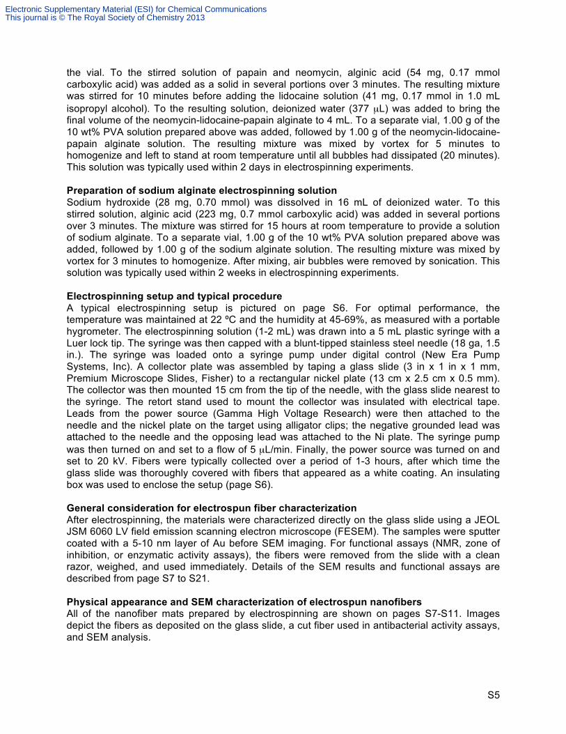
the vial. To the stirred solution of papain and neomycin, alginic acid (54 mg, 0.17 mmol carboxylic acid) was added as a solid in several portions over 3 minutes. The resulting mixture was stirred for 10 minutes before adding the lidocaine solution (41 mg, 0.17 mmol in 1.0 mL isopropyl alcohol). To the resulting solution, deionized water (377 µL) was added to bring the final volume of the neomycin-lidocaine-papain alginate to 4 mL. To a separate vial, 1.00 g of the 10 wt% PVA solution prepared above was added, followed by 1.00 g of the neomycin-lidocaine-papain alginate solution. The resulting mixture was mixed by vortex for 5 minutes to homogenize and left to stand at room temperature until all bubbles had dissipated (20 minutes). This solution was typically used within 2 days in electrospinning experiments. Preparation of sodium alginate electrospinning solution Sodium hydroxide (28 mg, 0.70 mmol) was dissolved in 16 mL of deionized water. To this stirred solution, alginic acid (223 mg, 0.7 mmol carboxylic acid) was added in several portions over 3 minutes. The mixture was stirred for 15 hours at room temperature to provide a solution of sodium alginate. To a separate vial, 1.00 g of the 10 wt% PVA solution prepared above was added, followed by 1.00 g of the sodium alginate solution. The resulting mixture was mixed by vortex for 3 minutes to homogenize. After mixing, air bubbles were removed by sonication. This solution was typically used within 2 weeks in electrospinning experiments. Electrospinning setup and typical procedure A typical electrospinning setup is pictured on page S6. For optimal performance, the temperature was maintained at 22 ºC and the humidity at 45-69%, as measured with a portable hygrometer. The electrospinning solution (1-2 mL) was drawn into a 5 mL plastic syringe with a Luer lock tip. The syringe was then capped with a blunt-tipped stainless steel needle (18 ga, 1.5 in.). The syringe was loaded onto a syringe pump under digital control (New Era Pump Systems, Inc). A collector plate was assembled by taping a glass slide (3 in x 1 in x 1 mm, Premium Microscope Slides, Fisher) to a rectangular nickel plate (13 cm x 2.5 cm x 0.5 mm). The collector was then mounted 15 cm from the tip of the needle, with the glass slide nearest to the syringe. The retort stand used to mount the collector was insulated with electrical tape. Leads from the power source (Gamma High Voltage Research) were then attached to the needle and the nickel plate on the target using alligator clips; the negative grounded lead was attached to the needle and the opposing lead was attached to the Ni plate. The syringe pump was then turned on and set to a flow of 5 µL/min. Finally, the power source was turned on and set to 20 kV. Fibers were typically collected over a period of 1-3 hours, after which time the glass slide was thoroughly covered with fibers that appeared as a white coating. An insulating box was used to enclose the setup (page S6). General consideration for electrospun fiber characterization After electrospinning, the materials were characterized directly on the glass slide using a JEOL JSM 6060 LV field emission scanning electron microscope (FESEM). The samples were sputter coated with a 5-10 nm layer of Au before SEM imaging. For functional assays (NMR, zone of inhibition, or enzymatic activity assays), the fibers were removed from the slide with a clean razor, weighed, and used immediately. Details of the SEM results and functional assays are described from page S7 to S21. Physical appearance and SEM characterization of electrospun nanofibers All of the nanofiber mats prepared by electrospinning are shown on pages S7-S11. Images depict the fibers as deposited on the glass slide, a cut fiber used in antibacterial activity assays, and SEM analysis.
Electronic Supplementary Material (ESI) for Chemical CommunicationsThis journal is © The Royal Society of Chemistry 2013

S6
Electrospinning setup used in this study:
Power supply
Ground wire
Syringe and syringe pump
Lead to needle
Lead to Collector plate
Glass slide affixed to Ni collector plate
Electronic Supplementary Material (ESI) for Chemical CommunicationsThis journal is © The Royal Society of Chemistry 2013

S7
Sodium alginate nanofibers
Electronic Supplementary Material (ESI) for Chemical CommunicationsThis journal is © The Royal Society of Chemistry 2013

S8
Lidocaine alginate nanofibers
Electronic Supplementary Material (ESI) for Chemical CommunicationsThis journal is © The Royal Society of Chemistry 2013

S9
Neomycin alginate nanofibers
Electronic Supplementary Material (ESI) for Chemical CommunicationsThis journal is © The Royal Society of Chemistry 2013

S10
Papain alginate nanofibers
Electronic Supplementary Material (ESI) for Chemical CommunicationsThis journal is © The Royal Society of Chemistry 2013

S11
Lidocaine-papain-neomycin-alginate trifunctional nanofibers
Electronic Supplementary Material (ESI) for Chemical CommunicationsThis journal is © The Royal Society of Chemistry 2013

S12
Characterization for lidocaine functionalized nanofibers Preparation of lidocaine acetate
Lidocaine acetate was prepared as a small molecule mimic of lidocaine alginate to obtain reference 1H NMR data. Lidocaine (234 mg, 1 mmol) was added to 100 mL round bottom flask and dissolved in CH2Cl2 (10 mL). To the stirred solution, AcOH (60 µL, 1 mmol) was added at room temperature. The solution was stirred at room temperature for 5 minutes. After this time, the solvent was removed by rotary evaporation. The resulting lidocaine salt was then dried under high vacuum and analyzed directly by 1H NMR. 1H NMR (400 MHz, D2O): δ = 1.14 (6H, t, J = 7.3, 2 x CH2CH3), 1.69 (3H, s, CH3CO2
-), 1.99 (6H, s, 2 x CH3), 3.11 (4H, q, J = 7.3, 2 x CH2CH3), 4.04 (2H, s, COCH2), 6.98-7.05 (3H, m, CHAr). The spectrum is shown below: 1H NMR (400 MHz, D2O) for Lidocaine acetate:
NH
ONEt2
NH
ONH
O
O
Lidocaine
AcOH (1 equiv.)
CH2Cl2
Lidocaine acetate
Electronic Supplementary Material (ESI) for Chemical CommunicationsThis journal is © The Royal Society of Chemistry 2013

S13
Control NMR for sodium alginate / PVA: sodium alginate nanofibers were peeled from glass slide after electrospinning with forceps and cut into small pieces with a razor. 20 mg of the sodium alginate fibers where then dissolved in 600 µL D2O. The solution was left to stand for 20 minutes so that bubbles could dissipate before transferring to the NMR tube. The NMR is shown below: 1H NMR (400 MHz, D2O) for sodium alginate nanofibers
Electronic Supplementary Material (ESI) for Chemical CommunicationsThis journal is © The Royal Society of Chemistry 2013

S14
NMR analysis of lidocaine alginate nanofibers: Lidocaine alginate was analyzed by 1H NMR and compared to the spectrum of lidocaine acetate shown above to verify the presence of lidocaine in the nanofibers. To prepare the NMR sample fibers were peeled from glass slide after electrospinning with forceps and cut into small portions with a razor. 20 mg of the cut fibers were then dissolved in 600 µL D2O. The solution was left to stand for 20 minutes to allow bubbles to dissipate before transferring to the NMR tube. The peaks corresponding to lidocaine and alginate / PVA are labeled in blue and red, respectively. The chemical shifts match the control NMR for lidocaine acetate and sodium alginate. 1H NMR (400 MHz, D2O) for lidocaine alginate nanofibers
Electronic Supplementary Material (ESI) for Chemical CommunicationsThis journal is © The Royal Society of Chemistry 2013

S15
NMR analysis of the trifunctional alginate nanofibers: The lidocaine-neomycin-papain nanofibers were peeled from glass slide with forceps after electrospinning and cut into small pieces with a razor. 20 mg of the trifunctional nanofibers were then dissolved in 600 µL D2O. The solution was left to stand for 20 minutes for bubbles to dissipate before transferring to the NMR tube. Lidocaine was again verified as a constituent in the trifunctional nanofibers by comparison to the NMR for lidocaine acetate. The 1H NMR is shown below:
1H NMR (400 MHz, D2O) for neomycin-lidocaine-papain (trifunctional) nanofibers
Electronic Supplementary Material (ESI) for Chemical CommunicationsThis journal is © The Royal Society of Chemistry 2013

S16
Antibacterial activity assays for neomycin functionalized nanofibers The neomycin-alginate nanofibers and the lidocaine-neomycin-papain (trifunctional) alginate nanofiber mats were peeled from the glass slide after electrospinning with sterile forceps and then cut with a razor into 1 cm x 1 cm square pieces. As a control, sodium alginate nanofiber mats (unfunctionalized) were prepared in the same fashion. Agar source plates were prepared using EDVOTEK sterile ReadyPourTM Luira Broth. No antibiotics were used in the agar. A starter culture was prepared by placing a single E. coli (BL21 DE3) BactoBeadTM (EDVOTEK) on a source plate and allowing to dissolve (10 mintues, room temperature). After the E. coli BactoBead had dissolved, primary and secondary streaks were made and the culture was left to grow for 24 hours at 25 ºC. After this time all colonies in a 2 cm2 area were transferred to sterile liquid growth media (2 mL). The optical density of this cell suspension was measured to be 1.0 (OD600 nm). This suspension of E. coli was then used to inoculate 18 agar plates used in zone of inhibition experiments. In the inoculation, 100 µL of the cell suspension was added to the center of the agar plate by sterile pipette. The cell solution was then streaked over an area of approximately 3 cm2. Electrospun fibers were placed on the inoculated plates 10 minutes after the inoculation using sterile forceps. The area occupied by the nanofibers was 1 cm2. The mass of the nanofiber samples were 2.5 mg or 7.5 mg, as indicated below. All samples were run in triplicate. The sodium alginate nanofibers typically dissolved completely over the course of 3 hours. The nanofibers functionalized with neomycin typically dissolved more slowly over the course of 24 hours. The plates were incubated at room temperature for a total of 24 hours after the nanofibers were added. At the end of this incubation period, the zone of inhibition was measured. Zone of inhibition was defined in these assays as the largest diameter of a circular area that contained no visible confluence of E. coli. Photographs of the plates after the 24 hour incubation are shown on pages S17-S18. Note that no inhibition was observed for control experiments where either no nanofibers were used (Samples 1-3) or the nanofibers were made up of only sodium alginate and PVA (Samples 4-9). In these controls, E. coli were observed to grow to confluence over the area of inoculation and over the area of the fibers. In contrast, all nanofibers containing neomycin (Samples 10-18) resulted in a significant zone of inhibited E. coli. growth. A summary of the measured zones of inhibition are tabulated on page S19.
Electronic Supplementary Material (ESI) for Chemical CommunicationsThis journal is © The Royal Society of Chemistry 2013

S17
Samples 1-3: Control: E. coli only (no nanofibers)
Samples 4-6: Control: sodium alginate nanofibers (2.5 mg, single layer of 1 cm2 fibers)
Samples 7-9: Control: sodium alginate nanofibers (7.5 mg, three layers of 1 cm2 fibers)
Electronic Supplementary Material (ESI) for Chemical CommunicationsThis journal is © The Royal Society of Chemistry 2013

S18
Samples 10-12: Neomycin alginate nanofibers (2.5 mg, single layer of 1 cm2 fibers)
Samples 13-15: Neomycin-papain-lidocaine alginate (2.5 mg, single layer of fibers)
Samples 16-18: Neomycin-papain-lidocaine alginate (7.5 mg, three layers of fibers)
Electronic Supplementary Material (ESI) for Chemical CommunicationsThis journal is © The Royal Society of Chemistry 2013

S19
Table S1: Zone of inhibition data (mm) for neomycin containing nanofibers
Sample Description Zone of inhibition (mm)
1-3 E. coli only -
4-6 Sodium alginate (2.5 mg fibers) -
7-9 Sodium alginate (7.5 mg fibers) -
10 Neomycin alginate 28
11 Neomycin alginate 30
12 Neomycin alginate 29
Average 29 (± 1) mm
13 Trifunctional (2.5 mg fibers) 16
14 Trifunctional (2.5 mg fibers) 15
15 Trifunctional (2.5 mg fibers) 13
Average 14.7 (± 1.5) mm
16 Trifunctional (7.5 mg fibers) 24
17 Trifunctional (7.5 mg fibers) 23
18 Trifunctional (7.5 mg fibers) 19
Average 22 (± 2.6) mm
Enzymatic assays for nanofibers containing papain Determination of papain concentration in papain alginate nanofibers Three 10 mg samples of papain-alginate monofunctionalized fibers were placed into three separate microcentrifuge tubes. To each tube was then added 1000 µL of 50 mM Na2HPO4 buffer (pH 7.5). Each sample was mixed by vortex for three minutes to dissolve the fibers. Another sample of 10 mg of sodium alginate fibers (unfunctionalized) was prepared in the same fashion. The solutions were then transferred to 1.5 mL cuvettes, and an absorbance spectrum of the papain samples was taken from 270 to 290 nm using the sodium alginate fibers as a protein-free reference. The concentration of protein in each sample was calculated using the absorbance at 280 nm and the extinction coefficient of papain at that wavelength (εpapain = 57600 M-1 cm-1 at 280 nm). The average concentration of these papain alginate samples was 15.0 ± 1.1 µM.
Electronic Supplementary Material (ESI) for Chemical CommunicationsThis journal is © The Royal Society of Chemistry 2013

S20
Enzymatic activity assay for papain alginate nanofibers The three papain alginate samples prepared above were used directly to assay for enzymatic activity. To each of these samples was first added 200 µL of 50 mM Na2HPO4 buffer solution (pH = 7.5). Next, dithiothreitol (DTT) was added as a solid (10 mg) and allowed to dissolve in the cuvette. Finally, 300 µL of a chromogenic papain substrate solution (pGluPheLeu-p-nitroanilide, Sigma-Aldrich P3169, prepared at 500 µM in DMSO) was added to the cuvette and the progress of the reaction was monitored by absorbance at 410 nm over 20 minutes. A reference solution of 1200 µL buffer and 300 µL DMSO was used in this experiment. This activity assay was carried out in triplicate. As a positive control, the same assay was also run in triplicate with native papain at the same protein concentration. As a negative control, the same assay was run in triplicate using 10 mg of sodium alginate fibers that do not contain papain. Results are plotted below using the average absorbance of the triplicate runs. Error bars indicate ± 1 standard deviation at each data point. The first data point was obtained at 60 seconds of reaction time. This assay indicates that the papain present in the papain-alginate fibers is enzymatically active. Furthermore, while the activity is less than than native papain at the same concentration, this experiment indicates that the protease is still largely compatible with the processing required in the nanofiber synthesis. The negative control (sodium alginate nanofibers) resulted in no increase in absorbance at 410 nm, indicating that the production of p-nitroaniline requires papain. Note that the cuvettes containing papain and papain alginate nanofibers became visibly yellow over the course of the reaction while the sodium alginate samples remained clear.
0"
0.1"
0.2"
0.3"
0.4"
0.5"
0.6"
0.7"
0.8"
0.9"
1"
1" 2" 3" 4" 5" 6" 7" 8" 9" 10" 11" 12" 13" 14" 15" 16" 17" 18" 19" 20"
Absorban
ce*units*(4
10*nm)*
Time*(minutes)*
Enzyma8c*assay*of*papain*alginate*nanofibers*
Na/ve"Papain"
Papain"alginate"nanofibers"
Sodium"alginate"nanofibers"
Electronic Supplementary Material (ESI) for Chemical CommunicationsThis journal is © The Royal Society of Chemistry 2013

S21
Enzymatic activity assay for lidocain-neomycin-papain (trifunctional) alginate nanofibers 10 mg of neomycin-lidocaine-papain alginate (trifunctionalized) nanofibers were dissolved in 1000 µL of 50 mM NaH2PO4 buffer (pH 7.5) followed by 10 mg of dithiothreitol (DTT). The DTT was allowed to dissolve and then the resulting solution was transferred to a 1.5 mL cuvette followed by 200 µL of the same buffer. 300 µL of the papain substrate (pGluPheLeu-p-nitroanilide, Sigma-Aldrich P3169, prepared at 500 µM in DMSO) was added to the cuvette, and the absorbance was monitored over 20 minutes. This assay was run in triplicate. As a negative control, the same assay was run in triplicate using 10 mg of sodium alginate fibers that do not contain papain. Results are plotted below using the average absorbance of the triplicate runs. Error bars indicate the standard error at each data point. The first data point was obtained at 120 seconds of reaction time. The increase in absorbance at 410 nm indicates the formation of p-nitroanilide by reaction of papain with the peptide substrate. Note that the cuvettes containing trifunctional nanofibers became visibly yellow over the course of the reaction while the sodium alginate samples remained clear. Therefore, the trifunctional nanofibers contain enzymatically active papain. The data is shown below.
0"
0.01"
0.02"
0.03"
0.04"
0.05"
0.06"
0.07"
0.08"
0.09"
0.1"
2" 3" 4" 5" 6" 7" 8" 9" 10" 11" 12" 13" 14" 15" 16" 17" 18" 19" 20"
Absorban
ce*units*(4
10*nm)*
Time*(minutes)*
Enzyma8c*assay*of*lidocain<neomycin<papain*trifunc8onal*nanofibers*
Lidocaine5neomycin5papain"trifunc=onal"nanofibers"
Sodium"alginate"nanofibers"
Electronic Supplementary Material (ESI) for Chemical CommunicationsThis journal is © The Royal Society of Chemistry 2013

S22
Controlled dissolution of lidocaine-alginate electrospun mats Preliminary assessment: Lidocaine-alginate electrospun mats were prepared as described on page S2. The fibrous mats were removed from the glass slide and cut into 1.0 cm2 squares. 50 mg of these square mats were layered together and then placed directly on an agar plate. The mat was imaged over the course of 24 hours. The images at 0, 12, and 24 hours are shown below. Note that the fibers dissolved to some extent but do not completely dissolve over the 24 hour period.
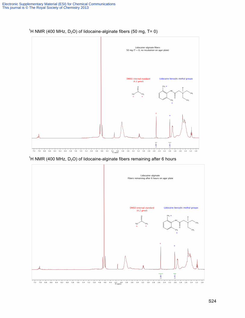
T = 0 hr T = 12 hr T = 24 hr Release profile of lidocaine from lidocaine-alginate electrospun mats The lidocaine-alginate electrospun mats were prepared as described in the previous experiment. Accordingly, the cut fibrous mats were layered into 50 mg samples and then placed on four separate agar plates to monitor both the dissolution of the fibers and lidocaine release. After 6 hours, the undissolved mat on plate 1 was removed with forceps and dried under vacuum for 1 hour and then weighed. This material then analyzed by 1H NMR: the mat was first dissolved in 600 µL of D2O and then 0.3 µL of non-deuterated DMSO (0.0042 mmol) was added by micropipette as an internal standard. Vigorous stirring was required to completely dissolve the recovered lidocaine-alginate mat. The amount of lidocaine was calculated using the relative integration values for the DMSO methyl groups and the benzylic methyl groups on lidocaine. This analysis—measurement of the mass of the undissolved mat and remaining lidocaine—was carried out 12, 18, and 24 hours for plates 2, 3, and 4, respectively. The same 1H NMR analysis was carried out with 50 mg of lidocaine-alginate fibers directly to determine the lidocaine content at t = 0 hours. The results are tabulated below on the following pages, along with NMR spectra.
Electronic Supplementary Material (ESI) for Chemical CommunicationsThis journal is © The Royal Society of Chemistry 2013

S23
Table S2: Dissolution of lidocaine-alginate on agar plate and release of lidocaine
Lidocaine release profile for 50 mg lidocaine-alginate fibers on agar plate:
Time on agar plate (hr) Undissolved Fiber Mass (mg) Lidocaine (µmol by NMR)0 50 7.76 33 6.0
12 32 4.818 29 4.424 21 3.3
0.0#
1.0#
2.0#
3.0#
4.0#
5.0#
6.0#
7.0#
8.0#
9.0#
10.0#
0# 6# 12# 18# 24#
Lido
caine)remaining)on)fib
ers)(µmol))
Time)on)agar)plate)(hours))
Lidocaine)release)profile))
Electronic Supplementary Material (ESI) for Chemical CommunicationsThis journal is © The Royal Society of Chemistry 2013
S24
1H NMR (400 MHz, D2O) of lidocaine-alginate fibers (50 mg, T= 0)
1H NMR (400 MHz, D2O) of lidocaine-alginate fibers remaining after 6 hours
�������������������������������������������������������������������������������������������� ����
����
����
���
�
�
���
����������������������
���������
!���"�����#��$%��"����&%��'��(��
))
!���"����*��'������+#���
���'� ,�-��.������"(#����������'���������
) )
)
)���
���
��
�
��
���
���
�
�������������������������������������������������������������������������������������������� ����
����
����
���
�
�
���
����������������������
���������
!���"�����#��$%��"����&%��'��(��
))
!���"����*��'������
+�#������������'���������&�(�������'��������
) )
)
)���
���
��
�
��
���
���
�
Electronic Supplementary Material (ESI) for Chemical CommunicationsThis journal is © The Royal Society of Chemistry 2013

S25
1H NMR (400 MHz, D2O) of lidocaine-alginate fibers remaining after 12 hours
1H NMR (400 MHz, D2O) of lidocaine-alginate fibers remaining after 18 hours
�������������������������������������������������������������������������������������������� ����
����
����
���
�
�
���
����������������������
���������
!���"�����#��$%��"����&%��'��(��
))
!���"����*��'������
+�#������������'����������&�(�������'��������
) )
)
)���
���
��
�
��
���
���
�
�������������������������������������������������������������������������������������������� ����
����
����
���
�
�
���
����������������������
���������
!���"�����#��$%��"����&%��'��(��
))
!���"����*��'������
+�#������������'����������&�(�������'��������
) )
)
)���
���
��
�
��
���
���
�
Electronic Supplementary Material (ESI) for Chemical CommunicationsThis journal is © The Royal Society of Chemistry 2013

S26
1H NMR (400 MHz, D2O) of lidocaine-alginate fibers remaining after 24 hours
�������������������������������������������������������������������������������������������� ����
���
����
���
�
�
���
����������������������
���������
!���"�����#��$%��"����&%��'��(��
)
)
!���"����*��'������
+�#������������'����������&�(�������'��������
) )
)
)���
���
��
�
��
���
���
�
Electronic Supplementary Material (ESI) for Chemical CommunicationsThis journal is © The Royal Society of Chemistry 2013

S27
Loading efficiency of lidocaine onto alginic acid
Alginic acid (14 mg, 0.044 mmol carboxylic acid) was added to a 1.5 mL microcentrifuge tube and suspended in 1.0 mL of D2O. Lidocaine (10 mg, 0.043 mmol) was added as a solid and the mixture was stirred at room temperature for 45 minutes. All material dissolved readily in this time period to provide lidocaine-alginate. A 0.3 µL aliquot of DMSO (0.0042 mmol) was added to the reaction mixture as an internal standard and the resulting solution was analyzed directly by 1H NMR. The amount of lidocaine in the NMR sample was calculated using the relative integration values for the DMSO methyl groups and the benzylic methyl groups on lidocaine. The relative integration was measured to be 9.97 lidocaine to 1.00 DMSO, indicating 0.042 mmol of lidocaine reacted with alginic acid to form lidocaine alginate. This is a 98% conversion of alginic acid to lidocaine-alginate. 1H NMR data is shown below.
OHOO
OHOH
OO
O
OHO
OH
OH
OLidocaine
m n
OHOO
OHO
OO
O
O
OOH
OH
O
m nR-NH3
RH3N
Alginic acid
D2O
98% conversion
HN
ONH
R-NH3 =
�������������������������������������������������������������������������������������������� ����
���
����
���
�
�
���
����������������������
������ ��!
"�� #�����$��%&��#����'&��(� )��
*
*
"�� #����+��(�����
, ������� �����#�� �� ����� #�����-��'���(���#��#��
* *
*
*���
���
��
�
��
���
���
�
Electronic Supplementary Material (ESI) for Chemical CommunicationsThis journal is © The Royal Society of Chemistry 2013

S28
Lidocaine loading efficiency onto sodium alginate
Sodium hydroxide (2.0 mg, 0.050 mmol) was added to 1 mL of D2O in a 1.5 mL microcentrifuge tube. Alginic acid (14 mg, 0.044 mmol carboxylic acid) was added to this solution and stirred to dissolve, forming sodium alginate. Once all material was dissolved, lidocaine (10 mg, 0.043 mmol) was added to the solution as a solid. The mixture was stirred at room temperature for 45 minutes. The majority of the lidocaine did not dissolve and was visible as a suspension in the mixture. A 0.3 µL aliquot of DMSO (0.0042 mmol) was added to the reaction mixture as an internal standard. The solution was analyzed directly by 1H NMR, and care was taken not to transfer undissolved solids to the NMR tube. In the 1H NMR spectrum, lidocaine was observed, but the chemical shift was consistent with free lidocaine, rather than the protonated amine. The amount of lidocaine in the NMR sample was calculated using the relative integration values for the DMSO methyl groups and the benzylic methyl groups on lidocaine. The relative integration was measured to be 1.59 lidocaine to 1.00 DMSO, indicating 0.0067 mmol of lidocaine in solution. This result indicates that only 16% of lidocaine is in solution. It is likely that this 16% is the amount of lidocaine that is soluble in the solution under these conditions. There is no evidence for formation of lidocaine-alginate starting from sodium alginate. Lidocaine-alginate is therefore most efficiently formed by reaction with alginic acid through a simple acid-base reaction. 1H NMR data is shown below.
OHOO
OHO
OO
O
O
OOH
OH
O
m n
Na
Na
Sodium alginate
LidocaineD2O O
HOO
OHO
OO
O
O
OOH
OH
O
m n
Na
Na
HN
ON
No reaction, 16% of the neutral lidocaine dissolved into D2O
+
�������������������������������������������������������������������������������������������� ����
���
����
���
�
�
���
����������������������
������ ��!
"�� #�����$��%&��#����'&��(� )��
���
���
��
�
�
���
���
* *
* *
*
*
� ��)�+��(�����������������������#�� ��,��'���� #����
Electronic Supplementary Material (ESI) for Chemical CommunicationsThis journal is © The Royal Society of Chemistry 2013