Embed Size (px)
Citation preview

Page 1/16
Effect of Interleukin-1 Blockade with Anakinra onLeukocyte Count in Patients with ST-SegmentElevation Acute Myocardial InfarctionMARCO GIUSEPPE DEL BUONO
Virginia Commonwealth UniversityJuan Ignacio Damonte
Virginia Commonwealth UniversityCory R Trankle
Virginia Commonwealth UniversityDinesh Kadariya
Virginia Commonwealth UniversitySalvatore Carbone
Virginia Commonwealth UniversityGeorgia K Thomas
Virginia Commonwealth UniversityJeremy Turlington
Virginia Commonwealth UniversityRoshanak Markley
Virginia Commonwealth UniversityJustin Canada
Virginia Commonwealth UniversityGiuseppe Biondi-Zoccai
Sapienza University of RomeMichael C. Kontos
Virginia Commonwealth UniversityBenjamin W. Van Tassell
Virginia Commonwealth UniversityAntonio Abbate ( [email protected] )
Virginia Commonwealth University
Research Article
Keywords: leukocyte, STEMI, neutrophils, anakinra, IL-1, cytokine, leukopoiesis, in�ammation

Page 2/16
Posted Date: October 15th, 2021
DOI: https://doi.org/10.21203/rs.3.rs-959200/v1
License: This work is licensed under a Creative Commons Attribution 4.0 International License. Read Full License

Page 3/16
AbstractBackground. Leukocytosis is a common �nding in patients with ST elevation myocardial infarction(STEMI) and portends a poor prognosis. Interleukin 1-b regulates leukopoiesis and pre-clinical studiessuggest that anakinra (recombinant human interleukin-1 [IL-1] receptor antagonist) suppressesleukocytosis in myocardial infarction. However, the effect of IL-1 blockade with anakinra on leukocytecount in patients with STEMI is unknown.
Methods. We reviewed the white blood cell (WBC) and differential count of 99 patients enrolled in aclinical trial of anakinra (n=64) versus placebo (n=35) for 14 days after STEMI. A complete blood cellcount with differential count were obtained at admission, and after 72 hours, 14 days and 3 months.
Results. After 72 hours from treatment, anakinra compared to placebo led to a statistically signi�cantgreater percent reduction in total WBC count (-35% [-48 to -24] vs -21% [-34 to -10], P=0.008), absoluteneutrophil count (-48% [-60 to -22] vs -27% [-46 to -5], P=0.004) and to an increase in absolute eosinophilcount (+50% [0 to +100] vs 0% [-50 to +62], P=0.022). These changes persisted while on treatment at 14days and were no longer apparent at 3 months after treatment discontinuation.
Conclusion. We found that in patients with STEMI IL-1 blockade with anakinra accelerates resolution ofleukocytosis and neutrophilia. This modulation may represent one of the mechanisms by which IL-1blockade improves clinical outcomes.
IntroductionST-segment–elevation myocardial infarction (STEMI) is a leading cause of morbidity and mortalityworldwide. Reperfusion strategies have revolutionized its treatment by reducing infarct size andimproving survival (1). However, an in�ammatory response is triggered by acute ischemia and ampli�edby the reperfusion, which contributes to cause further injury despite being essential for tissue healing (2).Elevated leukocyte count is a marker of in�ammation and is associated with greater infarct size, reducedsystolic function, and adverse clinical outcomes in patients with STEMI (3-6). In response to ischemicinjury, the number of neutrophils in the blood increases rapidly and begin to appear in the infarcted tissuewithin hours. While neutrophils contribute to the clearance of pathogens or debris as well as coordinatethe monocyte-derived macrophage in�ltration that activate reparative pathways necessary for scarformation (7-9), perturbation of this �nely regulated balance may lead to an exuberant and prolongedin�ammatory state that acutely worsens the infarct size and chronically contributes to post-myocardialinfarction adverse remodeling (10-13)
Interleukin 1-β regulates leukopoiesis favoring a sustained neutrophil production in the bone marrow (8).In this regard, pre-clinical studies reported that anakinra (recombinant human interleukin-1 [IL-1] receptorantagonist) suppressed leukocytosis in mouse models of myocardial infarction suggesting its potentialrole in suppressing the leukopoiesis in the bone marrow and neutrophil recruitment within the heart (14).

Page 4/16
Data on the role IL-1β in patients with STEMI are limited. In a small pilot trial of anakinra in 10 patientswith STEMI, patients treated with anakinra had signi�cantly lower leukocyte and neutrophil counts 24hours after the �rst injection compared to placebo-treated patients (15), and leukocytes from anakinra-treated patients had reduced production of pro-in�ammatory cytokines (16). In this study we describe theeffect of IL-1 blockade on leukocyte count in patients with STEMI treated with one of two different dosesof anakinra in the setting of a randomized double-blinded clinical trial.
Methods
Study designIn the Virginia Commonwealth University Anakinra Remodeling Trial (VCUART) 3 (www.clinicaltrials.govNCT01950299) (17,18), 99 patients were randomly assigned to receive anakinra 100 mg once daily(standard dose), alternating with placebo every 12 hours, for 14 days; anakinra, 100 mg twice daily, every12 hours (high dose) for 14 days; or placebo twice daily every 12 hours for 14 days, with the �rst doseadministered within 12 hours of coronary angiography. Patients were excluded from the study if they hadcontraindications to treatment with anakinra, chronic in�ammatory or infectious disease, or preexistingstructural or functional severe cardiac abnormalities. For the purpose of this analysis, we pooled the twoanakinra arms together, as previously presented for the clinical outcomes (17).
Laboratory dataThe methods have been described in detail elsewhere (17,18). A differential blood cell count wasobtained at admission, and after 72 hours, 14 days and 3 months. Total white blood cell (WBC) count andneutrophil, lymphocyte, monocyte and eosinophil counts were calculated using a hematology analyzer.Neutrophil to lymphocyte ratio, calculated as total neutrophil counts divided by total lymphocyte counts,was computed from the absolute values of neutrophils and lymphocyte (17,18).
Statistical analysisThe methods have been described in detail elsewhere (17,18). Descriptive statistics were used to describebaseline and clinical characteristics of the patients. Categorical variables are presented as frequency(percentage) and compared using Chi-Square or Fisher's exact test as appropriate. Continuous variablesare presented as median [interquartile range, IQR] and compared using Mann–Whitney U test orSpearman’s rank test for correlations. There were no differences in the missing data between the groups;no missing data imputation was used. All analyses were completed using SPSS, version 24.0 (SPSS;Chicago, IL) with signi�cance set at α = 0.05 (17,18).
Regulatory dataAll methods were carried out in accordance with relevant guidelines and regulations. All experimentalprotocols were approved by the Virginia Commonwealth University Institutional Review Board. All
Page 5/16
subjects provided written informed consent to be part of the trial in accordance with the VirginiaCommonwealth University Institutional Review Board.
Results
Baseline characteristicsBaseline characteristics of the 99 patients have been previously reported (17) and are summarized inTable 1. Patients were predominantly male (n=80, 81%) with a median age of 55 [49.0–62.0] years.Patients were randomized to anakinra standard dose daily (n=33, [33%]), anakinra high dose (n=31,[31%]), or placebo (n=35, [35%]). Clinical characteristics were well matched, without statisticallysigni�cant differences between anakinra and placebo groups except for a higher prevalence of diabetesmellitus in the placebo compared to anakinra group (15 [43%] vs 15 [23%], p=0.044). There were nodifferences between the two groups in time from symptom onset to percutaneous coronary intervention,time from symptom onset to investigational drug administration, or periprocedural drugs received. Therewas no difference in infarct size between the two groups, while the under the curve for high sensitivity C-Reactive Protein (CRP) was signi�cantly lower in patients receiving anakinra versus placebo (67 [39–120]vs 214 [131–394] mg·day/L, p<0.001) (Table 2).

Page 6/16
Table 1Clinical characteristics of the patients in anakinra and placebo groups.
Anakinra
(n=64)
Placebo
(n=35)
P-value
Age, y 55 [48-61] 56 [51-65] 0.174
Female sex 14 (22) 5 (4) 0.359
White 36 (56) 21 (60) 0.223
Black 21 (33) 6 (17)
Hispanic 2 (3) 3 (9)
Other 5 (8) 5 (14)
Procedural characteristics
Symptom onset to PCI, min 187 [106-333]
180 [130-347]
0.801
Symptom onset to investigational drugadministration, min
508 [348-718]
529 [403-716]
0.669
Fibrinolytic use before PCI 5 (8) 3 (9) 0.587
PCI type
Primary PCI 59 (92) 32 (91) 0.587
PCI after �brinolysis 5 (8) 3 (9) 0.587
Use of drug-eluting-stent 44 (69) 30 (86) 0.063
Use of thrombectomy 10 (16) 6 (17) 0.844
Use of P2Y12 inhibitor 64 (100) 35 (100) 1
Clopidogrel 9 (14) 7 (20)
Prasugrel 22 (34) 12 (34)
Ticagrelor 33 (52) 15 (46)
Clinical characteristics
Coronary artery disease 14 (22) 7 (20) 0.827
Diabetes mellitus 15 (23) 15 (43) 0.044
Systemic arterial hypertension 33 (52) 23 (66) 0.174
Abbreviations: STEMI: ST elevation myocardial infarction; PCI: percutaneous coronary intervention;Min: minutes; LVEF: left ventricle ejection fraction.
Page 7/16
Anakinra
(n=64)
Placebo
(n=35)
P-value
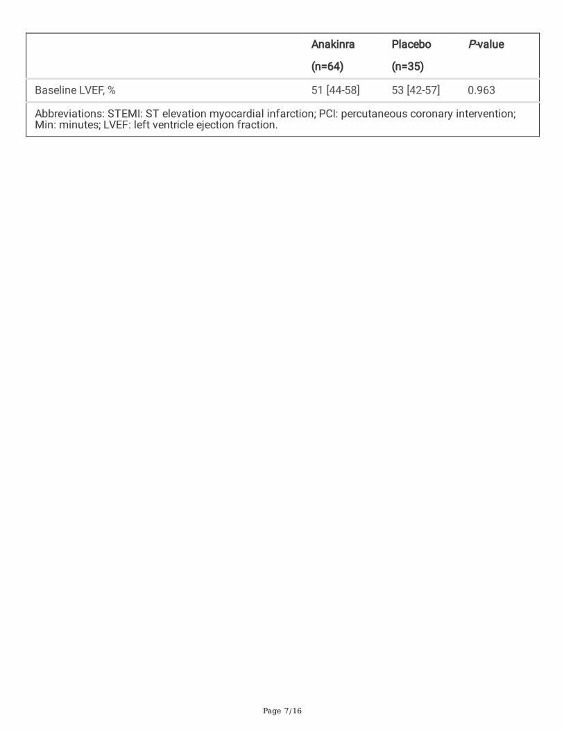
Baseline LVEF, % 51 [44-58] 53 [42-57] 0.963
Abbreviations: STEMI: ST elevation myocardial infarction; PCI: percutaneous coronary intervention;Min: minutes; LVEF: left ventricle ejection fraction.

Page 8/16
Table 2Laboratory data according to anakinra and placebo.
Anakinra
(n=64)
Placebo
(n=35)
P-value
CKMB-AUC, ng/mL*d 2219 [1130-3821] 2351 [765-4668] 0.859
At admission
Hemoglobin, g/dL 14.5 [13.4-15.3] 14.4 [13.6-15.6] 0.692
Hematocrit, % 43 [40-47] 42 [41-44] 0.558
White blood cell, 109/L 10.85 [8.52-13.90] 11.40 [9.20-15.07] 0.692
Absolute neutrophil count, 109/L 7.45 [4.72-11.05] 7.30 [5.20-12.50] 0.622
Absolute lymphocyte count, 109/L 2.05 [1.3-3.02] 1.8 [1.30-2.55] 0.699
Absolute monocyte count, 109/L 0.60 [0.40-0.80] 0.70 [.50-0.80] 0.185
Absolute eosinophil count, 109/L 0.10 [0.00-0.125] 0.10 [0.00-0.20] 0.279
Neutrophil to lymphocyte ratio 4.29 [1.99-7.51] 3.71 [2.08-8.37] 0.719
Creatinine, mg/dL 0.94 [0.78-1.10] 1.00 [0.89-1.34] 0.063
NTproBNP, pg/mL 52.50 [22.00-217.59] 95.5 [23.75-244.00] 0.614
At 72 hours
White blood cell, 109/L 7.50 [6.20-8.35] 8.30 [7.20-9.80]
% Change from baseline -35% [-48 to -24] -21% [-34 to -10] 0.008
Absolute neutrophil count, 109/L 4.15 [3.22-5.07] 5.40 [4.65-5.40]
% Change from baseline -48% [-60 to -22] -27% [-46 to -5] 0.004
Absolute lymphocyte count, 109/L 2.15 [1.50-2.60] 1.80 [1.40-2.20]
% Change from baseline -7% [-4% to +32] -6% [-38 to +22] 0.657
Absolute monocyte count, 109/L 0.60 [0.50-0.80] 0.80 [0.60-0.95]
% Change from baseline +11% [-29 to +51] +14% [-13 to + 47] 0.604
Absolute eosinophil count, 109/L 0.20 [0.10-0.30] 0.10 [0.10-0.30]
% Change from baseline +50% [0 to +100] 0% [-50 to +62] 0.022
Neutrophil to lymphocyte ratio 1.90 [1.37-3.22] 3.35 [2.6

Page 9/16
Anakinra
(n=64)
Placebo
(n=35)
P-value
% Change from baseline -54% [-13 to +70] -13% [-58 to +70] 0.047
At 14 days
White blood cell, 109/L 7.10 [5.70-9.40] 8.60 [6.92-10.5]
% Change from baseline -33% [-45 to – 22] -20% [-41 to -9] 0.044
Absolute neutrophil count, 109/L 4.60 [3.30-5.50] 5.50 [4.00-7.15]
% Change from baseline -42% [-61 to -25] -32% [-51% to -1] 0.067
Absolute lymphocyte count, 109/L 2.10 [1.50-2.60] 2.00 [1.70-2.27]
% Change from baseline 0% [-32 to +32] +10% [-22 to 40] 0.313
Absolute monocyte count, 109/L 0.60 [0.50-0.80] 0.65 [0.50-0.70]
% Change from baseline -5% [-30 to + 26] 0% [-20 to + 3] 0.989
Absolute eosinophil count, 109/L 0.20 [0.10-0.30] 0.20 [0.20-0.30]
% Change from baseline +100% [0 to + 200] 0% [-30 to +100] 0.043
Neutrophil to lymphocyte ratio 2.26 [1.34-3.25] 2.89 [1.93-3.68]
% Change from baseline -50% [-67 to 10] -36% [-65 to +3] 0.500
At 3 months
White blood cell, 109/L 6.90 [5.90-8.60] 7.55 [6.05-9.47]
% Change from baseline -41% [-49 to -16] -30% [-44 to -19] 0.301
Absolute neutrophil count, 109/L 4.20 [3.30-5.20] 4.35 [3.50-5.70]
% Change from baseline -53% [-61 to -25] - 43% [-54 to -33] 0.298
Absolute lymphocyte count, 109/L 2.10 [1.50-2.60] 2.00 [1.57-2.30]
% Change from baseline -3% [-29 to + 35] +8% [-28 to + 59] 0.584
Absolute monocyte count, 109/L 0.60 [0.50-0.70] 0.65 [0.50-0.70]
% Change from baseline -10% [-28 to + 24] 7% [-30 to + 25] 0.748
Absolute eosinophil count, 109/L 0.20 [0.10-0.20] 0.20 [0.10-0.32]
% Change from baseline 0% [0 to 100] +33% [o to 100%] 0.764

Page 10/16
Anakinra
(n=64)
Placebo
(n=35)
P-value
Neutrophil to lymphocyte ratio 2.09 [1.35-2.85] 2.61 [1.73-3.24]
% Change from baseline -51% [-70 to 17] - 42% [-75 to -9] 0.964
Data are expressed as median [interquartile range]. P-values in bold character indicate signi�cantvalues (<0.05) for difference between groups. Abbreviations: CKMB-AUC: Creatine kinase-MB areaunder the curve; CRP-AUC: C-reactive protein area under the curve; NTproBNP: N-terminal pro-brainnatriuretic peptide.
White Blood Cell Count and Differential Count during STEMITable 2 shows WBC count with differential and other laboratory parameters in the two groups. There wereno signi�cant differences in any of the laboratory parameters at baseline. A signi�cant reduction in WBC(11.40 [9.20-15.07] vs 8.30 [7.20-9.80] 109/L; -21% [-34 to -10], P<0.001 within placebo group) andneutrophil count (7.30 [5.20-12.50] vs 5.40 [4.65-5.40] 109/L; -27% [-46 to -5], P=0.003 within placebogroup) were seen in the placebo group at 72 hours (Table 2).
Effect of Treatment with Anakinra on White Blood CountAt 72 hours, when compared with placebo, treatment with anakinra led to a statistically signi�cant greaterpercentage reduction in total WBC count (-35% [-48 to -24] vs -21% [-34 to -10], P<0.001 within anakinragroup, and P=0.008 for between groups differences) (Figure 1, Table 2). A signi�cantly greater percentagereduction in WBC count persisted in the anakinra group compared to placebo group at 14 days, while ontreatment, and it was no longer seen at 90 days following discontinuation of treatment (Figure 1, Table2).
Effect of Treatment with Anakinra on Leukocyte DifferentialCountAt 72 hours, when compared with placebo, treatment with anakinra led to a statistically signi�cant greaterpercentage reduction in absolute neutrophil count (-48% [-60 to -22] vs -27% [-46 to -5], P<0.001 withinanakinra group, and P=0.004 for between groups differences), and neutrophil to lymphocyte ratio (-54%[-13 to +70] vs -13% [-58 to +70], P<0.001 within anakinra group, and P=0.047 for between groupsdifferences), and to an increase in absolute eosinophil count (+50% [0 to +100] vs 0% [-50 to +62],P<0.001 within anakinra group, and P=0.022 for between groups differences)(Figure 1, Table 2). Asigni�cantly greater percentage increase in absolute eosinophil count persisted in the anakinra group
Page 11/16
compared to placebo group at 14 days, while on treatment, and it was no longer seen at 90 daysfollowing discontinuation of treatment (Figure 1, Table 2).
DiscussionWe herein report, for the �rst time in literature, that IL-1 blockade with anakinra leads to a signi�cantlygreater reduction of leukocyte count in patients with STEMI, and that it drives a relative greater reductionin neutrophils and an increase in eosinophils. Considered a surrogate for the in�ltration of WBC intonecrotic tissue in response to ischemia and reperfusion, leukocytosis is a common �nding in patientswith acute STEMI and portends a poor prognosis (3-6,19-21). Leukocytosis is considered a surrogate forthe in�ltration of WBC into necrotic tissue in response to ischemia and reperfusion. Neutrophils are the�rst cells to arrive in the infarcted tissue attracted by the cellular debris and damage-associatedmolecular patterns generated by the necrotic cells (7-9). Upon arrival, leukocytes become activated andgenerate reactive oxygen species and proteolytic enzymes – thereby further expanding myocardial injury(7-9). Recently, Sreejit and colleagues showed that neutrophils play a key role in determining the natureand orchestrating the in�ammatory response in the heart (9). Once recruited in the myocardium, activatedneutrophils may release various proteins that prime the Nod-Like-Receptor (NLR) family Pyrin Domain-Containing 3 (NLRP3) in�ammasome—a multi-molecular platform crucial to induction of thein�ammatory response to cellular danger—on naïve neutrophils and stimulate them to produce IL-1βlocally. This local production interacts with the IL-1 receptor type I (IL-1RI) on hematopoietic stem cells inthe bone marrow, stimulating myelopoiesis in a cell-intrinsic manner and amplify the granulopoiesis (9).Unopposed IL-1 activity during myocardial infarctions mobilizes myeloid cells from bone marrow to theinfarction site inducing pathological myocardial healing and favoring cardiac rupture in experimentalmodels (5,20). During acute myocardial infarction, an increased expression of the IL-1RI was found,suggesting that the upregulation of IL-1RI may facilitate neutrophil proliferation and differentiation (9).Furthermore, IL-1 stimulates the calcium-dependent degranulation of neutrophils and release of proteases(e.g. cathepsin G, elastase, and proteinase) that may contribute to the proteolytic break down of necroticmyocytes and extracellular matrix, and to cleave and activate multiple IL-1 family members (22). Of note,IL-1 is su�cient to induce a cardiomyopathy phenotype in the mouse (23).
From a translational point of view, in the VCUART phase II clinical trial program that included 3 studies(n=139) (15,17,24,25) of patients with STEMI treated with anakinra resulted in a signi�cant improvementin left ventricular performance and a signi�cant reduction of new-onset heart failure and of heart failurehospitalization versus placebo. Similarly, the CANTOS trial enrolling patients with previous acutemyocardial infarction and persistent in�ammation found that canakinumab (an IL-1β blocker) resulted ina reduction in the hospitalizations for heart failure (26).
We also found that treatment with anakinra led to a signi�cant increase in circulating eosinophils.Eosinophilia is reported in 9% of patients using anakinra in clinical practice (27). The underlyingmechanisms are not known. However, eosinophils have recently emerged to play an important role ininfarct healing. Eosinophil recruitment within the myocardium may assist in mitigating the cardiac

Page 12/16
in�ammatory cell pro�le, limiting cardiomyocyte apoptosis, modulating �broblast activity, and regulatingpost myocardial infarction heart in�ammatory cell adhesion and in�ltration (28). Whether the bene�ts ofanakinra in modulating the in�ammatory response and preventing post-STEMI heart failure is alsorelated to an effect on eosinophils is unknown.
The small sample size of the study population and the post-hoc nature of the analysis represent themajor limitations of this report. The white blood cell count with differential count is also an approximatemeasure of leukocyte populations with no insight in the subpopulations thus limiting the ability to fullyunderstand the process. Furthermore, we did not measure values of WBC count and its differential countat 12 and 24 hours, when a peak in leukocytes and neutrophils is expected during STEMI (3), and we maybe therefore unable to assess the effect of anakinra on leukocyte in the early phase during STEMI and tofully appraise the effects of anakinra.
In conclusion, IL-1 blockade with anakinra resulted in a greater reduction of leukocyte count in patientswith STEMI, with a relative reduction in neutrophils and increase in eosinophils. These data support thepathophysiologic role of IL-1 in the leukopoiesis in acute myocardial infarction and support the role oftherapeutic strategies aiming at reducing IL-1 signaling or inhibiting the upstream in�ammasome totarget in�ammation and improve infarct healing and outcomes in STEMI.
DeclarationsAuthor contributions
Giuseppe G. Biondi‐Zoccai, Michael C. Kontos, Benjamin W. Van Tassell and Antonio Abbat contributed tothe study conception and design. Material preparation and data collection was performed by all theauthors. Analysis were performed by Marco Del Buono, Juani Damonte and An tonio Abbate. The �rstdraft of the manuscript was written by Marco G. Del Buono, Juani Damonte and all authors commentedon previous versions of the manuscript. All authors read and approved the �nal manuscript.
Funding VCUART3 The study was supported by a grant from the National Institutes of Health(1R34HL121402-01) to Drs. Abbate and Van Tassell. Swedish Orphan Biovitrum provided drug andplacebo for VCUART3. Dr. Abbate received support from the ‘Sapienza Visiting Professor Programme2020” of the Sapienza Università di Roma, Italy.
Disclosures: Drs. Abbate and Van Tassell have served as consultants to Swedish Orphan Biovitrum LLCin the past. The remaining authors have no disclosures to report.
References1. Kristensen SD, Laut KG, Fajadet J, et al. Reperfusion therapy for ST elevation acute myocardial
infarction 2010/2011: current status in 37 ESC countries [published correction appears in Eur HeartJ. 2014 Oct 7,35(38):2697]. Eur Heart J. 2014,35(29):1957-1970. doi:10.1093/eurheartj/eht529.

Page 13/16
2. Toldo S, Abbate A. The NLRP3 in�ammasome in acute myocardial infarction. Nat Rev Cardiol.2018,15(4):203-214. doi:10.1038/nrcardio.2017.161
3. Kirtane AJ, Bui A, Murphy SA, Barron HV, Gibson CM. Association of peripheral neutrophilia withadverse angiographic outcomes in ST-elevation myocardial infarction. Am J Cardiol. 2004,93(5):532-536. doi:10.1016/j.amjcard.2003.11.013
4. Kyne L, Hausdorff JM, Knight E, Dukas L, Azhar G, Wei JY. Neutrophilia and congestive heart failureafter acute myocardial infarction. Am Heart J. 2000,139(1 Pt 1):94-100. doi:10.1016/s0002-8703(00)90314-4
5. O'Donoghue M, Morrow DA, Cannon CP, et al. Association between baseline neutrophil count,clopidogrel therapy, and clinical and angiographic outcomes in patients with ST-elevation myocardialinfarction receiving �brinolytic therapy. Eur Heart J. 2008,29(8):984-991.doi:10.1093/eurheartj/ehn112
�. Seropian IM, Sonnino C, Van Tassell BW, Biasucci LM, Abbate A. In�ammatory markers in ST-elevation acute myocardial infarction. Eur Heart J Acute Cardiovasc Care. 2016 Aug,5(4):382-95. doi:10.1177/2048872615568965.
7. Nahrendorf M, Swirski FK, Aikawa E, et al. The healing myocardium sequentially mobilizes twomonocyte subsets with divergent and complementary functions. J Exp Med. 2007,204(12):3037-3047. doi:10.1084/jem.20070885
�. Horckmans M, Ring L, Duchene J, et al. Neutrophils orchestrate post-myocardial infarction healing bypolarizing macrophages towards a reparative phenotype. Eur Heart J. 2017,38(3):187-197.doi:10.1093/eurheartj/ehw002
9. Sreejit G, Abdel-Latif A, Athmanathan B, et al. Neutrophil-Derived S100A8/A9 Amplify GranulopoiesisAfter Myocardial Infarction. Circulation. 2020,141(13):1080-1094.doi:10.1161/CIRCULATIONAHA.119.043833
10. Frangogiannis NG. The in�ammatory response in myocardial injury, repair, and remodelling. Nat RevCardiol. 2014,11(5):255-265. doi:10.1038/nrcardio.2014.28
11. Toldo S, Mezzaroma E, Mauro AG, Salloum F, Van Tassell BW, Abbate A. The in�ammasome inmyocardial injury and cardiac remodeling. Antioxid Redox Signal. 2015,22(13):1146-1161.doi:10.1089/ars.2014.5989
12. Toldo S, Marchetti C, Mauro AG, et al. Inhibition of the NLRP3 in�ammasome limits the in�ammatoryinjury following myocardial ischemia-reperfusion in the mouse. Int J Cardiol. 2016,209:215-220.doi:10.1016/j.ijcard.2016.02.043
13. Toldo S, Mauro AG, Cutter Z, Abbate A. In�ammasome, pyroptosis, and cytokines in myocardialischemia-reperfusion injury. Am J Physiol Heart Circ Physiol. 2018,315(6):H1553-H1568.doi:10.1152/ajpheart.00158.2018
14. Sager HB, Heidt T, Hulsmans M, et al. Targeting Interleukin-1β Reduces Leukocyte Production AfterAcute Myocardial Infarction. Circulation. 2015,132(20):1880-1890.doi:10.1161/CIRCULATIONAHA.115.016160

Page 14/16
15. Abbate A, Kontos MC, Grizzard JD, et al. Interleukin-1 blockade with anakinra to prevent adversecardiac remodeling after acute myocardial infarction (Virginia Commonwealth University AnakinraRemodeling Trial [VCU-ART] Pilot study). Am J Cardiol. 2010,105(10):1371-1377.e1.doi:10.1016/j.amjcard.2009.12.059.
1�. Sonnino C, Christopher S, Oddi C, et al. Leukocyte activity in patients with ST-segment elevationacute myocardial infarction treated with anakinra. Mol Med. 2014,20(1):486-489..doi:10.2119/molmed.2014.00121
17. Abbate A, Trankle CR, Buckley LF, et al. Interleukin-1 Blockade Inhibits the Acute In�ammatoryResponse in Patients With ST-Segment-Elevation Myocardial Infarction. J Am Heart Assoc.2020,9(5):e014941. doi:10.1161/JAHA.119.014941
1�. Van Tassell BW, Lipinski MJ, Appleton D, et al. Rationale and design of the Virginia CommonwealthUniversity-Anakinra Remodeling Trial-3 (VCU-ART3): A randomized, placebo-controlled, double-blinded, multicenter study. Clin Cardiol. 2018,41(8):1004-1008. doi:10.1002/clc.22988
19. Chia S, Nagurney JT, Brown DF, et al. Association of leukocyte and neutrophil counts with infarctsize, left ventricular function and outcomes after percutaneous coronary intervention for ST-elevationmyocardial infarction. Am J Cardiol. 2009,103(3):333-337. doi:10.1016/j.amjcard.2008.09.085
20. Abbate A, Salloum FN, Van Tassell BW, et al. Alterations in the interleukin-1/interleukin-1 receptorantagonist balance modulate cardiac remodeling following myocardial infarction in the mouse.PLoS One. 2011,6(11):e27923. doi:10.1371/journal.pone.0027923
21. Núñez J, Núñez E, Bodí V, et al. Usefulness of the neutrophil to lymphocyte ratio in predicting long-term mortality in ST segment elevation myocardial infarction. Am J Cardiol. 2008,101(6):747-752.doi:10.1016/j.amjcard.2007.11.004
22. Smith RJ, Bowman BJ, Speziale SC. Interleukin-1 stimulates granule exocytosis from humanneutrophils. Int J Immunopharmacol. 1986,8(1):33-40. doi:10.1016/0192-0561(86)90070-6
23. Van Tassell BW, Seropian IM, Toldo S, Mezzaroma E, Abbate A. Interleukin-1β induces a reversiblecardiomyopathy in the mouse. In�amm Res. 2013,62(7):637-640. doi:10.1007/s00011-013-0625-0
24. Abbate A, Van Tassell BW, Biondi-Zoccai G, et al. Effects of interleukin-1 blockade with anakinra onadverse cardiac remodeling and heart failure after acute myocardial infarction [from the VirginiaCommonwealth University-Anakinra Remodeling Trial (2) (VCU-ART2) pilot study]. Am J Cardiol.2013,111(10):1394-1400. doi:10.1016/j.amjcard.2013.01.287
25. Abbate A, Kontos MC, Abouzaki NA, et al. Comparative safety of interleukin-1 blockade with anakinrain patients with ST-segment elevation acute myocardial infarction (from the VCU-ART and VCU-ART2pilot studies). Am J Cardiol. 2015,115(3):288-292. doi:10.1016/j.amjcard.2014.11.003
2�. Everett BM, Cornel JH, Lainscak M, et al. Anti-In�ammatory Therapy With Canakinumab for thePrevention of Hospitalization for Heart Failure. Circulation. 2019,139(10):1289-1299.doi:10.1161/CIRCULATIONAHA.118.038010
27. https://www.accessdata.fda.gov/drugsatfda_docs/label/2012/103950s5136lbl.pdf

Page 15/16
2�. Liu J, Yang C, Liu T, et al. Eosinophils improve cardiac function after myocardial infarction. NatCommun. 2020,11(1):6396. Published 2020 Dec 16. doi:10.1038/s41467-020-19297-5
Figures
Figure 1

Page 16/16
Percentage (%) change from baseline to 72 hours in white blood cell count (WBC, panel A), absoluteneutrophil count (NEU, panel B), absolute eosinophils count (EOS, panel C) and neutrophil to lymphocyteratio (NEU/LYM, panel D) in the anakinra (n=64) versus placebo group (n=35).