Embed Size (px)
Citation preview

Progress in Neuro-Psychopharmacology & Biological Psychiatry 53 (2014) 67–73
Contents lists available at ScienceDirect
Progress in Neuro-Psychopharmacology & BiologicalPsychiatry
j ourna l homepage: www.e lsev ie r .com/ locate /pnp
Elevation of rat brain tyrosine levels by phenelzine is mediated by itsactive metabolite β-phenylethylidenehydrazine
Dmitriy Matveychuk a, Emerson Nunes a, Nasir Ullah b, Fahad S. Aldawsari b,Carlos A. Velázquez-Martínez b, Glen B. Baker a,b,⁎a Neurochemical Research Unit, Department of Psychiatry, University of Alberta, Edmonton, Canadab Faculty of Pharmacy and Pharmaceutical Sciences, University of Alberta, Edmonton, Canada
Abbreviations: PEH, β-Phenylethylidenehydrazine; MPrimary amine oxidase; GABA, γ-Aminobutyric acid; LNTCP, Tranylcypromine; DMSO, Dimethyl sulfoxide; DA, DADR, Adrenaline; 5-HT, 5-Hydroxytryptamine (serotoninaminotransferase; TH, Tyrosine hydroxylase; AADC, L-AromPLP, Pyridoxal-5′-phosphate; HPA, Hypothalamic-pituiHydroxydopamine; HVA, Homovanillic acid.⁎ Corresponding author at: 12-105B Clinical Sciences Bu
Unit, Department of Psychiatry, University of Alberta, EdTel.: +1 780 492 5994; fax: +1 780 492 6841.
E-mail addresses: [email protected] (D. [email protected] (E. Nunes), nasirullah@[email protected] (F.S. Aldawsari), [email protected]@ualberta.ca (G.B. Baker).
http://dx.doi.org/10.1016/j.pnpbp.2014.02.0110278-5846/© 2014 Published by Elsevier Inc.
a b s t r a c t
a r t i c l e i n f oArticle history:Received 8 January 2014Received in revised form 25 February 2014Accepted 26 February 2014Available online 6 March 2014
Keywords:DepressionL-TyrosineNeurodegenerative disordersPhenelzineβ-Phenylethylidenehydrazine
Phenelzine, a non-selective irreversible inhibitor of monoamine oxidase (MAO), has been used in the treatmentof depression and anxiety disorders for several decades. It is a unique inhibitor of MAO as it is also a substrate forMAO, with one of the metabolites being β-phenylethylidenehydrazine (PEH), and it also inhibits several trans-aminases (e.g. GABA transaminase) in the brain when administered i.p. to rats. Administration of eitherphenelzine or PEH to rats has been reported to produce dramatic increases in rat brain levels of GABA and alaninewhile reducing levels of glutamine; these effects are abolished for phenelzine, but not for PEH, when the animalsare pre-treated with another MAO inhibitor, suggesting that they are mediated by the MAO-catalyzed formationof PEH from phenelzine. In the present report, we have found that phenelzine and E- and Z-geometric isomers ofPEH significantly increased rat whole brain concentrations of L-tyrosine. In a time-response study, acute admin-istration of phenelzine, E-PEH and Z-PEH (30 mg/kg i.p.) elevated rat whole brain L-tyrosine levels at 3 and 6 hfollowing injection, reaching approximately 265–305% of vehicle-treated controls at 3 h. To determine whetherthe effect on L-tyrosine is MAO-dependent, animals were pre-treated with the non-selective MAO inhibitortranylcypromine (1 mg/kg i.p.) prior to administration of phenelzine, racemic PEH or vehicle controls. Thispre-treatment reversed the effects of phenelzine, but not of PEH, on brain L-tyrosine levels, suggesting that thetyrosine-elevating property of phenelzine is largely the result of its active metabolite PEH. These results arediscussed in relation to possible therapeutic applications of these drugs.
© 2014 Published by Elsevier Inc.
1. Introduction
Phenelzine (β-phenylethylhydrazine) is amonoamine oxidase (MAO)inhibitor that has been used clinically since the 1960s for the treatment ofdepression and anxiety disorders, includingpanic disorder and social anx-iety disorder. It is an irreversible inhibitor of bothMAO-A andMAO-B, andis unique in that it is also a substrate for these enzymes, with one of themetabolites being the active metabolite β-phenylethylidenehydrazine
AO, Monoamine oxidase; PrAO,AA, Large neutral amino acid;opamine; NA, Noradrenaline;); TAT, Tyrosine transaminase/atic amino acid decarboxylase;
tary-adrenal axis; 6-OHDA, 6-
ilding, Neurochemical Researchmonton, AB T6G 2G3, Canada.
uk),hotmail.com (N. Ullah),.ca (C.A. Velázquez-Martínez),
(PEH) (Binda et al., 2008; MacKenzie, 2009; Tipton and Spires, 1972). Inrecent years, therehas been substantial interest inMAO inhibitors, includ-ing phenelzine, due to reports of their neuroprotective properties (Bakeret al., 2012; Song et al., 2013; Youdimet al., 2006). Phenelzinewas recent-ly demonstrated to improve functional outcomes in the experimental au-toimmune encephalomyelitis (EAE) mouse model of multiple sclerosis(Benson et al., 2013; Musgrave et al., 2011) and to provide neuroprotec-tion in a ratmodel of traumatic brain injury (Singh et al., 2013). It appearsthat PEH shares some of the neuroprotective properties of phenelzine asboth drugs were shown to reduce neuronal loss in a gerbil model of tran-sient forebrain ischemia (Todd et al., 1999; Wood et al., 2006).
Although PEH is a relatively poor MAO inhibitor in comparison tophenelzine (MacKenzie et al., 2008a; Paslawski et al., 2001), bothdrugs are potent inhibitors of human primary amine oxidase in vitro(PrAO, formerly called semicarbazide-sensitive amine oxidase [SSAO])(MacKenzie, 2009), an amine oxidase whose activity and/or expressionhas been reported to be increased in diabetes mellitus (Boomsma et al.,1995; Garpenstrand et al., 1999; Meszaros et al., 1999), cardiovasculardisorders (Boomsmaet al., 1997, 2000; Karadi et al., 2002), hemorrhagicand ischemic stroke (Airas et al., 2008; Hernandez-Guillamon et al.,2012) and Alzheimer's disease (del Mar Hernandez et al., 2005; Ferrer
68 D. Matveychuk et al. / Progress in Neuro-Psychopharmacology & Biological Psychiatry 53 (2014) 67–73
et al., 2002). PrAO is responsible for the formation of the toxic reactive al-dehydes formaldehyde and methylglyoxal (Boor et al., 1992; Lyles andChalmers, 1992; Yu et al., 2003), has been found to be co-localized withcerebrovascular β-amyloid deposits in Alzheimer's disease patients anddemonstrated to increase the deposition of β-amyloid onto blood vesselwalls (Jiang et al., 2008). Phenelzine and PEH both significantly elevaterat whole brain concentrations of methylamine (Matveychuk et al.,2013), a physiological substrate of PrAO that is converted to formalde-hyde (Boor et al., 1992; Precious and Lyles, 1988). In addition, bothphenelzine and PEH are able to sequester a variety of toxic reactivealdehydes, including formaldehyde, acrolein, 4-hydroxy-2-nonenal,malondialdehyde, 3-aminopropanal and methylglyoxal (MacKenzie,2009; Singh et al., 2013; Wood et al., 2006; Matveychuk and Baker, un-published), that are considered to contribute to the pathology of severalneurodegenerative disorders (see Matveychuk et al., 2011 for review).
Acute administration of phenelzine and PEH also produces compa-rable elevations in rat brain levels of γ-aminobutyric acid (GABA), ala-nine and ornithine, while reducing brain concentrations of glutamine(Baker et al., 1991; Kumpula, 2013; MacKenzie, 2009; Mackenzieet al., 2008b; Matveychuk et al., 2013; McManus et al., 1992; Popovand Matthies, 1969; Tanay et al., 2001; Todd and Baker, 1995, 2008;Wong et al., 1990). These effects of phenelzine appear to be mediatedby PEH since pre-treatment of the animals with an irreversible non-selective MAO inhibitor abolishes alterations in the aforementionedamino acids produced by phenelzine, but not by PEH (MacKenzie,2009; Mackenzie et al., 2008b; Popov and Matthies, 1969; Todd andBaker, 1995, 2008). As phenelzine has previously been reported to pro-duce acute elevations in rat striatal L-tyrosine (hereafter referred to astyrosine) (Dyck and Dewar, 1986), we wanted to investigate whetherPEH also has an effect on levels of this amino acid. A ubiquitous buildingblock for proteins, tyrosine is obtained from exogenous dietary sourcesor endogenous synthesis from phenylalanine in the liver. It istransported into the brain through a large neutral amino acid (LNAA)transporter and is a precursor for the production of the catecholamineneurotransmitters dopamine (DA), noradrenaline (NA) and adrenaline(ADR) (Fernstrom and Fernstrom, 2007). In addition, tyrosine is also aprecursor for the production of melanin in the skin and the brain, andfor the production of thyroid gland hormones (Cansev and Wurtman,2007).
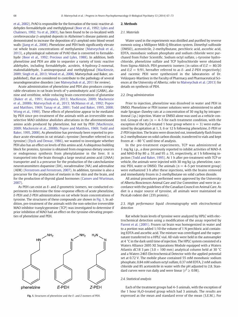
As PEH can exist as E- and Z-geometric isomers, we conducted ex-periments to determine the time-response effects of acute phenelzine,E-PEH and Z-PEH administration on rat whole brain concentrations oftyrosine. The structures of these compounds are shown in Fig. 1. In ad-dition, pre-treatment of the animals with the non-selective irreversibleMAO inhibitor tranylcypromine (TCP) was investigated to determine ifprior inhibition of MAO had an effect on the tyrosine-elevating proper-ties of phenelzine and PEH.
Fig. 1. Structures of phenelzine and the E- and Z-isomers of PEH.
2. Methods
2.1. Materials
Water used in the experiments was distilled and purified by reverseosmosis using a Millipore Milli-Q filtration system. Dimethyl sulfoxide(DMSO), acetonitrile, 2-methylbutane, perchloric acid, ascorbic acid,EDTA, monobasic sodium phosphate and sodium chloride were pur-chased from Fisher Scientific. Sodium octyl sulfate, L-tyrosine hydro-chloride, phenelzine sulfate and TCP hydrochloride were obtainedfrom Sigma-Aldrich. PEH geometric isomers (in ratios of E/Z = 80/20and E/Z = 9/91, hereafter referred to as E- and Z-PEH respectively)and racemic PEH were synthesized in the laboratories of Dr.Velázquez-Martínez in the Faculty of Pharmacy and Pharmaceutical Sci-ences at the University of Alberta; refer to Matveychuk et al. (2013) fordetails on synthesis of PEH.
2.2. Drug administration
Prior to injection, phenelzine was dissolved in water and PEH inDMSO. Phenelzine or PEH isomer solutions were administered to adultmale Sprague–Dawley rats at a concentration of 30 mg/kg by intraper-itoneal (i.p.) injection.Water or DMSO alone was used as a vehicle con-trol. Groups of rats (n = 4–5 for each treatment condition, with theexception of the H2O-treated 1 hour group where n = 3) were eutha-nized by decapitation at 1, 3, 6 or 12 h following phenelzine, E-PEH orZ-PEH injection. The brainswere dissected out, immediatelyflash frozenin 2-methylbutane on solid carbon dioxide, transferred to vials and keptfrozen at −80 °C until time of analysis.
In the pre-treatment experiments, TCP was administered at1 mg/kg i.p., a dose previously reported to inhibit activities of MAO-Aand MAO-B by 80 ± 5% and 95 ± 5%, respectively, at 1 h following in-jection (Todd and Baker, 1995). At 1 h after pre-treatment with TCP orvehicle, the animals were injected with 30 mg/kg i.p. phenelzine, race-mic PEH, water or DMSO. The animals (n = 4–5 per treatment group)were euthanized 3 h after these injections, with the brains removedand immediately frozen in 2-methylbutane on solid carbon dioxide.
All animal procedures performed were approved by the Universityof Alberta Biosciences Animal Care and Use Committee and were in ac-cordancewith the guidelines of theCanadianCouncil on Animal Care. Asdiet is a major source of tyrosine, all animals were maintained onPicoLab rodent diet (23% protein).
2.3. High performance liquid chromatography with electrochemicaldetection
Rat whole brain levels of tyrosine were analyzed by HPLCwith elec-trochemical detection using a modification of the assay reported byParent et al. (2001). Frozen rat brain was homogenized in water andto a portion was added 1/10 the volume of 1 N perchloric acid contain-ing EDTA and ascorbic acid. Themixture was centrifuged and the super-natant transferred to a HPLC vial. All vials were held in the autosamplerat 4 °C in the dark until time of injection. TheHPLC system consisted of aWaters Alliance 2695 XE Separations Module equipped with a WatersAtlantis dC18 3 μm (3.0 × 100 mm) analytical column held at 30 °Cand aWaters 2465 Electrochemical Detector with the applied potentialset at 0.72 V. The mobile phase contained 55 mM monobasic sodiumphosphate, 0.84mMsodiumoctyl sulfate, 0.37mMEDTA, 2mMsodiumchloride and 8% acetonitrile in water with the pH adjusted to 2.8. Stan-dard curves were run daily and were linear (r2 ≥ 0.98).
2.4. Statistical analysis
Each of the treatment groups had 4–5 animals, with the exception ofthe 1 hour H2O-treated group which had 3 animals. The results areexpressed as the mean and standard error of the mean (S.E.M.). For

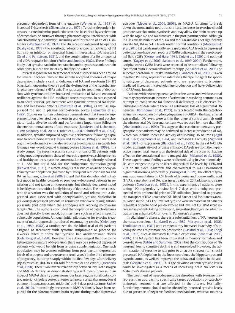
Fig. 3. Rat whole brain tyrosine levels (means ± S.E.M.) of animals pre-treated with TCP(1 mg/kg i.p.) or H2O for 1 h, followed by administration of 30 mg/kg i.p. phenelzine,PEH, H2O or DMSO. Each treatment group (n = 4–5) was analyzed at 3 h following thelast drug administration. Asterisks show that means are significantly different in compar-ison to TCP/H2O in the case of phenelzine and TCP/DMSO in the case of PEH, as assessed bythe Newman–Keuls multiple comparison test at * α = 0.05 and ** α = 0.01.
69D. Matveychuk et al. / Progress in Neuro-Psychopharmacology & Biological Psychiatry 53 (2014) 67–73
the time-response experiment, differences between groupswere evalu-ated by two-way analysis of variance (ANOVA) followed by analysis ofsimple main effects with a Bonferroni correction for multiple compari-sons. For the TCP pre-treatment experiment, differences were statisti-cally evaluated by one-way ANOVA followed by the Newman–Keulsmultiple comparison test. A P value of b 0.05 was considered to be sta-tistically significant.
3. Results
As shown in Fig. 2, phenelzine and the PEH isomers elevated ratwholebrain tyrosine levels compared to vehicle-treated values in the time-response study. There was a statistically significant effect of treatment(F [4,67] = 40.473, P b 0.001) and time following injection (F [3,67] =22.225, P b 0.001). As there was a significant interaction between treat-ment and time following injection (F [12,67]= 9.213, P b 0.001), analysisfor simplemain effects with a Bonferroni correction for multiple compar-isons was carried out. For vehicle-treated animals, brain tyrosine levelsdid not vary across the time points tested: F (3,67) = 0.791, P = 0.503for H2O and F (3,67) = 1.853, P= 0.146 for DMSO. In addition, brain ty-rosine concentrations for animals treated with H2O or DMSO were notsignificantly different from each other at any time point. When averagedacross all timepoints, tyrosine concentrations forH2O- andDMSO-treatedanimals (12.3±0.7 and12.6±0.9 μg/g tissue, respectively)were compa-rable to those previously reported by other researchers (Ablett et al.,1984; Dyck, 1987; Dyck and Dewar, 1986; Gibson and Wurtman, 1978;Wurtman et al., 1974). In comparison toH2O-treated controls, phenelzinesignificantly increased tyrosine levels at 3 and 6 h (P b 0.001). Both PEHisomers elevated brain tyrosine levels at 3 and 6 h (P b 0.001) in compar-ison toDMSO-treated controls, and E-PEH also increased tyrosine levels at12 h (P= 0.001). The effects of the PEH isomerswere comparable to eachother at all of the time points tested (P= 1.000 at 1–6 h and P= 0.706 at12 h). Phenelzine produced larger increases in brain tyrosine than eitherE-PEH or Z-PEH at 3 h (P b 0.001); however, E-PEH had elevated tyrosinelevels to a greater extent than phenelzine at 12 h (P= 0.001).
Since the PEH isomers produced comparable increases in tyrosinelevels for the time-response experiment, we used racemic PEH for theTCP pre-treatment experiment. Displayed in Fig. 3 are the results fromthe animals receivingpre-treatmentwith TCPor vehicle (H2O)prior to in-jection with phenelzine, PEH or vehicle (H2O or DMSO). There was a sta-tistically significant effect of treatment (F [5, 23]= 7.886, P b 0.001). TCPitself had no effect on rat brain tyrosine levels, as TCP/H2O and TCP/DMSOanimals had tyrosine levels (12.1±0.6 and 11.4±1.2 μg/g tissue, respec-tively) comparable to those of H2O and DMSO (12.3 ± 0.7 and 12.6 ±
Fig. 2. Changes in levels of rat whole brain tyrosine in animals treated with 30 mg/kg i.p. E-PEfollowing drug administration. Data are normalized relative to vehicle-treated controls. Valuesvalues, as assessed by analysis of simple main effects with a Bonferroni correction for multiple
0.9 μg/g tissue, respectively) from the time-response study. Pre-treatmentwith TCP reversed the phenelzine-induced increase in tyrosinelevels at 3 h after phenelzine injection. As assessed by theNewman–Keulsmultiple comparison test, both H2O/PLZ and H2O/PEH were significantlydifferent in comparison to TCP/DMSO, TCP/H2O and TCP/PLZ at α =0.05. Pre-treatment with TCP had no effect on PEH's elevating action ontyrosine levels, as brain tyrosine concentrations in the TCP/PEH groupwere also significantly different from TCP/DMSO, TCP/H2O and TCP/PLZat α= 0.01.
4. Discussion
The results of the present study demonstrate that a single i.p. injec-tion of the antidepressant/anxiolytic drug phenelzine and the E- or Z-isomers of its active metabolite PEH produced comparable time-dependent elevations in rat whole brain tyrosine levels. A similar effectof both PEH isomers on tyrosine is consistent with the previous findingthat E- and Z-PEH exhibit comparable neurochemical effects to eachother on other rat brain amino acids (Matveychuk et al., 2013). Thedata also support a previous report of increased rat striatal tyrosine fol-lowing an acute i.p. injection of 100 mg/kg, but not 5 mg/kg, phenelzine
H, Z-PEH or phenelzine. Each treatment group (n = 3–5) was analyzed at 1, 3, 6 or 12 hare means ± S.E.M. ** P b 0.01, *** P b 0.001 in comparison to matching vehicle-treatedcomparisons.

70 D. Matveychuk et al. / Progress in Neuro-Psychopharmacology & Biological Psychiatry 53 (2014) 67–73
(Dyck and Dewar, 1986). With the 100 mg/kg dose, striatal tyrosinewas significantly elevated at 12 h (to approximately 300% of controls)and was also increased at 1, 2 and 24 h following injection but notreaching statistical significance due to large variations in the data. Inthe current investigation, a 30 mg/kg i.p. injection of phenelzine orPEH isomers produced statistically significant increases in tyrosinewithin 3 to 6 h, reaching approximately 220–305% of controls at thesetime points. This increase was reversed for phenelzine by pre-treatment of the animalswith the non-selective irreversibleMAO inhib-itor TCP. Since functional MAO is required for the conversion ofphenelzine to PEH, pre-treatment with TCP should prevent the forma-tion of PEH via thismetabolic pathway. TCP pre-treatment did not affectthe changes in tyrosine induced by PEH. The data from these pre-treatment experiments support the notion that PEH is responsible forthe effects of phenelzine on rat brain tyrosine.
In earlier investigations by other researchers, chronic administrationof phenelzine via subcutaneous osmotic minipumps for 13 days (0.5 or2.5 mg/kg/day) or 28 days (10 mg/kg/day) was reported to have no ef-fect on tyrosine in the rat striatum, whole brain or plasma (Dyck et al.,1988; Paetsch and Greenshaw, 1991). As a 5 mg/kg acute injection ofphenelzine previously failed to evoke an effect on striatal tyrosine(Dyck and Dewar, 1986), it can be argued that the investigators usedtoo low a dose in the chronic experiments. Alternatively, long-term ad-ministration of phenelzine may inhibit MAO to an extent where only anegligible amount of PEH is formed by the action of the enzyme,resulting in the drop of tyrosine towards control levels. A study examin-ing the effects of chronic administration of PEH on tyrosine is nowwar-ranted, as are studies comparing brain levels of phenelzine and PEHwith the changes in tyrosine levels observed.
Although the exact mechanism for PEH-induced increases in tyro-sine has not yet been confirmed, the inhibition of tyrosine degradationis a likely candidate. There are several pathways for tyrosine metabo-lism: (1) conversion to 4-hydroxyphenylpyruvate by tyrosine transam-inase (TAT, also referred to as tyrosine aminotransferase in theliterature); (2) conversion to L-DOPA by tyrosine hydroxylase (TH);and (3) conversion to tyramine by L-aromatic amino acid decarboxylase(AADC).
PEH-mediated inhibition of TAT, a major metabolic pathway for ty-rosine (Fellman et al., 1976), is the most probable explanation.Phenelzine has previously been demonstrated to produce pronouncedelevations in rat brain levels GABA and alanine due to inhibition ofGABA transaminase and alanine transaminase, respectively (Bakeret al., 1991; Matveychuk et al., 2013; McManus et al., 1992; Popov andMatthies, 1969; Tanay et al., 2001; Todd and Baker, 1995, 2008; Wonget al., 1990). In addition, phenelzine also increased rat brain ornithineby as much as 6-fold, presumably due to inhibition of ornithine trans-aminase (Mackenzie et al., 2008b). The phenelzine-induced increasesin GABA, alanine and ornithine, as well as the inhibition of GABA trans-aminase and alanine transaminase can be abolished by pre-treatmentwith another MAO inhibitor (MacKenzie, 2009; Mackenzie et al.,2008b; Popov and Matthies, 1969; Todd and Baker, 1995, 2008), sug-gesting that a metabolite of phenelzine formed by MAO is responsiblefor these effects. PEH appears to be that metabolite, as administrationof PEH also increases brain GABA, alanine and ornithine levels(Kumpula, 2013;MacKenzie, 2009;Matveychuk et al., 2013) and causesinhibition of brain GABA transaminase (MacKenzie et al., 2008a;Paslawski et al., 2001); these properties of PEH are not affected byprior inhibition of MAO (MacKenzie, 2009). The transaminase enzymesfor GABA, alanine, ornithine and tyrosine are all dependent onpyridoxal-5′-phosphate (PLP) as a cofactor. It has been suggested thatthe binding of a hydrazine moiety, present on both phenelzine andPEH, to PLP may form an inactive hydrazone complex that will reducethe activity of associated enzymes and cause an accumulation of sub-strate (Yu and Boulton, 1991). Of interest, plasma levels of PLP in 19 pa-tients taking phenelzine were reduced to 54% of the values in thecontrol group (Malcolm et al., 1994). In fact, phenelzine has been
previously reported to inhibit rat liver TAT, an effect that may increasethe amount of tyrosine available for transport into the brain via theLNAA transporter from the periphery (Dyck, 1987; Dyck and Dewar,1986). As the assays for TAT function in those studies used liver homog-enate (likely containing MAO enzymes), the MAO-mediated metabo-lism of phenelzine to PEH and subsequent inactivation of TAT by thismetabolite may be a viable explanation for the results. A cerebralversion of the TAT is also present in the rat brain, with several notabledifferences from hepatic TAT: it is localized mainly on the inner mito-chondrial membrane in comparison to the soluble hepatic enzyme pri-marily in the cytosol; contains four versus two PLP molecules perenzyme; and has different amino acid composition and inhibition char-acteristics (Mandel and Aunis, 2008). Further investigations intowhether PEH can inhibit liver and rat brain TAT are needed.
It is unlikely that PEH is an inhibitor of TH, the enzyme responsiblefor the hydroxylation of tyrosine to L-DOPA (a rate-limiting step in cat-echolamine synthesis), as rat brain concentrations of DA andNA remainat control levels 1–12 h following acute administration of PEH(MacKenzie, 2009; Matveychuk et al., 2013). It should be noted thatthe expression of TH may be affected with long-term treatment as aform of a negative-feedback mechanism, although there is little experi-mental consensus on the topic. A study performedwith chronic admin-istration of phenelzine (15 mg/kg i.p.) in three replicate experimentsshowed a 15–20% reduction in rat striatal and hypothalamic TH activityat 3 and 6 weeks in one of the experiments, but failed to reproduce thisfinding in the other two replicate experiments (Robinson et al., 1979).Furthermore, chronic administration of phenelzine (for 14 days viasubcutaneously-implanted minipumps) did not produce a significantreduction in TH mRNA in several rat brain regions (Rovin et al., 2012).However, an additional investigation demonstrated that daily adminis-tration of phenelzine (5 mg/kg i.p.) increased THmRNA levels in the ratlocus coeruleus by 70–150% after 2 weeks and by 71–115% after8 weeks of treatment (Brady et al., 1992).
Another possibility to be considered is PEH-mediated inhibition ofAADC, another PLP-dependent enzyme responsible for the conversionof tyrosine to tyramine and the conversion of L-DOPA to DA. However,the decarboxylation of tyrosine by this enzyme is very slow and consid-ered to represent aminormetabolic pathway of tyrosine (Fellman et al.,1976; Lovenberg et al., 1962). In addition, inhibition of AADC shouldproduce a reduction in brain DA and NA levels, an effect not consistentwith PEH administration (MacKenzie, 2009; Matveychuk et al., 2013).Previous studies have suggested that phenelzine is an inhibitor of ratbrain AADC (Dyck, 1987; Dyck andDewar, 1986). However, administra-tion of the drugα-monofluoromethyldopa, a stronger inhibitor of brainAADC but a weaker inhibitor of liver TAT in comparison to phenelzine,did not have an effect on rat whole brain tyrosine levels (Dyck, 1987).
Can an increase in brain tyrosine affect production of catechol-amines? As the activity of TH is governed by an end-product inhibitionmechanism via DA, NA andADR (Nagatsu et al., 1964), increases in tyro-sine levels do not normally have a lasting effect on brain catecholamineconcentrations (see Cansev andWurtman, 2007 for review). This is sup-ported by our observation that a single administration of PEH does notsignificantly increase rat brain DA or NA at 1–12 h (Matveychuk et al.,2013) despite nearly a 3-fold increase in brain tyrosine at 3 h. However,experimental evidence suggests that increased tyrosine availability canenhance catecholamine production in monoaminergic neurons undercertain conditions, such as increased or sustained neuronal activation(Fernstrom and Fernstrom, 2007). This is illustrated by the observationthat supplementation with tyrosine in rats potentiated the tyrosine hy-droxylation rate in the retina (containing light-sensitive DA neurons)when the animals were exposed to light, but not dark environments(Fernstrom et al., 1986). There are several possible mechanisms for en-hanced catecholamine synthesis from tyrosine upon increased neuronalstimulation: the intraneuronal catecholamine levels are reduced andthereby diminish end-product inhibition of TH (Weiner and Rabadjija,1968), phosphorylation of TH resulting in a more active and
71D. Matveychuk et al. / Progress in Neuro-Psychopharmacology & Biological Psychiatry 53 (2014) 67–73
precursor-dependent form of the enzyme (Weiner et al., 1978) orincreased TH synthesis (Silberstein et al., 1972). Tyrosine-dependent in-creases in catecholamine production can also be elicited by accelerationof catecholamine turnover through pharmacological interference withcatecholaminergic pathways, including administration of an ADCC in-hibitor (Wurtman et al., 1974), the DA receptor antagonist haloperidol(Scally et al., 1977), the anesthetic γ-butyrolactone (an activator of THbut also an inhibitor of neuronal firing in nigrostriatal DA neurons)(Sved and Fernstrom, 1981), and the combination of a DA antagonistand a DA reuptake inhibitor (Fuller and Snoddy, 1982). These findingsimply that tyrosine can influence catecholamine synthesis under certainconditions, but can this be of any therapeutic value?
Interest in tyrosine for treatment ofmood disorders has been aroundfor several decades. Two of the widely accepted theories of majordepression include a central deficiency of NA and serotonin (5-HT)(classical monoamine theory) and the dysfunction of the hypothalam-ic–pituitary–adrenal (HPA) axis. The rationale for treatment of depres-sion with tyrosine includes increased production of NA and enhancedresilience against the HPA-mediated stress response. In rats exposedto an acute stressor, pre-treatment with tyrosine prevented NA deple-tion and behavioral deficits (Reinstein et al., 1984), as well as sup-pressed the rise in plasma corticosterone levels (Reinstein et al.,1985). Studies on human volunteers demonstrated that tyrosine sup-plementation alleviated decrements in working memory and psycho-motor tasks, adverse moods and performance impairment followingthe environmental stress of cold exposure (Banderet and Lieberman,1989; Mahoney et al., 2007; O'Brien et al., 2007; Shurtleff et al., 1994).In addition, tyrosine improved cognitive performance following expo-sure to acute noise stress (Deijen and Orlebeke, 1994) and increasedcognitive performance while also reducing blood pressure in cadets fol-lowing a one-week combat training course (Deijen et al., 1999). In astudy comparing tyrosine plasma concentrations of 38 patients withendogenous depression to those of neurotic depressives, schizophrenicsand healthy controls, tyrosine concentration was significantly reducedat 11 AM, but not 8 AM, for the endogenous depression group(Benkert et al., 1971). In a meta-analysis of 8 studies on acute phenylal-anine/tyrosine depletion (followed by subsequent reductions in NA andDA) in humans, Ruhe et al. (2007) found that this depletion did not af-fect mood in healthy controls or previously-depressed patients in re-mission and not taking antidepressants, but slightly decreased moodin healthy controls with a family history of depression. Themost consis-tent observation was the reduction of mood and relapse into a de-pressed state associated with phenylalanine/tyrosine depletion inpreviously-depressed patients in remission who were taking antide-pressants (but only when the antidepressant working mechanismtargets NA). The authors concluded that depletion of catecholaminesdoes not directly lower mood, but may have such an effect in specificvulnerable populations. Although initial pilot studies for tyrosine treat-ment of major depression produced encouraging results (Gelenberget al., 1980, 1982), a randomized double-blind trial of 65 patientsassigned to treatment with tyrosine, imipramine or placebo for4 weeks failed to show that tyrosine had antidepressant effects(Gelenberg et al., 1990). However, the authors suggest that due to theheterogeneous nature of depression, theremay be a subset of depressedpatients who would benefit from tyrosine supplementation. One suchpopulation may be women suffering from post-partum depression.Levels of estrogens and progesterone reach a peak in the third trimesterof pregnancy, but drop sharply within the first few days after delivery(by as much as 100- to 1000-fold for estradiol and estriol) (Hendricket al., 1998). There is an inverse relationship between levels of estrogenand MAO-A density, as demonstrated by a 43% mean increase in anindex of MAO-A density across numerous brain regions (prefrontal cor-tex, anterior cingulate cortex, anterior temporal cortex, thalamus, dorsalputamen, hippocampus andmidbrain) at 4–6 days post-partum(Sacheret al., 2010). Interestingly, increases in MAO-A density have been re-ported for the aforementioned brain regions during major depressive
episodes (Meyer et al., 2006, 2009). As MAO-A functions to breakdown the monoamine neurotransmitters, increases in tyrosine shouldpromote catecholamine synthesis and may allow the brain to keep upwith the rapid NA and DA turnover in the post-partumperiod. AlthoughPEH is a weak inhibitor of MAO-A andMAO-B and does not significantlyelevate NA, DA or 5-HT levels under normal conditions (Matveychuket al., 2013), it can dramatically increase brain GABA levels. In depressedpatients, there have been reports of GABAdeficiencies in the cerebrospi-nal fluid (CSF) (Gerner and Hare, 1981; Gold et al., 1980) and occipitalcortex (Kugaya et al., 2003; Sanacora et al., 1999, 2004). Furthermore,occipital cortex GABA levels were reported to be normalized followingtreatment with electroconvulsive therapy (Sanacora et al., 2003) orselective serotonin reuptake inhibitors (Sanacora et al., 2002). Takentogether, PEHmay represent an interesting therapeutic agent for specif-ic subtypes of depressed patients who are responsive to tyrosine-mediated increases in catecholamine production and have deficienciesin GABAergic function.
Patients with neurodegenerative disorders associatedwith neuronallossmay experience an increase of activation in surviving neurons as anattempt to compensate for functional deficiency, as is observed forParkinson's disease where there is a substantial loss of nigrostriatal DAneurons (Bernheimer et al., 1973). In rats treated with the catechol-aminergic neurotoxin 6-hydroxydopamine (6-OHDA), the basal striatalextracellular DA levels were within the range of control animals untilthe nigrostriatal DA neuronal content was reduced by more than 80%(Abercrombie et al., 1990). This suggests that certain compensatory pre-synaptic mechanisms may be activated to increase production of DA,which can include increased activity of surviving DA neurons (Agidet al., 1973; Zigmond et al., 1984) and increased TH activity (Zigmondet al., 1984) or expression (Blanchard et al., 1995). In the rat 6-OHDAmodel, administration of tyrosine enhanced DA release from the hyper-active nigrostriatal neurons on the lesioned side of the brain but had noeffect on the contralateral non-damaged side (Melamed et al., 1980).These experimental findings were replicated using in vivo microdialy-sis, with exogenous tyrosine increasing striatal DA levels by 139% and25% on the sides ipsilateral and contralateral to 6-OHDA-inducednigrostriatal lesions, respectively (During et al., 1989). The effect of tyro-sine supplementation on CSF levels of tyrosine and homovanillic acid(HVA), a metabolite of DA, was investigated in 23 Parkinson's diseasepatients (Growdon et al., 1982). In this experiment, all patients weretaking 100 mg/kg/day tyrosine for 4–7 days with a subgroup pre-treated with probenecid prior to CSF collection (intended to preventthe transport of HVA across the CSF–blood barrier and facilitate its accu-mulation in the CSF). CSF levels of tyrosinewere increased in all patientsregardless of probenecid pre-treatment and levels of CSF HVA were in-creased in patients taking probenecid, suggesting that tyrosine adminis-tration can enhance DA turnover in Parkinson's disease.
In Alzheimer's disease, there is a substantial loss of NA neurons inthe locus coeruleus (Bondareff et al., 1982; Marcyniuk et al., 1986;Tomlinson et al., 1981) with compensatory increases in activity of sur-viving neurons to promote NA production (Raskind et al., 1984; Tohgiet al., 1992), such as increased TH mRNA expression (Szot et al., 2000,2006). The NA system has been implicated in memory formation andconsolidation (Gibbs and Summers, 2002), but the contribution of NAneuronal loss to cognitive decline is still unresolved. However, the ad-ministration of tyrosine to rats prior to an acute stressor (tail shock)prevented NA depletion in the locus coeruleus, the hippocampus andhypothalamus, as well as improved the behavioral deficits in the ani-mals (Reinstein et al., 1984). Thus, the elevation of brain tyrosine levelscould be investigated as a means of increasing brain NA levels inAlzheimer's disease patients.
The treatment of neurodegenerative disorders with tyrosine mayrepresent an approach to specifically target populations of catechol-aminergic neurons that are affected in the disease. Normally-functioning neurons should not be affected by increased tyrosine levelsdue to presence of negative feedback mechanisms, whereas neurons

72 D. Matveychuk et al. / Progress in Neuro-Psychopharmacology & Biological Psychiatry 53 (2014) 67–73
that have been affected by the disorder (e.g. nigrostriatal DA neurons inParkinson's disease or locus coeruleus NA neurons in Alzheimer's dis-ease) have increased activity due to compensatory mechanisms andwould therefore undergo tyrosine-enhanced synthesis of catechol-amine neurotransmitters (Cansev and Wurtman, 2007).
5. Conclusion
Phenelzine and its active metabolite PEH were shown to producetime-dependent elevations in rat whole brain levels of tyrosine.These increases in brain tyrosine concentrations were abolished forphenelzine, but not for PEH, by pre-treatment of the animals withthe MAO inhibitor TCP, suggesting that the tyrosine-elevating prop-erties of phenelzine are MAO-mediated and are likely the result of PEHformation. As indicated by Matveychuk et al. (2013) and Baker et al.(2012), PEHmay be a useful adjunctive drug for a number of neurolog-ical and psychiatric disorders because of its ability to elevate brain GABAlevels, inhibit PrAO and sequester toxic aldehydes. The results from thepresent study suggest that elevation of tyrosine levels could also be abeneficial effect of PEH in the treatment of such disorders.
Acknowledgments
This workwas supported by funding from the Canadian Institutes ofHealth Research (CIHR: MOP-86712), the University of Alberta and theQueen Elizabeth II graduate studentship program. The authors aregrateful to Gail Rauw for expert technical assistance.
References
Abercrombie ED, Bonatz AE, Zigmond MJ. Effects of L-dopa on extracellular dopamine instriatum of normal and 6-hydroxydopamine-treated rats. Brain Res 1990;525:36–44.
Ablett RF, MacMillan M, Sole MJ, Toal CB, Anderson GH. Free tyrosine levels of rat brainand tissues with sympathetic innervation following administration of L-tyrosine inthe presence and absence of large neutral amino acids. J Nutr 1984;114:835–9.
Agid Y, Javoy F, Glowinski J. Hyperactivity of remaining dopaminergic neurones after par-tial destruction of the nigro-striatal dopaminergic system in the rat. Nat New Biol1973;245:150–1.
Airas L, Lindsberg PJ, Karjalainen-Lindsberg ML, Mononen I, Kotisaari K, Smith DJ, et al.Vascular adhesion protein-1 in human ischaemic stroke. Neuropathol Appl Neurobiol2008;34:394–402.
Baker GB,Wong JT, Yeung JM, Coutts RT. Effects of the antidepressant phenelzine on brainlevels of gamma-aminobutyric acid (GABA). J Affect Disord 1991;21:207–11.
Baker GB, Matveychuk D, MacKenzie EM, Dursun SM, Mousseau DD. Monoamine oxidaseinhibitors and neuroprotective mechanisms. Bull Clin Psychopharmacol 2012;22:293–6.
Banderet LE, Lieberman HR. Treatment with tyrosine, a neurotransmitter precursor, re-duces environmental stress in humans. Brain Res Bull 1989;22:759–62.
Benkert O, Renz A, Marano C, Matussek N. Altered tyrosine daytime plasma levels in en-dogenous depressive patients. Arch Gen Psychiatry 1971;25:359–63.
Benson CA, Wong G, Tenorio G, Baker GB, Kerr BJ. The MAO inhibitor phenelzine can im-prove functional outcomes inmice with established clinical signs in experimental au-toimmune encephalomyelitis (EAE). Behav Brain Res 2013;252:302–11.
Bernheimer H, Birkmayer W, Hornykiewicz O, Jellinger K, Seitelberger F. Brain dopamineand the syndromes of Parkinson and Huntington. Clinical, morphological and neuro-chemical correlations. J Neurol Sci 1973;20:415–55.
Binda C, Jin W, Min L, Hubalek F, Mattevi A, Edmondson DE. Structural and mechanisticstudies of arylalkylhydrazine inhibition of human monoamine oxidases A and B. Bio-chemistry 2008;47:5616–25.
Blanchard V, Chritin M, Vyas S, Savasta M, Feuerstein C, Agid Y, et al. Long-term inductionof tyrosine hydroxylase expression: compensatory response to partial degenerationof the dopaminergic nigrostriatal system in the rat brain. J Neurochem 1995;64:1669–79.
Bondareff W, Mountjoy CQ, Roth M. Loss of neurons of origin of the adrenergic projectionto cerebral cortex (nucleus locus ceruleus) in senile dementia. Neurology 1982;32:164–8.
Boomsma F, Derkx FH, van den Meiracker AH, Man in 't Veld AJ, Schalekamp MA. Plasmasemicarbazide-sensitive amine oxidase activity is elevated in diabetes mellitus andcorrelates with glycosylated haemoglobin. Clin Sci 1995;88:675–9.
Boomsma F, van Veldhuisen DJ, de Kam PJ, Man in't Veld AJ, Mosterd A, Lie KI, et al. Plas-ma semicarbazide-sensitive amine oxidase is elevated in patients with congestiveheart failure. Cardiovasc Res 1997;33:387–91.
Boomsma F, de Kam PJ, Tjeerdsma G, van den Meiracker AH, van Veldhuisen DJ. Plasmasemicarbazide-sensitive amine oxidase (SSAO) is an independent prognostic markerfor mortality in chronic heart failure. Eur Heart J 2000;21:1859–63.
Boor PJ, TrentMB, Lyles GA, TaoM, Ansari GA. Methylamine metabolism to formaldehydeby vascular semicarbazide-sensitive amine oxidase. Toxicology 1992;73:251–8.
Brady LS, Gold PW, HerkenhamM, LynnAB,Whitfield Jr HJ. The antidepressants fluoxetine,idazoxan and phenelzine alter corticotropin-releasing hormone and tyrosine hydroxy-lase mRNA levels in rat brain: therapeutic implications. Brain Res 1992;572:117–25.
Cansev M, Wurtman RJ. Chapter 4: aromatic amino acids in the brain. In: Lajtha A, Oja S,Schousboe A, Saransaari P, editors. Handbook of Neurochemistry and Molecular Neu-robiology. Springer US; 2007. p. 59–97.
Deijen JB, Orlebeke JF. Effect of tyrosine on cognitive function and blood pressure understress. Brain Res Bull 1994;33:319–23.
Deijen JB, Wientjes CJ, Vullinghs HF, Cloin PA, Langefeld JJ. Tyrosine improves cognitiveperformance and reduces blood pressure in cadets after one week of a combat train-ing course. Brain Res Bull 1999;48:203–9.
del Mar Hernandez M, Esteban M, Szabo P, Boada M, Unzeta M. Human plasmasemicarbazide sensitive amine oxidase (SSAO), beta-amyloid protein and aging.Neurosci Lett 2005;384:183–7.
DuringMJ, Acworth IN,Wurtman RJ. Dopamine release in rat striatum: physiological cou-pling to tyrosine supply. J Neurochem 1989;52:1449–54.
Dyck LE. Effect of decarboxylase inhibitors on brain p-tyrosine levels. Biochem Pharmacol1987;36:1373–6.
Dyck LE, Dewar KM. Inhibition of aromatic L-amino acid decarboxylase and tyrosine ami-notransferase by the monoamine oxidase inhibitor phenelzine. J Neurochem 1986;46:1899–903.
Dyck LE, Juorio AV, Durden DA, Boulton AA. Effect of chronic deuterated and non-deuterated phenelzine on rat brain monoamines and monoamine oxidase. NaunynSchmiedebergs Arch Pharmacol 1988;337:279–83.
Fellman JH, Roth ES, Fujita TS. Decarboxylation to tyramine is not a major route of tyro-sine metabolism in mammals. Arch Biochem Biophys 1976;174:562–7.
Fernstrom JD, Fernstrom MH. Tyrosine, phenylalanine, and catecholamine synthesis andfunction in the brain. J Nutr 2007;137:1539S–47S. [discussion 1548S].
Fernstrom MH, Volk EA, Fernstrom JD, Iuvone PM. Effect of tyrosine administration ondopa accumulation in light- and dark-adapted retinas from normal and diabeticrats. Life Sci 1986;39:2049–57.
Ferrer I, Lizcano JM, Hernandez M, Unzeta M. Overexpression of semicarbazide sensitiveamine oxidase in the cerebral blood vessels in patients with Alzheimer's diseaseand cerebral autosomal dominant arteriopathy with subcortical infarcts andleukoencephalopathy. Neurosci Lett 2002;321:21–4.
Fuller RW, Snoddy HD. L-Tyrosine enhancement of the elevation of 3,4-dihydroxyphenylacetic acid concentration in rat brain by spiperone andamfonelic acid. J Pharm Pharmacol 1982;34:117–8.
Garpenstrand H, Ekblom J, Backlund LB, Oreland L, Rosenqvist U. Elevated plasmasemicarbazide-sensitive amine oxidase (SSAO) activity in type 2 diabetes mellituscomplicated by retinopathy. Diabet Med 1999;16:514–21.
Gelenberg A, Wojcik J, Growdon J, Sved A, Wurtman R. Tyrosine for the treatment of de-pression. Am J Psychiatry 1980;137:622–3.
Gelenberg AJ, Wojcik JD, Gibson CJ, Wurtman RJ. Tyrosine for depression. J Psychiatr Res1982;17:175–80.
Gelenberg AJ, Wojcik JD, Falk WE, Baldessarini RJ, Zeisel SH, Schoenfeld D, et al. Tyrosinefor depression: a double-blind trial. J Affect Disord 1990;19:125–32.
Gerner RH, Hare TA. CSF GABA in normal subjects and patients with depression, schizo-phrenia, mania, and anorexia nervosa. Am J Psychiatry 1981;138:1098–101.
Gibbs ME, Summers RJ. Role of adrenoceptor subtypes in memory consolidation. ProgNeurobiol 2002;67:345–91.
Gibson CJ, Wurtman RJ. Physiological control of brain norepinephrine synthesis by braintyrosine concentration. Life Sci 1978;22:1399–405.
Gold BI, Bowers Jr MB, Roth RH, Sweeney DW. GABA levels in CSF of patients with psychi-atric disorders. Am J Psychiatry 1980;137:362–4.
Growdon JH, Melamed E, Logue M, Hefti F, Wurtman RJ. Effects of oral L-tyrosine admin-istration on CSF tyrosine and homovanillic acid levels in patients with Parkinson'sdisease. Life Sci 1982;30:827–32.
Hendrick V, Altshuler LL, Suri R. Hormonal changes in the postpartum and implicationsfor postpartum depression. Psychosomatics 1998;39:93–101.
Hernandez-GuillamonM, SoleM, Delgado P, Garcia-Bonilla L, Giralt D, Boada C, et al. VAP-1/SSAO plasma activity and brain expression in human hemorrhagic stroke.Cerebrovasc Dis 2012;33:55–63.
Jiang ZJ, Richardson JS, Yu PH. The contribution of cerebral vascular semicarbazide-sensitive amine oxidase to cerebral amyloid angiopathy in Alzheimer's disease.Neuropathol Appl Neurobiol 2008;34:194–204.
Karadi I, Meszaros Z, Csanyi A, Szombathy T, Hosszufalusi N, Romics L, et al. Serumsemicarbazide-sensitive amine oxidase (SSAO) activity is an independent marker ofcarotid atherosclerosis. Clin Chim Acta 2002;323:139–46.
Kugaya A, Sanacora G, Verhoeff NP, Fujita M, Mason GF, Seneca NM, et al. Cerebral benzo-diazepine receptors in depressed patients measured with [123I]iomazenil SPECT. BiolPsychiatry 2003;54:792–9.
Kumpula DJ. The antidepressant/antipanic/neuroprotective drug phenelzine: neurophar-macological and drug metabolism studies [MSc Thesis] University of Alberta; 2013.
Lovenberg W, Weissbach H, Udenfriend S. Aromatic L-amino acid decarboxylase. J BiolChem 1962;237:89–93.
Lyles GA, Chalmers J. The metabolism of aminoacetone to methylglyoxal bysemicarbazide-sensitive amine oxidase in human umbilical artery. BiochemPharmacol 1992;43:1409–14.
MacKenzie EM. Neurochemical and neuroprotective aspects of phenelzine and its activemetabolite β-phenylethylidenehydrazine [PhD Thesis] University of Alberta; 2009.
MacKenzie EM, Fassihi A, Davood A, Chen Q-H, RauwG, Rauw G, et al. N-Propynyl analogsof β-phenylethylidenehydrazines: synthesis and evaluation of effects on glycine,GABA, and monoamine oxidase. Bioorg Med Chem 2008a;16:8254–63.

73D. Matveychuk et al. / Progress in Neuro-Psychopharmacology & Biological Psychiatry 53 (2014) 67–73
Mackenzie EM, Grant SL, Baker GB, Wood PL. Phenelzine causes an increase in brain orni-thine that is prevented by prior monoamine oxidase inhibition. Neurochem Res2008b:430–6.
Mahoney CR, Castellani J, Kramer FM, Young A, Lieberman HR. Tyrosine supplementationmitigates working memory decrements during cold exposure. Physiol Behav 2007;92:575–82.
Malcolm DE, Yu PH, Bowen RC, O'Donovan C, Hawkes J, Hussein M. Phenelzine reducesplasma vitamin B6. J Psychiatry Neurosci 1994;19:332–4.
Mandel P, Aunis D. Tyrosine Aminotransferase in the Rat Brain, (Eds.), Ciba FoundationSymposium 22 — Aromatic Amino Acids in the Brain. John Wiley & Sons, Ltd., 2008.pp. 67–83.
Marcyniuk B, Mann DM, Yates PO. Loss of nerve cells from locus coeruleus in Alzheimer'sdisease is topographically arranged. Neurosci Lett 1986;64:247–52.
Matveychuk D, Dursun S, Wood P, Baker G. Reactive aldehydes and neurodegenerativedisorders. Bull Clin Psychopharmacol 2011;21:277–88.
Matveychuk D, Nunes E, Ullah N, Velazquez-Martinez CA, MacKenzie EM, Baker GB. Com-parison of phenelzine and geometric isomers of its active metabolite, beta-phenylethylidenehydrazine, on rat brain levels of amino acids, biogenic amine neuro-transmitters and methylamine. J Neural Transm 2013;120:987–96.
McManus DJ, Baker GB, Martin IL, Greenshaw AJ, McKenna KF. Effects of the antidepres-sant/antipanic drug phenelzine on GABA concentrations and GABA-transaminase ac-tivity in rat brain. Biochem Pharmacol 1992;43:2486–9.
Melamed E, Hefti F,Wurtman RJ. Tyrosine administration increases striatal dopamine releasein rats with partial nigrostriatal lesions. Proc Natl Acad Sci U S A 1980;77:4305–9.
Meszaros Z, Szombathy T, Raimondi L, Karadi I, Romics L, Magyar K. Elevated serumsemicarbazide-sensitive amine oxidase activity in non-insulin-dependent diabetesmellitus: correlation with body mass index and serum triglyceride. Metabolism1999;48:113–7.
Meyer JH, Ginovart N, Boovariwala A, Sagrati S, Hussey D, Garcia A, et al. Elevated mono-amine oxidase a levels in the brain: an explanation for the monoamine imbalance ofmajor depression. Arch Gen Psychiatry 2006;63:1209–16.
Meyer JH, Wilson AA, Sagrati S, Miler L, Rusjan P, Bloomfield PM, et al. Brain monoamineoxidase A binding in major depressive disorder: relationship to selective serotoninreuptake inhibitor treatment, recovery, and recurrence. Arch Gen Psychiatry 2009;66:1304–12.
Musgrave T, Benson C, Wong G, Browne I, Tenorio G, Rauw G, et al. The MAO inhibitorphenelzine improves functional outcomes in mice with experimental autoimmuneencephalomyelitis (EAE). Brain Behav Immun 2011;25:1677–88.
Nagatsu T, Levitt M, Udenfriend S. Tyrosine hydroxylase. The initial step in norepineph-rine biosynthesis. J Biol Chem 1964;239:2910–7.
O'Brien C,MahoneyC, TharionWJ, Sils IV, Castellani JW.Dietary tyrosine benefits cognitiveand psychomotor performance during body cooling. Physiol Behav 2007;90:301–7.
Paetsch PR, Greenshaw AJ. Beta-adrenergic effects on plasma and brain large neutralamino acids are unaltered by chronic administration of antidepressants. J Neurochem1991;56:2027–32.
ParentM, Bush D, RauwG,Master S, Vaccarino F, Baker G. Analysis of amino acids and cat-echolamines, 5-hydroxytryptamine and their metabolites in brain areas in the ratusing in vivo microdialysis. Methods 2001;23:11–20.
Paslawski T, Knaus E, Iqbal N, Coutts R, Baker G. β-Phenylethylidenehydrazine, a novel in-hibitor of GABA transaminase. Drug Dev Res 2001;54:35–9.
Popov N, Matthies H. Some effects of monoamine oxidase inhibitors on themetabolism ofgamma-aminobutyric acid in rat brain. J Neurochem 1969;16:899–907.
Precious E, Lyles GA. Properties of a semicarbazide-sensitive amine oxidase in human um-bilical artery. J Pharm Pharmacol 1988;40:627–33.
Raskind MA, Peskind ER, Halter JB, Jimerson DC. Norepinephrine and MHPG levels in CSFand plasma in Alzheimer's disease. Arch Gen Psychiatry 1984;41:343–6.
Reinstein DK, Lehnert H, Scott NA, Wurtman RJ. Tyrosine prevents behavioral and neuro-chemical correlates of an acute stress in rats. Life Sci 1984;34:2225–31.
Reinstein DK, Lehnert H, Wurtman RJ. Dietary tyrosine suppresses the rise in plasma cor-ticosterone following acute stress in rats. Life Sci 1985;37:2157–63.
Robinson DS, Campbell IC, Walker M, Statham NJ, Lovenberg W, Murphy DL. Effects ofchronic monoamine oxidase inhibitor treatment on biogenic amine metabolism inrat brain. Neuropharmacology 1979;18:771–6.
Rovin ML, Boss-Williams KA, Alisch RS, Ritchie JC, Weinshenker D, West CH, et al. Influ-ence of chronic administration of antidepressant drugs on mRNA for galanin, galaninreceptors, and tyrosine hydroxylase in catecholaminergic and serotonergic cell-bodyregions in rat brain. Neuropeptides 2012;46:81–91.
Ruhe HG, Mason NS, Schene AH. Mood is indirectly related to serotonin, norepinephrineand dopamine levels in humans: a meta-analysis of monoamine depletion studies.Mol Psychiatry 2007;12:331–59.
Sacher J, Wilson AA, Houle S, Rusjan P, Hassan S, Bloomfield PM, et al. Elevated brainmonoamine oxidase A binding in the early postpartum period. Arch Gen Psychiatry2010;67:468–74.
Sanacora G, Mason GF, Rothman DL, Behar KL, Hyder F, Petroff OA, et al. Reduced corticalgamma-aminobutyric acid levels in depressed patients determined by proton mag-netic resonance spectroscopy. Arch Gen Psychiatry 1999;56:1043–7.
Sanacora G, Mason GF, Rothman DL, Krystal JH. Increased occipital cortex GABA concen-trations in depressed patients after therapy with selective serotonin reuptake inhib-itors. Am J Psychiatry 2002;159:663–5.
Sanacora G, Mason GF, Rothman DL, Hyder F, Ciarcia JJ, Ostroff RB, et al. Increased corticalGABA concentrations in depressed patients receiving ECT. Am J Psychiatry 2003;160:577–9.
Sanacora G, Gueorguieva R, Epperson CN, Wu YT, Appel M, Rothman DL, et al. Subtype-specific alterations of gamma-aminobutyric acid and glutamate in patients withmajor depression. Arch Gen Psychiatry 2004;61:705–13.
Scally MC, Ulus I, Wurtman RJ. Brain tyrosine level controls striatal dopamine synthesis inhaloperidol-treated rats. J Neural Transm 1977;41:1–6.
Shurtleff D, Thomas JR, Schrot J, Kowalski K, Harford R. Tyrosine reverses a cold-induced working memory deficit in humans. Pharmacol Biochem Behav 1994;47:935–41.
Silberstein SD, Lemberger L, Klein DC, Axelrod J, Kopin IJ. Induction of adrenal tyrosine hy-droxylase in organ culture. Neuropharmacology 1972;11:721–6.
Singh IN, Gilmer LK, Miller DM, Cebak JE, Wang JA, Hall ED. Phenelzine mitochondrialfunctional preservation and neuroprotection after traumatic brain injury related toscavenging of the lipid peroxidation-derived aldehyde 4-hydroxy-2-nonenal. JCereb Blood Flow Metab 2013;33:593–9.
Song MS, Matveychuk D, MacKenzie EM, Duchcherer M, Mousseau DD, Baker GB. An up-date on amine oxidase inhibitors: multifaceted drugs. Prog NeuropsychopharmacolBiol Psychiatry 2013;44:118–24.
Sved A, Fernstrom J. Tyrosine availability and dopamine synthesis in the striatum: studieswith gamma-butyrolactone. Life Sci 1981;29:743–8.
Szot P, Leverenz JB, Peskind ER, Kiyasu E, Rohde K, Miller MA, et al. Tyrosine hydroxylaseand norepinephrine transporter mRNA expression in the locus coeruleus inAlzheimer's disease. Brain Res Mol Brain Res 2000;84:135–40.
Szot P, White SS, Greenup JL, Leverenz JB, Peskind ER, Raskind MA. Compensatory chang-es in the noradrenergic nervous system in the locus ceruleus and hippocampus ofpostmortem subjects with Alzheimer's disease and dementia with Lewy bodies. JNeurosci 2006;26:467–78.
Tanay AI, Parent MB,Wong JTF, Paslawski T, Martin IL, Baker GB. Effects of the antidepres-sant/antipanic drug phenelzine on alanine and alanine transaminase in rat brain. CellMol Neurobiol 2001;21:325–39.
Tipton KF, Spires IP. Oxidation of 2-phenylethylhydrazine by monoamine oxidase.Biochem Pharmacol 1972;21:268–70.
Todd KG, Baker GB. GABA-elevating effects of the antidepressant/antipanic drugphenelzine in brain: effects of pretreatment with tranylcypromine, (−)-deprenyland clorgyline. J Affect Disord 1995;35:125–9.
Todd KG, Baker GB. Neurochemical effects of the monoamine oxidase inhibitorphenelzine on brain GABA and alanine: a comparison with vigabatrin. J PharmPharm Sci 2008;11:14s–21s.
Todd KG, Banigesh AI, Baker GB, Coutts RT, Shuaib A. Phenylethylidenehydrazine, a novelGABA-T inhibitor, has neuroprotective actions in transient global ischemia. JNeurochem 1999;73:S202B. [Suppl.].
Tohgi H, Ueno M, Abe T, Takahashi S, Nozaki Y. Concentrations of monoamines and theirmetabolites in the cerebrospinal fluid from patients with senile dementia of theAlzheimer type and vascular dementia of the Binswanger type. J Neural TransmPark Dis Dement Sect 1992;4:69–77.
Tomlinson BE, Irving D, Blessed G. Cell loss in the locus coeruleus in senile dementia ofAlzheimer type. J Neurol Sci 1981;49:419–28.
Weiner N, Rabadjija M. The effect of nerve stimulation on the synthesis and metabolismof norepinephrine in the isolated guinea-pig hypogastric nerve–vas deferens prepa-ration. J Pharmacol Exp Ther 1968;160:61–71.
Weiner N, Lee FL, Dreyer E, Barnes E. The activation of tyrosine hydroxylase innoradrenergic neurons during acute nerve stimulation. Life Sci 1978;22:1197–215.
Wong JT, Baker GB, Coutts RT, Dewhurst WG. Long-lasting elevation of alanine in brainproduced by the antidepressant phenelzine. Brain Res Bull 1990;25:179–81.
Wood PL, Khan MA, Moskal JR, Todd KG, Tanay VA, Baker G. Aldehyde load in ischemia–reperfusion brain injury: neuroprotection by neutralization of reactive aldehydeswith phenelzine. Brain Res 2006;1122:184–90.
Wurtman RJ, Larin F, Mostafapour S, Fernstrom JD. Brain catechol synthesis: control bytrain tyrosine concentration. Science 1974;185:183–4.
Youdim MB, Edmondson D, Tipton KF. The therapeutic potential of monoamine oxidaseinhibitors. Nat Rev Neurosci 2006;7:295–309.
Yu PH, Boulton AA. A comparison of effect of brofaromine, phenelzine and tranylcypromineon the activities of some enzymes involved in the metabolism of different neurotrans-mitters. Res Commun Chem Pathol Pharmacol 1991;16:141–53.
Yu PH, Wright S, Fan EH, Lun ZR, Gubisne-Harberle D. Physiological and pathological im-plications of semicarbazide-sensitive amine oxidase. Biochim Biophys Acta 2003;1647:193–9.
Zigmond MJ, Acheson AL, Stachowiak MK, Stricker EM. Neurochemical compensationafter nigrostriatal bundle injury in an animal model of preclinical parkinsonism.Arch Neurol 1984;41:856–61.