Embed Size (px)
Citation preview
7/28/2019 Elispot assays: state-of-the-art tools for functional analysis of cellular immunology
http://slidepdf.com/reader/full/elispot-assays-state-of-the-art-tools-for-functional-analysis-of-cellular 1/12
7/28/2019 Elispot assays: state-of-the-art tools for functional analysis of cellular immunology
http://slidepdf.com/reader/full/elispot-assays-state-of-the-art-tools-for-functional-analysis-of-cellular 2/12
7/28/2019 Elispot assays: state-of-the-art tools for functional analysis of cellular immunology
http://slidepdf.com/reader/full/elispot-assays-state-of-the-art-tools-for-functional-analysis-of-cellular 3/12
target cell lysis20. Until the development o the Lysispot
assay, since most cytotoxicity assays are perormed on
bulk cultures21, IFNg Elispots were commonly used as
correlates o CD8+ cellular immunity22-24. Use o the
Lysispot in the study o HIV revealed that not all IFNg
producing cells were capable o killing25. This nding also
highlights the need or greater multiplicity o detection
in single cell immunoassays.
T cells occur in a wide range o eector classes, and
expression o one or more o these can vary greatly
depending on the type o pathogen and the subject’s
immune status. Cumulative ndings in the area o
tuberculosis (TB) diagnostics suggest that dierences in
the cytokine signature may provide a clearer distinction
between asymptomatic latent and active orms o the
inection. The rapid identication o active cases is most
critical as these individuals pose the greatest health risk
to the community26-30. While bead-based quantitation in
supernatants oers multiparameter analysis, it suersrom limitations precluding acceptance as a diagnostic
platorm or TB and other diseases. By contrast,
Elispots are amenable to multiplex analyses carried
out simultaneously (single well) or in parallel. Well-
established dual-color Elispots, using both enzymatic
and fuorescent approaches, are currently used in many
research settings. Fluorescent Elispots, or FluoroSpots,
oer signicant advantages over colorimetric ormats,
particularly in the areas o multiplexing and automated
spot detection. Moreover, as spot development is not
enzymatic, signal intensity is directly proportional to
the amount o analyte within the spot and thereore ar
more quantitative.
Increasing the multiplexing capacity beyond two colors
requires membrane suraces with minimal fuorescent
background signal. Due to their highly porous nature,
membrane suraces are very rough. For this reason, they
scatter light and exhibit high fuorescence background.
While PVDF membrane (Immobilon®-P membrane) is
purported to be a better surace than nitrocellulose or
FluoroSpots, the Immobilon®-FL PVDF membrane variant
was designed specically or fuorescence detection inWestern blotting applications and exhibits background
fuorescence signal that is nearly 1/100 that o standard
PVDF. Data showing the use o Immobilon®-FL
membrane in two-color IFNg/IL-2 FluoroSpots is
For the T cell repertoire to be capable o recognizing
a potentially innite number o inective agents while
simultaneously distinguishing them rom sel, the total
naive pool contains ≥ 1012 unique T-cell receptor (TCR)
specicities. Consequently, in the absence o inection,
the requency o circulating memory cells with specicity
to any one antigen is quite low, typically in the range o
1:10,000 -1,000,0009,10. Detection o such rare events can
present a signicant challenge to fow-based platorms,
where the lower limit o sensitivity is reported to be
0.02%11. Relative to Elispots, the sensitivity threshold
or cytokine measurements in culture supernatants is
urther diminished by analyte dilution in the surrounding
milieu, absorption by bystander cells, and enzymatic
degradation. By contrast, Elispot assays demonstrate
a detection threshold o less than 25 IFNg-producing
T cells per million PBMC (0.0025%)12,13; this equates
to a near 10-old increase in detection sensitivity.
The Elispot assay’s high sensitivity is also important
or allergy research, where identiying the very lowrequency Th2 cytokine-producing cells is critical or
both disease monitoring and development o immune
therapies14. Specically, both fow cytometry and ELISA
platorms demonstrate insucient detection o IL-4, the
predominant indicator o a Th2-driven response15.
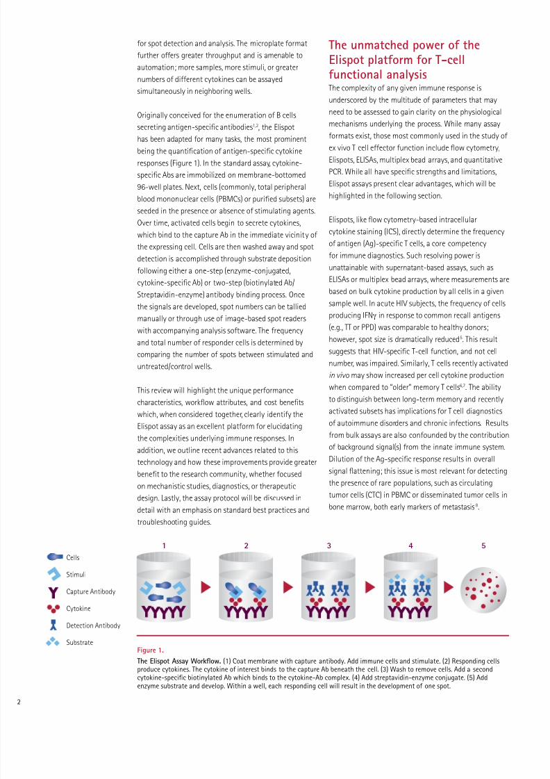
Elispot is one o the ew techniques permitting
quantitative single cell analysis o biological unction
(e.g., cytokine release). With intracellular cytokine
staining (ICS), where cytokine detection occurs prior
to release, there is the potential or misleading results
due to post-translational modulation beore or during
the secretory process16. The duration o an ICS assay is
limited by the toxicity o protein transport inhibitors such
as Breeldin A or Momensin. For quantitative RT-PCR,
detection is even urther removed rom actual unction,
since the target being measured is mRNA. Elispot assays
are also independent o secretion kinetics, a signicant
act given the unsynchronized nature o the responding
T cells pool. For ICS, all cells are killed via xation at a
pre-determined time. Cytolytic response mediators, such
as granzyme B and perorin, are stored in granules then
released upon proper stimuli17-19. Due to this uniqueregulatory mechanism, ICS will alsely identiy all eector
memory cells (~20% o total T cells) as perorin-positive.
Perhaps o greater signicance is the Lysispot assay, a
modied Elispot capable o enumerating Ag-specic
cytotoxic CD8+ T cell eector unction through direct
7/28/2019 Elispot assays: state-of-the-art tools for functional analysis of cellular immunology
http://slidepdf.com/reader/full/elispot-assays-state-of-the-art-tools-for-functional-analysis-of-cellular 4/12
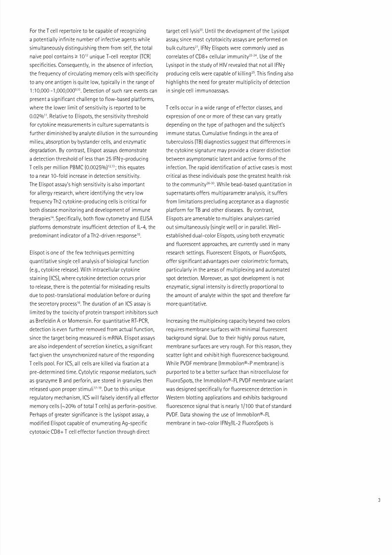
presented in Figure 2 (images) and Figure 5 (page 6,
spot counts). Beyond multiplexing, FluoroSpots permit
distinction o two simultaneously measured unctional
outputs. Multiplexing also serves to reduce required
sample size. The ever-expanding availability o discrete
fuorochromes, when combined with multi-fuorescent
imaging instrumentation and ully automated sample
acquisition and data analysis, provides the ramework or
unsurpassed polyunctional analysis o Ag-specic T cell
responses via Elispots.
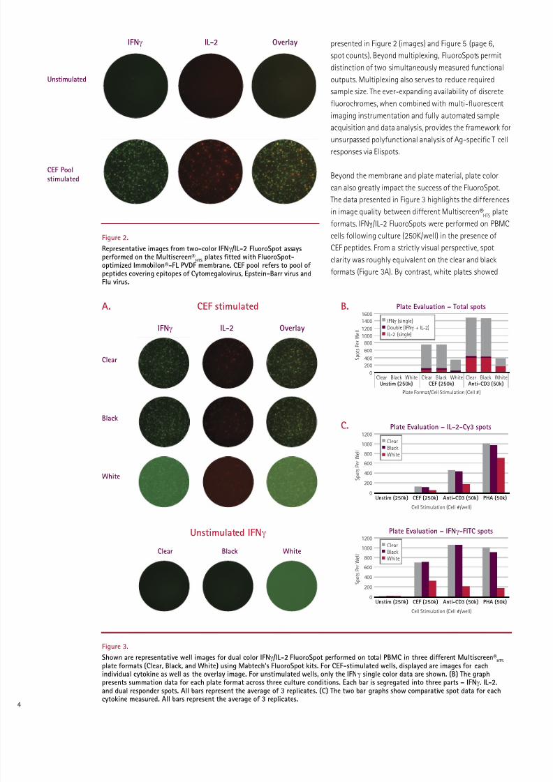
Beyond the membrane and plate material, plate color
can also greatly impact the success o the FluoroSpot.
The data presented in Figure 3 highlights the dierences
in image quality between dierent Multiscreen®HTS
plate
ormats. IFNg/IL-2 FluoroSpots were perormed on PBMC
cells ollowing culture (250K/well) in the presence o
CEF peptides. From a strictly visual perspective, spot
clarity was roughly equivalent on the clear and black
ormats (Figure 3A). By contrast, white plates showed
Figure 2.
Representative images rom two-color IFNg /IL-2 FluoroSpot assaysperormed on the Multiscreen®
HTSplates ftted with FluoroSpot-
optimized Immobilon®-FL PVDF membrane. CEF pool reers to pool o
peptides covering epitopes o Cytomegalovirus, Epstein-Barr virus andFlu virus.
Figure 3.
Shown are representative well images or dual color IFNg /IL-2 FluoroSpot perormed on total PBMC in three dierent Multiscreen®HTS
plate ormats (Clear, Black, and White) using Mabtech’s FluoroSpot kits. For CEF-stimulated wells, displayed are images or eachindividual cytokine as well as the overlay image. For unstimulated wells, only the IFNg single color data are shown. (B) The graph
presents summation data or each plate ormat across three culture conditions. Each bar is segregated into three parts – IFNg, IL-2,and dual responder spots. All bars represent the average o 3 replicates. (C) The two bar graphs show comparative spot data or eachcytokine measured. All bars represent the average o 3 replicates.
Unstimulated
CEF Pool
stimulated
Clear
Black
White
IFNg
IFNg
Clear
IL-2
IL-2
CEF stimulatedA. B.
C.
Unstimulated IFNg
Black
Overlay
Overlay
White
Plate Evaluation – Total spots
Plate Format/Cell Stimulation (Cell #)
S p o t s P e r W e l l
Clear
1600
BlackUnstim (250k)
White Clear BlackAnti-CD3 (50k)
WhiteClear BlackCEF (250k)
White
1400
1200
1000
800
600
400
200
0
IFNγ (single)Double (IFNγ + IL-2)IL-2 (single)
Plate Evaluation – IL-2-Cy3 spots
Cell Stimulation (Cell #/well)
S p o t s P e r W e l l
1200
Unstim (250k) Anti-CD3 (50k) PHA (50k)CEF (250k)
1000
800
600
400
200
0
Clear
Black
White
Plate Evaluation – IFNγ-FITC spots
Cell Stimulation (Cell #/well)
S p o t s P e r W e l l
1200
Unstim (250k) Anti-CD3 (50k) PHA (50k)CEF (250k)
1000
800
600
400
200
0
Clear
Black
White
7/28/2019 Elispot assays: state-of-the-art tools for functional analysis of cellular immunology
http://slidepdf.com/reader/full/elispot-assays-state-of-the-art-tools-for-functional-analysis-of-cellular 5/12
high background signal, making spot detection dicult,
particularly in the Green channel (IFNg-FITC). The high
background occurred even ater a signicant reduction
in exposure time (roughly 1/5). High background was
most likely due to increased refectance as compared
to black or clear rames where light is either absorbed
by or passes through the surrounding plate material,
respectively. A comparison o spot counts demonstrated
a signicant reduction in “spots counted” on white
plates when compared to either black or clear ormats
(Figure 3B-C). Once again, the discrepancy was more
signicant or IFNg spots, where almost 80% reduction
in total spots was observed. The background issue may
be eliminated i wells were punched out and analyzed
separately; however, this may not be practical i large
experiments are to be perormed. Given their similarities
in perormance, the clear plate ormat oers the more
practical option, as it also acilitates visually monitoring
reagent addition. It should be noted that all analyses
were perormed using the iSpot™ system (AID). Dueto dierences in perormance characteristics, other
fuorescent plate readers, may not demonstrate the same
plate preerence.
Unlike fow cytometry, where instrument priming can
result in sample loss, every cel l in an Elispot is measured.
Elispots also, on average, require one-tenth as many
cells per test, which provides a crucial advantage under
conditions where samples are precious (remote settings)
and/or limiting (pediatric or immunosuppressed test
subjects). One long-standing problem with the 96-well
microplates has been the waste o unused wells in
small-scale assays such as that occurring in diagnostic
analysis o a single patient sample. EMD Millipore oers
8-well strips (Catalogue No. M8IPS4510) designed or
the diagnostic community; this ormat is particularly
attractive to resource-limited countries where diseases
such as TB and HIV are most devastating (Figure 4A)28.
Constructed in a transparent ormat, the strips are
suitable or FluoroSpots, as well as standard enzymatic
options, and perorm comparably to the standard 96-well
plate (Figure 4B, 5). The 8-well strips are currently part o
Oxord Immunotech’s T-SPOT.TB Test, an FDA-approvedIFNg Elispot test designed specically or diagnosis o
tuberculosis inection.
Working with ar ewer cells per assay also means that
multiple replicates can be perormed, thereby increasing
statistical power. Such discriminatory capacity is not
possible with bulk assays. Although Elispots and fow
cytometry assays have similar protocol steps, Elispot
data acquisition/analysis is ar easier to perorm and
less time-consuming than fow cytometry. In act,
data rom a 96-well Elispot plate can be acquired and
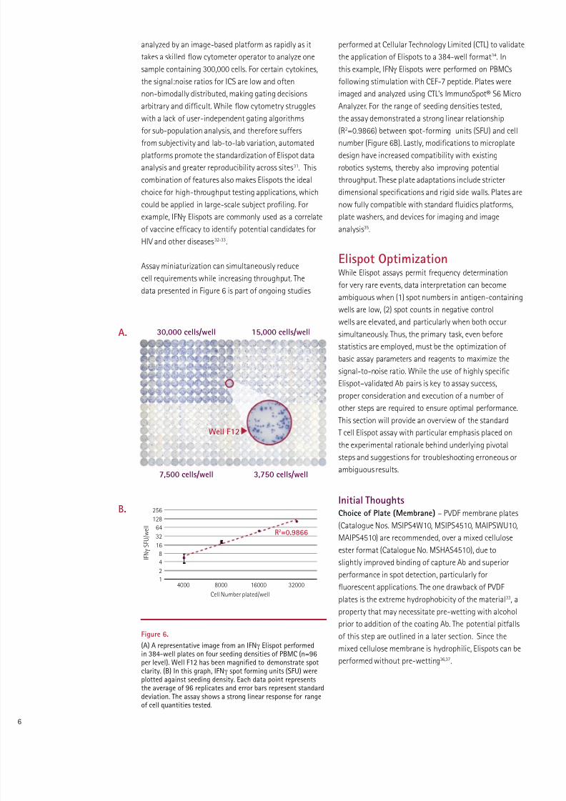
Figure 5.
8-well strips perorm similarly to the standard 96-wellMultiScreen® plates. The bar graph presents summation dataor each ormat across three culture conditions. Each bar issegregated into three parts – IFNg, IL-2, and dual responderspots. All bars represent the average o 3 replicates.
A.
B.
IFNg
IL-2
Unstimulated CEF Pool
Overlay
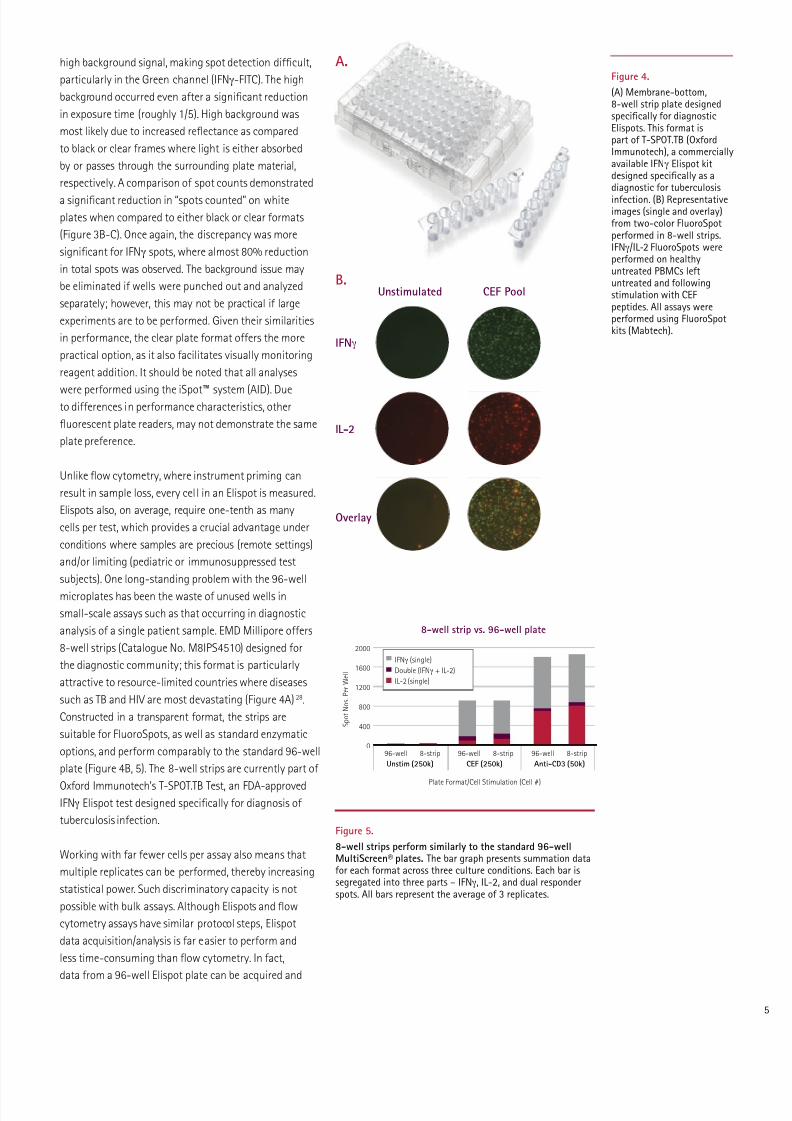
Figure 4.
(A) Membrane-bottom,8-well strip plate designedspecifcally or diagnosticElispots. This ormat ispart o T-SPOT.TB (OxordImmunotech), a commerciallyavailable IFNg Elispot kitdesigned specifcally as adiagnostic or tuberculosis
inection. (B) Representativeimages (single and overlay)rom two-color FluoroSpotperormed in 8-well strips.IFNg/IL-2 FluoroSpots wereperormed on healthyuntreated PBMCs letuntreated and ollowingstimulation with CEFpeptides. All assays wereperormed using FluoroSpotkits (Mabtech).
8-well strip vs. 96-well plate
Plate Format/Cell Stimulation (Cell #)
S p o t N o s . P e r W e l l
96-well
Unstim (250k)
8-strip 96-well 8-strip 96-well 8-strip
Anti-CD3 (50k)CEF (250k)
2000
1600
1200
800
400
0
IFNγ (single)
Double (IFNγ + IL-2)
IL-2 (single)
7/28/2019 Elispot assays: state-of-the-art tools for functional analysis of cellular immunology
http://slidepdf.com/reader/full/elispot-assays-state-of-the-art-tools-for-functional-analysis-of-cellular 6/12
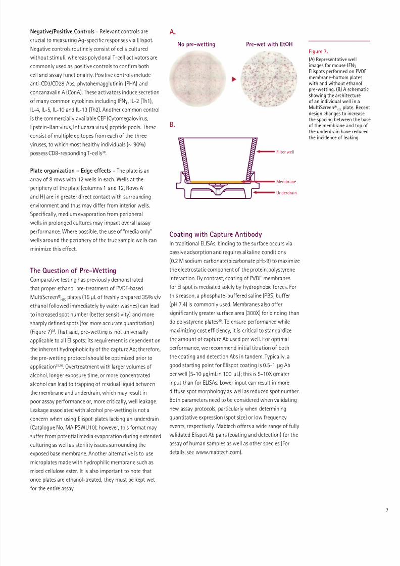
perormed at Cellular Technology Limited (CTL) to validate
the application o Elispots to a 384-well ormat34. In
this example, IFNg Elispots were perormed on PBMCs
ollowing stimulation with CEF-7 peptide. Plates were
imaged and analyzed using CTL’s ImmunoSpot® S6 Micro
Analyzer. For the range o seeding densities tested,
the assay demonstrated a strong linear relationship
(R2=0.9866) between spot-orming units (SFU) and cell
number (Figure 6B). Lastly, modications to microplate
design have increased compatibility with existing
robotics systems, thereby also improving potential
throughput. These plate adaptations include stricter
dimensional specications and rigid side walls. Plates are
now ully compatible with standard fuidics platorms,
plate washers, and devices or imaging and image
analysis35.
Elispot OptimizationWhile Elispot assays permit requency determination
or very rare events, data interpretation can become
ambiguous when (1) spot numbers in antigen-containing
wells are low, (2) spot counts in negative control
wells are elevated, and particularly when both occur
simultaneously. Thus, the primary task, even beore
statistics are employed, must be the optimization o
basic assay parameters and reagents to maximize the
signal-to-noise ratio. While the use o highly specic
Elispot–validated Ab pairs is key to assay success,
proper consideration and execution o a number o
other steps are required to ensure optimal perormance.
This section will provide an overview o the standardT cell Elispot assay with particular emphasis placed on
the experimental rationale behind underlying pivotal
steps and suggestions or troubleshooting erroneous or
ambiguous results.
Initial ThoughtsChoice o Plate (Membrane) – PVDF membrane plates
(Catalogue Nos. MSIPS4W10, MSIPS4510, MAIPSWU10,
MAIPS4510) are recommended, over a mixed cellulose
ester ormat (Catalogue No. MSHAS4510), due to
slightly improved binding o capture Ab and superiorperormance in spot detection, particularly or
fuorescent applications. The one drawback o PVDF
plates is the extreme hydrophobicity o the material33, a
property that may necessitate pre-wetting with alcohol
prior to addition o the coating Ab. The potential pitalls
o this step are outlined in a later section. Since the
mixed cellulose membrane is hydrophilic, Elispots can be
perormed without pre-wetting36,37.
analyzed by an image-based platorm as rapidly as it
takes a skilled fow cytometer operator to analyze one
sample containing 300,000 cells. For certain cytokines,
the signal:noise ratios or ICS are low and oten
non-bimodally distributed, making gating decisions
arbitrary and dicult. While fow cytometry struggles
with a lack o user-independent gating algorithms
or sub-population analysis, and thereore suers
rom subjectivity and lab-to-lab variation, automated
platorms promote the standardization o Elispot data
analysis and greater reproducibility across sites31. This
combination o eatures also makes Elispots the ideal
choice or high-throughput testing applications, which
could be applied in large-scale subject proling. For
example, IFNg Elispots are commonly used as a correlate
o vaccine ecacy to identiy potential candidates or
HIV and other diseases32-33.
Assay miniaturization can simultaneously reduce
cell requirements while increasing throughput. Thedata presented in Figure 6 is part o ongoing studies
Figure 6.
(A) A representative image rom an IFNg Elispot perormedin 384-well plates on our seeding densities o PBMC (n=96per level). Well F12 has been magnifed to demonstrate spotclarity. (B) In this graph, IFNg spot orming units (SFU) wereplotted against seeding density. Each data point representsthe average o 96 replicates and error bars represent standarddeviation. The assay shows a strong linear response or rangeo cell quantities tested.
A.
B.
30,000 cells/well
7,500 cells/well
15,000 cells/well
3,750 cells/well
Well F12
256
128
64
32
16
8
42
14000 8000 16000
R2=0.9866
Cell Number plated/well
I F N γ S
F U / w e l l
32000
7/28/2019 Elispot assays: state-of-the-art tools for functional analysis of cellular immunology
http://slidepdf.com/reader/full/elispot-assays-state-of-the-art-tools-for-functional-analysis-of-cellular 7/12
Coating with Capture AntibodyIn traditional ELISAs, binding to the surace occurs via
passive adsorption and requires alkaline conditions
(0.2 M sodium carbonate/bicarbonate pH>9) to maximize
the electrostatic component o the protein:polystyrene
interaction. By contrast, coating o PVDF membranes
or Elispot is mediated solely by hydrophobic orces. For
this reason, a phosphate-buered saline (PBS) buer(pH 7.4) is commonly used. Membranes also oer
signicantly greater surace area (300X) or binding than
do polystyrene plates39. To ensure perormance while
maximizing cost eciency, it is critical to standardize
the amount o capture Ab used per well. For optimal
perormance, we recommend initial titration o both
the coating and detection Abs in tandem. Typically, a
good starting point or Elispot coating is 0.5-1 µg Ab
per well (5-10 µg/mLin 100 µL); this is 5-10X greater
input than or ELISAs. Lower input can result in more
diuse spot morphology as well as reduced spot number.
Both parameters need to be considered when validating
new assay protocols, particularly when determining
quantitative expression (spot size) or low requency
events, respectively. Mabtech oers a wide range o ully
validated Elispot Ab pairs (coating and detection) or the
assay o human samples as well as other species (For
details, see www.mabtech.com).
Negative/Positive Controls – Relevant controls are
crucial to measuring Ag-specic responses via Elispot.
Negative controls routinely consist o cells cultured
without stimuli, whereas polyclonal T-cell activators are
commonly used as positive controls to conrm both
cell and assay unctionality. Positive controls include
anti-CD3/CD28 Abs, phytohemagglutinin (PHA) and
concanavalin A (ConA). These activators induce secretion
o many common cytokines including IFNg, IL-2 (Th1),
IL-4, IL-5, IL-10 and IL-13 (Th2). Another common control
is the commercially available CEF (Cytomegalovirus,
Epstein-Barr virus, Infuenza virus) peptide pools. These
consist o multiple epitopes rom each o the three
viruses, to which most healthy individuals (~ 90%)
possess CD8-responding T-cells38.
Plate organization - Edge eects – The plate is an
array o 8 rows with 12 wells in each. Wells at the
periphery o the plate (columns 1 and 12, Rows A
and H) are in greater direct contact with surroundingenvironment and thus may dier rom interior wells.
Specically, medium evaporation rom peripheral
wells in prolonged cultures may impact overall assay
perormance. Where possible, the use o “media only”
wells around the periphery o the true sample wells can
minimize this eect.
The Question o Pre-WettingComparative testing has previously demonstrated
that proper ethanol pre-treatment o PVDF-based
MultiScreen®HTS plates (15 µL o reshly prepared 35% v/vethanol ollowed immediately by water washes) can lead
to increased spot number (better sensitivity) and more
sharply dened spots (or more accurate quantitation)
(Figure 7)35. That said, pre-wetting is not universally
applicable to all Elispots; its requirement is dependent on
the inherent hydrophobicity o the capture Ab; thereore,
the pre-wetting protocol should be optimized prior to
application35,36. Overtreatment with larger volumes o
alcohol, longer exposure time, or more concentrated
alcohol can lead to trapping o residual liquid between
the membrane and underdrain, which may result in
poor assay perormance or, more critically, well leakage.
Leakage associated with alcohol pre-wetting is not a
concern when using Elispot plates lacking an underdrain
(Catalogue No. MAIPSWU10); however, this ormat may
suer rom potential media evaporation during extended
culturing as well as sterility issues surrounding the
exposed base membrane. Another alternative is to use
microplates made with hydrophilic membrane such as
mixed cellulose ester. It is also important to note that
once plates are ethanol-treated, they must be kept wet
or the entire assay.
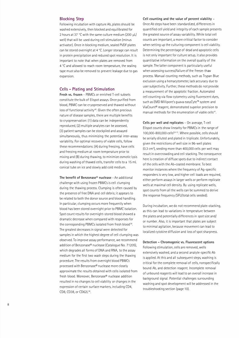
Figure 7.
(A) Representative wellimages or mouse IFNg Elispots perormed on PVDFmembrane-bottom plateswith and without ethanolpre-wetting. (B) A schematicshowing the architectureo an individual well in aMultiScreen®
HTSplate. Recent
design changes to increasethe spacing between the baseo the membrane and top o the underdrain have reducedthe incidence o leaking.
A.
B.
No pre-wetting Pre-wet with EtOH
Filter well
Membrane
Underdrain
7/28/2019 Elispot assays: state-of-the-art tools for functional analysis of cellular immunology
http://slidepdf.com/reader/full/elispot-assays-state-of-the-art-tools-for-functional-analysis-of-cellular 8/12
Cell counting and the value o percent viability –
Once Ab steps have been standardized, dierences in
quantied cell yield and integrity o each sample presents
the greatest source o assay variability. While total cell
counts are important, a more critical actor to consider
when setting up the culturing component is cell viability.
Determining the percentage o dead and apoptotic cells
is not only important or culture setup, it also provides
quantitative inormation on the overall quality o the
sample. The latter component is particularly useul
when assessing success/ailure o the reeze-thaw
process. Manual counting methods, such as Trypan Blue
exclusion using a hemacytometer, lack accuracy due to
user subjectivity. Further, these methods do not provide
a measurement o the apoptotic raction. Automated
cell counting via fow cytometry using fuorescent dyes,
such as EMD Millipore’s guava easyCyte™ system and
ViaCount® reagent, demonstrated superior precision to
manual methods or the enumeration o viable cells41.
Cells per well and replicates – On average, T-cell
Elispot counts show linearity or PBMCs in the range o
100,000-800,000 cells5,9-13. Where possible, cells should
be serially diluted and plated in triplicate. Unortunately,
given the restrictions o well size in 96-well plates
(0.3 cm2), seeding more than 400,000 cells per well may
result in overcrowding and cell stacking. The consequence
here is creation o diuse spots due to indirect contact
o the cells with the Ab-coated membrane. To best
monitor instances where the requency o Ag-specic
responders is very low, and higher cell loads are required,
either perorm assays in larger wells or perorm replicate
wells at maximal cell density. By using replicate wells,
spot counts rom all the wells can be summed to derive
the response requency (SFU/total cells seeded).
During incubation, we do not recommend plate-stacking,
as this can lead to variations in temperature between
the plates and potentially dierences in spot size and/
or number. Also, it is important that plates are subject
to minimal agitation, because movement can lead to
localized cytokine diusion and loss o spot sharpness.
Detection - Chromogenic vs. Fluorescent options
Following stimulation, cells are removed, wells
extensively washed, and a second analyte-specic Ab
is applied. At this and all subsequent steps, washing is
critical or the complete removal o cells, nonspecically
bound Ab, and detection reagent. Incomplete removal
o unbound reagents will lead to an overall increase in
background signal. Potential challenges surrounding
washing and spot development will be addressed in the
troubleshooting section (page 10).
Blocking StepFollowing incubation with capture Ab, plates should be
washed extensively, then blocked and equilibrated or
2 hours at 37 °C with the same culture medium (200 µL/
well) that will be used during cell stimulation (minus
activator). Once in blocking medium, sealed PVDF plates
can be stored overnight at 4 °C. Longer storage can result
in protein precipitation and reduced spot resolution. It is
important to note that when plates are removed rom
4 °C and allowed to reach room temperature, the sealing
tape must also be removed to prevent leakage due to gas
expansion.
Cells - Plating and StimulationFresh vs. rozen - PBMCs or enriched T-cell subsets
constitute the bulk o Elispot assays. Once puried rom
blood, PBMC can be cryopreserved and thawed without
loss o unctional activity40. Given the oten precious
nature o disease samples, there are multiple benets
to cryopreservation: (1) data can be independently
reproduced, (2) multiple analytes can be assessed,
(3) patient samples can be stockpiled and assayed
simultaneously, thus minimizing the potential inter-assay
variability. For optimal recovery o viable cells, ollow
these recommendations: (A) during reezing, have cells
and reezing medium at room temperature prior to
mixing and (B) during thawing, to minimize osmotic lysis
during washing o thawed cells, transer cells to a 15 mL
conical tube on ice and slowly add cold medium.
The beneft o Benzonase® nuclease - An additionalchallenge with using rozen PBMCs is cell clumping
during the thawing process. Clumping is oten caused by
the presence o ree DNA and cell debris; it appears to
be related to both the donor source and blood handling.
In particular, clumping occurs more requently when
blood has been stored overnight prior to PBMC isolation.
Spot count results or overnight-stored blood showed a
dramatic decrease when compared with responses or
the corresponding PBMCs isolated rom resh blood36.
The greatest decreases in signal were detected or
samples in which the highest degree o cell clumping was
observed. To improve assay perormance, we recommend
addition o Benzonase® nuclease (Catalogue No. 71205),
which degrades all orms o DNA and RNA, to the assay
medium or the rst two wash steps during the thawing
procedure. The results rom overnight blood PBMCs
processed with Benzonase® nuclease more closely
approximate the results obtained with cells isolated rom
resh blood. Moreover, Benzonase® nuclease addition
resulted in no changes to cell viability or changes in the
expression o certain surace markers, including CD4,
CD8, CD38, or CD62L36.
7/28/2019 Elispot assays: state-of-the-art tools for functional analysis of cellular immunology
http://slidepdf.com/reader/full/elispot-assays-state-of-the-art-tools-for-functional-analysis-of-cellular 9/12
Elispot assays may be perormed either with antibodies
directly conjugated to the detection moti (enzyme
or fuorochrome) or as a two-step process involving
a biotin/streptavidin-conjugated Ab pair. While the
two-step process oers greater intensity due to signal
amplication, and thereore may be preerable in cases
where cytokine production per cell is low (allergy/Th2
responses), this protocol also suers rom a greater
potential or background staining due to nonspecic
interaction with the coating Ab. With enzymes, such as
horseradish peroxidase (HRP), a precipitating substrate
(TMB or AEC) is used or spot detection. Due to HRP’s
high turnover rate, spot development is ast
(≤5 minutes). By contrast, spot development using
alkaline phosphatase-conjugated Abs is ar slower but
with apprecibly lower background. For chromogenic
assays perormed on MultiScreen®HTS
plates (those with
underdrains), it is recommended that the underdrain
be removed prior to substrate addition; ailure to do so
can result in high background staining. Once removed,plates should be propped up to minimize membrane
contact. To enhance spot visualization, plates should be
dried without a lid, upside down, at room temperature
or several hours. For long-term storage, plates should be
kept in a dark, dry place at room temperature to prevent
bleaching o spots.
As previously discussed, the use o fuorescent conjugates
oers signicant advantages over colorimetric schemes
especially or dual cytokine applications or where greater
quantitative assessments o individual spots is desired.
While FITC- and Cy3-conjugated Abs are commonly used,
the choice o fuorescent probe is limited only by the
availability o conjugates and detection platorms.
Spot Counting and Analysis – What is a real spot?
Each spot represents the ‘cytokine signature’ o a single
cell. Due to diusion properties, a true spot has a
densely colored center which ades toward the edges;
the size and/or color intensity o the spots is determined
by the amount o cytokine released. That said, due
to dierences in analyte measured, incubation time,
antibody concentration, enzyme activity, substrates andother materials used as well as the unctional state o the
cytokine-secreting cells, spot size and density can vary
greatly. Artiactual spots may appear and can be caused
by the aggregation o antibodies or the incomplete
removal o cells and cellular debris. Morphologically,
these spots can be dierentiated rom ‘true’ spots by
their homogeneity in color intensity and sharper (non-
rounded) edges.
From the above description, manual spot counting
by light microscopy would be classied as a highly
subjective process, raught with a great degree o
inter-user variability. Further, when considering the
sheer number o wells that may need to quantied
in a standard vaccine trials, the task o Elispot data
analysis becomes a ar too laborious task or human
eyes. The availability o sophisticated Elispot readers
oers a complete solution or precise evaluation o spot
data. These instruments include eatures to overcome
problems with variable background intensity and the
ability to distinguish true single cell spots rom artiacts.
The latter capability relies upon the use o minimum and
maximum threshold values or spot size and intensity,
permitting the exclusion o weak bystander responses
and clusters containing multiple cells, respectively.
Beyond speed, spot analysis sotware oers process
standardization, a critical component when studies are
perormed across sites, such as is the case or diagnostic
testing and vaccine trials. Moreover, Elispot readersand analysis sotware open the door or more precise
measurements o spots, permitting the quantitation o
secretion o multiple cytokines on a per-cell basis.
7/28/2019 Elispot assays: state-of-the-art tools for functional analysis of cellular immunology
http://slidepdf.com/reader/full/elispot-assays-state-of-the-art-tools-for-functional-analysis-of-cellular 10/12
0
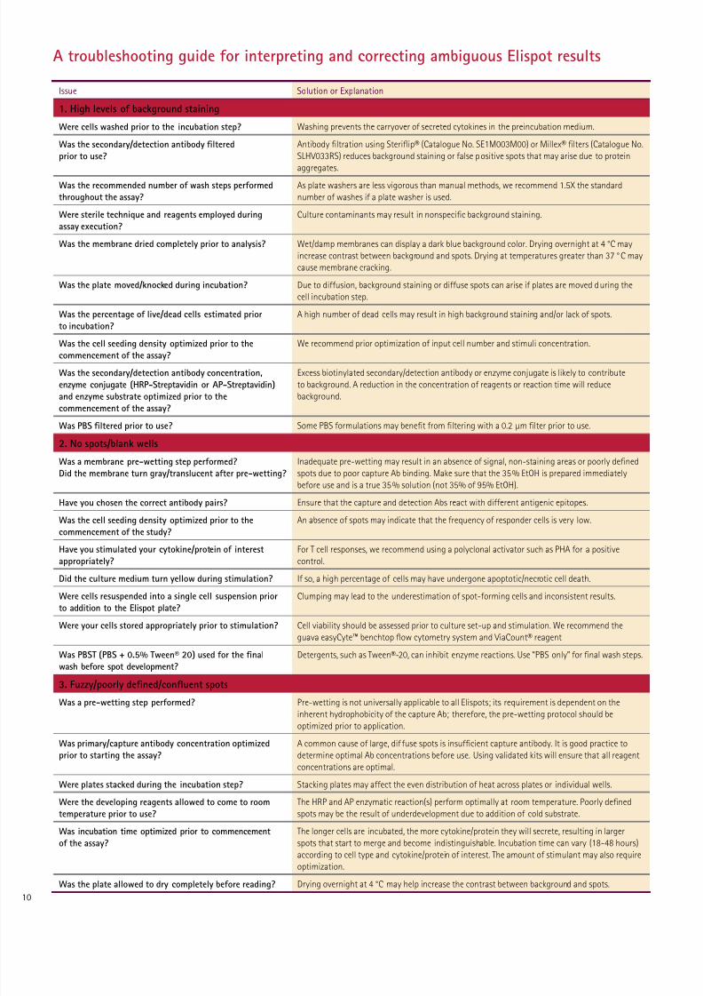
Issue Solution or Explanation
1. High levels o background staining
Were cells washed prior to the incubation step? Washing prevents the carryover o secreted cytokines in the preincubation medium.
Was the secondary/detection antibody fltered
prior to use?
Antibody ltration using Sterifip® (Catalogue No. SE1M003M00) or Millex® lters (Catalogue No.
SLHV033RS) reduces background staining or alse positive spots that may arise due to protein
aggregates.
Was the recommended number o wash steps perormed
throughout the assay?
As plate washers are less vigorous than manual methods, we recommend 1.5X the standard
number o washes i a plate washer is used.
Were sterile technique and reagents employed during
assay execution?
Culture contaminants may result in nonspecic background staining.
Was the membrane dried completely prior to analysis? Wet/damp membranes can display a dark blue background color. Drying overnight at 4 °C may
increase contrast between background and spots. Drying at temperatures greater than 37 °C may
cause membrane cracking.
Was the plate moved/knocked during incubation? Due to diusion, background staining or diuse spots can arise i plates are moved during the
cell incubation step.
Was the percentage o live/dead cells estimated prior
to incubation?
A high number o dead cells may result in high background staining and/or lack o spots.
Was the cell seeding density optimized prior to the
commencement o the assay?
We recommend prior optimization o input cell number and stimuli concentration.
Was the secondary/detection antibody concentration,
enzyme conjugate (HRP-Streptavidin or AP-Streptavidin)
and enzyme substrate optimized prior to the
commencement o the assay?
Excess biotinylated secondary/detection antibody or enzyme conjugate is likely to contribute
to background. A reduction in the concentration o reagents or reaction time will reduce
background.
Was PBS fltered prior to use? Some PBS ormulations may benet rom ltering with a 0.2 µm lter prior to use.
2. No spots/blank wells
Was a membrane pre-wetting step perormed?
Did the membrane turn gray/translucent ater pre-wetting?
Inadequate pre-wetting may result in an absence o signal, non-staining areas or poorly dened
spots due to poor capture Ab binding. Make sure that the 35% EtOH is prepared immediately
beore use and is a true 35% solution (not 35% o 95% EtOH).
Have you chosen the correct antibody pairs? Ensure that the capture and detection Abs react with dierent antigenic epitopes.
Was the cell seeding density optimized prior to thecommencement o the study? An absence o spots may indicate that the requency o responder cells is very low.
Have you stimulated your cytokine/protein o interest
appropriately?
For T cell responses, we recommend using a polyclonal activator such as PHA or a positive
control.
Did the culture medium turn yellow during stimulation? I so, a high percentage o cells may have undergone apoptotic/necrotic cell death.
Were cells resuspended into a single cell suspension prior
to addition to the Elispot plate?
Clumping may lead to the underestimation o spot-orming cells and inconsistent results.
Were your cells stored appropriately prior to stimulation? Cell viability should be assessed prior to culture set-up and stimulation. We recommend the
guava easyCyte™ benchtop fow cytometry system and ViaCount® reagent
Was PBST (PBS + 0.5% Tween® 20) used or the fnal
wash beore spot development?
Detergents, such as Tween®-20, can inhibit enzyme reactions. Use “PBS only” or nal wash steps.
3. Fuzzy/poorly defned/conuent spots
Was a pre-wetting step perormed? Pre-wetting is not universally applicable to all Elispots; its requirement is dependent on the
inherent hydrophobicity o the capture Ab; thereore, the pre-wetting protocol should be
optimized prior to application.
Was primary/capture antibody concentration optimized
prior to starting the assay?
A common cause o large, di use spots is insucient capture antibody. It is good practice to
determine optimal Ab concentrations beore use. Using validated kits will ensure that all reagent
concentrations are optimal.
Were plates stacked during the incubation step? Stacking plates may aect the even distribution o heat across plates or individual wells.
Were the developing reagents allowed to come to room
temperature prior to use?
The HRP and AP enzymatic reaction(s) perorm optimally at room temperature. Poorly dened
spots may be the result o underdevelopment due to addition o cold substrate.
Was incubation time optimized prior to commencement
o the assay?
The longer cells are incubated, the more cytokine/protein they will secrete, resulting in larger
spots that start to merge and become indistinguishable. Incubation time can vary (18-48 hours)
according to cell type and cytokine/protein o interest. The amount o stimulant may also require
optimization.
Was the plate allowed to dry completely beore reading? Drying overnight at 4 °C may help increase the contrast between background and spots.
A troubleshooting guide or interpreting and correcting ambiguous Elispot results
7/28/2019 Elispot assays: state-of-the-art tools for functional analysis of cellular immunology
http://slidepdf.com/reader/full/elispot-assays-state-of-the-art-tools-for-functional-analysis-of-cellular 11/12
1
Reerences1. Czerkinsky, C., Nilsson, L., Nygren, H., Ouchterlony, O.,and Tarkowski, A. (1983). A solid-phase enzyme-linked
immunospot (ELISPOT) assay or enumeration o speciic
antibody-secreting cells. J Immunol Methods 65 (1–2):
109–121.
2. Sedgewick, J. and Holt, P. (1983) A solid-phase
immunoenzymatic technique or the enumeration o
speciic antibody secreting cells. J. Immunol. Methods.
57:301-309.
3. Starita-Geribaldi, M. and Suduka, L. (1990) Fast
electrotranser o human serum proteins in their native
state rom polyacrylamide thin gradient gels reinorced
by textiles to Polyvinylidene diluoride membranes.
Bioseparations. 1:111-117.
4. Wahlgren, M. and Arnebrant, T. (1991) Protein
Absorption to solid suraces. Tibtech 9:201-208.
5. Helms, T., Boehm, B., Asaad, R., Trezza, M., Lehmann,
P., and Tary-Lehmann, M. (2000) Direct visualization
o cytokine-producing recall antigen-speciic CD4
memory T cells in healthy individuals and HIV patients.
J. Immunol. 164:3723-3732.
6. Schlingmann, T., Shlive, C., Targoni, O., Tary-Lehmann,
M., and Lehmann, P. (2009) Increased per cell IFN-g
productivity indicates recent in vivo activation o T cells.
Cellular Immunol. 258:131-137.
7. Hesse, M., Karulin, A., Boehm, B., Lehmann, P. and Tary-
Lehmann, M. (2001). A T-cell clone’s avidity is a unction
o its activation state. J. Immunol. 167:1353-1361.
8. Panabieres, C., Riethdor, S., and Pantel, K.
(2008) Circulating Tumor Cells and Bone Marrow
Micrometastasis. Clin. Cancer Res. 14(16):5013-5021.
9. Davis, M. and Bjorkman, P. (1988) T-cell antigen receptor
genes and T-cell recognition. Nature 334:395-399.
10. Arstila, T., Casrouge, A., Baron, V., Even, J., Kanellopoulos,
J., and Kourilsky, P. (1999). A direct estimate o the
human αβ T cell receptor diversity. Science 286,
958–961.
11. Sun, Y., Iglesias, E., Samri, A., Kamkamidze, G., Decoville,
T., Carcelain, G., and Autran, B. (2003) A systematice
comparison o methods to measure HIV-1 speciic CD8 T
cells. J. Immunol. Methods. 272:23-24.
12. Schmittel, A., Keilholz, U., and Scheibenbogen, C. (1997)
Evaluation o the intereron-gamma ELISPOT-assay or
quantiication o peptide speciic T lymphocytes rom
peripheral blood. J. Immunol. Methods. 210:160-174.
13. Streeck, H., Frahm, N., and Walker, B. (2009) The roleo IFN-g elispot assay in HIV vaccine research. Nature
Protoc. 4:461-469.
14. Jakobson, E., Masjedi, K., Ahlborg, N., Lundeberg,
L., Karlberg, A., and Scheynius, A. (2002) Cytokine
production in nickel-sensitized individuals analyzed
with enzyme-linked immunosorbent assay: possible
implications or diagnosis. Br. J. Dermatol. 147:442-449.
15. Ewen, C. and Baca-Estrada, M. (2001). Evaluation o
interleukin-4 concentration by ELISA is inluenced by
the consumption o IL-4 cultured cells. J. Intereron
Cytok. Res. 21:39-43.
16. Lehmann, P., and Zhang, W. (2012) Unique Strengths o
ELISPOT or T cell diagnostics. Methods Mol. Biol. 792:3-
23.
17. Rininsland, F., Helms, T., Assad, R., Boehm, B., and Tary-
Lehmann. (2000) Granzyme B ELISPOT assay or ex vivo
measurements o T cell immunity. J. Immunol. Methods.
240:143-155.
18. Kleen, T., Assad, R., Landry, S., Boehm, B., and Tary-
Lehmann, M. (2005) Tc1 eector diversity shows
dissociated expression o granzyme B and intereron-
gamma in HIV inection. AIDS. 18:383-392.
19. Kuerton, S., Kleen, T., Assad, R., Lehmann, P., and Tary-
Lehmann, M. (2007) Dissociated production o perorin,
granzyme B, and IFN-g by HIV-speciic CD8+ cells in HIV
inection. AIDS Res and Human Retroviruses. 24:62-71.
20. Snyder, J., Bowers, W., Livingstone, A., Lee, F., Federo,
H., and Mosmann T. (2003) Measuring the requency
o mouse and human cytotoxic T cells by the Lysispot
assay: independent regulation o cytokine secretion and
short-term killing. Nat. Med. 9: 231–235.
21. Brunner, K., Mauel, J., Cerottini, J., and Chapuis, B.
(1968) Quantitative assay o the lytic action o immune
lymphoid cells on 51-Cr-labeled allogeneic cells in vitro ;
inhibition by isoantibody and by drugs. Immunology.
14:181-196.
22. Ghanekar, S., Nomura, L., Suni, M., Picker, L., Maecker,
H., and Maino, V. (2001) Gamma intereron expression
in CD8+ T cells is a marker or circulating cytotoxic
T lymphocytes that recognize an HLA A2-restricted
epitope o human cytomegalovirus phosphoprotein
pp65. Clin. Vaccine Immunol. 8(3):628-631.
23. Horton, H., Russell, N., Moore, E., Frank, I., Baydo, R.,
Havenar-Daughton, C., Lee, D., Deers, M., Hudgens,
M., Weinhold, K., and McElrath, M. (2004) Correlation
between intereron-gamma secretion and cytotoxicity,
in virus-speciic memory T cells. J. Inect Dis.
190(9):1692-1696.
24. Betts, M., Nason, M., West, S., De Rosa, S., Migueles,
S., Abraham, J., Lederman, M., Benito, J., Goepert,
P., Connors, M., Roderer, M., and Koup, R. (2006) HIV
nonprogressors preerentially maintain highly unctional
HIV-speciic CD8+ T cells. Blood. 107:4781-4789.25. Snyder-Cappione, J., Divekar, A., Maupin, J., Demeter,
L., and Mosmann, T. (2006) HIV-Speciic Cytotoxic Cell
requencies Measured directly ex vivo by the Lysispot
Assay can be higher or Lower than the requencies
o IFN-g Secreting cells: Anti-HIV cytotoxicity is not
generally relative to other chronic virus responses. J.
Immunol. 176:2662-2668.
26. Casey R, Blumenkrantz D, Millington K, Montamat-
Sicotte D, Kon OM, Wickremasinghe M, Bremang
S, Magtoto M, Sridhar S, Connell D, and Lalvani A.
(2010) Enumeration o unctional T-cell subsets
by luorescence-immunospot deines signatures
o pathogen burden in tuberculosis. PLoS One.
5(12):e15619.
27. Sester, U., Fousse, M., Dirks, J., Mack, U., Prasses, A.,
Signh, M., Lalvani, A., and Sester, M. (2011) Whole-blood
low-cytometric analysis o antigen-speciic CD4 T-cell
cytokine proiles distinguishes active tuberculosis rom
non-active states. PloS One 6:e17813.
28. Hirsch, C., Hussain, R., Toossi, Z., Dawood, G., Shahid, F.,
and Ellner, J. (1996) Cross-modulation by transorming
growth actor beta in human tuberculosis: suppression
o antigen-driven blastogenesis and intereron gamma
production. Proc. Natl. Aca. Sci. USA. 93:3193-3198.
29. Harris, J., De Haro, S., Master, S., Keane, J., Roberts,
E., Delgado, M., and Deretic, V. (2007) T helper 2
cytokines inhibit autophagic control o intracellular
Mycobacterium tuberculosis. Immunity 27:505-517.
30. Tincati, C., Cappione, A., and Snyder-Cappione, J. (2012)
Distinguishing Latent rom Active Mycobacterium
tuberculosis Inection Using Elispot Assays: Looking
Beyond Intereron-gamma. Cells. 1:89-99.
31. Boaz, M., Hayes, P., Terragona, T., Seamons, L., Cooper, A.,
Birungi, J., Kitandwe, P., Semaganda, A., Kaleebu, P. et al.
(2009) Concordant Proiciency in Measurement o T-Cell
Immunity in Human Immunodeiciency Virus Vaccine
Clinical Trials by Peripheral Blood Mononuclear Cell and
Enzyme-Linked Immunospot Assays in Laboratories rom
Three Continents. Clin. Vaccine Immunol. 16(2):147-155.
32. Streeck, H., Lichtereld M, Alter G, Meier A, Teigen N,
Yassine-Diab B, Sidhu HK, Little S, Kelleher A, Routy JP,
Rosenberg ES, Sekaly RP, Walker BD, Alteld M. (2007)
Recognition o a deined region within p24 gag by
CD8+ T cells during primary human immunodeiciency
virus type 1 inection in individuals expressing
protective HLA class I alleles. J. Virol. 81:7725–7731.
33. Alteld, M., Kalie ET, Qi Y, Streeck H, Lichtereld M,
Johnston MN, Burgett N, Swartz ME, Yang A, Alter
G, Yu XG, Meier A, Rockstroh JK, Allen TM, Jessen H,
Rosenberg ES, Carrington M, Walker BD. (2006) HL A
alleles associated with delayed progression to AIDS
contribute strongly to the initial CD8(+) T cell response
against HIV-1. PLoS Med. 3:e403.
34. Hanson, J., Caspell, R., Karacsony, E., Sundararaman,
S., Lehmann, P., and Zhang, W. (2012) Validation o ELISPOT Assay in 384 Well Format: 400 T Cell Tests done
in 10ml Blood.http://www.immunospot.com/ileadmin/
Documents/Posters/CTL384Validation LR.pd
35. Weiss, A. (2012) Overview o Membranes and Membrane
devices used in Research and Diagnostic ELISPOT Assays.
Methods Mol. Biol. 792:243-256.
36. Smith, J., Liu, X., Kauhold, R., Clair, J., Caulield, M.
(2001) Development and Validation o a Gamma
Intereron ELISPOT Assay or Quantitation o Cellular
Immune responses to Varicella-Zoster Virus. Clin Diag.
Lab. Immunol. 8(5):871-879.
37. Kalyuzhny, A. (2009) ELISPOT assay on membrane
microplates. Methods Mol. Biol. 536:355-365.
38. Currier, J., Kuta, E., Turk, E., Earhart, L., Loomis-Price,
L., Janetzki, S., Ferrari, G., Brix, D., and Cox, J. (2002) Apanel o MHC class I restricted viral peptides or use
as a quality control or vaccine trails ELISPOT assays. J.
Immunol. Methods. 260:157-172.
39. Pitt, A.M. (1987). The nonspeciic protein binding
o polymeric microporous membranes. J. Parenteral
Science and Tech. 41(3): 110-113.
40. Kreher, C., Dittrich, M., Guerkov, R., Boehm, B., and
Tary-Lehmann, M. (2003) CD4+ and CD8+ cells in
crypreserved human PBMC maintain ull unctionality
in cytokine ELISPOTS. J. Immunol. Methods. 278:79-93.
41. Ferrari, G. and Pollara, J. (2004) Validating the Guava
ViaCount® Assay, an Automated Cell Counting/
Viability Method or Use in ELISpot Assays. EMD
Millipore Corporation. Application Note Literature No.
MK10441101.
AcknowledgementsWe would like to thank Tomas Ernemar (Mabtech) or provid-
ing all the FluoroSpot data as well as manuscript review. The
384-well data was kindly provided by Jodie Hansen (CTL). We
would also like to thank Sylvia Janetzki (ZellNet Consulting)
or manuscript review.
7/28/2019 Elispot assays: state-of-the-art tools for functional analysis of cellular immunology
http://slidepdf.com/reader/full/elispot-assays-state-of-the-art-tools-for-functional-analysis-of-cellular 12/12
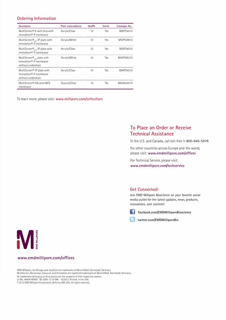
Ordering Inormation
Description Plate material/color Qty/Pk Sterile Catalogue No.
MultiScreen® 8-well strip with
Immobilon®-P membrane
Acrylic/Clear 10 Yes M8IPS4510
MultiScreen®HTS
-IP plate with
Immobilon®-P membrane
Acrylic/White 10 Yes MSIPS4W10
MultiScreen®HTS
-IP plate with
Immobilon®-P membrane
Acrylic/Clear 10 Yes MSIPS4510
MultiScreen®HTS
plate with
Immobilon®-P membrane
without underdrain
Acrylic/White 10 Yes MAIPSWU10
MultiScreen®-IP plate with
Immobilon®-P membrane
without underdrain
Acrylic/Clear 10 Yes MAIPS4510
MultiScreen®-HA with MCE
membrane
Styrene/Clear 10 Yes MAHAS4510
To learn more, please visit: www.millipore.com/cellculture
EMD Millipore, the M logo, and easyCyte are trademarks o Merck KGaA, Darmstadt, Germany.MultiScreen, Benzonase, Viacount, and Immobilon are registered trademarks o Merck KGaA, Darmstadt, Germany.
All trademarks belonging to third parties are the property o their respective owners.
Lit No. AN4474EN00 BS-GEN-12-07399 10/2012 Printed in the USA.© 2012 EMD Millipore Corporation, Billerica, MA USA. All rights reserved.
www.emdmillipore.com/oices
To Place an Order or ReceiveTechnical Assistance
In the U.S. and Canada, call toll-ree 1-800-645-5476
For other countries across Europe and the world,
please visit: www.emdmillipore.com/ofces
For Technical Service, please visit:
www.emdmillipore.com /techservice
Get Connected!Join EMD Millipore Bioscience on your avorite social
media outlet or the latest updates, news, products,
innovations, and contests!
acebook.com/EMDMilliporeBioscience
twitter.com/EMDMilliporeBio