Embed Size (px)
Citation preview
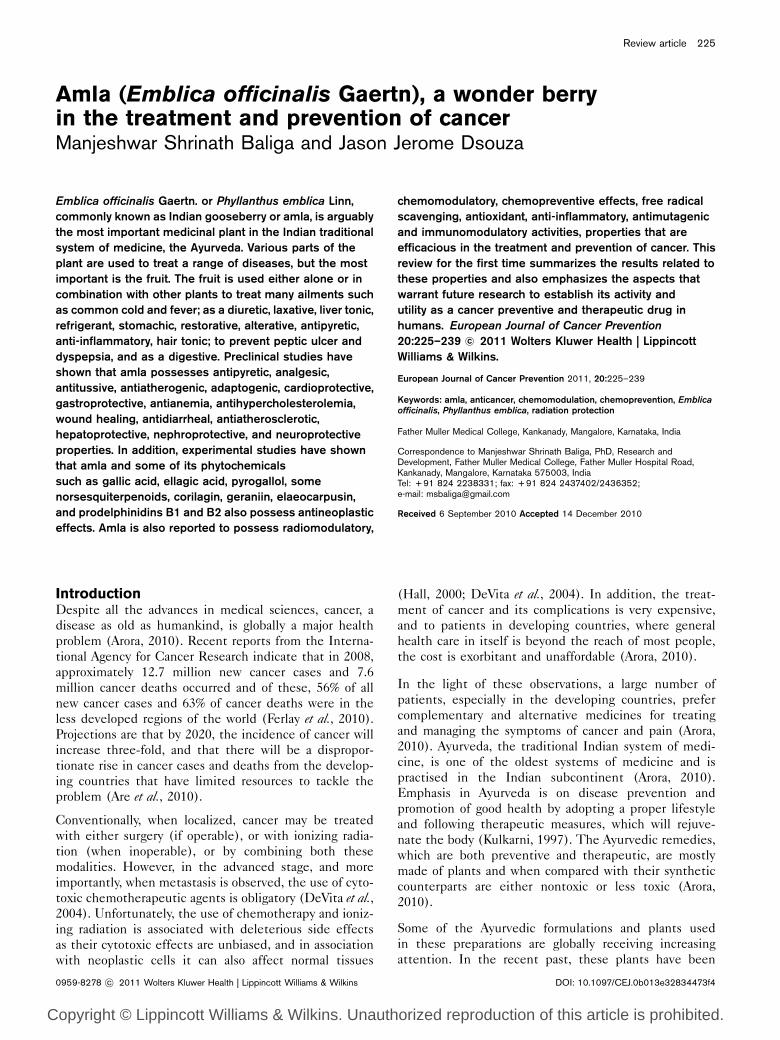
Amla (Emblica officinalis Gaertn), a wonder berryin the treatment and prevention of cancerManjeshwar Shrinath Baliga and Jason Jerome Dsouza
Emblica officinalis Gaertn. or Phyllanthus emblica Linn,
commonly known as Indian gooseberry or amla, is arguably
the most important medicinal plant in the Indian traditional
system of medicine, the Ayurveda. Various parts of the
plant are used to treat a range of diseases, but the most
important is the fruit. The fruit is used either alone or in
combination with other plants to treat many ailments such
as common cold and fever; as a diuretic, laxative, liver tonic,
refrigerant, stomachic, restorative, alterative, antipyretic,
anti-inflammatory, hair tonic; to prevent peptic ulcer and
dyspepsia, and as a digestive. Preclinical studies have
shown that amla possesses antipyretic, analgesic,
antitussive, antiatherogenic, adaptogenic, cardioprotective,
gastroprotective, antianemia, antihypercholesterolemia,
wound healing, antidiarrheal, antiatherosclerotic,
hepatoprotective, nephroprotective, and neuroprotective
properties. In addition, experimental studies have shown
that amla and some of its phytochemicals
such as gallic acid, ellagic acid, pyrogallol, some
norsesquiterpenoids, corilagin, geraniin, elaeocarpusin,
and prodelphinidins B1 and B2 also possess antineoplastic
effects. Amla is also reported to possess radiomodulatory,
chemomodulatory, chemopreventive effects, free radical
scavenging, antioxidant, anti-inflammatory, antimutagenic
and immunomodulatory activities, properties that are
efficacious in the treatment and prevention of cancer. This
review for the first time summarizes the results related to
these properties and also emphasizes the aspects that
warrant future research to establish its activity and
utility as a cancer preventive and therapeutic drug in
humans. European Journal of Cancer Prevention
20:225–239 �c 2011 Wolters Kluwer Health | Lippincott
Williams & Wilkins.
European Journal of Cancer Prevention 2011, 20:225–239
Keywords: amla, anticancer, chemomodulation, chemoprevention, Emblicaofficinalis, Phyllanthus emblica, radiation protection
Father Muller Medical College, Kankanady, Mangalore, Karnataka, India
Correspondence to Manjeshwar Shrinath Baliga, PhD, Research andDevelopment, Father Muller Medical College, Father Muller Hospital Road,Kankanady, Mangalore, Karnataka 575003, IndiaTel: + 91 824 2238331; fax: + 91 824 2437402/2436352;e-mail: [email protected]
Received 6 September 2010 Accepted 14 December 2010
IntroductionDespite all the advances in medical sciences, cancer, a
disease as old as humankind, is globally a major health
problem (Arora, 2010). Recent reports from the Interna-
tional Agency for Cancer Research indicate that in 2008,
approximately 12.7 million new cancer cases and 7.6
million cancer deaths occurred and of these, 56% of all
new cancer cases and 63% of cancer deaths were in the
less developed regions of the world (Ferlay et al., 2010).
Projections are that by 2020, the incidence of cancer will
increase three-fold, and that there will be a dispropor-
tionate rise in cancer cases and deaths from the develop-
ing countries that have limited resources to tackle the
problem (Are et al., 2010).
Conventionally, when localized, cancer may be treated
with either surgery (if operable), or with ionizing radia-
tion (when inoperable), or by combining both these
modalities. However, in the advanced stage, and more
importantly, when metastasis is observed, the use of cyto-
toxic chemotherapeutic agents is obligatory (DeVita et al.,2004). Unfortunately, the use of chemotherapy and ioniz-
ing radiation is associated with deleterious side effects
as their cytotoxic effects are unbiased, and in association
with neoplastic cells it can also affect normal tissues
(Hall, 2000; DeVita et al., 2004). In addition, the treat-
ment of cancer and its complications is very expensive,
and to patients in developing countries, where general
health care in itself is beyond the reach of most people,
the cost is exorbitant and unaffordable (Arora, 2010).
In the light of these observations, a large number of
patients, especially in the developing countries, prefer
complementary and alternative medicines for treating
and managing the symptoms of cancer and pain (Arora,
2010). Ayurveda, the traditional Indian system of medi-
cine, is one of the oldest systems of medicine and is
practised in the Indian subcontinent (Arora, 2010).
Emphasis in Ayurveda is on disease prevention and
promotion of good health by adopting a proper lifestyle
and following therapeutic measures, which will rejuve-
nate the body (Kulkarni, 1997). The Ayurvedic remedies,
which are both preventive and therapeutic, are mostly
made of plants and when compared with their synthetic
counterparts are either nontoxic or less toxic (Arora,
2010).
Some of the Ayurvedic formulations and plants used
in these preparations are globally receiving increasing
attention. In the recent past, these plants have been
Review article 225
0959-8278 �c 2011 Wolters Kluwer Health | Lippincott Williams & Wilkins DOI: 10.1097/CEJ.0b013e32834473f4
Copyright © Lippincott Williams & Wilkins. Unauthorized reproduction of this article is prohibited.
investigated for their pharmacological effects in accordance
with modern medicine (Arora, 2010). One such plant
that has been extensively studied is the medium-sized
deciduous tree Emblica officinalis Gaertn. or Phyllanthusemblica Linn belonging to the family Euphorbiaceae. The
plant species, which was originally native to India, is today
found growing in Pakistan, Uzbekistan, Sri Lanka, South-
East Asia, China, and Malaysia (Warrier et al., 1996; Zhang
et al., 2003; Khan, 2009). Colloquially, they are known as
Indian gooseberry tree, emblic myrobalans, and Malacca
tree in English and amla in Hindi. The other vernacular
names have been listed in Table 1.
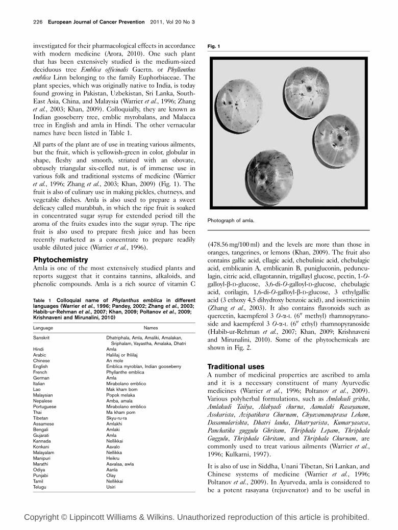
All parts of the plant are of use in treating various ailments,
but the fruit, which is yellowish-green in color, globular in
shape, fleshy and smooth, striated with an obovate,
obtusely triangular six-celled nut, is of immense use in
various folk and traditional systems of medicine (Warrier
et al., 1996; Zhang et al., 2003; Khan, 2009) (Fig. 1). The
fruit is also of culinary use in making pickles, chutneys, and
vegetable dishes. Amla is also used to prepare a sweet
delicacy called murabbah, in which the ripe fruit is soaked
in concentrated sugar syrup for extended period till the
aroma of the fruits exudes into the sugar syrup. The ripe
fruit is also used to prepare fresh juice and has been
recently marketed as a concentrate to prepare readily
usable diluted juice (Warrier et al., 1996).
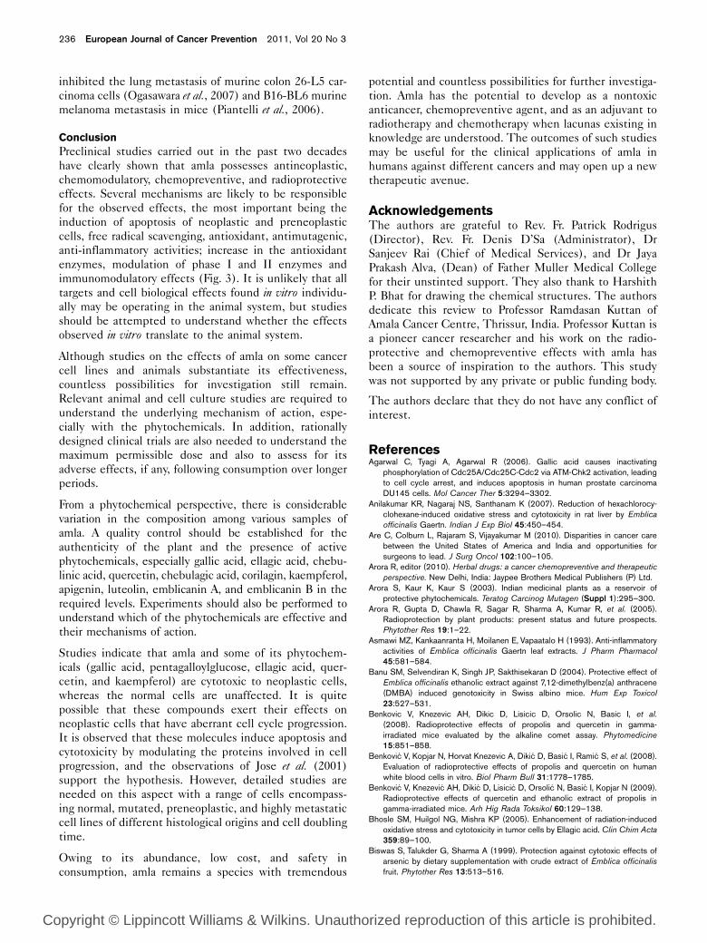
PhytochemistryAmla is one of the most extensively studied plants and
reports suggest that it contains tannins, alkaloids, and
phenolic compounds. Amla is a rich source of vitamin C
(478.56 mg/100 ml) and the levels are more than those in
oranges, tangerines, or lemons (Khan, 2009). The fruit also
contains gallic acid, ellagic acid, chebulinic acid, chebulagic
acid, emblicanin A, emblicanin B, punigluconin, peduncu-
lagin, citric acid, ellagotannin, trigallayl glucose, pectin, 1-O-
galloyl-b-D-glucose, 3,6-di-O-galloyl-D-glucose, chebulagic
acid, corilagin, 1,6-di-O-galloyl-b-D-glucose, 3 ethylgallic
acid (3 ethoxy 4,5 dihydroxy benzoic acid), and isostrictiniin
(Zhang et al., 2003). It also contains flavonoids such as
quercetin, kaempferol 3 O-a-L (600 methyl) rhamnopyrano-
side and kaempferol 3 O-a-L (600 ethyl) rhamnopyranoside
(Habib-ur-Rehman et al., 2007; Khan, 2009; Krishnaveni
and Mirunalini, 2010). Some of the phytochemicals are
shown in Fig. 2.
Traditional usesA number of medicinal properties are ascribed to amla
and it is a necessary constituent of many Ayurvedic
medicines (Warrier et al., 1996; Poltanov et al., 2009).
Various polyherbal formulations, such as Amlakadi gritha,
Amlakadi Tailya, Alakyadi churna, Aamalaki Rasayanam,
Asokarista, Avipatikara Churnam, Chyavananaprasa Leham,
Dasamularishta, Dhatri lauha, Dhatryarista, Kumaryasava,
Panchatika guggulu Ghritam, Thriphala Lepam, ThriphalaGuggulu, Thriphala Ghritam, and Thriphala Churnam, are
commonly used to treat various ailments (Warrier et al.,1996; Kulkarni, 1997).
It is also of use in Siddha, Unani Tibetan, Sri Lankan, and
Chinese systems of medicine (Warrier et al., 1996;
Poltanov et al., 2009). In Ayurveda, amla is considered to
be a potent rasayana (rejuvenator) and to be useful in
Table 1 Colloquial name of Phylanthus emblica in differentlanguages (Warrier et al., 1996; Pandey, 2002; Zhang et al., 2003;Habib-ur-Rehman et al., 2007; Khan, 2009; Poltanov et al., 2009;Krishnaveni and Mirunalini, 2010)
Language Names
Sanskrit Dhatriphala, Amla, Amaliki, Amalakan,Sriphalam, Vayastha, Amalaka, Dhatri
Hindi AmlaArabic Haliilaj or IhliilajChinese An moleEnglish Emblica myroblan, Indian gooseberryFrench Phyllanthe emblicaGerman AmlaItalian Mirabolano emblicoLao Mak kham bomMalaysian Popok melakaNepalese Amba, amalaPortuguese Mirabolano emblicoThai Ma kham pomTibetan Skyu-ru-raAssamese AmlakhiBengali AmlakiGujarati AmlaKannada NellikkaiKonkani AavaloMalayalam NellikkaManipuri HeikruMarathi Aavalaa, awlaOdiya AanlaPunjabi OlayTamil NellikkaiTelugu Usiri
Fig. 1
Photograph of amla.
226 European Journal of Cancer Prevention 2011, Vol 20 No 3
Copyright © Lippincott Williams & Wilkins. Unauthorized reproduction of this article is prohibited.

Fig. 2
HO
HO
HO
O
OO
OO
O
HO
HO
HOHO
HO
O
OO
O
O
O
O O
O
OO
OH
OH
Corilagin
Pedunculagin
OH
OH
OH
OH
OH
OH
HO
HO OHO OH
OH
OH
OH
OH
O
OH
(a) (b)
(c)
HO HO
HO
HO
HO
O
Gallic acid Ellagic acid
Pyrogallol
Quercetin Kaempferol
O
O
O
OOH
OH
OH
OH
OH
HOHO
O
O
O
OOH
OH
OH
OH
OH
OH
OH
OH
HO
HO
HOR
CO
HO
HO
HO
HO
OH
O
OO
O
OO
OO
OO
O
O
O OH
OH
OH
OHOH
O
HOHO
HO
HO
OH
H
O
O
O
O
COO
OOCO
OChebulic acid
Chebulinic acid
O OH
OH
OH
OH
OHR
COOCH2
HO
HO
HO
Some important phytochemicals of amla.
Amla (Emblica officinalis Gaertn) in cancer Baliga and Dsouza 227
Copyright © Lippincott Williams & Wilkins. Unauthorized reproduction of this article is prohibited.
stalling the degenerative and senescence process, to
promote longevity, enhance digestion, to treat constipa-
tion, reduce fever, purify the blood, reduce cough,
alleviate asthma, strengthen the heart, benefit the eyes,
stimulate hair growth, enliven the body, and enhance the
intellect (Pandey, 2002).
In various folk medicines the fruits, which are astringent,
are useful in treating ophthalmic problems, dyspepsia, gas-
tritis, hyperacidity, constipation, colitis, hemorrhoids,
hematuria, menorrhagia, treat anemia, diabetes, cough,
asthma, osteoporosis, premature graying of hair, weakness
and fatigue. Amla is also reported to possess hepatopro-
tective, cardioprotective, diuretic, laxative, refrigerant, sto-
machic, restorative, alterative, antipyretic, anti-inflammatory
properties, is a hair tonic, prevents peptic ulcer dyspepsia,
and is a digestive medicine (Pandey, 2002). It is used for
a variety of ailments such as anemia, hyperacidity,
diarrhea, eye inflammation, leucorrhea, jaundice, nerve
debility, liver complaints, cough, and anomalies of urine
(Pandey, 2002).
Scientifically validated studiesPreclinical studies carried out in the past three decades
have validated many of the traditional uses of amla.
Experiments have shown that amla possesses antibacte-
rial, antifungal, antiviral, antidiabetic, hypolipidemic,
antiulcerogenic, free radical scavenging, antioxidant, anti-
mutagenic, anti-inflammatory and immunomodulatory,
antipyretic, analgesic, antitussive, antiatherogenic, adap-
togenic, snake venom neutralizing, gastroprotective,
antianemia, antihypercholesterolemia, wound healing,
antidiarrheal, antiatherosclerotic, hepatoprotective, ne-
phroprotective, and neuroprotective properties (Khan,
2009; Krishnaveni and Mirunalini, 2010). Compelling
preclinical studies with both in-vitro and in-vivo systems
have shown that amla possesses anticancer, chemopreven-
tive, cytoprotective, and radioprotective effects. Here, an
attempt is made to analyze the role of amla in the
treatment and prevention of cancer.
Amla as an antineoplastic agentPreclinical studies have shown that the aqueous extract of
amla causes a concentration-dependent cytotoxic effect on
L 929 cells in vitro and that the IC50 was observed to be
16.5mg/ml (Jose et al., 2001). The extract also caused
apoptosis in Dalton’s lymphoma ascites and CeHa cell lines
(Rajeshkumar et al., 2003). Khan et al. (2002) studied the
antiproliferative activity of the extract in the human tumor
cell lines of different histological orgins (human erythro-
myeloid K562, B-lymphoid Raji, T-lymphoid Jurkat,
erythroleukemic HEL) and observed it to be effective.
Recently, Ngamkitidechakul et al. (2010) have observed
that the aqueous extract of amla, which contains tannins
(43%), uronic acid (11%), and gallic acid (21%), inhibited
the growth of A549 (lung), HepG2 (liver), HeLa (cervical),
MDA-MB-231 (breast), SK-OV3 (ovarian), and SW620
(colorectal) cells in vitro. However, at the same concentra-
tion the extract did not cause similar level of cytotoxicity
in the MRC5, normal lung fibroblast, suggesting it to be
safe for normal cells (Ngamkitidechakul et al., 2010). The
extract also induced apoptosis in HeLa, A549, MDA-MB-
231, and SK-OV3 cells (Ngamkitidechakul et al., 2010).
An amla extract possesses antiproliferative activity in
MCF7 and MDA-MB-231 breast cancer cell lines and
also induces an increase in ERamRNA in these cells
(Lambertini et al., 2004). The extract was devoid of
cytotoxic effects on the normal Chinese hamster ovary
cell line, suggesting it to be selectively cytotoxic to only
neoplastic cells (Sumantran et al., 2007). Administering
the extract to Dalton’s lymphoma-bearing mice caused a
reduction in ascitic volume (when the tumor cells were
inoculated in the peritoneum) and solid tumor growth
(when inoculated subcutaneously). The amla extract
significantly reduced the solid tumors and prolonged
survival time. At a molecular level, the extract was
observed to inhibit the cell cycle-regulating enzyme,
Cdc25 phosphatase, in a dose-dependent manner and the
IC50 was observed to be 5 mg/ml (Jose et al., 2001).
Studies have also shown that some of the compounds
present in amla are effective in inhibiting the prolifera-
tion of neoplastic cells in vitro and also in tumor-bearing
animals. The hydrolyzable tannins of amla are also re-
ported to possess selective cytotoxicity to the human oral
squamous cell carcinoma and salivary gland tumor cell
lines, while they were nontoxic to the normal human
gingival fibroblasts. The dimeric compounds, oenothein
B, woodfordin C, and woodfordin D, were more effective
than the monomeric compounds, while the macrocyclic
ellagitannin oligomers were more effective than gallic
acid and epigallocatechin gallate. These compounds also
induced apoptosis in the neoplastic cells and mechanistic
studies showed that the effect was mediated by the
prooxidant actions, but not through the generation of
hydrogen peroxide (Sakagami et al., 2000).
Zhang et al. (2004) evaluated the antiproliferative effects
of 18 phytochemicals of amla (norsesquiterpenoids,
phenolic compounds, and proanthocyanidin polymers)
in B16F10, HeLa, and MK-1 cells in vitro. Among the
norsesquiterpenoids, it was observed that the glycoside
phyllaemblicins B and C were highly potent in all the
three cells [B16F10 (GI50 at 2.0, 3.5 mg/ml, respectively),
HeLa (GI50 at 3.0, 12.0 mg/ml, respectively), and MK-1
(GI50 at 7.0 mg/ml for both compounds)]. However, with
respect to the phenolic compounds, all showed inhibi-
tory activity against the three tumor cell lines (at a
concentration of < 68 mg/ml), and were more effective
against B16F10 than against HeLa and MK-1 cells. The
highest activity was observed with corilagin, geraniin,
elaeocarpusin, and prodelphinidins B1 and B2 against
B16F10 (Zhang et al., 2004).
228 European Journal of Cancer Prevention 2011, Vol 20 No 3
Copyright © Lippincott Williams & Wilkins. Unauthorized reproduction of this article is prohibited.
Pyrogallol, a catechin compound of amla, is also reported
to possess a potent antiproliferative effect on human lung
cancer cell lines and, to a lesser degree, on the human
bronchial epithelium cell line. Detailed studies with the
human lung cancer cell lines H441 (lung adenocarcino-
ma) and H520 (lung squamous cell carcinoma) have
shown that pyrogallol inhibited the growth of these cells,
triggered apoptosis by increasing Bax and concomitantly
decreasing Bcl-2, arrested the cells in the G2/M phase by
affecting the cyclin B1, Cdc25C and increasing the
phosphorylation of Cdc2 (Thr14). The in-vitro observa-
tions also extended into in-vivo studies with xenograft
nude mice (Yang et al., 2009).
Gallic acid, another chief constituent of amla, is also
shown to cause a concentration- and time-dependent
inhibition of proliferation, and to induce apoptosis in
BEL-7404 cells (Zhong et al., 2009). Gallic acid is also
shown to cause apoptosis in human non-small-cell lung
cancer NCI-H460 cells (Ji et al., 2009), A375.S2 human
melanoma cells (Ji et al., 2009), human bladder transi-
tional carcinoma cell line (TSGH-8301 cell) (Lo et al.,2010) and HeLa cervical cancer cells (You et al., 2010).
Consuming gallic acid (0.3–1% in drinking water)
inhibited the growth of prostate cancer and retarded
the progression to advanced-stage adenocarcinoma in
mice with transgenic adenocarcinoma of the prostate by
suppressing cell cycle progression and cell proliferation
and, concomitantly, increasing apoptosis (Raina et al.,2008). Gallic acid also suppressed lung xenograft tumor
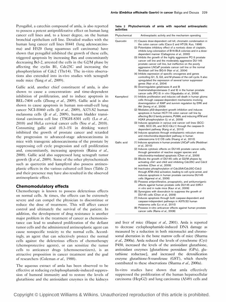
growth (Ji et al., 2009). Some of the other phytochemicals
such as quercetin and kampferol also possess antineo-
plastic effects in the various cultured cell lines (Table 2)
and their presence may have also resulted in the observed
antineoplastic effect.
Chemomodulatory effectsChemotherapy is known to possess deleterious effects
on normal cells. At times, the effects can be extremely
severe and can compel the physician to discontinue or
reduce the dose of treatment. This will affect cancer
control and ultimately the survival of the patient. In
addition, the development of drug resistance is another
major problem in the treatment of cancer as chemoresis-
tance can lead to unabated proliferation of the defiant
tumor cells and the administered antineoplastic agent can
cause nonspecific toxicity to the normal cells. Accord-
ingly, an agent that can selectively protect the normal
cells against the deleterious effects of chemotherapy
(chemoprotective agents), or can sensitize the tumor
cells to anticancer drugs (chemosensetizers), is an
attractive proposition in cancer treatment and the goal
of researchers (Coleman et al., 1988).
The aqueous extract of amla has been observed to be
effective at reducing cyclophosphamide-induced suppres-
sion of humoral immunity and to restore the levels of
glutathione and the antioxidant enzymes in the kidneys
and liver of mice (Haque et al., 2001). Amla is reported
to decrease cyclophosphamide-induced DNA damage as
measured by a reduction in both micronuclei and chromo-
somal aberration in the bone marrow cells of mice (Sharma
et al., 2000a). Amla reduced the levels of cytochrome (Cyt)
P450, increased the levels of the antioxidant glutathione,
antioxidant enzymes [glutathione peroxidase (GPx), glu-
tathione reductase], and increased the detoxification
enzyme glutathione-S-transferase (GST), which thereby
contributed to these observations (Sharma et al., 2000a).
In-vitro studies have shown that amla effectively
suppressed the proliferation of the human hepatocellular
carcinoma (HepG2) and lung carcinoma (A549) cells and
Table 2 Phytochemicals of amla with reported antineoplasticactivities
Phytochemical Antineoplastic activity and the mechanism operating
Quercetin (1) Causes dose-dependent cell kill, chromatin condensation inthe colon cancer cells (Caco-2 and HT-29; Kuo, 1996)
(2) Potentiates inhibitory effect of a nontoxic dose of cisplatin,inhibits lung colonization of B16-BL6 colonies and in a dose-dependent manner (Caltagirone et al., 2000)
(3) Inhibits the growth of the highly aggressive PC-3 prostatecancer cell line and the moderately aggressive DU-145prostate cancer cell line, but ineffective on the poorlyaggressive LNCaP prostate cancer cell line or the normalfibroblast cell line BG-9 (Nair et al., 2004)
(4) Inhibits expression of specific oncogenes and genescontrolling G1, S, G2, and M phases of the cell cycle. It alsoupregulated the expression of several tumor suppressorgenes (Nair et al., 2004)
(5) Downregulates gelatinases A and B(matrixmetalloproteinases 2 and 9) in the human prostatecancer cells (PC-3) in vitro (Vijayababu et al., 2006)
Kaempferol (1) Inhibits proliferation and induces cell death in human gliomacells through caspase-dependent mechanisms involvingdownregulation of XIAP and survivin regulation by ERK andAkt (Jeong et al., 2009)
(2) Mediates p53-dependent growth inhibition and inducesapoptosis in human HCT116 colon cancer cell line byaffecting Bcl-2 family proteins, PUMA, and inducing ATM andH2AX phosphorylation (Li et al., 2009)
(3) Induces apoptosis in various oral cancer cell lines (SCC-1483, SCC-25, and SCC-QLL1) through the caspase-3-dependent pathway (Kang et al., 2010)
(4) Induces apoptosis through endoplasmic reticulum stressand mitochondria-dependent pathway in humanosteosarcoma U-2 OS cells (Huang et al., 2010)
Gallic acid (1) Induces apoptosis in human prostate LNCaP cells (Reddivariet al., 2010)
(2) Induces cytotoxic effects on DU145 prostate cancer cells,through generation of reactive oxygen species andmitochondria-mediated apoptosis (Chen et al., 2009)
(3) Blocks the growth of DU145 cells at G2/M phases byactivating chk1 and chk2 and inhibiting Cdc25C and Cdc2activities (Chen et al., 2009)
(4) Inactivates phosphorylation of Cdc25A/Cdc25C-Cdc2through ATM-chk2 activation, leading to cell cycle arrest, andinduces apoptosis in human prostate carcinoma DU145cells (Agarwal et al., 2006)
(5) Possess antiproliferative, proapoptotic, and antitumorigeniceffects against human prostate cells DU145 and 22Rv1in vitro and in nude mice (Kaur et al., 2009)
(6) Synergizes with doxorubicin to suppress the growth ofDU145 cells (Chen et al., 2009)
(7) Induces apoptosis through both caspase-dependent andcaspase-independent pathways in A375.S2 humanmelanoma cells (Lo et al., 2010)
(8) Possess in-vitro anticancer effects against human prostatecancer cells (Raina et al., 2008)
Amla (Emblica officinalis Gaertn) in cancer Baliga and Dsouza 229
Copyright © Lippincott Williams & Wilkins. Unauthorized reproduction of this article is prohibited.
synergized the cytotoxic effects of doxorubicin and
cisplatin, two important clinically used antineoplastic
drugs (Pinmai et al., 2008). The ethanolic extract of amla
also protected the cardiac myoblasts H9c2 cells against
doxorubicin-induced toxicity (Wattanapitayakul et al.,2005). Together these observations suggest that it is
quite possible that amla prevents doxorubicin-induced
cardiotoxicity to the normal cardiac myoblasts and,
concomitantly, sensitizes the antineoplastic effects on
cancer cells. However, detailed studies are required for
this hypothesis to be validated, especially in the relevant
animal models of study.
Amla as a radioprotective agentSince the discovery of the deleterious effects of ionizing
radiation, studies have been focused on developing
chemical radioprotectors that have the ability to decrease
the ill effects of radiation on normal tissues (Arora et al.,2005). The thiol compound amifostine is credited with
being the only radioprotector to have been approved by
the Food and Drug Administration to reduce the
incidence and severity of xerostomia in head and neck
cancer patients undergoing radiotherapy (Arora et al.,2005). Unfortunately, the application of this drug has so
far been less than hoped for, owing to its untoward
toxicity often being evidenced at the optimal radio-
protective doses (Arora et al., 2005).
With regard to the radioprotective effects of amla, studies
have shown that administering (50, 100, 200, 400, and
800 mg/kg b.wt./day) amla once daily for 7 consecutive
days before exposure to sublethal dose of g-radiation
(9 Gy) protected mice against the radiation-induced
sickness and mortality (Singh et al., 2005). Among all
the doses studied, the optimal effect was observed at
100 mg/kg b.w. as it delayed the radiation-induced leth-
ality and caused a survival of 87.5% when compared with
placebo-treated irradiated cohorts in which no survivors
were observed (Singh et al., 2005).
Administration of amla (100 mg/kg b.wt.) ameliorated the
radiation (5 Gy)-induced gastrointestinal damage as
evaluated by the histopathological studies, by quantifying
the crypt cell population, mitotic figures, and villus
length at all the assay points (12 h–30 days). Reports also
suggest that amla ameliorated the radiation-induced
hemopoietic damage (Hari Kumar et al., 2004). Feeding
mice with 2.5 g/kg b.wt. of amla for 10 consecutive days
before exposure to a single dose of 7 Gy of radiation
increased the total leukocyte count, bone marrow viabi-
lity, and levels of hemoglobin. However, treatment with
amla after exposure to irradiation (continuously for an-
other 15 days) was not as effective when compared with
administeration before radiation, suggesting it to be of
use only when exposure to radiation is planned (Hari
Kumar et al., 2004).
Mechanistic studies have shown that feeding amla
enhanced the activity of the various antioxidant enzymes
(catalase, superoxide dismutase, and GPx), the phase II
detoxifying enzyme, GST, and the antioxidant thiol,
glutathione, in the blood, with a concomitant decrease in
the levels of lipid peroxides (Hari Kumar et al., 2004).
Similar results were also observed by Jindal et al. (2009) in
mice intestine and together both these studies confirm
that amla significantly reduces the deleterious effects of
radiation at least in part through its antioxidant and
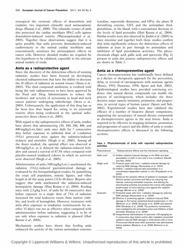
inhibition of lipid peroxidation activities. The phyto-
chemicals ellagic acid, gallic acid, and quercetin (Fig. 2)
present in amla also possess radioprotective effects and
are shown in Table 3.
Amla as a chemopreventive agentCancer chemoprevention has traditionally been defined
as a dietary or therapeutic approach for the prevention,
delay, or reversal of carcinogenesis with nontoxic agents
(Bonte, 1993; Pastorino, 1994; Sporn and Suh, 2002).
Epidemiological studies have provided convincing evi-
dence that natural dietary compounds can modify the
process of carcinogenesis, which includes the three
decisive steps: namely initiation, promotion, and progres-
sion, in several types of human cancer (Sporn and Suh,
2002). Experimental studies have also validated the
efficacy of a number of bioactive dietary components,
supporting the acceptance of natural dietary compounds
as chemopreventive agents in the near future. Amla is
reported to be effective in stopping initiation, promotion,
and progression of cancer and the ability of amla to render
chemopreventive effects is discussed in the following
sections.
Table 3 Phytochemicals of amla with reported radioprotectiveactivities
Phytochemical Radioprotective effects and the mechanism operating
Gallic acid (1) Inhibits radiation-induced damage to the DNA and lipidperoxidation in both in-vitro and in-vivo conditions (Gandhiand Nair, 2005)
Ellagic acid (1) Protects yeast cells from g-radiation-induced damage byreducing DNA damage (Nemavarkar et al., 2004)
(2) Inhibits g-radiation-induced lipid peroxidation in aconcentration-dependent manner in vitro (Priyadarsini et al.,2002)
(3) Enhances the cytotoxic effects of radiation in neoplastic cells(Ehrlich ascites carcinoma and HeLa) by inducing freeradicals, reducing antioxidant enzymes, and altering themitochondrial potential, but protects the normal cells (spleniclymphocytes) of tumor-bearing mice against the radiationdamage (Bhosle et al., 2005)
Quercetin (1) Protected yeast cells from g-radiation damage by reducingDNA damage (Nemavarkar et al., 2004)
(2) Effective in protecting against g-radiation-induced DNAdamage to the human peripheral blood lymphocytes in vitro(Benkovic et al., 2008; Devipriya et al., 2008) and plasmidDNA (Devipriya et al., 2008). The protective mechanismswere mediated by the antioxidant action and inhibition of lipidperoxides (Devipriya et al., 2008)
(3) Intraperitoneal administration of quercetin 100 mg kg/kg for3 consecutive days before and/or after irradiation preventedradiation-induced DNA damage in white blood cells of mice.Pronounced effects were when querecetin was administeredbefore radiation (Benkovic et al., 2008; Benkovic et al.,2009)
230 European Journal of Cancer Prevention 2011, Vol 20 No 3
Copyright © Lippincott Williams & Wilkins. Unauthorized reproduction of this article is prohibited.

Sancheti et al. (2005) investigated the chemopreventive
effects of amla in two-stage carcinogenesis {[7,12-
dimethylbenz(a)anthracene] (DMBA)-induced and croton
oil promoted} in mice by considering the delay in
tumorigenesis, cumulative number of papillomas, tumor
incidence, tumor yield, and tumor burden as the end
points. The researchers observed that feeding amla for
7 consecutive days before and after DMBA application was
less effective than when administered during the promo-
tion (starting from the time of croton oil treatment and
continued till the end of experiment for 16 weeks).
However, the best effect was observed when amla was fed
throughout the experimental period, that is, before and
after DMBA application and during the promotional stage.
These observations may be because of the various protective
mechanisms that were operating. When amla is administered
before DMBA treatment, there will be an increase in
the levels of antioxidant and phase II enzymes, with a
concomitant decrease in the phase I detoxifying enzymes,
which cumulatively may prevent/reduce the process of
carcinogenesis. However, when administered during the
promotion, amla may trigger the selective apoptosis of the
mutated and preneoplastic cells and decrease the carcino-
genesis (explained later). The phytochemicals, such as
ellagic acid, gallic acid, and quercetin, present in amla also
possess chemopreventive effects and may have been
responsible for the beneficial effects (Table 4).
Recently, Ngamkitidechakul et al. (2010) have also
observed that the aqueous extract of amla containing
tannins (43%), uronic acid (11%), and gallic acid (21%)
was effective in delaying and reducing DMBA-induced
and (12-otetradecanoylphorbol-13-acetate)-promoted skin
carcinogenesis in mice. The topical application of the
extract (1, 2, or 4 mg in 0.1 ml acetone) 1 h before each
(12-otetradecanoylphorbol-13-acetate) application until the
termination of the experiment caused a concentration-
dependent decrease in the appearance and incidence
of skin papillomas (Ngamkitidechakul et al., 2010). These
results clearly suggest the effectiveness of amla when
applied topically and also its possible use as a skin care
product.
In Ayurveda amla is considered to be a hepatoprotective
agent and scientific studies have validated this traditional
belief. Studies have shown that amla protects against
chemical-induced carcinogenesis and oxidative stress.
With regard to chemoprevention, studies by Rajeshkumar
et al. (2003) have shown that feeding amla decreased the
N-nitrosodiethylamine-induced liver tumors in rats. Amla
decreased the levels of serum g-glutamyl transpeptidase,
alkaline phosphatase, glutamate pyruvate transaminase,
and bilirubin (Rajeshkumar et al., 2003). Similar observa-
tions were also made when the chemopreventive effects
of amla were studied against diethylnitrosoamine-
induced and 2-acetylaminoflourine-promoted hepato-
carcinogenesis in rats (Sultana et al., 2008).
Table 4 Phytochemicals of amla with reported chemopreventiveeffects
Phytochemical Chemopreventive effects and the mechanism operating
Ellagic acid (1) Inhibitor of benzo[a]pyrene-induced pulmonary adenoma and7,12-dimethyl benz[a]anthracene-induced skin tumorigenesisin Swiss mice (Lesca, 1983)
(2) Topical application (Mukhtar et al., 1984a) and oral feeding ofellagic acid (Mukhtar et al., 1986) rendered protectionagainst 3-methylcholanthrene-induced skin tumorigenesis inmice and decreased tumor incidence, number of tumors,tumors per mouse, and tumors per tumor-bearing animal(Chang et al., 1985; Gali et al., 1992)
(3) Topical application of ellagic acid and oral before a tumor-initiation by B[a]P 7,8-diol-9,10-epoxide-2 and promotionwith 12-O-tetradecanoylphorbol-13-acetate reduced thenumber of skin tumors per mouse (Kaul and Khanduja, 1998)
(4) Ellagic acid applied topically to female CF-1 mice 20 minbefore each 12-O-tetradecanoylphorbol-13-acetatetreatment inhibited the induction of epidermal ornithinedecarboxylase activity, hydroperoxide production, and DNAsynthesis, and also inhibited the promotion of skin papillomasand carcinomas in the two-step initiation–promotion protocol(Del Tito et al., 1983)
(5) Topical application of ellagic acid simultaneously withphorbol-12-myristate-13-acetate or mezerein resulted insignificant protection against 7,12-dimethyl-benz[a]anthracene-induced skin tumors in mice (Makita et al.,1996)
(6) The levels of aryl hydrocarbon hydroxylase activity in skin andliver and the extent of 3H-BP-binding to skin, liver, and lungDNA were decreased (Mukhtar et al., 1984a)
(7) Is a potent inhibitor of benzo[a]pyrene metabolism and itssubsequent glucuronidation, sulfation, and covalent bindingto DNA in cultured BALB/C mouse keratinocytes. Carcino-genesis (Mukhtar et al., 1986)
(8) Inhibited the epidermal microsomal aryl hydrocarbonhydroxylase activity and benzo[a]pyrene (BP)-binding to bothcalf thymus DNA in vitro and to epidermal DNA in vivo (DelTito et al., 1983)
Gallic acid (1) Inhibits 12-O-tetradecanoylphorbol-13-acetate-inducedinduction of epidermal ornithine decarboxylase activity,hydroperoxide production, and DNA synthesis, and alsoinhibits the promotion of skin papillomas and carcinomas intwo-step initiation–promotion protocol (Gali et al., 1992)
(2) Administering gallic acid (0.3–1% gallic acid) for 20consecutive weeks from the age of 4 weeks to male TRAMPmice caused decreased tumors. Gallic acid brought aboutthis effect by decreasing the proliferative indexwith a concomitant increase in the apoptotic cells which wasdue to a decrease in the expression of Cdc2, CDK2, CDK4,CDK6, cyclin B1, and E which were also decreased by gallicacid feeding and increase in apoptosis (Raina et al., 2008)
Quercetin (1) Possesses chemopreventive effects against 4-nitroquinoline1-oxide-induction and its administration during eitherinitiation or postinitiation phases caused a significantreduction in the frequency of tongue carcinoma in rats. Itreduced the polyamine levels and the proliferation (Makitaet al., 1996)
(2) Prevents N-nitrosodiethylamine-induced lung tumorigenesisin mice (Khanduja et al., 1999)
(3) Prevents 20-methyl cholanthrene-induced cervical neoplasiain virgin Swiss albino mice by increasing the antioxidantenzymes, decreasing DNA damage and lipid peroxidation(De et al., 2000)
(4) Decreases 7,12-dimethyl benz[a]anthracene-induced DNAdamage (Sengupta et al., 2001)
(5) In a bioengineered human gingival epithelial tissue, quercetinwas observed to inhibit BaP-DNA binding, a precursor formutagenesis and carcinogenesis (Walle et al., 2006)
(6) Quercetin supplementation prevents benzo[a]pyrene-induced carcinogenesis by modulating the antioxidants anddecreasing lipid peroxidation, aryl hydrocarbon hydroxylase,g-glutamyl transpeptidase, 50-nucleotidase, lactatedehydrogenase, and adenosine deaminase (Kamaraj et al.,2007)
TRAMP, transgenic adenocarcinoma of the mouse prostate.
Amla (Emblica officinalis Gaertn) in cancer Baliga and Dsouza 231
Copyright © Lippincott Williams & Wilkins. Unauthorized reproduction of this article is prohibited.

Prophylactic treatment with amla for 7 consecutive days
before the single administration of thioacetamide re-
verses the thioacetamide-induced oxidative stress and
early promotional events of primary hepato-carcinogen-
esis in rats. Amla inhibited the serum levels of SGOT,
SGPT, and GGT; decreased levels of lipid peroxide, in-
hibited aberrant synthesis of DNA; decreased the acti-
vities of GST, GR, G6PD, and ornithine decarboxylase;
and concomitantly increased the glutathione content and
GPx activity in the liver (Sultana et al., 2004).
Studies have also shown that administering amla reduces
the cytotoxic effects of the proven carcinogens such as
3,4-benzo(a)pyrene (Nandi et al., 1997), benzo[a]pyrene
(Sharma et al., 2000a), DMBA (Banu et al., 2004) by
reducing the mutagenesis, oxidative stress, lipid per-
oxides, phase I enzymes [cytochrome (Cyt) P450 and Cyt
b5], and concomitantly increasing the antioxidants
(glutathione) and enzymes (GPx, glutathione reductase,
and phase II detoxifying enzyme GST (Nandi et al., 1997;
Sharma et al., 2000a; Banu et al., 2004).
In addition to these observations, amla has been scienti-
fically studied for its protective role against country liquor
(Gulati et al., 1995), ethanol (Pramyothin et al., 2006; Reddy
et al., 2009), carbon tetrachloride (Sultana et al., 2005; Lee
et al., 2006; Mir et al., 2007), ochratoxin (Verma and
Chakraborty, 2008), hexachlorocyclohexane (Anilakumar
et al., 2007), paracetamol (Gulati et al., 1995), and the
antituberculosis drugs (rifampicin, isoniazid, and pyrazina-
mide) (Tasduq et al., 2005; Panchabhai et al., 2008)-induced
oxidative stress and damage to the liver. Most of these
agents are known to be hepatotoxins and to initiate and
promote carcinogenesis. By preventing oxidative stress and
the resulting damage, amla protects against both hepato-
toxicity and possible carcinogenesis.
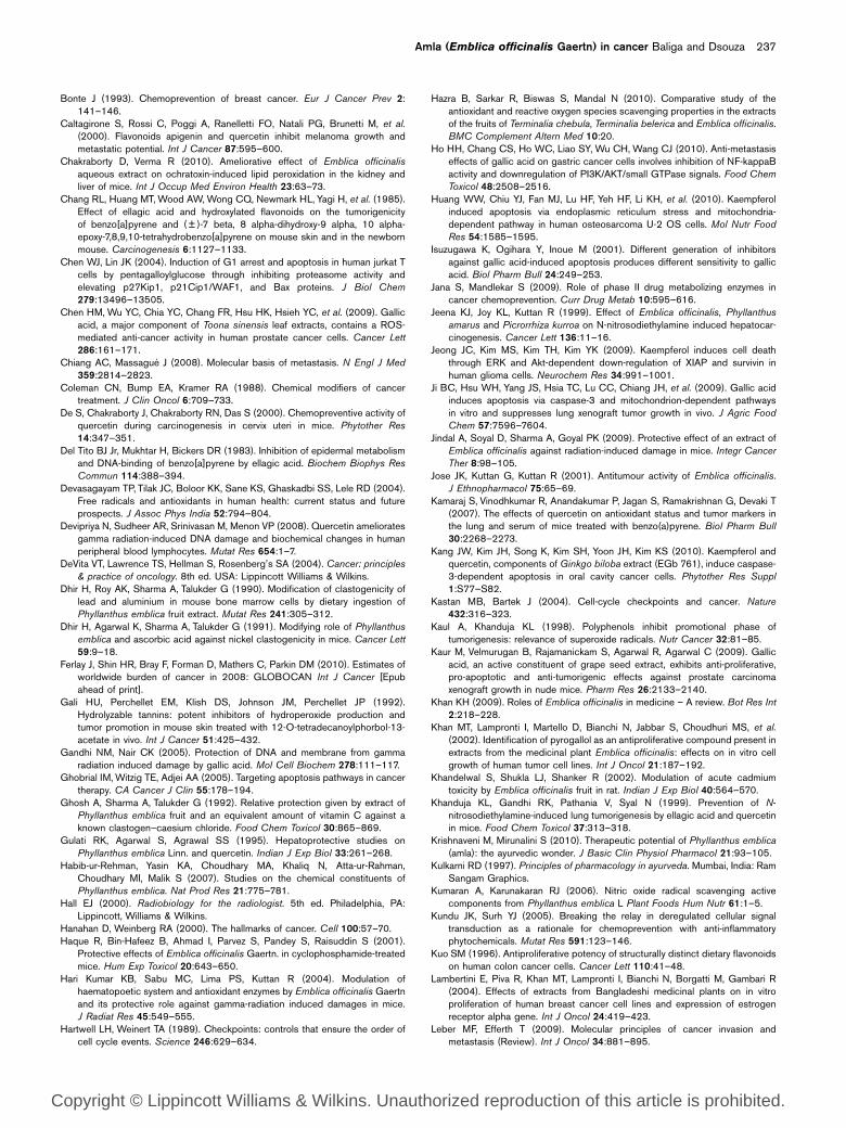
Mechanisms of action (Fig. 3)Amla is a free radical scavenger
Excess generation of free radicals, the reactive oxygen
species [ROS superoxide anion radical (O2K – ), hydroxyl
radical (OHK) and hydrogen peroxide (H2O2)], and the
reactive nitrogen species [RNS nitric oxide (NO),
peroxynitrite (ONOO – )], respectively, causes oxidative
stress and nitrosative stress. The free radicals that are
generated are highly reactive and cause damage to the
membrane lipids, proteins, and DNA (Devasagayam et al.,2004). Accordingly, their prevention is important in
preventing cell damage, mutagenesis, and carcinogenesis.
In-vitro studies have shown that amla scavenges 2,2-
diphenyl-1-picrylhydrazyl radicals (Naik et al., 2005;
Hazra et al., 2010), superoxide anions (Naik et al., 2005;
Hazra et al., 2010), hydroxyl radical (Hazra et al., 2010),
nitric oxide (Hazra et al., 2010), hydrogen peroxide (Hazra
et al., 2010), peroxynitrite (Hazra et al., 2010), singlet
oxygen (Hazra et al., 2010), and hypochlorous acid (Hazra
et al., 2010). The phytochemicals, such as gallic acid,
ellagic acids, emblicanin A, and emblicanin B, are also
reported to possess free-radical-scavenging effects in the
2,2-diphenyl-1-picrylhydrazyl assay and efficacy was as
follows: A emblicanin greater than B emblicanin greater
than gallic acid greater than ellagic acid greater than
ascorbic acid (Pozharitskaya et al., 2007).
Studies have also shown that the methanol extract of
amla and its various fractions (hexane, ethyl acetate, and
water fractions) possess NO scavenging effects. The
isolated compounds, such as gallic acid, methyl gallate,
corilagin, furosin, and geraniin, which were isolated from
the ethyl acetate fraction that possessed the best NO-
scavenging effect, were also effective. Gallic acid was
found to be a major compound in the ethyl acetate
extract and geraniin showed highest NO-scavenging
activity among the isolated compounds (Kumaran and
Karunakaran, 2006).
Amla decreases phase I enzymes
Phase I drug-metabolizing enzymes, especially the CYP
P450 mixed-function oxidases, which are involved in the
biotransformation of xenobiotics, can transform a non-
toxic chemical (procarcinogen) into a harmful toxic subs-
tance (ultimate carcinogen), which can induce damage to
the nucleic acids and other macromolecules (Percival,
1997). Studies have also shown that administering the
ethanolic extract of amla reduced the hepatic levels of
the activating enzymes, Cyt P450 and Cyt b5, which are
important in converting the procarcinogen DMBA into
ultimate carcinogen (Banu et al., 2004). In addition, the
inhibition of microsomal-activating enzymes, including
Cyt P450, was also responsible for the antimutagenic
effects of amla against 2-aminofluorene (Arora et al.,2003), aflatoxin B1, and benzo[a]pyrene-induced muta-
genesis in the Ames test (Sharma et al., 2000b).
Amla increases glutathione S-transferase, a phase II
enzyme
The reactive species formed by the phase I enzymes are
often detoxified by phase II drug-metabolizing enzymes.
In the reaction, the hydrophobic intermediates generated
by the phase I enzymes are converted to a water-soluble
group, thus decreasing their reactive nature, and allowing
subsequent excretion (Jana and Mandlekar, 2009).
A properly functioning and balanced phase II system
would detoxify the metabolically activated carcinogen,
thereby preventing mutagenesis and carcinogenesis.
Agents preferentially activating phase II over phase I
enzymes can be more beneficial as chemopreventive
agents (Percival, 1997; Jana and Mandlekar, 2009).
Studies have shown that amla increases the level of GST
and thereby reduces the toxic effects of N-nitrosodiethy-
lamine (Jeena et al., 1999; Rajeshkumar et al., 2003),
benzo[a]pyrene (Sharma et al., 2000a), cyclophosphamide
(Sharma et al., 2000a), thioacetamide (Sultana et al.,2004), CCl4 (Sultana et al., 2005), ionizing radiation (Hari
232 European Journal of Cancer Prevention 2011, Vol 20 No 3
Copyright © Lippincott Williams & Wilkins. Unauthorized reproduction of this article is prohibited.

Kumar et al., 2004), hexachlorocyclohexane (Anilakumar
et al., 2007), arsenic (Panchabhai et al., 2008), ethanol
(Reddy et al., 2009), and ochratoxin (Sultana et al., 2004).
Molecular studies have also shown that amla increased
GSTP1 expression (Niture et al., 2006), thereby valida-
ting the biochemical observation.
Amla decreases ornithine decarboxylase
Ornithine decarboxylase (ODC), the rate-limiting en-
zyme in polyamine synthesis, is important in polyamine
synthesis. High levels of ODC are an adverse prognostic
factor as it is observed to be important in tumor proli-
feration, progression, and metastasis and for the survival
of cancer patients (Manni et al., 2002).
Studies have shown that administering amla inhibited
thioacetamide-induced hyper-proliferation in rat liver
by decreasing the levels of ODC activity and thymidine
incorporation in DNA (Sultana et al., 2004). These obser-
vations clearly indicate the inhibitory effects of amla on
ODC and DNA replication, steps that are important in
tumor cell proliferation.
Amla increases the antioxidant enzymes
The antioxidant enzymes, superoxide dismutase, GPx,
and catalase, cooperate or, in a synergistic method, work
to protect cells against oxidative stress. The superoxide
dismutase catalyses the dismutation of superoxide
radicals, a major form of ROS, into hydrogen peroxide,
which is acted on by the GPx and catalase to give water.
When an appropriate balance exists between these three
enzymes, oxidative stress is reduced and the cells are
protected from the cytotoxic and mutagenic effects of the
ROS (Devasagayam et al., 2004).
Preclinical studies have conclusively shown that amla
ameliorates the oxidative and xenobiotic-induced stress,
mutagenesis, and carcinogenesis by increasing the anti-
oxidant enzymes. Reports suggest that amla increases
the antioxidant enzymes and prevents benzo[a]pyrene
(Sharma et al., 2000a), cyclophosphamide (Sharma et al.,2000a), DMBA (Banu et al., 2004), g-radiation (Hari Kumar
et al., 2004; Jindal et al., 2009), hexachlorocyclohexane
(Anilakumar et al., 2007), and ethanol (Pramyothin et al.,2006)-induced toxic effects.
Amla decreases lipid peroxidation
Lipid peroxidation is one of the most evaluated con-
sequences of free radicals on membrane structure. The
polyunsaturated fatty acids are vulnerable to peroxidative
attack and this can cause loss of fluidity, decreased
membrane potential, increased permeability for protons
and calcium ions and eventually loss of cell membranes,
and result in pathological and toxicological processes
(Devasagayam et al., 2004). The major aldehydic end
Fig. 3
Immune modulation
DNA damage
Mutagenesis
Ornithine decarboxylase
Phase II detoxificationenzymes
Phase I detoxificationenzymes
Antioxidant enzymes
Glutathione
Amla Inflammation
Oxidative stress
Lipid peroxidation
Free radical scavenging
Some of the protective mechanisms responsible for the radioprotective and chemoprotective effects of amla (arrow pointing up depicts increase,whereas down signifies decrease).
Amla (Emblica officinalis Gaertn) in cancer Baliga and Dsouza 233
Copyright © Lippincott Williams & Wilkins. Unauthorized reproduction of this article is prohibited.

product of lipid peroxidation is malondialdehyde and is
mutagenic in the bacterial and mammalian systems of
studies.
Multiple studies have shown that amla possesses inhibitory
effects on lipid peroxidation induced by various inducers.
In-vitro studies have shown that amla prevents radiation-
induced lipid peroxidation (Naik et al., 2005) and this effect
also extends to animal studies (Hari Kumar et al., 2004;
Jindal et al., 2009). Amla inhibits cadmium (Khandelwal
et al., 2002), carbon tetra chloride (Sultana et al., 2005),
arsenic (Panchabhai et al., 2008), ethanol (Reddy et al.,2009), ochratoxin (Chakraborty and Verma, 2010), N-
nitrosodiethylamine (Rajeshkumar et al., 2003), and thioa-
cetamide (Anilakumar et al., 2007)-induced lipid peroxida-
tion. By inhibiting lipid peroxidation amla may contribute
toward the observed beneficial effects, at least in part.
Amla possess anti-inflammatory effects
Chronic inflammation has been proved to cause free
radicals and the resulting oxidative and nitrosative stress
is known to directly or indirectly contribute toward mali-
gnant cell transformation by inducing genomic instabi-
lity, alterations in epigenetic events, inappropriate gene
expression, enhanced proliferation of mutated cells, resis-
tance to apoptosis, tumor neovascularization, and meta-
stasis (Kundu and Surh, 2005).
Experiments have shown that the aqueous fraction of
methanol extract of the leaves possesses anti-inflammatory
effects in carrageenan-induced and dextran-induced rat
hind paw edema. Mechanistically, it was observed that the
extract inhibited migration of human polymorphonuclear
cells and exerted its anti-inflammatory effects (Asmawi
et al., 1993). Studies have also shown that amla extract and
the phytochemical pyrogallol also possess anti-inflamma-
tory effects and inhibited the Pseudomonas aeruginosa labora-
tory strain PAO1-dependent expression of the neutrophil
chemokines IL-8, GRO-a, GRO-g, of the adhesion
molecule, ICAM-1, and of the pro-inflammatory cytokine,
IL-6 (Nicolis et al., 2008). Recently, Muthuraman et al.(2010) have also observed that the phenolic compounds
from amla possess anti-inflammatory effects in the
carrageenan and cotton pellet-induced acute and chronic
inflammatory response in animal models of study. The
effect was significant at high doses and was comparable to
the positive control, diclofenac (Muthuraman et al., 2010).
Antimutagenic effects
The initial step in the process of carcinogenesis is induc-
tion of mutation in the oncogenes or tumor-suppressor
genes of the genome of a somatic cell. Therefore, its
prevention is of great importance (Weisburger, 2001).
Multiple studies carried out in the last two decades have
conclusively shown that amla prevents DNA damage
against different carcinogens and mutagens. Using the
standard Ames test, Sharma et al. (2000b) observed for
the first time that the aqueous extract of amla inhibited
aflatoxin B1 and benzo[a]pyrene-induced mutagenesis in
the Salmonella typhimurium strains TA 98 and TA 100.
Amla is also reported to increase the levels and activities
of O6-methylguanine-DNA methyltransferase, an en-
zyme important for removing the highly mutagenic
adducts formed by alkylating agents in human lympho-
cytes (Niture et al., 2006). Amla was also effective in
preventing the radiation-induced damage in the plasmid
DNA assay (Naik et al., 2005), suggesting its effectiveness
against different classes of mutagens.
In addition, studies with experimental animals have shown
that amla prevents cadmium (Khandelwal et al., 2002), lead
(Dhir et al., 1990), aluminium (Dhir et al., 1990), nickel
(Dhir et al., 1991), cesium chloride (Ghosh et al., 1992),
arsenic (Biswas et al., 1999), chromium (Sai Ram et al.,2003), 3,4-benzo(a)pyrene (Nandi et al., 1997), benzo[a]-
pyrene (Sharma et al., 2000a), DMBA (Nandi et al., 1997),
and cyclophosphamide (Sharma et al., 2000a)-induced
DNA damage. Together these observations clearly suggest
the effectiveness of amla in preventing mutagenesis and
DNA damage, which would inhibit/reduce the incidence
and process of carcinogenesis, at least in part.
Amla possesses immunomodulatory effects
Immune activation is an effective protective approach
against emerging infectious diseases and certain cancers.
Immunostimulants enhance the overall immunity of the
host, present a nonspecific immune response against
microbial pathogens and increase humoral and cellular
immune responses, by either enhancing cytokine secretion,
or by directly stimulating B-lymphocytes or T-lymphocytes
(Spelman et al., 2006). In Ayurveda, amla is considered to
be an immunostimulatory agent and scientific studies have
validated this (Warrier et al., 1996; Kulkarni, 1997; Khan,
2009; Krishnaveni and Mirunalini, 2010).
Studies have shown that amla enhances natural killer
(NK) cell activity and antibody-dependent cellular
cytotoxicity in BALB/c mice bearing Dalton’s lymphoma
ascites tumor. Amla increases the life span of tumor-
bearing animals and this was because of the increase in
the activation of splenic NK cell activity and antibody
dependent cellular cytotoxicity. However, the increase in
survival was completely abrogated when the NK cell and
killer cell activities were depleted, either by cyclopho-
sphamide or anti-asialo-GM1 antibody treatment, validat-
ing that the observed effects were because of its immuno-
modulatory effects (Suresh and Vasudevan, 1994).
Amla and its phytochemicals modulate the levels
of proteins important in cell cycle progression
Cancer is frequently considered to be a disease of the cell
cycle and a convincing body of data has proved that the
disruption of the normal regulation of cell-cycle progres-
sion and division are important events in cancer deve-
lopment (Hanahan and Weinberg, 2000; Kastan and
234 European Journal of Cancer Prevention 2011, Vol 20 No 3
Copyright © Lippincott Williams & Wilkins. Unauthorized reproduction of this article is prohibited.
Bartek, 2004). The progression of the cell cycle is a
tightly regulated and highly ordered process involving
multiple checkpoints that assess extracellular growth
signals, cell size, and DNA integrity (Kastan and Bartek,
2004). The cyclin-dependent kinases (CDKs) and their
respective partners (cyclin) are responsible for the
progression of the cell cycle, whereas the CDK inhibitors
act as brakes to stop cell cycle progression (Hartwell and
Weinert, 1989). The genesis of cancer is principally because
of the derailed expression or activation of positive
regulators and functional suppression of negative regulators
(Hartwell and Weinert, 1989; Kastan and Bartek, 2004).
Studies by Jose et al. (2001) have shown for the first time
that amla extract caused a dose-dependent inhibition of
the cell cycle-regulating enzyme Cdc25 phosphatase
in vitro, with an IC50 of 5 mg/ml (Jose et al., 2001). The
phytochemical pentagalloylglucose is shown to cause G1
arrest in human Jurkat T cells by elevating p27Kip1 and
p21Cip1/WAF1 proteins (Chen and Lin, 2004). Gallic
acid induces cell cycle arrest by decreasing CDKs and
cyclins. It phosporylates Cip1/p21 and cell division cycle
2 (Cdc2), Cdc25A, and Cdc25C in DU145 cells (Sun
et al., 2004). It also induces G2/M phase cell cycle arrest
by regulating 14-3-3b release from Cdc25C; activation of
chk2; decreasing CDK1, cyclin B1, and Cdc25C; increas-
ing phosphorylation of p-Cdc2 (Tyr-15), Cip1/p21 and
Cdc25C in human bladder transitional carcinoma cells
(TSGH-8301cells) (Ou et al., 2010). Gallic acid feeding
also reduces Cdc2, CDK2, CDK4, CDK6, cyclin B1, and E
in the prostatic tissue of mice with transgenic adenocarci-
noma of the mouse prostate (Raina et al., 2008).
Amla and some of its constituents cause apoptosis
and cytotoxicity of neoplastic cells
Apoptosis, a process by which the cell is committed to
death by not initiating an inflammatory response, is
vital in regulating tissue homeostasis (Sun et al., 2004;
Ghobrial et al., 2005). A large body of evidence has proved
that the processes of neoplastic transformation, progres-
sion, and metastasis involve alterations of the normal
apoptotic pathway and that the number of cell deaths is
very low in these cells (Sun et al., 2004; Ghobrial et al.,2005). Therefore, the induction of apoptosis is arguably
the most potent defence against cancer as it effectively
eliminates the mutated and severely damaged cells.
Accordingly, agents that can eliminate mutated, preneo-
plastic, and neoplastic cells by sparing the normal cells
are supposed to be an effective chemopreventive agent
and to offer therapeutic advantage in the elimination of
cancer cells (Sun et al., 2004; Ghobrial et al., 2005).
The ability of the extract of amla and some of its
phytochemicals to induce apoptosis in cancer cells con-
tributes to the understanding of its anticancer and
chemopreventive potential. Studies have shown that
the aqueous extract of amla induces apoptosis and
inhibits the growth of HeLa, MDA-MB-231, and SK-
OV3 without affecting the normal lung fibroblast, MRC5
(Ngamkitidechakul et al., 2010). The hydrolyzable tannins
possess selective cytotoxicity to the human oral squamous
cell carcinoma and salivary gland tumor cell lines, whereas
they were nontoxic to the normal human gingival fibro-
blasts (Sakagami et al., 2000). Studies have also shown that
quercetin (Son et al., 2004), gallic acid (Isuzugawa et al.,2001), ellagic acid (Losso et al., 2004), and pyrogallol (Yang
et al., 2009) also possess cytotoxic and apoptogenic effects
on the neoplastic and transformed cells, but not in normal
cells. Together, these observations clearly suggest that the
presence of these compounds in amla resulted in the eli-
mination of the mutated and neoplastic cells and resulted
in the desired effects in both antineoplastic effects and
chemoprevention.
Amla and some of its constituents prevent
metastasis
Cancer cells differ from normal cells; the most important
being the loss of differentiation, self-sufficiency in growth
signals, limitless replicative potential, decreased drug
sensitivity, increased invasiveness, and metastasis (Hanahan
and Weinberg, 2000). Metastasis, the process by which
some of the neoplastic cells spread from the primary site
to distant tissue, is the life-threatening aspect of cancer.
It is the hallmark of cancer and is responsible for the
failure of treatment and death. The process of tumor
metastasis is extremely complex and involves myriad bio-
chemical interactions operating concurrently or sequen-
tially. The important steps in the process of metastasis
are (i) invasion and migration, (ii) intravasation, (iii)
circulation, (iv) extravasation, and (v) colonization,
proliferation, and angiogenesis (Chiang and Massague,
2008; Leber and Efferth, 2009). Cell invasion is one of
the fundamental processes required during tumor pro-
gression and metastasis and matrix metalloproteinases
(MMPs), a group of enzymes that regulate cell-matrix
composition, are important in this process (Chiang and
Massague, 2008; Leber and Efferth, 2009).
Recent studies have suggested that the aqueous extract
of amla was effective in preventing the invasion of MDA-
MB-231 cells in the in-vitro matrigel invasion assay
(Ngamkitidechakul et al., 2010). The amla phytochem-
ical, kaempferol, inhibited the expression of stromelysin 1
(MMP-3) in the MDA-MB-231 breast cancer cell line
(Phromnoi et al., 2009). The polyphenol gallic acid is also
reported to possess inhibitory effects on gastric adeno-
carcinoma cell migration, decreased expression of MMP-
2/9 in vitro (Ho et al., 2010), and metastasis of P815
mastocytoma cells to the liver of DBA/2 mice (Ohno et al.,2001). The flavanol, quercetin, decreased the expression
of gelatinases A and B (MMP-2 and MMP-9) in the
human metastatic prostate PC-3 cells (Vijayababu et al.,2006) and stromelysin 1 (MMP-3) in the MDA-MB-231
breast cancer cell line (Phromnoi et al., 2009) and
Amla (Emblica officinalis Gaertn) in cancer Baliga and Dsouza 235
Copyright © Lippincott Williams & Wilkins. Unauthorized reproduction of this article is prohibited.
inhibited the lung metastasis of murine colon 26-L5 car-
cinoma cells (Ogasawara et al., 2007) and B16-BL6 murine
melanoma metastasis in mice (Piantelli et al., 2006).
Conclusion
Preclinical studies carried out in the past two decades
have clearly shown that amla possesses antineoplastic,
chemomodulatory, chemopreventive, and radioprotective
effects. Several mechanisms are likely to be responsible
for the observed effects, the most important being the
induction of apoptosis of neoplastic and preneoplastic
cells, free radical scavenging, antioxidant, antimutagenic,
anti-inflammatory activities; increase in the antioxidant
enzymes, modulation of phase I and II enzymes and
immunomodulatory effects (Fig. 3). It is unlikely that all
targets and cell biological effects found in vitro individu-
ally may be operating in the animal system, but studies
should be attempted to understand whether the effects
observed in vitro translate to the animal system.
Although studies on the effects of amla on some cancer
cell lines and animals substantiate its effectiveness,
countless possibilities for investigation still remain.
Relevant animal and cell culture studies are required to
understand the underlying mechanism of action, espe-
cially with the phytochemicals. In addition, rationally
designed clinical trials are also needed to understand the
maximum permissible dose and also to assess for its
adverse effects, if any, following consumption over longer
periods.
From a phytochemical perspective, there is considerable
variation in the composition among various samples of
amla. A quality control should be established for the
authenticity of the plant and the presence of active
phytochemicals, especially gallic acid, ellagic acid, chebu-
linic acid, quercetin, chebulagic acid, corilagin, kaempferol,
apigenin, luteolin, emblicanin A, and emblicanin B in the
required levels. Experiments should also be performed to
understand which of the phytochemicals are effective and
their mechanisms of action.
Studies indicate that amla and some of its phytochem-
icals (gallic acid, pentagalloylglucose, ellagic acid, quer-
cetin, and kaempferol) are cytotoxic to neoplastic cells,
whereas the normal cells are unaffected. It is quite
possible that these compounds exert their effects on
neoplastic cells that have aberrant cell cycle progression.
It is observed that these molecules induce apoptosis and
cytotoxicity by modulating the proteins involved in cell
progression, and the observations of Jose et al. (2001)
support the hypothesis. However, detailed studies are
needed on this aspect with a range of cells encompass-
ing normal, mutated, preneoplastic, and highly metastatic
cell lines of different histological origins and cell doubling
time.
Owing to its abundance, low cost, and safety in
consumption, amla remains a species with tremendous
potential and countless possibilities for further investiga-
tion. Amla has the potential to develop as a nontoxic
anticancer, chemopreventive agent, and as an adjuvant to
radiotherapy and chemotherapy when lacunas existing in
knowledge are understood. The outcomes of such studies
may be useful for the clinical applications of amla in
humans against different cancers and may open up a new
therapeutic avenue.
AcknowledgementsThe authors are grateful to Rev. Fr. Patrick Rodrigus
(Director), Rev. Fr. Denis D’Sa (Administrator), Dr
Sanjeev Rai (Chief of Medical Services), and Dr Jaya
Prakash Alva, (Dean) of Father Muller Medical College
for their unstinted support. They also thank to Harshith
P. Bhat for drawing the chemical structures. The authors
dedicate this review to Professor Ramdasan Kuttan of
Amala Cancer Centre, Thrissur, India. Professor Kuttan is
a pioneer cancer researcher and his work on the radio-
protective and chemopreventive effects with amla has
been a source of inspiration to the authors. This study
was not supported by any private or public funding body.
The authors declare that they do not have any conflict of
interest.
ReferencesAgarwal C, Tyagi A, Agarwal R (2006). Gallic acid causes inactivating
phosphorylation of Cdc25A/Cdc25C-Cdc2 via ATM-Chk2 activation, leadingto cell cycle arrest, and induces apoptosis in human prostate carcinomaDU145 cells. Mol Cancer Ther 5:3294–3302.
Anilakumar KR, Nagaraj NS, Santhanam K (2007). Reduction of hexachlorocy-clohexane-induced oxidative stress and cytotoxicity in rat liver by Emblicaofficinalis Gaertn. Indian J Exp Biol 45:450–454.
Are C, Colburn L, Rajaram S, Vijayakumar M (2010). Disparities in cancer carebetween the United States of America and India and opportunities forsurgeons to lead. J Surg Oncol 102:100–105.
Arora R, editor (2010). Herbal drugs: a cancer chemopreventive and therapeuticperspective. New Delhi, India: Jaypee Brothers Medical Publishers (P) Ltd.
Arora S, Kaur K, Kaur S (2003). Indian medicinal plants as a reservoir ofprotective phytochemicals. Teratog Carcinog Mutagen (Suppl 1):295–300.
Arora R, Gupta D, Chawla R, Sagar R, Sharma A, Kumar R, et al. (2005).Radioprotection by plant products: present status and future prospects.Phytother Res 19:1–22.
Asmawi MZ, Kankaanranta H, Moilanen E, Vapaatalo H (1993). Anti-inflammatoryactivities of Emblica officinalis Gaertn leaf extracts. J Pharm Pharmacol45:581–584.
Banu SM, Selvendiran K, Singh JP, Sakthisekaran D (2004). Protective effect ofEmblica officinalis ethanolic extract against 7,12-dimethylbenz(a) anthracene(DMBA) induced genotoxicity in Swiss albino mice. Hum Exp Toxicol23:527–531.
Benkovic V, Knezevic AH, Dikic D, Lisicic D, Orsolic N, Basic I, et al.(2008). Radioprotective effects of propolis and quercetin in gamma-irradiated mice evaluated by the alkaline comet assay. Phytomedicine15:851–858.
Benkovic V, Kopjar N, Horvat Knezevic A, Dikic D, Basic I, Ramic S, et al. (2008).Evaluation of radioprotective effects of propolis and quercetin on humanwhite blood cells in vitro. Biol Pharm Bull 31:1778–1785.
Benkovic V, Knezevic AH, Dikic D, Lisicic D, Orsolic N, Basic I, Kopjar N (2009).Radioprotective effects of quercetin and ethanolic extract of propolis ingamma-irradiated mice. Arh Hig Rada Toksikol 60:129–138.
Bhosle SM, Huilgol NG, Mishra KP (2005). Enhancement of radiation-inducedoxidative stress and cytotoxicity in tumor cells by Ellagic acid. Clin Chim Acta359:89–100.
Biswas S, Talukder G, Sharma A (1999). Protection against cytotoxic effects ofarsenic by dietary supplementation with crude extract of Emblica officinalisfruit. Phytother Res 13:513–516.
236 European Journal of Cancer Prevention 2011, Vol 20 No 3
Copyright © Lippincott Williams & Wilkins. Unauthorized reproduction of this article is prohibited.
Bonte J (1993). Chemoprevention of breast cancer. Eur J Cancer Prev 2:141–146.
Caltagirone S, Rossi C, Poggi A, Ranelletti FO, Natali PG, Brunetti M, et al.(2000). Flavonoids apigenin and quercetin inhibit melanoma growth andmetastatic potential. Int J Cancer 87:595–600.
Chakraborty D, Verma R (2010). Ameliorative effect of Emblica officinalisaqueous extract on ochratoxin-induced lipid peroxidation in the kidney andliver of mice. Int J Occup Med Environ Health 23:63–73.
Chang RL, Huang MT, Wood AW, Wong CQ, Newmark HL, Yagi H, et al. (1985).Effect of ellagic acid and hydroxylated flavonoids on the tumorigenicityof benzo[a]pyrene and ( ± )-7 beta, 8 alpha-dihydroxy-9 alpha, 10 alpha-epoxy-7,8,9,10-tetrahydrobenzo[a]pyrene on mouse skin and in the newbornmouse. Carcinogenesis 6:1127–1133.
Chen WJ, Lin JK (2004). Induction of G1 arrest and apoptosis in human jurkat Tcells by pentagalloylglucose through inhibiting proteasome activity andelevating p27Kip1, p21Cip1/WAF1, and Bax proteins. J Biol Chem279:13496–13505.
Chen HM, Wu YC, Chia YC, Chang FR, Hsu HK, Hsieh YC, et al. (2009). Gallicacid, a major component of Toona sinensis leaf extracts, contains a ROS-mediated anti-cancer activity in human prostate cancer cells. Cancer Lett286:161–171.
Chiang AC, Massague J (2008). Molecular basis of metastasis. N Engl J Med359:2814–2823.
Coleman CN, Bump EA, Kramer RA (1988). Chemical modifiers of cancertreatment. J Clin Oncol 6:709–733.
De S, Chakraborty J, Chakraborty RN, Das S (2000). Chemopreventive activity ofquercetin during carcinogenesis in cervix uteri in mice. Phytother Res14:347–351.
Del Tito BJ Jr, Mukhtar H, Bickers DR (1983). Inhibition of epidermal metabolismand DNA-binding of benzo[a]pyrene by ellagic acid. Biochem Biophys ResCommun 114:388–394.
Devasagayam TP, Tilak JC, Boloor KK, Sane KS, Ghaskadbi SS, Lele RD (2004).Free radicals and antioxidants in human health: current status and futureprospects. J Assoc Phys India 52:794–804.
Devipriya N, Sudheer AR, Srinivasan M, Menon VP (2008). Quercetin amelioratesgamma radiation-induced DNA damage and biochemical changes in humanperipheral blood lymphocytes. Mutat Res 654:1–7.
DeVita VT, Lawrence TS, Hellman S, Rosenberg’s SA (2004). Cancer: principles& practice of oncology. 8th ed. USA: Lippincott Williams & Wilkins.
Dhir H, Roy AK, Sharma A, Talukder G (1990). Modification of clastogenicity oflead and aluminium in mouse bone marrow cells by dietary ingestion ofPhyllanthus emblica fruit extract. Mutat Res 241:305–312.
Dhir H, Agarwal K, Sharma A, Talukder G (1991). Modifying role of Phyllanthusemblica and ascorbic acid against nickel clastogenicity in mice. Cancer Lett59:9–18.
Ferlay J, Shin HR, Bray F, Forman D, Mathers C, Parkin DM (2010). Estimates ofworldwide burden of cancer in 2008: GLOBOCAN Int J Cancer [Epubahead of print].
Gali HU, Perchellet EM, Klish DS, Johnson JM, Perchellet JP (1992).Hydrolyzable tannins: potent inhibitors of hydroperoxide production andtumor promotion in mouse skin treated with 12-O-tetradecanoylphorbol-13-acetate in vivo. Int J Cancer 51:425–432.
Gandhi NM, Nair CK (2005). Protection of DNA and membrane from gammaradiation induced damage by gallic acid. Mol Cell Biochem 278:111–117.
Ghobrial IM, Witzig TE, Adjei AA (2005). Targeting apoptosis pathways in cancertherapy. CA Cancer J Clin 55:178–194.
Ghosh A, Sharma A, Talukder G (1992). Relative protection given by extract ofPhyllanthus emblica fruit and an equivalent amount of vitamin C against aknown clastogen–caesium chloride. Food Chem Toxicol 30:865–869.
Gulati RK, Agarwal S, Agrawal SS (1995). Hepatoprotective studies onPhyllanthus emblica Linn. and quercetin. Indian J Exp Biol 33:261–268.
Habib-ur-Rehman, Yasin KA, Choudhary MA, Khaliq N, Atta-ur-Rahman,Choudhary MI, Malik S (2007). Studies on the chemical constituents ofPhyllanthus emblica. Nat Prod Res 21:775–781.
Hall EJ (2000). Radiobiology for the radiologist. 5th ed. Philadelphia, PA:Lippincott, Williams & Wilkins.
Hanahan D, Weinberg RA (2000). The hallmarks of cancer. Cell 100:57–70.Haque R, Bin-Hafeez B, Ahmad I, Parvez S, Pandey S, Raisuddin S (2001).
Protective effects of Emblica officinalis Gaertn. in cyclophosphamide-treatedmice. Hum Exp Toxicol 20:643–650.
Hari Kumar KB, Sabu MC, Lima PS, Kuttan R (2004). Modulation ofhaematopoetic system and antioxidant enzymes by Emblica officinalis Gaertnand its protective role against gamma-radiation induced damages in mice.J Radiat Res 45:549–555.
Hartwell LH, Weinert TA (1989). Checkpoints: controls that ensure the order ofcell cycle events. Science 246:629–634.
Hazra B, Sarkar R, Biswas S, Mandal N (2010). Comparative study of theantioxidant and reactive oxygen species scavenging properties in the extractsof the fruits of Terminalia chebula, Terminalia belerica and Emblica officinalis.BMC Complement Altern Med 10:20.
Ho HH, Chang CS, Ho WC, Liao SY, Wu CH, Wang CJ (2010). Anti-metastasiseffects of gallic acid on gastric cancer cells involves inhibition of NF-kappaBactivity and downregulation of PI3K/AKT/small GTPase signals. Food ChemToxicol 48:2508–2516.
Huang WW, Chiu YJ, Fan MJ, Lu HF, Yeh HF, Li KH, et al. (2010). Kaempferolinduced apoptosis via endoplasmic reticulum stress and mitochondria-dependent pathway in human osteosarcoma U-2 OS cells. Mol Nutr FoodRes 54:1585–1595.
Isuzugawa K, Ogihara Y, Inoue M (2001). Different generation of inhibitorsagainst gallic acid-induced apoptosis produces different sensitivity to gallicacid. Biol Pharm Bull 24:249–253.
Jana S, Mandlekar S (2009). Role of phase II drug metabolizing enzymes incancer chemoprevention. Curr Drug Metab 10:595–616.
Jeena KJ, Joy KL, Kuttan R (1999). Effect of Emblica officinalis, Phyllanthusamarus and Picrorrhiza kurroa on N-nitrosodiethylamine induced hepatocar-cinogenesis. Cancer Lett 136:11–16.
Jeong JC, Kim MS, Kim TH, Kim YK (2009). Kaempferol induces cell deaththrough ERK and Akt-dependent down-regulation of XIAP and survivin inhuman glioma cells. Neurochem Res 34:991–1001.
Ji BC, Hsu WH, Yang JS, Hsia TC, Lu CC, Chiang JH, et al. (2009). Gallic acidinduces apoptosis via caspase-3 and mitochondrion-dependent pathwaysin vitro and suppresses lung xenograft tumor growth in vivo. J Agric FoodChem 57:7596–7604.
Jindal A, Soyal D, Sharma A, Goyal PK (2009). Protective effect of an extract ofEmblica officinalis against radiation-induced damage in mice. Integr CancerTher 8:98–105.
Jose JK, Kuttan G, Kuttan R (2001). Antitumour activity of Emblica officinalis.J Ethnopharmacol 75:65–69.
Kamaraj S, Vinodhkumar R, Anandakumar P, Jagan S, Ramakrishnan G, Devaki T(2007). The effects of quercetin on antioxidant status and tumor markers inthe lung and serum of mice treated with benzo(a)pyrene. Biol Pharm Bull30:2268–2273.
Kang JW, Kim JH, Song K, Kim SH, Yoon JH, Kim KS (2010). Kaempferol andquercetin, components of Ginkgo biloba extract (EGb 761), induce caspase-3-dependent apoptosis in oral cavity cancer cells. Phytother Res Suppl1:S77–S82.
Kastan MB, Bartek J (2004). Cell-cycle checkpoints and cancer. Nature432:316–323.
Kaul A, Khanduja KL (1998). Polyphenols inhibit promotional phase oftumorigenesis: relevance of superoxide radicals. Nutr Cancer 32:81–85.
Kaur M, Velmurugan B, Rajamanickam S, Agarwal R, Agarwal C (2009). Gallicacid, an active constituent of grape seed extract, exhibits anti-proliferative,pro-apoptotic and anti-tumorigenic effects against prostate carcinomaxenograft growth in nude mice. Pharm Res 26:2133–2140.
Khan KH (2009). Roles of Emblica officinalis in medicine – A review. Bot Res Int2:218–228.
Khan MT, Lampronti I, Martello D, Bianchi N, Jabbar S, Choudhuri MS, et al.(2002). Identification of pyrogallol as an antiproliferative compound present inextracts from the medicinal plant Emblica officinalis: effects on in vitro cellgrowth of human tumor cell lines. Int J Oncol 21:187–192.
Khandelwal S, Shukla LJ, Shanker R (2002). Modulation of acute cadmiumtoxicity by Emblica officinalis fruit in rat. Indian J Exp Biol 40:564–570.
Khanduja KL, Gandhi RK, Pathania V, Syal N (1999). Prevention of N-nitrosodiethylamine-induced lung tumorigenesis by ellagic acid and quercetinin mice. Food Chem Toxicol 37:313–318.
Krishnaveni M, Mirunalini S (2010). Therapeutic potential of Phyllanthus emblica(amla): the ayurvedic wonder. J Basic Clin Physiol Pharmacol 21:93–105.
Kulkarni RD (1997). Principles of pharmacology in ayurveda. Mumbai, India: RamSangam Graphics.
Kumaran A, Karunakaran RJ (2006). Nitric oxide radical scavenging activecomponents from Phyllanthus emblica L Plant Foods Hum Nutr 61:1–5.
Kundu JK, Surh YJ (2005). Breaking the relay in deregulated cellular signaltransduction as a rationale for chemoprevention with anti-inflammatoryphytochemicals. Mutat Res 591:123–146.
Kuo SM (1996). Antiproliferative potency of structurally distinct dietary flavonoidson human colon cancer cells. Cancer Lett 110:41–48.
Lambertini E, Piva R, Khan MT, Lampronti I, Bianchi N, Borgatti M, Gambari R(2004). Effects of extracts from Bangladeshi medicinal plants on in vitroproliferation of human breast cancer cell lines and expression of estrogenreceptor alpha gene. Int J Oncol 24:419–423.
Leber MF, Efferth T (2009). Molecular principles of cancer invasion andmetastasis (Review). Int J Oncol 34:881–895.
Amla (Emblica officinalis Gaertn) in cancer Baliga and Dsouza 237
Copyright © Lippincott Williams & Wilkins. Unauthorized reproduction of this article is prohibited.

Lee CY, Peng WH, Cheng HY, Chen FN, Lai MT, Chiu TH (2006).Hepatoprotective effect of Phyllanthus in Taiwan on acute liver damageinduced by carbon tetrachloride. Am J Chin Med 34:471–482.
Lesca P (1983). Protective effects of ellagic acid and other plant phenols onbenzo[a]pyrene-induced neoplasia in mice. Carcinogenesis 4:1651–1653.
Li W, Du B, Wang T, Wang S, Zhang J (2009). Kaempferol induces apoptosis inhuman HCT116 colon cancer cells via the ataxia-telangiectasia mutated-p53pathway with the involvement of p53 upregulated modulator of apoptosis.Chem Biol Interact 177:121–127.
Lo C, Lai TY, Yang JH, Yang JS, Ma YS, Weng SW, et al. (2010). Gallic acidinduces apoptosis in A375.S2 human melanoma cells through caspase-dependent and -independent pathways. Int J Oncol 37:377–385.
Losso JN, Bansode RR, Trappey A II, Bawadi HA, Truax R (2004). In vitro anti-proliferative activities of ellagic acid. J Nutr Biochem 15:672–678.
Makita H, Tanaka T, Fujitsuka H, Tatematsu N, Satoh K, Hara A, Mori H (1996).Chemoprevention of 4-nitroquinoline 1-oxide-induced rat oral carcinogenesisby the dietary flavonoids chalcone, 2-hydroxychalcone, and quercetin. CancerRes 56:4904–4909.
Manni A, Washington S, Griffith JW, Verderame MF, Mauger D, Demers LM, et al.(2002). Influence of polyamines on in vitro and in vivo features of aggressiveand metastatic behavior by human breast cancer cells. Clin Exp Metastasis19:95–105.
Mir AI, Kumar B, Tasduq SA, Gupta DK, Bhardwaj S, Johri RK (2007). Reversal ofhepatotoxin-induced pre-fibrogenic events by Emblica officinalis – ahistological study. Indian J Exp Biol 45:626–629.
Mukhtar H, Das M, Del Tito BJ Jr, Bickers DR (1984a). Protection against 3-methylcholanthrene-induced skin tumorigenesis in Balb/C mice by ellagicacid. Biochem Biophys Res Commun 119:751–757.
Mukhtar H, Das M, Bickers DR (1986). Inhibition of 3-methylcholanthrene-induced skin tumorigenicity in BALB/c mice by chronic oral feeding of traceamounts of ellagic acid in drinking water. Cancer Res 46:2262–2265.
Muthuraman A, Sood S, Singla SK (2010). The antiinflammatory potential ofphenolic compounds from Emblica officinalis L in rat. Inflammopharmacology[Epub ahead of print].
Naik GH, Priyadarsini KI, Bhagirathi RG, Mishra B, Mishra KP, Banavalikar MM,Mohan H (2005). In vitro antioxidant studies and free radical reactions of triphala,an ayurvedic formulation and its constituents. Phytother Res 19:582–586.
Nair HK, Rao KV, Aalinkeel R, Mahajan S, Chawda R, Schwartz SA (2004).Inhibition of prostate cancer cell colony formation by the flavonoid quercetincorrelates with modulation of specific regulatory genes. Clin Diagn LabImmunol 11:63–69.
Nandi P, Talukder G, Sharma A (1997). Dietary chemoprevention of clastogeniceffects of 3,4-benzo(a)pyrene by Emblica officinalis Gaertn. fruit extract. Br JCancer 76:1279–1283.
Nemavarkar P, Chourasia BK, Pasupathy K (2004). Evaluation of radioprotectiveaction of compounds using Saccharomyces cerevisiae. J Environ PatholToxicol Oncol 23:145–151.
Ngamkitidechakul C, Jaijoy K, Hansakul P, Soonthornchareonnon N, Sireerata-wong S (2010). Antitumour effects of Phyllanthus emblica L: induction ofcancer cell apoptosis and inhibition of in vivo tumour promotion and in vitroinvasion of human cancer cells. Phytother Res 24:1405–1413.
Nicolis E, Lampronti I, Dechecchi MC, Borgatti M, Tamanini A, Bianchi N, et al.(2008). Pyrogallol, an active compound from the medicinal plant Emblicaofficinalis, regulates expression of pro-inflammatory genes in bronchialepithelial cells. Int Immunopharmacol 8:1672–1680.
Niture SK, Rao US, Srivenugopal KS (2006). Chemopreventative strategiestargeting the MGMT repair protein: augmented expression in humanlymphocytes and tumor cells by ethanolic and aqueous extracts of severalIndian medicinal plants. Int J Oncol 29:1269–1278.
Ogasawara M, Matsunaga T, Suzuki H (2007). Differential effects of antioxidantson the in vitro invasion, growth and lung metastasis of murine colon cancercells. Biol Pharm Bull 30:200–204.
Ohno T, Inoue M, Ogihara Y (2001). Cytotoxic activity of gallic acid against livermetastasis of mastocytoma cells P-815. Anticancer Res 21:3875–3880.
Ou TT, Wang CJ, Lee YS, Wu CH, Lee HJ (2010). Gallic acid induces G2/Mphase cell cycle arrest via regulating 14-3-3beta release from Cdc25C andChk2 activation in human bladder transitional carcinoma cells. Mol Nutr FoodRes 54:1781–1790.
Panchabhai TS, Ambarkhane SV, Joshi AS, Samant BD, Rege NN (2008).Protective effect of Tinospora cordifolia, Phyllanthus emblica and theircombination against antitubercular drugs induced hepatic damage: anexperimental study. Phytother Res 22:646–650.
Pandey G (2002). Dravyaguna Vijnana (Materia Medica-Vegetable Drugs) Part IVol. 2. Varanasi, India: Krishnadas Academy; p. 102.
Pastorino U (1994). Lung cancer chemoprevention. Eur J Cancer Prev 3:371–372.
Percival M (1997). Phytonutrients and detoxification. Clin Nutr Insights 5:1–3.Phromnoi K, Yodkeeree S, Anuchapreeda S, Limtrakul P (2009). Inhibition
of MMP-3 activity and invasion of the MDA-MB-231 human invasivebreast carcinoma cell line by bioflavonoids. Acta Pharmacol Sin 30:1169–1176.
Piantelli M, Rossi C, Iezzi M, La Sorda R, Iacobelli S, Alberti S, Natali PG (2006).Flavonoids inhibit melanoma lung metastasis by impairing tumor cellsendothelium interactions. J Cell Physiol 207:23–29.
Pinmai K, Chunlaratthanabhorn S, Ngamkitidechakul C, Soonthornchareon N,Hahnvajanawong C (2008). Synergistic growth inhibitory effects ofPhyllanthus emblica and Terminalia bellerica extracts with conventionalcytotoxic agents: doxorubicin and cisplatin against human hepatocellularcarcinoma and lung cancer cells. World J Gastroenterol 14:1491–1497.
Poltanov EA, Shikov AN, Dorman HJ, Pozharitskaya ON, Makarov VG, TikhonovVP, Hiltunen R (2009). Chemical and antioxidant evaluation of Indiangooseberry (Emblica officinalis Gaertn., syn. Phyllanthus emblica L)supplements. Phytother Res 23:1309–1315.
Pozharitskaya ON, Ivanova SA, Shikov AN, Makarov VG (2007). Separation andevaluation of free radical-scavenging activity of phenol components ofEmblica officinalis extract by using an HPTLC-DPPH method. J Sep Sci30:1250–1254.
Pramyothin P, Samosorn P, Poungshompoo S, Chaichantipyuth C (2006). Theprotective effects of Phyllanthus emblica Linn. extract on ethanol induced rathepatic injury. J Ethnopharmacol 107:361–364.
Priyadarsini KI, Khopde SM, Kumar SS, Mohan H (2002). Free radical studiesof ellagic acid, a natural phenolic antioxidant. J Agric Food Chem 50:2200–2206.
Raina K, Rajamanickam S, Deep G, Singh M, Agarwal R, Agarwal C (2008).Chemopreventive effects of oral gallic acid feeding on tumor growth andprogression in TRAMP mice. Mol Cancer Ther 7:1258–1267.
Rajeshkumar NV, Pillai MR, Kuttan R (2003). Induction of apoptosis in mouse andhuman carcinoma cell lines by Emblica officinalis polyphenols and its effecton chemical carcinogenesis. J Exp Clin Cancer Res 22:201–212.
Reddivari L, Vanamala J, Safe SH, Miller JC Jr (2010). The bioactive compoundsalpha-chaconine and gallic acid in potato extracts decrease survival andinduce apoptosis in LNCaP and PC3 prostate cancer cells. Nutr Cancer62:601–610.
Reddy VD, Padmavathi P, Varadacharyulu NCh (2009). Emblica officinalisprotects against alcohol-induced liver mitochondrial dysfunction in rats. JMed Food 12:327–333.
Sai Ram M, Neetu D, Deepti P, Vandana M, Ilavazhagan G, Kumar D, SelvamurthyW (2003). Cytoprotective activity of amla (Emblica officinalis) againstchromium (VI) induced oxidative injury in murine macrophages. Phytother Res17:430–433.
Sakagami H, Jiang Y, Kusama K, Atsumi T, Ueha T, Toguchi M, et al. (2000).Cytotoxic activity of hydrolyzable tannins against human oral tumor cell lines –a possible mechanism. Phytomedicine 7:39–47.
Sancheti G, Jindal A, Kumari R, Goyal PK (2005). Chemopreventive action ofEmblica officinalis on skin carcinogenesis in mice. Asian Pac J Cancer Prev6:197–201.
Sengupta A, Ghosh S, Das S (2001). Modulation of DMBA induced genotoxicityin bone marrow by quercetin during skin carcinogenesis. J Exp Clin CancerRes 20:131–134.
Sharma N, Trikha P, Athar M, Raisuddin S (2000a). Inhibitory effect of Emblicaofficinalis on the in vivo clastogenicity of benzo[a]pyrene and cyclopho-sphamide in mice. Hum Exp Toxicol 19:377–384.
Sharma N, Trikha P, Athar M, Raisuddin S (2000b). In vitro inhibition ofcarcinogen-induced mutagenicity by Cassia occidentalis and Emblicaofficinalis. Drug Chem Toxicol 23:477–484.
Singh I, Sharma A, Nunia V, Goyal PK (2005). Radioprotection of Swiss albinomice by Emblica officinalis. Phytother Res 19:444–446.
Son YO, Lee KY, Kook SH, Lee JC, Kim JG, Jeon YM, Jang YS (2004). Selectiveeffects of quercetin on the cell growth and antioxidant defense system innormal versus transformed mouse hepatic cell lines. Eur J Pharmacol502:195–204.
Spelman K, Burns J, Nichols D, Winters N, Ottersberg S, Tenborg M (2006).Modulation of cytokine expression by traditional medicines: a review of herbalimmunomodulators. Altern Med Rev 11:128–150.
Sporn MB, Suh N (2002). Chemoprevention: an essential approach to controllingcancer. Nat Rev Cancer 2:537–543.
Sultana S, Ahmed S, Sharma S, Jahangir T (2004). Emblica officinalis reversesthioacetamide-induced oxidative stress and early promotional events ofprimary hepatocarcinogenesis. J Pharm Pharmacol 56:1573–1579.
Sultana S, Ahmad S, Khan N, Jahangir T (2005). Effect of Emblica officinalis(Gaertn) on CCl4 induced hepatic toxicity and DNA synthesis in Wistar rats.Ind J Exp Biol 43:430–436.
238 European Journal of Cancer Prevention 2011, Vol 20 No 3
Copyright © Lippincott Williams & Wilkins. Unauthorized reproduction of this article is prohibited.
Sultana S, Ahmed S, Jahangir T (2008). Emblica officinalis and hepatocarcino-genesis: a chemopreventive study in Wistar rats. J Ethnopharmacol 118:1–6.
Sumantran VN, Boddul S, Koppikar SJ, Dalvi M, Wele A, Gaire V, Wagh UV(2007). Differential growth inhibitory effects of W somnifera root and Eofficinalis fruits on CHO cells. Phytother Res 21:496–499.
Sun SY, Hail N Jr, Lotan N (2004). Apoptosis as a novel target for cancerchemoprevention. J Natl Cancer Inst 96:662–672.
Suresh K, Vasudevan DM (1994). Augmentation of murine natural killer cell andantibody dependent cellular cytotoxicity activities by Phyllanthus emblica, anew immunomodulator. J Ethnopharmacol 44:55–60.
Tasduq SA, Kaisar P, Gupta DK, Kapahi BK, Maheshwari HS, Jyotsna S, Johri RK(2005). Protective effect of a 50% hydroalcoholic fruit extract of Emblicaofficinalis against anti-tuberculosis drugs induced liver toxicity. Phytother Res19:193–197.
Verma R, Chakraborty D (2008). Emblica officinalis aqueous extract amelioratesochratoxin-induced lipid peroxidation in the testis of mice. Acta Pol Pharm65:187–194.
Vijayababu MR, Arunkumar A, Kanagaraj P, Venkataraman P, Krishnamoorthy G,Arunakaran J (2006). Quercetin downregulates matrix metalloproteinases 2and 9 proteins expression in prostate cancer cells (PC-3). Mol Cell Biochem287:109–116.
Walle T, Walle UK, Sedmera D, Klausner M (2006). Benzo[a]pyrene-induced oralcarcinogenesis and chemoprevention: studies in bioengineered humantissue. Drug Metab Dispos 34:346–350.
Warrier PK, Nambiar VPK, Ramankutty C (1996). Indian medicinal plants. Vol. 5.Hyderabad, India: Orient Longman Ltd; pp. 225–228.
Wattanapitayakul SK, Chularojmontri L, Herunsalee A, Charuchongkolwongse S,Niumsakul S, Bauer JA (2005). Screening of antioxidants from medicinalplants for cardioprotective effect against doxorubicin toxicity. Basic ClinPharmacol Toxicol 96:80–87.
Weisburger JH (2001). Antimutagenesis and anticarcinogenesis, from the past tothe future. Mutat Res 480–481:23–35.
Yang CJ, Wang CS, Hung JY, Huang HW, Chia YC, Wang PH, et al. (2009).Pyrogallol induces G2-M arrest in human lung cancer cells and inhibits tumorgrowth in an animal model. Lung Cancer 66:162–168.
You BR, Moon HJ, Han YH, Park WH (2010). Gallic acid inhibits the growth ofHeLa cervical cancer cells via apoptosis and/or necrosis. Food Chem Toxicol48:1334–1340.
Zhang LZ, Zhao WH, Guo YJ, Tu GZ, Lin S, Xin LG (2003). Studies on chemicalconstituents in fruits of Tibetan medicine: Phyllanthus emblica. ZhongguoZhong Yao Za Zhi 28:940–943.
Zhang YJ, Nagao T, Tanaka T, Yang CR, Okabe H, Kouno I (2004). Antiproliferativeactivity of the main constituents from Phyllanthus emblica. Biol Pharm Bull27:251–255.
Zhong ZG, Huang JL, Liang H, Zhong YN, Zhang WY, Wu DP, et al. (2009). Theeffect of gallic acid extracted from leaves of Phyllanthus emblica onapoptosis of human hepatocellular carcinoma BEL-7404 cells. Zhong YaoCai; 32:1097–1101.
Amla (Emblica officinalis Gaertn) in cancer Baliga and Dsouza 239
Copyright © Lippincott Williams & Wilkins. Unauthorized reproduction of this article is prohibited.
![FAOLEX Database | Food and Agriculture Organization of the ...faolex.fao.org/docs/texts/cro138211.doc · Web view(Emblica officinalis) Indijska kosmulja Phoenix dactylifera[1] Datulja](https://img.pdfslide.net/doc/110x75/60a1ac41f80a831e0130681e/faolex-database-food-and-agriculture-organization-of-the-web-view-emblica.jpg)