Embed Size (px)
Citation preview

Embryonic shell structure of Early–Middle Jurassicbelemnites, and its significance for belemnite expansion anddiversification in the Jurassic
LARISA A. DOGUZHAEVA, ROBERT WEIS, DOMINIQUE DELSATE AND NINO MARIOTTI
Doguzhaeva, L.A., Weis, R., Delsate, D. & Mariotti N. 2014: Embryonic shell structureof Early–Middle Jurassic belemnites, and its significance for belemnite expansion anddiversification in the Jurassic. Lethaia, Vol. 47, pp. 49–65.
Early Jurassic belemnites are of particular interest to the study of the evolution of skel-etal morphology in Lower Carboniferous to the uppermost Cretaceous belemnoids,because they signal the beginning of a global Jurassic–Cretaceous expansion and diver-sification of belemnitids. We investigated potentially relevant, to this evolutionary pat-tern, shell features of Sinemurian–Bajocian Nannobelus, Parapassaloteuthis, Holcobelusand Pachybelemnopsis from the Paris Basin. Our analysis of morphological, ultrastruc-tural and chemical traits of the earliest ontogenetic stages of the shell suggests thatmodified embryonic shell structure of Early–Middle Jurassic belemnites was a factorin their expansion and colonization of the pelagic zone and resulted in remarkablediversification of belemnites. Innovative traits of the embryonic shell of Sinemurian–Bajocian belemnites include: (1) an inorganic–organic primordial rostrumencapsulating the protoconch and the phragmocone, its non-biomineralized compo-nent, possibly chitin, is herein detected for the first time; (2) an organic rich closingmembrane which was under formation. It was yet perforated and possessed a foramen;and (3) an organic rich pro-ostracum earlier documented in an embryonic shell ofPliensbachian Passaloteuthis. The inorganic–organic primordial rostrum tightly coat-ing the protoconch and phragmocone supposedly enhanced protection, withoutincrease in shell weight, of the Early Jurassic belemnites against explosion in deep-water environment. This may have increased the depth and temperature ranges ofhatching eggs, accelerated the adaptation of hatchlings to a nektonic mode of life andpromoted increasing diversity of belemnoids. This study supports the hypothesis thatbelemnite hatchlings were ‘a miniature of the adults’. □ Belemnites, embryonic shell,expansion, inorganic–organic shell matter, Jurassic.
Larisa A. Doguzhaeva [[email protected]], Department of Palaeozoology, Swed-ish Museum of Natural History, PO Box 50007 Stockholm SE-104 05, Sweden; RobertWeis [[email protected]], Dominique Delsate [[email protected]], Mus�eenational d’histoire naturelle, 25, Rue M€unster Luxembourg L-2160, Luxembourg; NinoMariotti [[email protected]], Dipartimento di Scienze della Terra, Universit�a‘La Sapienza’, P. le A. Moro 5 RomaI-00185, Italy; manuscript received on 20/02/2013;manuscript accepted on 16/05/2013.
The scant records of the Permian (Chen & Sun1982) and Triassic (Zhu & Bian 1984; Iba et al.2012) belemnite rostra apparently provide evidencefor the limited habitats of pre-Jurassic belemnites,possibly caused by eustatically low sea levels. In theevolutionary history of belemnoids, the large-scaleTriassic⁄Jurassic boundary extinction was followedby the Early Jurassic recovery. The first Jurassic(Hettangian) belemnites have been rare in collectingand have small taxonomic diversity as well as limitedgeographical distribution; the subsequent Sinemuri-an–Pliensbachian belemnites expose higher diversityand had broader habitats (Sachs 1961; Gustomesov1977; Riegraf 1980; Chen 1982; Doyle 1987; Doyleet al. 1997; Weis & Delsate 2005, 2006; Mariottiet al. 2010); during Jurassic and Cretaceous belem-nites achieved a global expansion. The potential
traits of a shell that would favour the successfuladaptation of belemnites to a marine environmentof Jurassic and, contra versa, the traits of the shellthat would impede a global distribution of belem-noids in pre-Jurassic time have been inadequatelyknown. The pro-ostracum represented a sole shellelement that has been analysed in connection to glo-bal radiation of Jurassic belemnites (Doguzhaeva2012; Doguzhaeva & Summesberger 2012). Thepresent article has the purpose to clarify the embry-onic shell structure and its potential significance forthe expansion and diversification of belemnites inthe Jurassic. The embryonic shell structure has beendeduced from the internal structures of the apicalparts of shells and their chemical composition in theavailable Sinemurian–Bajocian (Early–Middle Juras-sic) genera from Belgium, Luxembourg and France.
DOI 10.1111/let.12037 © 2013 The Authors, Lethaia © 2013 The Lethaia Foundation
Historical background
Two existing hypotheses differently describe theearly ontogeny and the embryonic shell structureof belemnites. Quenstedt (1845–1849), Prell(1921), Naef (1922) and many authors after them(Jeletzky 1966; Bandel & Boletzky 1979; Bandelet al. 1984; Bandel 1985; Ward & Bandel 1987)assumed that the hatchlings of belemnites were‘miniatures’ of the adults (for more informationsee Bandel et al. 1984). This view was later sup-ported by direct observations on a pro-ostracumexposed in front of the protoconch in the juvenileshell of the Pliensbachian Passaloteuthis Lissajous1915 (Doguzhaeva et al. 2003). Recent discovery ofabout 2 mm large hatchlings of Spirula Lamarck1799 looking like adult individuals (Diekmannet al. 2002) showed that this extant shell-bearingdecapod mollusc has a developmental behaviourhypothesized for the extinct belemnites (seeabove). Contrarily to this hypothesis, Stolley(1911), M€uller-Stoll (1936), Hanai (1953) andBarskov (1973) assumed that the belemnites had alarval stage and a metamorphosis. According toBarskov (1973), the embryonic shell was external,formed by a protoconch with a single-layered shellwall; during the larval stage, the second layer ofthe protoconch wall was secreted and the larvalshell become internal; the metamorphosis was cor-related with the beginning of the rostrum secre-tion; the primordial rostrum was considered as adiagenetic formation.
Material and methods
Specimens
Among about a hundred tested Hettangian–Bajo-cian (Early–Middle Jurassic) belemnite shells, pre-serving an alveolus, from five localities of the ParisBasin (Fig. 1), 21 specimens of Nannobelus Pavlow1914 (Sinemurian), Parapassaloteuthis Riegraf 1980(Pliensbachian), Holcobelus Stolley 1927 (Aalenian–Bajocian) and Pachybelemnopsis Riegraf 1980(Bajocian) show the protoconch and the primor-dial rostrum, four of which preserved closingmembrane and beginning of siphuncle and fiveshowed partial pro-ostraca fused with the fossilizedmantle. The specimens are stored at the Museumof Natural History in Luxembourg (MNHNL),with the exception of three specimens of Nannobe-lus, which are housed at the Swedish Museum ofNatural History (NRM).
Morphological, ultrastructural and chemicalanalyses
The gross morphology of the apical parts of theavailable shells and the median shell sectionsthrough the protoconch were examined using a WildM400 light photo microscope (Zeiss, Germany).Then the ultrastructure and chemical compositionof the primordial rostrum, protoconch wall, proto-conch pockets, tubular and circular elements ofunknown origin in the protoconch, diagenetic fillingof protoconch, closing membrane, siphonal tubeand its diagenetic filling, cameral deposits and thegaps between them in the first three chambers of thephragmocone, pro-ostracum fused with the fossil-ized mantle, rostrum and apical channel along theapical line of the rostrum were studied by means of ascanning electron microscope Hitachi-4300equipped with an energy-dispersive spectrometer(EDS). For these purposes, the shell sections wereetched with 1–5% solution of hydrochloric acid for5–10 seconds, glued with colloidal silver to thestubs, dried, coated with gold. Chemical analyseswere performed at accelerating voltage of 15 kV, andenergy calibration as measured on standard mineralsof known accuracy was used. All elements were anal-ysed, and no peaks were omitted. Data obtained forthe listed above structures were compared.
Measuring protoconch size
The protoconch length is measured in three differentways: (1) on shell surface, as a distance between thetop of the protoconch and its apertural constriction(L1); (2) in median sections lacking a closing mem-brane, as a distance between the top of the proto-conch and a constriction between the protoconch
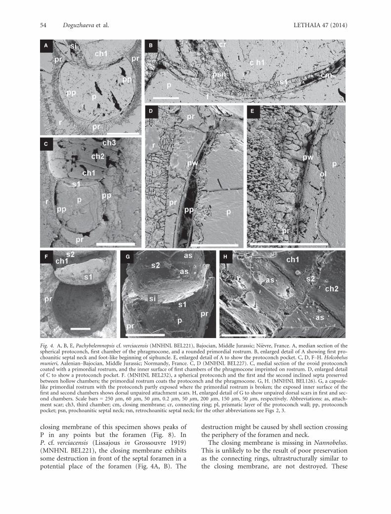
Fig. 1. Map of the Paris Basin showing the position of the citedlocalities marked by an asterisk (modified after Pom�erol 1974).
50 Doguzhaeva et al. LETHAIA 47 (2014)

and phragmocone (L2); (3) in median sectionsshowing a closing membrane, as a distance betweenthe top of the protoconch and a point of a maxi-mum curvature of closing membrane (L3). This iscarried out because the protoconch length (Table 1)measured on the outer surface (L1) is larger than thelength measured in median sections (L2, L3). Thedifference between the three measured lengths (L1,L2, L3) of the same protoconch achieves 30%(Table 1). These data are important for analysis ofprotoconch sizes in evolution of belemnoids (seeTables 1, 5; Discussion).
Geological setting and depositionalenvironment
The herein studied Early–Middle Jurassic belemnitesoriginate from the north-eastern and western mar-gins of Paris Basin (Fig. 1). The north-east ParisBasin corresponds today to the French and BelgianLorraine, including southern Luxembourg. In thisarea, the Lower Jurassic beds start with the alternat-ing carbonates and marls of the Hettangian and Sin-emurian age, deposited in the sedimentologicalregime of the littoral zone (Lathuili�ere 2010). TheHettangian belemnites, found in the nearby LorraineHettangian, are rare and fall in the single genusSchwegleria Riegraf 1980 (Weis & Delsate 2005; Del-sate 2008). The Early Sinemurian shells of Nannobe-lus directly follow the belemnites referred toSchwegleria. The Nannobelus shells were sampledfrom the blue-grey marls of the Strassen Member(Semicostatum and Turneri Zones in south-east Bel-gium, near Arlon (see Boulvain et al. 2001). The Pli-ensbachian Parapassaloteuthis shells were collected
from marls, containing carbonates and sandstones,which were deposited in the sedimentological regimesimilar to that in Hettangian and Sinemurian. Twooutcrops that yielded the Pliensbachian belemnites(Aubange–Ottemt, south-east Belgium, and Oe-trange, north-east France) are dated from the upper-most Pliensbachian, Spinatus Zone, by the means ofammonites Amauroceras ferrugineum (Simpson1855) and Pleuroceras sp. The Aalenian–BajocianHolcobelus and Bajocian Pachybelemnopsis shellswere collected from the ‘Oolithe ferrugineuse deBayeux’ Formation, at the ‘classic’, now inaccessiblelocalities Sully and St. Vigor (Pr�eat et al. 2000).Additional specimens have been recovered from thelower portion (Humphriesianum Zone) of the ‘Oo-lithe ferrugineuse de Bayeux’ Formation at Feuguer-olles-sur-Orne.
Observations
Internal apical shell structures
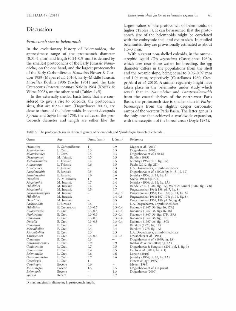
Protoconch. – The protoconchs observed in Nann-obelus (Fig. 2A–E), Parapassaloteuthis (Fig. 3A–F),Holcobelus (Fig. 4C, D, F) and Pachybelemnopsis(Figs 4A, B, E; 5A–F) are ovoid or spherical; theexternal surface is smooth. The protoconch wall isformed by the outer organic, prismatic and innerorganic layers (Fig. 2C); it frays out at the cornersbetween the protoconch and the phragmocone. Theaxes of the protoconch and the phragmocone forman angle. The range of the protoconch diameter is0.31–0.67 mm; the protoconchs with the smallestdiameter are those of Nannobelus and with the
Table 1. Protoconch maximum diameter (D) and length (L1, L2, L3) in Nannobelus, Parapassaloteuthis, Holcobelus and Pachybelemnopsisunder study.
Species Dmax Length L1/L2/L3 (mm) Age Locality
Nannobelus sp. 0.31 –/0.30/– Sinemurian Tontelange/BelgiumNannobelus sp. 0.32 –/0.24/– Sinemurian Tontelange/BelgiumNannobelus sp. 0.36 –/0.27/– Sinemurian Tontelange/BelgiumP. zieteni 0.37 –/0.35/– Pliensbachian Oetrange/ThionvilleP. zieteni 0.39 –/0.38/– Pliensbachian Oetrange/ThionvilleP. zieteni 0.42 –/0.44/– Pliensbachian Oetrange/ThionvilleP. zieteni 0.46 –/0.42/– Pliensbachian Oetrange/ThionvilleHolcobelus munieri 0.32 –/0.33/– Aalenian/Bajocian NormandyHolcobelus sp. 0.45 –/0.38/– Aalenian/Bajocian NormandyHolcobelus sp. 0.38 –/33/– Aalenian/Bajocian NormandyPachybelemnopsis sp. 0.65 0.6/0.46/– Bajocian FeuguerollesPachybelemnopsis sp. 0.67 –/0.59/– Bajocian FeuguerollesP. cf. baculiformis 0.46 –/0.36/– Bajocian SullyP. apiciconus 0.58 –/0.58/– Bajocian NormandyP. apiciconus 0.64 0.61/0.49/0.41 Bajocian NormandyP. apiciconus 0.65 –/0.53/– Bajocian NormandyP. apiciconus 0.65 0.54/–/0.51 Bajocian BelmontP. cf. verciacensis 0.61 –/0.45/0.42 Bajocian Ni�evre
LETHAIA 47 (2014) Embryonic shell factor in belemnite expansion 51

largest diameter are those of Pachybelemnopsis; theformer are about two times less than the latter(Table 1).
The protoconch pockets observed in Holcobelus(Fig. 4C, D) and Pachybelemnopsis (Fig. 4A, E) rep-resent a pair of longitudinal lens-like compartmentsformed by invagination of the inner organic layer ofthe protoconch wall in the middle part of the proto-conch length; they are hollow or filled with sedimentand have variable shape and size.
Numerous nano-tubes within the protoconch ofunknown origin observed in Nannobelus (Fig. 2B) –
in median shell section look like tubular, circular orelliptic structures about 20–30 lm in diameter; theirwall is about 2–3 lm thick and formed by grains ofcalcium phosphate; in this respect, they are similarto the apical channel (Fig. 2F).
Closing membrane. – The closing membrane,observed in four specimens of Pachybelemnopsis(Figs 4A, B; 5B–E; 6B, C; 8), represents a concavestructure above the aperture of the protoconchattached to the shell wall at the corners between theprotoconch and the phragmocone. It has an
A B
C D E F
Fig. 2. Nannobelus sp., Sinemurian, Early Jurassic; Tontelange, Belgian Province of Luxembourg. A, B, E (NRM–PZ Mo. 161/183). A,median section of the protoconch and first chamber of phragmocone. B, enlarged detail of A showing structure less first septum perfo-rated by a siphuncle, and numerous circular or tubular nano structures inside the protoconch. C, enlarged detail of B showing prismaticand originally organic layers of the protoconch wall. D (NRM–PZ Mo. 161/185), longitudinal section of a spherical protoconch and twosepta of phragmocone; crystals of iron-dioxide marks possible site of septal foramen in first septum. E (NRM PZ–Mo. 161/184), longitu-dinal section showing an ovoid protoconch and first septa of phragmocone. F (NRM–PZ Mo. 161/186), an overall view caused by a lon-gitudinal fracture of rostrum with the exposed apical channel with a granular wall inside the apical cavity of the rostrum. Abbreviations:ac, apical cavity; ap, apical channel; c, circular structures; ch1, first chamber of phragmocone; cr, connecting ring; ol, organic layer; p,protoconch; ph, phragmocone; pl, prismatic layer; pr, primordial rostrum; psn, prochoanitic septal neck; pw, protoconch wall; r, ros-trum; s1, s2; first and second septa; si, siphuncle; t, tubular structures. Scale bars = 100 lm, 100 lm, 25 lm, 250 lm, 250 lm, 2.5 lm,respectively.
52 Doguzhaeva et al. LETHAIA 47 (2014)

amorphous texture (Fig. 6B), a microglobular ultra-structure with globules being <1 lm in size (that is amicrobial size) (Fig. 9A). In Pachybelemnopsis apicic-onus (Blainville 1827) (MNHNL BEL224), the clos-ing membrane is strongly concave adapically andshows a tiny sub-ventral foramen edged with a retro-choanitic neck (Fig. 6B, C). The foramen is
0.02 mm in diameter, which is in 2.5 times less thanthe septal foramen in the first septum. In P. apicic-onus (MNHNL BEL220), the closing membraneshows a sub-ventral foramen lacking a neck(Fig. 5B–E). The foramen is 0.03 mm in diameter,which is slightly less than the diameter of the septalforamen of the first septum in this specimen. The
A B
C D
E F
Fig. 3. Parapassaloteuthis zieteni, Pliensbachian, Early Jurassic; Oetrange/Thionville, France. A–C (MNHNLBEL121a). A, median sectionexposing the hollow spherical protoconch, first chambers of the phragmocone, and a primordial rostrum. B, enlarged detail of A to showthe tubular first segment of siphuncle. C, enlarged detail of A to show finely laminated loosely mineralized primordial rostrum (top) andthin prismatic layer of the wall of protoconch (below). D–F (MNHNLBEL229). D, an overall view caused by a longitudinal fracture ofrostrum with the exposed ovoid protoconch and primordial rostrum; the protoconch exposes a smooth outer surface and a large diage-netical crystal inside. E, enlarged detail of D to show finely laminated primordial rostrum at the top of the protoconch. F, enlarged detailof E showing prismatic and irregularly mineralized apparently non-biomineralized outer layers of the protoconch wall. Scalebars = 200 lm, 50 lm, 50 lm, 250 lm, 50 lm, 10 lm, respectively. Abbreviations: ch2, second chamber of the phragmocone; g, gapbetween the cameral deposits in the second chamber; for the other abbreviations see Fig. 2.
LETHAIA 47 (2014) Embryonic shell factor in belemnite expansion 53
closing membrane of this specimen shows peaks ofP in any points but the foramen (Fig. 8). InP. cf. verciacensis (Lissajous in Grossouvre 1919)(MNHNL BEL221), the closing membrane exhibitssome destruction in front of the septal foramen in apotential place of the foramen (Fig. 4A, B). The
destruction might be caused by shell section crossingthe periphery of the foramen and neck.
The closing membrane is missing in Nannobelus.This is unlikely to be the result of poor preservationas the connecting rings, ultrastructurally similar tothe closing membrane, are not destroyed. These
A B
C
D E
F G H
Fig. 4. A, B, E, Pachybelemnopsis cf. verciacensis (MNHNL BEL221), Bajocian, Middle Jurassic; Ni�evre, France. A, median section of thespherical protoconch, first chamber of the phragmocone, and a rounded primordial rostrum. B, enlarged detail of A showing first pro-choanitic septal neck and foot-like beginning of siphuncle. E, enlarged detail of A to show the protoconch pocket. C, D, F–H. Holcobelusmunieri, Aalenian–Bajocian, Middle Jurassic; Normandy, France. C, D (MNHNL BEL227). C, medial section of the ovoid protoconchcoated with a primordial rostrum, and the inner surface of first chambers of the phragmocone imprinted on rostrum. D, enlarged detailof C to show a protoconch pocket. F. (MNHNL BEL232), a spherical protoconch and the first and the second inclined septa preservedbetween hollow chambers; the primordial rostrum coats the protoconch and the phragmocone. G, H. (MNHNL BEL126). G, a capsule-like primordial rostrum with the protoconch partly exposed where the primordial rostrum is broken; the exposed inner surface of thefirst and second chambers shows dorsal unpaired attachment scars. H, enlarged detail of G to show unpaired dorsal scars in first and sec-ond chambers. Scale bars = 250 lm, 60 lm, 50 lm, 0.2 lm, 50 lm, 200 lm, 150 lm, 50 lm, respectively. Abbreviations: as, attach-ment scar; ch3, third chamber; cm, closing membrane; cr, connecting ring; pl, prismatic layer of the protoconch wall; pp, protoconchpocket; psn, prochoanitic septal neck; rsn, retrochoanitic septal neck; for the other abbreviations see Figs 2, 3.
54 Doguzhaeva et al. LETHAIA 47 (2014)

observations favour the idea that the Early Sinemuri-an Nannobelus did not possess the closing mem-brane, and the protoconch of this genus was coveredwith the perforated septum (Fig. 2A, B, D, E). In theLate Pliensbachian Parapassaloteuthis, the first sep-tum is irregularly mineralized (Fig. 8A), the organiclayer on its adapical surface is distinct but the closingmembrane is not recognized and possibly missing(Fig. 3A, B). In the Aalenian–Bajocian Holcobelus,the shell shows well preserved originally organiclayer of the protoconch wall and the rarely preservedprotoconch pockets, however, the closing membraneis not distinguished (Fig. 4C). Therefore, in spite ofthe fact that the presumably organic shell structures
are well preserved in all studied genera, the closingmembrane is missing in Nannobelus, Parapassaloteu-this and Holcobelus and present only in Pachybel-emnopsis.
Phragmocone. – In Nannobelus (Fig. 2A, E) and inParapassaloteuthis (Fig. 3A), the first chamber of thephragmocone is longer than the second one and isabout 0.3 the length of the protoconch. The siphun-cle is sub-marginal in the first chamber; the first seg-ment of connecting rings is tubular. In Nannobelus,the connecting ring is attached on the outside of thefirst septal neck that is prochoanitic; next septal neckis retrochoanitic (Fig. 2B). In Parapassaloteuthis, the
A B C
D E
G
Fig. 5. Pachybelemnopsis apiciconus (MNHNL BEL220), Bajocian, Middle Jurassic; Belmont, France. A, an overall view caused by a longi-tudinal fracture of rostrum with the exposed globular protoconch and the apical part of the phragmocone. B, median section of shell ofA showing inclined septa and marginal siphuncle (to the left) in first chambers of the phragmocone and a closing membrane. C, enlargeddetail of B to show a primordial rostrum around a protoconch. D, enlarged detail of C to show the very beginning of siphuncle. E,enlarged detail of D showing the curved first segment of siphuncle in front of the foramen in closing membrane. Scale bars = 0.25 mm,0.25 mm, 0.5 mm, 125 lm, 50 lm, respectively. Abbreviations: f, foramen; ms, mural part of septum; sn1, first septal neck; for the otherabbreviations see Figs 2.
LETHAIA 47 (2014) Embryonic shell factor in belemnite expansion 55

siphuncle is swollen between the first and the secondsepta on the ventral side but straight cylindrical dor-sally (Fig. 3A, B). The septal foramen of the first sep-tum is about 0.05 mm in diameter. The first andseveral next septa are inclined dorsally; as a result,the chamber length on the dorsal side is longer thanon the ventral side (Fig. 3A, B). In Holcobelus(Fig. 4C), the length of the first and the secondchambers is about 0.4 protoconch length; the third
chamber is slightly shorter than the previous ones(Fig. 4C). The first four to five septa are inclineddorsally; the next ones are perpendicular to the axisof the phragmocone (Fig. 4C, F). The siphuncle isnot observed, however, the septal foramen of thefirst septum marks its ventro-marginal positionfrom the beginning of the phragmocone (Fig. 4G).The unpaired dorsal rectangular attachment scarsare present in the second and third chambers
A B
C
Fig. 6. Pachybelemnopsis apiciconus (MNHNL BEL224), Bajocian, Middle Jurassic; St. Vigor, Normandy, France. A, an overall viewcaused by a longitudinal fracture of rostrum with the exposed the globular protoconch and the phragmocone; first three septa areinclined; next septa are getting perpendicular to the phragmocone axis; and the fourth and fifth septa are not inclined. B, C, median sec-tion of the shell on A to show a contact between the protoconch (bottom) and phragmocone (top); a closing membrane exposes a ventralforamen in front of the foramen of the first septum; the foramen of the closing membrane is bordered with a retrochoanitic neck. Scalebars = 250 lm, 25 lm, 125 lm, respectively. Abbreviations: rn, reptrochoanitic neck; for the other abbreviations see Figs 2, 4, 5.
56 Doguzhaeva et al. LETHAIA 47 (2014)

(Fig. 4G, H). They are about as long as the chamber;the width is about three times less than their length.The attachment scars represent shallow depressionsrounded by the innermost layer of the chamber wall.The dorsal attachment scars of Holcobelus are similarin shape with the dorsal unpaired attachment scarsof Spirula (Doguzhaeva 2000, pl. 2, figs 7, 8; Bandel& Stinnesbeck 2006, fig. 3: 7). In Pachybelemnopsisapiciconus (with the exception of the specimenMNHNL BEL224), P. cf. verciacensis and Pachybel-emnopsis sp., the first septum and the closing mem-brane are located extremely close to each other; thespace between them can hardly be considered as achamber; siphuncle is foot-like there (Figs 4A, B; 8).The siphuncle is sub-marginal, ventral; the septalforamen is 0.03 mm there; it is equal to the foramenof the closing membrane. The septal neck of the sec-ond septum is long, slightly shorter than the cham-ber length (Fig. 4A). In P. cf. verciacensis, the septalforamen is 0.09 mm in diameter. The second–fifthsepta of the phragmocone are inclined dorsally; thefollowing ones are perpendicular to the axis of thephragmocone, therefore, the first chambers areshorter ventrally and longer dorsally. In Parapassalo-teuthis, the first and second chambers contain epi-and hyposeptal cameral deposits separated by theslit-like gaps (Fig. 3A, B). The cameral deposits donot belong to the embryonic stage but were appar-ently secreted at later ontogenetic stages.
Primordial rostrum. – The primordial rostrum isdistinguished from the rostrum by the ultrastructur-al differences between these two structures, visible inmedian shell sections. It tightly bounds the proto-conch and the apical part of the phragmocone(Figs 2A, D; 3A–E; 4A, C, F, G; 6A, C). In Parapassa-loteuthis (Fig. 3A–E), the primordial rostrum isrounded near the top of the protoconch where it hasa thickness of about 0.15 protoconch length. It isthin on sides of the protoconch but swollen in thecorners between the protoconch and the phragmo-cone; from this place forward the primordial ros-trum represents a prismatic layer. Around theprotoconch, the primordial rostrum is loosely min-eralized and has a fine laminated ultrastructure(Fig. 3C). In Holcobelus (Fig. 4C, F, G), the primor-dial rostrum is rounded or conical near the top ofthe protoconch; it is about as thick as 0.3 proto-conch length. In Pachybelemnopsis (Figs 4A, 6A), theprimordial rostrum is short conical or rounded.
Pro-ostracum. – The pro-ostracum is not distin-guished in the apical shell but is observed in about20th–25th chambers of the phragmocone in twospecimens of Parapassaloteuthis (MNHNL BEL235,
BEL059) and three specimens of Pachybelemnopsis(MNHNL BEL230, BEL224, BEL236). In Parapassa-loteuthis zieteni (Mayer-Eymar 1884), the pro-os-tracumis is distinguished due to longitudinalstriation of the lateral fields and criss-cross patternof the mantle attached to the lateral field (Fig. 9B–D). In longitudinal shell sections, the pro-ostracumis located between the mural parts of the septa andprimordial rostrum. The median shell section ofPachybelemnopsis (MNHNL BEL236) shows that thepro-ostracum is preserved in the middle part of thephragmocone but is destroyed in the apical direc-tion; a hollow space left at place of the destroyedpro-ostracum comes to the protoconch. This is anindirect indication of the presence of the pro-ostra-cum in the embryonic shell.
Energy-dispersive spectrometry data onchemical composition of shell
Tested shells show regular phosphatization of struc-tureless, apparently non-biomineralized, originallyorganic structures such as connecting rings, closingmembrane, inner and outer layers of protoconchwall, adoral and adapical layers of septa, wallof nano-tubular structures within the protoconch,wall of apical channel, originally organic rich pro-ostracum, and filling of siphuncle and gaps betweenepi- and hyposeptal cameral deposits. The listedabove structures are also sporadically iron-oxidized(Tables 2–4; Figs 7, 8).
The highest values of P are revealed in the closingmembrane (16.9%), gaps between the epi- and hyp-oseptal cameral deposits (16.1%), siphuncle in thefirst chamber (12.3%), connecting rings (12.2%),organic layer of protoconch wall (10.5%) andorganic layer of first septum (6.3%) (Tables 2–4).The shell elements and sites listed above show also avarying amount of iron-oxides (Tables 2–4). Thehighest values of Fe are revealed in the pro-ostracumfused with the mantle tunic (62.9%) (Tables 2, 4),siphuncle in the first chamber (62.8%) (Table 4)and large dark spots in the protoconch (56.7%)(Table 4). The highest values of S (4.4%) arerecorded in the siphuncle (the first chamber) inassociation with a comparatively lower content of Fe(9.5%) in Parapassaloteuthis (Table 2). One of threeprotoconchs of Holcobelus examined (MNHNLBEL227) is filled with transparent calcium carbonatebut contains also large clusters of iron-oxide(Table 3).
The pattern formed by dark-brown Fe-containingmaterial shows that it is distributed as if it wouldpenetrate in the protoconch via the closing mem-brane. Judging on colour images the dark ‘cloud’ of
LETHAIA 47 (2014) Embryonic shell factor in belemnite expansion 57

iron-oxide has varying concentration and heteroge-neous structure. A net formed by crossed, perpen-dicular tube-like elements is indistinctly visibleinside the ‘cloud’ as well as several solid spots. Thissupposedly indicates some organic material withinthe protoconch. The connecting rings and pro-ostra-cum show Z, As and Ba known to be associated withfossilization of organic rich or non-biomineralizedshell structures and soft tissues. These three chemicalelements have maximum values in the pro-ostra-cum: 1.02%, 0.27% and 0.27%, respectively. Peaks ofS in the connecting rings and pro-ostracum possiblyindicate minor amounts of barite. The gaps in the
cameral deposits as well as some other structureslisted above show peaks of Sn (Tables 2–4). Ba andSn were earlier reported in the organic capsule of theLate Cretaceous belemnite Gonioteuthis Bayle 1878(Doguzhaeva & Bengtson 2011; fig. 2). Thus, P, Fe,Ba, Sn, Zn, As and S are diagenetically precipitatedmarkers of the sites originally rich in organicmaterial.
Contrary to the shell elements discussed above,the primordial rostrum, rostrum and protoconchpockets lack P. The primordial rostrum and rostrumshow similar values of C, O, Na, Mg and Ca(Tables 2–4); the former however shows higher
Table 2. Chemical composition of the apical part of a shell (specimens from Oetrange/Thionville, France) and a pro-ostracum (speci-mens from Aubange, Belgian Province of Luxembourg) of Parapassaloteuthis zieteni, Pliensbachian, Early Jurassic (EDS data; in per centto total weight).
Element (%)
Site
Rostrum Primordial rostrum Septum Proostracum
Filling ofsiphunclein firstchamber
Cameraldeposits(first, secondchambers)
Gaps incameraldeposits
Carbon 6.9–10.3 7.4–11.2 7.3–10.6 10.0–13.0 6.7 8.6 no dataNitrogen 2.4–3.2 5.2–10.0 3.3–5.5 6.1–6.8 3.0 2.8 no dataOxygen 41.9–48.3 46.3–52.9 40.8–47.3 54–61.3 30.9 47.7 35.7Fluorine no data no data no data no data no data no data 7.7Sodium 0.1–0.3 0.1–0.5 0.4 0.3 no data no data no dataMagnesium 0.2–0.4 0.3–0.4 0.2–0.4 0.1 no data 0.6 0.2Aluminium no data no data no data no data no data no data no dataSilicon 0.2 no data 0.1 1.1 1.0–1.1 no data no dataPhosphorus no data no data no data no data 12.3 no data 16.1Sulphur no data no data no data no data 4.4 no data no dataPotassium 0.8 0.2 0.2 no data no data no data no dataCalcium 38.3–46.9 30.90–40.6 41.0–46.2 26.1–28.3 33.2 40.0 40.3Iron no data no data no data 62.9 9.5 no data no dataZinc no data no data no data no data no data no data no dataStrontium 0.2–0.4 0.2–1.3 0.1–0.5 0.4–0.7 no data 0.3 0.1Chlorine no data no data no data no data no data no data no dataNiobium no data no data no data no data no data no data 0.5Tin no data no data no data no data no data no data 6.3Platinum no data no data no data no data no data no data 0.9
Table 3. Chemical composition of the apical part of shell of the Aalenian–Bajocian Holcobelus munieri (EDS data; in per cent to totalweight).
Element
Site
Dark spots withinthe protoconch
Transparent fillingof protoconch
Primordialrostrum
Organic layer offirst septum
Protoconchpocket
Carbon 5. 8–6.7 10.4 14.4 12.3 11.7Oxygen 35.1–36.0 51.5 51.3 58.2 56.3Sodium no data no data no data no data 0.6Aluminium no data no data no data no data 0.6Silicon 1.0–1.1 no data no data no data 9.0Phosphorus no data no data no data 6.3 no dataCalcium 0.9–1.2 38.1 34.3 14.02 1.79Iron 55.3–56.7 no data no data 1.4 14.3Niobium no data no data no data 0.5 no dataTin no data no data no data 6.3 5.8Platinum no data no data no data 0.9 no data
58 Doguzhaeva et al. LETHAIA 47 (2014)

values of N and Sr that are three times higher thanin the rostrum, but lower value of K (Table 2).Moreover, the primordial rostrum shows the high-est, among all tested morphological structures, val-ues of C (14.38%), N (10.03%), O (53.89%), Mg(0.44%) and Sr (1.28%) (Tables 2, 3). Nitrogen evi-dences non-fossilized organic material of the pri-mordial rostrum. The protoconch pockets showpeaks of Si, Fe, Sn, Al, Na (Table 3) apparently indi-cating their postmortem filling.
In Parapassaloteuthis, the comparison of chemi-cal composition between the pro-ostracum, on theone hand, and the rostrum, primordial rostrum,and septa, on the other hand, reveals that in thepro-ostracum, the content of Ca (28.3%) is lessthan in the listed structures, but the content of C(13%), N (6.8%) and O (61.3%) is higher (seeTable 2). These data confirm the inorganic–organic composition of the pro-ostracum(Fig. 9B–D).
Table 4. Chemical composition of the internal shell structures and a pro-ostracum of the Bajocian Pachybelemnopsis. (EDS data; in percent to total weight).
Element
Site
Connectingring
Closingmembrane
Organic layers ofprotoconch walland septa Proostracum
Foramen ofclosing membrane
Filling ofsiphuncle
Filling ofprotoconchand firstchambers
Carbon 8.8–18.8 6.7–11.9 7.1–11.4 7.7–11.5 6.1–14.6 12.5 11.0–15.6Nitrogen 3.2–5.0 no data no data 0.6–3.9 no data no data no dataOxygen 33.7–42.7 20.3–48.0 30.0–54.0 20.9–47.2 34.3–51.7 33.8–46.9 38.6–56.2Fluorite 5.3–5.6 0–6.5 6.1–8.1 no data no data no data no dataMagnesium 0.1–0.8 no data no data 0.0–0.2 no data no data no dataAluminium 0.8–1.4 no data no data 0.5–1.4 1.3–1.6 1. 7 no dataSilicon 2.4–4.8 2.2 1.3 1.5–2.7 1.4–1.7 1.7 no dataPhosphorus 6.8–12.2 6.5–16.9 5.8–10.5 0.1–0.2 no data no data no dataSulphur 0.1–0.3 no data no data 0.1–0.2 no data no data no dataCalcium 23.4–29.6 16.7–50.1 20.6–37.2 0.9–19.3 2.9–43.4 40.6 28.2–50.4Manganese 0.1 no data no data 0.0–0.4 no data no data no dataIron 1.2–4.4 30 9.1 18.4–62.9 51.9–61.8 62.8 no dataZinc 0.1–0.3 no data no data 0.1–1.0 no data no data no dataArsenic 0.0–0.1 no data no data 0.1–0.3 no data no data no dataStrontium 0.9–1.23 no data no data 0.0–0.7 no data no data no dataNiobium 1.5–2.7 3.38 no data no data no data no data no dataTin no data 13.46 6.04 no data no data no data no dataPlatinum 1. 7 0.7–4.1 no data no data no data no data no dataBarium 0.1 no data no data 0.1–0.3 no data no data no data
A B
Fig. 7. Parapassaloteuthis zieteni (MNHNL BEL127), Pliensbachian, Early Jurassic; Oetrange. Energy-dispersive spectrometry data on theadapical organic layer of first septum of A, median section of the apical part of the shell with the marked point of the spectrum 2 taken.Scale bar = 50 lm. B, the spectrum 2 showing chemical composition of the adapical organic layer of first septum. Abbreviations: ol,organic layer of the first septum; for the other abbreviations see Fig. 2.
LETHAIA 47 (2014) Embryonic shell factor in belemnite expansion 59

Fig. 8. Pachybelemnopsis apiciconus (MNHNL BEL237), Bajocian, Middle Jurassic; Belmont, France. Energy-dispersive spectrometry datatracing distribution of P along the closing membrane and indicating a foramen of the closing membrane. Scale bar = 50 lm. Abbrevia-tions: +, points showing phosphorus; o, points lacking phosphorus; for the other abbreviations see Fig. 2.
A B
C D
Fig. 9. A, Pachybelemnopsis apiciconus (MNHNL BEL224), Bajocian, Middle Jurassic; St. Vigor, Normandy, France. The globular ultra-structure of the closing membrane with a microbial size of globules; enlarged detail of Fig. 6C. B, C, Parapassaloteuthis zieteni (MNHNLBEL235). Late Pliensbachian, Early Jurassic; Aubange, Belgian Province of Luxembourg. Pro-ostracum showing a longitudinal striationof the lateral field (to the wright)and a criss-cross pattern of the mantle attached to the pro-ostracum (to the left). Scale bars = 1 mm,250 lm, 200 lm, respectively. Abbreviations: ma, mantle; pro, pro-ostracum; for the other abbreviations see Fig. 2.
60 Doguzhaeva et al. LETHAIA 47 (2014)
Discussion
Protoconch size in belemnoids
In the evolutionary history of Belemnoidea, theapproximate range of the protoconch diameter(0.31–1 mm) and length (0.24–0.9 mm) is defined bythe smallest protoconchs of the Early Jurassic Nann-obelus, on the one hand, and the largest protoconchsof the Early Carboniferous Hematites Flower & Gor-don 1959 (Mapes et al. 2010), Early–Middle JurassicDicoelites Boehm 1906 (Sachs 1961) and the LateCretaceous Praeactinocamax Naidin 1964 (Ko�s�t�ak &Wiese 2008), on the other hand (Tables 1, 5).
In the externally shelled bactritoids that are con-sidered to give a rise to coleoids, the protoconchsizes, that are 0.27–1 mm (Doguzhaeva 2002), areclose to those of the belemnoids. In extant decapodsSpirula and Sepia Linn�e 1758, the values of the pro-toconch diameter and length are either like the
largest values of the protoconch of belemnoids, orhigher (Tables 5). It can be assumed that the proto-conch size of the belemnoids might be correlatedwith the embryonic shell and ovum sizes. In studiedbelemnites, they are provisionally estimated as about1.5–3 mm.
Within extant non-shelled coleoids, in the omma-strephid squid Illex argentines (Castellanos 1960),which uses near-shore waters for breeding, the eggdiameter differs in the populations from the shelfand the oceanic slope, being equal to 0.96–0.97 mmand 1.04 mm, respectively (Castellanos 1960; Cres-pi-Abril et al. 2010). A similar regularity might havetaken place in the belemnites under study whichreveal that in Nannobelus and Parapassaloteuthisfrom the coastal shelves of the north-west ParisBasin, the protoconch size is smaller than in Pachy-belemnopsis from the slightly deeper carbonaticramps of the western Paris Basin. The latter genus isthe only one that achieved a worldwide expansion,with the exception of the boreal areas (Doyle 1987).
Table 5. The protoconch size in different genera of belemnoids and Spirula/Sepia branch of coleoids.
Genus Age Dmax (mm) L (mm) Reference
Hematites E. Carboniferous 1 0.9 Mapes et al. (2010)Mutveiconites L. Carb. 0.3 0.3 Doguzhaeva (2002)Mutveiconites L. Carb. 0.5 0.3 Doguzhaeva et al. (2006)Dictyoconites M. Triassic 0.5 0.3 Bandel (1985)Metabelemnites L. Triassic 0.4 0.5 Jeletzky (1966; pl. 3; fig. 1A)Aulacoceras L. Triassic 0.6 0.5 Fuchs (2012; fig. 2D)Acrocoelites E. Jurassic – 0.3 L.A. Doguzhaeva, unpublished dataPassaloteuthis E. Jurassic 0.5 0.6 Doguzhaeva et al. (2003; figs 9, 13, 17, 19)Passaloteuthis E. Jurassic 0.6 0.6 Jeletzky (1966; pl. 11; fig. 1)Dicoelites E.–M. Jurassic 1 0.9 Sachs (1961; figs 7, 8)Megateuthis M. Jurassic 0.7 0.4 Jeletzky (1966; pl. 14; fig. 1A)Hibolithes M. Jurassic 0.4 0.5 Bandel et al. (1984; fig. 1A), Ward & Bandel (1987; fig. 17.8)Megateuthis M. Jurassic 0.5 0.7 Pugaczewska (1961; 138; pl. 7, fig. 8)Pachybelemnopsis M. Jurassic – 0.4–0.5 Pugaczewska (1961; 152, 160; pl. 14, fig. 6)Hibolithes M. Jurassic – 0.4–0.8 Pugaczewska (1961; 167, 176; pl. 19, fig. 8)Dicoelites M. Jurassic – 0.5 Pugaczewska (1961; 186, pl. 24, fig. 4)Pachyteuthis L. Jurassic 0.5 0.4 L.A. Doguzhaeva, unpublished dataHibolithes E. Cretaceous 0.3–0.5 0.3–0.4 Kabanov (1967; 36, figs 16, 17A)Aulacoteuthis E. Cret. 0.3–0.5 0.3–0.4 Kabanov (1967; 36, figs 16–18)Neohibolites E. Cret. 0.3–0.5 0.3–0.4 Kabanov (1967; 36, figs 17B, 18A)Conobelus E. Cret. 0.3–0.5 0.3–0.4 Kabanov (1967; 36, fig. 18B)Duvalia E. Cret. 0.3–0.5 0.3–0.4 Kabanov (1967: 36; fig. 18G)Conobelus E. Cret. 0.4 0.4 Barskov (1973; fig. 1E)Mesohibolites E. Cret. 0.4 0.4 Barskov (1973; fig. 1A)Mesohibolites E. Cret. 0.3 0.3 L.A. Doguzhaeva, unpublished dataTauriconites E. Cret. 0.5–0.6 0.4–0.5 Drushchits et al. (1984)Conobelus E. Cret. 0.3 ─ Doguzhaeva et al. (1999; fig. 1A)Praeactinocamax L. Cret. 0.9 0.9 Ko�s�t�ak & Wiese (2008; fig. 4A)Gonioteuthis L. Cret. 0.7 0.5 Doguzhaeva & Bengtson (2011; pl. 1, fig. 1)Conoteuthis L. Cret. 0.4 0.5 Fuchs et al. (2012; fig. 4D)Belemnitella L. Cret. 0.6 0.6 Larson (2010)Groenlandibelus L. Cret. 0.7 0.6 Jeletzky (1966; pl. 20, fig. 1A)Ceratisepia L. Cret. 1 – Hewitt & Jagt (1999)Ceratisepia Eocene 0.6 – Meyer (1993)Mississaepia Eocene 1.5 0.5 Doguzhaeva et al. (in press)Belemnosis Eocene – 1.3 Doguzhaeva (2000)Spirula Recent – 0.7
D max, maximum diameter; L, protoconch length.
LETHAIA 47 (2014) Embryonic shell factor in belemnite expansion 61
Embryonic shell structure of the Early–MiddleJurassic belemnites
The morphology of the juvenile shells of the Pli-ensbachian Passaloteuthis, that have been so farknown in more detail than in other belemnites,favours the idea that the embryonic shell of theJurassic belemnites represents an internal structureconsisted of the protoconch, one or two chambers ofthe phragmocone, and the organic-rich primordialrostrum and pro-ostracum; the latter possessingabroad central field with rounded growth rings andtwo narrow lateral fields with fine longitudinal striae(Doguzhaeva et al. 2003, figs 1–3).
The Passaloteuthis model is confirmed by theembryonic shell structure of the Early–MiddleJurassic belemnites from the Paris Basin hereinreported (see Observations). In these belemnites,the embryonic shell is distinguished in polishedmedian shell sections due to the ultrastructural dif-ferences between the loosely mineralized finely lam-inated primordial rostrum and the solid rostrum(Figs 2A; 3A–E; 4A, C, F, G; 5C; 6C). The primor-dial rostrum forms a capsule-like structure tightlysurrounding the protoconch and the phragmocone(Fig. 4G). Non-fossilized organic component of theprimordial rostrum is indicated with the detectednitrogen (Table 2) that is a reliable marker of non-fossilized organic material (Oehler et al. 2008).Among the diagenetic features suggesting the origi-nally organic-rich composition of the primordialrostrum are caverns observed within this shellstructure in the Middle Jurassic Hibolithes D�enysde Montfort 1808; (Bandel et al. 1984) and Passa-loteuthis (Doguzhaeva et al. 2003). The cavernswere probably formed at places of perishable mate-rial that was not preserved. The globular or ovoidprotoconchs of the belemnites under study havesmooth surfaces (Figs 3C; 5A); they have neitherthe net-like ornament of the protoconch secretedin some bactritoids (Doguzhaeva 2002), nor tuber-cles typical for the protoconchs of ammonoids(Landman 1988; Tanabe et al. 2010). The proto-conch wall is formed by two organic layers withthe prismatic layer in between (Fig. 2C); it fraysout in the corners between the protoconch and thephragmocone. In Holcobelus and Pachybelemnopsis,the inner organic layer of the protoconch walldiverges from the prismatic layer and forms thelens-like compartments (Fig. 4A–E), herein namedprotoconch pockets, which have postmortem filling(Table 3), and therefore, in lifetime, they mightcontain gas or liquid with gas; this would diminishthe weight of eggs and hatching. A similar structurehas previously been observed in the Late Jurassic
belemnite Pachyteuthis Bayle 1878 (Barskov 1973).It is not excluded that the protoconch containedduring life-time organic substance that was post-mortem sporadically iron-oxidized (Figs 2D, 3D;Table 3).
The observations on Nannobelus, Parapassaloteu-this, Holcobelus and Pachybelemnopsis suggest that anorganic rich closing membrane is under formation.In the Early Sinemurian Nannobelus, the aperture ofthe protoconch seems to be covered with a perfo-rated septum (Fig. 2A, B), like in bactritoids. Theassumption that the closing membrane was formedbut not preserved in the noted genus is less probablebecause the structures of similar composition, forinstance connecting rings, are preserved in the shellsthat lack the closing membrane. In Parapassaloteu-this, the first septum is irregularly mineralized andhas a prominent organic layer on its adapical surface(Fig. 7). In Pachybelemnopsis apiciconus, the closingmembrane has a small sub-ventral foramen boardedwith a retrochoanitic neck (Fig. 6A–C). The fora-men of the closing membrane is ten times less thanthe foramen of the first septum. Besides, two morespecimens of Pachybelemnopsis exhibit the foramenof the closing membrane (Figs 4A, B; 8). The latterobservation suggests that the process of formation ofclosing membrane was not yet finished in the belem-nites under study. However, a Bajocian specimen ofHolcobelus from Dorset (Jeletzky 1966; pl. 25, fig.1B) was shown to have a structure that resembles aclosing membrane and ‘…typically developed andwell preserved. However, foot of the siphuncle andproseptum almost completely obliterated.’ (Jeletzky1966, p. 96). The image on pl. 25, fig. 1B in Jeletzky(1966) shows that the interpretation of the blackstructure above the protoconch as a closing mem-brane is not the only possible one as similar black‘membranes’ are present at the next septa.
The observed perforated closing membrane pointsout that in Pachybelemnopsis, like in Spirula, the sip-huncle, or prosipho, may penetrate in the proto-conch. Such a structure of the closing membrane isreported for the first time and is not typical for theclosing membrane of the belemnites. The presenceof the retrochoanitic neck may indicate that the clos-ing membrane originated from the adapical organiclining of the first septum due to cancellation of thesecretion of its prismatic layer. A similar idea on theorigin of the closing membrane of belemnites wasintroduced by Bandel (1985) who thinks that it orig-inated due to decalcification of the first prismaticseptum. The development of the closing membranein the Late Carboniferous–Triassic aulacocerids hasnot been yet convincingly proved and the opinionson this structure in aulacocerids are discrepant
62 Doguzhaeva et al. LETHAIA 47 (2014)

(Jeletzky 1966; Dauphin 1983; Bandel 1985; Fuchs2012). The closing membrane might be absent in theEarly Carboniferous Hematites (Mapes et al. 2010).The other cephalopods with a spherical protoconch– spirulids, bactritoids, ammonoids and sphaerotho-cerids – evidently secreted the first prismatic septumperforated with a foramen for siphuncle attached tothe inner surface of the protoconch.
The embryonic shell of the studied belemnitesincludes one or more chambers of the phragmocone.In Nannobelus (Fig. 2A, C, F), Parapassaloteuthis,Holcobelus (Fig. 4C) and Pachybelemnopsis (Figs 4A,5C, 6A), the first chamber is comparatively long andapparently belonged to the embryonic shell. If issupposed that the adaptation of hatchling firstretarded shell growth, the first chambers of the post-embryonic shell might be shorter than the chambersof the embryonic shell. The first septum has the pro-choanitic septal neck, while septal necks of the nextseptum are retrochoanitic (Figs 2A, B; 3B; 4A, B;6C). The siphuncle starts either like a tube (Fig. 3A,B; 5A–D; 6C), or a foot-like structure covering thetotal surface of the closing membrane (Fig. 4A, B).The tube-like beginning adheres to the closing mem-brane. The cross-section of the tube gives an idea onthe size of a permeable spot providing exchange ofgas and liquid between the siphuncle and the proto-conch. It is reasonable to assume that the closingmembrane has a foramen in front of the tube-likebeginning of the siphuncle, and the rest of the sur-face is not permeable. In a case of a foot-like begin-ning of the siphuncle, the permeable area isessentially larger than in the previous case of a tube-like beginning of the siphuncle.
In studied shells, the pro-ostracum is recognizedon the phragmocone surface in adult shells of Para-passaloteuthis (Fig. 9B–D) and Pachybelemnopsis; itshows irregular mineralization indicating its organicrich original composition (Tables 2, 4). These obser-vations, together with earlier obtained data on theorganic-rich composition of the pro-ostracum inthe Sinemurian Nannobelus (Doguzhaeva 2012),are extrapolated on the composition of the pro-ostracum of the embryonic shell.
Thereby, it is suggested that, due to the pro-ostracum, primordial rostrum, and closing mem-brane which are rich in organic matter, possibly inchitin, the embryonic shell of the Early–MiddleJurassic belemnites was transformed into light butmechanically strong inorganic–organic structure.The inorganic–organic shell is secreted, forinstance, in extant Sepia; the chitin–protein com-plex makes up about 10% of total weight of thecuttlebone (Florek et al. 2009). The chitin compat-ible material was detected in the Eocene cuttlebone
as well (Weaver et al. 2011; Doguzhaeva et al. inpress). The increasing of the organic component ofthe shell matter took place in the Late Cretaceousbelemnite Gonioteuthis secreted the organic capsulearound the protoconch and phragmocone (Dog-uzhaeva & Bengtson 2011).
The inorganic–organic embryonic shell, with aproperly protected protoconch of the Early–MiddleJurassic belemnites, possibly allowed the hatching ofthe eggs in greater depths and lower temperatures, insome degree, like in Sepia. This is due to the charac-teristic properties of the chitin that is light, mechani-cally strong, physiologically inert natural shellmatter broadly using for protection and support ininvertebrate animals. Such an embryonic shell struc-ture might also accelerate the adaptation of hatch-lings to a nektonic mode of life and allowed easymigration of juveniles and adults down into deepand cold waters and come back to warmer waters.Sepia usually migrates to deeper water in winter andcan move rapidly when needed. Cuttlefishes revealdiverse skeletal morphology enabling representativesof this taxon to occupy a range of depths and habi-tats (Sherrard 2000). This may be one of the factorscontributing to higher species diversity in the lattergroup, comprising about 120 species (Reid et al.2005). The increase in the taxonomic diversity of theEarly–Middle Jurassic belemnites might be alsodetermined by the increasing ability to occupy abroad range of depths along the coastal shelves.Therefore, the structure of the embryonic shelldeveloped in the Early and Middle Jurassic belem-nites might be of major significance in the process ofcolonization of a broad range of depths along thecoastal shelves and broadening of geographical dis-tribution and increasing diversification of belemnitesin Early–Middle Jurassic.
Acknowledgements. – This study was carried out thanks to sup-port by the Royal Swedish Academy of Sciences and personallyby Prof. Jan Bergstrom and Prof. Stefan Bengtson, Departmentof Palaeozoology of the Swedish Museum of Natural History. JanStrugnell (Australia) donated several shells of Nannobelus col-lected during the Third International Symposium ‘Coleoidsthrough time’, Luxembourg. Neil Landman (American Museumof Natural History, New-York, USA) made stylistic correctionsto the previous version of the study. We are indebted to all thesepersons; also we are grateful to Martin Ko�s�t�ak and the anony-mous reviewer for help with improving this article.
References
Bandel, K. 1985: Composition and ontogeny of Dictyoconites(Aulacocerida, Cephalopoda). Pal€aontologische Zeitschrift 59,223–244.
Bandel, K. & Boletzky, S.V. 1979: A comparative study of thestructure, development and morphological relationship ofchambered cephalopod shells. The Veliger 21, 313–354.
LETHAIA 47 (2014) Embryonic shell factor in belemnite expansion 63

Bandel, K. & Stinnesbeck, W. 2006: Naefia Wetzel 1930 from theQuiriquina Formation (Late Maastrichtian, Chile): relation-ship to modern Spirula and ancient Coleoidea (Cephalopoda).Acta Universitatis Carolinae – Geologica 49, 21–32.
Bandel, K., Engeser, T. & Reitner, J. 1984: Die Embryonalent-wicklung von Hibolithes (Belemnitida, Cephalopoda). NeuesJahrbuch f€ur Geologie und Pal€aontologie 167, 275–303.
Barskov, I.S. 1973: About a structure of the protoconch andontogeny of belemnites (Coleoidea, Cephalopoda). DokladyAkademii Nauk SSSR 208, 439–442. [In Russian].
Bayle, E. 1878: Fossiles principaux des terrains. Explication de lacarte g�eologique de la France 4, atlas, pls.1–157. Imprimerie Na-tionale, Paris.
Blainville, H.M.D. de 1827:M�emoire sur les Belemnites consider�eeszoologiquement etg�eologiquement. 136 pp. Levrault, Paris.
Boehm, G. 1906: Geologische Mitteilungen aus dem Indo-aus-tralischen Archipel. 1. Neues aus dem Indo-australischenArchipel. Neues Jahrbuch f€ur Geologie, Mineralogie und Pal€aon-tologie – Abhandlungen 22, 385–412.
Boulvain, F., Belanger, I., Delsate, D., Dosquet, D., Ghysel, P.,Godefroit, P., Laloux, M., Roche, M., Teerlynck, H. & Thorez,J. 2001: New lithostratigraphical, sedimentological, mineralog-ical and palaeontological data on the Mesozoic of BelgiumLorraine: a progress report. Geologica Belgica 3, 3–33.
Castellanos, Z.J.A.de. 1960: Una nueva especie de calamar Argen-tino Ommastrephes argentinus sp. nov. (Mollusca, Cephalo-poda). Neotropica (La Plata) 6, 55–58.
Chen, T.E. 1982: Mesozoic coleoidea fauna from Xizang. Paleon-tology of Xizang 4, 280–325.
Chen, T.E. & Sun, Z.H. 1982: Discovery of Permian belemnoidsin South China with comments on the origin of the Coleoidea.Acta Palaeontological Sinica 21, 181–190.
Crespi-Abril, A., Dellatorre, F. & Bar�on, P. 2010: On the presenceof Illex argentinus (Castellanos, 1960) (Cephalopoda: Ommas-trephidae), paralarvae and juveniles in near-shore waters ofNuevo Gulf, Argentina. Latin American Journal of AquaticResearch 38, 297–301.
Dauphin, Y. 1983: Les subdivisions majeures de la classe C�ephalo-podes: bases de la syst�ematique actuelle – apport de l’analyse mi-crostructurale. Th�ese Universit�e de Paris–Sud, Bulletin de laSoci�et�e G�eologique de France, 284 pp. Paris.
Delsate, D. 2008: Lower Sinemurian coleoids at Metzert-Tonte-lange (Belgian Province of Luxembourg). 141–145. In Mus�eenational d’histoire naturelle de Luxembourg (ed.): Third Inter-national Symposium Coleoid cephalopods Through Time, Lux-embourg 08 - 11. 10. 2008. National Museum of NaturalHistory. 148 pp.
D�enys de Montfort, D. 1808: Conchyliologie systematique, etclassification methodique des Coquilles. In Schoell, F. (ed.):Coquilles univalves, cloisonn�ees, Vol. 1, 410 pp. Schoell, Paris.
Diekmann, R., Piatkowski, U. & Schneider, M. 2002: Early lifeand juvenile cephalopods around seamounts of the subtropicaleastern North Atlantic: illustrations and a key for their identi-fication. Berichte aus dem Institut f€ur Meereskunde an derChristian-Albrechts–University of Kiel, Germany 326, 1–42.
Doguzhaeva, L.A. 2000: The evolutionary morphology of siphun-cular tube in Spirulida (Cephalopoda; Coleoidea). Revue dePal�eobiologie, Gen�eve, M�emoire Sp�ecial No. 8. 83–94.
Doguzhaeva, L.A. 2002: Adolescent bactritoid, orthoceroid,ammonoid and coleoid shells from the Upper Carboniferousand Lower Permian of south Urals. Abhandlungen der Geolog-ischen Bundesanstalt Wien 57, 9–55.
Doguzhaeva, L.A. 2012: The original composition of the pro-os-tracum of an Early Sinemurian belemnite from Belgiumdeduced from mode of fossilization and ultra-structure. Palae-ontology 55, 249–260.
Doguzhaeva, L.A. & Bengtson, S. 2011: The capsule: an organicskeleton structure in the Late Cretaceous belemniteGonioteuthis from North-West Germany. Palaeontology 54,397–415.
Doguzhaeva, L.A. & Summesberger, H. 2012: Pro–ostraca of Tri-assic belemnoids (Cephalopoda) from Northern CalcareousAlps, with observations on their mode of preservation in an
environment of northern Tethys which allowed for carboniza-tion of non-biomineralized structures. Neues Jahrbuch f€ur Ge-ologie und Pal€aontologie–Abhandlungen 266, 31–38.
Doguzhaeva, L.A., Mutvei, H., Kabanov, G.K. & Donovan,D.T. 1999. Conch ultrastructure and septal neck ontogenyof the belemnite Conobelus (Duvaliidae) from the Valan-ginian of the Crimea (Black Sea). 223–232. In Oloriz, F. &Rodriguez-Tovar, F.J. (eds): Advancing Research on Livingand Fossil Cephalopods, 550 pp. Klumer Academic/PlenumPublishers, New-York, Boston, Dordrecht, London,Moscow.
Doguzhaeva, L.A., Mutvei, H. & Weitschat, W. 2003: The pro-os-tracum and primordial rostrum at early ontogeny of LowerJurassic belemnites from north-western Germany. BerlinerPal€aobiologische Abhandlungen 3, 79–89.
Doguzhaeva, L.A., Mapes, R.H. & Dunca, E. 2006: A Late Car-boniferous adolescent cephalopod from Texas (USA), with ashort rostrum and a long body chamber. Acta UniversitatisCarolonae – Geologica 49, 55–68.
Doguzhaeva, L.A., Weaver, P.G. & Ciampaglio, C.N. in press: Aunique late Eocene coleoid cephalopod Mississaepia from Mis-sissippi, USA: New data on cuttlebone morphology, ultra-structure, chemical composition, and their phylogeneticimplications. Acta Palaeontologica Polonica, doi:10.4202/app.2011.0208.
Doyle, P. 1987: Lower Jurassic-Lower Cretaceous belemnite bio-geography and the development of the Mesozoic boreal realm.Palaeography, Palaeoclimatology, Palaeoecology 61, 237–254.
Doyle, P., Kelly, S.R.A., Pirrie, D. & Riccardi, A.C. 1997: Jurassicbelemnite distribution patterns: implications of new data fromAntarctica and Argentina. Alcheringa 21, 219–228.
Drushchits, V.V., Kabanov, G.K. & Nerodenko, V.M. 1984: Thestructure of the protoconch and rostrum in Tauriconites gen.nov. (Coleoidea, Diplobelida). Palaeontologicheskij zhurnal 1,12–18. [In Russian].
Florek, M., Formal, E., G�omez-Romero, P., Zieba, E., Pas-zkowicz, W., Lekki, J., Nowak, J. & Kuczumow, A. 2009:Complementary microstructural and chemical analyses ofSepia officinalis endoskeleton. Materials Science andEngineering: C, Biomimetic and Supramolecular Systems 29,1220–1226.
Flower, R.H. & Gordon, M.J.R. 1959: More Mississippian belem-nites. Journal of Paleontology 33, 809–842.
Fuchs, D. 2012: The ‘rostrum’– problem in coleoid terminology– an attempt to clarify inconsistencies. Geobios 45, 29–39.
Fuchs, D., Keupp, H. & Wiese, F. 2012: Protoconch morphologyof Conoteuthis (Diplobelida, Coleoidea) and its implicationson the presumed origin of the Sepiida. Cretaceous Research 34,200–207.
Grossouvre, A.de. 1919: Bajocien-Bathonien dans la Ni�evre. Bul-letin de la Soci�et�e G�eologique de France 4, 337–459.
Gustomesov, V.A. 1977: Revision of Jurassic belemnites. Bulletinof Moscow Society of ispytatelej prirody, Section Geology 52,103–117. [In Russian].
Hanai, T. 1953: Lower Cretaceous belemnites from Miyako Dis-trict, Japan. Japanese Journal of Geology and Geography 23, 63–80.
Hewitt, R.A. & Jagt, J.W.M. 1999: Maastrichtian Ceratisepia andMesozoic cuttlebone homeomorphs. Acta Palaeontologica Pol-onica 44, 305–326.
Iba, Y., Sano, S., Mutterlose, J. & Kondo, Y. 2012: Belemnitesoriginated in the Triassic – a new look at an old group. Geology40, 911–914.
Jeletzky, J.A. 1966: Comparative Morphology, Phylogeny, andClassification of Fossil Coleoidea. University of Kansas Paleon-tological Contributions, Mollusca, Article 7, 162 pp.
Kabanov, G.K. 1967: The skeleton of belemnitids; its morphologyand biological analysis. Trudy Paleontologicheskogo InstitytaUSSR Academy of Sciences 114, 117 pp. [In Russian].
Ko�s�t�ak, M. & Wiese, F. 2008: Lower Turonian record of belem-nite Praeactinocamax from NW Siberia and its palaeogeo-graphic significance. Acta Palaeontologica Polonica 53, 669–678.
64 Doguzhaeva et al. LETHAIA 47 (2014)

Lamarck, J.B.P.A. 1799: Prodr°me d’une nouvelle classificationdes coquilles, comprenant une r�edaction appropri�ee des car-act�eres g�en�eriques, et l’�etablissement d’un grand nombre degenres nouveaux. M�emoires de la Soci�et�ed’histoire naturelle deParis 1, 63–91.
Landman, N. 1988: Early ontogeny of Mesozoic ammonites andnautiloids. 215–228. In Wiedmann J. & Kullmann J. (eds):Cephalopods – Present and Past, 765 pp. Schweizerbart’scheVerlangsbuchhandlung, Stuttgart.
Larson, N.L. 2010: Fossil coleoids from the Late Cretaceous(Campanian & Maastrichtian) of the Western Interior. Ferran-tia 59, 78–113.
Lathuili�ere, B. 2010. Jurassic. Eastern Paris Basin. 858–864. InMcCann, T. (ed.): The Geology of Central Europe. Volume 2:Mesozoic and Cenozoic, 1449 pp. The Geological Society, Lon-don.
Linn�e, C. 1758: Systema naturæ per regna tria naturæ, secundumclasses, ordines, genera, species, cum characteribus, differentiis,synonymis, locis. Systema naturæ per regna tria naturæ, secun-dum classes, ordines, genera, species, cum characteribus, differen-tiis, synonymis, locis. First volume, 10th revised edition, 824pp. Salvius, Stockholm,
Lissajous, M. 1915: Quelques remarques sur les b�elemnites juras-siques. Bulletin de la Soci�et�e d‘Histoire naturelle de Macon 6,1–32.
Mapes, R.H., Doguzhaeva, L.A., Mutvei, H., Landman, N. &Tanabe, K. 2010: The oldest known (Lower Carboniferous –Namurian) protoconch of a rostrum-bearing coleoid (Cepha-lopoda) from Arkansas, USA: phylogenetic and paleobiologicimplications. Ferrantia 59, 114–125.
Mariotti, N., Santantonio, M. & Weis, R. 2010: New data on thepaleobiogeographic and biostratigraphic distribution of Holc-obelus Stolley, 1927 and its allies (Belemnitida) in the MiddleJurassic. Ferrantia 59, 137–147.
Mayer-Eymar, K. 1884: Die Filiation der Belemnites acuti. Vier-teljahresschrift der naturforschenden Gesellschaft, Z€urich 1884,41–56.
Meyer, J.C. 1993: Un nouveau coleoide Sepioide. Ceratisepiaelongate nov. gen., nov. sp. du Pal�eoc�ene inf�erieur (Danien) deVigny. Implications taxonomiques et phylog�en�etiques. Geobi-os, M�emoire Sp�ecial 15, 287–304.
M€uller-Stoll, H. 1936: Beitr€age zur Anatomie der Belemnoidea.Nova Acta Leopoldina, N.F. 4, 159–217.
Naef, A. 1922: Die fossilen Tintenfische, 322 pp. Gustav Fischer,Jena.
Naidin, D.P. 1964: Upper Cretaceous belemnites from the RussianPlatform and adjacent areas: Actinocamax, Gonioteuthis, Belem-nellocamax, 190 pp. Moscow State University, Moscow. [InRussian].
Oehler, D.Z., Robert, F., Mostefaoui, S., Meibom, A., Selo, M.,Mckay, D.S. & Gibson, E.K. 2008: Nanoisms opens a new win-dow for deciphering organic matter in terrestrial and extrater-restrial samples. 3–23. In Seckbach J. & Walsh M. (eds): Fromfossils to astrobiology. Records of Life on Earth and Search forExtraterrestrial Biosignatures. 546pp. Springer.
Pavlow, A.P. 1914: Jurassic and Lower Cretaceous Cephalopodsof north Siberia. Zapiski Imperatorskoj Akademii Nauk, Phiz-iko-Matematicheskoe Otdelenie 21, 1–68. [In Russian].
Pom�erol, C. 1974: Le bassin de Paris. 230–258. In Debelmas, J.(ed.): G�eologie de la France volume 1. Vieux Massifs et GrandsBassins S�edimentaires, 293 pp. Doin, Paris.
Pr�eat, A., Mamet, B., De Ridder, C., Boulvain, F. & Gillan, D.2000: Iron bacterial and fungal mats, Bajocian stratotype(Mid-Jurassic, northern Normandy, France). SedimentaryGeology 137, 107–126.
Prell, H. 1921: Ǘber die Schale von Spirula und ihren Verwand-ten. Centralblatt f€ur Mineralogie, Geologie, Pal€aontologie 183–190, 215–222.
Pugaczewska, H. 1961: Belemnoids from the Jurassic of Poland.Acta Palaeontologica Polonica 6, 105–236.
Quenstedt, F.A.von. 1845–1849: Petrefaktenkunde Deutschlands.Die Cephalopoden. 1. T€ubingen, 580 pp.
Reid, A., Jereb, P. & Roper, C.F.E. 2005: Family Sepiidae. 56–62.In Jereb P., Roper C.F.E. (eds): Cephalopods of the World. AnAnnotated and Illustrated Catalogue of Species Known to Date.Volume 1. Chambered Nautiluses and Sepioids (Nautiloidea,Sepiidae, Sepiolidae, Sepiadariidae, Idiosepiidae and Spirulidae).FAO Species Catalogue for Fishery Purposes 4, 1,262 pp. Rome.
Riegraf, W. 1980: Revision der Belemniten des Schwl€abischenJura. Palaeontographica, Abhandlungen A 169, 128–208.
Sachs, V.N. 1961: Some questions on Jurassic palaeogeographyconnected to the study of the belemnite faunas of Siberia.Geology and Geophysics 10, 74–88. [In Russian].
Sherrard, K.M. 2000: Cuttlebone morphology limits habitatdepth in eleven species of Sepia (Cephalopoda: Sepiidae). TheBiological Bulletin 198, 404–414.
Simpson, M. 1855: The Fossils of the Yorkshire Lias, Describedfrom Nature, 1st edn. 149 pp. Whittaker & Company, London/Whitby.
Stolley, E. 1911: Beitr€age zur Kenntnis der Cephalopoden derNordeutschen Unteren Kreide. I. Die Belemnitiden der Nord-deuschen Untered Kreide. I. Die Belemnitiden des Norddeuts-chen Gaults (Aptiens und Albiens). Geologie und Pal€aontologieAbhandlungen, N.F. 10, 203–272.
Stolley, E. 1927: ZurSystematik und Stratigraphie median-gefurchter Belemniten. Nieders€achsischer geologischer VereinHannover, Jahresbericht 20, 111–136.
Tanabe, K., Kulicki, C., Landman, N. & Kaim, A. 2010: Tubercu-late micro-ornamentation on embryonic shells of Mesozoicammonoids: microstructure, taxonomic variation, and mor-phogenesis. 105–125. In Tanabe K., Shigeta Y., Sasaki T. &Hirano H. (eds): Cephalopods – Present and Past, 314 pp. To-kai University Press, Tokyo.
Ward, P. & Bandel, K. 1987: Life history strategies in fossil ceph-alopods. 329–350. In Boyle, P.A. (ed.): Cephalopod Life Cycles2, 441 pp. Academic Press, London.
Weaver, P.G., Doguzhaeva, L.A., Lawver, D.R., Tacker, R.C., Ci-ampaglio, C.N., Crate, J.M. & Zheng, W. 2011: Characteriza-tion of organics consistent with b-chitin preserved in the lateEocene cuttlefish Mississaepia mississippiensis. PLoS One 6,e28195, 1–9.
Weis, R. & Delsate, D. 2005: Pr�esence de B�elemnites pr�ecocesdans l’Hettangien de Belgique. In Delsate, D. (ed.): Biostratig-raphie et Pal�eontologie de l’Hettangien en Belgique et au Grand-Duch�e de Luxembourg. Royal Institute of Natural Sciences.Memoirs of the Geological Survey of Belgium 51, pp. 27–31.
Weis, R. & Delsate, D. 2006: The earliest belemnites: new recordsfrom the Hettangian of Belgium and Luxembourg. Acta Uni-versitatis Carolinae – Geologica 49, 181–184.
Zhu, K.Y. & Bian, Z. 1984: Sinobelemnitidae, a new family of Be-lemnitida from the Upper Triassic of Longmenshan, Sichuan.Acta Palaeontologica Sinica 23, 300–319.
LETHAIA 47 (2014) Embryonic shell factor in belemnite expansion 65