Embed Size (px)
Citation preview
Empirical evaluation of gastrocnemius and soleus functionduring walking
Rachel L. Lenhart a, Carrie A. Francis a, Amy L. Lenz b, Darryl G. Thelen a,c,d,n
a Department of Biomedical Engineering, University of Wisconsin-Madison, Madison, WI, USAb Mary Free Bed Rehabilitation Hospital, Grand Rapids, MI, USAc Department of Mechanical Engineering, University of Wisconsin-Madison, Madison, WI, USAd Department of Orthopedics and Rehabilitation, University of Wisconsin-Madison, Madison, WI, USA
a r t i c l e i n f o
Article history:Accepted 7 July 2014
Keywords:Biarticular muscleDynamic muscle functionElectrical stimulationInduced motionForward dynamics
a b s t r a c t
Distinguishing gastrocnemius and soleus muscle function is relevant for treating gait disorders in whichabnormal plantarflexor activity may contribute to pathological movement patterns. Our objective was touse experimental and computational analysis to determine the influence of gastrocnemius and soleusactivity on lower limb movement, and determine if anatomical variability of the gastrocnemius affectedits function. Our hypothesis was that these muscles exhibit distinct functions, with the gastrocnemiusinducing limb flexion and the soleus inducing limb extension. To test this hypothesis, the gastrocnemiusor soleus of 20 healthy participants was electrically stimulated for brief periods (90 ms) during mid- orterminal stance of a random gait cycle. Muscle function was characterized by the induced change insagittal pelvis, hip, knee, and ankle angles occurring during the 200 ms after stimulation onset. Resultswere corroborated with computational forward dynamic gait models, by perturbing gastrocnemius orsoleus activity during similar portions of the gait cycle. Mid- and terminal stance gastrocnemiusstimulation induced posterior pelvic tilt, hip flexion and knee flexion. Mid-stance gastrocnemiusstimulation also induced ankle dorsiflexion. In contrast mid-stance soleus stimulation induced anteriorpelvic tilt, knee extension and plantarflexion, while late-stance soleus stimulation induced relativelylittle change in motion. Model predictions of induced hip, knee, and ankle motion were generally in thesame direction as those of the experiments, though the gastrocnemius' results were shown to be quitesensitive to its knee-to-ankle moment arm ratio.
& 2014 Elsevier Ltd. All rights reserved.
1. Introduction
Distinguishing the relative function of the gastrocnemius andsoleus is relevant for treating gait disorders (e.g. equinus) in whichabnormal plantarflexor activity may contribute to pathologicalmovement patterns (Etnyre et al., 1993; Perry et al., 1974;Svehlik et al., 2010; Zwick et al., 2004). Due to their similaractivation profiles and distal insertion onto the Achilles tendon,the gastrocnemius and soleus were traditionally assumed to havesimilar function during gait. However, this may not be true due todifferences in architecture between the muscles. Most notably, thegastrocnemius is biarticular with the capacity to generate kneeflexion and ankle plantarflexion moments. The soleus, on the otherhand, is a uniarticular muscle, generating only a plantarflexionmoment. This distinction is important to consider in whole body
movement, where muscles have the ability to accelerate jointsthey do not span via dynamic coupling (Zajac, 1993).
Prior work has indeed suggested unique functions for thesoleus and gastrocnemius during gait, though conclusions differbetween studies. Models by Pandy et al. (2010) and Neptune et al.(2004) predicted that the muscles generate opposite accelerationsat the hip (gastrocnemius flexes, soleus extends), but accelerationsin the same direction at the knee (extension). In contrast, othermodels suggest the gastrocnemius may induce knee flexion(Kimmel and Schwartz, 2006; Neptune et al., 2001). The uniquefunction was confirmed experimentally by a group who found thatelectrical stimulation of the gastrocnemius and soleus inducedopposite motion at the knee and ankle during stance (Stewart etal., 2007). Surprisingly they found that the gastrocnemius inducedankle dorsiflexion, which may reflect the action of the knee flexionmoment of the muscle in more extended postures (Zajac andGordon, 1989). However a limited number of subjects were testedin the Stewart study and external muscle stimulation persistedthrough the majority of stance, making it challenging to delineatepostural effects.
Contents lists available at ScienceDirect
journal homepage: www.elsevier.com/locate/jbiomechwww.JBiomech.com
Journal of Biomechanics
http://dx.doi.org/10.1016/j.jbiomech.2014.07.0070021-9290/& 2014 Elsevier Ltd. All rights reserved.
n Corresponding author at: Department of Mechanical Engineering, University ofWisconsin-Madison, 3039 Mechanical Engineering Building, 1513 University Ave-nue, Madison, WI 53706. USA.. Tel.: þ1 608/262 1902; fax: þ1 608/265 2316.
E-mail address: [email protected] (D.G. Thelen).
Journal of Biomechanics 47 (2014) 2969–2974
Another important consideration is the effect of anatomicalvariation on muscle function. Zajac and Gordon (1989) showedthat, in standing, the motion generated at a joint by the gastro-cnemius will vary based on posture and the ratio of knee to anklemoment arms. However this phenomenon has not been exploredin the context of walking, where posture and support conditionscontinually change. Prior modeling studies of plantarflexor musclecontributions to gait (Kimmel and Schwartz, 2006; Liu et al., 2008;Neptune et al., 2001, 2004, 2008; Neptune and McGowan, 2011;Pandy et al., 2010) have used generic models without consideringthe influence that assumed geometry may have on results.
The first objective of this study was therefore to empiricallymeasure lower limb movement induced by the soleus and gastro-cnemius when activated at specific portions of the stance phase ofgait. Based on prior work we hypothesized that gastrocnemiusstimulation would induce limb flexion, whereas soleus stimulationwould induce extension. The second objective was to compareempirical measures to the predictions of a computational gaitmodel, and to assess the sensitivity of model predictions tovariations in gastrocnemius geometry.
2. Materials and methods
2.1. Subjects and experimental overview
Twenty healthy young adults (13 females, mean7standard deviation: age24.473.0 yr, mass 66.4710.5 kg, height 1.7170.10 m) with normal gait wererecruited for participation. Subjects were excluded if they had a history ofgastrocnemius/soleus muscle strain, bone fracture or knee injury within the past24 months, prior surgery of the lower extremity, a latex allergy, or the inability towalk on a treadmill for 30 min. This protocol was approved by the University ofWisconsin-Madison Health Sciences Institutional Review Board. Each participantprovided appropriate written informed consent prior to testing.
This study investigated the effect of electrically stimulating the gastrocnemiusor soleus at different stages of the gait cycle. For each trial, muscle (gastrocnemiusor soleus) and stimulation timings (20% or 30% of the gait cycle) were randomized.Trials were 90 s in duration, and included approximately 10 stimulations per trial.These results were then compared to computational modeling results of increasedmuscle activity at similar times in the gait cycle.
2.2. Muscle stimulation protocol
Stimulating surface electrodes were placed on the mid-muscle belly of themedial gastrocnemius and the distal lateral soleus (Fig. 1a) of the right leg.Stimulating pulses were generated by a dual-channel, current-controlled stimu-lator (Grass S88, Astro-Med, Inc., West Warwick, RI). Subject-specific placement ofthe electrodes was determined by moving a surface electrode to the point where amaximum twitch response was observed in the muscle of interest, without visiblecontractions of other muscles. The stimulating current (r50 mA) was adjusted foreach participant to a level that induced contractions and joint movement in arelaxed posture. During walking trials, stimulation was introduced to either thegastrocnemius or the soleus starting at 20% (referred to as mid-stance) or 30%(terminal stance) of the gait cycle (Perry, 1992). We note that we have previouslyshown these stimulation timings correspond well to the normal activation duringwalking (Francis et al., 2013).
Trials were randomized and stimulation occurred within a trial at randomintervals, every 5–10 strides. The stimulation pulse train consisted of four pulsesdelivered over 90 ms. Timing of stimulation was controlled by a custom LabView(National Instruments, Austin, TX) program that identified heel strike events fromthe vertical ground reactions. Gait cycle duration was estimated by a movingaverage of 3 successive heel strikes of the same limb. Muscle stimulation was thenintroduced starting at either 20% or 30% of random gait cycles. At least five non-stimulated cycles occurred after each stimulated cycle to allow the transient effectsto diminish.
2.3. Motion analysis
Forty four reflective surface markers were used to track and record 3D whole-body kinematics using an 8 camera motion capture system (Motion Analysis, SantaRosa, CA). Twenty five of these markers were placed on anatomical landmarks, and14 were placed on rigid plates strapped to the shanks and thighs. Subjects wereinstructed to walk at a self-selected pace (1.1470.10 m/s) on a split-belt instru-mented treadmill (Bertec Corp., Columbus, OH) (Fig. 1b). Kinematic data was
recorded at 100 Hz, and low-pass filtered at 6 Hz. The marker data were then usedto compute the pelvis, hip, knee, and ankle joint angles throughout the trials. Awhole body musculoskeletal model was scaled to align with anatomical markerpositions of each subject in a standing posture. The base segment was the pelvis,with 6 degrees of freedom (dof). The trunk was attached to the pelvis with a ball-in-socket joint with 3 dof. Each upper limb was modeled with 5 dof (shoulderadduction, flexion, rotation, elbow flexion, and supination/pronation). The hip wasmodeled as a ball-in-socket with 3 dof, and the ankle was allowed to plantar/dorsiflex with 1 dof. The 1 dof knee had translations and nonsagittal rotationsdefined as functions of knee flexion (Arnold et al., 2010). Hip joint center wascalculated based on a functional calibration (Leardini et al., 1999). Equations ofmotion were derived using SIMM/Dynamics Pipeline (Musculographics Inc, SantaRosa, CA) and SD/FAST (Parametric Technology Corporation, Needham, MA). Theinverse kinematics problem was solved using numerical optimization to minimizethe sum of weighted squared errors between measured and model markerpositions (Delp et al., 2007).
Pre-amplified, single differential electromyographic (EMG) electrodes (DE-2.1,Delsys Inc., Boston, MA) were placed over the medial and lateral gastrocnemius,soleus, tibialis anterior, vastus medialis, and medial hamstrings. The EMG activity,the stimulator's signal, and ground reaction forces were all sampled at 2000 Hz.During post-processing, EMG activities during cycles before and during stimulationwere rectified. To evaluate spill‐over, we quantified induced muscle activities byintegrating rectified EMG between stimulus pulses, after a brief time period toallow for the direct stimulation pulse effects to dissipate on each electrode
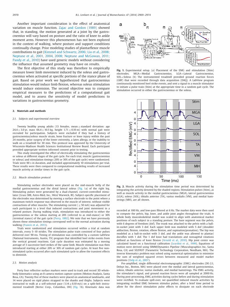
Fig. 1. Experimental setup. (a) Placement of the EMG and stimulation (Stim)electrodes. MGA¼Medial Gastrocnemius, LGA¼Lateral Gastrocnemius,SOL¼Soleus (b) The instrumented treadmill provided ground reaction forces(GRF) that were recorded through data acquisition (DAQ). A LabView programcontinuously monitored heel strike events, and sent a signal to a muscle stimulatorto initiate a pulse train (Stim) at the appropriate time in a random gait cycle. Thestimulation occurred in either the gastrocnemius or the soleus.
Fig. 2. Muscle activity during the stimulation time period was determined byintegrating the activity denoted by the shaded regions. Stimulation pulses (Stim), aswell as muscle activity in the medial gastrocnemius (MGA), lateral gastrocnemius(LGA), soleus (SOL), tibialis anterior (TA), vastus medialis (VM), and medial ham-strings (MH), are all shown.
R.L. Lenhart et al. / Journal of Biomechanics 47 (2014) 2969–29742970
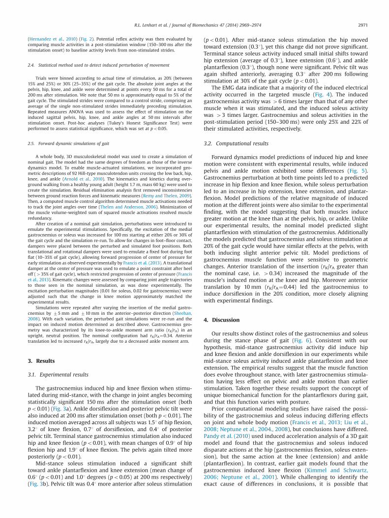
(Hernandez et al., 2010) (Fig. 2). Potential reflex activity was then evaluated bycomparing muscle activities in a post-stimulation window (150–300 ms after thestimulation onset) to baseline activity levels from non-stimulated strides.
2.4. Statistical method used to detect induced perturbation of movement
Trials were binned according to actual time of stimulation, as 20% (between15% and 25%) or 30% (25–35%) of the gait cycle. The absolute joint angles at thepelvis, hip, knee, and ankle were determined at points every 50 ms for a total of200 ms after stimulation. We note that 50 ms is approximately equal to 5% of thegait cycle. The stimulated strides were compared to a control stride, comprising anaverage of the single non-stimulated strides immediately preceding stimulation.Repeated measures ANOVA was used to assess the effect of stimulation on theinduced sagittal pelvis, hip, knee, and ankle angles at 50 ms intervals afterstimulation onset. Post-hoc analyses (Tukey's Honest Significance Test) wereperformed to assess statistical significance, which was set at po0.05.
2.5. Forward dynamic simulations of gait
A whole body, 3D musculoskeletal model was used to create a simulation ofnominal gait. The model had the same degrees of freedom as those of the inversedynamics model. To enable muscle-actuated simulations, we incorporated geo-metric descriptions of 92 Hill-type musculotendon units crossing the low back, hip,knee, and ankle (Arnold et al., 2010). The kinematics and kinetics during over-ground walking from a healthy young adult (height 1.7 m, mass 60 kg) were used tocreate the simulation. Residual elimination analysis first removed inconsistenciesbetween ground reaction forces and kinematic measures (Remy and Thelen, 2009).Then, a computed muscle control algorithm determined muscle activations neededto track the joint angles over time (Thelen and Anderson, 2006). Minimization ofthe muscle volume-weighted sum of squared muscle activations resolved muscleredundancy.
After creation of a nominal gait simulation, perturbations were introduced toemulate the experimental stimulations. Specifically, the excitation of the medialgastrocnemius or soleus was increased for 100 ms starting at either 20% or 30% ofthe gait cycle and the simulation re-run. To allow for changes in foot–floor contact,dampers were placed between the perturbed and simulated foot positions. Bothtranslational and rotational dampers were used to emulate a fixed foot during footflat (10–35% of gait cycle), allowing forward progression of center of pressure forearly stimulation as observed experimentally by Francis et al. (2013). A translationaldamper at the center of pressure was used to emulate a point constraint after heeloff (435% of gait cycle), which restricted progression of center of pressure (Franciset al., 2013). Kinematic changes were assessed by comparing joint angle trajectoriesto those seen in the nominal simulation, as was done experimentally. Theexcitation perturbation magnitudes (0.01 for soleus, 0.02 for gastrocnemius) wereadjusted such that the change in knee motion approximately matched theexperimental results.
Simulations were repeated after varying the insertion of the medial gastro-cnemius by 75 mm and 710 mm in the anterior–posterior direction (Sheehan,2008). With each variation, the perturbed gait simulations were re-run and theimpact on induced motion determined as described above. Gastrocnemius geo-metry was characterized by its knee-to-ankle moment arm ratio (rK/rA) in anupright, neutral position. The nominal configuration had rK/rA¼0.34. Anteriortranslation led to increased rK/rA, largely due to a decreased ankle moment arm.
3. Results
3.1. Experimental results
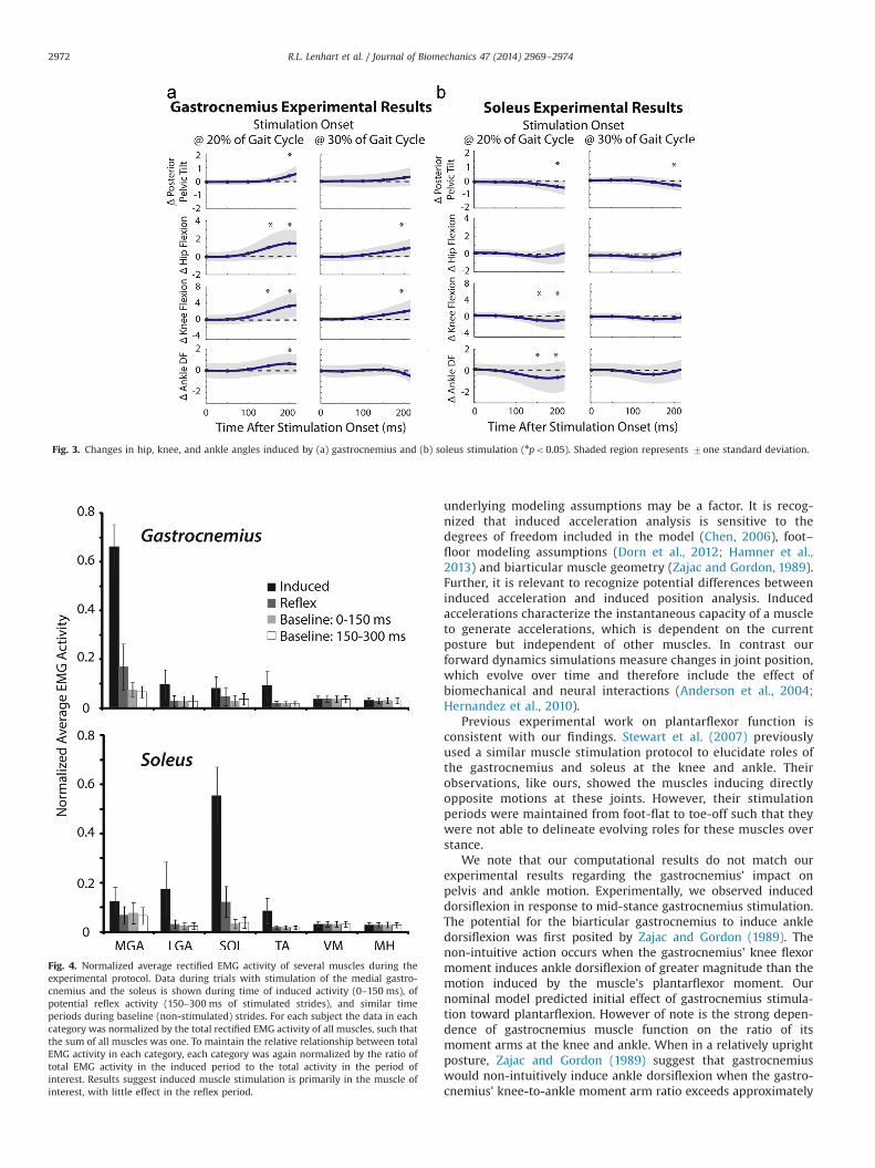
The gastrocnemius induced hip and knee flexion when stimu-lated during mid-stance, with the change in joint angles becomingstatistically significant 150 ms after the stimulation onset (bothpo0.01) (Fig. 3a). Ankle dorsiflexion and posterior pelvic tilt werealso induced at 200 ms after stimulation onset (both po0.01). Theinduced motion averaged across all subjects was 1.51 of hip flexion,3.21 of knee flexion, 0.71 of dorsiflexion, and 0.41 of posteriorpelvic tilt. Terminal stance gastrocnemius stimulation also inducedhip and knee flexion (po0.01), with mean changes of 0.91 of hipflexion hip and 1.91 of knee flexion. The pelvis again tilted moreposteriorly (po0.01).
Mid-stance soleus stimulation induced a significant shifttoward ankle plantarflexion and knee extension (mean change of0.61 (po0.01) and 1.01 degrees (po0.05) at 200 ms respectively)(Fig. 3b). Pelvic tilt was 0.41 more anterior after soleus stimulation
(po0.01). After mid-stance soleus stimulation the hip movedtoward extension (0.31), yet this change did not prove significant.Terminal stance soleus activity induced small initial shifts towardhip extension (average of 0.31), knee extension (0.61), and ankleplantarflexion (0.31), though none were significant. Pelvic tilt wasagain shifted anteriorly, averaging 0.31 after 200 ms followingstimulation at 30% of the gait cycle (po0.01).
The EMG data indicate that a majority of the induced electricalactivity occurred in the targeted muscle (Fig. 4). The inducedgastrocnemius activity was 46 times larger than that of any othermuscle when it was stimulated, and the induced soleus activitywas 43 times larger. Gastrocnemius and soleus activities in thepost-stimulation period (150–300 ms) were only 25% and 22% oftheir stimulated activities, respectively.
3.2. Computational results
Forward dynamics model predictions of induced hip and kneemotion were consistent with experimental results, while inducedpelvis and ankle motion exhibited some differences (Fig. 5).Gastrocnemius perturbation at both time points led to a predictedincrease in hip flexion and knee flexion, while soleus perturbationled to an increase in hip extension, knee extension, and plantar-flexion. Model predictions of the relative magnitude of inducedmotion at the different joints were also similar to the experimentalfinding, with the model suggesting that both muscles inducegreater motion at the knee than at the pelvis, hip, or ankle. Unlikeour experimental results, the nominal model predicted slightplantarflexion with stimulation of the gastrocnemius. Additionallythe models predicted that gastrocnemius and soleus stimulation at20% of the gait cycle would have similar effects at the pelvis, withboth inducing slight anterior pelvic tilt. Model predictions ofgastrocnemius muscle function were sensitive to geometricchanges. Anterior translation of the insertion (rK/rA greater thanthe nominal case, i.e. 40.34) increased the magnitude of themuscle's induced motion at the knee and hip. Moreover anteriortranslation by 10 mm (rK/rA¼0.44) led the gastrocnemius toinduce dorsiflexion in the 20% condition, more closely aligningwith experimental findings.
4. Discussion
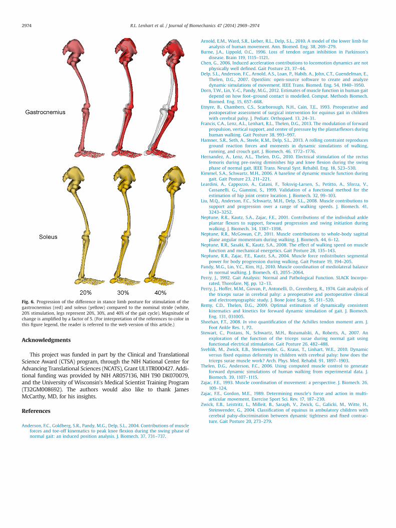
Our results show distinct roles of the gastrocnemius and soleusduring the stance phase of gait (Fig. 6). Consistent with ourhypothesis, mid-stance gastrocnemius activity did induce hipand knee flexion and ankle dorsiflexion in our experiments whilemid-stance soleus activity induced ankle plantarflexion and kneeextension. The empirical results suggest that the muscle functiondoes evolve throughout stance, with later gastrocnemius stimula-tion having less effect on pelvic and ankle motion than earlierstimulation. Taken together these results support the concept ofunique biomechanical function for the plantarflexors during gait,and that this function varies with posture.
Prior computational modeling studies have raised the possi-bility of the gastrocnemius and soleus inducing differing effectson joint and whole body motion (Francis et al., 2013; Liu et al.,2008; Neptune et al., 2004., 2008), but conclusions have differed.Pandy et al. (2010) used induced acceleration analysis of a 3D gaitmodel and found that the gastrocnemius and soleus induceddisparate actions at the hip (gastrocnemius flexion, soleus exten-sion), but the same action at the knee (extension) and ankle(plantarflexion). In contrast, earlier gait models found that thegastrocnemius induced knee flexion (Kimmel and Schwartz,2006; Neptune et al., 2001). While challenging to identify theexact cause of differences in conclusions, it is possible that
R.L. Lenhart et al. / Journal of Biomechanics 47 (2014) 2969–2974 2971
underlying modeling assumptions may be a factor. It is recog-nized that induced acceleration analysis is sensitive to thedegrees of freedom included in the model (Chen, 2006), foot–floor modeling assumptions (Dorn et al., 2012; Hamner et al.,2013) and biarticular muscle geometry (Zajac and Gordon, 1989).Further, it is relevant to recognize potential differences betweeninduced acceleration and induced position analysis. Inducedaccelerations characterize the instantaneous capacity of a muscleto generate accelerations, which is dependent on the currentposture but independent of other muscles. In contrast ourforward dynamics simulations measure changes in joint position,which evolve over time and therefore include the effect ofbiomechanical and neural interactions (Anderson et al., 2004;Hernandez et al., 2010).
Previous experimental work on plantarflexor function isconsistent with our findings. Stewart et al. (2007) previouslyused a similar muscle stimulation protocol to elucidate roles ofthe gastrocnemius and soleus at the knee and ankle. Theirobservations, like ours, showed the muscles inducing directlyopposite motions at these joints. However, their stimulationperiods were maintained from foot-flat to toe-off such that theywere not able to delineate evolving roles for these muscles overstance.
We note that our computational results do not match ourexperimental results regarding the gastrocnemius' impact onpelvis and ankle motion. Experimentally, we observed induceddorsiflexion in response to mid-stance gastrocnemius stimulation.The potential for the biarticular gastrocnemius to induce ankledorsiflexion was first posited by Zajac and Gordon (1989). Thenon-intuitive action occurs when the gastrocnemius' knee flexormoment induces ankle dorsiflexion of greater magnitude than themotion induced by the muscle's plantarflexor moment. Ournominal model predicted initial effect of gastrocnemius stimula-tion toward plantarflexion. However of note is the strong depen-dence of gastrocnemius muscle function on the ratio of itsmoment arms at the knee and ankle. When in a relatively uprightposture, Zajac and Gordon (1989) suggest that gastrocnemiuswould non-intuitively induce ankle dorsiflexion when the gastro-cnemius' knee-to-ankle moment arm ratio exceeds approximately
Fig. 3. Changes in hip, knee, and ankle angles induced by (a) gastrocnemius and (b) soleus stimulation (npo0.05). Shaded region represents 7one standard deviation.
Fig. 4. Normalized average rectified EMG activity of several muscles during theexperimental protocol. Data during trials with stimulation of the medial gastro-cnemius and the soleus is shown during time of induced activity (0–150 ms), ofpotential reflex activity (150–300 ms of stimulated strides), and similar timeperiods during baseline (non-stimulated) strides. For each subject the data in eachcategory was normalized by the total rectified EMG activity of all muscles, such thatthe sum of all muscles was one. To maintain the relative relationship between totalEMG activity in each category, each category was again normalized by the ratio oftotal EMG activity in the induced period to the total activity in the period ofinterest. Results suggest induced muscle stimulation is primarily in the muscle ofinterest, with little effect in the reflex period.
R.L. Lenhart et al. / Journal of Biomechanics 47 (2014) 2969–29742972
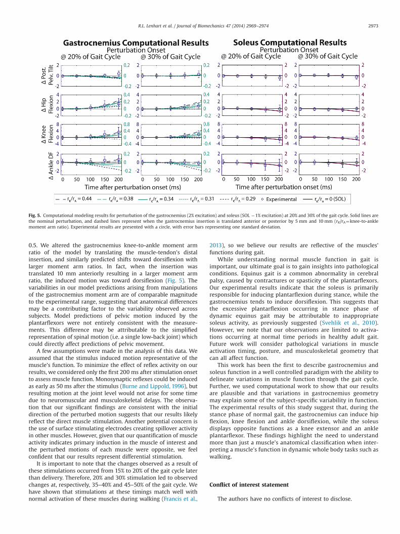
0.5. We altered the gastrocnemius knee-to-ankle moment armratio of the model by translating the muscle-tendon's distalinsertion, and similarly predicted shifts toward dorsiflexion withlarger moment arm ratios. In fact, when the insertion wastranslated 10 mm anteriorly resulting in a larger moment armratio, the induced motion was toward dorsiflexion (Fig. 5). Thevariabilities in our model predictions arising from manipulationsof the gastrocnemius moment arm are of comparable magnitudeto the experimental range, suggesting that anatomical differencesmay be a contributing factor to the variability observed acrosssubjects. Model predictions of pelvic motion induced by theplantarflexors were not entirely consistent with the measure-ments. This difference may be attributable to the simplifiedrepresentation of spinal motion (i.e. a single low-back joint) whichcould directly affect predictions of pelvic movement.
A few assumptions were made in the analysis of this data. Weassumed that the stimulus induced motion representative of themuscle’s function. To minimize the effect of reflex activity on ourresults, we considered only the first 200 ms after stimulation onsetto assess muscle function. Monosynaptic reflexes could be inducedas early as 50 ms after the stimulus (Burne and Lippold, 1996), butresulting motion at the joint level would not arise for some timedue to neuromuscular and musculoskeletal delays. The observa-tion that our significant findings are consistent with the initialdirection of the perturbed motion suggests that our results likelyreflect the direct muscle stimulation. Another potential concern isthe use of surface stimulating electrodes creating spillover activityin other muscles. However, given that our quantification of muscleactivity indicates primary induction in the muscle of interest andthe perturbed motions of each muscle were opposite, we feelconfident that our results represent differential stimulation.
It is important to note that the changes observed as a result ofthese stimulations occurred from 15% to 20% of the gait cycle laterthan delivery. Therefore, 20% and 30% stimulation led to observedchanges at, respectively, 35–40% and 45–50% of the gait cycle. Wehave shown that stimulations at these timings match well withnormal activation of these muscles during walking (Francis et al.,
2013), so we believe our results are reflective of the muscles’functions during gait.
While understanding normal muscle function in gait isimportant, our ultimate goal is to gain insights into pathologicalconditions. Equinus gait is a common abnormality in cerebralpalsy, caused by contractures or spasticity of the plantarflexors.Our experimental results indicate that the soleus is primarilyresponsible for inducing plantarflexion during stance, while thegastrocnemius tends to induce dorsiflexion. This suggests thatthe excessive plantarflexion occurring in stance phase ofdynamic equinus gait may be attributable to inappropriatesoleus activity, as previously suggested (Svehlik et al., 2010).However, we note that our observations are limited to activa-tions occurring at normal time periods in healthy adult gait.Future work will consider pathological variations in muscleactivation timing, posture, and musculoskeletal geometry thatcan all affect function.
This work has been the first to describe gastrocnemius andsoleus function in a well controlled paradigm with the ability todelineate variations in muscle function through the gait cycle.Further, we used computational work to show that our resultsare plausible and that variations in gastrocnemius geometrymay explain some of the subject-specific variability in function.The experimental results of this study suggest that, during thestance phase of normal gait, the gastrocnemius can induce hipflexion, knee flexion and ankle dorsiflexion, while the soleusdisplays opposite functions as a knee extensor and an ankleplantarflexor. These findings highlight the need to understandmore than just a muscle’s anatomical classification when inter-preting a muscle's function in dynamic whole body tasks such aswalking.
Conflict of interest statement
The authors have no conflicts of interest to disclose.
Fig. 5. Computational modeling results for perturbation of the gastrocnemius (2% excitation) and soleus (SOL �1% excitation) at 20% and 30% of the gait cycle. Solid lines arethe nominal perturbation, and dashed lines represent when the gastrocnemius insertion is translated anterior or posterior by 5 mm and 10 mm (rK/rA¼knee-to-anklemoment arm ratio). Experimental results are presented with a circle, with error bars representing one standard deviation.
R.L. Lenhart et al. / Journal of Biomechanics 47 (2014) 2969–2974 2973
Acknowledgments
This project was funded in part by the Clinical and TranslationalScience Award (CTSA) program, through the NIH National Center forAdvancing Translational Sciences (NCATS), Grant UL1TR000427. Addi-tional funding was provided by NIH AR057136, NIH T90 DK070079,and the University of Wisconsin's Medical Scientist Training Program(T32GM008692). The authors would also like to thank JamesMcCarthy, MD, for his insights.
References
Anderson, F.C., Goldberg, S.R., Pandy, M.G., Delp, S.L., 2004. Contributions of muscleforces and toe-off kinematics to peak knee flexion during the swing phase ofnormal gait: an induced position analysis. J. Biomech. 37, 731–737.
Arnold, E.M., Ward, S.R., Lieber, R.L., Delp, S.L., 2010. A model of the lower limb foranalysis of human movement. Ann. Biomed. Eng. 38, 269–279.
Burne, J.A., Lippold, O.C., 1996. Loss of tendon organ inhibition in Parkinson’sdisease. Brain 119, 1115–1121.
Chen, G., 2006. Induced acceleration contributions to locomotion dynamics are notphysically well defined. Gait Posture 23, 37–44.
Delp, S.L., Anderson, F.C., Arnold, A.S., Loan, P., Habib, A., John, C.T., Guendelman, E.,Thelen, D.G., 2007. OpenSim: open-source software to create and analyzedynamic simulations of movement. IEEE Trans. Biomed. Eng. 54, 1940–1950.
Dorn, T.W., Lin, Y.-C., Pandy, M.G., 2012. Estimates of muscle function in human gaitdepend on how foot–ground contact is modelled. Comput. Methods Biomech.Biomed. Eng. 15, 657–668.
Etnyre, B., Chambers, C.S., Scarborough, N.H., Cain, T.E., 1993. Preoperative andpostoperative assessment of surgical intervention for equinus gait in childrenwith cerebral palsy. J. Pediatr. Orthopaed. 13, 24–31.
Francis, C.A., Lenz, A.L., Lenhart, R.L., Thelen, D.G., 2013. The modulation of forwardpropulsion, vertical support, and center of pressure by the plantarflexors duringhuman walking. Gait Posture 38, 993–997.
Hamner, S.R., Seth, A., Steele, K.M., Delp, S.L., 2013. A rolling constraint reproducesground reaction forces and moments in dynamic simulations of walking,running, and crouch gait. J. Biomech. 46, 1772–1776.
Hernandez, A., Lenz, A.L., Thelen, D.G., 2010. Electrical stimulation of the rectusfemoris during pre-swing diminishes hip and knee flexion during the swingphase of normal gait. IEEE Trans. Neural Syst. Rehabil. Eng. 18, 523–530.
Kimmel, S.A., Schwartz, M.H., 2006. A baseline of dynamic muscle function duringgait. Gait Posture 23, 211–221.
Leardini, A., Cappozzo, A., Catani, F., Toksvig-Larsen, S., Petitto, A., Sforza, V.,Cassanelli, G., Giannini, S., 1999. Validation of a functional method for theestimation of hip joint centre location. J. Biomech. 32, 99–103.
Liu, M.Q., Anderson, F.C., Schwartz, M.H., Delp, S.L., 2008. Muscle contributions tosupport and progression over a range of walking speeds. J. Biomech. 41,3243–3252.
Neptune, R.R., Kautz, S.A., Zajac, F.E., 2001. Contributions of the individual ankleplantar flexors to support, forward progression and swing initiation duringwalking. J. Biomech. 34, 1387–1398.
Neptune, R.R., McGowan, C.P., 2011. Muscle contributions to whole-body sagittalplane angular momentum during walking. J. Biomech. 44, 6–12.
Neptune, R.R., Sasaki, K., Kautz, S.A., 2008. The effect of walking speed on musclefunction and mechanical energetics. Gait Posture 28, 135–143.
Neptune, R.R., Zajac, F.E., Kautz, S.A., 2004. Muscle force redistributes segmentalpower for body progression during walking. Gait Posture 19, 194–205.
Pandy, M.G., Lin, Y.C., Kim, H.J., 2010. Muscle coordination of mediolateral balancein normal walking. J. Biomech. 43, 2055–2064.
Perry, J., 1992. Gait Analysis: Normal and Pathological Function. SLACK Incorpo-rated, Thorofare, NJ, pp. 12–13.
Perry, J., Hoffer, M.M., Giovan, P., Antonelli, D., Greenberg, R., 1974. Gait analysis ofthe triceps surae in cerebral palsy: a preoperative and postoperative clinicaland electromyographic study. J. Bone Joint Surg. 56, 511–520.
Remy, C.D., Thelen, D.G., 2009. Optimal estimation of dynamically consistentkinematics and kinetics for forward dynamic simulation of gait. J. Biomech.Eng. 131, 031005.
Sheehan, F.T., 2008. In vivo quantification of the Achilles tendon moment arm. J.Foot Ankle Res. 1, P2.
Stewart, C., Postans, N., Schwartz, M.H., Rozumalski, A., Roberts, A., 2007. Anexploration of the function of the triceps surae during normal gait usingfunctional electrical stimulation. Gait Posture 26, 482–488.
Svehlik, M., Zwick, E.B., Steinwender, G., Kraus, T., Linhart, W.E., 2010. Dynamicversus fixed equinus deformity in children with cerebral palsy: how does thetriceps surae muscle work? Arch. Phys. Med. Rehabil. 91, 1897–1903.
Thelen, D.G., Anderson, F.C., 2006. Using computed muscle control to generateforward dynamic simulations of human walking from experimental data. J.Biomech. 39, 1107–1115.
Zajac, F.E., 1993. Muscle coordination of movement: a perspective. J. Biomech. 26,109–124.
Zajac, F.E., Gordon, M.E., 1989. Determining muscle's force and action in multi-articular movement. Exercise Sport Sci. Rev. 17, 187–230.
Zwick, E.B., Leistritz, L., Milleit, B., Saraph, V., Zwick, G., Galicki, M., Witte, H.,Steinwender, G., 2004. Classification of equinus in ambulatory children withcerebral palsy-discrimination between dynamic tightness and fixed contrac-ture. Gait Posture 20, 273–279.
Fig. 6. Progression of the difference in stance limb posture for stimulation of thegastrocnemius (red) and soleus (yellow) compared to the nominal stride (white,20% stimulation, legs represent 20%, 30%, and 40% of the gait cycle). Magnitude ofchange is amplified by a factor of 5. (For interpretation of the references to color inthis figure legend, the reader is referred to the web version of this article.)
R.L. Lenhart et al. / Journal of Biomechanics 47 (2014) 2969–29742974