Embed Size (px)
Citation preview

Enabling REcovERy fRom common TRaffic injuRiEs: a focus on ThE injuREd PERson | 1
Enabling REcovERy fRom common TRaffic injuRiEs: A focus on the Injured Person
OntariO PrOtOcOl fOr traffic injury ManageMent cOllabOratiOn

Enabling REcovERy fRom common TRaffic injuRiEs: a focus on ThE injuREd PERson | 2
ENABLING RECOVERY FROM COMMON TRAFFIC INJURIES: A FOCUS ON THE INJURED PERSON
OntariO PrOtOcOl fOr traffic injury ManageMent (OPtiMa) cOllabOratiOn
Chair Pierre Côté DC, PhD University of Ontario Institute of Technology
University of Toronto UOIT-CMCC Center for the Study of Disability Prevention and Rehabilitation
Project Manager Heather Shearer DC, MSc UOIT-CMCC Center for the Study of Disability
Prevention and Rehabilitation
Guideline Expert Panel Arthur Ameis MD, FRCPC DESS CFE DABPM&R SSC Private Practice
Université de Montréal
Carlo Ammendolia DC, PhD University of Toronto
Lynn Anderson BSc (non-voting member) Aviva Canada
Richard N. Bohay DMD, MSc, MRCD (C) Western University
Robert Brison MD, MPH, FRCPC, CCFPC Queen’s University
Linda Carroll PhD University of Alberta
David Cassidy PhD, DrMedSc University of South Denmark University of Toronto
Douglas Gross BScPT, PhD University of Alberta
Murray Krahn MD, MSc, FRCPC University of Toronto
Michel Lacerte MDCM, MSc, FRCPC Université de Montréal Western University
Gail M. Lindsay RN, PhD (Patient liaison) University of Ontario Institute of Technology
Patrick Loisel MD University of Toronto University of Ontario Institute of Technology
Shawn Marshall MD, MSc, FRCPC University of Ottawa

Enabling REcovERy fRom common TRaffic injuRiEs: a focus on ThE injuREd PERson | 3
Enabling REcovERY fRom common TRaffic injuRiEs: a focus on ThE injuREd PERson
Silvano Mior DC, PhD Canadian Memorial Chiropractic College
University of Ontario Institute of Technology Margareta Nordin Dr. Med. Sci., PT, CIE New York University
Mike Paulden MA, MSc University of Alberta
Viivi Riis BScPT, MSc (non-voting member) Health Service Management
HON. Roger Salhany Q.C., BA, LLB Retired Judge from the Ontario Superior Court of
Justice
John Stapleton (Consumer representative) Open Policy Ontario
Maja Stupar DC, PhD UOIT – CMCC Centre for the Study of Disability Prevention and Rehabilitation
Gabrielle van der Velde DC, PhD University of Toronto
TECHNICAL TEAM
(uOit–cMcc centre fOr the Study Of diSability PreventiOn and rehabilitatiOn)
Poonam Cardoso BHS
Craig Jacobs DC, MSc
Kristi Randhawa BHSc, MPH
Danielle Southerst BScH, DC
Deborah Sutton BSc, MEd, MSc, OT Reg. (Ont)
Anne Taylor-Vaisey BA, MLS
Sharanya Varatharajan BSc, MSc
Angela Verven BA
Leslie Verville BHSc
Jessica Wong BSc, DC, FCCS(C)
Hainan Yu BSc, MBBS, MSc

Enabling REcovERy fRom common TRaffic injuRiEs: a focus on ThE injuREd PERson | 4
GRADUATE STUDENTS
Sean Y. Abdulla BA(Hons), MSc Canadian Memorial Chiropractic College
Courtney Brown BSc, DC, MSc Canadian Memorial Chiropractic College
Karen Chrobak BHSc(Hons), DC Canadian Memorial Chiropractic College
Kevin D’Angelo Bsc(Hons), DC Canadian Memorial Chiropractic College
Sarah Dion DC Canadian Memorial Chiropractic College
Jocelyn Dresser BPhEd, DC Canadian Memorial Chiropractic College
Brad Ferguson BSc, DC Canadian Memorial Chiropractic College
Rachel Goldgrub BHSc University of Ontario Institute of Technology
Chantal James, BHsc University of Ontario Institute of Technology
Roger Menta BKin, DC Canadian Memorial Chiropractic College
Steven Piper DC Canadian Memorial Chiropractic College
Yaadwinder Shergill BSc(Hons), DC Canadian Memorial Chiropractic College
Thepikaa Varatharajan BSc University of Saskatchewan
Erin Woitzik BKin, DC Canadian Memorial Chiropractic College
CONSULTANTS
Eleanor Boyle PhD University of Southern Denmark
Brenda Gamble PhD University of Ontario Institute of Technology
Willie Handler Willie Handler and Associates
Paula Stern BSc, DC, FCCSC Canadian Memorial Chiropractic College (CMCC)
Citation: Côté P, Shearer H, Ameis A, Carroll L, Mior M, Nordin M and the OPTIMa Collaboration. Enabling recovery from common traffic injuries: A focus on the injured person. UOIT-CMCC Centre for the Study of Disability Prevention and Rehabilitation. January 31, 2015.

Enabling REcovERy fRom common TRaffic injuRiEs: a focus on ThE injuREd PERson | 5
PREFACE
The Ontario Protocol for Traffic Injury Management (OPTIMa) Collaboration includes a multidisciplinary team of expert clinicians (from medical, dental, physiotherapy, chiropractic, psychological, occupational therapy and nursing disciplines), academics and scientists (epidemiologists, clinical epidemiologists and health economists), a patient liaison, a consumer advocate, a retired judge and automobile insurance industry experts. Our objective was to develop Care Pathways* that promote recovery from common traffic injuries. This was achieved through carrying out a comprehensive and detailed review of the most current scientific literature and by conducting qualitative research with patients receiving health care for injuries from traffic collisions. Our research findings and recommendations are directed towards a specific goal: Enable and optimize the recovery of individuals injured in traffic collisions.
* The sequence and options of health care services a patient with traffic injuries receives during a particular episode of care.
While concentrating on the Why, What, and Who of evidence-based care+, we ensured that each of our recommendations respected the ethical principle of the shared decision-making process. To optimize and help inform the decision-making process, our recommendations were derived from evidence synthesized from high quality studies, thus maximizing the validity but limiting the uncertainty and biases inherent in lower quality studies. Decisions about health care must be the product of quality evidence and the unique contributions of both the patient and the attending health care professional.
+ According to Sackett et al (Sackett DL, Rosenberg WMC, Gray JAM, Haynes RB, Richardson WS. BMJ 1996;312:71): “Evidence based medicine is the conscientious, explicit, and judicious use of current best evidence in making decisions about the care of individual patients. The practice of evidence based medicine means integrating individual clinical expertise with the best available external clinical evidence from systematic research. By individual clinical expertise we mean the proficiency and judgment that individual clinicians acquire through clinical experience and clinical practice. Increased expertise is reflected in many ways, but especially in more effective and efficient diagnosis and in the more thoughtful identification and compassionate use of individual patients’ predicaments, rights, and preferences in making clinical decisions about their care. By best available external clinical evidence we mean clinically relevant research, often from the basic sciences of medicine, but especially from patient centred clinical research into the accuracy and precision of diagnostic tests (including the clinical examination), the power of prognostic markers, and the efficacy and safety of therapeutic, rehabilitative, and preventive regimens. External clinical evidence both invalidates previously accepted diagnostic tests and treatments and replaces them with new ones that are more powerful, more accurate, more efficacious, and safer.”
We addressed the Why in accordance with the ethical principle of primum non nocere (first do no harm). We asked: is treatment necessary to improve outcomes? If yes, then we asked: do the currently available interventions meaningfully accelerate the natural recovery time of an injury?
We looked at the What by asking whether there was high quality evidence indicating that any specific intervention improved recovery? If the answer was ‘yes’ then we asked: does this intervention improve long-term recovery or is the benefit restricted to short-term symptom relief?
We use the evidence to determine Who would benefit from specific interventions. We further focussed on the injured person by asking: what personal and societal factors can influence recovery?
The answers to these questions informed the development of Care Pathways that have the goal of improving the recovery of individuals injured in traffic collisions and facilitating their return to healthy and productive lives.
The Evidence Over the 2-year course of the OPTIMa Collaboration, we drew upon three sources of information concerning traffic injury rehabilitation.

Enabling REcovERy fRom common TRaffic injuRiEs: a focus on ThE injuREd PERson | 6
PREfacE
1. We critically reviewed the contents and evidentiary basis of published clinical practice guidelines for the management of traffic injuries.
2. We carried out an exhaustive search followed by a rigorous methodological evaluation of the current scientific literature concerning the management of traffic injuries published in peer-reviewed journals in the English language. We screened 234,995 abstracts and conducted in depth review of 597 scientific papers. This effort was summarized in 43 new systematic reviews of the literature.
3. We conducted a new study in which we gathered and carefully considered the narratives of Ontarians who have sustained injuries in traffic collisions and received health care.
This information was combined using a modified framework developed by the Ontario Health Technology Advisory Committee (OHTAC), a standing advisory subcommittee of the Health Quality Ontario (HQO) Board (an independent crown agency funded by the Government of Ontario through the Ministry of Health and Long-Term Care), responsible for making recommendations about the uptake, diffusion, distribution, or removal of health interventions in Ontario. The framework considers the overall clinical benefit; value for money; societal and ethical considerations; and the economic and organizational feasibility of the intervention.
Injury Classification Since 2010 in Ontario, common traffic injuries with a favourable natural history* have been legislatively classified as “minor injuries.” In the current Minor Injury Guideline (MIG), a minor injury is defined as a sprain, strain, whiplash associated disorder, contusion, abrasion, laceration or subluxation and any clinically associated sequelae. Over the course of our work, we have conducted qualitative research and carefully listened to the narratives, concerns and suggestions of injured persons who were actively receiving or who had received care under the current MIG. These injured persons consistently shared with us their belief that the term “minor injury” is unrepresentative of the actual experiences associated with traffic-related injuries. Many narratives emphasize the perception that vague terms such as “benign”, “temporary”, “transient”, and “non-serious”, and the categorization of “minor injury”, were not helpful; to the contrary they seemed to trivialize and dismiss very real experiences of distress or suffering. Injured persons described to us their experiences of unplanned, sudden onset intense pain, and subsequent occupational or domestic disability, sleep disruption and daytime exhaustion, family stress, and psychological and emotional distress. These persons also reported encountering frustration and uncertainty during the course of their recovery. We found it of particular importance that injured persons shared the belief that the provisions of the current MIG were not ensuring that they would receive what they needed; instead their concern was that guidelines seemed to limit what they would be permitted to receive, on the basis that their injuries and associated experiences were ‘minor’, and thus inconsequential.
* Natural history refers to the average course that an injury takes from its onset until its recovery, especially in the absence of treatment.
Having considered the narratives of persons who have experienced injuries and received care under the MIG, we have concluded that it is not appropriate to categorize either the injuries or their associated symptoms as minor injuries, inasmuch as they can be associated with a broad range of symptomatology and with some degree of disability for activities of daily life or work. It is our view that there is no scientific rationale or merit in continuing to employ the term “minor injury”. We propose a new classification that categorizes automobile collision injuries as Type I, Type II, or Type III injuries. Moreover, given the important temporal considerations outlined above, there is merit in further characterizing the injury, in order to optimize the approaches and interventions, by phase: Recent (0-3 months post-collision), or Persistent (4-6 months post-collision).

Enabling REcovERy fRom common TRaffic injuRiEs: a focus on ThE injuREd PERson | 7
PREfacE
Type I Injuries Type I injuries are those traffic injuries which have been shown in epidemiological studies to have a favourable natural history (recovery times ranging from days to a few months). These injuries include musculoskeletal injuries (such as Neck Pain and Associated Disorders Grades I-III, Grades I and II sprains and strains of the spine and limbs); traumatic radiculopathies*; mild traumatic brain injuries+; and post-traumatic psychological symptoms such as anxiety and stress. The proposed Care Pathways outlined in our report pertain to Type I injuries.
* A condition involving the nerve root(s) with symptoms of pain, numbness, and/or weakness in the muscles. + Mild traumatic brain injury denotes the acute neurophysiological effects of blunt impact or other mechanical energy applied to the head, such as from sudden acceleration, deceleration or rotational forces (Ontario Neurotrauma Foundation. Guidelines on concussion/mild traumatic brain injury and persistent symptoms. 2nd ed. Toronto: Ontario Neurotrauma Foundation; 2013.).
Type I injuries have a number of common features. There is typically either no significant loss of anatomical alignment or no loss of structural integrity. Most often, Type I injuries improve within days to a few months of the collision, leaving no permanent, serious impairment. Typically, the impact of even the most effective treatment for Type I injuries is modest, and usually limited to a reduction in symptom intensity. The evidence concerning the effectiveness of current interventions for Type I injuries can be summarized as follows:
(1) most interventions produce, at best, short-term benefits in the form of symptom relief and/or increased function;
(2) for such interventions, there is no evidence that effectiveness can be increased through higher dose intensity, more frequent attendance or prolongation of course of treatment;
(3) there is no evidence supporting a ‘piling on’ of complex combinations of clinicians, therapists, or therapies; and
(4) many commonly used interventions provide no more benefit than sham or placebo.
Common features are not confined to physical injuries alone. It is important for health care professionals and injured persons alike to understand that the experience of psychological symptoms such as anxiety, distress and anger is natural and not-atypical after a traffic collision; most psychological symptoms are temporary.
Our research also highlights that despite intervention, a small percentage of patients with Type I injuries will experience residual problems over the long term; and, a small proportion of these patients seem to develop chronic regional or more widespread pain, again regardless of the intervention they might have or continue to receive.
At present, there is no accurate tool to identify injured persons who may not recover. However, our research indicates that the prognosis for NAD (neck pain and its associated disorders), the most common type of injury that results from a traffic collision, may be less optimal for: 1) older individuals; 2) those with high levels of pain after the collision; and 3) those who demonstrate post-collision psychological symptoms involving depressed mood, anxiety, high levels of frustration or anger about the pain and those with poor expectation of recovery. Although the literature often refers to such patients as being “at-risk”, it is not known if any specific intervention can avert or significantly alter an adverse outcome.

Enabling REcovERy fRom common TRaffic injuRiEs: a focus on ThE injuREd PERson | 8
PREfacE
General Approach to the Management of Type I injuries As an overview, therefore, we propose that a consistent approach be adopted to manage Type I injuries over the entire course of their recovery process. The management should include education, advice, encouragement to stay active (including return to work), and reassurance that Type I injuries and their associated distress and discomfort are usually of a time-limited nature. Health care professionals should discuss with the injured person the range of effective interventions available for the management of their injuries. Supplementing self-management strategies with clinical care may be indicated for Type I injuries provided the intervention is likely to enable recovery through symptom relief and improvement in function.
Type II Injuries Type II injuries typically involve a substantial loss of anatomical alignment, structural integrity, psychological, cognitive, and/or physiological functioning. The majority of patients with such injuries will require (in addition to natural healing) a significant amount of medical, surgical, rehabilitation, and/or psychiatric/psychological intervention to ensure an optimal recovery. There is an evidentiary basis for major concern about both the extent of recovery and about the likelihood of complications developing and/or persisting in the absence of such expert care; significant impairment and disability are primary concerns. Examples of traffic collision-induced Type II injuries include fractures of the femur and hip, shoulder dislocation/fracture, facial fractures, depression or post-traumatic stress disorder.
The management of Type II injuries is not within the scope of our report.
Type III Injuries Type III injuries refer to the subset of Type II injuries which fall within the conceptual framework of catastrophic impairment within the Ontario Statutory Accident Benefits Schedule (SABS). In Ontario, there is a special set of entitlements available to patients whose injuries are extremely serious and permanent such as amputation, spinal cord injuries and severe brain injuries. Extended benefits are available for long term attendant care, and medical and rehabilitative goods and services.
The management of Type III injuries is not within the scope of our report.
Summary We recommend a new classification of traffic injuries. The natural history of the initial injury is the basis for classification. A Type I injury is likely to recover within days to a few months of the collision; but during the period of recovery the patient may benefit from education, advice, reassurance and time-limited evidence-based clinical care. Type I injuries are the focus of this report. A Type II injury is not likely to undergo spontaneous recovery, and the injured person may require medical, surgical and/or psychiatric/psychological care. Type III injuries are a subset of Type II injuries, that involve permanent catastrophic impairment or disability. The care for Type II and Type III injuries is not covered in this report.
Persons with Type I injuries should be educated and reassured from the outset that their own inherent healing capacities are likely to lead to a substantial recovery. They should also be informed that only a discrete set of treatments show evidence of any benefit; and that the same evidence shows that benefit is largely on the basis of pain alleviation. Healthcare professionals need to listen to the patient’s concerns and emphasize measures to assist them to cope, recognize and avoid complications.
Interventions for Type I injuries should only be provided in accordance with published evidence for effectiveness,

Enabling REcovERy fRom common TRaffic injuRiEs: a focus on ThE injuREd PERson | 9
PREfacE
including parameters of dosage, duration, and frequency; and within the most appropriate phase. The emphasis during the early phase (0-3 months) should be on education, advice, reassurance, activity and encouragement. Health care professionals should be reassured and encouraged to consider watchful waiting and clinical monitoring as evidence-based therapeutic options during the acute phase. For injured persons requiring therapy, time-limited and evidence-based intervention(s) should be implemented on a shared decision-making basis, an approach that equally applies to patients in the persistent phase (4-6 months).
Pierre Côté DC, PhD Chair Canada Research Chair in Disability Prevention and Rehabilitation Associate Professor, Faculty of Health Sciences, UOIT Director, UOIT-CMCC Centre for the Study of Disability Prevention and Rehabilitation
Arthur Ameis MD, FRCPC Private Practice Lecturer, Faculty of Medicine, University of Montreal
Linda Carroll PhD Professor, School of Public Health, University of Alberta
Gail M. Lindsay RN, PhD Associate Professor, Faculty of Health Sciences, UOIT
Silvano Mior DC, PhD Professor, Division of Research, Canadian Memorial Chiropractic College Adjunct Professor, Faculty of Health Sciences, University of Ontario Institute of Technology
Margareta Nordin Dr. Med. Sci., PT, CIE Professor (Research) Department of Orthopaedic Surgery and Environmental Medicine, New York University

Enabling REcovERy fRom common TRaffic injuRiEs: a focus on ThE injuREd PERson | 10
TABLE OF CONTENTS
Preface
1. Background . . . . . . . . . . . . . . . . . . . . . . . . . . . . . . . . . . . . . . . . . . . . . . . . . . . . . . . . . . . . . . . . . . . . . . . . . . . . . . 24 1.1 Disclaimer . . . . . . . . . . . . . . . . . . . . . . . . . . . . . . . . . . . . . . . . . . . . . . . . . . . . . . . . . . . . . . . . . . . . . . . . . 25
1.2 History of guidelines for the management of traffic injuries in Ontario . . . . . . . . . . . . . . . . . . . . 25 1.2.1 First-generation pre-approved framework guidelines . . . . . . . . . . . . . . . . . . . . . . . . . . . . 25 1.2.2 Second-generation pre-approved framework guidelines . . . . . . . . . . . . . . . . . . . . . . . . . . 26 1.2.3 Minor Injury Guideline . . . . . . . . . . . . . . . . . . . . . . . . . . . . . . . . . . . . . . . . . . . . . . . . . . . . . . . 27
1.3 Mandate for the development of evidence-based clinical practice guidelines for the management of traffic injuries . . . . . . . . . . . . . . . . . . . . . . . . . . . . . . . . . . . . . . . . . . . . . . . . 27
1.4 Guideline development group . . . . . . . . . . . . . . . . . . . . . . . . . . . . . . . . . . . . . . . . . . . . . . . . . . . . . . . 28 1.4.1 Guideline Expert Panel . . . . . . . . . . . . . . . . . . . . . . . . . . . . . . . . . . . . . . . . . . . . . . . . . . . . . . . 28 1.4.2 Core Scientific Team . . . . . . . . . . . . . . . . . . . . . . . . . . . . . . . . . . . . . . . . . . . . . . . . . . . . . . . . . 30 1.4.3 Technical team . . . . . . . . . . . . . . . . . . . . . . . . . . . . . . . . . . . . . . . . . . . . . . . . . . . . . . . . . . . . . . 31 1.4.4 Consultants . . . . . . . . . . . . . . . . . . . . . . . . . . . . . . . . . . . . . . . . . . . . . . . . . . . . . . . . . . . . . . . . . 32 1.4.5 Graduate students . . . . . . . . . . . . . . . . . . . . . . . . . . . . . . . . . . . . . . . . . . . . . . . . . . . . . . . . . . . 32
1.5 Scope of the project . . . . . . . . . . . . . . . . . . . . . . . . . . . . . . . . . . . . . . . . . . . . . . . . . . . . . . . . . . . . . . . . 33 1.5.1 Definition of clinical practice guideline . . . . . . . . . . . . . . . . . . . . . . . . . . . . . . . . . . . . . . . . . 33 1.5.2 Key background information . . . . . . . . . . . . . . . . . . . . . . . . . . . . . . . . . . . . . . . . . . . . . . . . . . 33 1.5.3 Population . . . . . . . . . . . . . . . . . . . . . . . . . . . . . . . . . . . . . . . . . . . . . . . . . . . . . . . . . . . . . . . . . . 34
1.5.3.1 Conditions covered by the guideline . . . . . . . . . . . . . . . . . . . . . . . . . . . . . . . . . . . . . 34 1.5.3.2 Conditions not covered by this guideline . . . . . . . . . . . . . . . . . . . . . . . . . . . . . . . . . 34
1.5.4 Health and delivery . . . . . . . . . . . . . . . . . . . . . . . . . . . . . . . . . . . . . . . . . . . . . . . . . . . . . . . . . . 35 1.5.5 Clinical management, rehabilitation, and self-management . . . . . . . . . . . . . . . . . . . . . . . 35
1.6 References . . . . . . . . . . . . . . . . . . . . . . . . . . . . . . . . . . . . . . . . . . . . . . . . . . . . . . . . . . . . . . . . . . . . . . . . . 36
2. Methodology for the development of clinical practice guidelines . . . . . . . . . . . . . . . . . . . . . . . . . . . . . . 37
2.1 Statements of conflicts of interest . . . . . . . . . . . . . . . . . . . . . . . . . . . . . . . . . . . . . . . . . . . . . . . . . . . . 38 2.1.1 Guideline Expert Panel . . . . . . . . . . . . . . . . . . . . . . . . . . . . . . . . . . . . . . . . . . . . . . . . . . . . . . . 39 2.1.2 Core Scientific Team . . . . . . . . . . . . . . . . . . . . . . . . . . . . . . . . . . . . . . . . . . . . . . . . . . . . . . . . . 42 2.1.3 Technical team . . . . . . . . . . . . . . . . . . . . . . . . . . . . . . . . . . . . . . . . . . . . . . . . . . . . . . . . . . . . . . . 45 2.1.4 Consultants . . . . . . . . . . . . . . . . . . . . . . . . . . . . . . . . . . . . . . . . . . . . . . . . . . . . . . . . . . . . . . . . . 46 2.1.5 Graduate students . . . . . . . . . . . . . . . . . . . . . . . . . . . . . . . . . . . . . . . . . . . . . . . . . . . . . . . . . . . 46
2.2 Systematic reviews of effectiveness of clinical interventions . . . . . . . . . . . . . . . . . . . . . . . . . . . . . . 47
2.3 Systematic reviews of the cost-effectiveness of clinical interventions for NAD . . . . . . . . . . . . . . 50
2.4 Review and approval of systematic reviews . . . . . . . . . . . . . . . . . . . . . . . . . . . . . . . . . . . . . . . . . . . . 52
2.5 Development of recommendations and care pathways . . . . . . . . . . . . . . . . . . . . . . . . . . . . . . . . . . 53 2.5.1 Contextualizing the evidence . . . . . . . . . . . . . . . . . . . . . . . . . . . . . . . . . . . . . . . . . . . . . . . . . 53 2.5.2 From evidence to recommendations . . . . . . . . . . . . . . . . . . . . . . . . . . . . . . . . . . . . . . . . . . . 56
2.5.2.1 Recommendation development subcommittee . . . . . . . . . . . . . . . . . . . . . . . . . . . . 57 2.5.2.2 Interpreting the evidence . . . . . . . . . . . . . . . . . . . . . . . . . . . . . . . . . . . . . . . . . . . . . . 57

Enabling REcovERy fRom common TRaffic injuRiEs: a focus on ThE injuREd PERson | 11
TablE of conTEnTs
2.5.2.3 Consideration of expected societal values and ethical values . . . . . . . . . . . . . . . 58 2.5.2.4 Wording of recommendations . . . . . . . . . . . . . . . . . . . . . . . . . . . . . . . . . . . . . . . . . . 58
2.5.2.4.1 Recommendations for interventions that must or must not be used . . . . . . . . . . . . . . . . . . . . . . . . . . . . . . . . . . . . . . . . 59
2.5.2.4.2 Recommendations for interventions that should or should not be used . . . . . . . . . . . . . . . . . . . . . . . . . . . . . . . . . . . . . . . 59
2.5.2.4.3 Recommendation for the interventions that could be used . . . . . . . . . . . . . . . . . . . . . . . . . . . . . . . . . . . . . . . . . . . . . . . . . 60
2.5.2.5 Reaching consensus on recommendations . . . . . . . . . . . . . . . . . . . . . . . . . . . . . . . 60 2.5.3 Integrating the recommendations . . . . . . . . . . . . . . . . . . . . . . . . . . . . . . . . . . . . . . . . . . . . . 61 2.5.4 Editorial independence . . . . . . . . . . . . . . . . . . . . . . . . . . . . . . . . . . . . . . . . . . . . . . . . . . . . . . . 61
2.6 Stakeholder consultation . . . . . . . . . . . . . . . . . . . . . . . . . . . . . . . . . . . . . . . . . . . . . . . . . . . . . . . . . . . . 61
2.7 Update of the care pathways . . . . . . . . . . . . . . . . . . . . . . . . . . . . . . . . . . . . . . . . . . . . . . . . . . . . . . . . 61
2.8 References . . . . . . . . . . . . . . . . . . . . . . . . . . . . . . . . . . . . . . . . . . . . . . . . . . . . . . . . . . . . . . . . . . . . . . . . 62
3. Summary of research informing the development of clinical practice guidelines . . . . . . . . . . . . . . . . . 65
3.1 “It wasn’t minor”: Injured persons’ experience under the current minor injury guideline . . . . . . . . . . . . . . . . . . . . . . . . . . . . . . . . . . . . . . . . . . . . . . . . . . . . . . . . . . . . . . . . . . . . . 66 3.1.1 Background . . . . . . . . . . . . . . . . . . . . . . . . . . . . . . . . . . . . . . . . . . . . . . . . . . . . . . . . . . . . . . . . . 66 3.1.2 What did the research focus on? . . . . . . . . . . . . . . . . . . . . . . . . . . . . . . . . . . . . . . . . . . . . . . . 67 3.1.3 Who was included in the research? . . . . . . . . . . . . . . . . . . . . . . . . . . . . . . . . . . . . . . . . . . . . . 67 3.1.4 How did we collect the data? . . . . . . . . . . . . . . . . . . . . . . . . . . . . . . . . . . . . . . . . . . . . . . . . . . 67 3.1.5 How did we analyze the data? . . . . . . . . . . . . . . . . . . . . . . . . . . . . . . . . . . . . . . . . . . . . . . . . . 68 3.1.6 What did we find? . . . . . . . . . . . . . . . . . . . . . . . . . . . . . . . . . . . . . . . . . . . . . . . . . . . . . . . . . . . . 68 3.1.7 Conclusions . . . . . . . . . . . . . . . . . . . . . . . . . . . . . . . . . . . . . . . . . . . . . . . . . . . . . . . . . . . . . . . . . 69 3.1.8 Recommended directions proposed by injured persons with minor
injuries sustained in motor vehicle collisions . . . . . . . . . . . . . . . . . . . . . . . . . . . . . . . . . . . . 70 3.1.9 References . . . . . . . . . . . . . . . . . . . . . . . . . . . . . . . . . . . . . . . . . . . . . . . . . . . . . . . . . . . . . . . . . 70
3.2 The road to recovery: how long does it take to recover from neck pain and its associated disorders? What influences recovery? . . . . . . . . . . . . . . . . . . . . . . . . . . . . . . . . 72 3.2.1 Background . . . . . . . . . . . . . . . . . . . . . . . . . . . . . . . . . . . . . . . . . . . . . . . . . . . . . . . . . . . . . . . . . 72 3.2.2 What did the research focus on? . . . . . . . . . . . . . . . . . . . . . . . . . . . . . . . . . . . . . . . . . . . . . . . 72 3.2.3 How did we do the research? . . . . . . . . . . . . . . . . . . . . . . . . . . . . . . . . . . . . . . . . . . . . . . . . . . 73 3.2.4 How did we integrate the data? . . . . . . . . . . . . . . . . . . . . . . . . . . . . . . . . . . . . . . . . . . . . . . . . 73 3.2.5 What did we find? . . . . . . . . . . . . . . . . . . . . . . . . . . . . . . . . . . . . . . . . . . . . . . . . . . . . . . . . . . . . 73 3.2.6 Conclusions . . . . . . . . . . . . . . . . . . . . . . . . . . . . . . . . . . . . . . . . . . . . . . . . . . . . . . . . . . . . . . . . . 75
3.3 A systematic review of peer-reviewed guidelines used in other jurisdictions . . . . . . . . . . . . . . . . 76 3.3.1 Background . . . . . . . . . . . . . . . . . . . . . . . . . . . . . . . . . . . . . . . . . . . . . . . . . . . . . . . . . . . . . . . . . 76 3.3.2 What was the purpose of the research? . . . . . . . . . . . . . . . . . . . . . . . . . . . . . . . . . . . . . . . . 76 3.3.3 How did we do the research? . . . . . . . . . . . . . . . . . . . . . . . . . . . . . . . . . . . . . . . . . . . . . . . . . 76 3.3.4 How did we synthesize the data? . . . . . . . . . . . . . . . . . . . . . . . . . . . . . . . . . . . . . . . . . . . . . . 77 3.3.5 What did we find? . . . . . . . . . . . . . . . . . . . . . . . . . . . . . . . . . . . . . . . . . . . . . . . . . . . . . . . . . . . 77 3.3.6 Conclusions . . . . . . . . . . . . . . . . . . . . . . . . . . . . . . . . . . . . . . . . . . . . . . . . . . . . . . . . . . . . . . . . . 77 3.3.7 References . . . . . . . . . . . . . . . . . . . . . . . . . . . . . . . . . . . . . . . . . . . . . . . . . . . . . . . . . . . . . . . . . . 78

Enabling REcovERy fRom common TRaffic injuRiEs: a focus on ThE injuREd PERson | 12
TablE of conTEnTs
3.4 Who is at risk of not recovering from neck pain and associated disorders? A clinical prediction model . . . . . . . . . . . . . . . . . . . . . . . . . . . . . . . . . . . . . . . . . . . . . . . . . . . . . . . . . . 79 3.4.1 Background . . . . . . . . . . . . . . . . . . . . . . . . . . . . . . . . . . . . . . . . . . . . . . . . . . . . . . . . . . . . . . . . . 79 3.4.2 What did the research focus on? . . . . . . . . . . . . . . . . . . . . . . . . . . . . . . . . . . . . . . . . . . . . . . . 79 3.4.3 How did we do the research? . . . . . . . . . . . . . . . . . . . . . . . . . . . . . . . . . . . . . . . . . . . . . . . . . . 79 3.4.4 What did we find? . . . . . . . . . . . . . . . . . . . . . . . . . . . . . . . . . . . . . . . . . . . . . . . . . . . . . . . . . . . . 80 3.4.5 Conclusions . . . . . . . . . . . . . . . . . . . . . . . . . . . . . . . . . . . . . . . . . . . . . . . . . . . . . . . . . . . . . . . . . 80
4. Guideline for the clinical management of neck pain and associated disorders (NAD) . . . . . . . . . . . . . 81
4.1 Management of NAD I-II . . . . . . . . . . . . . . . . . . . . . . . . . . . . . . . . . . . . . . . . . . . . . . . . . . . . . . . . . . . . . 85 4.1.1 Care pathway for recent onset NAD I-II (0-3 months post-collision) . . . . . . . . . . . . . . . . . 86 4.1.2 Care pathway for persistent NAD I-II (4-6 months post-collision) . . . . . . . . . . . . . . . . . . . 89 4.1.3 Key recommendations for the management of recent onset NAD I-II . . . . . . . . . . . . . . . 95
4.1.3.1 Structured patient education . . . . . . . . . . . . . . . . . . . . . . . . . . . . . . . . . . . . . . . . . . . 95 4.1.3.2 Exercise . . . . . . . . . . . . . . . . . . . . . . . . . . . . . . . . . . . . . . . . . . . . . . . . . . . . . . . . . . . . . . 96 4.1.3.3 Multimodal care . . . . . . . . . . . . . . . . . . . . . . . . . . . . . . . . . . . . . . . . . . . . . . . . . . . . . . 96 4.1.3.4 Soft tissue therapy . . . . . . . . . . . . . . . . . . . . . . . . . . . . . . . . . . . . . . . . . . . . . . . . . . . . 97 4.1.3.5 Passive physical modalities . . . . . . . . . . . . . . . . . . . . . . . . . . . . . . . . . . . . . . . . . . . . . 97 4.1.3.6 Acupuncture . . . . . . . . . . . . . . . . . . . . . . . . . . . . . . . . . . . . . . . . . . . . . . . . . . . . . . . . . . 98 4.1.3.7 Medication . . . . . . . . . . . . . . . . . . . . . . . . . . . . . . . . . . . . . . . . . . . . . . . . . . . . . . . . . . . 98
4.1.4 Key recommendations for the management of persistent NAD I-II . . . . . . . . . . . . . . . . . . 99 4.1.4.1 Structured patient education . . . . . . . . . . . . . . . . . . . . . . . . . . . . . . . . . . . . . . . . . . 99 4.1.4.2 Exercise . . . . . . . . . . . . . . . . . . . . . . . . . . . . . . . . . . . . . . . . . . . . . . . . . . . . . . . . . . . . . 100 4.1.4.3 Multimodal care . . . . . . . . . . . . . . . . . . . . . . . . . . . . . . . . . . . . . . . . . . . . . . . . . . . . . . 100 4.1.4.4 Soft tissue therapy . . . . . . . . . . . . . . . . . . . . . . . . . . . . . . . . . . . . . . . . . . . . . . . . . . . . 101 4.1.4.5 Passive physical modalities . . . . . . . . . . . . . . . . . . . . . . . . . . . . . . . . . . . . . . . . . . . . 102 4.1.4.6 Psychological intervention . . . . . . . . . . . . . . . . . . . . . . . . . . . . . . . . . . . . . . . . . . . . . 102 4.1.4.7 Acupuncture . . . . . . . . . . . . . . . . . . . . . . . . . . . . . . . . . . . . . . . . . . . . . . . . . . . . . . . . . 103 4.1.4.8 Medication . . . . . . . . . . . . . . . . . . . . . . . . . . . . . . . . . . . . . . . . . . . . . . . . . . . . . . . . . . 103
4.2 Management of NAD III . . . . . . . . . . . . . . . . . . . . . . . . . . . . . . . . . . . . . . . . . . . . . . . . . . . . . . . . . . . . 105 4.2.1 Care pathway for recent onset NAD III (0-3 months post-collision) . . . . . . . . . . . . . . . . . 106 4.2.2 Care pathway for persistent NAD III (4-6 months post-collision) . . . . . . . . . . . . . . . . . . . 109 4.2.3 Key recommendations for the management of recent onset NAD III . . . . . . . . . . . . . . . 110
4.2.3.1 Structured patient education . . . . . . . . . . . . . . . . . . . . . . . . . . . . . . . . . . . . . . . . . . 111 4.2.3.2 Exercise . . . . . . . . . . . . . . . . . . . . . . . . . . . . . . . . . . . . . . . . . . . . . . . . . . . . . . . . . . . . . 112 4.2.3.3 Passive physical modalities . . . . . . . . . . . . . . . . . . . . . . . . . . . . . . . . . . . . . . . . . . . . 112 4.2.3.4 Manual therapy . . . . . . . . . . . . . . . . . . . . . . . . . . . . . . . . . . . . . . . . . . . . . . . . . . . . . . 113
4.2.4 Key recommendations for the clinical management of persistent NAD III . . . . . . . . . . . 113 4.2.4.1 Passive physical modalities . . . . . . . . . . . . . . . . . . . . . . . . . . . . . . . . . . . . . . . . . . . . 113
5. Guideline for the clinical management of headaches associated with neck pain . . . . . . . . . . . . . . . . 119
5.1 Management of recent onset headaches associated with neck pain . . . . . . . . . . . . . . . . . . . . . 122
5.2 Management of persistent headaches associated with neck pain . . . . . . . . . . . . . . . . . . . . . . . . 122 5.2.1 Care pathway for episodic tension-type headaches (4-6 months post-collision) . . . . . 125

Enabling REcovERy fRom common TRaffic injuRiEs: a focus on ThE injuREd PERson | 13
TablE of conTEnTs
5.2.2 Key recommendations for the management of episodic tension-type headaches . . . . . . . . . . . . . . . . . . . . . . . . . . . . . . . . . . . . . . . . . . . . . . . . . . . . . . . . . . . . . . . . . 128 5.2.2.1 Structured Patient Education . . . . . . . . . . . . . . . . . . . . . . . . . . . . . . . . . . . . . . . . . . 128 5.2.2.2 Exercise . . . . . . . . . . . . . . . . . . . . . . . . . . . . . . . . . . . . . . . . . . . . . . . . . . . . . . . . . . . . . 129 5.2.2.3 Manual Therapy . . . . . . . . . . . . . . . . . . . . . . . . . . . . . . . . . . . . . . . . . . . . . . . . . . . . . 129
5.2.3 Care pathway for chronic tension-type headaches (4-6 months post-collision) . . . . . . 130 5.2.4 Key recommendations for the management of chronic tension-type
headaches . . . . . . . . . . . . . . . . . . . . . . . . . . . . . . . . . . . . . . . . . . . . . . . . . . . . . . . . . . . . . . . . . 133 5.2.4.1 Structured Patient Education . . . . . . . . . . . . . . . . . . . . . . . . . . . . . . . . . . . . . . . . . . 133 5.2.4.2 Exercise . . . . . . . . . . . . . . . . . . . . . . . . . . . . . . . . . . . . . . . . . . . . . . . . . . . . . . . . . . . . . 134 5.2.4.3 Multimodal Care . . . . . . . . . . . . . . . . . . . . . . . . . . . . . . . . . . . . . . . . . . . . . . . . . . . . . 135
5.2.5 Care pathway for cervicogenic headaches (4-6 months post-collision) . . . . . . . . . . . . . 136 5.2.6 Key recommendations for the management of cervicogenic headaches . . . . . . . . . . . . 139
5.2.6.1 Structured Patient Education . . . . . . . . . . . . . . . . . . . . . . . . . . . . . . . . . . . . . . . . . . 140 5.2.6.2 Exercise . . . . . . . . . . . . . . . . . . . . . . . . . . . . . . . . . . . . . . . . . . . . . . . . . . . . . . . . . . . . . 140 5.2.6.3 Multimodal Care . . . . . . . . . . . . . . . . . . . . . . . . . . . . . . . . . . . . . . . . . . . . . . . . . . . . . 140 5.2.6.4 Manual Therapy . . . . . . . . . . . . . . . . . . . . . . . . . . . . . . . . . . . . . . . . . . . . . . . . . . . . . 141
6. Guideline for the clinical management of soft tissue disorders of the upper extremity . . . . . . . . . . . 144
6.1 Management of epicondylitis . . . . . . . . . . . . . . . . . . . . . . . . . . . . . . . . . . . . . . . . . . . . . . . . . . . . . . . 148 6.1.1 Care pathway for recent onset epicondylitis (0-3 months post-collision) . . . . . . . . . . . 148 6.1.2 Care pathway for persistent epicondylitis (4-6 months post-collision) . . . . . . . . . . . . . 151 6.1.3 Key recommendations for the management of recent epicondylitis . . . . . . . . . . . . . . . 155
6.1.3.1 Multimodal Care . . . . . . . . . . . . . . . . . . . . . . . . . . . . . . . . . . . . . . . . . . . . . . . . . . . . . 155 6.1.3.2 Passive Physical Modalities . . . . . . . . . . . . . . . . . . . . . . . . . . . . . . . . . . . . . . . . . . . . 156
6.1.4 Key recommendations for the management of persistent epicondylitis . . . . . . . . . . . . 157 6.1.4.1 Exercise . . . . . . . . . . . . . . . . . . . . . . . . . . . . . . . . . . . . . . . . . . . . . . . . . . . . . . . . . . . . . 157 6.1.4.2 Multimodal Care . . . . . . . . . . . . . . . . . . . . . . . . . . . . . . . . . . . . . . . . . . . . . . . . . . . . . 158 6.1.4.3 Soft Tissue Therapy . . . . . . . . . . . . . . . . . . . . . . . . . . . . . . . . . . . . . . . . . . . . . . . . . . . 158 6.1.4.4 Passive Physical Modalities . . . . . . . . . . . . . . . . . . . . . . . . . . . . . . . . . . . . . . . . . . . . 159
6.2 Management of shoulder pain . . . . . . . . . . . . . . . . . . . . . . . . . . . . . . . . . . . . . . . . . . . . . . . . . . . . . . 160 6.2.1 Care pathway for recent onset shoulder pain (0-3 months post-collision) . . . . . . . . . . . 161 6.2.2 Care pathway for persistent shoulder pain (4-6 months post-collision) . . . . . . . . . . . . . 163 6.2.3 Key recommendations for the management of recent shoulder pain . . . . . . . . . . . . . . 167
6.2.3.1 Multimodal Care . . . . . . . . . . . . . . . . . . . . . . . . . . . . . . . . . . . . . . . . . . . . . . . . . . . . . 167 6.2.3.2 Soft Tissue Therapy . . . . . . . . . . . . . . . . . . . . . . . . . . . . . . . . . . . . . . . . . . . . . . . . . . . 168 6.2.3.3 Passive Physical Modalities . . . . . . . . . . . . . . . . . . . . . . . . . . . . . . . . . . . . . . . . . . . . 168 6.2.3.4 Manual Therapy . . . . . . . . . . . . . . . . . . . . . . . . . . . . . . . . . . . . . . . . . . . . . . . . . . . . . . 169
6.2.4 Key recommendations for the management of persistent shoulder pain . . . . . . . . . . . 169 6.2.4.1 Exercise . . . . . . . . . . . . . . . . . . . . . . . . . . . . . . . . . . . . . . . . . . . . . . . . . . . . . . . . . . . . . 170 6.2.4.2 Multimodal Care . . . . . . . . . . . . . . . . . . . . . . . . . . . . . . . . . . . . . . . . . . . . . . . . . . . . . 170 6.2.4.3 Soft Tissue Therapy . . . . . . . . . . . . . . . . . . . . . . . . . . . . . . . . . . . . . . . . . . . . . . . . . . . 171 6.2.4.4 Passive Physical Modalities . . . . . . . . . . . . . . . . . . . . . . . . . . . . . . . . . . . . . . . . . . . . 172 6.2.4.5 Manual Therapy . . . . . . . . . . . . . . . . . . . . . . . . . . . . . . . . . . . . . . . . . . . . . . . . . . . . . . 172

Enabling REcovERy fRom common TRaffic injuRiEs: a focus on ThE injuREd PERson | 14
TablE of conTEnTs
6.3 Management of shoulder pain with calcific tendinitis . . . . . . . . . . . . . . . . . . . . . . . . . . . . . . . . . . 173 6.3.1 Care pathway for shoulder pain with calcific tendinitis . . . . . . . . . . . . . . . . . . . . . . . . . . . 174 6.3.2 Key recommendations for the management of persistent shoulder pain
with calcific tendinitis . . . . . . . . . . . . . . . . . . . . . . . . . . . . . . . . . . . . . . . . . . . . . . . . . . . . . . . 177 6.3.2.1 Passive Physical Modalities . . . . . . . . . . . . . . . . . . . . . . . . . . . . . . . . . . . . . . . . . . . . 177
7. Guideline for the clinical management of soft tissue disorders of the lower extremity . . . . . . . . . . . 178
7.1 Management of patellofemoral pain . . . . . . . . . . . . . . . . . . . . . . . . . . . . . . . . . . . . . . . . . . . . . . . . . 182 7.1.1 Care pathway for recent onset patellofemoral pain (0-3 months post-collision) . . . . . 182 7.1.2 Care pathway for persistent patellofemoral pain (4-6 months post-collision) . . . . . . . 184 7.1.3 Key recommendations for the management of recent patellofemoral pain . . . . . . . . . 187 7.1.4 Key recommendations for the management of persistent patellofemoral pain . . . . . . 187
7.1.4.1 Exercise . . . . . . . . . . . . . . . . . . . . . . . . . . . . . . . . . . . . . . . . . . . . . . . . . . . . . . . . . . . . . 188
7.2 Management of ankle sprain . . . . . . . . . . . . . . . . . . . . . . . . . . . . . . . . . . . . . . . . . . . . . . . . . . . . . . . . 189 7.2.1 Care pathway for recent onset ankle sprain (0-3 months post-collision) . . . . . . . . . . . . 189 7.2.2 Care pathway for persistent ankle sprain (4-6 months post-collision) . . . . . . . . . . . . . . 192 7.2.3 Key recommendations for the management of recent ankle sprain . . . . . . . . . . . . . . . 195
7.2.3.1 Exercise . . . . . . . . . . . . . . . . . . . . . . . . . . . . . . . . . . . . . . . . . . . . . . . . . . . . . . . . . . . . . 195 7.2.3.2 Passive Physical Modalities . . . . . . . . . . . . . . . . . . . . . . . . . . . . . . . . . . . . . . . . . . . . 196 7.2.3.3 Manual Therapy . . . . . . . . . . . . . . . . . . . . . . . . . . . . . . . . . . . . . . . . . . . . . . . . . . . . . 197
7.2.4 Key recommendations for the management of persistent ankle sprain . . . . . . . . . . . . . 198 7.2.4.1 Manual Therapy . . . . . . . . . . . . . . . . . . . . . . . . . . . . . . . . . . . . . . . . . . . . . . . . . . . . . . 198
7.3 Management of achilles tendinopathy . . . . . . . . . . . . . . . . . . . . . . . . . . . . . . . . . . . . . . . . . . . . . . . 199 7.3.1 Care pathway for recent onset achilles tendinopathy (0-3 months
post-collision) . . . . . . . . . . . . . . . . . . . . . . . . . . . . . . . . . . . . . . . . . . . . . . . . . . . . . . . . . . . . . . 199 7.3.2 Care pathway for persistent achilles tendinopathy (4-6 months post-collision) . . . . . . 201 7.3.3 Key recommendations for the management of recent achilles tendinopathy . . . . . . . 204 7.3.4 Key recommendations for the management of persistent achilles
tendinopathy . . . . . . . . . . . . . . . . . . . . . . . . . . . . . . . . . . . . . . . . . . . . . . . . . . . . . . . . . . . . . . . 204 7.3.4.1 Passive Physical Modalities . . . . . . . . . . . . . . . . . . . . . . . . . . . . . . . . . . . . . . . . . . . . 205
7.4 Management of plantar fasciitis and heel pain . . . . . . . . . . . . . . . . . . . . . . . . . . . . . . . . . . . . . . . . 206 7.4.1 Care pathway for recent onset plantar fasciitis and heel pain (0-3 months
post-collision) . . . . . . . . . . . . . . . . . . . . . . . . . . . . . . . . . . . . . . . . . . . . . . . . . . . . . . . . . . . . . . 206 7.4.2 Care pathway for persistent plantar fasciitis and heel pain (4-6 months
post-collision) . . . . . . . . . . . . . . . . . . . . . . . . . . . . . . . . . . . . . . . . . . . . . . . . . . . . . . . . . . . . . . 208 7.4.3 Key recommendations for the management of recent plantar fasciitis
and heel pain . . . . . . . . . . . . . . . . . . . . . . . . . . . . . . . . . . . . . . . . . . . . . . . . . . . . . . . . . . . . . . 212 7.4.3.1 Exercise . . . . . . . . . . . . . . . . . . . . . . . . . . . . . . . . . . . . . . . . . . . . . . . . . . . . . . . . . . . . . 212 7.4.3.2 Soft Tissue Therapy . . . . . . . . . . . . . . . . . . . . . . . . . . . . . . . . . . . . . . . . . . . . . . . . . . . 213 7.4.3.3 Passive physical modalities . . . . . . . . . . . . . . . . . . . . . . . . . . . . . . . . . . . . . . . . . . . . 213
7.4.4 Key recommendations for the management of persistent plantar fasciitis and heel pain . . . . . . . . . . . . . . . . . . . . . . . . . . . . . . . . . . . . . . . . . . . . . . . . . . . . . . . . . . . . . . . 214 7.4.4.1 Exercise . . . . . . . . . . . . . . . . . . . . . . . . . . . . . . . . . . . . . . . . . . . . . . . . . . . . . . . . . . . . . 214 7.4.4.2 Soft Tissue Therapy . . . . . . . . . . . . . . . . . . . . . . . . . . . . . . . . . . . . . . . . . . . . . . . . . . . 215

Enabling REcovERy fRom common TRaffic injuRiEs: a focus on ThE injuREd PERson | 15
TablE of conTEnTs
7.4.4.3 Passive physical modalities . . . . . . . . . . . . . . . . . . . . . . . . . . . . . . . . . . . . . . . . . . . . 215 7.4.4.4 Multimodal care . . . . . . . . . . . . . . . . . . . . . . . . . . . . . . . . . . . . . . . . . . . . . . . . . . . . . 216
8. Guideline for the clinical management of temporomandibular disorders . . . . . . . . . . . . . . . . . . . . . . . 218
8.1 Management of temporomandibular disorders . . . . . . . . . . . . . . . . . . . . . . . . . . . . . . . . . . . . . . . . 221 8.1.1 Care pathway for recent temporomandibular disorders (0-3 months
post-collision) . . . . . . . . . . . . . . . . . . . . . . . . . . . . . . . . . . . . . . . . . . . . . . . . . . . . . . . . . . . . . . 221 8.1.2 Care pathway for persistent temporomandibular disorders (4-6 months
post-collision) . . . . . . . . . . . . . . . . . . . . . . . . . . . . . . . . . . . . . . . . . . . . . . . . . . . . . . . . . . . . . . 224 8.1.3 Key recommendations for the management of recent onset
temporomandibular disorders . . . . . . . . . . . . . . . . . . . . . . . . . . . . . . . . . . . . . . . . . . . . . . . . 227 8.1.4 Key recommendations for the management of persistent temporomandibular
disorders . . . . . . . . . . . . . . . . . . . . . . . . . . . . . . . . . . . . . . . . . . . . . . . . . . . . . . . . . . . . . . . . . . . 227 8.1.4.1 Self-management . . . . . . . . . . . . . . . . . . . . . . . . . . . . . . . . . . . . . . . . . . . . . . . . . . . . 228 8.1.4.2 Soft tissue therapy . . . . . . . . . . . . . . . . . . . . . . . . . . . . . . . . . . . . . . . . . . . . . . . . . . . 228 8.1.4.3 Psychological interventions . . . . . . . . . . . . . . . . . . . . . . . . . . . . . . . . . . . . . . . . . . . . 229 8.1.4.4 Passive physical modalities . . . . . . . . . . . . . . . . . . . . . . . . . . . . . . . . . . . . . . . . . . . . 229
9. Recommendation for the clinical management of mild traumatic brain injury (MTBI). . . . . . . . . . . . . . 230
9.1 Background . . . . . . . . . . . . . . . . . . . . . . . . . . . . . . . . . . . . . . . . . . . . . . . . . . . . . . . . . . . . . . . . . . . . . . . 231
9.2 Review Panel . . . . . . . . . . . . . . . . . . . . . . . . . . . . . . . . . . . . . . . . . . . . . . . . . . . . . . . . . . . . . . . . . . . . . 232
9.3 Critical Appraisal of the MTBI Guidelines . . . . . . . . . . . . . . . . . . . . . . . . . . . . . . . . . . . . . . . . . . . . . . 232
9.4 Results of the Review . . . . . . . . . . . . . . . . . . . . . . . . . . . . . . . . . . . . . . . . . . . . . . . . . . . . . . . . . . . . . . 232
9.5 Management of MTBI . . . . . . . . . . . . . . . . . . . . . . . . . . . . . . . . . . . . . . . . . . . . . . . . . . . . . . . . . . . . . . 232
9.6 References . . . . . . . . . . . . . . . . . . . . . . . . . . . . . . . . . . . . . . . . . . . . . . . . . . . . . . . . . . . . . . . . . . . . . . . . 233
10. Guideline for the clinical management of low back pain with and without radiculopathy . . . . . . . . . . 234
10.1 Management of non-specific low back pain . . . . . . . . . . . . . . . . . . . . . . . . . . . . . . . . . . . . . . . . . . . 237 10.1.1 Care pathway for recent onset non-specific low back pain (0-3 months
post-collision) . . . . . . . . . . . . . . . . . . . . . . . . . . . . . . . . . . . . . . . . . . . . . . . . . . . . . . . . . . . . . . 237 10.1.2 Care pathway for persistent non-specific low back pain (4-6 months
post-collision) . . . . . . . . . . . . . . . . . . . . . . . . . . . . . . . . . . . . . . . . . . . . . . . . . . . . . . . . . . . . . . 240 10.1.3 Key recommendations for the management of recent onset non-specific
low back pain . . . . . . . . . . . . . . . . . . . . . . . . . . . . . . . . . . . . . . . . . . . . . . . . . . . . . . . . . . . . . . . 245 10.1.3.1 Structured patient education . . . . . . . . . . . . . . . . . . . . . . . . . . . . . . . . . . . . 245 10.1.3.2 Manual therapy . . . . . . . . . . . . . . . . . . . . . . . . . . . . . . . . . . . . . . . . . . . . . . . 246 10.1.3.3 Medication . . . . . . . . . . . . . . . . . . . . . . . . . . . . . . . . . . . . . . . . . . . . . . . . . . . 247
10.1.4 Key recommendations for the management of persistent non-specific low back pain . . . . . . . . . . . . . . . . . . . . . . . . . . . . . . . . . . . . . . . . . . . . . . . . . . . . . . . . . . . . . . . . . . 247 10.1.4.1 Structured patient education . . . . . . . . . . . . . . . . . . . . . . . . . . . . . . . . . . . . 248 10.1.4.2 Exercise . . . . . . . . . . . . . . . . . . . . . . . . . . . . . . . . . . . . . . . . . . . . . . . . . . . . . . 249 10.1.4.3 Manual therapy . . . . . . . . . . . . . . . . . . . . . . . . . . . . . . . . . . . . . . . . . . . . . . . 249

Enabling REcovERy fRom common TRaffic injuRiEs: a focus on ThE injuREd PERson | 16
TablE of conTEnTs
10.1.4.4 Soft tissue therapy . . . . . . . . . . . . . . . . . . . . . . . . . . . . . . . . . . . . . . . . . . . . . 250 10.1.4.5 Medication . . . . . . . . . . . . . . . . . . . . . . . . . . . . . . . . . . . . . . . . . . . . . . . . . . . 250 10.1.4.6 Acupuncture . . . . . . . . . . . . . . . . . . . . . . . . . . . . . . . . . . . . . . . . . . . . . . . . . . 251 10.1.4.7 Multimodal care . . . . . . . . . . . . . . . . . . . . . . . . . . . . . . . . . . . . . . . . . . . . . . 252 10.1.4.8 Passive physical modalities . . . . . . . . . . . . . . . . . . . . . . . . . . . . . . . . . . . . . 252
10.2 Management of lumbar disc herniation with radiculopathy . . . . . . . . . . . . . . . . . . . . . . . . . . . . . 253 10.2.1 Care pathway for recent onset lumbar disc herniation with radiculopathy
(0-3 months post-collision) . . . . . . . . . . . . . . . . . . . . . . . . . . . . . . . . . . . . . . . . . . . . . . . . . . . 253 10.2.2 Care pathway for persistent lumbar disc herniation with radiculopathy
(4-6 months post-collision) . . . . . . . . . . . . . . . . . . . . . . . . . . . . . . . . . . . . . . . . . . . . . . . . . . . 256 10.2.3 Key recommendations for the management of recent onset lumbar disc
herniation with radiculopathy . . . . . . . . . . . . . . . . . . . . . . . . . . . . . . . . . . . . . . . . . . . . . . . . 258 10.2.3.1 Structured patient education . . . . . . . . . . . . . . . . . . . . . . . . . . . . . . . . . . . 258 10.2.3.2 Manual therapy . . . . . . . . . . . . . . . . . . . . . . . . . . . . . . . . . . . . . . . . . . . . . . . 259
10.2.4 Key recommendations for the management of persistent lumbar disc herniation with radiculopathy . . . . . . . . . . . . . . . . . . . . . . . . . . . . . . . . . . . . . . . . . . . . . . . . . 260
11. Interventions without evidence or inconclusive evidence . . . . . . . . . . . . . . . . . . . . . . . . . . . . . . . . . . . . 261
11.1 Background . . . . . . . . . . . . . . . . . . . . . . . . . . . . . . . . . . . . . . . . . . . . . . . . . . . . . . . . . . . . . . . . . . . . . . . 262
11.2 Interventions with inconclusive evidence . . . . . . . . . . . . . . . . . . . . . . . . . . . . . . . . . . . . . . . . . . . . . 263 11.2.1 Persistent NAD I-II . . . . . . . . . . . . . . . . . . . . . . . . . . . . . . . . . . . . . . . . . . . . . . . . . . . . . . . . . . 263 11.2.2 Persistent epicondylitis . . . . . . . . . . . . . . . . . . . . . . . . . . . . . . . . . . . . . . . . . . . . . . . . . . . . . . 263 11.2.3 Musculoskeletal injuries of the shoulder . . . . . . . . . . . . . . . . . . . . . . . . . . . . . . . . . . . . . . . 264 11.2.4 Persistent achilles tendinopathy . . . . . . . . . . . . . . . . . . . . . . . . . . . . . . . . . . . . . . . . . . . . . . 264 11.2.5 Persistent patellofemoral pain . . . . . . . . . . . . . . . . . . . . . . . . . . . . . . . . . . . . . . . . . . . . . . . 265 11.2.6 Persistent plantar fasciitis . . . . . . . . . . . . . . . . . . . . . . . . . . . . . . . . . . . . . . . . . . . . . . . . . . . 265 11.2.7 Persistent temporomandibular disorders . . . . . . . . . . . . . . . . . . . . . . . . . . . . . . . . . . . . . . 266
11.3 Evidence that could not be used to make recommendations . . . . . . . . . . . . . . . . . . . . . . . . . . . . 266
11.4 Interventions without evidence . . . . . . . . . . . . . . . . . . . . . . . . . . . . . . . . . . . . . . . . . . . . . . . . . . . . . 267 11.4.1 Headache interventions with no evidence . . . . . . . . . . . . . . . . . . . . . . . . . . . . . . . . . . . . . 267 11.4.2 Upper extremity injury interventions with no evidence . . . . . . . . . . . . . . . . . . . . . . . . . . 267 11.4.3 Lower extremity injury interventions with no evidence . . . . . . . . . . . . . . . . . . . . . . . . . . 268 11.4.4 Temporomandibular disorder interventions with no evidence . . . . . . . . . . . . . . . . . . . . 268
Glossary . . . . . . . . . . . . . . . . . . . . . . . . . . . . . . . . . . . . . . . . . . . . . . . . . . . . . . . . . . . . . . . . . . . . . . . . . . . . . . . . . . . . . . 269 Technical Appendix . . . . . . . . . . . . . . . . . . . . . . . . . . . . . . . . . . . . . . . . . . . . . . . . . . . . . . . . . . . . . . . . . . . . . . . . . . . . 280

Enabling REcovERy fRom common TRaffic injuRiEs: a focus on ThE injuREd PERson | 17
LIST OF TABLES
Table 2.A OHTAC Decision Determinants Tool
Table 2.B Modified OHTAC Decision Determinants Tool
Table 2.C Summary of Wording Used to Develop Recommendations
Table 3.A Recommended Directions Proposed by Injured Persons with Minor Injuries Sustained in Motor Vehicle Collisions
Table 4.A The 2000-2010 Bone and Joint Decade Task Force on Neck Pain and its Associated Disorders Classification of NAD
Table 4.B Risk factors for serious pathology (red flags) for neck pain
Table 4.C Structured patient education for recent onset NAD I-II
Table 4.D Exercise for recent onset NAD I-II
Table 4.E Multimodal care for recent onset NAD I-II
Table 4.F Soft tissue therapy for recent onset NAD I-II
Table 4.G Passive physical modalities for recent onset NAD I-II
Table 4.H Acupuncture for recent onset NAD I-II
Table 4.I Medication for recent onset NAD I-II
Table 4.J Structured patient education for persistent NAD I-II
Table 4.K Exercise for persistent NAD I-II
Table 4.L Multimodal care for persistent NAD I-II
Table 4.M Soft tissue therapy for persistent NAD I-II
Table 4.N Passive physical modalities for persistent NAD I-II
Table 4.O Psychological interventions for persistent NAD I-II
Table 4.P Acupuncture for persistent NAD I-II
Table 4.Q Medication for persistent NAD I-II
Table 4.R Structured patient education for recent onset NAD III
Table 4.S Exercise for recent onset NAD III
Table 4.T Passive physical modalities for recent onset NAD III
Table 4.U Manual therapy for recent onset NAD III

Enabling REcovERy fRom common TRaffic injuRiEs: a focus on ThE injuREd PERson | 18
lisT of TablEs
Table 4.V Passive physical modalities for persistent NAD III
Table 5.A Risk factors of serious pathology (red flags) for headaches associated with neck pain
Table 5.B Structured Patient Education for Episodic Tension-type Headaches
Table 5.C Exercise for Episodic Tension-type Headaches
Table 5.D Manual Therapy for Episodic Tension-type Headaches
Table 5.E Structured Patient Education for Chronic Tension-type Headaches
Table 5.F Exercise for Chronic Tension-type Headaches
Table 5.G Multimodal Care for Chronic Tension-type Headaches
Table 5.H Structured Patient Education for Cervicogenic Headaches
Table 5.I Exercise for Cervicogenic Headaches
Table 5.J Multimodal Care for Cervicogenic Headaches
Table 5.K Manual Therapy for Cervicogenic Headaches
Table 6.A The American Academy of Orthopaedic Surgeons Classification of Sprains
Table 6.B The American Academy of Orthopaedic Surgeons of Strains
Table 6.C Risk factors of serious pathology (red flags) for Epicondylitis
Table 6.D Multimodal care for recent onset epicondylitis
Table 6.E Passive physical modalities for recent onset epicondylitis
Table 6.F Exercise for persistent epicondylitis
Table 6.G Multimodal care for persistent epicondylitis
Table 6.H Soft tissue therapy for persistent epicondylitis
Table 6.I Passive physical modalities for persistent epicondylitis
Table 6.J Risk factors of serious pathology (red flags) for shoulder pain
Table 6.K Multimodal care for recent shoulder pain
Table 6.L Soft tissue therapy for recent shoulder pain
Table 6.M Passive physical modalities for recent shoulder pain
Table 6.N Manual Therapy for recent shoulder pain

lisT of TablEs
Enabling REcovERy fRom common TRaffic injuRiEs: a focus on ThE injuREd PERson | 19
Table 6.O Exercise for persistent shoulder pain
Table 6.P Multimodal care for persistent shoulder pain
Table 6.Q Soft tissue therapy for persistent shoulder pain
Table 6.R Passive physical modalities for persistent shoulder pain
Table 6.S Manual Therapy for persistent shoulder pain
Table 6.T Passive physical modalities for persistent shoulder pain with calcific tendinitis
Table 7.A The American Academy of Orthopaedic Surgeons Classification of Sprains
Table 7.B The American Academy of Orthopaedic Surgeons Classification of Strains
Table 7.C Risk factors of serious pathology (red flags) for patellofemoral pain
Table 7.D Exercise for persistent patellofemoral pain
Table 7.E Risk factors for serious pathology (red flags) for ankle sprain
Table 7.F Exercise for recent ankle sprain
Table 7.G Passive physical modalities for recent ankle sprain
Table 7.H Manual therapy for recent ankle sprain
Table 7.I Manual therapy for persistent ankle sprain
Table 7.J Risk factors of serious pathology (red flags) for achilles tendinopathy
Table 7.K Passive physical modalities for persistent achilles tendinopathy
Table 7.L Risk factors of serious pathology (red flags) for plantar fasciitis and heel pain
Table 7.M Exercise for recent plantar fasciitis and heel pain
Table 7.N Soft tissue therapy for recent plantar fasciitis and heel pain
Table 7.O Passive physical modalities for recent plantar fasciitis and heel pain
Table 7.P Exercise for persistent plantar fasciitis and heel pain
Table 7.Q Soft tissue therapy for persistent plantar fasciitis and heel pain
Table 7.R Passive physical modalities for persistent plantar fasciitis and heel pain
Table 7.S Multimodal care for persistent plantar fasciitis and heel pain

lisT of TablEs
Enabling REcovERy fRom common TRaffic injuRiEs: a focus on ThE injuREd PERson | 20
Table 8.A Risk factors of serious pathology (red flags) for temporomandibular disorders
Table 8.B Self-care management for persistent temporomandibular disorders
Table 8.C Soft tissue therapy for persistent temporomandibular disorders
Table 8.D Psychological interventions for persistent temporomandibular disorders
Table 8.E Passive physical modalities for persistent temporomandibular disorders
Table 10.A Risk factors of serious pathology (red flags) for low back pain
Table 10.B Structured patient education for recent onset non-specific low back pain
Table 10.C Manual therapy for recent onset non-specific low back pain
Table 10.D Medication for recent onset non-specific low back pain
Table. 10.E Structured patient education for persistent non-specific low back pain
Table 10.F Exercise for persistent non-specific low back pain
Table 10.G Manual therapy for persistent non-specific low back pain
Table 10.H Soft tissue therapy for persistent non-specific low back pain
Table 10.I Medication for persistent non-specific low back pain
Table 10.J Acupuncture for persistent non-specific low back pain
Table 10.K Multimodal care for persistent non-specific low back pain
Table 10.L Passive physical modalities for persistent non-specific low back pain
Table 10.M Structure patient education for recent onset lumbar disc herniation with radiculopathy
Table 10.N Manual therapy for recent onset lumbar disc herniation with radiculopathy
Table 11.A Inclusive evidence for persistent NAD I-II
Table 11.B Inconclusive evidence for persistent epicondylitis
Table 11.C Inconclusive evidence for musculoskeletal injuries of the shoulder
Table 11.D Inconclusive evidence for persistent achilles tendinopathy
Table 11.E Inconclusive evidence for persistent patellofemoral pain
Table 11.F Inconclusive evidence for persistent plantar fasciitis
Table 11.G Inconclusive Evidence for Persistent Temporomandibular Disorders

lisT of TablEs
Enabling REcovERy fRom common TRaffic injuRiEs: a focus on ThE injuREd PERson | 21
Table 11.H Evidence for adductor-related groin pain
Table 11.I Headache interventions with no evidence
Table 11.J Upper extremity injury interventions with no evidence
Table 11.K Lower extremity injury interventions with no evidence
Table 11.L Temporomandibular disorder interventions with no evidence
Enabling REcovERy fRom common TRaffic injuRiEs: a focus on ThE injuREd PERson | 22
LIST OF FIGURES
Figure 4.1 Body Mannequin
Figure 4.2 Care pathway for the management of NAD Grade I and II
Figure 4.3 Care pathway for the management of NAD Grade III
Figure 5.1 Care pathway for the management of headaches
Figure 5.2 Care pathway for the management of episodic tension-type headaches
Figure 5.3 Care pathway for the management of chronic tension-type headaches
Figure 5.4 Care pathway for the management of cervicogenic headaches
Figure 6.1 Care pathway for the management of epicondylitis
Figure 6.2 Care pathway for the management of shoulder pain
Figure 6.3 Care pathway for the management of shoulder pain with calcific tendinitis
Figure 7.1 Care pathway for the management of patellofemoral pain
Figure 7.2 Care pathway for the management of ankle sprain
Figure 7.3 Care pathway for the management of achilles tendinopathy
Figure 7.4 Care pathway for the management of plantar fasciitis and heel pain
Figure 8.1 Care pathway for the management of injured persons with temporomandibular disorders
Figure 10.1 Care pathway for the management of non-specific low back pain
Figure 10.2 Care pathway for the management of lumbar disc herniation with radiculopathy

Enabling REcovERy fRom common TRaffic injuRiEs: a focus on ThE injuREd PERson | 23
LIST OF CHAPTER APPENDICES
Guideline for the Clinical Management of Neck Pain and Associated Disorders (NAD):
Appendix 4.A Canadian C-spine Rule Appendix 4.B Examples of Questions or questionnaires to assess prognostic factors for delayed recovery Appendix 4.C Graded neck strengthening exercises
Guideline for the Clinical Management of Headaches Associated with Neck Pain:
Appendix 5.A International Classification of Headache Disorders, Second Edition (ICHD-2) Criteria for the Diagnosis of Tension-type and Cervicogenic Headaches
Guideline for the Clinical Management of Lower Extremity Injuries:
Appendix 7.A Ottawa Ankle Rules

Enabling REcovERy fRom common TRaffic injuRiEs: a focus on ThE injuREd PERson | 24
SECTION 1.0
BACkGROUND

SECTION 1.0
bacKgrOund
Enabling REcovERy fRom common TRaffic injuRiEs: a focus on ThE injuREd PERson | 25
1.1 Disclaimer 1.2 History of guidelines for the management on traffic injuries in Ontario 1.3 Mandate for the development of evidence-based clinical practice guidelines for the management of
traffic injuries 1.4 Guideline development group 1.5 Scope of the project 1.6 References
SECTION 1.1
diSclaiMer
The information included in this report does not represent the views of the Ontario Ministry of Finance or the Financial Services Commission of Ontario (FSCO). It represents the position developed from the research conducted by Dr. Pierre Côté, the Core Scientific Team at the UOIT-CMCC Centre for Disability Management and Rehabilitation and the Guideline Expert Panel (the GEP). The research formed the basis for recommendations to the Superintendent of the FSCO (the Superintendent) and the Ministry of Finance. Dr. Pierre Côté, the scientific team at the UOIT-CMCC Centre for Disability Management and Rehabilitation and the GEP acknowledge that proposed changes to regulations are at the sole discretion of the Ontario Government.
SECTION 1.2
hiStOry Of guidelineS fOr the ManageMent Of traffic injurieS in OntariO
In 2003, the Superintendent issued the first guidelines for the management of whiplash-associated disorders (WAD). The original guideline described the goods and services that could be provided, to an insured person with WAD, without the approval of the insurer. The Superintendent issued a revised guideline in 2005 and a new guideline was implemented in 2010. These guidelines were not evidence-based clinical practice guidelines.
SECTION 1.2.1
firSt-generatiOn Pre-aPPrOved fraMewOrK guidelineS
The first generation of guidelines, issued in 2003, consisted of two separate Pre-approved Framework (PAF) guidelines for the treatment of acute and sub-acute WAD Grades I and II.[1, 2] The PAF guidelines provided for block fees (flat fees charged for predetermined sets of services) and a pre-approved treatment approach.

1.2.1 fiRsT-gEnERaTion PRE-aPPRovEd fRamEwoRk guidElinEs
Enabling REcovERy fRom common TRaffic injuRiEs: a focus on ThE injuREd PERson | 26
The original guidelines were developed through a consensus methodology that involved regulated health professionals and insurers. The guidelines aimed to ensure timely access to rehabilitation services, improve the utilization of health care resources, and establish consistent fee schedules for insurers and health care providers. [1, 2] The first guideline addressed the management of patients with Grade I WAD assessed within 21 days of the injury. [1] The recommended interventions included education, reassurance, activation, manipulation/mobilization and pain control. According to the guideline, the frequency of care should decrease as the patient’s condition improves. The PAF recommended that patients should receive up to four treatments during the first two weeks of care and five in the subsequent two weeks. The duration of care for patients with Grade I WAD could not exceed 28 days. Patients were to be discharged from treatment when recovery had occurred.
The second guideline applied to patients with Grade II WAD who were assessed within 28 days of the injury.[2] The Grade II WAD guideline only differed from the Grade I WAD guideline with respect to the expected number of treatment sessions and duration of care. Specifically, if treatment was initiated within the first seven days of the injury then the treatment could last for up to seven weeks. However, if treatment was initiated between the 8th and 28th day following the injury then the maximum duration of care was 6 weeks. The expected number of treatment sessions was three in the 1st week; 2-4/week in the 2nd and 3rd weeks; and 1-3/week in the 4th to 6th weeks. Discharge from treatment occurred when the patient had recovered. If clinically indicated, health care providers could request an additional 4 treatment sessions over a two-week period to complete the care.
SECTION 1.2.2
SecOnd-generatiOn Pre-aPPrOved fraMewOrK guidelineS
In 2005, the Superintendent called for a revision of the guidelines.[3] Recommended revisions were based on: 1) consultations with stakeholders (health care providers, insurers and lawyers); 2) feedback and recommendations from a committee that evaluated the original PAF guidelines; and 3) a non-systematic narrative review of the scientific literature.[4, 5] The review concluded that patients with Grade I and II WAD should receive a course of treatment that includes: 1) education on self-management of acute WAD; 2) reassurance; 3) activation; 4) mobilization/manipulation combined with exercise; and 5) exercise including active range of motion, stretches and a home exercise regime (stretching and isometric strengthening).[4] Finally, the review stated that rest and continuous use of a soft collar may be harmful to patients.[4]
The revised PAF guidelines were implemented in 2007.[6] In the revised guideline, the management of Grade I and II WAD was combined into one guideline; the time limit for eligibility was eliminated; and the maximum number of treatments was set at 10 during the first three weeks of care and nine during the subsequent three weeks of care (the frequency of care was left to the discretion of the clinician). Finally, patients with significant functional limitations could receive a functional assessment and intervention by an occupational therapist. Patients who had not recovered but reported significant improvement during the first six weeks were eligible to receive four additional treatments over a two-week period. Those who failed to recover within that period were to be re-evaluated and a new plan of management was to be submitted to the insurer for approval.

Enabling REcovERy fRom common TRaffic injuRiEs: a focus on ThE injuREd PERson | 27
SECTION 1.2.3
MinOr injury guideline
In March 2009, the Superintendent released his report on the Five Year Review of Automobile Insurance in Ontario.[7] In his report, the Superintendent recommended that the Pre-approved Framework be expanded to provide a more extensive continuum of care for minor injuries. On September 1, 2010, the current Minor Injury Guideline replaced the Pre-approved Framework Guideline for Grade I and II WAD.[8] The Minor Injury Guideline was designed as an interim measure until an evidence-based treatment protocol is developed.[9] The Minor Injury Guideline covers a wider range of injuries and allows for a longer duration of treatment than its predecessor. The scope of the guideline was broadened to cover all soft tissue injuries as well as their clinically associated sequelae. Finally, the Minor Injury Guideline did not put a limit on the number of health care visits that can be provided during the 12 weeks of care.
SECTION 1.3
Mandate fOr the develOPMent Of evidence-baSed clinical Practice guidelineS fOr the ManageMent Of traffic injurieS
On November 23, 2011, the Ministry of Finance and Financial Services Commission of Ontario issued a Request for Proposals (No.: OSS_00267175) for consulting services for the development of a new treatment protocol.[9] The team led by Dr. Pierre Côté was awarded the research contract on July 16, 2012.
According to the agreement between the Ministry of Finance, the Financial Services Commission of Ontario and the University of Ontario Institute of Technology, Dr. Pierre Côté (Chair of the project) was mandated to develop:
• A new protocol to be used by insurers and health care providers for the treatment of the full range of traffic injuries resulting from automobile collisions.
• A clinical prediction rule to screen for patients who may be at higher risk for developing chronic pain and disability.
The deliverables for the project included:
• The development of a methodology for the creation of a new guideline covering the treatment of injuries that regularly result from motor vehicle collisions.
• The preparation and submission of a report that updates the research conducted by the 2000-2010 Bone and Joint Task Force on Neck Pain and Its Associated Disorders from 2008 to 2013.
• The conduct of research into the treatment of other injuries regularly resulting from motor vehicle accidents (using a methodology similar to the one used by the 2000-2010 Bone and Joint Task Force on Neck Pain and Its Associated Disorders on Neck Pain and its Associated Disorders).
• The identification of best practices for treatment of traffic injuries where there is insufficient published material.

1.3 mandaTE foR ThE dEvEloPmEnT of EvidEncE-basEd clinical PRacTicE guidElinEs foR ThE managEmEnT of TRaffic injuRiEs
Enabling REcovERy fRom common TRaffic injuRiEs: a focus on ThE injuREd PERson | 28
• Research of clinical practice guidelines for the treatment of traffic injuries in other jurisdictions (and have regard to these guidelines as necessary in developing the traffic injury protocol).
• The development of a clinical prediction tool, supported by appropriate and documented research, to be included in the new traffic injury protocol to enable insurers and clinicians to screen patients who may be at higher risk of developing chronic pain and disability.
• The Development of a traffic injury protocol suitable for incorporation by FSCO into a new Guideline for the treatment of traffic injuries regularly resulting from motor vehicle accidents based on best evidence as identified through appropriate research.
The mandate from the Ontario Government and the Financial Services Commission of Ontario did not require that the recommendations included in the new guideline be constrained by costs.
SECTION 1.4
guideline develOPMent grOuP
SECTION 1.4.1
guideline exPert Panel
The guideline expert panel included a multidisciplinary panel of scientist and clinicians:
• Pierre Côté DC, PhD (Chair): Canada Research Chair in Disability Prevention and Rehabilitation; Associate Professor, Faculty of Health Sciences, University of Ontario Institute of Technology (UOIT); Director, UOIT-CMCC Center for the Study of Disability Prevention and Rehabilitation
• Arthur Ameis MD, FRCPC DESS CFE DABPM&R SSC-Pain Medicine: Physiatrist in Private Practice; Lecturer, Insurance Medicine & MedicoLegal Expertise, Faculty of Medicine, Université de Montréal
• Carlo Ammendolia DC, PhD: Assistant Professor, University of Toronto; Clinical Researcher, Mount Sinai Hospital; Associate Scientist, Institute for Work & Health
• Lynn Anderson BSc - Vice President, Aviva Canada (non-voting member) • Richard N. Bohay DMD, MSc, MRCD (C): Associate Professor Dentistry & Epidemiology & Biostatistics;
Assistant Director, Academic – Dentistry Schulich School of Medicine & Dentistry, Western University • Robert Brison MD, MPH, FRCPC, CCFPC: Director of Clinical Research, Kingston General Hospital; Professor,
Department of Emergency Medicine, School of Medicine, Queen’s University • Linda Carroll PhD: Professor, School of Public Health, University of Alberta • David Cassidy PhD, DrMedSc: Professor, Institute for Sports Science and Clinical Biomechanics, University
of South Denmark; Professor, Dalla Lana School of Public Health, University of Toronto; Senior Scientist, University Health Network, Toronto Western Hospital
• Douglas Gross BScPT, PhD: Professor and Interim Chair, Faculty of Rehabilitation Medicine, University of Alberta;

1.4.1 guidElinE ExPERT PanEl
Enabling REcovERy fRom common TRaffic injuRiEs: a focus on ThE injuREd PERson | 29
• Murray Krahn MD, MSc, FRCPC – Director, Toronto Health Economics and Technology Assessment Collaborative (THETA), University of Toronto; Professor, University Of Toronto
• Michel Lacerte MDCM, MSc, FRCPC: Associate Director, Faculty of Medicine, Université de Montréal; Associate Professor, Department of Physical Medicine and Rehabilitation, Faculty of Medicine, Western University
• Gail M. Lindsay RN, PhD: Patient liaison; Associate Professor, Faculty of Health Sciences, UOIT – Oshawa • Patrick Loisel MD: Professor, Dalla Lana School of Public Health, University of Toronto; Researcher,
Department of Surgery, University Health Network; Director Founder, Work Disability Prevention CIHR Strategic Training Program; Assistant Professor, Université de Sherbrooke
• Shawn Marshall MD, MSc, FRCPC: Medical Director, Acquired Brain Injury Rehabilitation Program, The Ottawa Hospital Rehabilitation Centre; Associate Professor in the Department of Medicine, University of Ottawa; Director of Electromyography Laboratory, The Ottawa Hospital Rehabilitation Centre; Clinical Investigator, Clinical Epidemiology, Ottawa Hospital Research Institute; Investigator, Institute for Rehabilitation Research and Development, The Ottawa Hospital Rehabilitation Centre
• Silvano Mior DC, PhD: Professor, Division of Research, Canadian Memorial Chiropractic College; Adjunct Professor, Faculty of Health Sciences, University of Ontario Institute of Technology
• Margareta Nordin Dr. Med. Sci., PT, CIE: Director, Occupational and Industrial Orthopaedic Center, Hospital for joint Diseases, New York University Medical Center; Program Director, Program of Ergonomics and Biomechanics, New York University; Professor (Research) Department of Orthopaedic Surgery and Environmental Medicine, New York University
• Mike Paulden MA, MSc: Senior Research Associate, Faculty of Medicine and Dentistry, University of Alberta
• Viivi Riis BScPT, MSc: President, Health Service Management (HSM) (non-voting member) • HON. Roger Salhany Q.C., BA, LLB: Retired Judge from the Ontario Superior Court of Justice • John Stapleton: Consumer Representative: Open Ontario Policy • Maja Stupar DC, PhD: Post-doctoral Fellow, University of Ontario Institute of Technology (UOIT) – CMCC
Centre for the Study of Disability Prevention and Rehabilitation • Gabrielle van der Velde DC, PhD: Scientist, Toronto Health Economics and Technology Assessment (THETA)
Collaborative, University of Toronto; Assistant Professor, Leslie Dan Faculty of Pharmacy, University of Toronto
Roles and Responsibilities of the Guideline Expert Panel
• Approve the scope of the new evidence-based clinical practice guidelines
• Provide guidance to the Core Scientific Team in carrying out the research
• Contribute to the conduct of the research
• Contribute to the development of evidence-based recommendations
• Approve evidence-based recommendations
• Assist with the identification of best practices when research evidence is absent, weak or equivocal
• Approve the clinical practice guidelines

Enabling REcovERy fRom common TRaffic injuRiEs: a focus on ThE injuREd PERson | 30
SECTION 1.4.2
cOre Scientific teaM
The Core Scientific Team included experienced scientists and clinicians who directly oversaw the scientific work of the Technical Team.
• Pierre Côté DC, PhD (Chair): Epidemiologist • Arthur Ameis MD, FRCPC: Physical Medicine & Rehabilitation • Linda Carroll PhD: Clinical health psychologist and epidemiologist • David Cassidy PhD, DrMedSc: Epidemiologist • Gail M. Lindsay RN, PhD: Qualitative researcher and patient liaison • Silvano Mior DC, PhD – Professor, Division of Research, Canadian Memorial Chiropractic College; Adjunct
Professor, Faculty of Health Sciences, University of Ontario Institute of Technology • Margareta Nordin Dr. Med. Sci., PT, CIE: Rehabilitation scientist • Maja Stupar BSc, DC, PhD: Clinical epidemiologist and Post-doctoral fellow
• Gabrielle van der Velde DC, PhD: Clinical epidemiologist
Roles and Responsibilities of the Core Scientific Team
• Assist with the development of the scope for the guideline
• Contribute to searching the literature for the systematic reviews
• Critically appraise and synthesize the scientific literature
• Prepare progress reports and scientific papers
• Assist with the conduct of the qualitative study of patients’ experiences
• Prepare the evidence briefs for the guideline expert panel
• Assist with the development of draft evidence-based recommendations for the guideline expert panel
• Contribute to the development of the clinical prediction rules
• Assist with the redaction of the clinical practice guidelines

Enabling REcovERy fRom common TRaffic injuRiEs: a focus on ThE injuREd PERson | 31
SECTION 1.4.3
technical teaM
The Technical Team included research and administrative staff responsible for the conduct of research and administrative activities for the project.
• Poonam Cardoso BHS: Administrative coordinator • Craig Jacobs DC, MSc: Research coordinator • Kristi Randhawa BHSc, MPH: Research associate • Heather Shearer DC, MSc: Research manager • Danielle Southerst BScH, DC: Research associate • Maja Stupar BSc, DC, PhD: Clinical epidemiologist and Post-doctoral fellow • Deborah Sutton BSc, Med, MSc, OT Reg. (Ont): Research associate • Anne Taylor-Vaisey BA, MLS: Librarian • Sharanya Varatharajan BSc, MSc: Research associate • Angela Verven BA: Research associate • Leslie Verville BHSc: Administrative assistant • Jessica Wong BSc, DC, FCCS(C): Research associate • Hainan Yu MBBS, MSc: Research associate
Roles and Responsibilities of the Technical Team
a. Search the scientific literature
b. Critically appraise and synthesize the scientific literature
c. Prepare scientific papers
d. Prepare evidence briefs for the guideline expert panel
e. Assist with the development of draft evidence-based recommendations for the guideline expert
panel
f. Assist with the preparation of the clinical practice guidelines

Enabling REcovERy fRom common TRaffic injuRiEs: a focus on ThE injuREd PERson | 32
SECTION 1.4.4
cOnSultantS
• Eleanor Boyle PhD: Associate Professor, Institute of Sports Science and Clinical Biomechanics, University of Southern Denmark
• Brenda Gamble PhD: Associate Professor, Faculty of Health Sciences, University of Ontario Institute of Technology
• Willie Handler: Willie Handler and Associates • Paula Stern BSc, DC, FCCSC: Director of Graduate Studies, Canadian Memorial Chiropractic College
SECTION 1.4.5
graduate StudentS
• Sean Y. Abdulla BA(Hons), MSc, DC: Department of Graduate Studies, Canadian Memorial Chiropractic College
• Courtney Brown BSc, DC, MSc: Department of Graduate Studies, Canadian Memorial Chiropractic College • Karen Chrobak BHSc(Hons), DC: Department of Graduate Studies, Canadian Memorial Chiropractic
College • Kevin D’Angelo Bsc(Hons), DC: Department of Graduate Studies, Canadian Memorial Chiropractic College • Sarah Dion DC: Department of Graduate Studies, Canadian Memorial Chiropractic College • Jocelyn Dresser BPhEd, DC: Department of Graduate Studies, Canadian Memorial Chiropractic College • Brad Ferguson BSc, DC: Department of Graduate Studies, Canadian Memorial Chiropractic College • Rachel Goldgrub BHSc: Candidate, Master of Health Sciences, Faculty of Health Sciences, University of
Ontario Institute of Technology • Chantal James, BHsc: Faculty of Health Sciences, University of Ontario Institute of Technology • Roger Menta BKin, DC: Department of Graduate Studies, Canadian Memorial Chiropractic College • Steven Piper DC: Department of Graduate Studies, Canadian Memorial Chiropractic College • Yaadwinder Shergill BSc(Hons), DC: Department of Graduate Studies, Canadian Memorial Chiropractic
College • Thepikaa Varatharajan BSc: Candidate, Master of Public Health, School of Public Health, University of
Saskatchewan • Erin Woitzik BKin, DC: Department of Graduate Studies, Canadian Memorial Chiropractic College

Enabling REcovERy fRom common TRaffic injuRiEs: a focus on ThE injuREd PERson | 33
SECTION 1.5
ScOPe Of the PrOject
SECTION 1.5.1
definitiOn Of clinical Practice guideline
The GEP adopted the definition proposed in the Canadian Medical Association Handbook on Clinical Practice Guidelines.[10] Accordingly, clinical practice guidelines are “systematically developed statements to assist practitioner and patient decisions about appropriate health care for specific clinical circumstances”.[10]
SECTION 1.5.2
Key bacKgrOund infOrMatiOn
• According to a population-based study conducted by Cassidy et al. 2007 in Saskatchewan, between 600 and 800 per 100,000 people are injured annually in traffic collisions.[11]
• 95% of individuals injured in traffic collisions experience pain in the spine and surrounding areas (neck, shoulder, mid-back or low back and buttock areas).[12]
• Injuries resulting from traffic collisions often present as clusters of physical, mental and psychological impairments. Research indicates that neck pain and its associated disorders (NAD) is the most common condition experienced after a motor vehicle collision: 86.2% of those injured in traffic collisions report NAD, with soft tissue injuries of the neck in association with comorbidities including sprains and strains to the back and extremities, headache, psychological symptomatology, and mild traumatic brain injury.[12]
• Injuries resulting from traffic collisions are a leading cause of disability and health care use and expenditures in Ontario. According to the Ontario Government’s Auto Insurance Anti-Fraud Task Force, the growing burden related to these injuries puts an enormous stress on the Ontario automobile insurance system, which paid nearly $4.5 billion in accident benefits in 2010.[13]
• The overall prognosis of traffic injuries that result in NAD is good. Nevertheless, a small proportion of injured persons develop recurrent or persistent (chronic) pain. The effects of these recurrent or chronic symptoms can adversely impact health-related quality of life; work attendance, capacity and income; activities of daily living, and psychological wellbeing.[14]
• The clinical management of post-automobile collision NAD aims to: 1) reassure the injured person about the favorable prognosis of these injuries; 2) reduce the intensity of symptoms; 3) assist injured persons to cope with their condition; 4) restore function by promoting activity; and 5) prevent chronic pain and disability.
• Several forms of clinical interventions are currently available in Ontario to treat and rehabilitate traffic injuries. These include pharmacological treatments, manual therapies, psychological interventions, exercise programs, patient education, and acupuncture. However, no evidence-based clinical practice guideline is currently in use to inform the management of traffic injuries in Ontario.

Enabling REcovERy fRom common TRaffic injuRiEs: a focus on ThE injuREd PERson | 34
SECTION 1.5.3
POPulatiOn
For the purpose of the development of this guideline, the population of interest included injured persons with injuries commonly caused or exacerbated by a traffic collision. These are injuries that leads to a physical, mental, or psychological impairment for which the scientific evidence suggests that at least 50% of patients recover within six months.
SECTION 1.5.3.1
cOnditiOnS cOvered by the guideline
• Physical impairments: grades I to III NAD; headaches associated with neck pain; non-specific thoracic andlumbar spine pain, thoracic and lumbar radiculopathy [nerve root injury]; grades I and II girdle and limbsprains and strains; grades I and II sprains and strains of the temporomandibular joint; skin and musclecontusions, abrasions and skin lacerations (which do not extend beneath the dermis).
• Mental impairments: concussion/mild traumatic brain injury as defined by the American Congress ofRehabilitation Medicine (MTBI is defined by loss of consciousness of less than 30 minutes, with alteredconsciousness < 24 hours, and post-traumatic amnesia < 1 day, and a Glasgow Coma Scale of 13 to 15)and normal structural imaging.
• Psychological impairments: early psychological signs and symptoms that include poor expectations ofrecovery, post-collision depressive symptomatology, fear, anger and frustration.
SECTION 1.5.3.2
cOnditiOnS nOt cOvered by thiS guideline
• Injuries caused by the traffic collision, such as:• Spinal cord injuries;• Moderate and severe traumatic brain injuries;• Amputations;• Blindness;• Injuries resulting in a complete or partial joint dislocation; (this definition encompasses the term
subluxation which, as defined by the American Academy of Orthopedic Surgeons (AAOS), is apartial or incomplete dislocation of a joint);
• All fractures.

1.5.3.2 condiTions noT covEREd by This guidElinE
Enabling REcovERy fRom common TRaffic injuRiEs: a focus on ThE injuREd PERson | 35
• Disabling conditions that interfere with recovery, which are either pre-existing or that develop during thecourse of patientmanagement, such as:• Neurological disorders (for example, cervical spondylotic myelopathy);• Autoimmune arthritides in an uncontrolled state (for example, rheumatoid arthritis);• Other autoimmune disorders and Type I Diabetes;• Disabling psychiatric conditions (for example disabling psychoses, disabling PTSD).• Other pathologies (for example, cancer/neoplasms, systemic infections);
SECTION 1.5.4
health care delivery
The treatment and rehabilitation recommended in this guideline is provided as primary and secondary care. Referral to tertiary care is indicated by symptom presentation, progression, and objective physical findings and testing, including imaging. Health care professionals eligible to provide care under this guideline are any health practitioners, as defined by the SABS, who are authorized by law to treat the injury and who have the legislative authority to deliver the interventions referred to in this guideline. The health practitioner may also co-ordinate or directly supervise the provision of services to the insured person by other appropriate health care providers.
SECTION 1.5.5
clinical ManageMent, rehabilitatiOn, and Self-ManageMent
The interventions considered in the new clinical practice guidelines include:
• Acupuncture• Education and self-management• Exercise• Manual therapy• Multi-modal care• Passive physical modalities• Pharmacologic treatments (analgesics, non-steroidal anti-inflammatory drugs (NSAIDs) and muscle
relaxants)• Soft tissue therapy

Enabling REcovERy fRom common TRaffic injuRiEs: a focus on ThE injuREd PERson | 36
SECTION 1.6
referenceS
1. Financial Services Commission of Ontario. Pre-approved Framework Guideline for Whiplash Associated Disorder Grade I Injuries With or Without Complaint of Back Symptoms. Financial Services Commission of Ontario 2003a. 2003.
2. Financial Services Commission of Ontario. Pre-approved Framework Guideline for Whiplash Associated Disorder Grade II Injuries With or Without Complaint of Back Symptoms. Financial Services Commission of Ontario 2003b. 2003.
3. Financial Services Commission of Ontario. PAF Guidelines Project Newsletter. Edition 1: Fall 2005; 10-14-2005.
4. Corporate Health Group Inc. Background paper for the preparation of the Ontario PAF Guideline for Whiplash-associated Disorders I & II. 2006.
5. Financial Services Commission of Ontario. PAF Guidelines Project Newsletter. Edition 4: Fall 2006; 2006b. 11-9-2006.
6. Financial Services Commission of Ontario. Pre-approved Framework Guideline for Whiplash Associated Disorder Grade II Injuries With or Without Complaint of Back Symptoms. 2007.
7. Financial Services Commission of Ontario. Report on the Five Year Review of Automobile Insurance, March 31, 2009.
8. Financial Services Commission of Ontario. Minor Injury Guideline. Superintendent’s Guideline No. 02/10. 2010.
9. Ministry of Finance – Financial Services Commission of Ontario. Request for Proposals For Consulting Services for the Development of a Minor Injury Treatment Protocol. Request for Proposals No.: OSS_00267175. November 23, 2011.
10. Davis DG, J.; Palda, V.A. Handbook on clinical practice guidelines: Canadian Medical Association; 2007. 11. Cassidy JD, Carroll LJ, Côté P, Lemstra M, Berglund A, Nygren A. Effect of eliminating compensation for
pain and suffering on the outcome of insurance claims for whiplash injury. The New England journal of medicine. 2000;342(16):1179-86.
12. Hincapie CA, Cassidy JD, Côté P, Carroll LJ, Guzman J. Whiplash injury is more than neck pain: a population-based study of pain localization after traffic injury. Journal of occupational and environmental medicine / American College of Occupational and Environmental Medicine. 2010;52(4):434-40.
13. Ontario Auto Insurance Anti-Fraud Task Force Interim Report. 2011. 14. Carroll LJC, P.; Southerst, D.; Shearer, H.M.; Varatharajan, S.; et al. . The Course of Recovery of WAD I-III: A
systematic review by the Ontario Protocol for Traffic Injury Management (OPTIMa) Collaboration. Report to the Superintendent of the Financial Services Commission of Ontario. 2013.

Enabling REcovERy fRom common TRaffic injuRiEs: a focus on ThE injuREd PERson | 37
SECTION 2.0
METHODOLOGY FOR THE DEVELOPMENT OF CLINICAL PRACTICE GUIDELINES

SECTION 2.0
MethOdOlOgy fOr the develOPMent Of clinical Practice guidelineS
Enabling REcovERy fRom common TRaffic injuRiEs: a focus on ThE injuREd PERson | 38
2.1 Statements of conflicts of interest 2.2 Systematic reviews of the effectiveness of clinical interventions 2.3 Systematic reviews of the cost-effectiveness of clinical interventions for NAD 2.4 Review and approval of systematic reviews 2.5 Development of recommendations and care pathways 2.6 Stakeholder Consultation 2.7 Update of the Care Pathways 2.8 References
SECTION 2.1
StateMentS Of cOnflictS Of intereSt
The following questions were designed to allow members of the Guideline Expert Panel, Core Scientific Team, Technical Team and Consultants to disclose any real or apparent conflict(s) of interest with respect to their activities in guideline development. Conflicts of interest include the appraisers’ participation in the development or endorsement of any of the guidelines that are being reviewed for the purpose of this project. They may also involve relationships with pharmaceutical companies or other corporations whose products or services are related to the guideline topics. Financial interests or relationships requiring disclosure include but are not limited to honoraria, consultancies, employment, or stock ownership.
The intent of the disclosure of declaration is to have the participants identify any potential conflict(s) in relation to any of the guidelines that are under consideration so appraisal group members can form their own judgements, while taking the conflict(s) of interest of other group members into consideration.
The conflict of interest disclosure of declaration form was completed by all participants involved with the Minor Injury Treatment Protocol project (adapted from the ADAPTE Collaboration).[1] The form was completed and submitted within the first three months of the project start date. Additionally, the conflict of interest disclosure declaration form was re-administered in the last month of the project to update participant information. The form included the following questions:
I. Participation in a Guideline Development: Have you been involved in the development of any of the guidelines under review (e.g., a member of the guideline development committee)?
II. Guideline Endorsement: Have you directly participated in any processes to formally endorse any of the guidelines under review?
III. Employment: Are you or have you been employed by a guideline developer or an entity having a commercial interest in any of the guidelines under consideration?

2.1 sTaTEmEnTs of conflicTs of inTEREsT
Enabling REcovERy fRom common TRaffic injuRiEs: a focus on ThE injuREd PERson | 39
IV. Consultancy: Have you served as a consultant for any guideline developer or any entity having a commercial interest in any of the guidelines under construction?
V. Ownership Interests – Part A: Do you have any ownership interests (including stock options) in any entity, the stock of which is not publically traded, which has a commercial interest in any of the guidelines under consideration?
VI. Ownership Interests – Part B: Do you have any ownership interests (including stock option but excluding indirect investments through mutual funds and the like) valued at $1500 or more in any entity that has commercial interest in any of the guidelines under consideration?
VII. Research Funding: Are you currently receiving or have you received research funding from any entity that has a commercial interest in any of the guidelines under consideration?
VIII. Honoraria: Have you been paid honoraria or received gifts of value equal to or greater than $3500 per year or $7500 over a three-year period from a guideline developer or an entity having a commercial interest in any of the guidelines under consideration or from the developers of any of the guidelines under consideration?
IX. Other Potential Conflict(s) of Interest.
SECTION 2.1.1
guideline exPert Panel
Lynn Anderson (non-voting member)
I. No II. No
III. Yes – Aviva Canada IV. No V. No
VI. No VII. No
VIII. No IX. Yes - Independent consulting to FSCO for the Accident Benefits Policy implementation project from 1998 – 2001.
Robert Brison
I. Yes – Canadian C-Spine Rule, Canadian CT Head Rule, Ottawa Ankle Rule – Co-investigator on the research team that developed these rules.
II. No III. Yes – No commercial interest. My university role incorporates research activities related to guideline development. IV. No V. No
VI. No VII. No
VIII. No IX. No

2.1.1 guidElinE ExPERT PanEl
Enabling REcovERy fRom common TRaffic injuRiEs: a focus on ThE injuREd PERson | 40
Douglas Gross
I. No II. No
III. No IV. No V. No
VI. No VII. Alberta Innovates – Health Solutions, Canadian Institutes of Health Research, Workers’ Compensation Board of
Alberta, Workers’ Compensation Board of Manitoba, WorkSafeBC, Institute for Health Economics, Physiotherapy Foundation of Canada, Canadian Occupational Therapy Foundation, and the University of Alberta Faculty of Rehabilitation Medicine.
VIII. No IX. Contract work with the Institute for Health Economics, which developed the Alberta Ambassador Low Back Pain
Guidelines.
Michel Lacerte
I. Yes – Professional Fee Guidelines – Consultant for IBC in 1996. II. No
III. Yes – I have a very active clinical practice and these guidelines will directly affect many of my patients and their representatives. I also do medicolegal assessments or reports for plaintiff lawyers (mostly on my own patients) and on rare occasions IMEs for lawyers and insurers.
IV. Yes - Catastrophic Impairment Expert Panel: Mandate to conduct a review of the definition of Catastrophic Impairment included in the Statutory Accident Benefits Schedule and make recommendations to the Superintendent of Financial Services Commission of Ontario (Non-paid Consultancy).
V. No VI. No
VII. No VIII. No
IX. I have received free meals from plaintiff lawyers. However, I have refused any gifts, tips or other gratuities. I have collaborated with lawyers and insurers on many conferences, seminars, and presentations but did not receive any remuneration.
Patrick Loisel
I. Yes II. No
III. No IV. Yes - Catastrophic Impairment Expert Panel: Mandate to conduct a review of the definition of Catastrophic Impairment
included in the Statutory Accident Benefits Schedule and make recommendations to the Superintendent of Financial Services Commission of Ontario (Non-paid Consultancy).
V. No VI. No
VII. No VIII. No
IX. None. My grants as principal or co-investigator come from public funding agencies following a peer review process. The funding agencies having funded my research work as PI or co-investigator during the past 10 years are: CIHR, IRSST, SSHRC, FRQS, Government of Quebec, WSIB. Also, I detained from 2002 to 2008 at the Université de Sherbrooke a Research Chair in Work Disability Prevention, funded by J. Armand Bombardier – Pratt &Whitney Canada

2.1.1 guidElinE ExPERT PanEl
Enabling REcovERy fRom common TRaffic injuRiEs: a focus on ThE injuREd PERson | 41
Shawn Marshall
I. Yes - Guide for the Management of concussion/ mild TBI and persistent symptoms II. Yes – the guideline identified above (I am the primary developer of guideline)
III. No IV. No V. No
VI. No VII. No
VIII. No IX. None
Viivi Riis (non-voting member)
I. Yes – Minor injury Guideline (Ontario), Diagnostic and Treatment Protocols (Alberta). II. Yes – Participated in education of insurers and health professionals about Diagnostic and Treatment Protocols and
Minor Injury Guideline. III. Yes – My own business, Health Service Management, does not presently deliver treatment services to persons injured
in automobile collisions but it is conceivable that it might at some point in the future. I was previously employed by Dynamic Rehabilitation, which was part of the CBI Health Group. CBI Health operates as a rehabilitation goods and service provider in Canada, including Ontario.
IV. Yes – I have in the past and presently consult to Insurance Bureau of Canada (IBC) on injury claims issues. IBC is the National Trade Association of the Property & Casualty Insurance sector. In addition, I have been consulted by health care organizations who deliver goods and services to persons injured in automobile collisions.
V. Yes – My business, Health Service Management (HSM) is a sole ownership business registered in Ontario. I am the sole owner. While HSM does not currently deliver clinical treatment services to persons injured after automobile accidents, it is conceivable that in the future it might.
VI. No VII. No
VIII. No IX. None
The following Guideline Expert Panel members did not identify any conflicts of interest:
• Carlo Ammendolia • Richard Bohay • Murray Krahn • Mike Paulden • Roger Salhany • John Stapleton

Enabling REcovERy fRom common TRaffic injuRiEs: a focus on ThE injuREd PERson | 42
SECTION 2.1.2
cOre Scientific teaM
Pierre Côté
I. No II. No
III. No IV. Yes - Catastrophic Impairment Expert Panel: Mandate to conduct a review of the definition of Catastrophic Impairment
included in the Statutory Accident Benefits Schedule and make recommendations to the Superintendent of Financial Services Commission of Ontario (Non-paid Consultancy).
V. No VI. No
VII. Yes – Research funding provided to the Institute for Work and Health and University Health Network for research studies. Funding sources as follows: Canadian Institutes of Health Research, Ontario Ministry of Finance, Ontario Ministry of Health and Long Term Care, Workplace Safety and Insurance Board, Ontario Neurotrauma Foundation, Canadian Chiropractic Protective Association, Aviva Canada, Financial Services Commission of Ontario, Worksafe BC, The Arthritis Society, National Chiropractic Mutual Insurance Company, The Swedish Research Council and the Physician Services Incorporated (PSI) Foundation.
VIII. I have received honorariums from a plaintiff lawyer, the Canadian Chiropractic Protective Association and NCMIC to appear as an expert witness in medical-legal cases.
IX. None
Arthur Ameis
I. No II. No
III. No IV. Yes - Catastrophic Impairment Expert Panel: Mandate to conduct a review of the definition of Catastrophic Impairment
included in the Statutory Accident Benefits Schedule and make recommendations to the Superintendent of Financial Services Commission of Ontario (Non-paid Consultancy).
V. No VI. No
VII. No VIII. No
IX. In the past I was medical director to MDAC, a division of Granite Health Services. I am currently senior medical consultant to GHS, on a fee for service basis. Granite Health Services’ business includes the brokering of medico-legal assessment services to both plaintiff and insurer referral sources across Canada; in Ontario their assessment services include automobile insurance casework, with referral questions that may involve entitlement under the Minor Injury Guidelines. I directly and indirectly own less than a 1% equity interest in privately owned Granite Global Solutions Holdings, the parent company of Granite Health Services.

2.1.2 coRE sciEnTific TEam
Enabling REcovERy fRom common TRaffic injuRiEs: a focus on ThE injuREd PERson | 43
Linda Carroll
I. No II. No
III. No IV. Yes - Contracted to produce the following report: Current Diagnostic and Treatment Protocols Regulations (DTPR)
and Current Best Evidence Literature Review Gap Analysis. Report submitted to the Alberta Department of Finance (Insurance Branch). April 2, 2008 (Paid Consultancy.).
V. Yes - Advisory Committee Member, Concussion Definition Consortium, Brain Trauma Foundation (USA). Funded by CDC – Atlanta, United States Navy, and the Brain Trauma Foundation. (Non-paid Consultancy.)
VI. Yes - Member, Catastrophic Impairment Expert Panel: Mandate to conduct a review of Statutory Accident Benefits Schedule and make recommendations to the Superintendant of Financial Services, Financial Services Commission of Ontario (Non-paid Consultancy).
VII. Yes - Member of the Technical Expert Panel (TEP) for an evidence synthesis report being conducted by the Portland Evidence-Based Synthesis Program in Portland, Oregon, whose mandate is to examine long-term outcomes and costs associated with MTBI in Veteran/military populations. This project was commissioned by the Department of Veterans Affairs (VA), Veterans Health Administration, Office of Research and Development and Quality Enhancement Research Initiative (QUERI) (Non-paid Consultancy).
VIII. No IX. No X. Yes – Research funding provided to University of Saskatchewan or University of Alberta for research studies. Funding
sources as follows: XI. SGI, ICBC, SAAQ, Trygg-Hansa (Sweden), Saskatchewan Health: Drug Plan, WBC Alberta, WCB BC, WCB Manitoba, State
Farm (US), IBC, NCMIC, CCPA, Länsförsäkringar (Sweden), and Government of Alberta Transportation Board: Traffic Safety.
XII. No XIII. None
J. David Cassidy
I. Yes - Bone and Joint Decade 2000-2010 Task Force and its Associated Disorders and I was the PI and Scientific Secretary.
II. WHO Collaborating Centre Task Force on Mild Traumatic Brain Injury and I was the PI and Scientific Secretary. III. Quebec Task Force on Whiplash-Associated Disorders and I was the scientific editor. IV. Yes- For all three guidelines listed above, I gave multiple presentations at clinical and scientific meetings. V. No
VI. Yes - Catastrophic Impairment Expert Panel: Mandate to conduct a review of the definition of Catastrophic Impairment included in the Statutory Accident Benefits Schedule and make recommendations to the Superintendent of Financial Services Commission of Ontario (Non-paid Consultancy).
VII. No VIII. No
IX. Yes – I have received grants in aid of research from Saskatchewan Government Insurance, State Farm Insurance, the Insurance Corporation of British Columbia, Aviva Canada, the National Chiropractic Mutual Insurance Company, the Canadian Chiropractic Protective Association, and the Societe d’assurance automobile du Quebec.
X. Yes – I have been paid by the Canadian Chiropractic Protective Association to appear as an expert witness in medical malpractice court actions.
XI. None

2.1.2 coRE sciEnTific TEam
Enabling REcovERy fRom common TRaffic injuRiEs: a focus on ThE injuREd PERson | 44
Silvano Mior
I. No II. No
III. No IV. Yes – Chiropractic Consultant in Primary Care Project. Ontario Chiropractic Association May 2011 – June 2012 V. No
VI. No VII. No
VIII. Yes – Chiropractic Consultant in Primary Care Project. Ontario Chiropractic Association May 2011 – June 2012 IX. None
Margareta Nordin
I. Yes - Acute Low Back Pain (AHCPR); Whiplash Associated Disorders; Paris Task Force on Exercises; The Bone and Joint Decade Neck Pain Task Force; Veteran Administration Neck and Low Back Pain Guidelines.
II. Yes - Acute Low Back Pain (AHCPR); Whiplash Associated Disorders (Quebec Auto Insurance); Paris Task Force on Exercises; The Bone and Joint Decade Neck Pain Task Force; US Veteran Administration Neck and Low Back Pain Guidelines: For all these guidelines I have lectured and participated in evidence discussions to endorse the guidelines.
III. No IV. Yes - Palladian Health; A health care provider and insurance companies. I am a member of the Scientific Board to
develop clinical evidence based treatment recommendations. V. No
VI. No VII. No
VIII. No IX. None
Maja Stupar
I. No II. No
III. No IV. No V. No
VI. No VII. Yes - I received two recognition awards from societies that may have an interest in the topic of this guideline but
were not related to any guideline production or work towards a guideline. They were awards in recognition of my doctoral thesis work that focused on determining measurement properties of an outcome measure: 2011 Canadian Chiropractic Association Young Investigator Award 2009 Ontario Rehabilitation Research Advisory Network (ORRAN) Musculoskeletal Fellowship for PhD students.
VIII. No IX. None
The following Core Scientific Team members did not identify any conflicts of interest:
• Gail Lindsay • Gabrielle van der Velde

Enabling REcovERy fRom common TRaffic injuRiEs: a focus on ThE injuREd PERson | 45
SECTION 2.1.3
technical teaM
Danielle Southerst
I. No II. No
III. No IV. No V. No
VI. No VII. No
VIII. Yes – OCA/CMCC Student Research Assistantship Award – Winter 2010 IX. None
Anne Taylor-Vaisey
I. Yes – I was on the literature search team for the following guideline: Canadian Chiropractic Association; Canadian Federation of Chiropractic Regulatory Boards; Clinical Practice Guidelines Development Initiative; Guideline Development Committee (GDC)
II. No III. No IV. No V. No
VI. No VII. No
VIII. No IX. None
The following Technical Team members did not identify any conflicts of interest:
• Poonam Cardoso • Craig Jacobs • Kristi Randhawa • Heather Shearer • Deborah Sutton • Sharanya Varatharajan • Angela Verven • Leslie Verville • Jessica Wong • Hainan Yu

Enabling REcovERy fRom common TRaffic injuRiEs: a focus on ThE injuREd PERson | 46
SECTION 2.1.4
cOnSultantS
Eleanor Boyle
I. No II. No
III. No IV. No V. No
VI. No VII. Yes –Aviva Canada.
VIII. No IX. None
Willie Handler
I. Yes – While employed at FSCO, I participated in the development of the current Minor Injury Guideline II. No
III. No IV. No V. No
VI. No VII. No
VIII. No IX. None
The following consultants did not identify any conflicts of interest:
• Brenda Gamble • Paula Stern
SECTION 2.1.5
graduate StudentS
The following students did not identify any conflicts of interest:
• Sean Y. Abdulla • Courtney Brown • Karen Chrobak • Kevin D’Angelo • Sarah Dion • Jocelyn Dresser • Brad Ferguson • Rachel Goldgrub • Chantal James • Roger Menta

2.1.5 gRaduaTE sTudEnTs
Enabling REcovERy fRom common TRaffic injuRiEs: a focus on ThE injuREd PERson | 47
• Steven Piper • Yaad Shergill • Thepikaa Varatharajan • Erin Woitzik
SECTION 2.2
SySteMatic reviewS Of effectiveneSS Of clinical interventiOnS
Systematic reviews were conducted to determine the effectiveness of interventions available to manage traffic injuries. A systematic review is a research design used to identify, critically appraise and synthesize the scientific literature according to a predetermined methodology.[2]
Registration All systematic reviews were prospectively registered with the International Prospective Register of Systematic Reviews (PROSPERO) at www.crd.york.ac.uk/PROSPERO/. PROSPERO is an international registry of systematic reviews in health and social care. Registration to PROSPERO provides transparency in the review process and helps prevent reporting biases; thereby improving quality and confidence that policy or practice informed by a systematic review is developed based on the best-quality evidence.
Eligibility Criteria Populat on The systematic reviews identified studies of individuals diagnosed with the impairments described in the Scope of the Project (section 1.5). These include:
• Physical impairments: neck pain and its associated disorders (NAD) grades I to III.[3] The 2000-2010 Bone and Joint Decade Task Force on Neck Pain and its Associated Disorders recommended that all types of neck pain, including whiplash-associated disorders, be included under the classification of NAD; headaches associated with neck pain; non-specific thoracic and lumbar spine pain; thoracic and lumbar radiculopathy (nerve root injury); grades I and II sprains and strains of the temporomandibular joint, and of the upper and lower extremity.[4]
• Mental impairments: concussion/mild traumatic brain injury (MTBI) as defined by the American Congress of Rehabilitation Medicine[5] (MTBI is defined by loss of consciousness of less than 30 minutes, with altered consciousness < 24 hours, and post-traumatic amnesia < 1 day, and a Glasgow Coma Scale of 13 to 15) and normal structural imaging.
• Psychological impairments: early psychological signs and symptoms that include poor expectations of recovery, post-collision depressive symptomatology, fear, anger and frustration.
Interventions The systematic reviews targeted the following interventions:
• Acupuncture: Acupuncture is a therapeutic technique that utilizes a thin metal needle to puncture the skin and stimulate specific points. Various acupuncture techniques exist, as well as the use of other types of stimulation in combination with or instead of a needle. Acupuncture interventions include body needling, moxibustion, electroacupuncture, laser acupuncture, microsystem acupuncture and acupressure.[6]

2.2 sysTEmaTic REviEws of EffEcTivEnEss of clinical inTERvEnTions
Enabling REcovERy fRom common TRaffic injuRiEs: a focus on ThE injuREd PERson | 48
• Exercise: Exercise refers to any series of movements with the aim of training or developing the body by routine practice or as physical training to promote good physical health. Exercise therapy includes a wide variety of techniques.[7]
• Manual Therapy: Manual therapy refers to techniques that involve the application of hands-on and/or mechanically assisted treatments, including manipulation, mobilization and traction.[8]
• Medication: Our reviews investigated the effectiveness of three classes of medication: analgesics, non-steroidal anti-inflammatory drugs (NSAIDs), and muscle relaxants. Analgesics are drugs that are used to reduce or relieve pain without blocking the conduction of nerve impulses, significantly altering sensory perception or producing a loss of consciousness. An example of a non-opioid analgesic drug is acetaminophen. NSAIDs are medications that block the action of cyclooxygenase (Cox)-1 and/or Cox-2 to help reduce inflammation. Muscle relaxants are a broad range of drugs with different chemical structures and mechanisms of action, which fall into three groups according to their actions along the voluntary motor control: muscle decoupler, neuromuscular blockers, and spasmolytics.[9, 10]
• Multimodal: Multimodal care includes at least two distinct therapeutic modalities provided by one or more health care professionals.[11]
• Passive Physical Modalities: Passive physical modalities include two categories of interventions: physico-chemical and structural. Physico-chemical modalities use a thermal or electromagnetic agent to affect the body at or beneath the skin level. Structural modalities include functional or non-functional assistive devices. Functional assistive devices intend to align, support or otherwise indirectly facilitate function in the affected region. Non-functional devices intend to achieve a state of rest in specific anatomic positions or prevent movement.[12]
• Psychological Interventions: Psychological interventions are methods used to treat psychological distress, consequences of musculoskeletal injuries (such as pain), or psychological disorders; primarily (but not exclusively) by verbal or non-verbal communication. Psychological interventions can be broadly subdivided into several theoretical orientations, including but not limited to psychodynamic, psychoanalytic, behavioural/cognitive behavioural, humanistic and existential, family/systems approaches and combinations of these approaches. Psychological interventions can include (but are not limited to) in-person psycho-education; booklet/written material that includes a psycho-educational component; cognitive-behavioural interventions, or a guided psychological self-help intervention.[13]
• Soft-Tissue Therapy: Soft tissue therapy is a mechanical therapy in which muscles, tendons, and ligaments are passively pressed or kneaded by hand or with mechanical devices.[14] It includes relaxation massage, clinical massage, movement re-education and energy work.[15]
• Structured Patient Education: Structured education refers to standardized interventions such as scripted discussion, pamphlets or videos. The content of the structured education interventions may include (but is not limited to): reassurance about the prognosis of a condition; advice on return to usual activities, including work; instruction of exercise; discussion of expected pain and pain mechanism; discussion of prognosis; pain coping skills; discussion of workplace ergonomics; and self-care strategies or general health.[16]
Comparison groups

2.2 sysTEmaTic REviEws of EffEcTivEnEss of clinical inTERvEnTions
Enabling REcovERy fRom common TRaffic injuRiEs: a focus on ThE injuREd PERson | 49
Eligible control interventions included non-invasive interventions, placebo or sham, waiting list, or no intervention.
Outcomes Eligible studies had to include at least one of the following: 1) self-rated recovery; 2) functional recovery (e.g. return to activities at work or school); 3) disability; 4) pain intensity; 5) health-related quality of life; 6) psychological outcomes (e.g. depression, fear); or 7) adverse events.
Study characteristics Inclusion criteria: 1) English language; 2) published between January 1st, 1990 and April 30, 2014 (the exact search dates varied between reviews); 3) clinical practice guidelines, systematic reviews, randomized controlled trials, cohort studies, or case-control studies; and 4) included an inception cohort of at least 30 participants per treatment arm with the condition for randomized controlled trials, or 100 subjects per group with the condition in cohort studies or case-control studies (no sample size restriction was used for studies of effectiveness of psychological interventions; which in general are small).
Exclusion criteria: 1) letters, editorials, commentaries, unpublished manuscripts, dissertations, government reports, books and book chapters, conference proceedings, meeting abstracts, lectures and addresses, consensus development statements, or guideline statements; 2) pilot studies, cross-sectional studies, case reports, case series, qualitative studies, narrative reviews, systematic reviews, clinical practice guidelines, biomechanical studies, or laboratory studies; or 3) cadaveric or animal studies.
Information sources The search strategies used to retrieve the scientific literature were developed by a health sciences librarian and at least one clinical expert. A second librarian reviewed the search for completeness and accuracy using the Peer Review of Electronic Search Strategies (PRESS) checklist.[17, 18] The following databases were searched: MEDLINE, EMBASE, CINAHL, PsycINFO, and the Cochrane Central Register of Controlled Trials. Additional databases were searched if required, such as economic databases for the cost-effectiveness searches.
The search strategy was first developed in MEDLINE and subsequently adapted to the other databases. The search terms included subject headings (MeSH) specific to each database and free text words specific to each systematic review. Databases containing the results of the searches were created using EndNote X6 (http:// endnote.com/if/online-user-manual).
Study Selection – Screening A two-phase screening process was used. In phase one, randomly paired reviewers independently screened titles and abstracts to determine eligibility. Studies were classified as relevant, possibly relevant or irrelevant. In phase two, the same reviewers independently reviewed the full text of possibly relevant studies to make a final determination of eligibility. Reviewers met to resolve disagreements and reach consensus in both phases. A third independent reviewer was used if consensus could not be reached.
Assessment of Risk of Bias Random pairs of independent reviewers critically appraised the internal validity of eligible studies using the Scottish Intercollegiate Guidelines Network (SIGN) criteria for systematic reviews, randomized controlled trials, cohort studies and case-control studies.[19] The SIGN criteria assist with the evaluation of the impact of selection bias, information bias, and confounding on the results of a study. High-quality studies were included in the synthesis of the evidence.

2.2 sysTEmaTic REviEws of EffEcTivEnEss of clinical inTERvEnTions
Enabling REcovERy fRom common TRaffic injuRiEs: a focus on ThE injuREd PERson | 50
Clinical practice guidelines were appraised by random pairs of independent reviewers using the Appraisal of Guidelines for Research and Evaluation version II (AGREE II) instrument.[20, 21] The AGREE II instrument is widely used to assess the development and reporting of guidelines. It is used to evaluate six quality-related domains: scope and purpose, stakeholder involvement, rigour of development, clarity of presentation, applicability and editorial independence of guidelines. Discussions were held between pairs of reviewers to reach consensus on: 1) individual AGREE II items; 2) overall quality of the guideline; 3) whether the guideline was of high quality; and 4) whether modifications to the guideline would be needed for use in specific jurisdictions. High-quality guidelines were used in the synthesis of the evidence.
Data Extraction and Synthesis of Results The lead author of each systematic review extracted data from high-quality studies and built evidence tables. A second reviewer independently checked the extracted data. Meta-analyses were not performed due to clinical heterogeneity of studies. A qualitative synthesis of the scientifically admissible studies was performed according to principles of best evidence synthesis.[22] Minimally clinically important difference (MCID) thresholds were used to determine the clinical significance of reported results. MCIDs specific to each reviews were obtained from the literature. The evidence was synthesized by disorder type and duration of the disorder [i.e. recent (≤ 3 months), persistent (> 3 months), variable (all durations)].
Reporting All systematic reviews were structured and reported based on the Preferred Reporting Items for Systematic Reviews and Meta-Analyses (PRISMA) statement.[23] The PRISMA statement is an internationally adopted evidence-based minimum set of items for reporting in systematic reviews and meta-analyses.
SECTION 2.3
SySteMatic review Of the cOSt-effectiveneSS Of clinical interventiOnS fOr nad
A systematic review on the cost-effectiveness of non-invasive interventions for the treatment of grade I-III NAD was conducted to update the 2008 report of the 2000-2010 Bone and Joint Task Force on Neck Pain and Its Associated Disorders.
Registration The systematic review protocol was registered with PROSPERO on April 17, 2013 (Registration No.: CRD42013004354).
Eligibility Criteria Population The systematic review targeted individuals diagnosed with grade I-III WAD or NAD. The Québec Task Force on Whiplash-associated Disorders’ classification was used to define WAD and the Neck Pain Task Force’s classification was used to define NAD.[3, 24]
* Internal validity is the extent to which observed treatment effects can be ascribed to differences in treatment and not confounding, thereby allowing the inference of causality to be ascribed to a treatment. (External and internal validity in clinical trials. BMJ 2012;344:e1004)

2.3 sysTEmaTic REviEw of ThE cosT-EffEcTivEnEss of clinical inTERvEnTions foR nad
Enabling REcovERy fRom common TRaffic injuRiEs: a focus on ThE injuREd PERson | 51
Interventions All interventions described in Section 2.2 were considered.
Comparison groups Eligible studies compared the above interventions to non-invasive interventions, placebo or sham interventions, or no intervention.
Outcomes Eligible studies reported health outcomes expressed as quality-adjusted life years (QALYs), a standard health economic outcome measure.
Study characteristics Inclusion criteria: 1) English language; 2) published between January 1st, 2000 and April 1, 2013; 3) full economic evaluations of any non-invasive intervention for treating grade I-III WAD or NAD in children and adults. Full economic evaluations were defined as comparisons that jointly analyzed costs (resource use) and consequences (health outcomes), and expressed cost-effectiveness using incremental cost-effectiveness ratio (ICER) and/or incremental net benefit statistics.
Exclusion criteria: 1) evaluations that only included costs; 2) evaluations of mixed populations where grade I–III WAD or NAD-specific results could not be extracted; 3) evaluations of subjects with grade IV WAD or NAD or injuries due to major pathologies (spinal cord injuries, dislocations, fractures, pre-existing disabling conditions including neurological disorders, serious pathologies, autoimmune arthritides, or systemic diseases); or 4) evaluations based on studies with fewer than 30 subjects per intervention arm for randomized controlled trials, or 100 subjects per intervention arm for quasi-randomized controlled trials, cohort or case-control studies. The exception to the final criterion was evaluations based on an economic model (e.g., Markov model or a decision tree).
Information sources A systematic search was developed with a health sciences librarian (ATV) and an expert in health economic evaluations. The search strategy was developed in MEDLINE, adapted for searching in other bibliographic databases, and reviewed by a second librarian using the Peer Review of Electronic Search Strategies (PRESS) checklist.[17, 18] The following databases were searched: EBSCO, Cochrane Health Technology Assessment Database, EconLit, EMBASE, Medline, National Health Services Economic Evaluation Database, PsychINFO, and Tufts Medical Center Cost-effectiveness Analysis Register. Economic evaluations of WAD and NAD interventions considered in the Neck Pain Task Force systematic review were also included.
Study Selection – Screening Random pairs of reviewers independently screened titles and abstracts for eligibility. The full text of an article was reviewed in the case of uncertainty about eligibility. Disagreements were resolved through consensus and a third reviewer was used if a disagreement persisted.
Assessment of Risk of Bias Random pairs of independent reviewers critically appraised the internal validity of eligible studies using the SIGN Methodology Checklist for Economic Evaluations.[25] In this system, the quality of the evidence is assessed according to nine criteria based on the British Medical Journal requirements for authors submitting economic

2.3 sysTEmaTic REviEw of ThE cosT-EffEcTivEnEss of clinical inTERvEnTions foR nad
Enabling REcovERy fRom common TRaffic injuRiEs: a focus on ThE injuREd PERson | 52
studies for publication. The criteria were used to qualitatively evaluate the presence and impact of bias on study results. Reviewers reached consensus through discussion with an independent third reviewer where necessary. Authors were contacted when additional information was needed. Studies with high internal validity (low risk of bias) were included in the best-evidence synthesis.[22] Studies with low internal validity (most criteria not met or significant flaws related to key aspects of study design) were excluded.
For economic evaluations that were conducted alongside a randomized controlled trial, first the trial was critically appraised using the methodology described in Section 2.2. If a trial was determined to have low internal validity, the associated economic evaluation was assumed to have inadequate internal validity and was not critically appraised nor included in the best-evidence synthesis.
Data Extraction and Synthesis of Results One reviewer summarized the methodological quality and characteristics of scientifically admissible studies. A second reviewer independently checked the accuracy of the extracted data. The evidence synthesis was stratified by type of neck pain (WAD or NAD), duration of the disorder, and perspective of the evaluation.
Cost-effectiveness estimates were not statistically pooled because of heterogeneity across studies.[26] Costs were standardized to 2013 Canadian dollars.[27, 28] Within each study, the ICER and incremental net benefit was calculated for each intervention. The cost-effectiveness of each intervention was determined using a conventional threshold of CAD $50,000 per QALY.[29-31] Evidence statements were developed according to principles of best-evidence synthesis.[22]
Reporting The systematic review was conducted and reported in accordance with the PRISMA statement[23] and the Cochrane Handbook for Systematic Reviews of Interventions.[32]
SECTION 2.4
review and aPPrOval Of SySteMatic reviewS
All systematic reviews underwent three phases of review. In the first phase, the co-authors approved the content, style and format of the review. In phase two, the revised systematic review was submitted to the Core Scientific Team for appraisal of its clarity, methodology and analysis. Members of the Core Scientific Team were asked to review each systematic review and to vote on whether they accept its methodology and content. Consensus was reached when 75% of the Core Scientific Team approved the manuscript. In the third phase, an evidence brief (summary of the systematic review) was prepared by the review’s lead author in collaboration with the Chair of the project. The evidence brief and the systematic review were submitted to members of the Guideline Expert Panel who were asked to review and approve the content and accuracy of the evidence brief. Consensus was reached when 75% of the Guideline Expert Panel approved the evidence brief. The critical appraisal and approval of the systematic reviews by the Core Scientific Team and Guideline Expert Panel were conducted using on-line surveys.

Enabling REcovERy fRom common TRaffic injuRiEs: a focus on ThE injuREd PERson | 53
SECTION 2.5
develOPMent Of recOMMendatiOnS and care PathwayS
The evidence-based recommendations included in this guideline aim to facilitate the recovery of persons who sustain common injuries following a traffic collision. The recommendations were developed according to three decision determinants:
• Overall clinical benefit (effectiveness, safety, burden of illness and need) • Value for money (evidence of cost-effectiveness where available) • Consistency with expected societal and ethical values
The recommendations were developed in collaboration with the Guideline Expert Panel. The wording of recommendations aimed to provide clear and concise guidance to all readers. This section describes the process used to develop recommendations.
SECTION 2.5.1
cOntextualizing the evidence
To be recommended as an evidence-based intervention, a treatment must be safe and effective. However, safety and effectiveness are not sufficient and a treatment must possess other attributes. In Ontario, the Ontario Health Technology Advisory Committee (OHTAC), a standing advisory subcommittee of the Health Quality Ontario Board (an independent crown agency funded by the Government of Ontario through the Ministry of Health and Long-Term Care), proposed a framework to develop recommendations for health care interventions.[33] The framework was inspired by a review of the literature and discussions with key informants specialized in evidence-based medicine, health economics, decisions analysis, bioethics, and health policy (Table 2.A). The decision determinants proposed by OHTAC include:
• Overall clinical benefit • Value for money • Consistency with expected societal and ethical values • Feasibility of adoption into the health system
We modified the OHTAC framework to develop recommendations for the Superintendent of the Financial Services Commission of Ontario (FSCO) (Table 2.B). We made changes to reflect the uniqueness of our scope and the Ontario automobile insurance system. For example, the sub-determinant “burden of illness and need” was deleted because all the recommendations relate to traffic injuries, which are known to put a large burden on our population. Finally, we did not address the feasibility of adopting a particular recommendation into the Ontario health system. Instead, the Superintendent of FSCO will determine whether the recommendations will be adopted.
In summary, the recommendations proposed in this guideline were developed using three decision determinants:
• Overall clinical benefit (evidence of effectiveness and safety) • Value for money (evidence of cost-effectiveness where available) • Consistency with expected societal and ethical values

2.5.1 conTExTualizing ThE EvidEncE
Enabling REcovERy fRom common TRaffic injuRiEs: a focus on ThE injuREd PERson | 54
Table 2.A: OHTAC Decision Determinants Tool
Determinant Definition Evaluation of Criterion Sub-Determinant
Overall clinical benefit A measure of the net health benefit of a technology to diagnose or manage a disease, condition (i.e. heart failure) or health care related issue (e.g. infection control).
The overall clinical benefit of the technology should be determined after evaluating its effectiveness and safety, as well as the burden of the target illness for which the technology is used.
The need for the technology should also be assessed in comparison to effective alternatives.
• Effectiveness • Safety • Burden of illness • Need
Consistency with expected societal and ethical values
May include measured preferences or ethical principles relevant to the use of the technology.
Consistency is a balanced judgment made after considering all reasonable sources of high quality information about the societal and ethical values associated with aspects of the use of the technology (for whom and for what it will be used).
• Expected societal values
• Expected ethical values
Value for money A measure of the net cost or efficiency of the health technology compared to available alternatives.
Value for money is determined after completing one or more appropriate economic evaluations, for example: the incremental cost-effectiveness utility ratio (ICEUR) in terms of quality of life years gained (QALY) or life years gained (LYG), cost effectiveness acceptability curves, or cost consequence analysis. OHTAC does not use a value for money threshold.
• Incremental cost-effectiveness utility ratio
• Cost effectiveness acceptability curves
• Cost-consequence analysis
• Other appropriate economic analysis determined by OHTAC

2.5.1 conTExTualizing ThE EvidEncE
Table 2.A: OHTAC Decision Determinants Tool
Determinant Definition Evaluation of Criterion Sub-Determinant
Enabling REcovERy fRom common TRaffic injuRiEs: a focus on ThE injuREd PERson | 55
Feasibility of adoption A measure of the ease with which a health technology can be adopted into the Ontario health care system through the identification of specific issues likely to arise from implementation.
Feasibility will be evaluated by assessing the economic and organizational feasibility of adopting the technology.
Economic feasibility evaluates the net budget impact of adopting the technology.
Organizational feasibility evaluates the impact of the technology on existing infrastructure (operational, capital and human resources) of the health care environment. This includes assessing the health system enablers that will encourage adoption of the technology, as well as any barriers.
• Economic feasibility • Organizational
feasibility
We adapted the OHTAC framework to reflect the scope of this project. The sub-determinant “burden of illness and need” was deleted because the project focuses on the management of common injuries resulting from traffic collisions in Ontario (a known burden to our population). We did not address the feasibility of adopting recommendations into the Ontario health system because the Superintendent and government will determine whether the recommendations will be adopted (Table 2.B).

2.5.1 conTExTualizing ThE EvidEncE
Enabling REcovERy fRom common TRaffic injuRiEs: a focus on ThE injuREd PERson | 56
Table 2.B: Modified OHTAC Decisions Determinants Tool
Decision Criteria Sub-Criteria Decision Determinants Consideration
Overall clinical benefit Effectiveness • Health impact
• Magnitude and direction of effect Safety • Frequency and severity of adverse effects
compared to other interventions
Value for money Economic evaluation • Cost-effectiveness analysis
Consistent expected societal and ethical Values
• Broadly shared values in society related to appropriate use and impact of a technology
• Potential ethical issues inherent in using or not using the technology
• Relevant ethical issues
In summary, the recommendations proposed in this guideline were developed using three decision determinants:
• Overall clinical benefit (evidence of effectiveness and safety) • Value for money (evidence of cost-effectiveness where available) • Consistency with expected societal and ethical values.
SECTION 2.5.2
frOM evidence tO recOMMendatiOnS
The translation of evidence into draft recommendations included five steps:
• The lead author of each systematic review developed draft recommendations based on the best-evidence synthesis.
• The lead author presented evidence-based draft recommendations and supporting evidence to the Recommendation Subcommittee. The Recommendation Subcommittee reviewed the draft recommendation, debated its validity and, if necessary, modified according to the evidence.
• The Recommendation Subcommittee used the decision determinants to further modify the draft recommendation, if necessary. Specifically, the merit of each recommendation and its consistency with expected societal and ethical values was evaluated.
• The Recommendation Subcommittee reached consensus on the draft recommendation. • The draft recommendations (along with the supporting decision determinants) were presented to the
Guideline Expert Panel at quarterly meetings or through pre-recorded online videos. The Guideline Expert

2.5.1 conTExTualizing ThE EvidEncE
Enabling REcovERy fRom common TRaffic injuRiEs: a focus on ThE injuREd PERson | 57
Panel provided feedback and voted to approve, reject or modify the recommendations. Voting was done through secret ballot. Consensus was reached when 75% of the Guideline Expert Panel approved a recommendation.
SECTION 2.5.2.1
recOMMendatiOn develOPMent SubcOMMittee
A Recommendation Development Subcommittee was formed to review the evidence and develop draft recommendations. The Recommendation Subcommittee included Dr. Arthur Ameis, Dr. Pierre Côté, Dr. Gail Lindsay (patient liaison), Dr. Silvano Mior, Ms. Kristi Randhawa, Dr. Heather Shearer and Dr. Gabrielle van der Velde. Draft recommendations were developed during face-to-face consensus meetings. The draft recommendations were subsequently submitted to the Guideline Expert Panel for discussion and approval.
SECTION 2.5.2.2
interPreting the evidence
The evidence used to inform recommendations originated from systematic reviews conducted for the purpose of developing this guideline. The systematic reviews synthesized results from high-quality studies. The results of low-quality studies (i.e. studies with major methodological limitations) were excluded to limit uncertainty and prevent the development of biased recommendations. This exclusion was necessary to maximize the validity of the recommendations and ensure that health care professionals, patients, insurers and policy makers trust and adopt the recommendations.
Using the results from the systematic reviews, the Recommendation Subcommittee interpreted the evidence on the effectiveness and safety of interventions by determining whether an intervention was superior, equal or inferior to placebo/sham or a control intervention. An intervention was deemed superior if the evidence indicated that it provided statistically significant and clinically important benefits compared to its comparator. An intervention was deemed to provide equal benefit as its comparator if the differences between the two interventions were not statistically significant and not clinically important. Finally, an intervention was deemed to be inferior to its comparator if the evidence indicated that it was associated with statistically significant and clinically important worse results. The frequency and severity of adverse events associated with an intervention were also used to interpret the results. The trade-off between the clinical benefit and risk of harm was assessed qualitatively through discussion.
The results of the systematic reviews on cost-effectiveness were also used to inform the development of recommendations. Value for money was evaluated by assessing the overall costs associated with the delivery of an intervention, the incremental cost-effectiveness ratio and the incremental net-benefit statistics.
The systematic reviews used to develop recommendations for this guideline include studies conducted on a range of populations. These populations included persons with conditions related to traffic collisions, work or other etiologies. These populations are explicitly defined in the systematic reviews.

2.5.2.2 inTERPRETing ThE EvidEncE
Enabling REcovERy fRom common TRaffic injuRiEs: a focus on ThE injuREd PERson | 58
Although the members of the Recommendation Subcommittee and Guideline Expert Panel used evidence from high-quality studies to develop recommendations, it was clear that no study was flawless. Therefore, the Recommendation Subcommittee and Guideline Expert Panel also used their clinical and scientific judgement to interpret the findings of studies and consider the potential impact of methodological biases on the results. For example, the results of a cohort study with less than perfect control for confounding would have received less weight than a large randomized clinical trial. Moreover, the Recommendation Subcommittee and Guideline Expert Panel used their collective judgment to synthesize the results of multiple studies investigating the effectiveness or cost-effectiveness of an intervention. For example, the Recommendation Subcommittee and Guideline Expert Panel could recommend that the effectiveness of an intervention is inconclusive when multiple studies of similar quality reported conflicting results. Finally, the Recommendation Subcommittee gave more weight to evaluation (effectiveness) studies than exploratory (efficacy studies).
SECTION 2.5.2.3
cOnSideratiOn Of exPected SOcietal valueS and ethical valueS
In addition to weighing safety and effectiveness, patients’ experiences and recommended directions, societal values and ethical values were considered. Where appropriate and where evidence was available, consideration was given to whether an intervention was consistent with expected societal values and norms. Inherent in this decision was the explicit attention to relevant ethical issues of using or not using the intervention at the public health and/or patient levels.
SECTION 2.5.2.4
wOrding Of recOMMendatiOnS
Most users of clinical practice guidelines do not have time to review the methodology involved in developing recommendations. Therefore, recommendations must be clear, reflect the quality of the evidence, and be contextualized within a referenced framework.
This guideline adapted the methodology proposed by the National Institute for Health and Care Excellence (NICE) to develop recommendations for clinical practice guidelines.[34] NICE is an organization that provides guidance to the National Health Service in the United Kingdom. The methodology used by NICE reflects the strength in the wording of the recommendation rather than using a parallel system that links the grades of evidence to the recommendation. The wording of the recommendation must reflect the preponderance of evidence and emphasize the involvement of the patient.
The NICE methodology suggests that some recommendations can be made with more certainty than others (Table 2.C). Three levels of certainty are recommended:
• Recommendations for interventions that must (or must not) be used • Recommendations for interventions that should (or should not) be used • Recommendations for interventions that could be used

2.5.2.4 woRding of REcommEndaTions
Enabling REcovERy fRom common TRaffic injuRiEs: a focus on ThE injuREd PERson | 59
Table 2.C: Summary of Wording Used to Develop Recommendations
Strength of the Recommendation
Definitions Wording used to convey the strength
Example
MUST • Legal duty to apply the recommendation
“must” Hands must be washed before performing surgery
SHOULD • Intervention has superior outcomes compared to other interventions, placebo/sham interventions, or no intervention
“offer”
“do not offer”: interventions that should not be used as they do not offer sufficient benefit to most patients
Offer antidepressant medication to individuals with moderate depression
COULD • Interventions have similar outcomes
• Offers a choice of interventions or whether to have an intervention at all
“consider” Consider combination chemotherapy to treat patients with advanced breast cancer who understand and are likely to tolerate the additional toxicity
SECTION 2.5.2.4.1
recOMMendatiOnS fOr interventiOnS that MuSt Or MuSt nOt be uSed
Interventions that must or must not be used are those for which there is a legal duty to apply the intervention. However, the wording ‘must or must not’ can be used if there are serious consequences of not following the recommendation.
SECTION 2.5.2.4.2
recOMMendatiOnS fOr interventiOnS that ShOuld Or ShOuld nOt be uSed
Interventions that should be used are interventions demonstrated to be clearly superior to other interventions, placebo/sham interventions, or no intervention. For example, evidence from randomized controlled trials suggests that “Treatment A” leads to greater pain reduction and improvement in function than placebo or usual care. For these interventions, the Guideline Expert Panel is confident that the treatment will do more good than harm. The recommendation for these intervention starts with the word “offer”.

2.5.2.4.2 REcommEndaTions foR inTERvEnTions ThaT should oR should noT bE usEd
Enabling REcovERy fRom common TRaffic injuRiEs: a focus on ThE injuREd PERson | 60
Some interventions should not be offered because they do not provide benefits above and beyond placebo/sham or because they are harmful. For example, evidence from randomized controlled trials suggests that “Treatment A” leads to similar or smaller pain reduction and equal or slower improvement in function than placebo. For these interventions, the Guideline Expert Panel is confident that the treatment will not benefit patients. The recommendation for these interventions starts with the words “do not offer”.
SECTION 2.5.2.4.3
recOMMendatiOnS that cOuld be uSed
Interventions that could be used are interventions of similar effectiveness. For example, evidence from randomized controlled trials suggests that “Treatment A” and “Treatment B” lead to similar pain reduction and improvement in function. However, there is no clear evidence that “Treatment A” is superior to “Treatment B”. For these interventions, the Guideline Expert Panel is confident that the treatment will do more good than harm. The recommendation for these interventions starts with the word “consider”. In these cases, the choice of interventions should be influenced by a patient’s values and preferences.
SECTION 2.5.2.5
reaching cOnSenSuS On recOMMendatiOnS
Recommendations are consensus-based. They consider a balance between potential harm and benefit, economic considerations, current practices, recommendations made in other relevant guidelines, patient preferences, and equality issues. Members of the Guideline Expert Panel had the opportunity to comment and independently vote on all recommendations.
At each consensus meeting, the Guideline Expert Panel was provided a summary of available evidence (i.e. manuscripts, scatterplots, evidence briefs and recommendation briefs). The Guideline Expert Panel used a modified Delphi methodology to refine the draft recommendation and reach consensus on the appropriateness of the recommendation. The recommendations were refined until the Guideline Expert Panel reached consensus. Consensus was reached when 75% of the Guideline Expert Panel agreed with a recommendation.
During the Guideline Expert Panel meetings, members discussed the content, wording and suggested refinement to the recommendations, in the event consensus could not be reached. If consensus was not reached, the modified recommendation was reviewed by the Recommendation Subgroup and re-submitted to the Guideline Expert Panel for a further round of electronic voting.

Enabling REcovERy fRom common TRaffic injuRiEs: a focus on ThE injuREd PERson | 61
SECTION 2.5.3
integrating the recOMMendatiOnS
Each recommendation was integrated into the care pathways. Care pathways were created by synthesizing recommendations and by sorting them by type and duration of condition. The algorithms (flow diagrams) are organized to display a continuum of care leading to desired clinical outcomes.
The Recommendation Subcommittee developed draft algorithms and care pathways. The drafts were subsequently reviewed, discussed and approved by the Guideline Expert Panel according to the methodology described in section 2.5.
SECTION 2.5.4
editOrial indePendence
The Ministry of Finance and FSCO were not involved in the design, conduct or interpretation of the research that informed the development of the care pathways included in this report. Similarly, the development of the care pathways by the Technical Team, Core Scientific Team and Guideline Expert Panel was not influenced by the Ministry of Finance or FSCO; the views and interests of the funding body did not influence the final recommendations. Moreover, all members of the Technical Team, Core Scientific Team and Guideline Expert Panel declared whether they have any competing interests.
SECTION 2.6
StaKehOlder cOnSultatiOn
The methodology used to conduct the systematic review was presented to stakeholders at a meeting held at the LHEARN, Lakeridge Health in Oshawa on January 17, 2014. The Ministry of Finance and FSCO reserved the right to conduct a public consultation following the submission of the report on Januray 31, 2015.
SECTION 2.7
uPdate Of the care PathwayS
Every year, new evidence on the management of traffic injuries becomes available in the scientific literature. It is important that the new evidence be used to update clinical practice guidelines and ensure that patients receive the most effective clinical care.[35-37] Therefore, we recommend that the systematic reviews and the care pathways included in this report be updated every five years using a methodology similar to the one outlined in this chapter.[37]

Enabling REcovERy fRom common TRaffic injuRiEs: a focus on ThE injuREd PERson | 62
SECTION 2.8
referenceS
1. The ADAPTE Collaboration (2009). The ADAPTE Process: Resource Toolkit for Guideline Adaptation. Version 2.0.
2. Preferred Reporting Items for Systematic Reviews and Meta-analyses. The PRISMA Statement. 2009 [December 15 2014]; Available from: http://www.prisma-statement.org/statement.htm.
3. Guzman J, Haldeman S, Carroll LJ, et al. Clinical practice implications of the Bone and Joint Decade 2000-2010 Task Force on Neck Pain and Its Associated Disorders: from concepts and findings to recommendations. Spine. 2008;33(4 Suppl):S199-213.
4. American Academy of Orthopaedic Surgeons (AAOS). Sprains and strains. [December 15, 2014]; Available from: http://www.aaos.org.
5. American Congress of Rehabilitation Medicine (ACRM). Definition of mild traumatic brain injury. J Head Trauma Rehabil. 1993;8(3):86-7.
6. World Health Organization. Acupuncture: review and analysis of reports on controlled clinical trials. Geneva: World Health Organization. 2002.
7. Abenhaim L, Rossignol M, Valat JP, et al. The role of activity in the therapeutic management of back pain. Report of the International Paris Task Force on Back Pain. Spine. 2000;25(4 Suppl):1s-33s.
8. Wong JJ, Shearer HM, Mior SA, Jacobs CL. Are manual therapies, passive physical modalities, or acupuncture effective for the management of patients with whiplash-associated disorders or neck pain and associated disorders? An update of the Bone and Joint Decade Task Force on Neck Pain and its Associated Disorders by the OPTIMa Collaboration. 2014.
9. Wong JJ, Côté P, Ameis A, et al. Are non-steroidal anti-inflammatory drugs effective for the management of neck pain and associated disorders, whiplash-associated disorders, or non-specific low back pain? A systematic review of systematic reviews by the Ontario Protocol for Traffic Injury Management (OPTIMa) Collaboration. Report submitted to the Financial Services Commission of Ontario. 2014.
10. Varatharajan S, Côté P, Ameis A, Wong JJ. Are muscle relaxants effective for the management of neck pain and associated disorders, whiplash-associated disorders, or non-specific low back pain? A systematic review of systematic reviews by the Ontario Protocol for Traffic Injury Management (OPTIMa) Collaboration. Report submitted to the Financial Services Commission of Ontario. 2014.
11. Sutton DA, Côté P, Wong JJ, et al. Is multimodal care effective for the management of patients with whiplash-associated disorders or neck pain and associated disorders? A systematic review by the Ontario Protocol for Traffic Injury Management (OPTIMa) Collaboration. The spine journal : official journal of the North American Spine Society. 2014.
12. Yu H, Côté P, Shearer HM, et al. Effectiveness of passive physical modalities for shoulder pain: A systematic review by the Ontario Protocol for Traffic Injury Management Collaboration. Physical therapy. 2014.
13. Shearer HM, Carroll LJ, Wong JJ, et al. Are psychological interventions effective for the management of neck pain and whiplash-associated disorders: A systematic review by the Ontario Protocol for Traffic Injury Management (OPTIMa) Collaboration. Report submitted to the Financial Services Commission of Ontario. 2014.
14. Australian Acute Musculoskeletal Pain Guidelines Group. Evidence-based management of acute musculoskeletal pain. Brisbane, Australian Academic Press Pty. Ltd. 2003.
15. Sherman KJ, Dixon MW, Thompson D, Cherkin DC. Development of a taxonomy to describe massage

2.6 REfEREncEs
Enabling REcovERy fRom common TRaffic injuRiEs: a focus on ThE injuREd PERson | 63
treatments for musculoskeletal pain. BMC complementary and alternative medicine. 2006;6:24. 16. Yu H, Côté P, Southerst D, et al. Does structured patient education improve the recovery and clinical
outcomes of patients with neck pain? A systematic review from the Ontario Protocol for Traffic Injury Management (OPTIMa) Collaboration. The spine journal : official journal of the North American Spine Society. 2014.
17. McGowan J, Sampson M, Lefebvre C. An evidence based checklist for the peer review of electronic search strategies (PRESS EBC). Evidence Based Library and Information Practice 2010;5(1):149-54.
18. Sampson M, McGowan J, Cogo E, Grimshaw J, Moher D, Lefebvre C. An evidence-based practice guideline for the peer review of electronic search strategies. Journal of clinical epidemiology. 2009;62(9):944-52.
19. Scottish Intercollegiate Guidelines Network (SIGN). Critical appraisal: notes and checklists. 2014 [December 16, 2014]; Available from: http://www.sign.ac.uk/methodology/checklists.html.
20. Brouwers MC, Kho ME, Browman GP, et al. AGREE II: advancing guideline development, reporting and evaluation in health care. CMAJ : Canadian Medical Association journal = journal de l’Association medicale canadienne. 2010;182(18):E839-42.
21. Brouwers MC, Kho ME, Browman GP, et al. Development of the AGREE II, part 2: assessment of validity of items and tools to support application. CMAJ : Canadian Medical Association journal = journal de l’Association medicale canadienne. 2010;182(10):E472-8.
22. Slavin RE. Best evidence synthesis: an intelligent alternative to meta-analysis. Journal of clinical epidemiology. 1995;48(1):9-18.
23. Moher D, Liberati A, Tetzlaff J, Altman DG. Preferred reporting items for systematic reviews and meta-analyses: the PRISMA statement. BMJ (Clinical research ed). 2009;339:b2535.
24. Spitzer WO, Skovron ML, Salmi LR, et al. Scientific monograph of the Quebec Task Force on Whiplash-Associated Disorders: redefining “whiplash” and its management. Spine. 1995;20(8 Suppl):1s-73s.
25. Harbour R, Miller J. A new system for grading recommendations in evidence based guidelines. BMJ (Clinical research ed). 2001;323(7308):334-6.
26. Anderson R. Systematic reviews of economic evaluations: utility or futility? Health economics. 2010;19(3):350-64.
27. The Bank of Canada Currency Converter. [December 2, 2014]; Available from: http://www.bankofcanada.ca/rates/exchange.
28. The Statistics Canada Consumer Price Index for Health Care [December 2, 2014]; Available from: http://www.statcan.gc.ca/tables-tableaux/sum-som/l01/cst01/econ161a-eng.htm.
29. Shiroiwa T, Sung YK, Fukuda T, Lang HC, Bae SC, Tsutani K. International survey on willingness-to-pay (WTP) for one additional QALY gained: what is the threshold of cost effectiveness? Health economics. 2010;19(4):422-37.
30. Rocchi A, Menon D, Verma S, Miller E. The role of economic evidence in Canadian oncology reimbursement decision-making: to lambda and beyond. Value in health : the journal of the International Society for Pharmacoeconomics and Outcomes Research. 2008;11(4):771-83.
31. Guidelines for the economic evaluation of health technologies: Canada. [3rd edition]. 2006. 32. Shemilt I, Mugford M, Byford S. Incorporating economics evidence. In: Higgins JPT GSe, ed. Cochrane
Handbook for Systematic Reviews of Interventions Version 501 [updated September 2008]. The Cochrane Collaboration; 2008; Available from: www.cochrane-handbook.org.

2.6 REfEREncEs
Enabling REcovERy fRom common TRaffic injuRiEs: a focus on ThE injuREd PERson | 64
33. Johnson AP, Sikich NJ, Evans G, et al. Health technology assessment: a comprehensive framework for evidence-based recommendations in Ontario. International journal of technology assessment in health care. 2009;25(2):141-50.
34. National Institute for Health and Care Excellence (NICE). Developing NICE clinical guidelines. 2014. 35. Kung J, Miller RR, Mackowiak PA. Failure of clinical practice guidelines to meet institute of medicine
standards: Two more decades of little, if any, progress. Archives of internal medicine. 2012;172(21):1628-33.
36. Shaneyfelt TM, Centor RM. Reassessment of clinical practice guidelines: go gently into that good night. Jama. 2009;301(8):868-9.
37. Shojania KG, Sampson M, Ansari MT, Ji J, Doucette S, Moher D. How quickly do systematic reviews go out of date? A survival analysis. Annals of internal medicine. 2007;147(4):224-33.

Enabling REcovERy fRom common TRaffic injuRiEs: a focus on ThE injuREd PERson | 65
SECTION 3.0
SUMMARY OF RESEARCH INFORMING THE DEVELOPMENT OF CLINICAL PRACTICE GUIDELINES

SECTION 3.0
SuMMary Of reSearch infOrMing the develOPMent Of clinical Practice guidelineS
Enabling REcovERy fRom common TRaffic injuRiEs: a focus on ThE injuREd PERson | 66
3.1 It wasn’t my fault & it wasn’t minor: a qualitative study of patients’ experience under the current minor injury guideline
3.2 The road to recovery: how long does it take to recover from neck pain and its associated disorders? What influences recovery?
3.3 A systematic review of peer-reviewed guidelines used in other jurisdictions 3.4 Who is at risk of not recovering from neck pain and associated disorders? A clinical prediction model
SECTION 3.1
“it waSn’t MinOr”: injured PerSOnS’ exPerience under the current MinOr injury guideline
SECTION 3.1.1
bacKgrOund
Since 2010, Ontarians injured in traffic collisions have been managed according to the Minor Injury Guideline (MIG). However, little is known about the experiences of injured persons (patients) who receive health care services under the MIG. Understanding patients’ experiences and giving a voice to their recommended directions are important when developing patient-centred, evidence-informed clinical practice guidelines.
We begin with the position that people have the right to contribute to the creation of knowledge used to make decisions about their health.[1] When developing clinical practice guidelines, qualitative research can provide an understanding of what is important and relevant to patients. Knowledge pooled from injured persons and that from scientific evidence on the effectiveness of clinical interventions provides a strong foundation for guideline development.
These two forms of evidence complement each other to ensure that guideline recommendations are informed by the experiences of injured persons. Consequently, the recommended clinical care becomes an evidence-informed experience and a partnership between providers and patients.[2, 3]
One type of qualitative research, narrative inquiry, is based on the assumption that people learn from reconstructing their experience and discerning new choices for their lives.[4, 5] We used narrative inquiry to inform recommendations developed in a new evidence-based clinical practice guideline for the management of traffic injuries.

Enabling REcovERy fRom common TRaffic injuRiEs: a focus on ThE injuREd PERson | 67
SECTION 3.1.2
what did the reSearch fOcuS On?
We asked injured persons: “What is your experience with healthcare following your motor vehicle collision-caused injury?” and “What would you want a group of experts (healthcare professionals, insurers, government) to know about your experience as they make decisions about care for people with injuries after motor vehicle collisions?”
SECTION 3.1.3
whO waS included in the reSearch?
We included injured persons within three months of a motor vehicle collision, whose injuries were classified as minor, over 18 years of age, and English-speaking. Eleven participants were randomly recruited from rehabilitation clinics across Ontario: 4 from the Greater Toronto Area, 3 from the Kingston Area, 2 from the Niagara Region, and 2 from the Sudbury Area. All injured persons provided informed consent. The research was approved by the University of Ontario Institute of Technology Research Ethics Board. Each person was interviewed twice between August and November 2013. Consistent with this methodology, the number of participants and interviews was sufficient to reach saturation (i.e. no new information emerged).[6-8]
SECTION 3.1.4
hOw did we cOllect the data?
The lead researcher met individually with injured persons in locations of their choice in their home community. Each interview was guided by the research questions, audiotaped, reviewed and transcribed. Using the interview questions as a template, the researcher created a story of the collision and its aftermath. The story captured the experience and recommendations of each participant. Each participant then reviewed the story and extended the conversation about what happened and edited as they wished. A final version of their story was sent to each participant. All the stories were then combined to create a composite narrative based wholly on the interview data.[9] Findings are reported in the form of a person narrating experiences of the motor vehicle collision and the sequel of the injury sustained in that collision. This narrative includes what helped and hindered recovery, and their recommendations for consideration in guideline revision.

Enabling REcovERy fRom common TRaffic injuRiEs: a focus on ThE injuREd PERson | 68
SECTION 3.1.5
hOw did we analyze the data?
To ensure trustworthiness of the composite, the research team read matched pairs of transcripts and stories. The researchers then met and contrasted their independent reviews against the composite. In this audit process, the composite was confirmed as capturing the experiences as lived by the participants. Relevant academic literature was also used to inform and critique the research. Evaluative criteria of transparency, authenticity and transferability are applicable to our data analysis process and findings.[10, 11]
SECTION 3.1.6
what did we find?
The language used to label and categorize injuries is very important to injured persons. They saw the word ‘minor’ as trivializing the extent and impact of the injuries they suffered from the collision. It matters that their experience is acknowledged.[12] This issue encompasses more than semantics. Language reflects how people are conceptualized and therefore how relationships are constructed.
This is highlighted in the following quote:
“The title of the guideline makes you feel this is just minor. It’s like you are not credible. I don’t compare myself to those who are almost killed, but there are many ways to be injured.”
Injured persons drew attention to the importance of developing a partnership/relationship between them and their healthcare providers; a key component in shared decision-making. The quality of this relationship is consistent with the precept of patient centred clinical care [13] and is an opportunity for clinicians to learn from patient experiences[14-16]. Healthcare providers should offer explanations, choices, and anticipatory teaching about treatment options. Our participants suggested that care offered by practitioners who know about injured persons’ life context and values is more likely to be accepted and followed through. For example:
“The key was getting to the clinic and getting care right away. My chiropractor went through things slowly, teaching me as she goes. She listened, not just telling me what to do, and offered alternatives. She also coached me about talking with the surgeon about the amount of Advil I’m taking and possible stomach issues. She’s proactive”.
Injured persons talked of being emotionally distressed by the collision and the injury. Furthermore, they talked about the distress experienced by their family, especially those also in the vehicle during the collision. They talked about flashbacks of the impact and feelings of anxiety and depression. Thus, injured persons stressed the need for emotional and psychological support for those involved in the collision. However, they also noted that sufficient resources should be offered when required, so that they would not be limited to having to choose between emotional support and physical care. For example, injured persons need to understand the expected course of recovery and that flashbacks about the collision are not uncommon. They also need to be monitored for signs and symptoms of depression and anxiety, as exemplified in the following comment:

3.1.6 whaT did wE find?
Enabling REcovERy fRom common TRaffic injuRiEs: a focus on ThE injuREd PERson | 69
“There is no post-trauma counselling available for people after a collision. I was very worried about my younger son who wouldn’t leave my side for several days. I kept him home from school and we rested together. He was not physically hurt but he was so afraid of what could have happened and that I was alright. People should be offered counselling”.
The need for support is also underscored by patients’ uncertainty about the future.
“I don’t know what the future holds or what to expect”.
Injured persons pointed out the need for insurers to understand and be guided by claimants’ health care and vehicle care preferences. Most people had little or no experience with the insurance industry prior to their collision. They want to be provided with information, options and choice from insurance companies. For example:
“The advice I’d give is for the insurance company to give more choices. I already have a relationship where I go for physio and the dealership where I take my car. They know me and my vehicle so why can’t I continue to use them?”
They also expressed concern about how resources were expensed and allocated to their extended healthcare rather than their automobile insurance coverage.
“My main suggestion is to change how people have to exhaust their own personal resources, including extended healthcare, before insurance money kicks in. It wasn’t my fault in any way but it is costing me to recover. What if I fall off a ladder this fall? I’d have no extended healthcare left. That’s not fair”.
Finally, injured persons felt it is important that appropriate information was made available to assist them in navigating the complex insurance and healthcare systems. In addition, such information should also help inform them of relevant policies and benefits.
“Resources should be offered to people instead of leaving it for traumatized people to seek help”.
SECTION 3.1.7
cOncluSiOnS
Injured persons’ experiences and suggested recommendations about the first three months after a vehicle collision must be considered in guideline development and updates. Our study suggests that injured persons need to know that their injuries are seen as legitimate by all stakeholders. Understanding injured persons’ experiences is very important in ensuring the relevance, applicability and uptake of clinical practice guidelines, as well as in making policy decisions. The experiences and recommendations of injured persons were part of all deliberations by the Guideline Expert Panel.

Enabling REcovERy fRom common TRaffic injuRiEs: a focus on ThE injuREd PERson | 70
SECTION 3.1.8
recOMMended directiOnS PrOPOSed by injured PerSOnS with MinOr injurieS SuStained in MOtOr vehicle cOlliSiOnS
People who experience minor injuries and who are managed under the MIG provided recommendations for the Guideline Expert Panel and policy makers to consider. Recommendations have been extracted from the composite story that fully illustrates the experience of injured persons.
Table 3.A Recommended Directions Proposed by Injured Persons with Minor Injuries Sustained in Motor Vehicle Collisions
Recommended Direction
3.1.8.1 Change the language used to label and categorize injuries, since the word ‘minor’ is seen as trivializing.
3.1.8.2 Promote the development of a partnership between injured persons and their healthcare providers for the purpose of shared decision-making.
3.1.8.3 Make available emotional and psychological support for those involved in the collision. Also, provide sufficient resources so that injured persons do not have to choose between emotional support and physical care.
3.1.8.4 Insurers need to understand and be guided by claimants’ health care and vehicle care preferences.
3.1.8.5 Develop a guide to help injured persons navigate the insurance and healthcare systems, as well as informing them of relevant policies and benefits.
SECTION 3.1.9
referenceS
1. Heron J. Human Inquiry in Action: Developments in New Paradigm Research. In: Reason P, ed. Validity in co-operative inquiry. London: Sage; 1988.
2. Mior S. Patients perceptions of the primary care characteristics in a model of interprofessional patient-centred collaboration between chiropractors and physicians. Unpublished PhD dissertation.: Health Policy Management and Evaluation, University of Toronto; 2010.
3. Nevo I S-NV. The myth of evidence-based practice: Towards evidence-informed practice. British Journal of Social Work 2011. 2011;41:1176-97.
4. Dewey J. Experience and Education. New York: Macmillan; 1938.

3.1.9 REfEREncEs
Enabling REcovERy fRom common TRaffic injuRiEs: a focus on ThE injuREd PERson | 71
5. Lindsay G. Constructing a nursing identity: Reflecting on and reconstructing experience Reflecting Practice 2006;7(1):59-72.
6. Clandinin DJC, F.M. Narrative inquiry: Experience and story in qualitative research. San Francisco: Jossey-Bass; 2000.
7. Guest G BA, Johson L. How many interviews are enough? Field Methods 2006;18:59-82. 8. Newman M. Health as expanding consciousness. 3rd ed. Boston, MA: Jones & Bartlett 1994. 9. Lindsay G CN, Ives-Baine L. . Narratives of NICU nuses: Experience with end-of-life care. [In: Theme
issue with Pregnancy Loss and Infant Death Alliance] Illness, Crisis & Loss 2012;20:239-53. 10. Butler-Kisber L. Qualitative inquiry: Thematic, narrative and arts-informed perpectives. Los Angeles, CA:
Sage; 2010. 11. Denzin NL, Y. Introduction: The discipline and practice of qualitative research, In The SAGE handbook of
qualitative research. 4th ed. ed. Los Angeles Sage; 2011. 12. Dewar AL, Gregg K, White MI, Lander J. Navigating the health care system: perceptions of patients with
chronic pain. Chronic Dis Can. 2009;29(4):162-8. 13. Osborn M, Smith JA. Living with a body separate from the self. The experience of the body in chronic
benign low back pain: an interpretative phenomenological analysis. Scand J Caring Sci. 2006;20(2):216-22.
14. Bostick GP, Brown CA, Carroll LJ, Gross DP. If they can put a man on the moon, they should be able to fix a neck injury: a mixed-method study characterizing and explaining pain beliefs about WAD. Disabil Rehabil. 2012;34(19):1617-32.
15. Richmond TS, Guo W, Ackerson T, et al. The effect of postinjury depression on quality of life following minor injury. J Nurs Scholarsh. 2014;46(2):116-24.
16. Truog R. Patients & doctors: The evolution of a relationship. New England Journal of Medicine 2012;366(7):581-5.

Enabling REcovERy fRom common TRaffic injuRiEs: a focus on ThE injuREd PERson | 72
SECTION 3.2
the rOad tO recOvery: hOw lOng dOeS it taKe tO recOver frOM necK Pain and itS aSSOciated diSOrderS? what influenceS recOvery?
SECTION 3.2.1
bacKgrOund
In 2008, the Bone and Joint Decade 2000-2010 Task Force on Neck Pain and Its Associated Disorders (Neck Pain Task Force: NPTF) presented a systematic review on the course of recovery in whiplash-associated disorders (WAD) and factors predicting that course. At that time, the best available evidence suggested that recovery from neck pain and associated disorders (NAD) subsequent to a traffic collision was prolonged, with only half of those affected recovering within 6 months to a year. However, there were few studies following recovery during the initial months after a traffic collision and follow-up points were infrequent, leading to lack of clarity in our knowledge of how long it takes to recover.
The NPTF review also reported that collision characteristics (such as self-reported speed or collision severity) were not associated with recovery time, but that those with greater initial symptom severity (such as greater initial pain, greater initial disability and/or more initial symptoms) recovered more slowly. Post-collision psychological factors (such as depressed mood or passive coping) were also implicated in slower recovery, although the number of studies in this area was very limited, which again led to lack of certainty about their prognostic role.
Since the NPTF review, there has been a great deal of new evidence generated and this report updates those earlier findings.
SECTION 3.2.2
what did the reSearch fOcuS On?
We asked the following questions: How long on average does it take to recover from NAD subsequent to a traffic collision? What are the determinants or influences on the course of NAD recovery?

Enabling REcovERy fRom common TRaffic injuRiEs: a focus on ThE injuREd PERson | 73
SECTION 3.2.3
hOw did we dO the reSearch?
We conducted a systematic search of electronic databases to identify studies published up to 2013, which had not been included in the NPTF review. Those studies which related to our research questions were subjected to a structured critical appraisal by two independent reviewers. This critical appraisal was conducted to judge methodological quality, and through this, we identified studies with low risk of bias, and which would therefore provide valid information. Information from these studies was summarized in evidence tables. This evidence was integrated with evidence presented in the earlier NPTF report.
SECTION 3.2.4
hOw did we integrate the data?
Where the findings of studies were discordant, we integrated the evidence using scientific judgment, based on methodological quality, to weight the findings. For the question of how long it takes to recover, low risk of bias studies were classified according to the representativeness of the population from which they were sampled. Population-based studies are most representative of all those with traffic-related NAD, and were given the most credence. For the question of determinants or influences on recovery, low risk of bias studies were categorized by their design and analysis into those which were (a) descriptive/hypothesis generating; (b) exploratory studies; and (c) hypothesis driven/confirmatory. Hypothesis driven/confirmatory studies were given the most weight (i.e., considered “stronger” evidence), followed by exploratory studies. Descriptive studies were given the least weight in the integration of findings. We used the following terms to describe the integrated findings. Preliminary evidence was used when all findings came from descriptive and/or exploratory studies only, while the term evidence was used when at least some findings were from confirmatory studies. Where three or fewer studies provided findings on a particular topic, evidence was considered limited. Where there was variability among study findings, but stronger studies agreed or the majority of studies of similar strength agreed on the presence or absence of an association, we reported what the preponderance of evidence showed. Where there was no consistency in studies of similar strength on the presence or absence of an association, we referred to the evidence as variable.
SECTION 3.2.5
what did we find?
Our search yielded 6765 articles. We removed 760 duplicates and screened 6005 articles for eligibility. Seventy-seven articles were relevant to our research questions and had not been reviewed by the NPTF. After critical appraisal, 49 studies were judged to have low risk of bias and were included in our synthesis. Twenty-four studies related to length of time to recovery and 41 studies related to determinants or influences on recovery (some studies related to both topics). This adds to the 20 studies on length of time to recovery and 29 studies on determinants or influences on recovery reported by the NPTF. Only one study in the NPTF reported on children, and no new studies did so; as a consequence, the below findings relate to adults only.

3.2.5 whaT did wE find?
Enabling REcovERy fRom common TRaffic injuRiEs: a focus on ThE injuREd PERson | 74
Course of recovery: The term “recovery” is defined by studies in many different ways, and this has an impact on their conclusions about the average time to reach that criterion. A conservative criterion for recovery used in some studies was complete or almost complete cessation of pain and disability. Other studies used a less conservative criterion for recovery, for example, the criterion of recovery might have allowed for the presence of mild pain or disability; and being classified as having failed to recover might have required that participants be work disabled or to have frequent pain that limits activities. Average time to recovery is longer where the criterion for recovery is conservative and shorter where the criterion is less conservative.
In the general population of persons with traffic-related NAD, half recovered between 3 months (using a less conservative definition of recovery) and 6 months (using a more conservative definition of recovery) post-collision. In studies of persons making personal injury claims to selected insurers (which may not be representative of all insurance claimants in that jurisdiction, and is unlikely to be representative of all those with traffic-related NAD), recovery took 6 to 12 months. Criterion for recovery in those studies was generally conservative. In studies of persons reporting to an emergency room after a traffic collision, there was an average recovery time of 3 months for a less conservative definition of recovery (e.g., frequent pain that interferes with activities) and up to 6 or 12 months for a more conservative definition of recovery. It is unclear whether those studies of persons reporting to an emergency room after a traffic collision reflect a more seriously injured group.
Determinants or Influences on Recovery: These findings are reported in five categories: (1) factors which are indicators of poorer recovery, that is, there is evidence of an association between listed factors and poorer recovery; (2) factors which may be indicators of poorer recovery, that is, there is preliminary evidence of an association between listed factors and poorer recovery; (3) factors with conflicting evidence, that is, findings from studies of equal strength varied; (4) factors which are unlikely to be indicators of poorer recovery, that is, there is preliminary evidence of no association between listed factors and poorer recovery and (5) factors which are not indicators of poorer recovery, that is, there is evidence of no association between listed factors and poorer recovery.
1. These factors are indicators of poor recovery: Poorer expectations for recovery; coping style; high levels of initial health care utilization; post-collision pain-related fear, anxiety, anger, frustration and depression (strong studies but limited in number). History of traffic-related NAD increases the risk of future neck pain.
2. These factors may be indicators of poorer recovery: Greater post-collision neck pain and/or self-rated disability; post-collision acute stress disorder/post-traumatic stress disorder; post-collision anxiety, worry or kinesophobia; older age and disability recovery; Québec Classification Grade III WAD; poor initial post-collision concentration (limited number of studies); 5 or more initial post-collision symptoms (limited number of studies); reduced time to peak pain threshold and lower post-collision cold pain threshold (limited number of studies); claiming under a tort system (limited number of studies).
3. These factors have conflicting evidence so should not be used to predict recovery: Being female and pain recovery; initial neck range of motion; post-collision general psychological health.
4. These factors are likely unrelated to poorer recovery: Self-reported collision factors (e.g., speed, collision severity); post-collision MRI findings; being female and disability recovery; older age and pain recovery; education; pre-collision neck pain; pre-collision physical health; pre-collision psychological health; Québec Classification Grade II vs. Grade I WAD; initial post-collision dizziness (limited number of studies); post-collision pressure pain threshold, sympathetic vasoconstrictor response, smooth pursuit eye movement (limited number of studies); seniority of treating physician (limited number of studies)

3.2.5 whaT did wE find?
Enabling REcovERy fRom common TRaffic injuRiEs: a focus on ThE injuREd PERson | 75
5. These factors are not indicators of poorer recovery: Body Mass Index (strong studies but limited in number)
SECTION 3.2.6
cOncluSiOnS
In the general population of those with traffic-related NAD, recovery appears to take between 3 and 6 months, depending on the criterion used to indicate recovery. There is strong evidence that a history of traffic-related NAD increases the risk of future neck pain, and that high level of health care utilization in the first weeks after the collision is associated with poorer recovery. There is also strong evidence that post-collision psychological factors are associated with poorer recovery. These factors include injured persons having poor expectations about recovery; having a poor pain coping style; having high levels of post-collision fear, anxiety, anger and/or frustration related to their pain; and having post-collision depression/depressive symptoms.

Enabling REcovERy fRom common TRaffic injuRiEs: a focus on ThE injuREd PERson | 76
SECTION 3.3
a SySteMatic review Of Peer-reviewed guidelineS uSed in Other juriSdictiOnS
SECTION 3.3.1
bacKgrOund
Traffic collisions are a common cause of musculoskeletal injuries, psychological distress and mild traumatic brain injuries (also known as concussion). The clinical management of patients injured in traffic collisions is often challenging, and recommendations from clinical practice guidelines aim to inform clinical management. However, the quality of clinical practice guidelines varies, and recommendations that are not evidence-based are unlikely to assist patients recovering from their injuries.
SECTION 3.3.2
what waS the PurPOSe Of the reSearch?
We evaluated the methodological quality of currently available evidence-based clinical practice guidelines and synthesized the recommendations for the management of common conditions related to traffic collisions (i.e., common physical, mental, or psychological injuries, including whiplash-associated disorder).
SECTION 3.3.3
hOw did we dO the reSearch?
We conducted a systematic review of the literature to evaluate published clinical practice guidelines for the management of common traffic injuries. We systematically reviewed guidelines on musculoskeletal injuries, psychological disorders, and mild traumatic brain injuries that were published from January 1, 1995 to October 25, 2012. All eligible guidelines were evaluated for methodological quality by two independent reviewers. This critical appraisal was conducted to identify high quality clinical practice guidelines that would provide valid information. The recommendations from these high quality guidelines were summarized.

Enabling REcovERy fRom common TRaffic injuRiEs: a focus on ThE injuREd PERson | 77
SECTION 3.3.4
hOw did we SyntheSize the data?
We qualitatively synthesized recommendations from high quality guidelines using evidence tables and summary statements. We did not derive recommendations from the synthesis, but instead descriptively reported their content. Recommendations from each guideline were organized by specific interventions in a table to facilitate comparisons of recommendations across guidelines.
SECTION 3.3.5
what did we find?
We evaluated sixteen guidelines. Of those, eight were high quality evidence-based clinical practice guidelines. The remaining guidelines had methodological flaws including inadequate literature searches, lack of explicit links between evidence and recommendations, and/or unclear recommendations. Four of the eight high quality guidelines addressed whiplash-associated disorders, one targeted anxiety and three guidelines addressed mild traumatic brain injuries. Half (4/8) of the high quality guidelines were outdated (i.e., more than five years old) based on their literature search dates.[1] The high quality guidelines recommended that:
• Advice, education and reassurance be offered to patients to manage whiplash-associated disorders, anxiety and mild traumatic brain injuries;
• Exercise, return-to-activity, mobilization/manipulation, and analgesics be used to manage whiplash-associated disorders;
• Collars should not be used to treat whiplash-associated disorders; • Support (e.g. provide comfort, information, and give opportunity to discuss the experience),
pharmacotherapy and cognitive behavioural therapy be used as first-line interventions for anxiety; • Patients with mild traumatic brain injuries be monitored for complications and provided advice (about
common symptoms and strategies to manage symptoms and resume activities) upon discharge from the emergency room;
• Patients with mild traumatic brain injuries be followed every 2-4 weeks until symptom resolution/ reassessment;
• Patients with mild traumatic brain injuries should be referred to a specialist if symptoms persist for more than three months.
SECTION 3.3.6
cOncluSiOnS
Half (8/16) of the retrieved clinical practice guidelines were of adequate quality. Half of the high quality guidelines are out of date (i.e., more than five years old) and need to be updated with recent scientific evidence. Recommendations described within the high quality guidelines were limited to whiplash-associated disorders, anxiety, and mild traumatic brain injuries. There is a need for an up-to-date guideline of adequate methodological quality to provide comprehensive recommendations on a wide range of consequences from traffic collisions.

Enabling REcovERy fRom common TRaffic injuRiEs: a focus on ThE injuREd PERson | 78
SECTION 3.3.7
referenceS
1. Kung J, Miller RR, Mackowiak PA. Failure of clinical practice guidelines to meet Institute of Medicine standards: two more decades of little, if any, progress. Arch Intern Med 2012;172:1–6

Enabling REcovERy fRom common TRaffic injuRiEs: a focus on ThE injuREd PERson | 79
SECTION 3.4
whO iS at riSK Of nOt recOvering frOM necK Pain and aSSOciated diSOrderS? a clinical PredictiOn MOdel
SECTION 3.4.1
bacKgrOund
Predicting recovery from neck pain and associated disorders (NAD) that is caused or aggravated by a traffic collision is challenging. In 2008, the Bone and Joint Decade 2000-2010 Task Force on Neck Pain and Its Associated Disorders (Neck Pain Task Force: NPTF) highlighted that this was in part due to a lack of evidence about the course and prognostic factors for NAD. A recent update of the NPTF work suggests that half of individuals with traffic collision-related NAD recover within 3 to 6 months following symptom onset. Furthermore, recovery may be delayed by factors such as post-collision expectations for recovery, coping and pain-related emotional symptoms.
Despite this evidence, there is a lack of clinical prediction models to assist clinicians in predicting recovery from traffic collision-related NAD. This inability to predict recovery has important implications including: 1) impacting clinician-patient interactions regarding education and prognosis of NAD; and 2) limiting the capability of insurers to estimate and appropriately allocate the necessary funds to manage common traffic injury claims. The development of a valid clinical prediction model could improve patient care by assisting clinicians to help patients modify recovery expectations and tailor care to their needs.
SECTION 3.4.2
what did the reSearch fOcuS On?
Our objective was to develop a clinical prediction model for self-reported recovery and insurance claim closure in persons with neck pain and associated disorders (NAD) that was caused or aggravated by a traffic collision.
SECTION 3.4.3
hOw did we dO the reSearch?
The selection of predictors was informed by a systematic review of the literature. We used Cox regression to build models in a cohort of Saskatchewan adults (n=4923). The models were internally validated using bootstrapping and externally validated using data from a randomized controlled trial conducted in Ontario (n=340). We used C-statistics to describe predictive ability.

Enabling REcovERy fRom common TRaffic injuRiEs: a focus on ThE injuREd PERson | 80
SECTION 3.4.4
what did we find?
Participants from Saskatchewan and Ontario were similar at baseline. Our prediction model for self-rated recovery included prior traffic-related neck injury claim, expectation of recovery, age, percentage of body in pain, disability, neck pain intensity and headache intensity (C=0.64). The prediction model for claim closure included prior traffic-related neck injury claim, expectation of recovery, age, percentage of body in pain, disability, neck pain intensity, headache intensity and depressive symptoms (C=0.64).
SECTION 3.4.5
cOncluSiOnS
We developed a clinical prediction model that is predictive of recovery and claim closure in individuals with NAD following traffic collisions. Prognostic factors included in this evidence-based prediction model were expectation of recovery, age, having a prior neck injury claim, percentage of body in pain, baseline neck pain and headache intensity, and disability. In addition to these factors, depressive symptoms included in the model were predictive of claim closure. Our model only showed modest predictive ability. Future research needs to focus on improving the predictive ability of the models prior to creating a useful prediction rule for clinical use.

Enabling REcovERy fRom common TRaffic injuRiEs: a focus on ThE injuREd PERson | 81
SECTION 4.0
GUIDELINE FOR THE CLINICAL MANAGEMENT OF NECk PAIN AND ITS ASSOCIATED DISORDERS (NAD)

SECTION 4.0
guideline fOr the clinical ManageMent Of necK Pain and itS aSSOciated diSOrderS (nad)
Enabling REcovERy fRom common TRaffic injuRiEs: a focus on ThE injuREd PERson | 82
4.1 Management of NAD I-II 4.1.1 Care pathway for recent onset NAD I-II (0-3 months post-collision) 4.1.2 Care pathway for persistent NAD I-II (4-6 months post-collision) 4.1.3 Key recommendations for the management of recent onset NAD I-II 4.1.4 Key recommendations for the management of persistent NAD I-II 4.2 Management of NAD III 4.2.1 Care pathway for recent onset NAD III (0-3 months post-collision) 4.2.2 Care pathway for persistent NAD III (4-6 months post-collision) 4.2.3 Key recommendations for the management of recent onset NAD III 4.2.4 Key recommendations for the management of persistent NAD III
This evidence-based guideline establishes the best practice for the clinical management of neck pain and its associated disorders (NAD)* that is caused or exacerbated by a motor vehicle collision. This guideline covers recent onset (0-3 months post-collision) and persistent (4-6 months post-collision) NAD grades I-III; it does not cover NAD that persists for more than 6 months post-collision. This guideline encompasses recommendations for the management of musculoskeletal thoracic spine and chest wall pain.
* According to the Bone and Joint Decade 2000-2010 Task Force on Neck Pain and its Associated Disorders, NAD includes Whiplash-associated Disorders (WAD). Guzman J, Hurwitz E, Carroll L, Haldeman S, Côté P, Carragee E, Peloso P, van der Velde G, Holm L, Hogg-Johnson S, Nordin M, Cassidy JD. A New Conceptual Model of Neck Pain: Linking Onset, Course and Care. The Bone and Joint Decade 2000-2010 Task Force on Neck Pain and its Associated Disorders. Spine. 2008; 33 (4S): S14-S23.
In this guideline, the neck is defined as the region that extends from the base of the skull to top of the shoulder blades and the mid-thoracic spine (Figure 4.1). Moreover, this guideline addresses symptoms that radiate or are referred from the neck to the head, arms or trunk.
NAD I-III refers to neck pain, stiffness or tenderness not attributed to pathology such as fractures, dislocations, infections or tumours*. This guideline is not indicated for conditions that include the presence of major structural or other pathological causes of NAD.
Figure 4.1 Body mannequin
NAD can be classified into four grades, distinguished by the severity of symptoms, signs and impact on activities of daily life (Table 4.A).
NAD is the most common condition resulting from motor vehicle collisions. In Canada, 86.2%+ of people involved in motor vehicle collisions develop NAD. Although the primary symptom of NAD is neck pain, it also includes physical and psychological symptoms, such as back pain, headaches, arm pain, temporomandibular disorders and depressive symptomatology. Most people recover from NAD.
+ Hincapié C, Cassidy JD, Côté P, Carroll LJ, Guzmán J. Whiplash injury is more than neck pain: a population-based study of pain localization after traffic injury. JOEM. 2010; 52:434-440.

4.0 guidElinE foR ThE clinical managEmEnT of nEck Pain and iTs associaTEd disoRdERs (nad)
Enabling REcovERy fRom common TRaffic injuRiEs: a focus on ThE injuREd PERson | 83
Table 4.A The 2000-2010 Bone and Joint Decade Task Force on Neck Pain and its Associated Disorders Classification of NAD
Grade Definition
I No signs or symptoms suggestive of major structural pathology and no or minor interference with activities of daily living
II No signs or symptoms of major structural pathology, but major interference with activities of daily living
III No signs or symptoms of major structural pathology, but presence of neurologic signs such as decreased deep tendon reflexes, weakness or sensory deficits
IV Signs or symptoms of major structural pathology
The clinical management recommended in this guideline aims to: 1) accelerate recovery; 2) reduce the intensity of symptoms; 3) promote early restoration of function; 4) prevent chronic pain and disability; 5) improve health-related quality of life; 6) reduce recurrences; and 7) promote active participation of patients in their care.
Patient-centered care is an internationally recognized principle that was fundamental to the development of this guideline. This guideline reinforces the importance of communication and partnership between patients and health care professionals.
Patients with multiple injuries should be managed using all appropriate care pathways. For example, a patient who suffers from neck and low back pain should also be managed according to the recommendations included in the NAD and low back pain care pathways.
All recommendations included in this guideline are based on studies with a low risk of bias.
Interventions not described in this guideline are not recommended for the management of patients with NAD because of a lack of evidence about their effectiveness and safety.
Health care professionals eligible to provide care under this guideline are those defined by the Statutory Accident Benefits Schedules (SABS).
This guideline is organized into two sections. Each section provides evidence-based recommendations for the clinical management of various grades and durations of NAD:
• Section 4.1 - Management of NAD I-II • Section 4.2 - Management of NAD III

4.0 guidElinE foR ThE clinical managEmEnT of nEck Pain and iTs associaTEd disoRdERs (nad)
Enabling REcovERy fRom common TRaffic injuRiEs: a focus on ThE injuREd PERson | 84
All recommendations presented in this guideline integrate the:
• Key decision determinants based upon the framework developed by the Ontario Health Technology Advisory Committee (OHTAC);
• Best evidence obtained from a critical review of current scientific literature; and • Qualitative research exploring the experiences of persons treated for traffic injuries in Ontario
All background documents and references are available at www.fsco.gov.on.ca

Quick Reference Guide – Management of NAD Grade I and II
Enabling REcovERy fRom common TRaffic injuRiEs: a focus on ThE injuREd PERson | 85
SECTION 4.1
ManageMent Of nad i-ii
Quick Reference Guide – Management of NAD Grade I and II
Symptoms ≤ 3 months post-collision Symptoms > 3 months post-collision For all injured persons with NAD I and II: Rule out risk factors for serious pathologiesa
Assess for factors delaying recovery: prior history of collision-related NAD, older age, high levels of initial pain, post-crash psychological factors [poor recovery expectation, depressed mood, anxiety or fear about pain, kinesiophobia, acute stress disorder (symptoms ≤ 4 weeks from injury), post-traumatic stress disorder (symptoms > 4 weeks), high levels of frustration or anger about pain, passive coping] Offer information on nature, management, course of collision-related NAD as a framework for initiation of a program of care Conduct ongoing assessment for symptom improvement or progression during intervention and refer accordingly Reassess and Monitor for presence of acute stress disorder, post-traumatic stress disorder, kinesiophobia, passive coping, depression, anxiety, anger, frustration, and fear Discharge injured person as appropriate at any point during intervention and recovery
Based upon shared decision making between the patient and provider, the following therapeutic interventions are recommended:b,c
Home and clinic based interventions: Structured education (advice to stay active), reassurance and one of the following:
1. Unsupervised neck range of motion exercises 2. Multimodal care that includes the combination of:
a) unsupervised neck range of motion exercises b) manipulation or mobilization
3. Muscle relaxantsd
Refer to specific recommendation for treatment details (Section 4.1.3)
Based upon shared decision making between the patient and provider, the following therapeutic interventions are recommended:b,c
Home and clinic based interventions: Structured education (advice to stay active) reassurance and one of the following:
1. Supervised combined exercises 2. Supervised qigong exercises 3. Iyengar yoga 4. Multimodal care that includes the combination of (if not previously given in 1st 3 months of care):
a) Neck range of motion exercises b)Manipulation or mobilization
5. Clinical massage 6. Low-level laser therapy 7. Non-steroidal anti-inflammatory drugsd
Refer to specific recommendation for treatment details (Section 4.1.4)
Do Not Offer:e
Structured patient education alone, in either verbal or written formats Strain-counterstrain or relaxation massage Cervical collar Electroacupuncture (electrical stimulation of acupuncture points with acupuncture needles
or electrotherapy applied to the skin) EMS, heat (clinic-based)
Do Not Offer:e
Programs solely of clinic-based supervised high dose strengthening exercises Strain-counterstrain or relaxation massage Relaxation therapy for pain or disability outcomes TENS, EMS, pulsed shortwave diathermy, heat (clinic-based) Electroacupuncture (electrical stimulation of acupuncture points with acupuncture needles
or electrotherapy applied to the skin) Botulinum toxin injections
Outcome: Recovered → Discharge Unrecovered: Incomplete recovery → Initiate persistent protocol
Signs progress to Grade III → NAD III care pathway Development of serious pathology (new or worsening physical, mental or psychological symptoms) → Refer to physician
Outcome: Recovered → Discharge Unrecovered: Incomplete recovery → Refer to physician
Signs progress to Grade III → NAD III care pathway Development of serious pathology (new or worsening physical, mental or psychological symptoms) → Refer to physician
a Risk factors for serious pathologies (also known as red flags): Cancer (history of cancer, unexplained weight loss, nocturnal pain, age >50), vertebral infection (fever, intravenous drug use, recent infection), osteoporotic fractures (history of osteoporosis, use of corticosteroid, older age), traumatic fracture (positive Canadian C-Spine rule), myelopathy – severe/progressive neurological deficits (painful stiff neck, arm pain and weakness, sensory changes in lower extremity, motor weakness and atrophy, hyper-reflexia, spastic gait), carotid/vertebral artery dissection (sudden and intense onset of headache or neck pain), brain haemorrhage/mass lesion (sudden and intense onset of headache), inflammatory arthritis (morning stiffness, swelling in multiple joints) b This guideline does not include interventions for which there is a lack of evidence of effectiveness c The ordering of interventions does not reflect superiority of effectiveness d The evidence indicates that analgesia is the primary therapeutic benefit of the muscle relaxant and NSAID classes of medicati on. Pain reduction should be apparent during the initial period of usage; in the absence of therapeutic benefit, prolongation of usage is not warranted. There is no evidence of differential efficacy for the various drugs within each class. There is also no evidence that any combination of these medications provides added benefit . There are potentially significant adverse effects associated with use of these classes of medications . Finally, the non-opioid first ‘step’ in the Analgesic Ladder includes NSAIDs, muscle relaxant and acetaminophen (Vargas-Schaffer G. Is the WHO analgesic ladder still valid? Twenty-four years of experience. Vol 56: June 2010 Canadian Family Physician). However, the evidence does not indicate that acetaminophen is an effective analgesic for either NAD or low back pain ; therefore, the use of acetaminophen is not recommended. e Based on evidence of no benefit to patients

Enabling REcovERy fRom common TRaffic injuRiEs: a focus on ThE injuREd PERson | 86
SECTION 4.1.1
care Pathway fOr recent OnSet nad i-ii (0-3 MOnthS POSt-cOlliSiOn)
The care pathway is presented in Figure 4.2.
At initial contact, health care professionals should educate and reassure the patient that NAD will resolve within a few months of symptom onset. Patients greatly improve their recovery by actively participating in their care. Clinical care aims to accelerate recovery by reducing pain and improving function. The care pathway recommended for the first three months of care for NAD I-II is described below.
Assess the Patient and Classify NAD
Conduct an appropriate clinical evaluation to rule out major structural or other pathologies as the cause of the symptoms. Cervical spine fractures and dislocations can be ruled out using the Canadian C-spine rule (Appendix 4.A). The presence of a risk factor for serious pathologies (also known as red flags) identified during the history and examination warrants further investigation and referral to the appropriate health care professional. However, once pathology has been ruled out, the patient should be treated according to the NAD care pathway.
Assess neurological signs (decreased deep tendon reflexes, muscle weakness or sensory deficits).
If neurological signs are present, the patient should be managed under the “Care Pathway for the Management of NAD III” (see section 4.2).
Classify the grade of NAD as grade I or II (Table 4.A).
Assess the Prognostic Factors
Assess the prognostic factors for delayed recovery. Most patients recover from their injury. Patients with Grade I NAD are expected to recover the most quickly, while those with NAD III are expected to recover the most slowly. Patients with the following prognostic factors may have a higher risk for delayed recovery:
• Prior history of NAD related to a motor vehicle collision • Older age • High levels of initial pain • Post-collision psychological factors:
• Poor expectation of recovery • Depressed mood, feelings of depression about the pain • Anxiety or fear about pain, kinesiophobia or avoiding activities due to fear of pain • Symptoms of acute stress disorder (symptoms exhibited within 4 weeks of the injury)/post
-traumatic stress disorder (symptoms lasting at least 4 weeks) • High levels of frustration or anger about the pain • Passive coping
Examples of questions or questionnaires to assess the prognostic factors for delayed recovery can be found in Appendix 4.B.

4.1.1 caRE PaThway foR REcEnT onsET nad i-ii (0-3 monThs PosT-collision)
Enabling REcovERy fRom common TRaffic injuRiEs: a focus on ThE injuREd PERson | 87
Table 4.B Risk factors of serious pathology (red flags) for neck pain
Possible cause Risk factors of serious pathology identified during history or physical examination*
Fracture/dislocation • Positive Canadian C-spine rule Cancer • History of cancer
• Unexplained weight loss • Nocturnal pain • Age > 50
Vertebral infection • Fever • Intravenous drug use • Recent infection
Osteoporotic fractures • History of osteoporosis • Use of corticosteroid • Older age
Myelopathy - Severe/progressive neurological deficits
• Painful stiff neck • Arm pain and weakness • Sensory changes in lower extremity • Motor weakness and atrophy • Hyper-reflexia • Spastic gait
Carotid/vertebral artery dissection • Sudden and intense onset of headache or neck pain Brain haemorrhage/mass lesion • Sudden and intense onset of headache Inflammatory arthritis • Morning stiffness
• Swelling in multiple joints
* This list of risk factors of serious pathology was informed from the following peer reviewed articles rather than being developed from a systematic review of the literature on “red flags”: Chou R, Qaseem A, Snow S, Casey D, Cross JT, Shekelle P, Owens DK for the Clinical Efficacy Assessment Subcommittee of the American College of Physicians and the American College of Physicians/American Pain Society Low Back Pain Guidelines Panel. Diagnosis and Treatment of Low Back Pain: A Joint Clinical Practice Guideline from the American College of Physicians and the American Pain Society. Ann Intern Med. 2007;147: 478-491. Downie A, Williams CM, Henschke N, Hancock MJ, Ostelo RWJG, de Vet HC, Macaskill P, Irwig L, van Tulder MW, Koes BW, Maher CG. Red flags to screen for malignancy and fracture in patients with low back pain: systematic review. BMJ 2013;347:f7095 doi: 10.1136/bmj.f7095); 75. Nordin M, Carragee, EJ, Hogg-Johnson S, Schecter Weiner S, Hurwitz EL, Peloso PM, Guzman J, van der Velde G, Carroll LJ, Holm LW, Côté P, Cassidy JD, Haldeman S. Assessment of neck pain and its associated disorders. Results of the Bone and Joint Decade 2000-2010 Task Force on Neck Pain and its Associated Disorders. Spine. 2008; 33 (4S): S101-S122.
Educate and Reassure the Patient
Develop a patient-centred care plan in partnership with the patient.
Health care professionals need to reassure patients that there are no major structural or progressive pathologies (NAD IV) in their neck.
Prognostic factors for poor recovery should be addressed when present. The care should start with education
4.1.1 caRE PaThway foR REcEnT onsET nad i-ii (0-3 monThs PosT-collision)
Enabling REcovERy fRom common TRaffic injuRiEs: a focus on ThE injuREd PERson | 88
and reassurance about the benign and self-limited nature of NAD I-II and the importance of maintaining activity and movement. This is particularly important when the patient reports poor expectation of recovery.
It is also important to reassure patients that it is normal to feel some anxiety, distress or anger following a traffic collision. In the presence of such symptoms or emotions, the health care professional should listen to the patient’s concerns, discuss them and adjust the care plan accordingly.
Determine if Ongoing Clinical Care is Necessary
Health care professionals should first determine if the patient requires ongoing clinical care. Patients with Grade I - II NAD may not require ongoing clinical care. Rather, patients can be managed with reassurance, education, home stretching, and neck range of motion exercises.
Deliver the Care Plan for Recent onset NAD (0-3 months post-collision)
Patients who require ongoing clinical care should be encouraged to actively participate in their care by staying active, doing neck stretching, and range of motion exercises on a regular basis. Based upon shared decision making between the patient and provider, structured education (advice to stay active) reassurance and one of the following therapeutic interventions are recommended:
• Unsupervised neck range of motion exercise alone; or • A short course of multimodal care that includes the combination of manipulation or mobilization and
unsupervised neck range of motion exercises • Muscle relaxants
Interventions that are not recommended include:
• Structured patient education alone (either verbal or written) • Strain-counterstrain or relaxation massage • Cervical collar • Electroacupuncture (electrical stimulation of acupuncture points with acupuncture needles or
electrotherapy applied to the skin) • EMS, Heat (Clinic-based)
Discuss the risks and benefits of the care plan with the patient.
Reassess and Take the Indicated Course of Action
Reassess the patient at every visit to determine if additional care is necessary, or if the condition is worsening.
Patients should be discharged as soon as they report significant improvement or recovery. It is recommended that health care professionals use the self-rated recovery question to measure patient recovery: “How well do you feel you are recovering from your injuries?” The response options include: 1) completely better, 2) much improved, 3) slightly improved, 4) no change, 5) slightly worse, 6) much worse, 7) worse than ever. Patients reporting to be ‘completely better’ or ‘much improved’ should be considered recovered. Patients who have not recovered should follow the care pathway outlined in the guideline.*
* The use of a valid and reliable condition-specific instrument (e.g., Neck Disability Index) is encouraged but should not be used to measure overall recovery.
4.1.1 caRE PaThway foR REcEnT onsET nad i-ii (0-3 monThs PosT-collision)
Enabling REcovERy fRom common TRaffic injuRiEs: a focus on ThE injuREd PERson | 89
Patients who develop NAD III should be managed according to the care pathway for the management of NAD III (section 4.2).
Patients with worsening of symptoms and those who develop new symptoms (other than NAD III) should be referred to a physician for further evaluation.
Patients who have not significantly improved or recovered within the first 3 months after the traffic collision should enter the care pathway for persistent NAD I-II described in section 4.1.2.
SECTION 4.1.2
care Pathway fOr PerSiStent nad i-ii (4-6 MOnthS POSt-cOlliSiOn)
The care pathway is presented in Figure 4.2.
Patients who still experience symptoms and disability more than 3 months after the injury may benefit from receiving additional clinical care. The primary goals of the clinical care are to promote recovery by reducing symptoms and return patients to their normal activities of daily living. The care plan should focus on exercise and movement, but can be supplemented by a short course of passive care.
Assess the Patient and Classify NAD
Conduct an appropriate clinical evaluation to rule out major structural or other pathologies as the cause of the symptoms. Cervical spine fractures and dislocations can be ruled out using the Canadian C-spine rule (Appendix 4.A). The presence of a risk factor for serious pathologies (also known as red flags) identified during the history and examination warrants further investigation and referral to the appropriate health care professional. However, once pathology has been ruled out, the patient should be treated according to the NAD care pathway.
Assess neurological signs (decreased deep tendon reflexes, muscle weakness or sensory deficits).
If neurological signs are present, the patient should be managed under the “Care Pathway for the Management of NAD III” (see section 4.2).
Classify the grade of NAD as grade I or II (see Table 4.A).
Assess the Prognostic Factors
Assess the prognostic factors for delayed recovery. Most patients recover from their injury. Patients with Grade I NAD are expected to recover the most quickly, while those with NAD III are expected to recover the most slowly. However, patients with the following prognostic factors may have a higher risk for delayed recovery:
• Prior history of NAD related to a motor vehicle collision • Older age • High levels of initial pain
4.1.2 caRE PaThway foR PERsisTEnT nad i-ii (4-6 monThs PosT-collision)
Enabling REcovERy fRom common TRaffic injuRiEs: a focus on ThE injuREd PERson | 90
• Post-collision psychological factors: • Poor expectation of recovery • Depressed mood, feelings of depression about the pain • Anxiety or fear about pain, kinesiophobia or avoiding activities due to fear of pain • Symptoms of acute stress disorder (symptoms exhibited within 4 weeks of the injury)/post-
traumatic stress disorder (symptoms lasting at least 4 weeks) • High levels of frustration or anger about the pain • Passive coping
Examples of questions or questionnaires recommended to assess the prognostic factors for delayed recovery are available in Appendix 4.B.
Educate and Reassure the Patient
Develop a patient-centred care plan in partnership with the patient.
Health care professionals need to reassure patients that there are no major structural or progressive pathologies (NAD IV) in their neck.
Prognostic factors for poor recovery should be addressed when present. The care should start with education and reassurance about the benign and self-limited nature of NAD I-II and the importance of maintaining activity and movement. This is particularly important when the patient reports poor expectation of recovery.
It is also important to reassure patients that it is normal to feel some anxiety, distress or anger following a traffic collision. In the presence of such symptoms or emotions, the health care professional should listen to the patient’s concerns, discuss them and adjust the care plan accordingly.
Deliver the Care Plan
The goal of the care plan is to promote activity through exercise and clinical interventions that promote resolution of symptoms and restoration of function. Patients requiring clinical care should be encouraged to participate in their program of care by remaining active and doing neck stretching and range of motion exercises on a regular basis.
Health care professionals should discuss treatment options with their patients and, through a process of shared decision making, determine which therapeutic option they wish to pursue. Based upon the shared decision making between the patient and provider, structured education (advice to stay active) reassurance and one of the following therapeutic interventions are recommended:
• Supervised combined exercises for the neck (range of motion, strengthening, and flexibility) • Qigong • Iyengar yoga • A short course of multimodal care that includes the combination of manipulation or mobilization and
unsupervised neck range of motion exercises. Multimodal care should not be offered to those patients who had previously received multimodal care in the first 3 months post-collision. However, a second course could be indicated if the patient demonstrates ongoing and significant improvement.

4.1.2 caRE PaThway foR PERsisTEnT nad i-ii (4-6 monThs PosT-collision)
Enabling REcovERy fRom common TRaffic injuRiEs: a focus on ThE injuREd PERson | 91
• A short course of clinical massage • Low level laser therapy • Non-steroidal anti-inflammatory drugs
Interventions that are not recommended include:
• Programs solely of clinic-based supervised high dose strengthening exercises • Strain-counterstrain or relaxation massage • Relaxation therapy for pain or disability outcomes • TENS, EMS, pulsed shortwave diathermy, heat (clinic-based) • Electroacupuncture (electrical stimulation of acupuncture points with acupuncture needles or
electrotherapy applied to the skin) • Botulinum toxin injections
Discuss the risks and benefits of the care plan with the patient.
Reassess and Take the Indicated Course of Action
Reassess the patient at every visit to determine if additional care is necessary, or if the condition is worsening.
Patients should be discharged as soon as they report significant improvement or recovery. It is recommended that health care professionals use the self-rated recovery question to measure patient recovery: “How well do you feel you are recovering from your injuries?” The response options include: 1) completely better, 2) much improved, 3) slightly improved, 4) no change, 5) slightly worse, 6) much worse, 7) worse than ever. Patients reporting to be ‘completely better’ or ‘much improved’ should be considered recovered. Patients who have not recovered should follow the care pathway outlined in the guideline.*
* The use of a valid and reliable condition-specific instrument (e.g., Neck Disability Index) is encouraged but should not be used to measure overall recovery.
Patients who develop NAD III should be managed according to the care pathway for the management of NAD III (section 4.2).
Patients with worsening of symptoms and those who develop new physical, mental or psychological symptoms (other than NAD III) should be referred to a physician for further evaluation at any time point during their care. Patients who have not improved significantly or recovered should be referred to their physician for further evaluation.
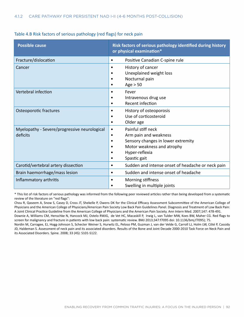
4.1.2 caRE PaThway foR PERsisTEnT nad i-ii (4-6 monThs PosT-collision)
Enabling REcovERy fRom common TRaffic injuRiEs: a focus on ThE injuREd PERson | 92
Table 4.B Risk factors of serious pathology (red flags) for neck pain
Possible cause Risk factors of serious pathology identified during history or physical examination*
Fracture/dislocation • Positive Canadian C-spine rule Cancer • History of cancer
• Unexplained weight loss • Nocturnal pain • Age > 50
Vertebral infection • Fever • Intravenous drug use • Recent infection
Osteoporotic fractures • History of osteoporosis • Use of corticosteroid • Older age
Myelopathy - Severe/progressive neurological deficits
• Painful stiff neck • Arm pain and weakness • Sensory changes in lower extremity • Motor weakness and atrophy • Hyper-reflexia • Spastic gait
Carotid/vertebral artery dissection • Sudden and intense onset of headache or neck pain Brain haemorrhage/mass lesion • Sudden and intense onset of headache Inflammatory arthritis • Morning stiffness
• Swelling in multiple joints
* This list of risk factors of serious pathology was informed from the following peer reviewed articles rather than being developed from a systematic review of the literature on “red flags”: Chou R, Qaseem A, Snow S, Casey D, Cross JT, Shekelle P, Owens DK for the Clinical Efficacy Assessment Subcommittee of the American College of Physicians and the American College of Physicians/American Pain Society Low Back Pain Guidelines Panel. Diagnosis and Treatment of Low Back Pain: A Joint Clinical Practice Guideline from the American College of Physicians and the American Pain Society. Ann Intern Med. 2007;147: 478-491. Downie A, Williams CM, Henschke N, Hancock MJ, Ostelo RWJG, de Vet HC, Macaskill P, Irwig L, van Tulder MW, Koes BW, Maher CG. Red flags to screen for malignancy and fracture in patients with low back pain: systematic review. BMJ 2013;347:f7095 doi: 10.1136/bmj.f7095); 75. Nordin M, Carragee, EJ, Hogg-Johnson S, Schecter Weiner S, Hurwitz EL, Peloso PM, Guzman J, van der Velde G, Carroll LJ, Holm LW, Côté P, Cassidy JD, Haldeman S. Assessment of neck pain and its associated disorders. Results of the Bone and Joint Decade 2000-2010 Task Force on Neck Pain and its Associated Disorders. Spine. 2008; 33 (4S): S101-S122.
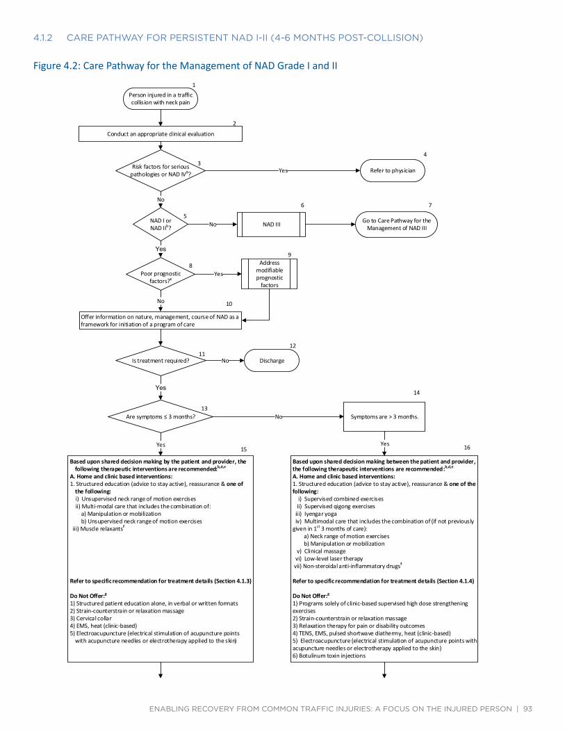
4.1.2 caRE PaThway foR PERsisTEnT nad i-ii (4-6 monThs PosT-collision)
Yes
No
No
Yes
Yes
No
No
Yes
No
Yes Yes
Enabling REcovERy fRom common TRaffic injuRiEs: a focus on ThE injuREd PERson | 93
Figure 4.2: Care Pathway for the Management of NAD Grade I and II
1 Person injured in a traffic collision with neck pain
2
Conduct an appropriate clinical evaluation
3Risk factors for serious pathologies or NAD IVa?
4
Refer to physician
5 NAD I or NAD IIb?
6
NAD III
7
Go to Care Pathway for the Management of NAD III
8 Poor prognostic
factors?c
9 Address
modifiable prognostic
factors
10
Offer information on nature, management, course of NAD as a framework for initiation of a program of care
11 Is treatment required?
12
Discharge
13 Are symptoms ≤ 3 months?
14
Symptoms are ˃3 months.
15
Based upon shared decision making by the patient and provider, the following therapeutic interventions are recommended:b,d,e
A. Home and clinic based interventions: 1. Structured education (advice to stay active), reassurance & one of
the following: i) Unsupervised neck range of motion exercises ii) Multi-modal care that includes the combination of:
a) Manipulation or mobilization b) Unsupervised neck range of motion exercises
iii) Muscle relaxantsf
Refer to specific recommendation for treatment details (Section 4.1.3)
Do Not Offer:g
1) Structured patient education alone, in verbal or written formats 2) Strain-counterstrain or relaxation massage 3) Cervical collar 4) EMS, heat (clinic-based) 5) Electroacupuncture (electrical stimulation of acupuncture points
with acupuncture needles or electrotherapy applied to the skin)
16
Based upon shared decision making between the patient and provider, the following therapeutic interventions are recommended:b,d,e
A. Home and clinic based interventions: 1. Structured education (advice to stay active), reassurance & one of the following:
i) Supervised combined exercises Ii) Supervised qigong exercises iii) Iyengar yoga iv) Multimodal care that includes the combination of (if not previously
given in 1st 3 months of care): a) Neck range of motion exercises b) Manipulation or mobilization
v) Clinical massage vi) Low-level laser therapy vii) Non-steroidal anti-inflammatory drugsf
Refer to specific recommendation for treatment details (Section 4.1.4)
Do Not Offer:g
1) Programs solely of clinic-based supervised high dose strengthening exercises 2) Strain-counterstrain or relaxation massage 3) Relaxation therapy for pain or disability outcomes 4) TENS, EMS, pulsed shortwave diathermy, heat (clinic-based) 5) Electroacupuncture (electrical stimulation of acupuncture points with acupuncture needles or electrotherapy applied to the skin) 6) Botulinum toxin injections

4.1.2 caRE PaThway foR PERsisTEnT nad i-ii (4-6 monThs PosT-collision)
Yes Yes
No No
Enabling REcovERy fRom common TRaffic injuRiEs: a focus on ThE injuREd PERson | 94
17 Reassess and Monitor for the presence of acute stress disorder, post-traumatic stress disorder, kinesiophobia, passive coping, depression, anxiety, anger, frustration and fear
18 Reassess and Monitor for the presence of acute stress disorder, post-traumatic stress disorder, kinesiophobia, passive coping, depression, anxiety, anger, frustration and fear
19 Is injured person
recovered?
20
Discharge
21 Is injured person
recovered?
22 1) Incomplete recovery: initiate persistent protocol (Box 16) 2) Signs progress to Grade III: proceed to NAD III care pathway 3) Development of serious pathology (new or worsening physical,
mental or psychological symptoms): refer to physician
23 1) Incomplete recovery: refer to physician 2) Signs progress to Grade III: proceed to NAD III care pathway 3) Development of serious pathology (new or worsening physical,
mental or psychological symptoms): refer to physician
a Risk factors for serious pathologies (also known as red flags): Cancer (history of cancer, unexplained weight loss, nocturnal pain, age >50), vertebral infection (fever, intravenous drug use, recent infection), osteoporotic fractures (history of osteoporosis, use of corticosteroid, older age), traumatic fracture (positive Canadian C-Spine rule), myelopathy – severe/progressive neurological deficits (painful stiff neck, arm pain and weakness, sensory changes in lower extremity, motor weakness and atrophy, hyper-reflexia, spastic gait), carotid/vertebral artery dissection (sudden and intense onset of headache or neck pain), brain haemorrhage/mass lesion (sudden and intense onset of headache), inflammatory arthritis (morning stiffness, swelling in multiple joints) b If symptoms progress proceed to NAD III protocol or refer. c Factors delaying recovery: prior history of collision-related NAD, older age, high levels of initial pain, post-crash psychological factors [poor recovery expectation, depressed mood, anxiety or fear about pain, kinesiophobia, acute stress disorder (symptoms ≤ 4 weeks of the injury), post-traumatic stress disorder (symptoms ˃ 4 weeks), high levels of frustration or anger about pain, passive coping] d This guideline does not include interventions for which there is a lack of evidence of effectiveness e The ordering of interventions does not reflect superiority of effectiveness f The evidence indicates that analgesia is the primary therapeutic benefit of the muscle relaxant and NSAID classes of medication. Pain reduction should be apparent during the initial period of usage; in the absence of therapeutic benefit, prolongation of usage is not warranted. There is no evidence of differential efficacy for the various drugs within each class. There is also no evidence that any combination of these medications provides added benefit. There are potentially significant adverse effects associated with use of these classes of medications. Finally, the non-opioid first ‘step’ in the Analgesic Ladder includes NSAIDs, muscle relaxant and acetaminophen (Vargas-Schaffer G. Is the WHO analgesic ladder still valid? Twenty-four years of experience. Vol 56: June 2010 Canadian Family Physician). However, the evidence does not indicate that acetaminophen is an effective analgesic for either NAD or low back pain; therefore, the use of acetaminophen is not recommended. g Based on evidence of no benefit to patients
SECTION 4.1.3
Key recOMMendatiOnS fOr the ManageMent Of recent OnSet nad i-ii
This section summarizes the key recommendations for the management of NAD I-II for the first 3 months post-collision. The wording of recommendations follows the guidance from the National Institute for Health and Care Excellence (NICE). Recommendations beginning with “offer” indicate that, according to the evidence, an intervention is associated with outcomes that were superior to other interventions, placebo/sham, or no intervention. The wording “consider” indicates that an intervention is as effective as another one. The wording “do not offer” indicates, according to the evidence, an intervention does not benefit patients. A detailed explanation of the wording of recommendations is presented in section 2.5.2.4 of this report.

4.1.3 kEy REcommEndaTions foR ThE managEmEnT of REcEnT onsET nad i-ii
Enabling REcovERy fRom common TRaffic injuRiEs: a focus on ThE injuREd PERson | 95
• Provide care in partnership with the patient. Involve the patient in care planning and decision-making.
• Reassure patients about the benign and self-limited nature of their pain.
• Educate patients about the benefits of being actively engaged and participating in their care plan by remaining active and continuing movement.
• Emphasize active rather than passive treatments.
• Deliver time-limited care.
• Do not provide ineffective or experimental treatments.
SECTION 4.1.3.1
Structured Patient educatiOn
Structured patient education aims to enable individuals to make informed decisions about their personal health-related behaviour. Structured education strategies refer to standardized interventions such as scripted discussion, pamphlets or videos. Educational interventions should begin with an assessment of the person’s knowledge of the injury and their health goals. The content of the structured education interventions may include (but is not limited to): reassurance about the favourable prognosis of NAD I-II; advice on return to usual activities, including work; instruction of exercise; discussion of expected pain and pain mechanism; discussion of prognosis; pain coping skills; discussion of workplace ergonomics; and self-care strategies or general health.
Table 4.C: Structured patient education for recent onset NAD I-II
Recommendation 4.1.3.1.1
Provide information about the nature, management, and course of NAD as a framework for the initiation of the program of care.
4.1.3.1.2 Consider a structured patient education program as an adjunct to an effec-tive program of care based on individual patient presentation.*
4.1.3.1.3 Do not offer structured patient education alone, in either verbal or written formats.
References: • Decision Determinants and Evidence Table for NAD – Report 1 – Appendix 2
* The structured education program should focus on providing advice to stay active and reassuring the patient by addressing the expectation of recovery.

Enabling REcovERy fRom common TRaffic injuRiEs: a focus on ThE injuREd PERson | 96
SECTION 4.1.3.2
exerciSe
Exercise refers to any series of movements with the aim of training or developing the body by routine practice or as physical training to promote good physical health. Exercise therapy includes a wide variety of techniques common to the treatment and rehabilitation of neck pain.
Table 4.D: Exercise for recent onset NAD I-II
Recommendation 4.1.3.2.1
Consider unsupervised range of motion exercises (5 to 10 repetitions of each exercise with no resistance, up to 6 to 8 times per day).*
References: • Decision Determinants and Evidence Table for NAD – Report 2 – Appendix 2
* Daily home unsupervised, gentle and controlled range of motion of exercise of the neck and shoulder joints, including neck retraction, extension, flexion, rotation, lateral bending motions, and scapular retraction. The exercise program should be instructed by a health care professional.
SECTION 4.1.3.3
MultiMOdal care
Multimodal care includes at least two distinct therapeutic modalities, provided by one or more health care disciplines.
Table 4.E: Multimodal care for recent onset NAD I-II
Recommendation 4.1.3.3.1
Consider a maximum of 6 sessions over 8 weeks of multimodal care that includes exercise* and manual therapy.**
References: • Decision Determinants and Evidence Table for NAD – Report 3 – Appendix 2
* Exercise refers to any series of movements with the aim of training or developing the body by routine practice or as physical training to promote good physical health. Exercise therapy includes a wide variety of techniques common for the treatment and rehabilitation of neck pain. Exercise interventions could include any prescribed movements with the intent of affecting clinical outcomes with respect to neck pain. ** Manual therapy refers to techniques that involve the application of hands-on and/or mechanically assisted treatments, including manipulation and mobilization. Manipulation is a high velocity, low amplitude impulse or thrust applied at or near the end of a joint’s passive range of motion. Mobilization refers to a low velocity and small or large amplitude oscillatory movement, within a joint’s passive range of motion. For the purpose of this recommendation, manual therapy refers to manipulation or mobilization to the cervical and/or the thoracic spine as clinically indicated.

Enabling REcovERy fRom common TRaffic injuRiEs: a focus on ThE injuREd PERson | 97
SECTION 4.1.3.4
SOft tiSSue theraPy
Soft tissue therapy is a mechanical therapy in which muscles, tendons, and ligaments are passively pressed or kneaded by hand or with mechanical devices. It includes relaxation massage, clinical massage, movement re-education and energy work.
Table 4.F: Soft tissue therapy for recent onset NAD I-II
Recommendation 4.1.3.4.1 Do not offer strain-counterstrain.*
4.1.3.4.2 Do not offer relaxation massage.**
References: • Decision Determinants and Evidence Table for NAD – Report 4 – Appendix 2
* Strain-counterstrain is a soft tissue therapy (clinical massage and movement re-education technique) that involves applied pressure to a muscle with positioning of the neck to provide a small stretch to a muscle. ** Relaxation massage refers to a group of soft tissue therapies intended to relax muscles. Examples of relaxation massage techniques are effleurage, petrissage, and tapotement.
SECTION 4.1.3.5
PaSSive PhySical MOdalitieS
Passive physical modalities include two categories of interventions: physico-chemical and structural. Physico-chemical modalities use a thermal or electromagnetic agent to affect the body at or beneath the skin level. Structural modalities include functional or non-functional assistive devices. Functional assistive devices intend to align, support or otherwise indirectly facilitate function in the affected region. Non-functional devices intend to achieve a state of rest in specific anatomic positions or prevent movement.
Table 4.G: Passive physical modalities for recent onset NAD I-II
Recommendation 4.1.3.5.1 Do not offer a cervical collar.
4.1.3.5.2 Do not offer moist heat as an intervention in the clinic.
4.1.3.5.3 Do not offer electrical muscle stimulation.*
References: • Decision Determinants and Evidence Table for NAD – Report 5 – Appendix 2
* Electrical muscle stimulation transmits electrical impulses to muscles via electrodes placed superficially on the skin.

Enabling REcovERy fRom common TRaffic injuRiEs: a focus on ThE injuREd PERson | 98
SECTION 4.1.3.6
acuPuncture
Acupuncture is a therapeutic technique that utilizes a thin metal needle to puncture the skin and stimulate specific points. Various acupuncture techniques exist, as well as the use of other types of stimulation in combination with or instead of a needle. Acupuncture interventions include body needling, moxibustion, electroacupuncture, laser acupuncture, microsystem acupuncture and acupressure.
Table 4.H: Acupuncture for recent onset NAD I-II
Recommendation 4.1.3.6.1 Do not offer electroacupuncture.*
References: • Decision Determinants and Evidence Table for NAD – Report 6 – Appendix 2
* Electroacupuncture refers to the electrical stimulation of acupuncture points with acupuncture needles or electrotherapy applied to the skin.
SECTION 4.1.3.7
MedicatiOn
Our reviews investigated the effectiveness of three classes of medication: analgesics, non-steroidal anti-inflammatory drugs (NSAIDs), and muscle relaxant. Analgesics are drugs that are used to reduce or relieve pain without blocking the conduction of nerve impulses, significantly altering sensory perception or producing a loss of consciousness. An example of a non-opioid analgesic drug is acetaminophen. NSAIDs are medications that block the action of cyclooxygenase (Cox)-1 and/or Cox-2 to help reduce inflammation. Muscle relaxants are a broad range of drugs with different chemical structures and mechanisms of action, which fall into three groups according to their actions along the voluntary motor control: muscle decoupler, neuromuscular blockers, and spasmolytics.
Table 4.I: Medication for recent onset NAD I-II
Recommendation 4.1.3.7.1 Consider muscle relaxants*
References: • Decision Determinants and Evidence Table for the Systematic Review on Muscle Relaxants for Neck
Pain and Associated Disorders – Report 11 – Appendix 2 • Decision Determinants and Evidence Table for the Systematic Review of Non-opioid Analgesic Drugs
for Neck and Associated Disorders – Report 9 - Appendix 2 • Decision Determinants and Evidence Table for the Systematic Review of Non-steroidal Anti-
inflammatory Drugs for Neck and Associated Disorders – Report 10 - Appendix 2
* The evidence indicates that analgesia is the primary therapeutic benefit, prolongation of usage is not warranted. There is no evidence of differential efficacy for the various drugs within each class. There is also no evidence that any combination of these medications provides added benefit. There are potentially significant adverse effects associated with use of these classes of medications. Finally, the non-opioid first ‘step’ in the Analgesic Ladder includes NSAIDs, muscle relaxant and acetaminophen (Vargas-Schaffer G. Is the WHO analgesic ladder still valid? Twenty-four years of experience. Vol 56: June 2010 Canadian Family Physician). However, the evidence does not indicate that acetaminophen is an effective analgesic for either NAD or low back pain; therefore, the use of acetaminophen is not recommended.

Enabling REcovERy fRom common TRaffic injuRiEs: a focus on ThE injuREd PERson | 99
SECTION 4.1.4
Key recOMMendatiOnS fOr the ManageMent Of PerSiStent nad i-ii
This section summarizes the key recommendations for the management of NAD I-II for the period extending from 4 to 6 months post-collision. The wording of recommendations follows the guidance from the National Institute for Health and Care Excellence (NICE). Recommendations beginning with “offer” indicate that, according to the evidence, an intervention is associated with outcomes that were superior to other interventions, placebo/sham, or no intervention. The wording “consider” indicates that an intervention is as effective as another one. The wording “do not offer” indicates, according to the evidence, an intervention does not benefit patients. A detailed explanation of the wording of recommendations is presented in section 2.5.2.4 of this report.
• Provide care in partnership with the patient. Involve the patient in care planning and decision-making.
• Reassure patients about the benign and self-limited nature of their pain.
• Educate patients about the benefits of being actively engaged and participating in their care plan by remaining active and continuing movements.
• Emphasize active rather than passive treatments.
• Deliver time-limited care.
• Do not provide ineffective or experimental treatments.
SECTION 4.1.4.1
Structured Patient educatiOn
Structured patient education aims to enable individuals to make informed decisions about their personal health-related behaviour. Structured education strategies refer to standardized interventions such as scripted discussion, pamphlets, or videos. Educational interventions should begin with an assessment of the person’s knowledge of the injury and their health goals. The content of the structured education interventions may include (but is not limited to): reassurance about the favourable prognosis of NAD I-II; advice on return to usual activities, including work; instruction of exercise; discussion of expected pain and pain mechanism; discussion of prognosis; stress-coping skills; discussion of workplace ergonomics; and self-care strategies or general health.
Table 4.J: Structured patient education for persistent NAD I-II
Recommendation 4.1.4.1.1
Provide information about the nature, management, and course of NAD as a framework for the initiation of the program of care.
4.1.4.1.2 Consider a structured patient education program as an adjunct to an effec-tive program of care based on individual patient presentation.*
References: • Decision Determinants and Evidence Table for NAD – Report 1 – Appendix 2
* The structured education program should focus on providing advice to stay active and reassuring the patient by addressing the expectation of recovery.

Enabling REcovERy fRom common TRaffic injuRiEs: a focus on ThE injuREd PERson | 100
SECTION 4.1.4.2
exerciSe
Exercise refers to any series of movements with the aim of training or developing the body by routine practice or as physical training to promote good physical health. Exercise therapy includes a wide variety of techniques common for the treatment and rehabilitation of neck pain.
Table 4.K: Exercise for persistent NAD I-II
Recommendation 4.1.4.2.1
Offer a program of supervised combined exercise* (strengthening, range of motion, and flexibility exercises). The program should be limited to a maxi-mum of 2 sessions/week for 12 weeks.
4.1.4.2.2 Offer a program of qigong** exercises supervised by a certified qigong in-structor. The program should be limited to a maximum of 2 sessions/week for 12 weeks.
4.1.4.2.3 Offer a program of Iyengar yoga*** supervised by a certified Iyengar yoga teacher. The program should be limited to a maximum of 9 sessions over 9 weeks.
4.1.4.2.4 Do not offer programs consisting solely of clinic-based supervised high dose strengthening exercises.****
References: • Decision Determinants and Evidence Table for NAD – Report 2 – Appendix 2
* Supervised combined exercise refers to a supervised and standardized group of exercise developed to improve persistent neck pain and consists of active cervical rotation, strengthening and flexibility exercises. ** Qigong refers to gentle, focused exercises for mind and body to increase and restore the flow of qi energy and encourage healing. *** Iyengar yoga refers to a range of classical yoga poses adapted with the use of modified poses or supportive props for individuals with specific health issues. **** Clinic-based supervised high dose strengthening exercises refers to a high frequency of supervised in-clinic sessions over a short time period, incorporating neck and upper body dynamic resistance strengthening.
SECTION 4.1.4.3
MultiMOdal care
Multimodal care includes at least two distinct therapeutic modalities, provided by one or more health care disciplines.

4.1.4.3 mulTimodal caRE
Enabling REcovERy fRom common TRaffic injuRiEs: a focus on ThE injuREd PERson | 101
Table 4.L: Multimodal care for persistent NAD I-II
Recommendation 4.1.4.3.1
Consider a maximum of 6 sessions over 8 weeks of multimodal care that includes exercise* and manual therapy.**Multimodal care for persistent Grade I-II NAD should only be considered if not previously given in the first three months of care. However, a second course could be indicated if the patient demonstrates ongoing and signifi-cant improvement.
References: • Decision Determinants and Evidence Table for NAD – Report 3 – Appendix 2
* Exercise refers to any series of movements with the aim of training or developing the body by routine practice or as physical training to promote good physical health. Exercise therapy includes a wide variety of techniques common for the treatment and rehabilitation of neck pain. Exercise interventions could include any prescribed movements with the intent of affecting clinical outcomes with respect to neck pain. ** Manual therapy refers to techniques that involve the application of hands-on and/or mechanically assisted treatments, including manipulation and mobilization. Manipulation is a high velocity, low amplitude impulse or thrust applied at or near the end of a joint’s passive range of motion. Mobilization refers to a low velocity and small or large amplitude oscillatory movement, within a joint’s passive range of motion. For the purpose of this recommendation, manual therapy refers to manipulation or mobilization to the cervical and/or the thoracic spine as clinically indicated.
SECTION 4.1.4.4
SOft tiSSue theraPy
Soft tissue therapy is a mechanical therapy in which muscles, tendons, and ligaments are passively pressed or kneaded by hand or with mechanical devices. It includes relaxation massage, clinical massage, movement re-education and energy work.
Table 4.M: Soft tissue therapy for persistent NAD I-II
Recommendation 4.1.4.4.1
Consider up to 10 sessions over 10 weeks of clinical massage. This treat-ment is expected to provide short-term benefits only.*
4.1.4.4.2 Do not offer strain-counterstrain.**
4.1.4.4.3 Do not offer relaxation massage.***
References: • Decision Determinants and Evidence Table for NAD – Report 4 – Appendix 2
* Clinical massage refers to a group of soft tissue therapies that targets muscles with specific goals such as relieving pain, releasing muscle spasms or improving restricted motion. An example of clinical massage is myofascial trigger point therapy. ** Strain-counterstrain is a soft tissue therapy (clinical massage and movement re-education) that involves applied pressure to a muscle with positioning of the neck to provide a small stretch a muscle. *** Relaxation massage refers to a group of soft tissue therapies intended to relax muscles. Examples of relaxation massage techniques are effleurage, petrissage, and tapotement.

Enabling REcovERy fRom common TRaffic injuRiEs: a focus on ThE injuREd PERson | 102
SECTION 4.1.4.5
PaSSive PhySical MOdalitieS
Passive physical modalities include two categories of interventions: physico-chemical and structural. Physico-chemical modalities use a thermal or electromagnetic agent to affect the body at or beneath the skin level. Structural modalities include functional or non-functional assistive devices. Functional assistive devices intend to align, support or otherwise indirectly facilitate function in the affected region. Non-functional devices intend to achieve a state of rest in specific anatomic positions or prevent movement.
Table 4.N: Passive physical modalities for persistent NAD I-II
Recommendation 4.1.4.5.1
Consider up to 12 sessions over 4 weeks of clinic-based low level laser ther-apy (LLLT)* (continuous or pulsed application; wavelength = 830 or 904 nm).
4.1.4.5.2 Do not offer transcutaneous electrical nerve stimulation (TENS).**
4.1.4.5.3 Do not offer pulsed short-wave diathermy.***
4.1.4.5.4 Do not offer moist heat as an intervention in the clinic.
4.1.4.5.5 Do not offer electrical muscular stimulation.****
References: • Decision Determinants and Evidence Table for NAD – Report 5 – Appendix 2
* Low-level laser therapy is the application of a coherent light beam (laser) to a region for the purpose of reducing local pain or promoting local healing. ** TENS is a passive physical modality connected to the skin, using two or more electrodes to apply low-level electrical current. It is typically used with the intent to help pain management. *** Pulsed short-wave diathermy uses electromagnetic energy to heat underlying tissues with the intent to help inflammatory and repair phases in soft tissues. **** Electrical muscle stimulation transmits electrical impulses to muscles via electrodes placed superficially on the skin.
SECTION 4.1.4.6
PSychOlOgical interventiOn
A psychological intervention is a method used to treat psychological distress, consequences of musculoskeletal injuries (such as pain), or psychological disorders; primarily (but not exclusively) by verbal or non-verbal communication. Psychological interventions can be broadly subdivided into several theoretical orientations, including but not limited to psychodynamic, psychoanalytic, behavioural/cognitive behavioural, humanistic and existential, family/systems approaches and combinations of these approaches. Psychological interventions can include (but are not limited to) in-person psycho-education; booklet/written material that includes a psycho-educational component; cognitive-behavioural interventions, or a guided psychological self-help intervention.
4.1.4.6 Psychological inTERvEnTion
Enabling REcovERy fRom common TRaffic injuRiEs: a focus on ThE injuREd PERson | 103
Table 4.O: Psychological interventions for persistent NAD I-II
Recommendation 4.1.4.6.1
Do not offer a standalone course of relaxation training for pain intensity or disability outcomes.
References: • Decision Determinants and Evidence Table for NAD – Report 7 – Appendix 2
SECTION 4.1.4.7
acuPuncture
Acupuncture is a therapeutic technique that utilizes a thin metal needle to puncture the skin and stimulate specific points. Various acupuncture techniques exist, as well as the use of other types of stimulation in combination with or instead of a needle. Acupuncture interventions include body needling, moxibustion, electroacupuncture, laser acupuncture, microsystem acupuncture and acupressure.
Table 4.P: Acupuncture for persistent NAD I-II
Recommendation 4.1.4.7.1 Do not offer electroacupuncture.*
References: • Decision Determinants and Evidence Table for NAD – Report 6 – Appendix 2
* Electroacupuncture refers to the electrical stimulation of acupuncture points with acupuncture needles or electrotherapy applied to the skin.
SECTION 4.1.4.8
MedicatiOn
Our reviews investigated the effectiveness of three classes of medication: analgesics, non-steroidal anti-inflammatory drugs (NSAIDs), and muscle relaxant. Analgesics are drugs that are used to reduce or relieve pain without blocking the conduction of nerve impulses, significantly altering sensory perception or producing a loss of consciousness. An example of a non-opioid analgesic drug is acetaminophen. NSAIDs are medications that block the action of cyclooxygenase (Cox)-1 and/or Cox-2 to help reduce inflammation. Muscle relaxants are a broad range of drugs with different chemical structures and mechanisms of action, which fall into three groups according to their actions along the voluntary motor control: muscle decoupler, neuromuscular blockers, and spasmolytics.

4.1.4.8 mEdicaTion
Enabling REcovERy fRom common TRaffic injuRiEs: a focus on ThE injuREd PERson | 104
Table 4.Q: Medication for persistent NAD I-II
Recommendation 4.1.4.8.1 Consider non-steroidal anti-inflammatory drugs (NSAIDs)*
4.1.4.8.2 Do not offer botulinum toxin injections
References: • Decision Determinants and Evidence Table for the Systematic Review on Muscle Relaxants for Neck
Pain and Associated Disorders – Report 11 – Appendix 2 • Decision Determinants and Evidence Table for the Systematic Review of Non-opioid Analgesic Drugs
for Neck and Associated Disorders – Report 9 - Appendix 2 • Decision Determinants and Evidence Table for the Systematic Review of Non-steroidal Anti-
inflammatory Drugs for Neck and Associated Disorders – Report 10 - Appendix 2
* The evidence indicates that analgesia is the primary therapeutic benefit of the muscle relaxant and NSAID classes of medication. Pain reduction should be apparent during the initial period of usage; in the absence of therapeutic benefit, prolongation of usage is not warranted. There is no evidence of differential efficacy for the various drugs within each class. There is also no evidence that any combination of these medications provides added benefit. There are potentially significant adverse effects associated with use of these classes of medications. Finally, the non-opioid first ‘step’ in the Analgesic Ladder includes NSAIDs, muscle relaxant and acetaminophen (Vargas-Schaffer G. Is the WHO analgesic ladder still valid? Twenty-four years of experience. Vol 56: June 2010 Canadian Family Physician). However, the evidence does not indicate that acetaminophen is an effective analgesic for either NAD or low back pain; therefore, the use of acetaminophen is not recommended.

Quick Reference Guide – Management of NAD III
Enabling REcovERy fRom common TRaffic injuRiEs: a focus on ThE injuREd PERson | 105
SECTION 4.2
ManageMent Of nad iii
Quick Reference Guide – Management of NAD Grade III
Symptoms ≤ 3 months post‐collision Symptoms > 3 months post ‐collision For all injured persons with NAD III: Rule out risk factors for serious pathologiesa
Assess for factors delaying recovery: prior history of collision‐related NAD, older age, high levels of initial pain, post‐crash psychological factors [poor recovery expectation, depressed mood, anxiety or fear about p ai n, kinesiophobia, acute stress disorder (symptoms ≤ 4 weeks from injury), post‐traumatic stress disorder (symptoms > 4 weeks), high levels of frustration or anger about p ai n, passive coping] Offer information on nature, management, course of NAD as a framework for initiation of a program of care Conduct ongoing assessment for symptom improvement or worsening/progress during intervention period and refer accordingly Reassess and Monitor the presence of acute stress disorder, post‐traumatic stress disorder, kinesiophobia, passive coping, depression, anxiety, anger, frustration and fear Discharge injured person as appropriate at any point during intervention and recovery
Based upon shared decision making betw een t h e patient and provider, the following therapeutic interventions are recommended:b , c
1) Structured education, reassurance 2) Supervised graded neck strengthening exercise
Refer to specific recommendation for treatment details (Section 4.2.3)
Refer to medical physician for consideration of further investigation of the neurological deficits b , c
* No admissible evidence of effective management of persistent NAD III
Do Not Offer:d
Cervical collar Structured patient education alone, in either verbal or written formats Low level laser therapy Intermittent traction
Do Not Offer:d
Cervical collar
Outcome: Recovered → Discharge Improvement (neurological signs no longer present) → Refer to NAD I/II care pathway Incomplete recovery → Refer to physician Major symptom change or development of serious pathology (new or worsening physical, mental or psychological symptoms) → Refer to physician
a Risk factors for serious pathologies (also known as red flags): Cancer (history of cancer, unexplained weight loss, nocturnal pain, age >50), vertebral infection (fever, intravenous drug use, recent infection), osteoporotic fractures (history of osteoporosis, use of corticosteroid, older age), traumatic fracture (positive Canadian C‐Spine rule), myelopathy – severe/progressive neurological deficits (painful stiff neck, arm pain and weakness, sensory changes in lower extremity, motor weakness and atrophy, h yp er ‐reflexia, spastic gait), carotid/vertebral artery dissection (sudden and intense onset of headache or neck pain), brain haemorrhage/mass lesion (sudden and i nt e n se onset of headache), inflammatory arthritis (morning stiffness, swelling in multiple joints) b This guideline does not include interventions for which there is a lack of evidence of effectiveness c The ordering of interventions does not reflect superiority of effectiveness d Base d on evidence of n o benefit to patien ts
Symptoms > 3 months post‐collisionSymptoms ≤ 3 months post‐collisionFor all injured persons with NAD III:Rule out risk factors for serious pathologiesa
Assess for factors delaying recovery: prior history of collision‐related NAD, older age, high levels of initial pain, post‐crash psychological factors [poor recovery expectation, depressed mood, anxiety or fear about pain, kinesiophobia, acute stress disorder (symptoms ≤ 4 weeks from injury), post‐traumatic stress disorder (symptoms > 4 weeks), high levels of frustration or anger about pain, passive coping]Offer information on nature, management, course of NAD as a framework for initiation of a program of careConduct ongoing assessment for symptom improvement or worsening/progress during intervention period and refer accordinglyReassess and Monitor the presence of acute stress disorder, post‐traumatic stress disorder, kinesiophobia, passive coping, depression, anxiety, anger, frustration and fearDischarge injured person as appropriate at any point during intervention and recovery
Based upon shared decision making between the patient and provider, the following therapeutic interventions are recommended:b,c
1) Structured education, reassurance 2) Supervised graded neck strengthening exercise
Refer to specific recommendation for treatment details (Section 4.2.3)
Refer to medical physician for consideration of further investigation of the neurological deficitsb,c
* No admissible evidence of effective management of persistent NAD III
Do Not Offer:d
Cervical collar Structured patient education alone, in either verbal or written formats Low level laser therapy Intermittent traction
Do Not Offer:d
Cervical collar
Outcome: Recovered → Discharge Improvement (neurological signs no longer present) → Refer to NAD I/II care pathway Incomplete recovery → Refer to physician Major symptom change or development of serious pathology (new or worsening physical, mental or psychological symptoms) → Refer to physician
a Risk factors for serious pathologies (also known as red flags): Cancer (history of cancer, unexplained weight loss, nocturnal pain, age >50), vertebral infection (fever, intravenous drug use, recent infection), osteoporotic fractures (history of osteoporosis, use of corticosteroid, older age), traumatic fracture (positive Canadian C‐Spine rule), myelopathy – severe/progressive neurological deficits (painful stiff neck, arm pain and weakness, sensory changes in lower extremity, motor weakness and atrophy, hyper‐reflexia, spastic gait), carotid/vertebral artery dissection (sudden and intense onset of headache or neck pain), brain haemorrhage/mass lesion (sudden and intense onset of headache), inflammatory arthritis (morning stiffness, swelling in multiple joints)b This guideline does not include interventions for which there is a lack of evidence of effectivenessc The ordering of interventions does not reflect superiority of effectivenessd Based on evidence of no benefit to patients

Enabling REcovERy fRom common TRaffic injuRiEs: a focus on ThE injuREd PERson | 106
SECTION 4.2.1
care Pathway fOr recent OnSet nad iii (0-3 MOnthS POSt-cOlliSiOn)
The care pathway is presented in Figure 4.3.
At initial contact, health care professionals should educate and reassure the patient that neck and arm pain will resolve within a few months of symptom onset. Patients greatly improve their recovery by actively engaging in their care. Clinical care aims to accelerate recovery by reducing pain and improving function. The care pathway recommended for the first 3 months of care for NAD III is described below.
Assess the Patient and Classify NAD
Conduct an appropriate clinical evaluation to rule out major structural or other pathologies as the cause of the symptoms. Cervical spine fractures and dislocations can be ruled out using the Canadian C-spine rule (Appendix 4.A). The presence of a risk factor for serious pathologies (also known as red flags) identified during the history and examination warrants further investigation and referral to the appropriate health care professional.* However, once pathology has been ruled out, the patient should be treated according to the NAD care pathway.
Assess neurological signs (decreased deep tendon reflexes, muscle weakness or sensory deficits).
Classify the grade of NAD as grade III (Table 4.A).
Patients without neurological signs should be managed under the “Care Pathway for the Management of NAD I-II” (see section 4.1).
Assess the Prognostic Factors
Assess the prognostic factors for delayed recovery. Most patients recover from their injury. Patients with Grade I NAD are expected to recover the most quickly, while those with NAD III are expected to recover the slowest. However, patients with the following prognostic factors may have a higher risk for delayed recovery:
• Prior history of NAD related to a motor vehicle collision • Older age • High levels of initial pain • Post-collision psychological factors:
• Poor expectation of recovery • Depressed mood, feelings of depression about the pain • Anxiety or fear about pain, kinesiophobia or avoiding activities due to fear of pain • Symptoms of acute stress disorder (symptoms exhibited within 4 weeks of the injury)/post-
traumatic stress disorder (symptoms lasting at least one month) • High levels of frustration or anger about the pain • Passive coping
Examples of questions or tools recommended to assess the prognostic factors for delayed recovery are available in Appendix 4.B.

4.2.1 caRE PaThway foR REcEnT onsET nad iii (0-3 monThs PosT-collision)
Enabling REcovERy fRom common TRaffic injuRiEs: a focus on ThE injuREd PERson | 107
Educate and Reassure the Patient
Develop a patient-centred care plan in partnership with the patient.
Health care professionals need to reassure patients that there are no major structural or progressive pathologies (NAD IV) in their neck.
Prognostic factors for poor recovery should be addressed when present. The care should start with education and reassurance about the benign and self-limited nature of NAD III and the importance of maintaining activity and movement. This is particularly important when the patient reports poor expectation of recovery.
It is also important to reassure patients that it is normal to feel some anxiety, distress or anger following a traffic collision. In the presence of such symptoms or emotions, the health care professional should listen to the patient’s concerns, discuss them and adjust the care plan accordingly.
Deliver the Clinical Care
The goal of the care plan is to promote activity through exercise and clinical interventions that promote resolution of symptoms and restoration of function. Based upon shared decision making between the patient and provider, the following therapeutic interventions are recommended:
• Structured education (advice to stay active) reassurance, and • Supervised graded neck strengthening exercises supplemented by home exercises and acetaminophen
or a non-steroidal anti-inflammatory drug. It is important that the health care professional encourages the patient to participate in their care by stretching their neck at home on a daily basis.
Interventions that are not recommended include:
• Cervical collar • Structured patient education alone (either verbal or written) • Low level laser therapy • Intermittent traction
Discuss the risks and benefits of the care plan with the patient.
Reassess and Take the Indicated Course of Action
Reassess the patient at every visit to determine if additional care is necessary, or if the condition is worsening.
Patients should be discharged as soon as they report significant improvement or recovery. It is recommended that health care professionals use the self-rated recovery question to measure patient recovery: “How well do you feel you are recovering from your injuries?” The response options include: 1) completely better, 2) much improved, 3) slightly improved, 4) no change, 5) slightly worse, 6) much worse, 7) worse than ever. Patients reporting to be ‘completely better’ or ‘much improved’ should be considered recovered. Patients who have not recovered should follow the care pathway outlined in the guideline.*
* The use of a valid and reliable condition-specific instrument (e.g., Neck Disability Index) is encouraged but should not be used to measure overall recovery.

4.2.1 caRE PaThway foR REcEnT onsET nad iii (0-3 monThs PosT-collision)
Enabling REcovERy fRom common TRaffic injuRiEs: a focus on ThE injuREd PERson | 108
Patients who improve and no longer report arm pain but still experience neck pain should be managed according to the care pathway for the management of NAD I-II (section 4.1)
Patients with worsening of symptoms and those who develop new physical, mental or psychological symptoms should be referred to a physician for further evaluation at any time point during their care.
Patients who still suffer from neurological signs after the first 3 months of care should be referred to a physician for further evaluation.
Table 4.B Risk factors of serious pathology (red flags) for neck pain
Possible cause Risk factors of serious pathology identified during history or physical examination*
Fracture/dislocation • Positive Canadian C-spine rule Cancer • History of cancer
• Unexplained weight loss • Nocturnal pain • Age > 50
Vertebral infection • Fever • Intravenous drug use • Recent infection
Osteoporotic fractures • History of osteoporosis • Use of corticosteroid • Older age
Myelopathy - Severe/progressive neurological deficits
• Painful stiff neck • Arm pain and weakness • Sensory changes in lower extremity • Motor weakness and atrophy • Hyper-reflexia • Spastic gait
Carotid/vertebral artery dissection • Sudden and intense onset of headache or neck pain Brain haemorrhage/mass lesion • Sudden and intense onset of headache Inflammatory arthritis • Morning stiffness
• Swelling in multiple joints
* This list of risk factors of serious pathology was informed from the following peer reviewed articles rather than being developed from a systematic review of the literature on “red flags”: Chou R, Qaseem A, Snow S, Casey D, Cross JT, Shekelle P, Owens DK for the Clinical Efficacy Assessment Subcommittee of the American College of Physicians and the American College of Physicians/American Pain Society Low Back Pain Guidelines Panel. Diagnosis and Treatment of Low Back Pain: A Joint Clinical Practice Guideline from the American College of Physicians and the American Pain Society. Ann Intern Med. 2007;147: 478-491. Downie A, Williams CM, Henschke N, Hancock MJ, Ostelo RWJG, de Vet HC, Macaskill P, Irwig L, van Tulder MW, Koes BW, Maher CG. Red flags to screen for malignancy and fracture in patients with low back pain: systematic review. BMJ 2013;347:f7095 doi: 10.1136/bmj.f7095); 75. Nordin M, Carragee, EJ, Hogg-Johnson S, Schecter Weiner S, Hurwitz EL, Peloso PM, Guzman J, van der Velde G, Carroll LJ, Holm LW, Côté P, Cassidy JD, Haldeman S. Assessment of neck pain and its associated disorders. Results of the Bone and Joint Decade 2000-2010 Task Force on Neck Pain and its Associated Disorders. Spine. 2008; 33 (4S): S101-S122.

Care Pathway for the Management of NAD Grade III
Yes
No
No Yes
Yes
Yes
No
No
Yes
Enabling REcovERy fRom common TRaffic injuRiEs: a focus on ThE injuREd PERson | 109
SECTION 4.2.2
care Pathway fOr PerSiStent nad iii (4-6 MOnthS POSt-cOlliSiOn)
The care pathway is presented in Figure 4.3.
Patients who still experience neurological signs and disability more than 3 months after the injury should be referred to a physician for further investigation of neurological deficits.
Figure 4.3: Care Pathway for the Management of NAD Grade III 1
Persons i n j u r e d i n a traffi c collision with neck p a i n
2
Conduct an appropriate clinical evaluation
3Risk factors for serious pathologies or NAD IVa ?
4Refer to p h ysi ci a n
5
NAD IIIb
6
NAD I or II
8P o o r prognostic
factors? c
7
Go to care p a t h w a y for the management of NAD I/II
9Address modifiable prognostic factors
10
Offer information on nature, management, course of NAD III as a framework for initiation of a program of care.
11A r e symptoms ≤ 3 months?
12
Symptoms are ˃ 3 m o n t hs .
13B a s e d upon shared decision making between the patient and provid er, the f o l lo w i n g therapeutic in terventions are recommended:b,d,e
1 ) Structured education, reassurance2 ) Supervised graded neck strengthening exercise
Refer to specific recommendation for treatment details (Section 4 .2 .3 )
Do not o f f e r: f
1 ) Structured patient education alone, i n either verbal or written form ats
2 ) Low level laser therapy 3 ) Inte rm ittent traction 4 ) Cervical col l ar
141) Refer to medical physician for consideration of furt her investigatio n of the neurological deficits b,d
Do not o f f e r: f
1) Cervical col l ar
15Reassess and Monitor for the p r e se n c e of acute stress disorder, post‐t ra u m at i c stress d i s o r d er , kinesiophobia, passive coping, depression, anxiety, a n g e r , frustrat ion a n d fear
16Reassess and Monitor for the p r e se n c e of acute stress disorder, post‐t ra u m at i c stress d i s o r d e r, kinesiophobia, passive coping, depression, anxiety, anger, frustrat ion a n d fear
Persons injured in a traffic collision with neck pain
Poor prognostic factors?c
Address modifiable prognostic factorsYes
Are symptoms ≤ 3 months? No
Based upon shared decision making between the patient and provider, the following therapeutic interventions are recommended:b,d,e
1) Structured education, reassurance 2) Supervised graded neck strengthening exercise
Refer to specific recommendation for treatment details (Section 4.2.3)
Do not offer:f 1) Structured patient education alone, in either verbal or written formats
2) Low level laser therapy3) Intermittent traction4) Cervical collar
Yes
Go to care pathway for the management of NAD I/II
1) Refer to medical physician for consideration of furtherinvestigation of the neurological deficitsb,d
Do not offer:f 1) Cervical collar
Offer information on nature, management, course of NAD III as a framework for initiation of a program of care.
No
Yes
7
1
8 9
11
13 14
Reassess and Monitor for the presence of acute stress disorder, post‐traumatic stress disorder, kinesiophobia, passive coping, depression, anxiety, anger, frustration and fear
Conduct an appropriate clinical evaluation
Risk factors for serious pathologies or NAD IVa? Yes Refer to physician
No
No
Yes
Reassess and Monitor for the presence of acute stress disorder, post‐traumatic stress disorder, kinesiophobia, passive coping, depression, anxiety, anger, frustration and fear
NAD IIIb NAD I or II
Symptoms are ˃ 3 months.
15
2
3 4
10
16
5 6
12

4.2.2 caRE PaThway foR PERsisTEnT nad iii (4-6 monThs PosT-collision)
Yes
No
Yes
No
Enabling REcovERy fRom common TRaffic injuRiEs: a focus on ThE injuREd PERson | 110
17Is injured person
recovered?
20
Refer to p h ysi ci a n
18
Discharge
19Are there neurological signs, major symptom change or development of serious
pathology?
21
Refer to NAD I/II care p a t h w a y
a Risk factors for serious pathologies (also known as red flags): Cancer (history of cancer, unexplained weight loss, nocturnal pain, age >50), vertebral infection (fever, iinnttrraavveennoouuss drdrugug use, recent infection), osteoporotic fractures (history of osteoporosis, use of corticosteroid, older age), traumatic fracture (positive Canadian C‐Spine rule), myelopathy – severe/progressive neurological deficits (painful stiff neck, arm pain and weakness, sensory changes in lower extremity, motor weakness and atrophy, hyper‐ reflexia, spastic gait), carotid/vertebral artery dissection (sudden and intense onset of headache or neck pain), brain haemorrhage/mass lesion (sudden and intense onset of headache), inflammatory arthritis (morning stiffness, swelling in multiple joints)b Conduct ongoing assessment for Improvement or worsening/progress of symptoms during intervention and refer accordingly.c Assess for factors delaying recovery: prior history of collision‐ related NAD, older age, high levels of initial pain, post‐ crash psychological factors [poor recovery expectation, depressed mood, anxiety or fear about pain, kinesiophobia, acute stress disorder (symptoms ≤ 4 weeks from injury), post‐ traumatic stress disorder (symptoms > 4 weeks), high levels of frustration or anger about pain, passive coping]d This guideline does not include interventions for which there is a lack of evidence of effectiveness.e The ordering of interventions does not reflect superiority of effectivenessf Based on evidence of no benefit to patients
SECTION 4.2.3
Key recOMMendatiOnS fOr the ManageMent Of recent OnSet nad iii
This section summarizes the key recommendations for the management of NAD III for the first 3 months post-collision. The wording of recommendations follows the guidance from the National Institute for Health and Care Excellence (NICE). Recommendations beginning with “offer” indicate that, according to the evidence, an intervention is associated with outcomes that were superior to other interventions, placebo/sham, or no intervention. The wording “consider” indicates that an intervention is as effective as another one. The wording “do not offer” indicates, according to the evidence, an intervention does not benefit patients. A detailed explanation of the wording of recommendations is presented in section 2.5.2.4 of this report.
explained weight loss, nocturnal p (history of cancer, un as flags): Cancere rd known a ni , age >50) v, ter reb la i efn ction (fever,
osla( iesgolothpa souiser rof factors Risk
Is injured person recovered? DischargeYes
a
use, recent infection), osteoporotic fractures (history of osteoporosis, use of corticosteroid, older age), traumatic fracture (positive Canadian C‐Spine rule), myelopathy – severe/progressive neurological deficits (painful stiff neck, arm pain and weakness, sensory changes in lower extremity, motor weakness and atrophy, hyper‐reflexia, spastic gait), carotid/vertebral artery dissection (sudden and intense onset of headache or neck pain), brain haemorrhage/mass lesion (sudden and intense onset of headache), inflammatory arthritis (morning stiffness, swelling in multiple joints)b Conduct ongoing assessment for Improvement or worsening/progress of symptoms during intervention and refer accordingly.c Assess for factors delaying recovery: prior history of collision‐related NAD, older age, high levels of initial pain, post‐crash psychological factors [poor recovery expectation, depressed mood, anxiety or fear about pain, kinesiophobia, acute stress disorder (symptoms ≤ 4 weeks from injury), post‐traumatic stress disorder (symptoms > 4 weeks), high levels of frustration or anger about pain, passive coping]d This guideline does not include interventions for which there is a lack of evidence of effectiveness.e The ordering of interventions does not reflect superiority of effectivenessf Based on evidence of no benefit to patients
Refer to physician
Refer to NAD I/II care pathway
No
Are there neurological signs, major symptom change or development of serious
pathology?
Yes
No
17 18
21
2019

4.2.3 kEy REcommEndaTions foR ThE managEmEnT of REcEnT onsET nad iii
Enabling REcovERy fRom common TRaffic injuRiEs: a focus on ThE injuREd PERson | 111
• Provide care in partnership with the patient. Involve the patient in care planning and decision-making.
• Reassure patients about the benign and self-limiting nature of their pain.
• Educate patients about the benefits of being actively engaged and participating in their care plan by remaining active and continuing movement. Emphasize active rather than passive treatments.
• Deliver a time-limited program of care.
• Do not provide ineffective or experimental treatments.
SECTION 4.2.3.1
Structured Patient educatiOn
Structured patient education aims to enable individuals to make informed decisions about their personal health-related behaviour. Structured education strategies refer to standardized interventions such as scripted discussion, pamphlets or videos. Educational interventions should begin with an assessment of the person’s knowledge of the injury and their health goals. The content of the structured education interventions may include (but is not limited to): reassurance about the favourable prognosis of most NAD III; advice on return to usual activities, including work; instruction of exercise; discussion of expected pain and pain mechanism; discussion of prognosis; stress-coping skills; discussion of workplace ergonomics; and self-care strategies or general health.
Table 4.R: Structured patient education for recent onset NAD III
Recommendation 4.2.3.1.1
Provide information about the nature, management, and course of NAD as a framework for the initiation of the program of care.
4.2.3.1.2 Consider a structured patient education program as an adjunct to an effective program of care based on individual patient presentation.*
4.2.3.1.3 Do not offer structured patient education alone; either in verbal or written formats.
References: • Decision Determinants and Evidence Table for NAD – Report 1 – Appendix 2
* The structured education program should focus on providing advice to stay active and reassuring the patient by addressing the expectation of recovery.

Enabling REcovERy fRom common TRaffic injuRiEs: a focus on ThE injuREd PERson | 112
SECTION 4.2.3.2
exerciSe
Exercise refers to any series of movements with the aim of training or developing the body by routine practice or as physical training to promote good physical health. Exercise therapy includes a wide variety of techniques common for the treatment and rehabilitation of neck pain.
Table 4.S: Exercise for recent onset NAD III
Recommendation 4.2.3.2.1
Consider 2 sessions/week for six weeks of supervised graded neck strength-ening exercises.*
References: • Decision Determinants and Evidence Table for NAD – Report 2 – Appendix 2
* Graded neck strengthening exercises refers to standardized activity exercises intended to strengthen the superficial and deep neck musculature. The home exercise program includes daily range of motion, strengthening and relaxation exercises and may be supplemented by acetaminophen or a non-steroidal anti-inflammatory. Please see Appendix 4.C for a detailed description of the exercise program.
SECTION 4.2.3.3
PaSSive PhySical MOdalitieS
Passive physical modalities include two categories of interventions: physico-chemical and structural. Physico-chemical modalities use a thermal or electromagnetic agent to affect the body at or beneath the skin level. Structural modalities include functional or non-functional assistive devices. Functional assistive devices intend to align, support or otherwise indirectly facilitate function in the affected region. Non-functional devices intend to achieve a state of rest in specific anatomic positions or prevent movement.
Table 4.T: Passive physical modalities for recent onset NAD III
Recommendation 4.2.3.3.1 Do not offer a cervical collar.
4.2.3.3.2 Do not offer low level laser therapy (LLLT).*
References: • Decision Determinants and Evidence Table for NAD – Report 5 – Appendix 2
* Low level laser therapy is the application of a coherent light beam (laser) to a region for the purpose of reducing local pain or promoting local healing.

Enabling REcovERy fRom common TRaffic injuRiEs: a focus on ThE injuREd PERson | 113
SECTION 4.2.3.4
Manual theraPy
Manual therapy refers to techniques that involve the application of hands-on and/or mechanically assisted treatments, including manipulation, mobilization and traction.
Table 4.U: Manual therapy for recent onset NAD III
Recommendation 4.2.3.4.1 Do not offer traction.*
References: • Decision Determinants and Evidence Table for NAD – Report 8 – Appendix 2
* Traction is defined as a manual or mechanically assisted application of an intermittent or continuous distractive force.
SECTION 4.2.4
Key recOMMendatiOnS fOr the clinical ManageMent Of PerSiStent nad iii
The wording of recommendations follows the guidance from the National Institute for Health and Care Excellence (NICE). Recommendations beginning with “offer” indicate that, according to the evidence, an intervention is associated with outcomes that were superior to other interventions, placebo/sham, or no intervention. The wording “consider” indicates that an intervention is as effective as another one. The wording “do not offer” indicates, according to the evidence, an intervention does not benefit patients. A detailed explanation of the wording of recommendations is presented in section 2.5.2.4 of this report.
• Patients who still suffer from neurological deficits (decreased deep tendon reflexes, muscle weakness or sensory deficits) three months after their injury should be referred to a physician for further evaluation.
SECTION 4.2.4.1
PaSSive PhySical MOdalitieS
Passive physical modalities include two categories of interventions: physico-chemical and structural. Physico-chemical modalities use a thermal or electromagnetic agent to affect the body at or beneath the skin level. Structural modalities include functional or non-functional assistive devices. Functional assistive devices intend to align, support or otherwise indirectly facilitate function in the affected region. Non-functional devices intend to achieve a state of rest in specific anatomic positions or prevent movement.

4.2.4.1 PassivE Physical modaliTiEs
Enabling REcovERy fRom common TRaffic injuRiEs: a focus on ThE injuREd PERson | 114
Table 4.V: Passive physical modalities for persistent NAD III
Recommendation 4.2.4.1.1 Do not offer a cervical collar.
References: • Decision Determinants and Evidence Table for NAD – Report 5 – Appendix 2

Enabling REcovERy fRom common TRaffic injuRiEs: a focus on ThE injuREd PERson | 115
Appendix 4.A
canadian c-SPine rule
Adopted with permission from Stiell et al. The Canadian C-Spine Rule for Radiogra-phy in Alert and Stable Trauma Patients. JAMA. 2001;286(15):1841-1848. doi:10.1001/ jama.286.15.1841.

Enabling REcovERy fRom common TRaffic injuRiEs: a focus on ThE injuREd PERson | 116
Appendix 4.B
exaMPle Of queStiOnS Or tOOlS tO aSSeSS PrOgnOStic factOrS fOr delayed recOvery
These are examples of measures that may be helpful. This is not meant to represent a comprehensive list of measures.
A.1 Poor expectation of recovery • Do you think that your injury will…a) get better soon; b) get better slowly; c) never get better; d) don’t
know
A.2 Depressed mood, feelings of depression about the pain • Patient Health Questionnaire-9 (PHQ-9) • Center for Epidemiologic Studies Depression Scale Revised (CESD-R) • Depression scale of the Hospital Anxiety and Depression Scale (HADS) • Beck Depression Inventory-II
A.3 Anxiety or fear about the pain, kinesiophobia or avoidance of activities due to fear of pain • Tampa Scale of Kinesiophobia • Fear Avoidance of Pain Scale
A.4 Symptoms of Acute Stress Disorder (symptoms exhibited within 4 weeks of the injury)/Post-Traumatic Stress Disorder (symptoms lasting at least one month)
• Impact of Events Scale – Revised • Trauma Screening Questionnaire
A.5 High levels of frustration or anger about the pain • Stand-alone question such as: How frustrated (angry) do you feel about your pain (0 means no
frustration/anger - 10 means as frustrated/angry as you can imagine)
A.6 Passive coping • Passive coping scale of the Pain Management Inventory • Pain Catastrophizing Scale

Enabling REcovERy fRom common TRaffic injuRiEs: a focus on ThE injuREd PERson | 117
Appendix 4.C
graded necK Strengthening exerciSeS
Graded neck strengthening exercises were prescribed as follows:*
* Kuijper B, Tans JT, Beelen A, Nollet F, de Visser M. Cervical collar or physiotherapy versus wait and see policy for recent onset cervical radiculopathy: randomised trial. BMJ 2009;339:b3883
Supervised hands-off exercise therapy: Exercise 1: Chest press, sitting position
• Purpose: warming-up • 2 x 10 repetitions with 5 kg.
Exercise 2: Lateral pull-down, sitting position • Purpose: warming-up • 2 x 10 repetitions with 20 kg
Exercise 3: Low-back flies • Purpose: warming-up • Dumbbells in both hands and make ‘flying movements’, bent forward standing position or in roman
chair • 2 x 10 repetitions with 1 kg
Exercise 4: Neck-press • Purpose: stability • Push dumbbells from the shoulder above the head, standing position • 2 x 10 repetitions with 1 kg
Exercise 5: Front-raises • Purpose: stability • Elevate dumbbells forward to shoulder height, standing position • 2 x 10 repetitions with 1 kg
Exercise 6: Upright row • Purpose: strength • ‘Rowing up’ bar with weights; elbows finish above shoulder height and wrists finish at shoulder height,
standing position • 2 x 10 repetitions with 7.5 kg
Exercise 7: Weight rotation • Purpose: strength and stability • In standing position, keep bar with weights on top in vertical position, bottom part stays on the ground,
with stretched arms and rotate to left and right • 3 repetitions: 5 x to left, 5 x to right with 7.5 kg

aPPEndix 4.c gRadEd nEck sTREngThEning ExERcisEs
Enabling REcovERy fRom common TRaffic injuRiEs: a focus on ThE injuREd PERson | 118
Home exercises prescribed in addition to supervised exercises: Once a day 2x10 repetitions.
Exercise 1. Purpose: mobility • Standing position. In neutral position: withdraw chin.
Exercise 2. Purpose: mobility • Lying on the back. Withdraw chin while keeping head on the ground.
Exercise 3. Purpose: mobility • Standing position. Withdraw chin and turn head to one side as far as possible. Repeat in the opposite
direction.
Exercise 4. Purpose: stability and muscle strength • Standing position. Withdraw chin, place the palm of the hand against the head (left or right side of the
forehead), and give resistance against the hand with the head (do not allow any movements of the head).
Exercise 5. Purpose: stability and muscle strength • Standing position. Place right hand against the head behind the right ear, left hand on the left side of
the forehead. Rotate the head to the right against the resistance of the hands. Reverse hand positions and repeat to the left. No movements of the head.
Exercise 6. Purpose: stability and muscle strength • Standing position. Withdraw chin, place both hands on the back of the head, and push the head against
the hands. No movement of the head allowed.
Exercise 7. Purpose: stability and muscle strength • Standing position. Withdraw chin, place the right hand on the right side of the head and move the head
to the right against resistance. Repeat to the left.
Exercise 8. Purpose: stability and muscle strength • Lying on the back. Lift the head a little from the ground and move the chin just a little bit towards the
chest.
Exercise 9. Purpose: stability and muscle strength • Lying on the back, lift the head a little from the ground and turn the head to the right. Repeat to the
left.
Exercise 10. Purpose: relaxation. • Sitting on a chair. Keep both arms down. Pull back the shoulders and relax again.

Enabling REcovERy fRom common TRaffic injuRiEs: a focus on ThE injuREd PERson | 119
SECTION 5.0
GUIDELINE FOR THE CLINICAL MANAGEMENT OF PERSISTENT HEADACHES ASSOCIATED wITH NECk PAIN
SECTION 5.0
guideline fOr the clinical ManageMent Of PerSiStent headacheS aSSOciated with necK Pain
Enabling REcovERy fRom common TRaffic injuRiEs: a focus on ThE injuREd PERson | 120
5.1 Management of recent onset headaches associated with neck pain 5.2 Management of persistent headaches associated with neck pain 5.2.1 Care pathway for episodic tension-type headaches (4-6 months post-collision) 5.2.2 Key recommendations for the management of episodic tension-type headaches 5.2.3 Care pathway for chronic tension-type headaches (4-6 months post-collision) 5.2.4 Key recommendations for the management of chronic tension-type headaches 5.2.5 Care pathway for cervicogenic headaches (4-6 months post-collision) 5.2.6 Key recommendations for the management of cervicogenic headaches
This evidence-based guideline establishes the best practice for the clinical management of persistent headaches that are associated with neck pain caused or exacerbated by a motor vehicle collision. Specifically, the guideline covers management of headaches associated with neck pain that persist for more than 3 months post-collision. These headaches include persistent tension-type (episodic and chronic) and cervicogenic headaches.
Recent onset headaches (0-3 months post-collision) that are associated with neck pain should be managed under the Care Pathway for the Management of Recent Onset NAD I-II (Chapter 4).
This guideline does not cover the management of headaches that persist for more than 6 months post-collision. Moreover, it does not cover the management of headaches associated with mild traumatic brain injury. The recommendations for the management of mild traumatic brain injury are presented in Chapter 9. This guideline is not indicated for headaches that are associated with major structural or other pathological causes.
In 2008, the Bone and Joint Decade 2000-2010 Task Force on Neck Pain and Its Associated Disorders stated that headaches are commonly associated with neck pain and can originate from the neck. In Canada, individuals with neck pain are up to 10 times* more likely to suffer from headaches than those without neck pain. Moreover, more than 80%† of individuals who experience headaches after a motor vehicle collision also experience neck pain.
* Côté P, Cassidy JD, Carroll L. The factors associated with neck pain and its related disability in the Saskatchewan population. Spine (Phila Pa 1976) 2000;25(9):1109-17. † Cassidy JD, Carroll LJ, Côté P, Lemstra M, Berglund A, Nygren A. Effect of eliminating compensation for pain and suffering on the outcome of insurance claims for whiplash injury. N Engl J Med 2000;342(16):1179-86.
In this guideline, the diagnosis of tension-type and cervicogenic headaches follows the International Classification of Headache Disorders, 2nd edition‡ (Appendix 5.A).
‡ Headache Classification Subcommittee of the International Headache Society. The International Classification of Headache Disorders: 2nd edition. Cephalagia 2004; 24 Suppl 1:9-160.
The clinical management recommended in this guideline aims to: 1) accelerate recovery; 2) reduce the intensity of symptoms; 3) promote early restoration of function; 4) prevent chronic pain and disability; 5) improve health-related quality of life; 6) reduce recurrences; and 7) promote active participation of patients in their care.

5.0 guidElinE foR ThE clinical managEmEnT of PERsisTEnT hEadachEs associaTEd wiTh nEck Pain
Enabling REcovERy fRom common TRaffic injuRiEs: a focus on ThE injuREd PERson | 121
Patients with multiple injuries should be managed using all appropriate care pathways. For example, a patient who suffers from cervicogenic headaches and low back pain should be managed according to the recommendations included in both the cervicogenic headache and low back pain care pathways.
Patient-centered care is an internationally recognized principle that was fundamental to the development of this guideline. This guideline reinforces the importance of communication and partnership between patients and health care professionals.
All recommendations included in this guideline are based on studies with low risk of bias.
Interventions not described in this guideline are not recommended for the management of patients with headaches associated with neck pain because of a lack of evidence about their effectiveness and safety.
Health care professionals eligible to provide care under this guideline are those defined by the Statutory Accident Benefits Schedules (SABS).
All recommendations presented in this guideline integrate the:
• Key decision determinants based upon the framework developed by the Ontario Health Technology Advisory Committee (OHTAC);
• Best evidence obtained from a critical review of current scientific literature; and • Qualitative research exploring the experiences of persons treated for traffic injuries in Ontario
All background documents and references are available at http://www.fsco.gov.on.ca

Enabling REcovERy fRom common TRaffic injuRiEs: a focus on ThE injuREd PERson | 122
SECTION 5.1
ManageMent Of recent OnSet headacheS aSSOciated with necK Pain
Headaches are commonly associated with a new episode of neck pain. These headaches (0-3 months post-collision) should be managed under the Care Pathway for the Management of Recent Onset NAD I-II (Chapter 4).
SECTION 5.2
ManageMent Of PerSiStent headacheS aSSOciated with necK Pain
The care pathway for the management of headaches is presented in Figure 5.1
Assess the Patient and Classify Headache
Conduct an appropriate clinical evaluation to rule out major structural or other pathologies as the cause of the symptoms. The presence of any risk factors for serious pathologies (also known as red flags) identified during the history and examination warrants further investigation and referral to the appropriate health care professional (Table 5.A). However, once pathology has been ruled out, the patient should be treated according to the appropriate care pathway for the management of headaches associated with neck pain.

5.2 managEmEnT of PERsisTEnT hEadachEs associaTEd wiTh nEck Pain
Enabling REcovERy fRom common TRaffic injuRiEs: a focus on ThE injuREd PERson | 123
Table 5.A Risk factors of serious pathology (red flags) for headaches associated with neck pain
Risk factors of serious pathology identified during history or physical examination*
* This list of risk factors of serious pathology was informed by the following two clinical practice guidelines: Headaches: Diagnosis and management of headaches in young people and adults. Issued: September 2012. NICE clinical guideline 150. guidance.nice.org.uk/cg150. Diagnosis and management of headache in adults: A national clinical guideline. November 2008. www.sign.ac.uk.
• Worsening headache with fever • Sudden-onset headache (thunderclap) reaching maximum intensity within 5 minutes • New-onset neurological deficit • New-onset cognitive dysfunction • Change in personality • Impaired level of consciousness • Recent (typically within the past 3 months) head trauma • Headache triggered by cough, valsalva maneuver (trying to breathe out with nose and mouth
blocked), or sneeze • Headache triggered by exercise • Headache that changes with posture • Symptoms suggestive of giant cell arteritis • Symptoms and signs of acute narrow-angle glaucoma • A substantial change in the characteristics of a patient’s headache • New onset or change in headache in patients who are over 50 years old • Headache waking the patient up • Patients with risk factors for cerebral venous sinus thrombosis • Jaw claudication or visual disturbance • Neck stiffness • New onset headache in patients with a history of human immunodeficiency virus (HIV) infection • New onset headache in patients with a history of cancer

Care Pathway for the Management of Headaches
5.2 managEmEnT of PERsisTEnT hEadachEs associaTEd wiTh nEck Pain
Yes
Yes
Yes Yes YesYes
No No
No
Yes
No
Yes
No
Yes
NNoo
Enabling REcovERy fRom common TRaffic injuRiEs: a focus on ThE injuREd PERson | 124
Figure 5.1: Care Pathway for the Management of Headaches
1
Persons injured in a traffic collision with headaches
2
Conduct an appropriate clinical evaluation
3Risk factors for serious
pathologies?a
4
Refer to physician
5Are symptoms ≤3 months?
6Accompanied by
NAD?
12Is this cervicogenic
headache?
7NAD I or NAD II?
9
Go to Box 18
13
Refer to Care Pathway for the Management of Cervicogenic Headache
14Is this episodic tension
type headache?
8
Refer to Care Pathway for Management of NAD Grade I, II
10
NAD III
15
Refer to Care Pathway for Management of Episodic Tension‐type Headache
16Is this chronic tension type
headache?
11
Refer to Care Pathway for Management of NAD Grade III
17Refer to Care Pathway for the Management of
Chronic Tension‐type Headache
18
Headache is of another classification
19
Refer to appropriate health care provider
a Risk factors for serious pathologies (also known as red flags): worsening headache with fever; sudden‐ onset headache (thunderclap) reaching maximum intensity within 5 minutes; new‐onset neurological deficit; new‐onset cognitive dysfunction; change in personality; impaired level of consciousness; recent (typically within the past 3 months) head trauma; headache triggered by cough, valsalva maneuver (trying to breathe out with nose and mouth blocked) or sneeze; headache triggered by exercise; headache that changes with posture; symptoms suggestive of giant cell arteritis; symptoms and signs of acute narrow‐ angle glaucoma; a substantial change in the characteristics of the patient’s headache; new onset or change in headache in patients who are aged over 50; headache wakening the patient up (migraine is the most frequent cause of morning headache); patients with risk factors for cerebral venous sinus thrombosis; jaw claudication or visual disturbance; neck stiffness; new onset headache in patients with a history of human immunodeficiency virus (HIV) infection; new onset headache in patients with a history of cancer

5.2 managEmEnT of PERsisTEnT hEadachEs associaTEd wiTh nEck Pain
Enabling REcovERy fRom common TRaffic injuRiEs: a focus on ThE injuREd PERson | 125
The diagnostic criteria for tension-type (episodic and chronic) and cervicogenic headaches are described in Appendix 5.A.
Patients diagnosed with episodic tension-type headaches should be managed according to the care pathway described in section 5.2.1.
Patients diagnosed with chronic tension-type headaches should be managed according to the care pathway described in section 5.2.3.
Patients diagnosed with cervicogenic headaches should be managed according to the pathway care described in section 5.2.5.
SECTION 5.2.1
care Pathway fOr ePiSOdic tenSiOn-tyPe headacheS (4-6 MOnthS POSt-cOlliSiOn)
Quick Reference Guide – Management of Episodic Tension-type Headaches
Symptoms > 3 months post‐collision
For all injured persons with episodic tension‐type headaches, after ruling out risk factors of serious pathologiesa : Offer information on nature, management, course of episodic tension‐type headaches as a framework for initiation of a program of care Conduct ongoing assessment for symptom improvement or worsening/progress during intervention and refer accordingly Reassess and Monitor the presence of acute stress disorder, post‐traumatic stress disorder, kinesiophobia, passive coping, depression, anxiety, anger, frustration and fear Discharge injured person as appropriate at any point during intervention and recovery
Based upon shared decision making between the patient and provider, the following therapeutic intervention is recommended: b
Home and clinic‐based interventions: 1. Low load endurance craniocervical and cervicoscapular exercises
Refer to specific recommendation for treatment details (Section 5.2.2)
Do Not Offer:d
Manipulation of the cervical spine
Outcome: Recovered → Discharge Unrecovered/Incomplete recovery or major symptom change (new or worsening physical, mental or psychological symptoms) → Refer to physician
a Risk factors for serious pathologies (also known as red flags): worsening headache with fever; sudden‐onset headache (thunderclap) reaching maximum intensity within 5 minutes; new‐onset neurological deficit; new‐onset cognitive dysfunction; change in personality; impaired level of consciousness; recent (typically within the past 3 months) head trauma; headache triggered by cough, valsalva maneuver (trying to breathe out with nose and mouth blocked) or sneeze; headache triggered by exercise; headache that changes with posture; symptoms suggestive of giant cell arteritis; symptoms and signs of acute narrow‐angle glaucoma; a substantial change in the characteristics of the patient’s headache; new onset or change in headache in patients who are aged over 50; headache wakening the patient up; patients with risk factors for cerebral venous sinus thrombosis; jaw claudication or visual disturbance; neck stiffness; new onset headache in patients with a history of human immunodeficiency virus (HIV) infection; new onset headache in patients with a history of cancer b This guideline does not include interventions for which there is a lack of evidence of effectiveness c The ordering of interventions does not reflect superiority of effectiveness d Based on evidence of no benefit to patients

5.2.1 caRE PaThway foR EPisodic TEnsion-TyPE hEadachEs (4-6 monThs PosT-collision)
Enabling REcovERy fRom common TRaffic injuRiEs: a focus on ThE injuREd PERson | 126
The care pathway for episodic tension-type headache is presented in Figure 5.2.
Educate and Reassure the Patient
The health care professional should aim to understand the patient’s beliefs and expectations about headaches and address any misunderstandings or apprehension through education and reassurance. The health care professional needs to educate and reassure the patient about the nature and course of episodic tension-type headaches. In the presence of prognostic factors for delayed recovery, the health care professional should discuss them with the patient and adjust their care plan accordingly.
Determine if Ongoing Clinical Care is Necessary
Health care professionals should first determine if the patient requires ongoing clinical care. Patients with episodic tension-type headache may not require ongoing clinical care. Rather, patients can be managed with reassurance and education.
Deliver the Care Plan
Patients requiring clinical care should be encouraged to participate in their program of care by remaining active and doing home exercises on a regular basis. Based upon shared decision making between the patient and provider, the following therapeutic intervention is recommended:
• Clinic-based low load endurance craniocervical and cervicoscapular exercises. The exercise program should also be done at home.
Interventions that are not recommended include:
• Manipulation of the cervical spine.
Reassess and Take the Indicated Course of Action
Reassess the patient at every visit to determine if additional care is necessary, or if the condition is worsening.
Patients should be discharged as soon as they report significant improvement or recovery. It is recommended that health care professionals use the self-rated recovery question to measure patient recovery: “How well do you feel you are recovering from your injuries?” The response options include: 1) completely better, 2) much improved, 3) slightly improved, 4) no change, 5) slightly worse, 6) much worse, and 7) worse than ever. Patients reporting to be ‘completely better’ or ‘much improved’ should be considered recovered. Patients who have not recovered should follow the care pathway outlined in this guideline.*
* The use of a valid and reliable condition-specific instrument (e.g., Visual Analogue Scale for headache intensity) is encouraged but should not be used to measure overall recovery.
Patients with worsening symptoms and those who develop new physical, mental or psychological symptoms should be referred to their physician for further evaluation at any time point during their care.
Patients who have not improved significantly or recovered should be referred to their physician for further evaluation.

5.2.1 caRE PaThway foR EPisodic TEnsion-TyPE hEadachEs (4-6 monThs PosT-collision)
Yes
No
No
Yes
Yes
No
Enabling REcovERy fRom common TRaffic injuRiEs: a focus on ThE injuREd PERson | 127
Figure 5.2: Care Pathway for the Management of Episodic Tension-type Headaches
1
Persons injured in a traffic collision with headaches
2
Conduct an appropriate clinical evaluation
3Risk factors for serious pathologies?a
4
Refer to physician
5 Offer information on nature, management, course of episodic tension‐type headaches as a framework for
initiation of a program of care.
6
Is treatment required?
7
Discharge
8 Based upon shared decision making between the patient and provider, the following therapeutic intervention is recommended:b
A. Home and clinic-based interventions: 1) Low load endurance craniocervical and cervicoscapular
exercises.
Do not offer:"1) Manipulation of the cervical spine
Refer to specific recommendation for treatment details (Section 5.2.2)
9 Is injured person recovered?
10
Discharge
11 1) Incomplete recovery: refer to physician 2) Major symptom change (new or worsening physical, mental or psychological symptoms): proceed to appropriate flowchart or refer to physician
a Risk factors for serious pathologies (also known as red flags): worsening headache with fever; sudden‐onset headache (thunderclap) reaching maximum intensity within 5 minutes; new‐onset neurological deficit; new‐onset cognitive dysfunction; change in personality; impaired level of consciousness; recent (typically within the past 3 months) head trauma; headache triggered by cough, valsalva Maneuver (trying to breathe out with nose and mouth blocked) or sneeze; headache triggered by exercise; headache that changes with posture; symptoms suggestive of giant cell arteritis; symptoms and signs of acute narrow‐angle glaucoma; a substantial change in the characteristics of the patient’s headache; new onset or change in headache in patients who are aged over 50; headache wakening the patient up; patients with risk factors for cerebral venous sinus thrombosis; jaw claudication or visual disturbance; neck stiffness; new onset headache in patients with a history of human immunodeficiency virus (HIV) infection; new onset headache in patients with a history of cancer b This guideline does not include interventions for which there is a lack of evidence of effectiveness c The ordering of interventions does not reflect superiority of effectiveness d Based on evidence of no benefit to patients

Enabling REcovERy fRom common TRaffic injuRiEs: a focus on ThE injuREd PERson | 128
SECTION 5.2.2
Key recOMMendatiOnS fOr the ManageMent Of ePiSOdic tenSiOn-tyPe headacheS
This section summarizes the key recommendations for the management of episodic tension-type headaches for the period extending from 4 to 6 months post-collision. The wording of recommendations follows the guidance from the National Institute for Health and Care Excellence (NICE). Recommendations beginning with “offer” indicate that, according to the evidence, an intervention is associated with outcomes that were superior to other interventions, placebo/sham, or no intervention. The wording “consider” indicates that an intervention is as effective as another intervention. The wording “do not offer” indicates that, according to the evidence, an intervention does not benefit patients. A detailed explanation of the wording of recommendations is presented in section 2.5.2.4 of this report.
• Provide care in partnership with the patient. Involve the patient in care planning and decision-making.
• Reassure patients about the benign and self-limited nature of their pain.
• Educate patients about the benefits of being actively engaged and participating in their care plan by remaining active and continuing movements.
• Emphasize active rather than passive treatments.
• Deliver time-limited care.
• Do not provide ineffective or experimental treatments.
SECTION 5.2.2.1
Structured Patient educatiOn
Structured patient education aims to enable individuals to make informed decisions about their personal health-related behaviour. Structured education strategies refer to standardized interventions such as scripted discussion, pamphlets or videos. Educational interventions should begin with an assessment of the person’s knowledge of the injury and their health goals. The content of the structured education interventions may include (but is not limited to): education about the nature and course of episodic tension-type headaches; advice on return to activities; instruction on exercise; discussion of expected pain and pain mechanisms; discussion of prognosis; pain coping skills; discussion of workplace ergonomics; and self-care strategies or general health.

5.2.2.1 sTRucTuREd PaTiEnT EducaTion
Enabling REcovERy fRom common TRaffic injuRiEs: a focus on ThE injuREd PERson | 129
Table 5.B: Structured Patient Education for Episodic Tension-type Headaches
Recommendation 5.2.2.1.1
Provide information about the nature, management, and course of episodic tension-types headaches as a framework for the initiation of the program of care*
References: • Decision Determinants and Evidence Table for Headaches Associated with Neck Pain – Report 5 –
Appendix 3
* The structured education program should focus on providing advice to stay active and reassuring the patient by addressing the expectation of recovery.
SECTION 5.2.2.2
exerciSe
Exercise refers to any series of movements with the aim of training or developing the body by routine practice or as physical training to promote good physical health. Exercise therapy includes a wide variety of techniques common to the treatment and rehabilitation of headaches associated with neck pain.
Table 5.C: Exercise for Episodic Tension-type Headaches
Recommendation 5.2.2.2.1
Consider a maximum of 8 sessions over 6 weeks of low load endurance craniocervical and cervicocapsular exercises, with resistance*
References: • Decision Determinants and Evidence Table for Headaches Associated with Neck Pain – Report 1 –
Appendix 3
* Low load endurance exercises intend to strengthen the muscles against resistance over time. These exercises should be performed for 1-8 visits over a period of 6 weeks in a supervised clinical environment. Moreover, the exercises should be done twice per day at home.
SECTION 5.2.2.3
Manual theraPy
Manual therapy refers to techniques that involve the application of hands-on and/or mechanically assisted treatments, including manipulation, mobilization and traction.
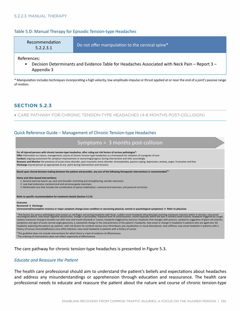
Quick Reference Guide – Management of Chronic Tension‐type Headaches
5.2.2.3 manual ThERaPy
Enabling REcovERy fRom common TRaffic injuRiEs: a focus on ThE injuREd PERson | 130
Table 5.D: Manual Therapy for Episodic Tension-type Headaches
Recommendation 5.2.2.3.1 Do not offer manipulation to the cervical spine*
References: • Decision Determinants and Evidence Table for Headaches Associated with Neck Pain – Report 3 –
Appendix 3
* Manipulation includes techniques incorporating a high velocity, low amplitude impulse or thrust applied at or near the end of a joint’s passive range of motion.
SECTION 5.2.3
care Pathway fOr chrOnic tenSiOn-tyPe headacheS (4-6 MOnthS POSt-cOlliSiOn)
Quick Reference Guide – Management of Chronic Tension-type Headaches
Symptoms > 3 months post‐collision For all injured persons with chronic tension‐type headaches, after ruling out risk factors of serious pathologiesa: Offer information on nature, management, course of chronic tension‐type headaches as a framework for initiation of a program of care Conduct ongoing assessment for symptom improvement or worsening/progress during intervention and refer accordingly Reassess and Monitor the presence of acute stress disorder, post‐traumatic stress disorder, kinesiophobia, passive coping, depression, anxiety, anger, frustration and fear Discharge injured person as appropriate at any point during intervention and recovery
Based upon shared decision making between the patient and provider, any one of the following therapeutic interventions is recommended:b,c
Home and clinic‐based interventions: 1. General exercise (warm‐up, neck and shoulder stretching and strengthening, aerobic exercises); 2. Low load endurance craniocervical and cervicoscapular exercises; 3. Multimodal care that includes the combination of spinal mobilization, craniocervical exercises, and postural correction
Refer to specific recommendation for treatment details (Section 5.2.4)
Outcome: Recovered → Discharge Unrecovered/Incomplete recovery or major symptom change (new condition or worsening physical, mental or psychological symptoms) → Refer to physician
a Risk factors for serious pathologies (also known as red flags): worsening headache with fever; sudden‐onset headache (thunderclap) reaching maximum intensity within 5 minutes; new‐onset neurological deficit; new‐onset cognitive dysfunction; change in personality; impaired level of consciousness; recent (typically within the past 3 months) head trauma; headache triggered by cough, valsalva maneuver (trying to breathe out with nose and mouth blocked) or sneeze; headache triggered by exercise; headache that changes with posture; symptoms suggestive of giant cell arteritis; symptoms and signs of acute narrow‐angle glaucoma; a substantial change in the characteristics of the patient’s headache; new onset or change in headache in patients who are aged over 50; headache wakening the patient up; patients with risk factors for cerebral venous sinus thrombosis; jaw claudication or visual disturbance; neck stiffness; new onset headache in patients with a history of human immunodeficiency virus (HIV) infection; new onset headache in patients with a history of cancer b This guideline does not include interventions for which there is a lack of evidence of effectiveness c The ordering of interventions does not reflect superiority of effectiveness
The care pathway for chronic tension-type headaches is presented in Figure 5.3.
Educate and Reassure the Patient
The health care professional should aim to understand the patient’s beliefs and expectations about headaches and address any misunderstandings or apprehension through education and reassurance. The health care professional needs to educate and reassure the patient about the nature and course of chronic tension-type

5.2.3 caRE PaThway foR chRonic TEnsion-TyPE hEadachEs (4-6 monThs PosT-collision)
Enabling REcovERy fRom common TRaffic injuRiEs: a focus on ThE injuREd PERson | 131
headaches. In the presence of prognostic factors for delayed recovery, the health care professional should discuss them with the patient and adjust their care plan accordingly.
Determine if Ongoing Clinical Care is Necessary
Health care professionals should first determine if the patient requires ongoing clinical care. Patients with chronic tension-type headache may not require ongoing clinical care. Rather, patients can be managed with reassurance and education.
Deliver the Care Plan
Patients with chronic tension-type headaches requiring clinical care should be encouraged to participate in their program of care by remaining active and doing home exercises on a regular basis. Based upon shared decision making between the patient and provider, any one of the following therapeutic interventions is recommended:
• A general exercise program that includes warm-up, neck and shoulder stretching and strengthening, aerobic exercise.
• Low load endurance craniocervical and cervicoscapular exercises. • Multimodal care that includes the combination of spinal mobilization, craniocervical exercises, and
postural correction.
Reassess and Take the Indicated Course of Action
Reassess the patient at every visit to determine if additional care is necessary, or if the condition is worsening.
Patients should be discharged as soon as they report significant improvement or recovery. It is recommended that health care professionals use the self-rated recovery question to measure patient recovery: “How well do you feel you are recovering from your injuries?” The response options include: 1) completely better, 2) much improved, 3) slightly improved, 4) no change, 5) slightly worse, 6) much worse, and 7) worse than ever. Patients reporting to be ‘completely better’ or ‘much improved’ should be considered recovered. Patients who have not recovered should follow the care pathway outlined in this guideline.*
* The use of a valid and reliable condition-specific instrument (e.g., Visual Analogue Scale for headache intensity) is encouraged but should not be used to measure overall recovery.
Patients with worsening symptoms and those who develop new physical, mental or psychological symptoms should be referred to their physician for further evaluation at any time point during their care.
Patients who have not improved significantly or recovered should be referred to their physician for further evaluation.

Care Pathway for the Management of Chronic Tension‐type Headaches
5.2.3 caRE PaThway foR chRonic TEnsion-TyPE hEadachEs (4-6 monThs PosT-collision)
Yes
No
No
Yes
No
Enabling REcovERy fRom common TRaffic injuRiEs: a focus on ThE injuREd PERson | 132
Figure 5.3: Care Pathway for the Management of Chronic Tension-type Headaches
1
Persons injured in a traffic collision with headaches
2
Conduct an appropriate clinical evaluation
3Risk factors for serious pathologies?a
4 Refer to physician
5 Offer information on nature, management, course of
chronic tension‐type headaches as a framework for initiation of a program of care.
6
Is treatment required?
7
Discharge
8 Based upon shared decision making between the patient and provider, any one of the following therapeutic interventions is recommended:b
A. Home and clinic-based interventions: 1)General exercise (warm-up, neck and shoulder stretching and strengthening, aerobic excercises)
2)Low load endurance craniocervical and cervicoscapular exercises;
3)Multimodal care that includes the combination of spinal mobilization, craniocervical exercises, and postural correction
Refer to specific recommendation for treatment details (Section 5.2.4)
9
Is injured person recovered?
10
Discharge
11 1) Incomplete recovery: refer to physician 2) Major symptom change (new or worsening physical, mental or psychological symptoms): proceed to appropriate flowchart or refer to physician
a Risk factors for serious pathologies (also known as red flags): worsening headache with fever; sudden‐onset headache (thunderclap) reaching maximum intensity within 5 minutes; new‐onset neurological deficit; new‐onset cognitive dysfunction; change in personality; impaired level of consciousness; recent (typically within the past 3 months) head trauma; headache triggered by cough, valsalva Maneuver (trying to breathe out with nose and mouth blocked) or sneeze; headache triggered by exercise; headache that changes with posture; symptoms suggestive of giant cell arteritis; symptoms and signs of acute narrow‐angle glaucoma; a substantial change in the characteristics of the patient’s headache; new onset or change in headache in patients who are aged over 50; headache wakening the patient up; patients with risk factors for cerebral venous sinus thrombosis; jaw claudication or visual disturbance; neck stiffness; new onset headache in patients with a history of human immunodeficiency virus (HIV) infection; new onset headache in patients with a history of cancer b This guideline does not include interventions for which there is a lack of evidence of effectiveness c The ordering of interventions does not reflect superiority of effectiveness

Enabling REcovERy fRom common TRaffic injuRiEs: a focus on ThE injuREd PERson | 133
SECTION 5.2.4
Key recOMMendatiOnS fOr the clinical ManageMent Of chrOnic tenSiOn-tyPe headacheS
This section summarizes the key recommendations for the management of chronic tension-type headaches for the period extending from 4 to 6 months post-collision. The wording of recommendations follows the guidance from the National Institute for Health and Care Excellence (NICE). Recommendations beginning with “offer” indicate that, according to the evidence, an intervention is associated with outcomes that were superior to other interventions, placebo/sham, or no intervention. The wording “consider” indicates that an intervention is as effective as another intervention. The wording “do not offer” indicates that, according to the evidence, an intervention does not benefit patients. A detailed explanation of the wording of recommendations is presented in section 2.5.2.4 of this report.
• Provide care in partnership with the patient. Involve the patient in care planning and decision-making.
• Reassure patients about the benign and self-limited nature of their pain.
• Educate patients about the benefits of being actively engaged and participating in their care plan by remaining active and continuing movements.
• Emphasize active rather than passive treatments.
• Deliver time-limited care.
• Do not provide ineffective or experimental treatments.
SECTION 5.2.4.1
Structured Patient educatiOn
Structured patient education aims to enable individuals to make informed decisions about their personal health-related behaviour. Structured education strategies refer to standardized interventions such as scripted discussion, pamphlets or videos. Educational interventions should begin with an assessment of the person’s knowledge of the injury and their health goals. The content of the structured education interventions may include (but is not limited to): education about the nature and course of chronic tension-type headaches; advice on return to activities; instruction of exercise; discussion of expected pain and pain mechanisms; discussion of prognosis; pain coping skills; discussion of workplace ergonomics; and self-care strategies or general health.

5.2.4.1 sTRucTuREd PaTiEnT EducaTion
Enabling REcovERy fRom common TRaffic injuRiEs: a focus on ThE injuREd PERson | 134
Table 5.E: Structured Patient Education for Chronic Tension-type Headaches
Recommendation 5.2.4.1.1
Provide information about the nature, management, and course of chronic tension-type headaches as a framework for the initiation of the program of care*
References: • Decision Determinants and Evidence Table for Headaches Associated with Neck Pain – Report 5 –
Appendix 3
* The structured education program should focus on providing advice to stay active and reassuring the patient by addressing the expectation of recovery.
SECTION 5.2.4.2
exerciSe
Exercise refers to any series of movements with the aim of training or developing the body by routine practice or as physical training to promote good physical health. Exercise therapy includes a wide variety of techniques common to the treatment and rehabilitation of headaches associated with neck pain.
Table 5.F: Exercise for Chronic Tension-type Headaches
Recommendation 5.2.4.2.1
Consider a maximum of 25 sessions over 12 weeks of general exercise (warm-up, neck and shoulder stretching and strengthening, aerobic exercise)*
5.2.4.2.2 Consider a maximum of 8 sessions over 6 weeks of low load endurance craniocervical and cervicocapsular exercises, with resistance**
References: • Decision Determinants and Evidence Table for Headaches Associated with Neck Pain – Report 1 –
Appendix 3
* These exercises should be performed for 1-25 visits over a period of 12 weeks in a supervised clinical environment and at home. ** Low load endurance exercises intend to strengthen the muscles against resistance over time. These exercises should be performed for 1-8 visits over a period of 6 weeks in a supervised clinical environment. Moreover, the exercises should be done twice per day at home.

Enabling REcovERy fRom common TRaffic injuRiEs: a focus on ThE injuREd PERson | 135
SECTION 5.2.4.3
MultiMOdal care
Multimodal care includes at least two distinct therapeutic modalities, provided by one or more health care disciplines. The evidence suggests that three interventions should be included in multimodal care: exercise, spinal mobilization, and postural correction.
Table 5.G: Multimodal Care for Chronic Tension-type Headaches
Recommendation 5.2.4.3.1
Offer a maximum of 9 sessions over 8 weeks of multimodal care that includes spinal mobilization*, craniocervical exercises, and postural correction**
References: • Decision Determinants and Evidence Table for Headaches Associated with Neck Pain – Report 4 –
Appendix 3
* Spinal mobilization refers to techniques incorporating a low velocity and small or large amplitude oscillatory movement, within a joint’s passive range of motion. ** Multimodal care should be performed for 1-9 visits over a period of 8 weeks.

Quick Reference Guide – Management of Cervicogenic Headaches
Enabling REcovERy fRom common TRaffic injuRiEs: a focus on ThE injuREd PERson | 136
SECTION 5.2.5
care Pathway fOr cervicOgenic headacheS (4-6 MOnthS POSt-cOlliSiOn)
Quick Reference Guide – Management of Cervicogenic Headaches
Symptoms > 3 months post‐collision For all injured persons with cervicogenic headaches, after ruling out risk factors of serious pathologiesa : Offer information on nature, management, course of cervicogenic headaches as a framework for initiation of a program of care Conduct ongoing assessment for symptom improvement or worsening/progress during intervention and refer accordingly Reassess and Monitor the presence of acute stress disorder, post‐traumatic stress disorder, kinesiophobia, passive coping, depression, anxiety, anger, frustration and fear Discharge injured person as appropriate at any point during intervention and recovery
Based upon shared decision making between the patient and provider, any one of the following therapeutic interventions is recommended:b,c
Home and clinic‐based interventionsb,c, : 1. Low load endurance craniocervical and cervicoscapular exercises; 2. Manual therapy (manipulation with or without mobilization) to the cervical and thoracic spine
Refer to specific recommendation for treatment details (Section 5.2.6)
Do Not Offer:d
Multimodal program of care that includes the combination of spinal manipulation, spinal mobilization, and low load endurance exercises
Outcome: Recovered → Discharge Unrecovered/Incomplete recovery or major symptom change (new condition or worsening physical, mental or psychological symptoms) → Refer to physician
a Risk factors for serious pathologies (also known as red flags): worsening headache with fever; sudden‐onset headache (thunderclap) reaching maximum intensity within 5 minutes; new‐onset neurological deficit; new‐onset cognitive dysfunction; change in personality; impaired level of consciousness; recent (typically within the past 3 months) head trauma; headache triggered by cough, valsalva maneuver (trying to breathe out with nose and mouth blocked) or sneeze; headache triggered by exercise; headache that changes with posture; symptoms suggestive of giant cell arteritis; symptoms and signs of acute narrow‐angle glaucoma; a substantial change in the characteristics of the patient’s headache; new onset or change in headache in patients who are aged over 50; headache wakening the patient up; patients with risk factors for cerebral venous sinus thrombosis; jaw claudication or visual disturbance; neck stiffness; new onset headache in patients with a history of human immunodeficiency virus (HIV) infection; new onset headache in patients with a history of cancer b This guideline does not include interventions for which there is a lack of evidence of effectiveness c The ordering of interventions does not reflect superiority of effectiveness d Based on evidence of no benefit to patients
The care pathway for cervicogenic headaches is presented in Figure 5.4.
Educate and Reassure the Patient
The health care professional should aim to understand the patient’s beliefs and expectations about headaches and address any misunderstandings or apprehension through education and reassurance. The health care professional needs to educate and reassure the patient about the nature and course of cervicogenic headaches. In the presence of prognostic factors for delayed recovery, the health care professional should discuss them with the patient and adjust their care plan accordingly.
Determine if Ongoing Clinical Care is Necessary
Health care professionals should first determine if the patient requires ongoing clinical care. Patients with cervicogenic headaches may not require ongoing clinical care. Rather, patients can be managed with reassurance and education.
Deliver the Care Plan
Patients with cervicogenic headaches requiring clinical care should be encouraged to participate in their program

5.2.5 caRE PaThway foR cERvicogEnic hEadachEs (4-6 monThs PosT-collision)
Enabling REcovERy fRom common TRaffic injuRiEs: a focus on ThE injuREd PERson | 137
of care by remaining active and doing home exercises on a regular basis. Based upon shared decision making between the patient and provider, any one of the following therapeutic interventions is recommended:
• Low load endurance craniocervical and cervicoscapular exercises • Manual therapy (manipulation with or without mobilization) to the cervical and thoracic spine
Interventions that are not recommended include:
• Manual therapy and low load endurance craniocervical and cervicoscapular exercises are effective on their own. However, combining these interventions does not add benefit to the patients. Therefore, multimodal program of care that combines spinal manipulation, spinal mobilization, and low load endurance exercises should not be offered to these patients.
Discuss the risks and benefits of the care plan with the patient.
Reassess and Take the Indicated Course of Action
Reassess the patient at every visit to determine if additional care is necessary, or if the condition is worsening. Patients should be discharged as soon as they report significant improvement or recovery. It is recommended that health care professionals use the self-rated recovery question to measure patient recovery: “How well do you feel you are recovering from your injuries?” The response options include: 1) completely better, 2) much improved, 3) slightly improved, 4) no change, 5) slightly worse, 6) much worse, and 7) worse than ever. Patients reporting to be ‘completely better’ or ‘much improved’ should be considered recovered. Patients who have not recovered should follow the care pathway outlined in this guideline.*
* The use of a valid and reliable condition-specific instrument (e.g., Visual Analogue Scale for headache intensity) is encouraged but should not be used to measure overall recovery.
Patients with worsening symptoms and those who develop new physical, mental or psychological symptoms should be referred to their physician for further evaluation at any time point during their care.
Patients who have not improved significantly or recovered should be referred to their physician for further evaluation.

Care Pathway for the Management of Cervicogenic Headaches
5.2.5 caRE PaThway foR cERvicogEnic hEadachEs (4-6 monThs PosT-collision)
Yes
No
No
Yes
Yes
No
Enabling REcovERy fRom common TRaffic injuRiEs: a focus on ThE injuREd PERson | 138
Figure 5.4: Care Pathway for the Management of Cervicogenic Headaches 1
Persons injured in a traffic collision with headaches
2
Conduct an appropriate clinical evaluation
3Risk factors for serious pathologies?a
4 Refer to physician
5 Offer information on nature, management, course of
cervicogenic headaches as a framework for initiation of a program of care.
6
Is treatment required? 7
Discharge
8 Based upon shared decision making between the patient and provider, any one of the following therapeutic interventions is recommended'
A. Home and clinic-based interventions: 1)Low load endurance craniocervical and cervicoscapular
exercises; 2)Manual therapy (manipulation with or without
mobilization) to the cervical and thoracic spine
Do not offer:d1)Multimodal program of care that includes the combination of spinal manipulation, spinal mobilization, and low load endurance exercises
Refer to specific recommendation for treatment details (Section 5.2.6)
9
Is injured person recovered?
10
Discharge
11 1) Incomplete recovery: refer to physician 2) Major symptom change (new or worsening physical, mental or psychological symptoms): proceed to appropriate flowchart or refer to physician
a Risk factors for serious pathologies (also known as red flags): worsening headache with fever; sudden‐onset headache (thunderclap) reaching maximum intensity within 5 minutes; new‐onset neurological deficit; new‐onset cognitive dysfunction; change in personality; impaired level of consciousness; recent (typically within the past 3 months) head trauma; headache triggered by cough, valsalva maneuver (trying to breathe out with nose and mouth blocked) or sneeze; headache triggered by exercise; headache that changes with posture; symptoms suggestive of giant cell arteritis; symptoms and signs of acute narrow‐ angle glaucoma; a substantial change in the characteristics of the patient’s headache; new onset or change in headache in patients who are aged over 50; headache wakening the patient up; patients with risk factors for cerebral venous sinus thrombosis; jaw claudication or visual disturbance; neck stiffness; new onset headache in patients with a history of human immunodeficiency virus (HIV) infection; new onset headache in patients with a history of cancer b This guideline does not include interventions for which there is a lack of evidence of effectiveness c The ordering of interventions does not reflect superiority of effectiveness d Based on evidence of no benefit to patients

Enabling REcovERy fRom common TRaffic injuRiEs: a focus on ThE injuREd PERson | 139
SECTION 5.2.6
Key recOMMendatiOnS fOr the ManageMent Of cervicOgenic headacheS
This section summarizes the key recommendations for the management of persistent cervicogenic headaches for the period extending from 4 to 6 months post-collision. The wording of recommendations follows the guidance from the National Institute for Health and Care Excellence (NICE). Recommendations beginning with “offer” indicate that, according to the evidence, an intervention is associated with outcomes that were superior to other interventions, placebo/sham, or no intervention. The wording “consider” indicates that an intervention is as effective as another one. The wording “do not offer” indicates that, according to the evidence, an intervention does not benefit patients. A detailed explanation of the wording of recommendations is presented in section 2.5.2.4 of this report.
• Provide care in partnership with the patient. Involve the patient in care planning and decision-making.
• Reassure patients about the benign and self-limited nature of their pain.
• Educate patients about the benefits of being actively engaged and participating in their care plan by remaining active and continuing movements.
• Emphasize active rather than passive treatments.
• Deliver time-limited care.
• Do not provide ineffective or experimental treatments.
SECTION 5.2.6.1
Structured Patient educatiOn
Structured patient education aims to enable individuals to make informed decisions about their personal health-related behaviour. Structured education strategies refer to standardized interventions such as scripted discussion, pamphlets or videos. Educational interventions should begin with an assessment of the person’s knowledge of the injury and their health goals. The content of the structured education interventions may include (but is not limited to): education about the nature and course of persistent cervicogenic headaches; advice on return to activities; instruction of exercise; discussion of expected pain and pain mechanism; discussion of prognosis; pain coping skills; discussion of workplace ergonomics; and self-care strategies or general health.

5.2.6.1 sTRucTuREd PaTiEnT EducaTion
Enabling REcovERy fRom common TRaffic injuRiEs: a focus on ThE injuREd PERson | 140
Table 5.H: Structured Patient Education for Cervicogenic Headaches
Recommendation 5.2.6.1.1
Provide information about the nature, management, and course of persistent cervicogenic headaches as a framework for the initiation of the program of care*
References: • Decision Determinants and Evidence Table for Headaches Associated with Neck Pain – Report 5 –
Apendix 3
* The structured education program should focus on providing advice to stay active and reassuring the patient by addressing the expectation of recovery.
SECTION 5.2.6.2
exerciSe
Exercise refers to any series of movements with the aim of training or developing the body by routine practice or as physical training to promote good physical health. Exercise therapy includes a wide variety of techniques common to the treatment and rehabilitation of headaches associated with neck pain.
Table 5. I: Exercise for Cervicogenic Headaches
Recommendation 5.2.6.2.1
Consider a maximum of 8 sessions over 6 weeks of low load endurance craniocervical and cervicoscapular exercise, with resistance*
References: • Decision Determinants and Evidence Table for Headaches Associated with Neck Pain – Report 1 –
Appendix 3
* Low load endurance exercises intend to strengthen the muscles against resistance over time. These exercises should be performed for 1-8 visits over a period of 6 weeks in a supervised clinical environment. Moreover, the exercises should be done twice per day at home.
SECTION 5.2.6.3
MultiMOdal care
Multimodal care includes at least two distinct therapeutic modalities, provided by one or more health care disciplines.

5.2.6.3 mulTimodal caRE
Enabling REcovERy fRom common TRaffic injuRiEs: a focus on ThE injuREd PERson | 141
Table 5.J: Multimodal Care for Cervicogenic Headaches
Recommendation 5.2.6.3.1
Do not offer a multimodal program of care that combines spinal manipulation*, spinal mobilization**, and low load endurance exercises***
References: • Decision Determinants and Evidence Table for Headaches Associated with Neck Pain – Report 4 –
Appendix 3
* Spinal manipulation refers to techniques incorporating a high velocity, low amplitude impulse or thrust applied at or near the end of a joint’s passive range of motion. ** Spinal mobilization refers to techniques incorporating a low velocity and small or large amplitude oscillatory movement, within a joint’s passive range of motion. *** Low load endurance exercises intend to strengthen the muscles against resistance over time.
SECTION 5.2.6.4
Manual theraPy
Manual therapy refers to techniques that involve the application of hands-on and/or mechanically assisted treatments, including manipulation, mobilization and traction.
Table 5.K: Manual Therapy for Cervicogenic Headaches
Recommendation 5.2.6.4.1
Consider a maximum of 12 sessions over 7 weeks of manual therapy (manipulation with or without mobilization)* to the cervical and thoracic spine*
References: • Decision Determinants and Evidence Table for Headaches Associated with Neck Pain – Report 3 –
Appendix 3
* Manual therapy refers to techniques that involve the application of hands-on and/or mechanically assisted treatments, including manipulation and mobilization. Manipulation refers to techniques incorporating a high velocity, low amplitude impulse or thrust applied at or near the end of a joint’s passive range of motion. Mobilization refers to techniques incorporating a low velocity and small or large amplitude oscillatory movement, within a joint’s passive range of motion. Manual therapy should be performed for 1-12 visits over a period of 7 weeks.

Enabling REcovERy fRom common TRaffic injuRiEs: a focus on ThE injuREd PERson | 142
Appendix 5.A
internatiOnal claSSificatiOn Of headache diSOrderS SecOnd editiOn (ichd-2) criteria fOr the diagnOSiS Of tenSiOn-tyPe and cervicOgenic headacheS*
* Headache Classification Subcommittee of the International Headache Society. The International Classification of Headache Disorders: 2nd edition. Cephalagia 2004; 24 Suppl 1:9-160.
Headache Type Classification Criteria
Tension-Type ICHD-2 Criteria: Tension-type headaches can be classified as: 1) frequent episodic tension-type head-ache with or without pericranial tenderness; or 2) chronic tension-type headache with or without pericranial tenderness. The presence of pericranial tenderness is indicated by increased tenderness on manual palpation of head and neck muscles, which include, but may not be limited to the following: frontal, temporal, masseter, pterygoid, sternocleidomastoid, splenius and trapezius muscles.
Diagnostic criteria for frequent episodic tension-type headache:
A. At least 10 episodes occurring on ≥1 but <15 days per month for at least 3 months (≥12 and <180 days per year) and fulfilling criteria B-D
B. Headache lasting from 30 minutes to 7 days C. Headache has at least two of the following characteristics:
1. bilateral location 2. pressing, tightening or non-pulsating quality 3. mild or moderate intensity 4. not aggravated by routine physical activity such as walking or climbing stairs
D. Both of the following: 1. no nausea or vomiting (but anorexia may occur) 2. no more than one of photophobia or phonophobia
E. Not attributed to another disorder
Diagnostic criteria for chronic tension-type headache:
A. Headache occurring on ≥15 days per month on average for >3 months (≥180 days per year) and fulfilling criteria B-D
B. Headache lasts hours or may be continuous C. Headache has at least two of the following characteristics:
1. bilateral location 2. pressing, tightening or non-pulsating quality 3. mild or moderate intensity 4. not aggravated by routine physical activity such as walking or climbing stairs.
D. Both of the following: 1. no more than one of photophobia, phonophobia or mild nausea 2. neither moderate or severe nausea nor vomiting
E. Not attributed to another disorder

aPPEndix 5.a inTERnaTional classificaTion of hEadachE disoRdERs sEcond EdiTion (ichd-2) cRiTERia foR ThE diagnosis of TEnsion-TyPE and cERvicogEnic hEadachEs
Enabling REcovERy fRom common TRaffic injuRiEs: a focus on ThE injuREd PERson | 143
Headache Type Classification Criteria
Cervicogenic Diagnostic criteria for cervicogenic headache:
A. Pain, referred from a source in the neck and perceived in one or more regions of the head and/or face, and fulfilling criteria A and Da
B. Clinical, laboratory and/or imaging evidence of a disorder or lesion within the cervical spine or soft tissues of the neck known to be, or generally accepted as, a valid cause of headache
C. Evidence that the pain can be attributed to the neck disorder or lesion based on at least one of the following: 1. demonstration of clinical signs that implicate a source of pain in the neck.b
2. abolition of headache following diagnostic blockade of a cervical structure or its nerve supply using placebo- or other adequate controls.c
D. Pain resolves within 3 months after successful treatment of the causative disorder or lesion.
a. Tumours, fractures, infections and rheumatoid arthritis of the upper cervical spine have not been validated formally as causes of headache, but are nevertheless accepted as valid causes when demonstrated to be so in individual cases. Cervical spondylosis and osteochondritis are NOT accepted as valid causes fulfilling criterion B. When myofascial tender spots are the cause, the headache should be coded under 2. Tension-type headache.
b. Clinical signs acceptable for criterion C1 must have demonstrated reliability and validity. The future task is the identification of such reliable and valid operational tests. Clinical features such as neck pain, focal neck tenderness, history of neck trauma, mechanical exacerbation of pain, unilaterality, coexisting shoulder pain, reduced range of motion in the neck, nuchal onset, nausea, vomiting, photophobia etc are not unique to cervicogenic headache. These may be features of cervicogenic headache, but they do not define the relationship between the disorder and the source of the headache.
c. Abolition of headache means complete relief of headache, indicated by a score of zero on a visual analogue scale (VAS). Nevertheless, acceptable as fulfilling criterion C2 is ≥90% reduction in pain to a level of <5 on a 100-point VAS.

Enabling REcovERy fRom common TRaffic injuRiEs: a focus on ThE injuREd PERson | 144
SECTION 6.0
GUIDELINE FOR THE CLINICAL MANAGEMENT OF SOFT TISSUE DISORDERS OF THE UPPER ExTREMITY

SECTION 6.0
guideline fOr the clinical ManageMent Of SOft tiSSue diSOrderS Of the uPPer extreMity
Enabling REcovERy fRom common TRaffic injuRiEs: a focus on ThE injuREd PERson | 145
6.1 Management of epicondylitis 6.1.1 Care pathway for recent onset epicondylitis (0-3 months post-collision) 6.1.2 Care pathway for persistent epicondylitis (4-6 months post-collision) 6.1.3 Key recommendations for the management of recent onset epicondylitis 6.1.4 Key recommendations for the management of persistent epicondylitis 6.2 Management of shoulder pain 6.2.1 Care pathway for recent onset shoulder pain (0-3 months post-collision) 6.2.2 Care pathway for persistent shoulder pain (4-6 months post-collision) 6.2.3 Key recommendations for the management of recent onset shoulder pain 6.2.4 Key recommendations for the management of persistent shoulder pain 6.3 Management of shoulder pain with calcific tendinitis 6.3.1 Care pathway for shoulder pain with calcific tendinitis 6.3.2 Key recommendations for the management of shoulder pain with calcific tendinitis
This evidence-based guideline establishes the best practice for the clinical management of soft tissue disorders of the upper extremity caused or exacerbated by a motor vehicle collision. This guideline covers recent onset (0-3 months post-collision) and persistent (4-6 months post-collision) epicondylitis (medial and lateral)*, shoulder pain, and shoulder pain with calcific tendinitis; it does not cover disorders that persist for more than 6 months post-collision.
* The evidence used to develop the care pathways for epicondylitis was obtained from randomized controlled trials on the management of lateral epicondylitis. The recommendations were extended to the management of medial epicondylitis because of the patho-anatomic similarities between the two injuries.
In this guideline, the upper extremity is defined as the region that includes the shoulder, arm, elbow, forearm, wrist and hand.
Upper extremity soft tissue disorders refer to grades I and II sprains or strains, bursitis and tendinitis of the upper extremity. Strains and sprains can be classified into three grades, distinguished by the severity of signs and symptoms, and structural disruption (Table 6.A and Table 6.B). This guideline is not indicated for conditions that include the presence of major structural or other pathological causes of the upper extremity such as fractures, dislocations, osteoarthritis, neuropathies, inflammatory disorders, systemic diseases, infections, tumors and grade III sprains/strains.
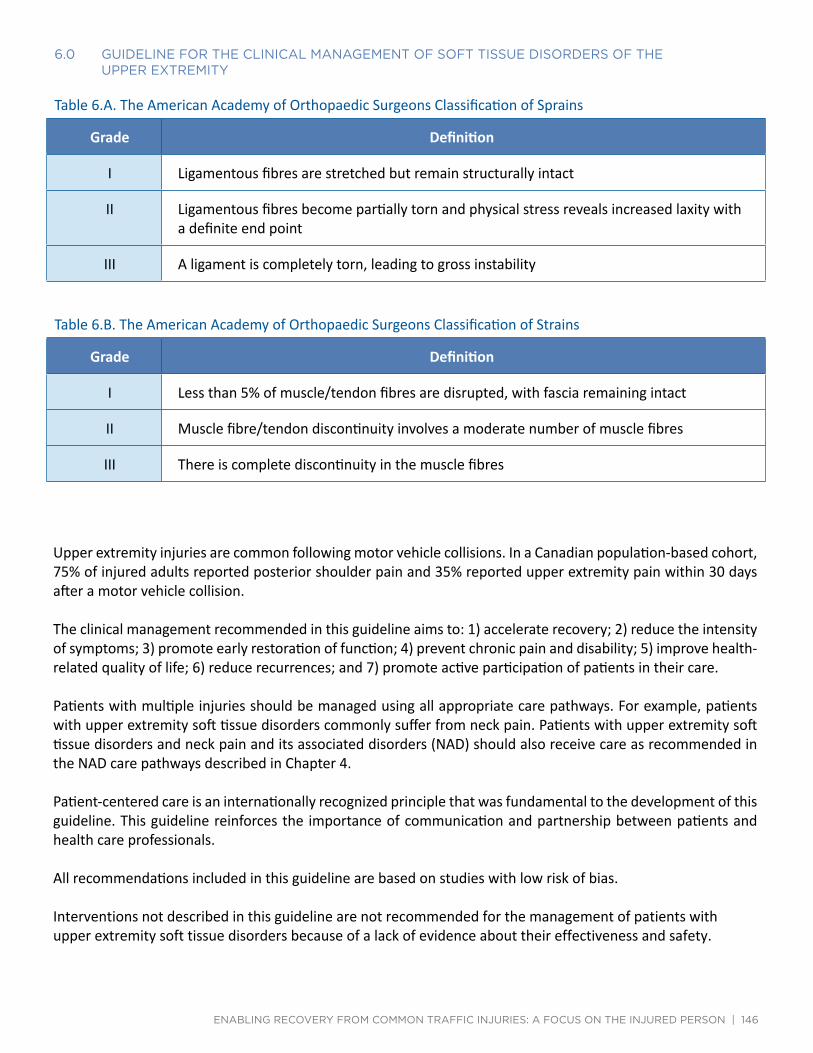
6.0 guidElinE foR ThE clinical managEmEnT of sofT TissuE disoRdERs of ThE uPPER ExTREmiTy
Enabling REcovERy fRom common TRaffic injuRiEs: a focus on ThE injuREd PERson | 146
Table 6.A. The American Academy of Orthopaedic Surgeons Classification of Sprains
Grade Definition
I Ligamentous fibres are stretched but remain structurally intact
II Ligamentous fibres become partially torn and physical stress reveals increased laxity with a definite end point
III A ligament is completely torn, leading to gross instability
Table 6.B. The American Academy of Orthopaedic Surgeons Classification of Strains
Grade Definition
I Less than 5% of muscle/tendon fibres are disrupted, with fascia remaining intact
II Muscle fibre/tendon discontinuity involves a moderate number of muscle fibres
III There is complete discontinuity in the muscle fibres
Upper extremity injuries are common following motor vehicle collisions. In a Canadian population-based cohort, 75% of injured adults reported posterior shoulder pain and 35% reported upper extremity pain within 30 days after a motor vehicle collision.
The clinical management recommended in this guideline aims to: 1) accelerate recovery; 2) reduce the intensity of symptoms; 3) promote early restoration of function; 4) prevent chronic pain and disability; 5) improve health-related quality of life; 6) reduce recurrences; and 7) promote active participation of patients in their care.
Patients with multiple injuries should be managed using all appropriate care pathways. For example, patients with upper extremity soft tissue disorders commonly suffer from neck pain. Patients with upper extremity soft tissue disorders and neck pain and its associated disorders (NAD) should also receive care as recommended in the NAD care pathways described in Chapter 4.
Patient-centered care is an internationally recognized principle that was fundamental to the development of this guideline. This guideline reinforces the importance of communication and partnership between patients and health care professionals.
All recommendations included in this guideline are based on studies with low risk of bias.
Interventions not described in this guideline are not recommended for the management of patients with upper extremity soft tissue disorders because of a lack of evidence about their effectiveness and safety.

6.0 guidElinE foR ThE clinical managEmEnT of sofT TissuE disoRdERs of ThE uPPER ExTREmiTy
Enabling REcovERy fRom common TRaffic injuRiEs: a focus on ThE injuREd PERson | 147
Health care professionals eligible to provide care under this guideline are those defined by the Statutory Accident Benefits Schedules (SABS).
This guideline is organized in three sections. Each section provides evidence-based recommendations for the clinical management of various types and durations of upper extremity soft tissue disorders:
• Section 6.1 - Management of epicondylitis • Section 6.2 - Management of shoulder pain • Section 6.3 - Management of shoulder pain with calcific tendinitis
All recommendations presented in this guideline integrate the:
• Key decision determinants based upon the framework developed by the Ontario Health Technology Advisory Committee (OHTAC);
• Best evidence obtained from a critical review of current scientific literature; and • Qualitative research exploring the experiences of persons treated for traffic injuries in Ontario
All background documents and references are available at http://www.fsco.gov.on.ca

Quick Reference Guide – Management of Epicondylitis
Enabling REcovERy fRom common TRaffic injuRiEs: a focus on ThE injuREd PERson | 148
SECTION 6.1
ManageMent Of ePicOndylitiS
Quick Reference Guide – Management of Epicondylitis
Symptoms ≤ 3 months post-collision Symptoms > 3 months post-collision For all injured persons with epicondylitis: Rule out risk factors for serious pathologiesa
Offer information on nature, management, course of epicondylitis as a framework for initiation of a program of care Conduct ongoing assessment for symptom improvement or worsening/progress during intervention and refer accordingly Discharge injured person as appropriate at any point during intervention and recovery
Based upon shared decision making between the patient and provider, any one of the following therapeutic interventions is recommended: Home and clinic based interventions:b,c,d
1. Elbow brace (lateral epicondylitis) 2. Multimodal care that includes the combination of:
a) Elbow manipulation or mobilization b) Deep tissue massage c) Forearm strengthening and stretching exercise d) Advice to stay active, and ergonomic and activity modification to avoid symptom
provocation
Refer to specific recommendation for treatment details (Section 6.1.3)
Based upon shared decision making between the patient and provider, any one of the following therapeutic interventions is recommended: Home and clinic based interventions:b,c,d
1. Muscle energy technique 2. Myofascial release 3. Elbow brace (lateral epicondylitis) 4. Home-based strengthening and/or stretching exercise 5. Multimodal care that includes the combination of (if not previously given in 1st 3 months
of care): a) Elbow manipulation or mobilization b) Deep tissue massage c) Forearm strengthening and stretching exercise d) Advice to stay active, and ergonomic and activity modification to avoid symptom
provocation
Refer to specific recommendation for treatment details (Section 6.1.4)
Do Not Offer:e
Transcutaneous electrical nerve stimulation (TENS) Elbow brace added to multimodal physical therapy (lateral epicondylitis)
Do Not Offer: e
Transcutaneous electrical nerve stimulation (TENS) Elbow brace added to multimodal physical therapy (lateral epicondylitis)
Outcome: Recovered → Discharge Unrecovered: Incomplete recovery → Initiate persistent protocol
Major symptom change (new or worsening physical, mental or psychological symptoms) → Refer to physician
Outcome: Recovered → Discharge Unrecovered: Incomplete recovery → Refer to physician
Major symptom change (new or worsening physical, mental or psychological symptoms) → Refer to physician
a Risk factors for serious pathologies (also known as red flags): History of significant trauma; history of inflammatory arthritis; history of unexplained, significant weight loss; fever; painful, swollen joints; progressive/widespread neurological symptoms/signs; severe, unremitting, night-time pain; widespread, unexplained pain; unremitting pain when at rest b For medial epicondylitis, special caution should be exercised to protect the ulnar nerve c This guideline does not include interventions for which there is a lack of evidence of effectiveness d The ordering of interventions does not reflect superiority of effectiveness e Based on evidence of no benefit to patients
SECTION 6.1.1
care Pathway fOr recent OnSet ePicOndylitiS (0 - 3 MOnthS POSt-cOlliSiOn)
The care pathway is presented in Figure 6.1
At initial contact, health care professionals should educate and reassure the patient that epicondylitis will resolve within a few months of symptom onset. Patients greatly improve their recovery by actively participating in their care. Clinical care aims to accelerate recovery by reducing pain and improving function. The care pathway recommended for the first 3 months of care for epicondylitis is described below.*
* Special caution should be exercised to protect the ulnar nerve when treating medial epicondylitis

6.1.1 caRE PaThway foR REcEnT onsET EPicondyliTis (0 - 3 monThs PosT-collision)
Enabling REcovERy fRom common TRaffic injuRiEs: a focus on ThE injuREd PERson | 149
Assess the Patient with Epicondylitis
Conduct an appropriate clinical evaluation to rule out major structural or other pathologies as the cause of the symptoms. The presence of a risk factor for serious pathologies (also known as red flags) identified during the history and examination warrants further investigation and referral to the appropriate health care professional. However, once pathology has been ruled out, the patient should be treated according to the epicondylitis care pathway.
Table 6.C Risk factors of serious pathology (red flags) for epicondylitis
Risk factors of serious pathology identified during history or physical examination*
* This list of risk factors of serious pathology was informed by the following clinical practice guideline: Accident Compensation Corporation (ACC). Distal upper limb. Guidelines for management of some common musculoskeletal disorders. New Zealand; 2009. Available from: http://www.acc.co.nz/PRD_EXT_CSMP/groups/external_communications/documents/guide/prd_ctrb112931.pdf [Last accessed 14 Dec 2014]
• History of significant trauma • History of inflammatory arthritis • History of unexplained, significant weight loss • Fever • Painful, swollen joints • Progressive/widespread neurological symptoms/signs • Severe, unremitting night-time pain • Widespread, unexplained pain • Unremitting pain when at rest
Patients who also have neck pain and associated disorders or other injuries should be managed using the appropriate care pathways.
Patients with multiple injuries should be managed using all appropriate care pathways.
Educate and Reassure the Patient
A patient-centred care plan should be developed in partnership with the patient. It is important that the health care professional reassures and explains to patients that most individuals recover spontaneously from epicondylitis. Patients need to be reassured about the benign and self-limited nature of epicondylitis. Health care professionals also need to reassure patients that there are no major structural or progressive pathologies (e.g., dislocations, fractures or infection) in the elbow.
Determine if Ongoing Clinical Care is Necessary
Health care professionals should first determine if the patient requires clinical care.

6.1.1 caRE PaThway foR REcEnT onsET EPicondyliTis (0 - 3 monThs PosT-collision)
Enabling REcovERy fRom common TRaffic injuRiEs: a focus on ThE injuREd PERson | 150
Deliver the Care Plan for Recent Onset Epicondylitis (0-3 months post-collision)
Patients who require clinical care should be encouraged to actively participate in their care by staying active.
Health care professionals should discuss treatment options with their patients and, through a process of shared decision making, determine which therapeutic options they wish to pursue. Based upon shared decision making between the patient and provider, any one of the following therapeutic interventions is recommended:
• Elbow brace (lateral epicondylitis) • Multimodal care that includes the combination of:
i. Elbow manipulation or mobilization ii. Deep tissue massage iii. Forearm strengthening and stretching exercise iv. Advice to stay active, and ergonomic and activity modification to avoid symptom provocation
Interventions that are not recommended include:
• Transcutaneous electrical nerve stimulation (TENS) • Elbow brace added to multimodal physical therapy (lateral epicondylitis)
Discuss the risks and benefits of the care plan with the patient.
Reassess and Take the Indicated Course of Action
Reassess the patient at every visit to determine if additional care is necessary, or if the condition is worsening.
Patients should be discharged as soon as they report significant improvement or recovery. It is recommended that health care professionals use the self-rated recovery question to measure patient recovery: “How well do you feel you are recovering from your injuries?” The response options include: 1) completely better, 2) much improved, 3) slightly improved, 4) no change, 5) slightly worse, 6) much worse, and 7) worse than ever. Patients reporting to be ‘completely better’ or ‘much improved’ should be considered recovered. Patients who have not recovered should follow the care pathway outlined in this guideline*.
* The use of a valid and reliable condition-specific instrument (e.g. Disabilities of the Arm, Shoulder and Hand (DASH)) is encouraged but should not be used to measure overall recovery.
Patients with worsening of symptoms and those who develop new physical, mental or psychological symptoms should be referred to a physician for further evaluation at any time point during their care.
Patients who have not significantly improved or recovered within the first 3 months after the injury should enter the care pathway for persistent epicondylitis described in section 6.1.2.

Enabling REcovERy fRom common TRaffic injuRiEs: a focus on ThE injuREd PERson | 151
SECTION 6.1.2
care Pathway fOr PerSiStent ePicOndylitiS (4 - 6 MOnthS POSt-cOlliSiOn)
The care pathway is presented in Figure 6.1
Patients who still experience symptoms and disability more than 3 months after the injury may benefit from receiving additional clinical care. The primary goals of clinical care are to promote recovery by reducing symptoms and return patients to their normal activities of daily living.*
* Special caution should be exercised to protect the ulnar nerve when treating medial epicondylitis.
Assess the Patient with Epicondylitis
Conduct an appropriate clinical evaluation to rule out major structural or other pathologies as the cause of symptoms. The presence of a risk factor for serious pathologies (also known as red flags) identified during the history and examination warrants further investigation and referral to the appropriate health care professional. However, once pathology has been ruled out, the patient should be treated according to the epicondylitis care pathway.
Table 6.C Risk factors of serious pathology (red flags) for epicondylitis
Risk factors of serious pathology identified during history or physical examination+
+ This list of risk factors of serious pathology was informed by the following clinical practice guideline: Accident Compensation Corporation (ACC). Distal upper limb. Guidelines for management of some common musculoskeletal disorders. New Zealand; 2009. Available from: http://www.acc. co.nz/PRD_EXT_CSMP/groups/external_communications/documents/guide/prd_ctrb112931.pdf [Last accessed 14 Dec 2014]
• History of significant trauma • History of inflammatory arthritis • History of unexplained, significant weight loss • Fever • Painful, swollen joints • Progressive/widespread neurological symptoms/signs • Severe, unremitting night-time pain • Widespread, unexplained pain • Unremitting pain when at rest
Patients who also have neck pain and associated disorders or other injuries should be managed using the appropriate care pathways.
Patients with multiple injuries should be managed using all appropriate care pathways.
Educate and Reassure the Patient
The health care professional should aim to understand the patient’s beliefs and expectations about epicondylitis and address any misunderstandings or apprehension through education and reassurance. The health care

6.1.2 caRE PaThway foR PERsisTEnT EPicondyliTis (4 - 6 monThs PosT-collision)
Enabling REcovERy fRom common TRaffic injuRiEs: a focus on ThE injuREd PERson | 152
professional needs to educate and reassure the patient about the benign and self-limited nature of epicondylitis and reinforce the importance of maintaining activities of daily living.
Deliver the Care Plan for Persistent Epicondylitis (4-6 months post-collision)
The goal of the care plan is to provide clinical interventions that promote resolution of symptoms and restoration of function. Patients requiring clinical care should be encouraged to participate in their program of care by remaining active.
Health care professionals should discuss treatment options with their patients and, through a process of shared decision making, determine which therapeutic options they wish to pursue. Based upon shared decision making between the patient and provider, any one of the following therapeutic interventions is recommended:
• Muscle energy technique • Myofascial release • Elbow brace (lateral epicondylitis) • Home-based strengthening and/or stretching exercise • Multimodal care that includes the combination of (if not previously given in 1st 3 months of care):
i. Elbow manipulation or mobilization ii. Deep tissue massage iii. Forearm strengthening and stretching exercise iv. Advice to stay active, and ergonomic and activity modification to avoid symptom provocation
Interventions that are not recommended include: • Transcutaneous electrical nerve stimulation (TENS) • Elbow brace added to multimodal physical therapy (lateral epicondylitis)
Discuss the risks and benefits of the care plan with the patient.
Reassess and Take the Indicated Course of Action
Reassess the patient at every visit to determine if additional care is necessary, or if the condition is worsening.
Patients should be discharged as soon as they report significant improvement or recovery. It is recommended that health care professionals use the self-rated recovery question to measure patient recovery: “How well do you feel you are recovering from your injuries?” The response options include: 1) completely better, 2) much improved, 3) slightly improved, 4) no change, 5) slightly worse, 6) much worse, and 7) worse than ever. Patients reporting to be ‘completely better’ or ‘much improved’ should be considered recovered. Patients who have not recovered should follow the care pathway outlined in this guideline*.
* The use of a valid and reliable condition-specific instrument (e.g. Disabilities of the Arm, Shoulder and Hand (DASH)) is encouraged but should not be used to measure overall recovery.
Patients with worsening of symptoms and those who develop new physical, mental or psychological symptoms should be referred to a physician for further evaluation at any time point during their care.

6.1.2 caRE PaThway foR PERsisTEnT EPicondyliTis (4 - 6 monThs PosT-collision)
Enabling REcovERy fRom common TRaffic injuRiEs: a focus on ThE injuREd PERson | 153
Patients who have not improved significantly or recovered should be referred to their physician for further evaluation.
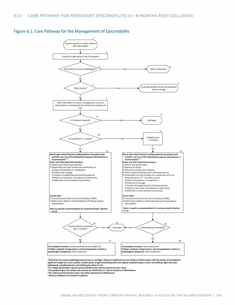
6.1.2 caRE PaThway foR PERsisTEnT EPicondyliTis (4 - 6 monThs PosT-collision)
Yes
No
Yes
No
No
Yes
No
Yes Yes
Yes Yes
NoNo
Enabling REcovERy fRom common TRaffic injuRiEs: a focus on ThE injuREd PERson | 154
Figure 6.1: Care Pathway for the Management of Epicondylitis 1
Persons injured in a traffic collision with epicondylitis
2
Conduct an appropriate clinical evaluation
3 Risk factors for serious pathologies?a
4 Refer to physician
5 Other injuries?
6 Go to appropriate clinical care pathways
and co-manage
7
Offer information on nature, management, course of epicondylitis as a framework for initiation of a program of
care.
8
Is treatment required?
9
Discharge
10
Are symptoms ≤3 months?
11
Symptoms are > 3 months.
12 Based upon shared decision making between the patient and
provider, any one of the following therapeutic interventions is recommended:b,c,d
Home and clinic-based interventions: 1) Elbow brace (lateral epicondylitis) 2) Multimodal care that includes the combination of:
a) Elbow manipulation or mobilization b) Deep tissue massage c) Forearm strengthening and stretching exercise d) Advice to stay active, and ergonomic and activity modification to avoid symptom provocation
Do not offer:e
1) Transcutaneous electrical nerve stimulation (TENS) 2) Elbow brace added to multimodal physical therapy (lateral
epicondylitis)
Refer to specific recommendation for treatment details (Section 6.1.3)
13 Based upon shared decision making between the patient and
provider, anyone of thefollowing therapeutk interventions is recommended:'444
Home and clinic-based interventions: 1)Musde energy technique 2)Myofascial release 3)Elbow brace (lateral epicondylitis) 4)Home-based stretching and/or stretching exerdse 5)Multimodal care that includes the combination of (if not
previously given in 1°3 months of care): a)Elbow manipulation or mobilization b) Deep tissue massage c)Forearm strengthening and stretching exercise d)Advice to stay active, and ergonomic and activity modification to avoid symptom provocation
Do not offer:` 1)Transcutaneous electrical nerve stimulation (TENS) 2)Elbow brace added to muhimodal physical therapy (lateral
epicondylitis)
Refer to specific recommendation for treatment details (Section 6.1.4)
14 Is injured person recovered
after 3 months?
16
Is injured person recovered?
15
Discharge
17
1) Incomplete recovery: Initiate persistent protocol (Box 13) 2) Major symptom change (new or worsening physical, mental or psychological symptoms): refer to physician
18
1) Incomplete recovery: refer to physician 2) Major symptom change (new or worsening physical, mental or psychological symptoms): refer to physician
a Risk factors for serious pathologies (also known as red flags): History of significant trauma; history of inflammatory arthritis; history of unexplained significant weight loss; fever; painful, swollen joints; progressive/widespread neurological symptoms/signs; severe, unremitting, night-time pain; widespread, unexplained pain; unremitting pain when at rest b For medial epicondylitis, special caution should be exercised to protect the ulnar nerve c This guideline does not include interventions for which there is a lack of evidence of effectiveness d The ordering of interventions does not reflect superiority of effectiveness e Based on evidence of no benefit to patients

Enabling REcovERy fRom common TRaffic injuRiEs: a focus on ThE injuREd PERson | 155
SECTION 6.1.3
Key recOMMendatiOnS fOr the ManageMent Of recent OnSet ePicOndylitiS
This section summarizes the key recommendations for the management of recent onset epicondylitis for the period extending from 0 to 3 months post-collision. The wording of recommendations follows the guidance from the National Institute for Health and Care Excellence (NICE). Recommendations beginning with “offer” indicate that, according to the evidence, an intervention is associated with outcomes that were superior to other interventions, placebo/sham, or no intervention. The wording “consider” indicates that an intervention is as effective as another one. The wording “do not offer” indicates that, according to the evidence, an intervention does not benefit patients. A detailed explanation of the wording of recommendations is presented in section 2.5.2.4 of this report.
• Provide care in partnership with the patient. Involve the patient in care planning and decision-making.
• Reassure patients about the benign and self-limited nature of their pain.
• Educate patients about the benefits of being actively engaged and participating in their care plan by remaining active and continuing movement.
• Emphasize active rather than passive treatments.
• Deliver time-limited care.
• Do not provide ineffective or experimental treatments.
SECTION 6.1.3.1
MultiMOdal care
Multimodal care includes at least two distinct therapeutic modalities, provided by one or more health care disciplines.
Table 6.D: Multimodal Care for Recent Onset Epicondylitis
Recommendation 6.1.3.1.1
Consider a maximum of 10 visits over 5 weeks of multimodal care that includes manual therapy*, deep tissue massage**, exercise*** and education****.
References: • Decision Determinants and Evidence Table for Upper Extremity Injuries – Report 5 – Appendix 4
* Mobilization: a) Sustained Lateral Glide With Pain-Free Grip: a sustained lateral glide across the elbow joint while the patient performs a gripping action; b) Sustained Lateral Glide with Movement: If there is also reproduction of pain with elbow movement, perform the lateral glide while the movement is repeated; c) Sustained Posterior-Anterior Glide with Pain-Free Grip: In the event (a) and (b) are not effective, attempt a sustained posteror-anterior glide of the radio-humeral joint. Manipulation: patient seated with upper extremity in 90 degrees of abduction with internal rotation so that the olecranon faces up. Stabilize wrist
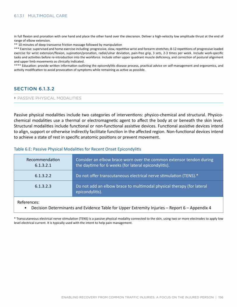
6.1.3.1 mulTimodal caRE
Enabling REcovERy fRom common TRaffic injuRiEs: a focus on ThE injuREd PERson | 156
in full flexion and pronation with one hand and place the other hand over the olecranon. Deliver a high-velocity low amplitude thrust at the end of range of elbow extension. ** 10 minutes of deep transverse friction massage followed by manipulation *** Exercise: supervised and home exercise including: progressive, slow, repetitive wrist and forearm stretches; 8-12 repetitions of progressive loaded exercise for wrist extension/flexion, supination/pronation, radial/ulnar deviation, pain-free grip, 3 sets, 2-3 times per week. Include work-specific tasks and activities before re-introduction into the workforce. Include other upper quadrant muscle deficiency, and correction of postural alignment and upper limb movements as clinically indicated. **** Education: provide written information outlining the epicondylitis disease process, practical advice on self-management and ergonomics, and activity modification to avoid provocation of symptoms while remaining as active as possible.
SECTION 6.1.3.2
PaSSive PhySical MOdalitieS
Passive physical modalities include two categories of interventions: physico-chemical and structural. Physico-chemical modalities use a thermal or electromagnetic agent to affect the body at or beneath the skin level. Structural modalities include functional or non-functional assistive devices. Functional assistive devices intend to align, support or otherwise indirectly facilitate function in the affected region. Non-functional devices intend to achieve a state of rest in specific anatomic positions or prevent movement.
Table 6.E: Passive Physical Modalities for Recent Onset Epicondylitis
Recommendation 6.1.3.2.1
Consider an elbow brace worn over the common extensor tendon during the daytime for 6 weeks (for lateral epicondylitis).
6.1.3.2.2 Do not offer transcutaneous electrical nerve stimulation (TENS).*
6.1.3.2.3 Do not add an elbow brace to multimodal physical therapy (for lateral epicondylitis).
References: • Decision Determinants and Evidence Table for Upper Extremity Injuries – Report 6 – Appendix 4
* Transcutaneous electrical nerve stimulation (TENS) is a passive physical modality connected to the skin, using two or more electrodes to apply low level electrical current. It is typically used with the intent to help pain management.

Enabling REcovERy fRom common TRaffic injuRiEs: a focus on ThE injuREd PERson | 157
SECTION 6.1.4
Key recOMMendatiOnS fOr the ManageMent Of PerSiStent ePicOndylitiS
This section summarizes the key recommendations for the management of persistent epicondylitis for the period extending from 4 to 6 months post-collision. The wording of recommendations follows the guidance from the National Institute for Health and Care Excellence (NICE). Recommendations beginning with “offer” indicate that, according to the evidence, an intervention is associated with outcomes that were superior to other interventions, placebo/sham, or no intervention. The wording “consider” indicates that an intervention is as effective as another one. The wording “do not offer” indicates that, according to the evidence, an intervention does not benefit patients. A detailed explanation of the wording of recommendations is presented in section 2.5.2.4 of this report.
• Provide care in partnership with the patient. Involve the patient in care planning and decision-making.
• Reassure patients about the benign and self-limited nature of their pain.
• Educate patients about the benefits of being actively engaged and participating in their care plan by remaining active and continuing movement.
• Emphasize active rather than passive treatments.
• Deliver time-limited care.
• Do not provide ineffective or experimental treatments.
SECTION 6.1.4.1
exerciSe
Exercise refers to any series of movements with the aim of training or developing the body by routine practice or as physical training to promote good physical health. Exercise therapy includes a wide variety of techniques common to the treatment and rehabilitation of elbow pain.
Table 6.F: Exercise for Persistent Epicondylitis
Recommendation 6.1.4.1.1
Consider home-based stretching and/or strengthening exercise.
The program should consist of 15 repetitions of progressive incremental loading exercises for forearm extensors, 3 sets daily for 3 months; and/or 3 repetitions of wrist extensor stretches, twice daily for 6 weeks.
References: • Decision Determinants and Evidence Table for Upper Extremity Injuries – Report 4 – Appendix 4

Enabling REcovERy fRom common TRaffic injuRiEs: a focus on ThE injuREd PERson | 158
SECTION 6.1.4.2
MultiMOdal care
Multimodal care includes at least two distinct therapeutic modalities, provided by one or more health care disciplines.
Table 6.G: Multimodal Care for Persistent Epicondylitis
Recommendation 6.1.4.2.1
Consider a maximum of 10 visits over 5 weeks of multimodal care that includes manual therapy*, deep tissue massage**, exercise*** and education****.
References: • Decision Determinants and Evidence Table for Upper Extremity Injuries – Report 5 – Appendix 4
* Mobilization: a) Sustained Lateral Glide With Pain-Free Grip: a sustained lateral glide across the elbow joint while the patient performs a gripping action; b) Sustained Lateral Glide with Movement: If there is also reproduction of pain with elbow movement, perform the lateral glide while the movement is repeated; c) Sustained Posterior-Anterior Glide with Pain-Free Grip: In the event (a) and (b) are not effective, attempt a sustained posterior-anterior glide of the radio-humeral joint. Manipulation: patient seated with upper extremity in 90 degrees of abduction with internal rotation so that the olecranon faces up. Stabilize wrist in full flexion and pronation with one hand and place the other hand over the olecranon. Deliver a high-velocity low amplitude thrust at the end or range of elbow extension. ** 10 minutes of deep transverse friction massage followed by manipulation *** Exercise: supervised and home exercise including: progressive, slow, repetitive wrist and forearm stretches; 8-12 repetitions of progressive loaded exercise for wrist extension/flexion, supination/pronation, radial/ulnar deviation, pain-free grip, 3 sets, 2-3 times per week. Include work-specific tasks and activities before re-introduction into the workforce. Include other upper quadrant muscle deficiency, and correction of postural alignment and upper limb movements as clinically indicated. **** Education: provide written information outlining the epicondylitis disease process, practical advice on self-management and ergonomics, and activity modification to avoid provocation of symptoms while remaining as active as possible.
SECTION 6.1.4.3
SOft tiSSue theraPy
Soft tissue therapy is a mechanical therapy in which muscles, tendons, and ligaments are passively pressed or kneaded by hand or with mechanical devices. It includes relaxation massage, clinical massage, movement re-education and energy work.

6.1.4.3 sofT TissuE ThERaPy
Enabling REcovERy fRom common TRaffic injuRiEs: a focus on ThE injuREd PERson | 159
Table 6.H: Soft Tissue Therapy for Persistent Epicondylitis
Recommendation 6.1.4.3.1
Offer muscle energy technique.*
The program should include 5 repetitions (twice per week for 4 weeks) of resisted forearm pronation from an initial maximally supinated position to passively stretch the pronator muscles.
6.1.4.3.2 Offer myofascial release** to the forearm for a maximum of 12 sessions over 4 weeks.
References: • Decision Determinants and Evidence Table for Upper Extremity Injuries – Report 10 – Appendix 4
* Muscle energy technique is a soft tissue therapy performed by a health care professional that involves a stretch to the muscle after the muscle was contracted against resistance. ** Myofascial release is a hands-on technique that involves applying gentle sustained pressure into the myofascial connective tissue.
SECTION 6.1.4.4
PaSSive PhySical MOdalitieS
Passive physical modalities include two categories of interventions: physico-chemical and structural. Physico-chemical modalities use a thermal or electromagnetic agent to affect the body at or beneath the skin level. Structural modalities include functional or non-functional assistive devices. Functional assistive devices intend to align, support or otherwise indirectly facilitate function in the affected region. Non-functional devices intend to achieve a state of rest in specific anatomic positions or prevent movement.
Table 6.I: Passive Physical Modalities for Persistent Epicondylitis
Recommendation 6.1.4.4.1
Consider an elbow brace worn over the common extensor tendon during the daytime for 6 weeks (for lateral epicondylitis).
6.1.4.3.2 Do not offer transcutaneous electrical nerve stimulation (TENS).*
6.1.4.3.3 Do not add an elbow brace to multimodal physical therapy (for lateral epicondylitis).
References: • Decision Determinants and Evidence Table for Upper Extremity Injuries – Report 6 – Appendix 4
* Transcutaneous electrical nerve stimulation (TENS) is a passive physical modality connected to the skin, using two or more electrodes to apply low level electrical current. It is typically used with the intent to help pain management.

Quick Reference Guide – Management of Shoulder Pain
Enabling REcovERy fRom common TRaffic injuRiEs: a focus on ThE injuREd PERson | 160
SECTION 6.2
ManageMent Of ShOulder Pain
Soft tissue disorders of the shoulder managed using this guideline include grades I and II sprains or strains, tendinitis, bursitis and impingement syndrome affecting the gleno-humeral and acromio-clavicular joints.
Quick Reference Guide – Management of Shoulder Pain
Symptoms ≤ 3 months post-collision Symptoms > 3 months post-collision For all injured persons with shoulder pain: Rule out risk factors for serious pathologiesa
Offer information on nature, management, course of shoulder pain as a framework for initiation of a program of care Conduct ongoing assessment for symptom improvement or progression during intervention and refer accordingly Discharge injured person as appropriate at any point during intervention and recovery
Based upon shared decision making between the patient and provider, any one of the following therapeutic interventions is recommended:
Home and clinic based interventions:b,c
1. Low-level laser therapy for short-term pain reduction 2. Spinal manipulation and mobilization as an adjunct to usual care for shoulder pain with
associated pain or restricted movement of the cervico-thoracic spine 3. Multimodal care that includes the combination of:
a) Heat/Cold b) Joint mobilization c) Range of motion exercise
Refer to specific recommendation for treatment details (Section 6.2.3)
Based upon shared decision making between the patient and provider, any one of the following therapeutic interventions is recommended:
Home and clinic based interventions:b,c
1. Low-level laser therapy for short-term pain reduction 2. Strengthening and stretching exercises 3. Usual GP care (information, recommendation, and pain contingent medical or
pharmaceutical therapy) 4. Spinal manipulation and mobilization as an adjunct to usual care for shoulder pain with
associated pain or restricted movement of the cervico-thoracic spine 5. Supervised combined strengthening and stretching exercises 6. Multimodal care that includes the combination of (if not previously given in 1st 3 months
of care): a) Heat/Cold b) Joint mobilization c) Range of motion exercise
Refer to specific recommendation for treatment details (Section 6.2.4)
Do Not Offer:d
Diacutaneous fibrolysis Ultrasound Interferential current therapy
Do Not Offer: d
Diacutaneous fibrolysis Shock-wave therapy Cervical mobilizations Multimodal care that includes the combination of exercise, mobilization, taping,
psychological interventions and massage Ultrasound Interferential current therapy
Outcome: Recovered → Discharge Unrecovered: Incomplete recovery → Initiate persistent protocol
Major symptom change (new or worsening physical, mental or psychological symptoms) → Refer to physician
Outcome: Recovered → Discharge Unrecovered: Incomplete recovery → Refer to physician
Major symptom change (new or worsening physical, mental or psychological symptoms) → Refer to physician
a Risk factors for serious pathologies (also known as red flags): Unexplained deformity or swelling or erythema of the skin; significant weakness not due to pain; past history of malignancy; suspected malignancy (e.g., weight loss or loss of appetite); fever/chills/malaise; significant unexplained sensory/motor deficits; pulmonary or vascular compromise; inability to perform any movements; pain at rest b This guideline does not include interventions for which there is a lack of evidence of effectiveness c The ordering of interventions does not reflect superiority of effectiveness d Based on evidence of no benefit to patients

Enabling REcovERy fRom common TRaffic injuRiEs: a focus on ThE injuREd PERson | 161
SECTION 6.2.1
care Pathway fOr recent OnSet ShOulder Pain (0 - 3 MOnthS POSt-cOlliSiOn)
The care pathway is presented in Figure 6.2
At initial contact, health care professionals should educate and reassure the patient that shoulder pain will resolve within a few months of symptoms onset. Patients greatly improve their recovery by actively participating in their care. Clinical care aims to accelerate recovery by reducing pain and improving function. The care pathway recommended for the first 3 months of care for shoulder pain is described below.
Assess the Patient with Shoulder Pain
Conduct an appropriate clinical evaluation to rule out major structural or other pathologies as the cause of the symptoms. The presence of a risk factor for serious pathologies (also known as red flags) identified during the history and examination warrants further investigation and referral to the appropriate health care professional. However, once pathology has been ruled out, the patient should be treated according to the shoulder pain care pathway.
Table 6.J Risk factors of serious pathology (red flags) for shoulder pain
Risk factors of serious pathology identified during history or physical examination+
* This list of risk factors of serious pathology was informed by the following clinical practice guidelines: Hopman K, Krahe L, Lukersmith S, McColl AR, Vine K. Clinical practice guidelines for the management of rotator cuff syndrome in the workplace. Port Macquarie (Australia): University of New South Wales; 2013. Washington State Department of Labor and Industries. Conservative care options for work-related mechanical shoulder conditions. Olympia (WA): Washington State Department of Labor and Industries; 2014 Apr 17.
• Unexplained deformity or swelling or erythema of the skin • Significant weakness not due to pain • Past history of malignancy • Suspected malignancy (e.g., weight loss or loss of appetite) • Fever/chills/malaise • Significant unexplained sensory/motor deficits • Pulmonary or vascular compromise • Inability to perform any movements • Pain at rest
Patients who also have neck pain and associated disorders or other injuries should be managed using the appropriate care pathways.
Patients with multiple injuries should be managed using all appropriate care pathways.
Educate and Reassure the Patient
A patient-centred care plan should be developed in partnership with the patient. It is important that the health

6.2.1 caRE PaThway foR REcEnT onsET shouldER Pain (0 - 3 monThs PosT-collision)
Enabling REcovERy fRom common TRaffic injuRiEs: a focus on ThE injuREd PERson | 162
care professional reassures and explains to patients that most individuals recover spontaneously from shoulder pain. Patients need to be reassured about the benign and self-limited nature of shoulder pain. Health care professionals also need to reassure patients if there are no major structural or progressive pathologies (e.g., dislocations, fractures or infection) in the shoulder. Risks and benefits of the care plan should be discussed with the patient.
Determine if Ongoing Clinical Care is Necessary
Health care professionals should first determine if the patient requires clinical care.
Deliver the Care Plan for Recent Onset Shoulder Pain (0-3 months post-collision)
Patients who require clinical care should be encouraged to actively participate in their care.
Health care professionals should discuss treatment options with their patients and, through a process of shared decision making, determine which therapeutic options they wish to pursue. Based upon shared decision making between the patient and provider, any one of the following therapeutic interventions is recommended:
• Low-level laser therapy for short-term pain reduction • Spinal manipulation and mobilization as an adjunct to usual care for shoulder pain with associated pain
or restricted movement of the cervico-thoracic spine • Multimodal care that includes the combination of:
i. Heat/Cold ii. Joint mobilization iii. Range of motion exercise
Interventions that are not recommended include:
• Diacutaneous fibrolysis • Ultrasound • Interferential current therapy
Discuss the risks and benefits of the care plan with the patient.
Reassess and Take the Indicated Course of Action
Reassess the patient at every visit to determine if additional care is necessary, or if the condition is worsening.
Patients should be discharged as soon as they report significant improvement or recovery. It is recommended that health care professionals use the self-rated recovery question to measure patient recovery: “How well do you feel you are recovering from your injuries?” The response options include: 1) completely better, 2) much improved, 3) slightly improved, 4) no change, 5) slightly worse, 6) much worse, and 7) worse than ever. Patients reporting to be ‘completely better’ or ‘much improved’ should be considered recovered*. Patients who have not
* The use of a valid and reliable condition-specific instrument (e.g. Disabilities of the Arm, Shoulder and Hand (DASH)) is encouraged but should not be used to measure overall recovery.

6.2.1 caRE PaThway foR REcEnT onsET shouldER Pain (0 - 3 monThs PosT-collision)
Enabling REcovERy fRom common TRaffic injuRiEs: a focus on ThE injuREd PERson | 163
recovered should follow the care pathway outlined in this guideline.
Patients with worsening of symptoms and those who develop new physical, mental or psychological symptoms should be referred to a physician for further evaluation at any time point during their care.
Patients who have not significantly improved or recovered within the first 3 months after the injury should enter the care pathway for persistent shoulder pain described in section 6.2.2.
SECTION 6.2.2
care Pathway fOr PerSiStent ShOulder Pain (4 - 6 MOnthS POSt-cOlliSiOn)
The care pathway is presented in Figure 6.2
Patients who still experience symptoms and disability more than 3 months after the injury may benefit from receiving additional clinical care. The primary goals of clinical care are to promote recovery by reducing symptoms and return patients to their normal activities of daily living.
Assess the Patient with Shoulder Pain
Conduct an appropriate clinical evaluation to rule out major structural or other pathologies as the cause of symptoms. The presence of a risk factor for serious pathologies (also known as red flags) identified during the history and examination warrants further investigation and referral to the appropriate health care professional. However, once pathology has been ruled out, the patient should be treated according to the shoulder pain care pathway.
Patients who also have neck pain and associated disorders or other injuries should be managed using the appropriate care pathways.
Patients with multiple injuries should be managed using all appropriate care pathways.
Educate and Reassure the Patient
The health care professional should aim to understand the patient’s beliefs and expectations about shoulder pain and address any misunderstandings or apprehension through education and reassurance. The health care professional needs to educate and reassure the patient about the benign and self-limited nature of shoulder pain and reinforce the importance of maintaining activities of daily living.
Deliver the Care Plan for Persistent Shoulder Pain (4-6 months post-collision)
The goal of the care plan is to provide clinical interventions that promote resolution of symptoms and restoration of function. Patients requiring clinical care should be encouraged to participate in their program of care by remaining active.
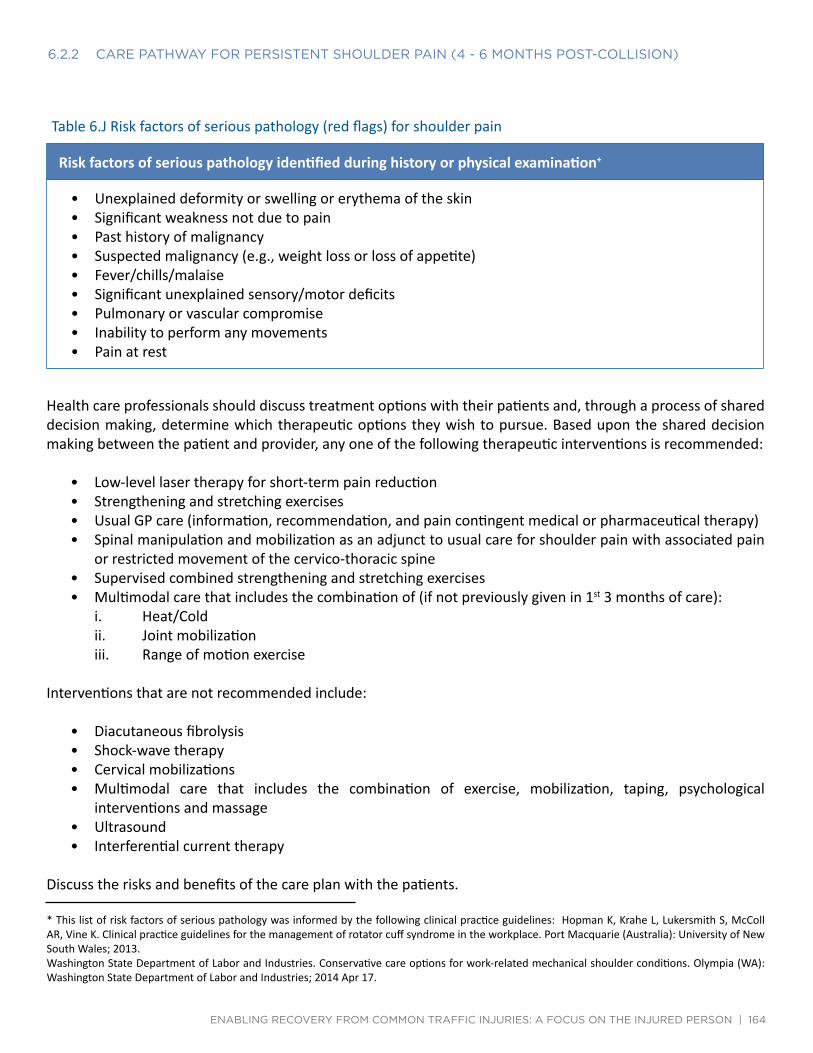
6.2.2 caRE PaThway foR PERsisTEnT shouldER Pain (4 - 6 monThs PosT-collision)
Enabling REcovERy fRom common TRaffic injuRiEs: a focus on ThE injuREd PERson | 164
Table 6.J Risk factors of serious pathology (red flags) for shoulder pain
Risk factors of serious pathology identified during history or physical examination+
* This list of risk factors of serious pathology was informed by the following clinical practice guidelines: Hopman K, Krahe L, Lukersmith S, McColl AR, Vine K. Clinical practice guidelines for the management of rotator cuff syndrome in the workplace. Port Macquarie (Australia): University of New South Wales; 2013. Washington State Department of Labor and Industries. Conservative care options for work-related mechanical shoulder conditions. Olympia (WA): Washington State Department of Labor and Industries; 2014 Apr 17.
• Unexplained deformity or swelling or erythema of the skin • Significant weakness not due to pain • Past history of malignancy • Suspected malignancy (e.g., weight loss or loss of appetite) • Fever/chills/malaise • Significant unexplained sensory/motor deficits • Pulmonary or vascular compromise • Inability to perform any movements • Pain at rest
Health care professionals should discuss treatment options with their patients and, through a process of shared decision making, determine which therapeutic options they wish to pursue. Based upon the shared decision making between the patient and provider, any one of the following therapeutic interventions is recommended:
• Low-level laser therapy for short-term pain reduction • Strengthening and stretching exercises • Usual GP care (information, recommendation, and pain contingent medical or pharmaceutical therapy) • Spinal manipulation and mobilization as an adjunct to usual care for shoulder pain with associated pain
or restricted movement of the cervico-thoracic spine • Supervised combined strengthening and stretching exercises • Multimodal care that includes the combination of (if not previously given in 1st 3 months of care):
i. Heat/Cold ii. Joint mobilization iii. Range of motion exercise
Interventions that are not recommended include:
• Diacutaneous fibrolysis • Shock-wave therapy • Cervical mobilizations • Multimodal care that includes the combination of exercise, mobilization, taping, psychological
interventions and massage • Ultrasound • Interferential current therapy
Discuss the risks and benefits of the care plan with the patients.

6.2.2 caRE PaThway foR PERsisTEnT shouldER Pain (4 - 6 monThs PosT-collision)
Enabling REcovERy fRom common TRaffic injuRiEs: a focus on ThE injuREd PERson | 165
Reassess and Take the Indicated Course of Action
Reassess the patient at every visit to determine if additional care is necessary, or if the condition is worsening.
Patients should be discharged as soon as they have made significant improvement or recovered. It is recommended that health care professionals use the self-rated recovery question to measure patient recovery: “How well do you feel you are recovering from your injuries?” The response options include: 1) completely better, 2) much improved, 3) slightly improved, 4) no change, 5) slightly worse, 6) much worse, and 7) worse than ever. Patients reporting to be ‘completely better’ or ‘much improved’ should be considered recovered.* Patients who have not recovered should follow the care pathway outlined in this guideline.
* The use of a valid and reliable condition-specific instrument (e.g. Disabilities of the Arm, Shoulder and Hand (DASH)) is encouraged but should not be used to measure overall recovery.
Patients with worsening of symptoms and those who develop new physical, mental or psychological symptoms should be referred to a physician for further evaluation at any time point during their care.
Patients who have not significantly improved or recovered should be referred to the physician for further evaluation.

6.2.2 caRE PaThway foR PERsisTEnT shouldER Pain (4 - 6 monThs PosT-collision)
Yes
No
Yes
No
No
Yes
No
Yes Yes
Yes Yes
No No
Enabling REcovERy fRom common TRaffic injuRiEs: a focus on ThE injuREd PERson | 166
Figure 6.2: Care Pathway for the Management of Shoulder Pain
1Persons injured in a traffic collision with
shoulder pain
2
Conduct an appropriate clinical evaluation
3
Risk factors for serious pathologies?a4
Refer to physician
5Other injuries?
6Go to appropriate clinical care pathways
and co‐manage
7
Offer information on nature, management, course of shoulder pain as a framework for initiation of a program of care.
8
Is treatment required?
9
Discharge
10Are symptoms ≤ 3 months?
11
Symptoms are > 3 months.
12
Based on shared decision making between the patient and provider, any one of the following therapeutic interventions is recommended:b,c
Home and clinic based interventions: 1) Low‐level laser therapy for short‐ term pain reduction2) Spinal manipulation and mobilization as an adjunct to usual care for shoulder pain with associated pain or restricted movement of the cervico‐thoracic spine
3) Multimodal care that includes the combination of:a) Heat/Coldb) Joint mobilizationc) Range of motion exercise
Do not offer:d
1) Diacutaneous fibrolysis2) Ultrasound3) Interferential current therapy
Refer to specific recommendation for treatment details (Section 6.2.3)
13
Based on shared decision making between the patient and provider, any one of the following therapeutic interventions is recommended:b,c
Home and clinic based interventions: 1) Low‐ level laser therapy for short‐term pain reduction2) Strengthening and stretching exercises3) Usual GP care (information, recommendation, and pain contingent medical or pharmaceutical therapy)
4) Spinal manipulation and mobilization as an adjunct to usual care for shoulder pain with associated pain or restricted movement of the cervico‐ thoracic spine
5) Supervised combined strengthening and stretching exercises6) Multimodal care that includes the combination of:a) Heat/Coldb) Joint mobilizationc) Range of motion exercise
Do not offer:d
1) Diacutaneous fibrolysis2) Shock‐ wave therapy3) Cervical mobilizations4) Multimodal care that includes the combination of exercise, mobilization, taping, psychological interventions and massage
5) Ultrasound6) Interferential current therapy
Refer to specific recommendation for treatment details (Section 6.2.4)
14
Is injured person recovered after 3 months?
16
Is injured person recovered?
15
Discharge
17
1) Incomplete recovery: Initiate persistent protocol (Box 13) 2) Major symptom change (new or worsening physical, mental or psychological symptoms) : refer to physician
18
1) Incomplete recovery: refer to physician 2) Major symptom change (new or worsening physical, mental or psychological symptoms): refer to physician
a Risk factors for serious pathologies (also known as red flags): Unexplained deformity or swelling or erythema of the skin; significant weakness not due to pain; past history of malignancy; suspected malignancy (e.g., weight loss or loss of appetite); fever/chills/malaise; significant unexplained sensory/ motor deficits; pulmonary or vascular compromise; inability to perform any movements; pain at rest b This guideline does not include interventions for which there is a lack of evidence of effectiveness c The ordering of interventions does not reflect superiority of effectiveness d Based on evidence of no benefit to patients

Enabling REcovERy fRom common TRaffic injuRiEs: a focus on ThE injuREd PERson | 167
SECTION 6.2.3
Key recOMMendatiOnS fOr the ManageMent Of recent ShOulder Pain
This section summarizes the key recommendations for the management of recent shoulder pain for the period extending from 0 to 3 months post-collision. The wording of recommendations follows the guidance from the National Institute for Health and Care Excellence (NICE). Recommendations beginning with “offer” indicate that, according to the evidence, an intervention is associated with outcomes that were superior to other interventions, placebo/sham, or no intervention. The wording “consider” indicates that an intervention is as effective as another one. The wording “do not offer” indicates that, according to the evidence, an intervention does not benefit patients. A detailed explanation of the wording of recommendations is presented in section 2.5.2.4 of this report.
• Provide care in partnership with the patient. Involve the patient in care planning and decision-making.
• Reassure patients about the benign and self-limiting nature of their pain.
• Educate patients about the benefits of being actively engaged and participating in their care plan by remaining active and continuing movement.
• Emphasize active rather than passive treatments.
• Deliver a time-limited program of care.
• Do not provide ineffective or experimental treatments.
SECTION 6.2.3.1
MultiMOdal care
Multimodal care includes at least two distinct therapeutic modalities, provided by one or more health care disciplines.
Table 6.K: Multimodal Care for Recent Shoulder Pain
Recommendation 6.2.3.1.1
Consider multimodal care that includes heat*, cold*, joint mobilizations**, and range of motion exercises*** provided in 8-10 sessions over a maximum 5-6 weeks.
References: • Decision Determinants and Evidence Table for Upper Extremity Injuries – Report 3 – Appendix 4
* Twice weekly supervised application of hot packs and cold packs. ** Passive joint mobilization at the shoulder, sternoclavicular and acromioclavicular joints twice a week. *** Daily home range of motion exercises entail progressively loaded functional movements of the arm, incorporating free weights or elastic resistance as required. Range of movement includes: shoulder abduction, flexion, extension, horizontal flexion and extension, hand-behind-back.

Enabling REcovERy fRom common TRaffic injuRiEs: a focus on ThE injuREd PERson | 168
SECTION 6.2.3.2
SOft tiSSue theraPy
Soft tissue therapy is a mechanical therapy in which muscles, tendons, and ligaments are passively pressed or kneaded by hand or with mechanical devices. It includes relaxation massage, clinical massage, movement re-education and energy work.
Table 6.L: Soft Tissue Therapy for Recent Shoulder Pain
Recommendation 6.2.3.2.1 Do not offer diacutaneous fibrolysis.*
References: • Decision Determinants and Evidence Table for Upper Extremity Injuries – Report 10 – Appendix 4
* Diacutaneous fibrolysis is a non-invasive physiotherapeutic technique applied by means of a set of metallic hooks to release adherences between the different musculoskeletal structures.
SECTION 6.2.3.3
PaSSive PhySical MOdalitieS
Passive physical modalities include two categories of interventions: physico-chemical and structural. Physico-chemical modalities use a thermal or electromagnetic agent to affect the body at or beneath the skin level. Structural modalities include functional or non-functional assistive devices. Functional assistive devices intend to align, support or otherwise indirectly facilitate function in the affected region. Non-functional devices intend to achieve a state of rest in specific anatomic positions or prevent movement.
Table 6.M: Passive Physical Modalities for Recent Shoulder Pain
Recommendation 6.2.3.3.1
Offer low-level laser therapy (LLLT)* for short-term pain reduction (pulsed laser, 10 sessions over 2 weeks: 1) peak power = 1 kW, average power = 6 W, maximum energy of single impulse = 150 mJ, duration of single impulse <150 ms, fluency = 760 mJ/cm2, wavelength = 1064 nm; or 2) wavelength = 890 nm, time = 2 minute/point, power 2-4 j/cm2 in each point).
6.2.3.3.2 Do not offer ultrasound.**
6.2.3.3.3 Do not offer interferential current therapy.***
References: • Decision Determinants and Evidence Table for Upper Extremity Injuries – Report 2 – Appendix 4
* Low-level laser therapy is the application of a coherent light beam (laser) to a region for the purpose of reducing local pain or promoting local healing. ** Ultrasound is an oscillating sound pressure wave affecting structures beneath the skin surface. *** Interferential current therapy produces current to selectively excite large diameter nerve fibres and temporarily inhibit transmission of nociceptive signals in the spinal dorsal horn from pain mediating small diameter nerve fibres.
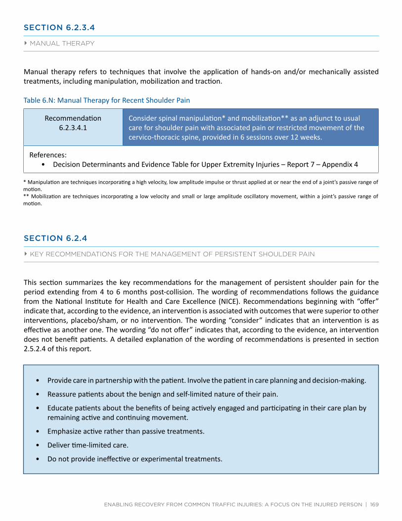
Enabling REcovERy fRom common TRaffic injuRiEs: a focus on ThE injuREd PERson | 169
SECTION 6.2.3.4
Manual theraPy
Manual therapy refers to techniques that involve the application of hands-on and/or mechanically assisted treatments, including manipulation, mobilization and traction.
Table 6.N: Manual Therapy for Recent Shoulder Pain
Recommendation 6.2.3.4.1
Consider spinal manipulation* and mobilization** as an adjunct to usual care for shoulder pain with associated pain or restricted movement of the cervico-thoracic spine, provided in 6 sessions over 12 weeks.
References: • Decision Determinants and Evidence Table for Upper Extremity Injuries – Report 7 – Appendix 4
* Manipulation are techniques incorporating a high velocity, low amplitude impulse or thrust applied at or near the end of a joint’s passive range of motion. ** Mobilization are techniques incorporating a low velocity and small or large amplitude oscillatory movement, within a joint’s passive range of motion.
SECTION 6.2.4
Key recOMMendatiOnS fOr the ManageMent Of PerSiStent ShOulder Pain
This section summarizes the key recommendations for the management of persistent shoulder pain for the period extending from 4 to 6 months post-collision. The wording of recommendations follows the guidance from the National Institute for Health and Care Excellence (NICE). Recommendations beginning with “offer” indicate that, according to the evidence, an intervention is associated with outcomes that were superior to other interventions, placebo/sham, or no intervention. The wording “consider” indicates that an intervention is as effective as another one. The wording “do not offer” indicates that, according to the evidence, an intervention does not benefit patients. A detailed explanation of the wording of recommendations is presented in section 2.5.2.4 of this report.
• Provide care in partnership with the patient. Involve the patient in care planning and decision-making.
• Reassure patients about the benign and self-limited nature of their pain.
• Educate patients about the benefits of being actively engaged and participating in their care plan by remaining active and continuing movement.
• Emphasize active rather than passive treatments.
• Deliver time-limited care.
• Do not provide ineffective or experimental treatments.

Enabling REcovERy fRom common TRaffic injuRiEs: a focus on ThE injuREd PERson | 170
SECTION 6.2.4.1
exerciSe
Exercise refers to any series of movements with the aim of training or developing the body by routine practice or as physical training to promote good physical health. Exercise therapy includes a wide variety of techniques common to the treatment and rehabilitation of shoulder pain.
Table 6.O: Exercise for Persistent Shoulder Pain
Recommendation 6.2.4.1.1
Offer strengthening and stretching exercises (home-based strengthening and stretching of the rotator cuff and scapulohumeral muscles, supervised weekly for 5 weeks).
6.2.4.1.2 Consider supervised combined strengthening and stretching exercises (8 repetitions of progressive shoulder flexion/extension/medial rotation/ lateral rotation strengthening, 2 sets, twice a week for 8 weeks; or home-based 5 repetitions of stretching of pectoralis minor and posterior shoulder per day, 10-20 repetitions of progressive strengthening for rotator cuff and serratus anterior, 3 sets per week for 8 weeks).
*For low-grade non-specific shoulder pain
References: • Decision Determinants and Evidence Table for Upper Extremity Injuries – Report 1 – Appendix 4
* Low-grade pain: pain intensity <3/10 cm or 30/100 mm on Visual Analog Scale.
SECTION 6.2.4.2
MultiMOdal care
Multimodal care includes at least two distinct therapeutic modalities, provided by one or more health care disciplines.

6.2.4.2 mulTimodal caRE
Enabling REcovERy fRom common TRaffic injuRiEs: a focus on ThE injuREd PERson | 171
Table 6.P: Multimodal Care for Persistent Shoulder Pain
Recommendation 6.2.4.2.1
Consider usual GP care (information, recommendation, and pain contingent medical or pharmaceutical therapy).
6.2.4.2.2 Consider multimodal care that includes heat*, cold*, joint mobilizations**, and range of motion exercises*** provided in 8-10 sessions over a maximum of 5-6 weeks.
6.2.4.2.3 Do not offer multimodal care that includes exercise, mobilization, taping, psychological interventions, and massage.
References: • Decision Determinants and Evidence Table for Upper Extremity Injuries – Report 3 – Appendix 4
* Twice weekly supervised application of hot packs and cold packs. ** Passive joint mobilization at the shoulder, sternoclavicular and acromioclavicular joints twice a week. *** Daily home range of motion exercises entail progressively loaded functional movements of the arm, incorporating free weights or elastic resistance as required. Range of movement includes: shoulder abduction, flexion, extension, horizontal flexion and extension, hand-behind-back.
SECTION 6.2.4.3
SOft tiSSue theraPy
Soft tissue therapy is a mechanical therapy in which muscles, tendons, and ligaments are passively pressed or kneaded by hand or with mechanical devices. It includes relaxation massage, clinical massage, movement re-education and energy work.
Table 6.Q: Soft Tissue Therapy for Persistent Shoulder Pain
Recommendation 6.2.4.3.1 Do not offer diacutaneous fibrolysis*
References: • Decision Determinants and Evidence Table for Upper Extremity Injuries – Report 10 – Appendix 4
* Diacutaneous fibrolysis is a non-invasive physiotherapeutic technique applied by means of a set of metallic hooks to release adherences between the different musculoskeletal structures.
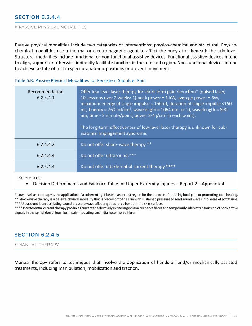
Enabling REcovERy fRom common TRaffic injuRiEs: a focus on ThE injuREd PERson | 172
SECTION 6.2.4.4
PaSSive PhySical MOdalitieS
Passive physical modalities include two categories of interventions: physico-chemical and structural. Physico-chemical modalities use a thermal or electromagnetic agent to affect the body at or beneath the skin level. Structural modalities include functional or non-functional assistive devices. Functional assistive devices intend to align, support or otherwise indirectly facilitate function in the affected region. Non-functional devices intend to achieve a state of rest in specific anatomic positions or prevent movement.
Table 6.R: Passive Physical Modalities for Persistent Shoulder Pain
Recommendation 6.2.4.4.1
Offer low-level laser therapy for short-term pain reduction* (pulsed laser, 10 sessions over 2 weeks: 1) peak power = 1 kW, average power = 6W, maximum energy of single impulse = 150mJ, duration of single impulse <150 ms, fluency = 760 mJ/cm2, wavelength = 1064 nm; or 2), wavelength = 890 nm, time - 2 minute/point, power 2-4 j/cm2 in each point).
The long-term effectiveness of low-level laser therapy is unknown for sub-acromial impingement syndrome.
6.2.4.4.2 Do not offer shock-wave therapy.**
6.2.4.4.4 Do not offer ultrasound.***
6.2.4.4.4 Do not offer interferential current therapy.****
References: • Decision Determinants and Evidence Table for Upper Extremity Injuries – Report 2 – Appendix 4
* Low-level laser therapy is the application of a coherent light beam (laser) to a region for the purpose of reducing local pain or promoting local healing. ** Shock-wave therapy is a passive physical modality that is placed onto the skin with sustained pressure to send sound waves into areas of soft tissue. *** Ultrasound is an oscillating sound pressure wave affecting structures beneath the skin surface. **** Interferential current therapy produces current to selectively excite large diameter nerve fibres and temporarily inhibit transmission of nociceptive signals in the spinal dorsal horn form pain mediating small diameter nerve fibres.
SECTION 6.2.4.5
Manual theraPy
Manual therapy refers to techniques that involve the application of hands-on and/or mechanically assisted treatments, including manipulation, mobilization and traction.

Quick Reference Guide – Management of Shoulder Pain with Calcific Tendinitis
6.2.4.5 manual ThERaPy
Enabling REcovERy fRom common TRaffic injuRiEs: a focus on ThE injuREd PERson | 173
Table 6.S: Manual Therapy for Persistent Shoulder Pain
Recommendation 6.2.4.5.1
Consider spinal manipulation* and mobilization** as an adjunct to usual care for shoulder pain with associated pain or restricted movement of the cervico-thoracic spine provided in 6 sessions over 12 weeks.
6.2.4.5.2 Do not offer cervical mobilizations.*
References: • Decision Determinants and Evidence Table for Upper Extremity Injuries – Report 7 – Appendix 4
* Manipulation are techniques incorporating a high velocity, low amplitude impulse or thrust applied at or near the end of a joint’s passive range of motion. ** Mobilizations are techniques incorporating a low velocity and small or large amplitude oscillatory movement, within a joint’s passive range of motion.
SECTION 6.3
ManageMent Of ShOulder Pain with calcific tendinitiS
Quick Reference Guide – Management of shoulder pain with calcific tendinitis
Management of Calcific Tendinitis For all injured persons with shoulder pain with calcific tendinitis: Rule out risk factors for serious pathologiesa
Offer information on nature, management, course of shoulder pain with calcific tendinitis as a framework for initiation of a program of care Conduct ongoing assessment for symptom improvement or progression during intervention and refer accordingly Discharge injured person as appropriate at any point during intervention and recovery
Based upon shared decision making between the patient and provider, the following therapeutic intervention is recommended:b
1. Shock‐wave therapy with an amplitude ranging from 0.08mJ/mm2 ‐0.06mJ/mm2
Refer to specific recommendation for treatment details (Section 6.3.2)
Outcome: Recovered → Discharge Unrecovered: Incomplete recovery → Refer to physician
Major symptom change (new or worsening physical, mental or psychological symptoms) → Refer to physician
a Risk factors for serious pathologies (also known as red flags): Unexplained deformity or swelling or erythema of the skin; significant weakness not due to pain; past history of malignancy; suspected malignancy (e.g., weight loss or loss of appetite); fever/chills/malaise; significant unexplained sensory/motor deficits; pulmonary or vascular compromise; inability to perform any movements; pain at rest b This guideline does not include interventions for which there is a lack of evidence of effectiveness

Enabling REcovERy fRom common TRaffic injuRiEs: a focus on ThE injuREd PERson | 174
SECTION 6.3.1
care Pathway fOr ShOulder Pain with calcific tendinitiS
The care pathway is presented in Figure 6.3
The primary goals of clinical care are to promote recovery by reducing symptoms and return patients to their normal activities of daily living.
Assess the Patient with Shoulder Pain with Calcific Tendinitis
Conduct an appropriate clinical evaluation to rule out major structural or other pathologies as the cause of symptoms. The presence of a risk factor for serious pathologies (also known as red flags) identified during the history and examination warrants further investigation and referral to the appropriate health care professional. However, once pathology has been ruled out, the patient should be treated according to the shoulder pain with calcific tendinitis care pathway.
Patients who also have neck pain and associated disorders or other injuries should be managed using the appropriate care pathways.
Table 6.J Risk factors of serious pathology (red flags) for shoulder pain
Risk factors of serious pathology identified during history or physical examination+
* This list of risk factors of serious pathology was informed by the following clinical practice guidelines: Hopman K, Krahe L, Lukersmith S, McColl AR, Vine K. Clinical practice guidelines for the management of rotator cuff syndrome in the workplace. Port Macquarie (Australia): University of New South Wales; 2013. Washington State Department of Labor and Industries. Conservative care options for work-related mechanical shoulder conditions. Olympia (WA): Washington State Department of Labor and Industries; 2014 Apr 17.
• Unexplained deformity or swelling or erythema of the skin • Significant weakness not due to pain • Past history of malignancy • Suspected malignancy (e.g., weight loss or loss of appetite) • Fever/chills/malaise • Significant unexplained sensory/motor deficits • Pulmonary or vascular compromise • Inability to perform any movements • Pain at rest
Educate and Reassure the Patient
The health care professional should aim to understand the patient’s beliefs and expectations about shoulder pain with calcific tendinitis and address any misunderstandings or apprehension through education and reassurance. The health care professional needs to educate and reassure the patient about the benign and self-limited nature of shoulder pain with calcific tendinitis and reinforce the importance of maintaining activities of daily living.

6.3.1 caRE PaThway foR shouldER Pain wiTh calcific TEndiniTis
Enabling REcovERy fRom common TRaffic injuRiEs: a focus on ThE injuREd PERson | 175
Deliver the Care Plan for Persistent Shoulder Pain with Calcific Tendinitis (4-6 months post-collision)
The goal of the care plan is to provide clinical interventions that promote resolution of symptoms and restoration of function. Patients requiring clinical care should be encouraged to participate in their program of care by remaining active.
Health care professionals should discuss the treatment plan with their patients, emphasizing the risk and benefits of the care plan. Based upon shared decision making between the patient and provider, the following therapeutic intervention is recommended:
• Shock-wave therapy with an amplitude ranging from 0.08mJ/mm2-0.06mJ/mm2
Reassess and Take the Indicated Course of Action
Reassess the patient at every visit to determine if additional care is necessary, or if the condition is worsening.
Patients should be discharged as soon as they report significant improvement or recovery. It is recommended that health care professionals use the self-rated recovery question to measure patient recovery: “How well do you feel you are recovering from your injuries?” The response options include: 1) completely better, 2) much improved, 3) slightly improved, 4) no change, 5) slightly worse, 6) much worse, and 7) worse than ever. Patients reporting to be ‘completely better’ or ‘much improved’ should be considered recovered.* Patients who have not recovered should follow the care pathway outlined in this guideline.
* The use of a valid and reliable condition-specific instrument (e.g. Disabilities of the Arm, Shoulder and Hand (DASH)) is encouraged but should not be used to measure overall recovery.
Patients with worsening of symptoms and those who develop new physical, mental or psychological symptoms (other than shoulder pain with calcific tendinitis) should be referred to a physician for further evaluation at any time point during their care.
Patients who have not improved significantly or recovered should be referred to the physician for further evaluation.

Care Pathway for the Management of Shoulder Pain with Calcific Tendinitis
6.3.1 caRE PaThway foR shouldER Pain wiTh calcific TEndiniTis
Yes
No
Yes
No
No
Yes
Yes
No
Enabling REcovERy fRom common TRaffic injuRiEs: a focus on ThE injuREd PERson | 176
Figure 6.3: Care Pathway for the Management of Shoulder Pain with Calcific Tendinitis
1
Persons injured in a traffic collision with shoulder pain with calcific tendinitis
2
Conduct an appropriate clinical evaluation
3 Risk factors for serious pathologies?a
4
Refer to physician
5 Other injuries?
6
Go to appropriate clinical care pathways and co-manage
7
Offer information on nature, management, course of shoulder pain with calcific tendinitis as a framework for initiation of a
program of care.
8
Is treatment required?
9
Discharge
10
Based on shared decision making between the patient and provider, the following therapeutic intervention is recommended:b
1) Shock-wave therapy with an amplitude ranging from 0.08 mJ/mm2-0.0gmJ/mm2
Refer to specific recommendation for treatment details (Section 6.3.2)
11 Is injured person recovered?
12
Discharge
13
1) Incomplete recovery: refer to physician 2) Major symptom change (new or worsening physical, mental or psychological symptoms): proceed to appropriate flowchart or refer to physician
a Risk factors for serious pathologies (also known as red flags): Unexplained deformity or swelling or erythema of the skin; significant weakness not due to pain; past history of malignancy; suspected malignancy (e.g., weight loss or loss of appetite); fever/chills/malaise; significant unexplained sensory/ motor deficits; pulmonary or vascular compromise; inability to perform any movements; pain at rest b This guideline does not include interventions for which there is a lack of evidence of effectiveness

Enabling REcovERy fRom common TRaffic injuRiEs: a focus on ThE injuREd PERson | 177
SECTION 6.3.2
Key recOMMendatiOnS fOr the ManageMent Of ShOulder Pain with calcific tendinitiS
This section summarizes the key recommendations for the management of shoulder pain with calcific tendinitis. The wording of recommendations follows the guidance from the National Institute for Health and Care Excellence (NICE). Recommendations beginning with “offer” indicate that, according to the evidence, an intervention is associated with outcomes that were superior to other interventions, placebo/sham, or no intervention. The wording “consider” indicates that an intervention is as effective as another one. The wording “do not offer” indicates that, according to the evidence, an intervention does not benefit patients. A detailed explanation of the wording of recommendations is presented in section 2.5.2.4 of this report.
• Provide care in partnership with the patient. Involve the patient in care planning and decision-making.
• Reassure patients about the benign and self-limited nature of their pain.
• Educate patients about the benefits of being actively engaged and participating in their care plan by remaining active and continuing movement.
• Emphasize active rather than passive treatments.
• Deliver time-limited care.
• Do not provide ineffective or experimental treatments.
SECTION 6.3.2.1
PaSSive PhySical MOdalitieS
Passive physical modalities include two categories of interventions: physico-chemical and structural. Physico-chemical modalities use a thermal or electromagnetic agent to affect the body at or beneath the skin level. Structural modalities include functional or non-functional assistive devices. Functional assistive devices intend to align, support or otherwise indirectly facilitate function in the affected region. Non-functional devices intend to achieve a state of rest in specific anatomic positions or prevent movement.
Table 6.T: Passive Physical Modalities for Shoulder Pain with Calcific Tendinitis
Recommendation 6.3.2.1.1
Offer shock-wave therapy* with an amplitude ranging from 0.08mJ/mm2-0.60mJ/mm2 (a maximum of 4 sessions over 4 weeks).
References: • Decision Determinants and Evidence Table or Upper Extremity Injuries – Report 2 – Appendix 4
* Shock-wave therapy is a passive physical modality that is placed onto the skin with sustained pressure to send sound waves into areas of soft tissue.

Enabling REcovERy fRom common TRaffic injuRiEs: a focus on ThE injuREd PERson | 178
SECTION 7.0
GUIDELINE FOR THE CLINICAL MANAGEMENT OF LOwER ExTREMITY SOFT TISSUE DISORDERS

SECTION 7.0
guideline fOr the clinical ManageMent Of lOwer extreMity SOft tiSSue diSOrderS
Enabling REcovERy fRom common TRaffic injuRiEs: a focus on ThE injuREd PERson | 179
7.1 Management of patellofemoral pain 7.1.1 Care pathway for recent onset patellofemoral pain (0-3 months post-collision) 7.1.2 Care pathway for persistent patellofemoral pain (4-6 months post-collision) 7.1.3 Key recommendations for the management of recent onset patellofemoral pain 7.1.4 Key recommendations for the management of persistent patellofemoral pain 7.2 Management of ankle sprain 7.2.1 Care pathway for recent onset ankle sprain (0-3 months post-collision) 7.2.2 Care pathway for persistent ankle sprain (4-6 months post-collision) 7.2.3 Key recommendations for the management of recent onset ankle sprain 7.2.4 Key recommendations for the management of persistent ankle sprain 7.3 Management of Achilles tendinopathy 7.3.1 Care pathway for recent onset Achilles tendinopathy (0-3 months post-collision) 7.3.2 Care pathway for persistent Achilles tendinopathy (4-6 months post-collision) 7.3.3 Key recommendations for the management of recent onset Achilles tendinopathy 7.3.4 Key recommendations for the management of persistent Achilles tendinopathy 7.4 Management of plantar fasciitis and heel pain 7.4.1 Care pathway for recent onset plantar fasciitis and heel pain (0-3 months post-collision) 7.4.2 Care pathway for persistent plantar fasciitis and heel pain (4-6 months post-collision) 7.4.3 Key recommendations for the management of recent onset plantar fasciitis and heel pain 7.4.4 Key recommendations for the management of persistent plantar fasciitis and heel pain
This evidence-based guideline establishes the best practice for the clinical management of lower extremity disorders caused or exacerbated by a motor vehicle collision. This guideline covers recent onset (0-3 months post-collision) and persistent (4-6 months post-collision) patellofemoral pain, ankle sprain, Achilles tendinopathy, plantar fasciitis and plantar heel pain; it does not cover disorders that persist for more than 6 months post-collision.
Lower extremity soft tissue disorders refer to grade I and II sprains or strains, tendonitis, tendinopathy, tendinosis, patellofemoral pain (syndrome), and non-specific pain of the hip, thigh, knee, leg, ankle and foot. Strains and sprains can be classified into three grades, distinguished by the severity of signs and symptoms and structural disruption (Table 7.A and Table 7.B). This guideline is not indicated for conditions that include the presence of major structural or other pathological causes of lower extremity disorders such as fractures, dislocations, osteoarthritis, inflammatory disorders, systemic disease, infections, tumors and Grade III sprains/strains. However, studies of ankle injuries that included Grade I-III sprains/strains were reviewed when the evidence was stratified by injury severity.
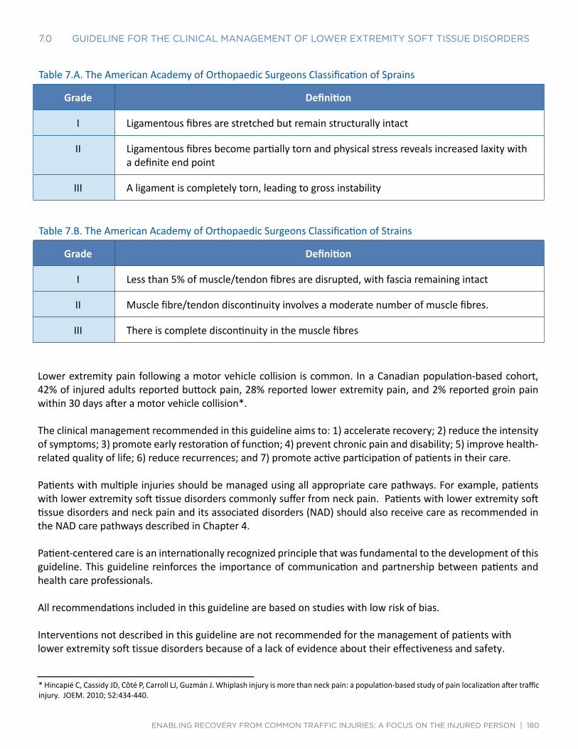
7.0 guidElinE foR ThE clinical managEmEnT of lowER ExTREmiTy sofT TissuE disoRdERs
Enabling REcovERy fRom common TRaffic injuRiEs: a focus on ThE injuREd PERson | 180
Table 7.A. The American Academy of Orthopaedic Surgeons Classification of Sprains
Grade Definition
I Ligamentous fibres are stretched but remain structurally intact
II Ligamentous fibres become partially torn and physical stress reveals increased laxity with a definite end point
III A ligament is completely torn, leading to gross instability
Table 7.B. The American Academy of Orthopaedic Surgeons Classification of Strains
Grade Definition
I Less than 5% of muscle/tendon fibres are disrupted, with fascia remaining intact
II Muscle fibre/tendon discontinuity involves a moderate number of muscle fibres.
III There is complete discontinuity in the muscle fibres
Lower extremity pain following a motor vehicle collision is common. In a Canadian population-based cohort, 42% of injured adults reported buttock pain, 28% reported lower extremity pain, and 2% reported groin pain within 30 days after a motor vehicle collision*.
* Hincapié C, Cassidy JD, Côté P, Carroll LJ, Guzmán J. Whiplash injury is more than neck pain: a population-based study of pain localization after traffic injury. JOEM. 2010; 52:434-440.
The clinical management recommended in this guideline aims to: 1) accelerate recovery; 2) reduce the intensity of symptoms; 3) promote early restoration of function; 4) prevent chronic pain and disability; 5) improve health-related quality of life; 6) reduce recurrences; and 7) promote active participation of patients in their care.
Patients with multiple injuries should be managed using all appropriate care pathways. For example, patients with lower extremity soft tissue disorders commonly suffer from neck pain. Patients with lower extremity soft tissue disorders and neck pain and its associated disorders (NAD) should also receive care as recommended in the NAD care pathways described in Chapter 4.
Patient-centered care is an internationally recognized principle that was fundamental to the development of this guideline. This guideline reinforces the importance of communication and partnership between patients and health care professionals.
All recommendations included in this guideline are based on studies with low risk of bias.
Interventions not described in this guideline are not recommended for the management of patients with lower extremity soft tissue disorders because of a lack of evidence about their effectiveness and safety.

7.0 guidElinE foR ThE clinical managEmEnT of lowER ExTREmiTy sofT TissuE disoRdERs
Enabling REcovERy fRom common TRaffic injuRiEs: a focus on ThE injuREd PERson | 181
Health care professionals eligible to provide care under this guideline are those defined by the Statutory Accident Benefits Schedule (SABS).
This guideline is organized in four sections. Each section provides evidence-based recommendations for the clinical management of various types and durations of lower extremity soft tissue disorders:
• Section 7.1 - Management of patellofemoral pain • Section 7.2 - Management of ankle sprain • Section 7.3 - Management of Achilles tendinopathy • Section 7.4 - Management of plantar fasciitis and heel pain
All recommendations presented in this guideline integrate the:
• Key decision determinants based upon the framework developed by the Ontario Health Technology Advisory Committee (OHTAC);
• Best evidence obtained from a critical review of current scientific literature; and • Qualitative research exploring the experiences of persons treated for traffic injuries in Ontario
All background documents and references are available at http://www.fsco.gov.on.ca

Quick Reference Guide – Management of Patellofemoral Pain
Enabling REcovERy fRom common TRaffic injuRiEs: a focus on ThE injuREd PERson | 182
SECTION 7.1
ManageMent Of PatellOfeMOral Pain
Quick Reference Guide – Management of Patellofemoral Pain
Symptoms ≤ 3 months post‐collision Symptoms > 3 months post‐collision For all injured persons with patellofemoral pain: Rule out risk factors for serious pathologiesa
Offer information on nature, management, course of collision‐related patellofemoral pain as a framework for initiation of a program of care Conduct ongoing assessment for symptom improvement or progression during intervention and refer accordingly Discharge injured person as appropriate at any point during intervention and recovery
1. Monitor and reassure
Refer to specific recommendation for treatment details (Section 7.1.3)
Based upon shared decision making between the patient and provider, the following therapeutic intervention is recommended:b
1. Supervised clinic‐based combined exercise
Refer to specific recommendation for treatment details (Section 7.1.4)
Outcome: Recovered → Discharge Unrecovered: Incomplete recovery → Initiate persistent protocol
Major symptom change (new or worsening physical, mental or psychological symptoms) → Refer to physician
Outcome: Recovered → Discharge Unrecovered: Incomplete recovery → Refer to physician
Major symptom change (new or worsening physical, mental or psychological symptoms) → Refer to physician
a Risk factors for serious pathologies (also known as red flags): history of major trauma; minor trauma (if >50 years, history of osteoporosis and taking corticosteroids); erythema, warmth, effusion and decreased range of motion; high velocity injury, absent pulses, foot drop, multiple plane laxity; past history of malignancy, unexplained weight loss, pain at multiple sites, night pain, pain at rest b This guideline does not include interventions for which there is a lack of evidence of effectiveness
SECTION 7.1.1
care Pathway fOr recent OnSet PatellOfeMOral Pain (0 - 3 MOnthS POSt-cOlliSiOn)
The care pathway is presented in Figure 7.1.
At initial contact, health care professionals should educate and reassure the patient that patellofemoral pain will resolve within weeks to months of symptom onset. Patients greatly improve their recovery by actively participating in their care. Clinical care aims to accelerate recovery by reducing pain and improving function. The care pathway recommended for the first 3 months of care for patellofemoral pain is described below.
Assess the Patient
Conduct an appropriate clinical evaluation to rule out major structural or other pathologies as the cause of the symptoms. The presence of a risk factor for serious pathologies (also known as red flags) identified during the history and examination warrants further investigation and referral to the appropriate health care professional. However, once pathology has been ruled out, the patient should be treated according to the patellofemoral pain care pathway.

7.1.1 caRE PaThway foR REcEnT onsET PaTEllofEmoRal Pain (0 - 3 monThs PosT-collision)
Enabling REcovERy fRom common TRaffic injuRiEs: a focus on ThE injuREd PERson | 183
Table 7.C Risk factors of serious pathology (red flags) for patellofemoral pain
Risk factors of serious pathology identified during history or physical examination*
* This list of risk factors of serious pathology was informed by the following clinical practice guidelines: Australian Acute Musculoskeletal Pain Guidelines Group. Evidence-based management of Acute Musculoskeletal Pain. A Guide for Clinicians. Queensland, Australia: Australian Academic Press; 2004. New Zealand Guidelines Group. The Diagnosis and Management of Soft Tissue Knee Injuries: Internal Derangements. Best Practice Evidence-Based Guideline. Available from: http://www.health.govt.nz/system/files/documents/publications/acc_soft_tissue_knee_injury_fulltext.pdf Bennett DL, Nelson JW, Weissman BN, Kransdorf MJ, Appel M, Bencardino JT, Fries IB, Hayes CW, Hochman MG, Jacobson JA, Luchs JS, Math KR, Murphey MD, Newman JS, Rubin DA, Scharf SC, Small KM, Expert Panel on Musculoskeletal Imaging. ACR Appropriateness Criteria® nontraumatic knee pain. http://www.guideline.gov/content.aspx?id=43872 [online publication]. Reston (VA): American College of Radiology (ACR); 2012.
• History of major trauma • Minor trauma (if >50 years, history of osteoporosis and taking corticosteroids) • Erythema, warmth, effusion and decreased range of motion • High velocity injury, absent pulses, foot drop, multiple plane laxity • Past history of malignancy, unexplained weight loss, pain at multiple sites, night pain, pain at rest
Patients with multiple injuries should be managed using the appropriate care pathways.
Monitor and Reassure the Patient
Patients with recent onset patellofemoral pain syndrome resulting from a traffic collision suffer from a minor trauma to the knee. Patients need to be reassured about the benign and self-limited nature of patellofemoral pain. Health care professionals also need to reassure patients if there are no major structural or progressive pathologies (e.g., dislocations, fractures or infection) in their knee. Clinicians should monitor the progression of patellofemoral pain syndrome and ensure that patients are effectively coping with their symptoms.
Reassess and Take the Indicated Course of Action
Reassess the patient at every visit to determine if additional care is necessary, or if the condition is worsening.
Patients should be discharged as soon as they report significant improvement or recovery. It is recommended that health care professionals use the self-rated recovery question to measure patient recovery: “How well do you feel you are recovering from your injuries?” The response options include: 1) completely better, 2) much improved, 3) slightly improved, 4) no change, 5) slightly worse, 6) much worse, and 7) worse than ever. Patients reporting to be ‘completely better’ or ‘much improved’ should be considered recovered.+
+ The use of a valid and reliable condition-specific instrument [e.g., Anterior Knee Pain Scale (AKPS)] is encouraged but should not be used to measure overall recovery.
Patients with worsening of symptoms and those who develop new physical, mental or psychological symptoms should be referred to their physician for further evaluation at any time point during their care.
Patients who have not significantly improved or recovered within the first 3 months after the injury should enter the care pathway for persistent patellofemoral pain described in section 7.1.2.

Enabling REcovERy fRom common TRaffic injuRiEs: a focus on ThE injuREd PERson | 184
SECTION 7.1.2
care Pathway fOr PerSiStent PatellOfeMOral Pain (4 - 6 MOnthS POSt-cOlliSiOn)
The care pathway is presented in Figure 7.1.
Patients who still experience symptoms and disability more than 3 months after the injury may benefit from receiving additional clinical care. The primary goals of clinical care are to promote recovery by reducing symptoms and return patients to their normal activities of daily living.
Assess the Patient
Conduct an appropriate clinical evaluation to rule out major structural or other pathologies as the cause of symptoms. The presence of a risk factor for serious pathologies (also known as red flags) identified during the history and examination warrants further investigation and referral to the appropriate health care professional. However, once a pathology has been ruled out, the patient should be treated according to the patellofemoral pain care pathway.
Table 7.C Risk factors of serious pathology (red flags) for patellofemoral pain
Risk factors of serious pathology identified during history or physical examination+
* This list of risk factors of serious pathology was informed by the following clinical practice guidelines: Australian Acute Musculoskeletal Pain Guidelines Group. Evidence-based management of Acute Musculoskeletal Pain. A Guide for Clinicians. Queensland, Australia: Australian Academic Press; 2004. New Zealand Guidelines Group. The Diagnosis and Management of Soft Tissue Knee Injuries: Internal Derangements. Best Practice Evidence-Based Guideline. Available from: http://www.health.govt.nz/system/files/documents/publications/acc_soft_tissue_knee_injury_fulltext.pdf Bennett DL, Nelson JW, Weissman BN, Kransdorf MJ, Appel M, Bencardino JT, Fries IB, Hayes CW, Hochman MG, Jacobson JA, Luchs JS, Math KR, Murphey MD, Newman JS, Rubin DA, Scharf SC, Small KM, Expert Panel on Musculoskeletal Imaging. ACR Appropriateness Criteria® nontraumatic knee pain. http://www.guideline.gov/content.aspx?id=43872 [online publication]. Reston (VA): American College of Radiology (ACR); 2012.
• History of major trauma • Minor trauma (if >50 years, history of osteoporosis and taking corticosteroids) • Erythema, warmth, effusion and decreased range of motion • High velocity injury, absent pulses, foot drop, multiple plane laxity • Past history of malignancy, unexplained weight loss, pain at multiple sites, night pain, pain at rest
Patients with multiple injuries should be managed using the appropriate care pathways.
Educate and Reassure the Patient
The health care professional should aim to understand the patient’s beliefs and expectations about patellofemoral pain and address any misunderstandings or apprehension through education and reassurance. The health care professional needs to educate and reassure the patient about the benign and self-limited nature of patellofemoral pain and reinforce the importance of maintaining activities of daily living.

7.1.2 caRE PaThway foR PERsisTEnT PaTEllofEmoRal Pain (4 - 6 monThs PosT-collision)
Enabling REcovERy fRom common TRaffic injuRiEs: a focus on ThE injuREd PERson | 185
Deliver the Care Plan for Persistent Patellofemoral Pain (4-6 months post-collision)
The goal of the care plan is to provide clinical interventions that promote resolution of symptoms and restoration of function. Patients requiring clinical care should be encouraged to participate in their program of care by remaining active. Health care professionals should discuss the treatment plan with their patients, emphasizing the risks and benefits of the care plan. Based on shared decision making between the patient and provider, the following therapeutic intervention is recommended:
• Supervised clinic-based combined exercise
Reassess and Take the Indicated Course of Action
Reassess the patient at every visit to determine if additional care is necessary, or if the condition is worsening.
Patients should be discharged as soon as they report significant improvement or recovery. It is recommended that health care professionals use the self-rated recovery question to measure patient recovery: “How well do you feel you are recovering from your injuries?” The response options include: 1) completely better, 2) much improved, 3) slightly improved, 4) no change, 5) slightly worse, 6) much worse, and 7) worse than ever. Patients reporting to be ‘completely better’ or ‘much improved’ should be considered recovered*.
* The use of a valid and reliable condition-specific instrument [e.g., Anterior Knee Pain Scale (AKPS)] is encouraged but should not be used to measure overall recovery.
Patients with worsening of symptoms and those who develop new physical, mental or psychological symptoms should be referred to their physician for further evaluation at any time point during their care.
Patients who have not improved significantly or recovered should be referred to their physician for further evaluation.

Care Pathway for the Management of Patellofemoral Pain
7.1.2 caRE PaThway foR PERsisTEnT PaTEllofEmoRal Pain (4 - 6 monThs PosT-collision)
Yes
No
Yes
No
No
Yes
No
Yes Yes
Yes Yes
NoNo
Enabling REcovERy fRom common TRaffic injuRiEs: a focus on ThE injuREd PERson | 186
Figure 7.1: Care Pathway for the Management of Patellofemoral Pain
1
Persons injured in a traffic collision with patellofemoral pain
2
Conduct an appropriate clinical evaluation
3
Risk factors for serious pathologies?a
4
Refer to physician
5 Other injuries?
6
Go to appropriate clinical care pathways and co-manage
7
Offer information on nature, management, course of patellofemoral pain as a framework for initiation of a
program of care.
8
Is treatment required?
9
Discharge
10
Are symptoms ≤3 months?
11
Symptoms are > 3 months.
12
Monitor and Reassure
13
Based upon shared decision making between the patient and provider, the following therapeutic intervention is recommended:b
1) Supervised clinic-based combined exercise
Refer to specific recommendation for treatment details (Section 7.1.4)
14
Is injured person recovered after 3 months?
16
Is injured person recovered?
15
Discharge
17
1) Incomplete recovery: Initiate persistent protocol (Box 13) 2) Major symptom change (new or worsening physical, mental or psychological symptoms): refer to physician
18
1) Incomplete recovery: refer to physician 2) Major symptom change (new or worsening physical, mental or psychological symptoms): refer to physician
a Risk factors for serious pathologies (also known as red flags):History of major trauma; minor trauma (if >50 years, history of osteoporosis and taking corticosteroids); erythema, warmth, effusion and decreased range of motion; high velocity injury, absent pulses, foot drop, multiple plane laxity; past history of malignancy, unexplained weight loss, pain at multiple sites, night pain, pain at rest b This guideline does not include interventions for which there is a lack of evidence of effectiveness

Enabling REcovERy fRom common TRaffic injuRiEs: a focus on ThE injuREd PERson | 187
SECTION 7.1.3
Key recOMMendatiOnS fOr the ManageMent Of recent OnSet PatellOfeMOral Pain
Most individuals with patellofemoral pain recover from their injury. However, it is recommended that the following be performed as a component of standard clinical care.
• Monitor the symptoms.
• Reassure patients about the benign and self-limited nature of their pain.
• Educate patients about the nature of their pain.
• Encourage patients to maintain their activities of daily living.
• Do not provide ineffective or experimental treatments.
SECTION 7.1.4
Key recOMMendatiOnS fOr the ManageMent Of PerSiStent PatellOfeMOral Pain
This section summarizes the key recommendations for the management of persistent patellofemoral pain for the period extending from 4 to 6 months post-collision. The wording of recommendations follows the guidance from the National Institute for Health and Care Excellence (NICE). Recommendations beginning with “offer” indicate that, according to the evidence, an intervention is associated with outcomes that were superior to other interventions, placebo/sham, or no intervention. The wording “consider” indicates that an intervention is as effective as another one. The wording “do not offer” indicates that, according to the evidence, an intervention does not benefit patients. A detailed explanation of the wording of recommendations is presented in section 2.5.2.4 of this report.
• Provide care in partnership with the patient. Involve the patient in care planning and decision-making.
• Reassure patients about the benign and self-limited nature of their pain.
• Educate patients about the benefits of being actively engaged and participating in their care plan by remaining active and continuing movement.
• Emphasize active rather than passive treatments.
• Deliver time-limited care.
• Do not provide ineffective or experimental treatments.

Enabling REcovERy fRom common TRaffic injuRiEs: a focus on ThE injuREd PERson | 188
SECTION 7.1.4.1
exerciSe
Exercise refers to any series of movements with the aim of training or developing the body by routine practice or as physical training to promote good physical health. Exercise therapy includes a wide variety of techniques common to the treatment and rehabilitation of knee pain.
Table 7.D: Exercise for persistent patellofemoral pain
Recommendation 7.1.4.1.1
Consider supervised clinic-based combined exercise (25 minutes of progressive loaded exercise for the quadriceps, adductor and gluteal muscles, 9 visits over 6 weeks; 25 minutes of home exercise daily for 3 months).
References: • Decision Determinants and Evidence Table for Lower Extremity Injuries – Report 1 - Appendix 5

Enabling REcovERy fRom common TRaffic injuRiEs: a focus on ThE injuREd PERson | 189
SECTION 7.2
ManageMent Of anKle SPrain
Quick Reference Guide – Management of Ankle Sprain
Symptoms ≤ 3 months post‐collision Symptoms > 3 months post‐collision
For all injured persons with ankle sprain:Rule out risk factors for serious pathologiesa
Offer information on nature, management, course of collision‐related ankle sprain as a framework for initiation of a program of careConduct ongoing assessment for symptom improvement or progression during intervention and refer accordinglyDischarge injured person as appropriate at any point during intervention and recovery
Based upon shared decision making between the patient and provider, any one of the following therapeutic interventions is recommended:
Home and clinic‐ based interventions:b,c1. Initiate a home exercise program within one week post‐collision based on patient
preference 2. For grades I/II ankle sprains: Home‐based cryotherapy 3. For grades II/III ankle sprains: Semi‐rigid brace, semi‐rigid boot or below‐knee
immobilization walking cast4. Mobilization of the distal and proximal tibiofibular joints, talocrural, and subtalar joints
Refer to specific recommendation for treatment details (Section 7.2.3)
Based upon shared decision making between the patient and provider, the following therapeutic intervention is recommended: b
1. Mobilization of the distal and proximal tibiofibular joints, talocrural, and subtalar joints
Refer to specific recommendation for treatment details (Section 7.2.4)
Do Not Offer: d
Supervised progressive exercise program Low‐level laser therapy (includes high‐ or low‐dose laser which stimulates tissue and alters
its function)
Outcome:Recovered → Discharge Unrecovered: Incomplete recovery → Initiate persistent protocol
Major symptom change (new or worsening physical, mental or psychological symptoms) → Refer to physician
Outcome:Recovered → Discharge Unrecovered: Incomplete recovery → Refer to physician
Major symptom change (new or worsening physical, mental or psychological symptoms) → Refer to physician
a Risk factors for serious pathologies (also known as red flags): positive Ottawa Ankle Rules; children <12 years of age, elderly patients; erythema, warmth; fever, chills, prolonged pain, swelling, catching and/or instability of the ankle joint; pain at rest, awakening due to pain at night, bilateral pain b This guideline does not include interventions for which there is a lack of evidence of effectiveness c The ordering of interventions does not reflect superiority of effectivenessd Based on evidence of no benefit to patients
SECTION 7.2.1
care Pathway fOr recent OnSet anKle SPrain (0 - 3 MOnthS POSt-cOlliSiOn)
The care pathway is presented in Figure 7.2.
At initial contact, health care professionals should educate and reassure the patient that most ankle sprains resolve within a few months of symptom onset. Patients greatly improve their recovery by actively participating in their care. Clinical care aims to accelerate recovery by reducing pain and improving function. The care pathway recommended for the first three months of care for ankle sprains is described below.
Assess the Patient
Conduct an appropriate clinical evaluation to rule out major structural or other pathologies as the cause of the symptoms. Fractures can be ruled out using the Ottawa Ankle Rules (Appendix 7.A). The presence of a risk factor for serious pathologies (also known as red flags) identified during the history and examination warrants further

7.2.1 caRE PaThway foR REcEnT onsET anklE sPRain (0 - 3 monThs PosT-collision)
Enabling REcovERy fRom common TRaffic injuRiEs: a focus on ThE injuREd PERson | 190
investigation and referral to the appropriate health care professional. However, once pathology has been ruled out, the patient should be treated according to the ankle sprain care pathway.
Patients with multiple injuries should be managed using the appropriate care pathways.
Educate and Reassure the Patient
A patient-centred care plan should be developed in partnership with the patient. It is important that the health care professional reassures and explains to patients that most individuals recover from an ankle sprain. Patients need to be reassured about the benign and self-limited nature of ankle sprains. Health care professionals also need to reassure patients if there are no major structural or progressive pathologies (e.g., fractures or infection) in the ankle.
Determine if Ongoing Clinical Care is Necessary
Health care professionals should first determine if the patient requires clinical care.
Deliver the Care Plan for recent onset ankle sprain (0-3 months post-collision)
Patients who require clinical care should be encouraged to actively participate in their care by staying active.
Health care professionals should discuss treatment options with their patients and, through a process of shared decision making, determine which therapeutic options they wish to pursue. Based upon shared decision making between the patient and provider, any one of the following therapeutic intervention is recommended:
• Home exercise program initiated within the first week post-collision • Home-based cryotherapy for grades I/II ankle sprains • Semi-rigid brace, semi-rigid boot or below-knee immobilization walking cast for grades II/III ankle sprains • Mobilization of the distal and proximal tibiofibular joints, talocrural, and subtalar joints
Interventions that are not recommended include:
• Supervised progressive exercise program • Low-level laser therapy
Discuss the risks and benefits of the care plan with the patient.
Reassess and Take the Indicated Course of Action
Reassess the patient at every visit to determine if additional care is necessary, or if the condition is worsening.
Patients should be discharged as soon as they report significant improvement or recovery. It is recommended that health care professionals use the self-rated recovery question to measure patient recovery: “How well do you feel you are recovering from your injuries?” The response options include: 1) completely better, 2) much improved, 3) slightly improved, 4) no change, 5) slightly worse, 6) much worse, and 7) worse than ever. Patients

7.2.1 caRE PaThway foR REcEnT onsET anklE sPRain (0 - 3 monThs PosT-collision)
Enabling REcovERy fRom common TRaffic injuRiEs: a focus on ThE injuREd PERson | 191
reporting to be ‘completely better’ or ‘much improved’ should be considered recovered.*
* The use of a valid and reliable condition-specific instrument e.g., Lower Extremity Functional Scale (LEFS) is encouraged but should not be used to measure overall recovery.
Patients with worsening of symptoms and those who develop new physical, mental or psychological symptoms should be referred to their physician for further evaluation at any time point during their care.
Patients who have not significantly improved or recovered within the first 3 months after the injury should enter the care pathway for persistent ankle sprain described in section 7.2.2.
Table 7.E Risk factors of serious pathology (red flags) for ankle sprain
Risk factors of serious pathology identified during history or physical examination+
+ This list of risk factors of serious pathology was informed by the following text and clinical practice guidelines: Royal Dutch Society of Physical Therapy (KNGF). KNGF—Guideline for Physical Therapy in patients with acute ankle sprain. Supplement to the Dutch Journal of Physical Therapy. 116 (5); 2006. New Zealand Guidelines Group. Chiropractic Treatment Profiles 2003. Available from: http://www.acc.co.nz/PRD_EXT_CSMP/groups/external_providers/documents/guide/dis_ctrb093423.pdf Hoffman MR, Daniels JM (Eds.). Common Musculoskeletal Problems: A Handbook. London: Springer; 2010.
• Positive Ottawa Ankle Rules • Children <12 years of age; Elderly patients • Erythema, warmth • Fever, chills • Prolonged pain, swelling, catching and/or instability of the ankle joint • Pain at rest, awakening due to pain at night, bilateral pain

Enabling REcovERy fRom common TRaffic injuRiEs: a focus on ThE injuREd PERson | 192
SECTION 7.2.2
care Pathway fOr PerSiStent anKle SPrain (4 - 6 MOnthS POSt-cOlliSiOn)
The care pathway is presented in Figure 7.2.
Patients who still experience symptoms and disability more than 3 months after the injury may benefit from receiving additional clinical care. The primary goals of clinical care are to promote recovery by reducing symptoms and return patients to their normal activities of daily living.
Assess the Patient
Conduct an appropriate clinical evaluation to rule out major structural or other pathologies as the cause of the symptoms. Fractures can be ruled out using the Ottawa Ankle Rules (Appendix 7.A). The presence of a risk factor for serious pathologies (also known as red flags) identified during the history and examination warrants further investigation and referral to the appropriate health care professional. However, once a pathology has been ruled out, the patient should be treated according to the ankle sprain clinical pathway.
Table 7.E Risk factors of serious pathology (red flags) for ankle sprain
Risk factors of serious pathology identified during history or physical examination*
* This list of risk factors of serious pathology was informed by the following text and clinical practice guidelines: Royal Dutch Society of Physical Therapy (KNGF). KNGF—Guideline for Physical Therapy in patients with acute ankle sprain. Supplement to the Dutch Journal of Physical Therapy. 116 (5); 2006. New Zealand Guidelines Group. Chiropractic Treatment Profiles 2003. Available from: http://www.acc.co.nz/PRD_EXT_CSMP/groups/external_providers/documents/guide/dis_ctrb093423.pdf Hoffman MR, Daniels JM (Eds.). Common Musculoskeletal Problems: A Handbook. London: Springer; 2010.
• Positive Ottawa Ankle Rules • Children <12 years of age; Elderly patients • Erythema, warmth • Fever, chills • Prolonged pain, swelling, catching and/or instability of the ankle joint • Pain at rest, awakening due to pain at night, bilateral pain
Patients with multiple injuries should be managed using the appropriate care pathways.
Educate and Reassure the Patient
The health care professional should aim to understand the patient’s beliefs and expectations about an ankle sprain and address any misunderstandings or apprehension through education and reassurance. The health care professional needs to educate and reassure the patient about the benign and self-limited nature of an ankle sprain and reinforce the importance of maintaining activities of daily living.
Deliver the Care Plan for Persistent Ankle Sprain (4-6 months post-collision)
The goal of the care plan is to provide clinical interventions that promote resolution of symptoms and restoration

7.2.2 caRE PaThway foR PERsisTEnT anklE sPRain (4 - 6 monThs PosT-collision)
Enabling REcovERy fRom common TRaffic injuRiEs: a focus on ThE injuREd PERson | 193
of function. Patients requiring clinical care should be encouraged to participate in their program of care by remaining active.
Health care professionals should discuss the treatment plan with their patients, emphasizing the risks and benefits of the care plan.
Based upon shared decision making between the patient and provider, the following therapeutic intervention is recommended:
• Mobilization of the distal and proximal tibiofibular joints, talocrural, and subtalar joints.
Reassess and Take the Indicated Course of Action
Reassess the patient at every visit to determine if additional care is necessary, or if the condition is worsening.
Patients should be discharged as soon as they report significant improvement or recovery. It is recommended that health care professionals use the self-rated recovery question to measure patient recovery: “How well do you feel you are recovering from your injuries?” The response options include: 1) completely better, 2) much improved, 3) slightly improved, 4) no change, 5) slightly worse, 6) much worse, and 7) worse than ever. Patients reporting to be ‘completely better’ or ‘much improved’ should be considered recovered.*
* The use of a valid and reliable condition-specific instrument [e.g., Lower Extremity Functional Scale (LEFS)] is encouraged but should not be used to measure overall recovery.
Patients with worsening of symptoms and those who develop new physical, mental or psychological symptoms should be referred to their physician for further evaluation at any time point during their care.
Patients who have not improved significantly or recovered should be referred to their physician for further evaluation.

7.2.2 caRE PaThway foR PERsisTEnT anklE sPRain (4 - 6 monThs PosT-collision)
Yes
No
Yes
No
No
Yes
No
Yes Yes
Yes Yes
No No
Enabling REcovERy fRom common TRaffic injuRiEs: a focus on ThE injuREd PERson | 194
Figure 7.2: Care Pathway for the Management of Ankle Sprain
1
Persons injured in a traffic collision with ankle sprain
2
Conduct an appropriate clinical evaluation
3
Risk factors for serious pathologies?a
4
Refer to physician
5 Other injuries?
6
Go to appropriate clinical care pathways and co-manage
7
Offer information on nature, management, course of ankle sprain as a framework for initiation of a program of care.
8
Is treatment required?
9
Discharge
10
Are symptoms ≤3 months?
11
Symptoms are > 3 months.
12
Based upon shared decision making between the patient and provider, any one of the following therapeutic interventions is recommended:b,c
Home and clinic-based interventions: 1) Initiate a home exercise program within one week post-injury based
on patient preference 2) For grades I/II ankle sprains: Home-based cryotherapy 3) For grades II/III ankle sprains: Semi-rigid brace, semi-rigid boot or
below-knee immobilization walking cast 4) Mobilization of the distal and proximal tibiofibular joints, talocrural,
and subtalar joints
Do not offer:d
1) Supervised progressive exercise program 2) Low-level laser therapy (includes high- or low-dose laser which
stimulates tissue and alters its function)
Refer to specific recommendation for treatment details (Section 7.2.3)
13
Based upon shared decision making between the patient and provider, the following therapeutic intervention is recommended:b
1) Mobilization of the distal and proximal tibiofibular joints, talocrural, and subtalar joints
Refer to specific recommendation for treatment details (Section 7.2.4)
14Is injured person recovered after 3 months?
16
Is injured person recovered?
15
Discharge
17
1) Incomplete recovery: Initiate persistent protocol (Box 13) 2) Major symptom change (new or worsening physical, mental or psychological symptoms): refer to physician
18
1) Incomplete recovery: refer to physician 2) Major symptom change (new or worsening physical, mental or psychological symptoms): refer to physician
a Risk factors for serious pathologies (also known as red flags): Positive Ottawa Ankle Rules; children <12 years of age, elderly patients; erythema, warmth; fever, chills; prolonged pain, swelling, catching and/or instability of the ankle joint; pain at rest, awakening due to pain at night, bilateral pain b This guideline does not include interventions for which there is a lack of evidence of effectiveness c The ordering of interventions does not reflect superiority of effectiveness d Based on evidence of no benefit to patients
Enabling REcovERy fRom common TRaffic injuRiEs: a focus on ThE injuREd PERson | 195
SECTION 7.2.3
Key recOMMendatiOnS fOr the ManageMent Of recent OnSet anKle SPrain
This section summarizes the key recommendations for the management of recent ankle sprain for the period extending from 0 to 3 months post-collision. The wording of recommendations follows the guidance from the National Institute for Health and Care Excellence (NICE). Recommendations beginning with “offer” indicate that, according to the evidence, an intervention is associated with outcomes that were superior to other interventions, placebo/sham, or no intervention. The wording “consider” indicates that an intervention is as effective as another one. The wording “do not offer” indicates, according to the evidence, an intervention does not benefit patients. A detailed explanation of the wording of recommendations is presented in section 2.5.2.4 of this report.
• Provide care in partnership with the patient. Involve the patient in care planning and decision-making.
• Reassure patients about the benign and self-limiting nature of their pain.
• Educate patients about the benefits of being actively engaged and participating in their care plan by remaining active and continuing movement.
• Emphasize active rather than passive treatments.
• Deliver time-limited care.
• Do not provide ineffective or experimental treatments.
SECTION 7.2.3.1
exerciSe
Exercise refers to any series of movements with the aim of training or developing the body by routine practice or as physical training to promote good physical health. Exercise therapy includes a wide variety of techniques common to the treatment and rehabilitation of ankle pain.

7.2.3.1 ExERcisE
Enabling REcovERy fRom common TRaffic injuRiEs: a focus on ThE injuREd PERson | 196
Table 7.F: Exercise for recent ankle sprain
Recommendation 7.2.3.1.1
Consider initiating a home exercise program within one week post-collision based on patient tolerance.
The program should include therapeutic exercises with cryotherapy adapted from a standard protocol that includes: active circumduction mobility (20 repetitions), active plantar flexion/dorsiflexion mobility (20 repetitions); static muscle strengthening: eversion, inversion, plantar flexion, dorsiflexion (5 repetitions each); functional movement pattern (lower limb triple flexion/ extension; 30 repetitions); and triceps surae stretch (3 repetitions) 4 times per week for 4 weeks.
7.2.3.1.2 Do not offer a supervised progressive exercise program.
References: • Decision Determinants and Evidence Table for Lower Extremity Injuries – Report 1 - Appendix 5
SECTION 7.2.3.2
PaSSive PhySical MOdalitieS
Passive physical modalities include two categories of interventions: physico-chemical and structural. Physico-chemical modalities use a thermal or electromagnetic agent to affect the body at or beneath the skin level. Structural modalities include functional or non-functional assistive devices. Functional assistive devices intend to align, support or otherwise indirectly facilitate function in the affected region. Non-functional devices intend to achieve a state of rest in specific anatomic positions or prevent movement.

7.2.3.2 PassivE Physical modaliTiEs
Enabling REcovERy fRom common TRaffic injuRiEs: a focus on ThE injuREd PERson | 197
Table 7.G: Passive physical modalities for recent ankle sprain
Recommendation 7.2.3.2.1
For grades I/II ankle sprains, consider home-based cryotherapy.
The program should include standard application of 20 minutes of continuous ice treatment performed every two hours; or, ice applied for 10 minutes, the ankle is rested at room temperature for 7 minutes, ice is reapplied for 10 minutes and performed every two hours; over the first 72 hours.
7.2.3.2.2 For grades II/III ankle sprains, consider semi-rigid brace during the daytime (4 weeks), semi-rigid boot during the daytime (4 weeks) or below-knee immobilization walking cast (10 days).
7.2.3.2.3 Do not offer low-level laser therapy.*
References: • Decision Determinants and Evidence Table for Lower Extremity Injuries – Report 5 - Appendix 5
* Low-level laser therapy is the application of a coherent light beam (laser) to a region for the purpose of reducing local pain or promoting local healing.
SECTION 7.2.3.3
Manual theraPy
Manual therapy refers to techniques that involve the application of hands-on and/or mechanically assisted treatments, including manipulation, mobilization and traction.
Table 7.H: Manual therapy for recent ankle sprain
Recommendation 7.2.3.3.1
Consider mobilization of the distal and proximal tibiofibular joints, talocrural, and subtalar joints.*
The program should include 5 repetitions (30 seconds; grades I-IV mobilization at the therapist’s discretion), twice per week for 4 weeks.
References: • Decision Determinants and Evidence Table for Lower Extremity Injuries – Report 3 - Appendix 5
* Mobilizations are techniques incorporating a low velocity and small or large amplitude oscillatory movement, within a joint’s passive range of motion.
Enabling REcovERy fRom common TRaffic injuRiEs: a focus on ThE injuREd PERson | 198
SECTION 7.2.4
Key recOMMendatiOnS fOr the ManageMent Of PerSiStent anKle SPrain
This section summarizes the key recommendations for the management of persistent ankle sprain for the period extending from 4 to 6 months post-collision. The wording of recommendations follows the guidance from the National Institute for Health and Care Excellence (NICE). Recommendations beginning with “offer” indicate that, according to the evidence, an intervention is associated with outcomes that were superior to other interventions, placebo/sham, or no intervention. The wording “consider” indicates that an intervention is as effective as another one. The wording “do not offer” indicates, according to the evidence, an intervention does not benefit patients. A detailed explanation of the wording of recommendations is presented in section 2.5.2.4 of this report.
• Provide care in partnership with the patient. Involve the patient in care planning and decision-making.
• Reassure patients about the benign and self-limited nature of their pain.
• Educate patients about the benefits of being actively engaged and participating in their care plan by remaining active and continuing movement.
• Emphasize active rather than passive treatments.
• Deliver time-limited care.
• Do not provide ineffective or experimental treatments.
SECTION 7.2.4.1
Manual theraPy
Manual therapy refers to techniques that involve the application of hands-on and/or mechanically assisted treatments, including manipulation, mobilization and traction.
Table 7.I: Manual therapy for persistent ankle sprain
Recommendation 7.2.4.1.1
Consider mobilization of the distal and proximal tibiofibular joints, talocrural, and subtalar joints.*
The program should include 5 repetitions (30 seconds; grades I-IV mobilization at the therapist’s discretion), twice per week for 4 weeks.
References: • Decision Determinants and Evidence Table for Lower Extremity Injuries – Report 3 - Appendix 5
* Mobilizations are techniques incorporating a low velocity and small or large amplitude oscillatory movement, within a joint’s passive range of motion.

Quick Reference Guide – Management of Achilles Tendinopathy
Enabling REcovERy fRom common TRaffic injuRiEs: a focus on ThE injuREd PERson | 199
SECTION 7.3
ManageMent Of achilleS tendinOPathy
Quick Reference Guide – Management of Achilles Tendinopathy
Symptoms ≤ 3 months post‐collision Symptoms > 3 months post‐collisionFor all injured persons with Achilles tendinopathy:Rule out risk factors for serious pathologiesa
Offer information on nature, management, course of collision‐related Achilles tendinopathy as a framework for initiation of a program of careConduct ongoing assessment for symptom improvement or progression during intervention and refer accordinglyDischarge injured person as appropriate at any point during intervention and recovery
1. Monitor and reassure
Refer to Specific recommendation for treatment details (Section 7.3.3)
Based upon shared decision making between the patient and provider, the following therapeutic intervention is recommended: b
1. Shock‐wave therapy
Refer to specific recommendation for treatment details (Section 7.3.4)
Do Not Offer: c
Night splint Semi‐rigid brace
Outcome:Recovered → Discharge Unrecovered: Incomplete recovery → Initiate persistent protocol
Major symptom change (new or worsening physical, mental or psychological symptoms) → Refer to physician
Outcome:Recovered → Discharge Unrecovered: Incomplete recovery → Refer to physician
Major symptom change (new or worsening physical, mental or psychological symptoms) → Refer to physician
a Risk factors for serious pathologies (also known as red flags): positive Ottawa Ankle Rules; sudden snap or sharp pain in the Achilles region (Achilles tendon rupture); inability to plantar flex ankle; gap above the heelb This guideline does not include interventions for which there is a lack of evidence of effectiveness c Based on evidence of no benefit to patients
SECTION 7.3.1
care Pathway fOr recent OnSet achilleS tendinOPathy (0 - 3 MOnthS POSt-cOlliSiOn)
The care pathway is presented in Figure 7.3.
At initial contact, health care professionals should educate and reassure the patient that Achilles tendinopathy will resolve in most patients. Patients greatly improve their recovery by actively participating in their care. Clinical care aims to accelerate recovery by reducing pain and improving function. The care pathway recommended for the first 3 months of care for Achilles tendinopathy is described below.
Assess the Patient
Conduct an appropriate clinical evaluation to rule out major structural or other pathologies as the cause of the symptoms. Fractures can be ruled out using the Ottawa Ankle Rules (Appendix 7.A). The presence of a risk factor for serious pathologies (also known as red flags) identified during the history and examination warrants further investigation and referral to the appropriate health care professional. However, once pathology has been ruled out, the patient should be treated according to the Achilles tendinopathy care pathway.

7.3.1 caRE PaThway foR REcEnT onsET achillEs TEndinoPaThy (0 - 3 monThs PosT-collision)
Enabling REcovERy fRom common TRaffic injuRiEs: a focus on ThE injuREd PERson | 200
Table 7.J Risk factors of serious pathology (red flags) for Achilles tendinopathy
Risk factors of serious pathology identified during history or physical examination*
* This list of risk factors of serious pathology was informed by the following references: Carcia CR, Martin RL, Houck J, Wukich DK, Orthopaedic Section of the American Physical Therapy Association. Achilles pain, stiffness, and muscle power deficitis: Achilles tendinitis. J Orthop Sports Phys Ther. 2010 Sep; 40(9): A1-26. Buschbacher R, Michael W (Eds.). Musculoskeletal, Sports and Occupational Medicine. New York: Demos Medical Publishing; 2010.
• Positive Ottawa Ankle Rules • Sudden snap or sharp pain in the Achilles region (Achilles tendon rupture) • Inability to plantar flex ankle • Gap above the heel
Patients with multiple injuries should be managed using all appropriate care pathways.
Monitor and Reassure the Patient
Patients with recent onset Achilles tendinopathy that results from a traffic collision suffer from a minor trauma to the leg. Patients need to be reassured about the benign and self-limited nature of recent onset Achilles tendinopathy. Health care professionals also need to reassure patients if there are no major structural or progressive pathologies (e.g., dislocations, fractures or infection) with their leg. Clinicians should monitor the progression of recent onset Achilles tendinopathy and ensure that patients are effectively coping with their symptoms.
Reassess and Take the Indicated Course of Action
Reassess the patient at every visit to determine if additional care is necessary, or if the condition is worsening.
Patients should be discharged as soon as they report significant improvement or recovery. It is recommended that health care professionals use the self-rated recovery question to measure patient recovery: “How well do you feel you are recovering from your injuries?” The response options include: 1) completely better, 2) much improved, 3) slightly improved, 4) no change, 5) slightly worse, 6) much worse, and 7) worse than ever. Patients reporting to be ‘completely better’ or ‘much improved’ should be considered recovered.+
+ The use of a valid and reliable condition-specific instrument (e.g. Victorian Institute of Sports Assessment – Achilles Questionnaire (VISA-A)) is encouraged but should not be used to measure overall recovery.
Patients with worsening of symptoms and those who develop new physical, mental or psychological symptoms should be referred to their physician for further evaluation at any time point during their care.
Patients who have not significantly improved or recovered within the first 3 months after the injury should enter the care pathway for persistent Achilles tendinopathy described in section 7.3.2.

Enabling REcovERy fRom common TRaffic injuRiEs: a focus on ThE injuREd PERson | 201
SECTION 7.3.2
care Pathway fOr PerSiStent achilleS tendinOPathy (4 - 6 MOnthS POSt-cOlliSiOn)
The care pathway is presented in Figure 7.3.
Patients who still experience symptoms and disability more than 3 months after the injury may benefit from receiving additional clinical care. The primary goals of clinical care are to promote recovery by reducing symptoms and return patients to their normal activities of daily living.
Assess the Patient
Conduct an appropriate clinical evaluation to rule out major structural or other pathologies as the cause of the symptoms. Fractures can be ruled out using the Ottawa Ankle Rules (Appendix 7.A). The presence of a risk factor for serious pathologies (also known as red flags) identified during the history and examination warrants further investigation and referral to the appropriate health care professional. However, once pathology has been ruled out, the patient should be treated according to the ankle sprain care pathway.
Table 7.J Risk factors of serious pathology (red flags) for Achilles tendinopathy
Risk factors of serious pathology identified during history or physical examination*
* This list of risk factors of serious pathology was informed by the following references: Carcia CR, Martin RL, Houck J, Wukich DK, Orthopaedic Section of the American Physical Therapy Association. Achilles pain, stiffness, and muscle power deficitis: Achilles tendinitis. J Orthop Sports Phys Ther. 2010 Sep; 40(9): A1-26. Buschbacher R, Michael W (Eds.). Musculoskeletal, Sports and Occupational Medicine. New York: Demos Medical Publishing; 2010.
• Positive Ottawa Ankle Rules • Sudden snap or sharp pain in the Achilles region (Achilles tendon rupture) • Inability to plantar flex ankle • Gap above the heel
Patients with multiple injuries should be managed using all appropriate care pathways.
Educate and Reassure the Patient
The health care professional should aim to understand the patient’s beliefs and expectations about Achilles tendinopathy and address any misunderstandings or apprehension through education and reassurance. The health care professional needs to educate and reassure the patient about the benign and self-limited nature of Achilles tendinopathy and reinforce the importance of maintaining activities of daily living.
Deliver the Care Plan for Persistent Achilles Tendinopathy (4-6 months post-collision)
The goal of the care plan is to provide clinical interventions that promote resolution of symptoms and restoration of function. Patients requiring clinical care should be encouraged to participate in their program of care by remaining active.

7.3.2 caRE PaThway foR PERsisTEnT achillEs TEndinoPaThy (4 - 6 monThs PosT-collision)
Enabling REcovERy fRom common TRaffic injuRiEs: a focus on ThE injuREd PERson | 202
Health care professionals should discuss the treatment plan with their patients, emphasizing the risks and benefits of the care plan. Based upon the shared decision making by the patient and provider, the following therapeutic intervention is recommended:
• Shock-wave therapy
Interventions that are not recommended include:
• Night splint • Semi-rigid brace
Reassess and Take the Indicated Course of Action
Reassess the patient at every visit to determine if additional care is necessary, or if the condition is worsening.
Patients should be discharged as soon as they have made significant improvement or recovered. It is recommended that health care professionals use the self-rated recovery question to measure patient recovery: “How well do you feel you are recovering from your injuries?” The response options include: 1) completely better, 2) much improved, 3) slightly improved, 4) no change, 5) slightly worse, 6) much worse, and 7) worse than ever. Patients reporting to be ‘completely better’ or ‘much improved’ should be considered recovered*.
* The use of a valid and reliable condition-specific instrument [e.g., Victorian Institute of Sports Assessment – Achilles Questionnaire (VISA-A)] is encouraged but should not be used to measure overall recovery.
Patients with worsening of symptoms and those who develop new physical, mental or psychological symptoms should be referred to their physician for further evaluation at any time point during their care.
Patients who have not improved significantly or recovered should be referred to their physician for further evaluation.

7.3.2 caRE PaThway foR PERsisTEnT achillEs TEndinoPaThy (4 - 6 monThs PosT-collision)
Yes
No
Yes
No
No
Yes
No
Yes Yes
Yes Yes
NoNo
Enabling REcovERy fRom common TRaffic injuRiEs: a focus on ThE injuREd PERson | 203
Figure 7.3: Care Pathway for the Management of Achilles Tendinopathy
1
Persons injured in a traffic collision with Achilles tendinopathy
2 Conduct an appropriate clinical evaluation
3 Risk factors for serious pathologies?a
4
Refer to physician
5 Other injuries?
6 Go to appropriate clinical care pathways
and co-manage
7
Offer information on nature, management, course of Achilles tendinopathy as a framework for initiation of a program of
care.
8 Is treatment required?
9
Discharge
10 Are symptoms ≤3 months?
11
Symptoms are > 3 months.
12 Monitor and Reassure
13 Based upon shared decision making between the patient and
provider, the following therapeutic intervention is recommended:b
1) Shock-wave therapy
Do not offer:c
1) Night splint 2) Semi-rigid brace
Refer to specific recommendation for treatment details (Section 7.3.4)
14 Is injured person recovered
after 3 months?
15
Discharge
16
Is injured person recovered?
17 1) Incomplete recovery: Initiate persistent protocol (Box 13) 2) Major symptom change (new or worsening physical, mental or psychological symptoms): refer to physician
18 1) Incomplete recovery: refer to physician 2) Major symptom change (new or worsening physical, mental or psychological symptoms): refer to physician
a Risk factors for serious pathologies (also known as red flags): positive Ottawa Ankle Rules; sudden snap or sharp pain in the Achilles region (Achilles tendon rupture); inability to plantar flex ankle; gap above the heel b This guideline does not include interventions for which there is a lack of evidence of effectiveness c Based on evidence of no benefit to patients

Enabling REcovERy fRom common TRaffic injuRiEs: a focus on ThE injuREd PERson | 204
SECTION 7.3.3
Key recOMMendatiOnS fOr the ManageMent Of recent OnSet achilleS tendinOPathy
Most individuals with Achilles tendinopathy recover from their injury. However, it is recommended that the following be performed as a component of standard clinical care.
• Monitor the symptoms.
• Reassure patients about the benign and self-limited nature of their pain.
• Educate patients about the nature of their pain.
• Encourage patients to maintain their activities of daily living.
• Do not provide ineffective or experimental treatments.
SECTION 7.3.4
Key recOMMendatiOnS fOr the ManageMent Of PerSiStent achilleS tendinOPathy
This section summarizes the key recommendations for the management of persistent Achilles tendinopathy for the period extending from 4 to 6 months post-collision. The wording of recommendations follows the guidance from the National Institute for Health and Care Excellence (NICE). Recommendations beginning with “offer” indicate that, according to the evidence, an intervention is associated with outcomes that were superior to other interventions, placebo/sham, or no intervention. The wording “consider” indicates that an intervention is as effective as another one. The wording “do not offer” indicates, according to the evidence, an intervention does not benefit patients. A detailed explanation of the wording of recommendations is presented in section 2.5.2.4 of this report.
• Provide care in partnership with the patient. Involve the patient in care planning and decision-making.
• Reassure patients about the benign and self-limited nature of their pain.
• Educate patients about the benefits of being actively engaged and participating in their care plan by remaining active and continuing movement.
• Emphasize active rather than passive treatments.
• Deliver time-limited care.
• Do not provide ineffective or experimental treatments.

Enabling REcovERy fRom common TRaffic injuRiEs: a focus on ThE injuREd PERson | 205
SECTION 7.3.4.1
PaSSive PhySical MOdalitieS
Passive physical modalities include two categories of interventions: physico-chemical and structural. Physico-chemical modalities use a thermal or electromagnetic agent to affect the body at or beneath the skin level. Structural modalities include functional or non-functional assistive devices. Functional assistive devices intend to align, support or otherwise indirectly facilitate function in the affected region. Non-functional devices intend to achieve a state of rest in specific anatomic positions or prevent movement.
Table 7.K: Passive physical modalities for persistent Achilles tendinopathy
Recommendation 7.3.4.1.1
Offer shock-wave therapy*
The program should include 2000 pulses/session (8 pulses/second, energy flux density=0.1mJ/mm2, targeted circumferentially at area of maximum tenderness) provided 1 session per week for 3 weeks.
7.3.4.1.2 Do not offer night splint
7.3.4.1.3 Do not offer semi-rigid brace**
References: • Decision Determinants and Evidence Table for Lower Extremity Injuries – Report 5 - Appendix 5
* Shock-wave therapy is a passive physical modality that is placed onto the skin with sustained pressure to send sound waves into areas of soft tissue. ** Semi-rigid brace is not recommended for mid-portion Achilles tendinopathy.

Quick Reference Guide – Management of Plantar Fasciitis and Heel Pain
Enabling REcovERy fRom common TRaffic injuRiEs: a focus on ThE injuREd PERson | 206
SECTION 7.4
ManageMent Of Plantar faSciitiS and heel Pain
Quick Reference Guide – Management of Plantar Fasciitis and Heel Pain
Symptoms ≤ 3 months post‐collision Symptoms > 3 months post‐collisionFor all injured persons with plantar fasciitis and heel pain: Rule out risk factors for serious pathologiesa
Offer information on nature, management, course of collision‐related plantar fasciitis and heel pain as a framework for initiation of a program of careConduct ongoing assessment for symptom improvement or progression during intervention and refer accordinglyDischarge injured person as appropriate at any point during intervention and recovery
Based upon shared decision making between the patient and provider, the following therapeutic intervention is recommended:b
1. Plantar fascia stretching
Refer to specific recommendation for treatment details (Section 7.4.3)
Based upon shared decision making between the patient and provider, any one of the following therapeutic interventions is recommended:b,c
Home and clinic based interventions: 1. Prefabricated foot orthoses for short‐term improvement in function2. Multimodal care that includes the combination of:
a) Manipulation or mobilization of the hip, knee and ankle as indicatedb) Clinical massagec) Home exercise
Refer to specific recommendation for treatment details (Section 7.4.4)
Do Not Offer: d
Trigger point therapy to the gastrocnemii Radial shock‐wave therapy
Do Not Offer: d
Trigger point therapy to the gastrocnemii Home‐based static stretching of calf muscles Low‐Dye taping
Outcome: Recovered → Discharge Unrecovered: Incomplete recovery → Initiate persistent protocol
Major symptom change (new or worsening physical, mental or psychological symptoms) → Refer to physician
Outcome: Recovered → Discharge Unrecovered: Incomplete recovery → Refer to physician
Major symptom change (new or worsening physical, mental or psychological symptoms) → Refer to physician
a Risk factors for serious pathologies (also known as red flags): positive Ottawa Ankle Rules; bruising, redness, edema; pain and/or burning in medial plantar region; atrophy of plantar pad; multiple joint pain, bilateral heel pain; acute injury with intense tearing sensation on the plantar surface of the foot; pain not relieved by rest b This guideline does not include interventions for which there is a lack of evidence of effectivenessc The ordering of interventions does not reflect superiority of effectivenessd Based on evidence of no benefit to patients
SECTION 7.4.1
care Pathway fOr recent OnSet Plantar faSciitiS and heel Pain (0 - 3 MOnthS POSt-cOlliSiOn)
The care pathway is presented in Figure 7.4.
At initial contact, health care professionals should educate and reassure the patient that plantar fasciitis and heel pain will resolve in most patients. Patients greatly improve their recovery by actively participating in their care. Clinical care aims to accelerate recovery by reducing pain and improving function. The care pathway recommended for the first 3 months of care for plantar fasciitis is described below.
Assess the Patient
Conduct an appropriate clinical evaluation to rule out major structural or other pathologies as the cause of the symptoms. Fractures can be ruled out using the Ottawa Ankle Rules (Appendix 7.A). The presence of a risk factor

7.4.1 caRE PaThway foR REcEnT onsET PlanTaR fasciiTis and hEEl Pain (0 - 3 monThs PosT-collision)
Enabling REcovERy fRom common TRaffic injuRiEs: a focus on ThE injuREd PERson | 207
for serious pathologies (also known as red flags) identified during the history and examination warrants further investigation and referral to the appropriate health care professional. However, once pathology has been ruled out, the patient should be treated according to the plantar fasciitis and heel pain care pathway.
Table 7.L Risk factors of serious pathology (red flags) for plantar fasciitis and heel pain
Risk factors of serious pathology identified during history or physical examination*
* This list of risk factors of serious pathology was informed by the following clinical practice guidelines and peer reviewed manuscripts: Goff JD, Crawford R. Diagnosis and treatment of plantar fasciitis. American Family Physician. Sep 15 2011;84(6):676-682. McPoil TG, Martin RL, Cornwall MW, Wukich DK, Irrgang JJ, Godges JJ. Heel pain--plantar fasciitis: clinical practice guildelines linked to the international classification of function, disability, and health from the orthopaedic section of the American Physical Therapy Association. The Journal of Orthopaedic and Sports Physical Therapy. Apr 2008;38(4):A1-A18. Roddy E, Myers H, Thomas MJ, et al. The clinical assessment study of the foot (CASF): study protocol for a prospective observational study of foot pain and foot osteoarthritis in the general population. Journal of Foot and Ankle Research. 2011;4:22.
• Positive Ottawa Ankle Rules • Bruising, redness, edema • Pain and/or burning in medial plantar region • Atrophy of plantar pad • Multiple joint pain, bilateral heel pain • Acute injury with intense tearing sensation on the plantar surface of the foot • Pain not relieved by rest
Patients with multiple injuries should be managed using all appropriate care pathways.
Educate and Reassure the Patient
A patient-centred care plan should be developed in partnership with the patient. Patients need to be reassured about the benign and self-limited nature of plantar fasciitis and the importance of maintaining activity and movement. Health care professionals also need to reassure patients that there are no major structural or progressive pathologies (e.g., fractures or infection) in the heel. Risks and benefits of the care plan should be discussed with the patient.
Determine if Ongoing Clinical Care is Necessary
Health care professionals should first determine if the patient requires ongoing clinical care.
Deliver the Care Plan for recent onset plantar fasciitis (0-3 months post-collision)
Patients who require clinical care should be encouraged to actively participate in their care by staying active.
Health care professionals should discuss the treatment plan with their patients, emphasizing the risks and benefits of the care plan.

7.4.1 caRE PaThway foR REcEnT onsET PlanTaR fasciiTis and hEEl Pain (0 - 3 monThs PosT-collision)
Enabling REcovERy fRom common TRaffic injuRiEs: a focus on ThE injuREd PERson | 208
Based upon shared decision making between the patient and provider, the following therapeutic intervention is recommended:
• Home-based plantar fascia stretching
Interventions that are not recommended include:
• Trigger point therapy to the gastrocnemii • Radial shock-wave therapy
Reassess and Take the Indicated Course of Action
Reassess the patient at every visit to determine if additional care is necessary, or if the condition is worsening.
Patients should be discharged as soon as they report significant improvement or recovery. It is recommended that health care professionals use the self-rated recovery question to measure patient recovery: “How well do you feel you are recovering from your injuries?” The response options include: 1) completely better, 2) much improved, 3) slightly improved, 4) no change, 5) slightly worse, 6) much worse, and 7) worse than ever. Patients reporting to be ‘completely better’ or ‘much improved’ should be considered recovered*.
* The use of a valid and reliable condition-specific instrument [e.g., Victorian Institute of Sports Assessment – Achilles Questionnaire (VISA-A)] is encouraged but should not be used to measure overall recovery.
Patients with worsening of symptoms and those who develop new physical, mental or psychological symptoms should be referred to their physician for further evaluation at any time point during their care.
Patients who have not significantly improved or recovered within the first 3 months after the injury should enter the care pathway for persistent plantar fasciitis and heel pain described in section 7.4.2.
SECTION 7.4.2
care Pathway fOr PerSiStent Plantar faSciitiS and heel Pain (4 - 6 MOnthS POSt-cOlliSiOn)
The care pathway is presented in Figure 7.4.
Patients who still experience symptoms and disability more than 3 months after the injury may benefit from receiving additional clinical care. The primary goals of clinical care are to promote recovery by reducing symptoms and return patients to their normal activities of daily living.
Assess the Patient
Conduct an appropriate clinical evaluation to rule out major structural or other pathologies as the cause of the symptoms. Fractures can be ruled out using the Ottawa Ankle Rules (Appendix 7.A). The presence of a risk factor

7.4.2 caRE PaThway foR PERsisTEnT PlanTaR fasciiTis and hEEl Pain (4 - 6 monThs PosT-collision)
Enabling REcovERy fRom common TRaffic injuRiEs: a focus on ThE injuREd PERson | 209
for serious pathologies (also known as red flags) identified during the history and examination warrants further investigation and referral to the appropriate health care professional. However, once pathology has been ruled out, the patient should be treated according to the plantar fasciitis and heel pain care pathway.
Table 7.L Risk factors of serious pathology (red flags) for plantar fasciitis and heel pain
Risk factors of serious pathology identified during history or physical examination*
* This list of risk factors of serious pathology was informed by the following clinical practice guidelines and peer reviewed manuscripts: Goff JD, Crawford R. Diagnosis and treatment of plantar fasciitis. American Family Physician. Sep 15 2011;84(6):676-682. McPoil TG, Martin RL, Cornwall MW, Wukich DK, Irrgang JJ, Godges JJ. Heel pain--plantar fasciitis: clinical practice guildelines linked to the international classification of function, disability, and health from the orthopaedic section of the American Physical Therapy Association. The Journal of Orthopaedic and Sports Physical Therapy. Apr 2008;38(4):A1-A18. Roddy E, Myers H, Thomas MJ, et al. The clinical assessment study of the foot (CASF): study protocol for a prospective observational study of foot pain and foot osteoarthritis in the general population. Journal of Foot and Ankle Research. 2011;4:22.
• Positive Ottawa Ankle Rules • Bruising, redness, edema • Pain and/or burning in medial plantar region • Atrophy of plantar pad • Multiple joint pain, bilateral heel pain • Acute injury with intense tearing sensation on the plantar surface of the foot • Pain not relieved by rest
Patients with multiple injuries should be managed using all appropriate care pathways.
Educate and Reassure the Patient
The health care professional should aim to understand the patient’s beliefs and expectations about plantar fasciitis and heel pain and address any misunderstandings or apprehension through education and reassurance. The health care professional needs to educate and reassure the patient about the benign and self-limited nature of plantar fasciitis and heel pain and reinforce the importance of maintaining activities of daily living.
Deliver the Care Plan for Persistent Plantar Heel Pain (4-6 months post-collision)
The goal of the care plan is to provide clinical interventions that promote resolution of symptoms and restoration of function. Patients requiring clinical care should be encouraged to participate in their program of care by remaining active.
Health care professionals should discuss treatment options with their patients and through a process of shared decision making, determine which therapeutic options they wish to pursue. Based upon shared decision making between the patient and provider, any one of the following therapeutic interventions is recommended:
• Prefabricated foot orthoses for short-term improvement in function • Multimodal care that includes the combination of:

7.4.2 caRE PaThway foR PERsisTEnT PlanTaR fasciiTis and hEEl Pain (4 - 6 monThs PosT-collision)
Enabling REcovERy fRom common TRaffic injuRiEs: a focus on ThE injuREd PERson | 210
a) Manipulation or mobilization of the hip, knee and ankle as indicated b) Clinical massage c) Home exercise
Interventions that are not recommended include:
• Trigger point therapy to the gastrocnemii • Home-based static stretching of calf muscles • Low-Dye taping
Discuss the risks and benefits of the care plan with the patients.
Reassess and Take the Indicated Course of Action
Reassess the patient at every visit to determine if additional care is necessary, or if the condition is worsening.
Patients should be discharged as soon as they report significant improvement or recovery. It is recommended that health care professionals use the self-rated recovery question to measure patient recovery: “How well do you feel you are recovering from your injuries?” The response options include: 1) completely better, 2) much improved, 3) slightly improved, 4) no change, 5) slightly worse, 6) much worse, and 7) worse than ever. Patients reporting to be ‘completely better’ or ‘much improved’ should be considered recovered.*
* The use of a valid and reliable condition-specific instrument [e.g., Lower Extremity Function Scale (LEFS)] is encouraged but should not be used to measure overall recovery.
Patients with worsening of symptoms and those who develop new physical, mental or psychological symptoms should be referred to their physician for further evaluation at any time point during their care.

Care Pathway for the Management of Plantar Fasciitis and Heel Pain
7.4.2 caRE PaThway foR PERsisTEnT PlanTaR fasciiTis and hEEl Pain (4 - 6 monThs PosT-collision)
Yes
No
Yes
No
No
Yes
No
Yes Yes
Yes Yes
No No
Enabling REcovERy fRom common TRaffic injuRiEs: a focus on ThE injuREd PERson | 211
Figure 7.4: Care Pathway for the Management of Plantar Fasciitis and Heel Pain
1
Persons injured in a traffic collision with plantar fasciitis and heel pain
2
Conduct an appropriate clinical evaluation
3
Risk factors for serious pathologies?a
4
Refer to physician
5 Other injuries?
6
Go to appropriate clinical care pathways and co-manage
7
Offer information on nature, management, course of plantar fasciitis and heel pain as a framework for initiation of a
program of care.
8
Is treatment required?
9
Discharge
10
Are symptoms ≤3 months?
11
Symptoms are > 3 months.
12 Based upon shared decision making between the patient and
provider, the following therapeutic intervention is recommended:b
1) Plantar fascia stretching
Do not offer:d
1) Trigger point therapy to the gastrocnemii 2) Radial shock-wave therapy
Refer to specific recommendation for treatment details (Section 7.4.3)
13 Based upon shared decision making between the patient and provider,
any one of the following therapeutic interventions is recommeded:b,c
1) Prefabricated foot orthoses for short-term improvement in function 2) Multimodal care that includes the combination of:
a) Manipulation or mobilization of the hip, knee and ankle as indicated b) Clinical massage c) Home exercise
Do not offer:d
1) Trigger point therapy to the gastrocnemii 2) Home-based stretching of calf muscles alone 3) Low-Dye taping
Refer to specific recommendation for treatment details (Section 7.4.4)
14 Is injured person recovered
after 3 months?
15
Discharge
16
Is injured person recovered?
17
1) Incomplete recovery: Initiate persistent protocol (Box 13) 2) Major symptom change (new or worsening physical, mental or psychological symptoms): refer to physician
18
1) Incomplete recovery: refer to physician 2) Major symptom change (new or worsening physical, mental or psychological symptoms): refer to physician
a Risk factors for serious pathologies (also known as red flags): positive Ottawa Ankle Rules; bruising, redness, edema; pain and/or burning in medial plantar region; atrophy of plantar pad; multiple joint pain, bilateral heel pain; acute injury with intense tearing sensation on the plantar surface of the foot; pain not relieved by rest b This guideline does not include interventions for which there is a lack of evidence of effectiveness c The ordering of interventions does not reflect superiority of effectiveness d Based on evidence of no benefit to patients

Enabling REcovERy fRom common TRaffic injuRiEs: a focus on ThE injuREd PERson | 212
SECTION 7.4.3
Key recOMMendatiOnS fOr the ManageMent Of recent OnSet Plantar faSciitiS and heel Pain
This section summarizes the key recommendations for the management of recent plantar fasciitis and heel pain for the period extending from 0 to 3 months post-collision. The wording of recommendations follows the guidance from the National Institute for Health and Care Excellence (NICE). Recommendations beginning with “offer” indicate that, according to the evidence, an intervention is associated with outcomes that were superior to other interventions, placebo/sham, or no intervention. The wording “consider” indicates that an intervention is as effective as another one. The wording “do not offer” indicates that, according to the evidence, an intervention does not benefit patients. A detailed explanation of the wording of recommendations is presented in section 2.5.2.4 of this report.
• Provide care in partnership with the patient. Involve the patient in care planning and decision-making.
• Reassure patients about the benign and self-limiting nature of their pain.
• Educate patients about the benefits of being actively engaged and participating in their care plan by remaining active and continuing movements.
• Deliver time-limited care.
• Do not provide ineffective or experimental treatments.
SECTION 7.4.3.1
exerciSe
Exercise refers to any series of movements with the aim of training or developing the body by routine practice or as physical training to promote good physical health. Exercise therapy includes a wide variety of techniques common for the treatment and rehabilitation of heel pain.
Table 7.M: Exercise for recent plantar fasciitis and heel pain
Recommendation 7.4.3.1.1
Consider a home program of plantar fascia stretching (10 repetitions, 3 times daily, for 8 weeks)*
References: • Decision Determinants and Evidence Table for Lower Extremity Injuries – Report 1 - Appendix 5
* While seated cross the affected leg over the contralateral leg. Then, while using the hand of the affected side, place the fingers across the base of the toes on the sole of the foot (distal to the metatarsophalangeal joints) and pull the toes back toward the shin until feeling a stretch in the arch of the foot. Confirm that the stretching is correct by palpating the tension in the plantar fascia with the opposite hand while performing the stretching. In addition, take the heel with the opposite hand and impose an additional longitudinal stretch on the plantar fascia. Hold each stretch for a count of 10. The first stretch is to be done before taking the first step in the morning.

Enabling REcovERy fRom common TRaffic injuRiEs: a focus on ThE injuREd PERson | 213
SECTION 7.4.3.2
SOft tiSSue theraPy
Soft tissue therapy is a mechanical therapy in which muscles, tendons, and ligaments are passively pressed or kneaded by hand or with mechanical devices. It includes relaxation massage, clinical massage, movement re-education and energy work.
Table 7.N: Soft tissue therapy for recent plantar fasciitis and heel pain
Recommendation 7.4.3.2.1 Do not offer trigger point therapy to the gastrocnemii
References: • Decision Determinants and Evidence Table for Lower Extremity Injuries – Report 8 - Appendix 5
SECTION 7.4.3.3
PaSSive PhySical MOdalitieS
Passive physical modalities include two categories of interventions: physico-chemical and structural. Physico-chemical modalities use a thermal or electromagnetic agent to affect the body at or beneath the skin level. Structural modalities include functional or non-functional assistive devices. Functional assistive devices intend to align, support or otherwise indirectly facilitate function in the affected region. Non-functional devices intend to achieve a state of rest in specific anatomic positions or prevent movement.
Table 7.O: Exercise for recent plantar fasciitis and heel pain
Recommendation 7.4.3.3.1 Do not offer radial shock-wave therapy*
References: • Decision Determinants and Evidence Table for Lower Extremity Injuries – Report 5 - Appendix 5
* Shock-wave therapy is a passive physical modality that is placed onto the skin with sustained pressure to send sound waves into areas of soft tissue.

Enabling REcovERy fRom common TRaffic injuRiEs: a focus on ThE injuREd PERson | 214
SECTION 7.4.4
Key recOMMendatiOnS fOr the ManageMent Of PerSiStent Plantar faSciitiS and heel Pain
This section summarizes the key recommendations for the management of persistent plantar fasciitis and heel pain for the period extending from 4 to 6 months post-collision. The wording of recommendations follows the guidance from the National Institute for Health and Care Excellence (NICE). Recommendations beginning with “offer” indicate that, according to the evidence, an intervention is associated with outcomes that were superior to other interventions, placebo/sham, or no intervention. The wording “consider” indicates that an intervention is as effective as another one. The wording “do not offer” indicates, according to the evidence, an intervention does not benefit patients. A detailed explanation of the wording of recommendations is presented in section 2.5.2.4 of this report.
• Provide care in partnership with the patient. Involve the patient in care planning and decision-making.
• Reassure patients about the benign and self-limited nature of their pain.
• Educate patients about the benefits of being actively engaged and participating in their care plan by remaining active and continuing movements.
• Deliver time-limited care.
• Avoid providing ineffective or experimental treatments.
SECTION 7.4.4.1
exerciSe
Exercise refers to any series of movements with the aim of training or developing the body by routine practice or as physical training to promote good physical health. Exercise therapy includes a wide variety of techniques common for the treatment and rehabilitation of heel pain.
Table 7.P: Exercise for persistent plantar fasciitis and heel pain
Recommendation 7.4.4.1.1 Do not offer home-based static stretching of calf muscles alone
References: • Decision Determinants and Evidence Table for Lower Extremity Injuries – Report 1 - Appendix 5

Enabling REcovERy fRom common TRaffic injuRiEs: a focus on ThE injuREd PERson | 215
SECTION 7.4.4.2
SOft tiSSue theraPy
Soft tissue therapy is a mechanical therapy in which muscles, tendons, and ligaments are passively pressed or kneaded by hand or with mechanical devices. It includes relaxation massage, clinical massage, movement re-education and energy work.
Table 7.Q: Soft tissue therapy for persistent plantar fasciitis and heel pain
Recommendation 7.4.4.2.1 Do not offer trigger point therapy to the gastrocnemii
References: • Decision Determinants and Evidence Table for Lower Extremity Injuries – Report 8 - Appendix 5
SECTION 7.4.4.3
PaSSive PhySical MOdalitieS
Passive physical modalities include two categories of interventions: physico-chemical and structural. Physico-chemical modalities use a thermal or electromagnetic agent to affect the body at or beneath the skin level. Structural modalities include functional or non-functional assistive devices. Functional assistive devices intend to align, support or otherwise indirectly facilitate function in the affected region. Non-functional devices intend to achieve a state of rest in specific anatomic positions or prevent movement.
Table 7.R: Exercise for recent plantar fasciitis and heel pain
Recommendation 7.4.4.3.1
Offer prefabricated foot orthoses for short-term improvement in function for 8-10 weeks.
7.4.4.3.2 Do not offer low-Dye taping*
References: • Decision Determinants and Evidence Table for Lower Extremity Injuries – Report 5 - Appendix 5
* An orthopaedic/sports adhesive strapping technique which is isolated to the foot and aims to support the medial longitudinal arch of the foot and limit foot pronation.
Enabling REcovERy fRom common TRaffic injuRiEs: a focus on ThE injuREd PERson | 216
SECTION 7.4.4.4
MultiMOdal care
Multimodal care includes at least two distinct therapeutic modalities. Our review of the evidence suggests that three interventions should be included in multimodal care: manual therapy (manipulation and mobilization), clinical massage and exercise.
Table 7.S: Multimodal care for persistent plantar fasciitis and heel pain
Recommendation 7.4.4.4.1
Consider a multimodal program of care that includes mobilization* and manipulation* (of the hip, knee, and ankle as indicated), as well as clinical massage** and home exercise***.
Provide a maximum of 6 visits over 4 weeks
References: • Decision Determinants and Evidence Table for Lower Extremity Injuries – Report 4 - Appendix 5
* Manipulation is a high velocity, low amplitude impulse or thrust applied at or near the end of a joint’s passive range of motion. Mobilization refers to a low velocity and small or large amplitude oscillatory movement, within a joint’s passive range of motion. For the purpose of this recommendation, manual therapy refers to manipulation or mobilization to the hip, knee and ankle as clinically indicated. ** Clinical massage is soft tissue therapies intended to target muscles with specific goals such as relieving pain, releasing muscle spasms or improving restricted motion, performed by a practitioner. *** Exercise: gastrocnemius and soleus stretches; 2 repetitions, held for 30 seconds, 3 times daily.

Enabling REcovERy fRom common TRaffic injuRiEs: a focus on ThE injuREd PERson | 217
Appendix 7.A
Ottawa anKle ruleS
Ottawa ankle rules for use of radiography in acute ankle injuries
An ankle x ray series is required only if there is any pain in malleolar zone and any of these findings
• Bone tenderness at A
• Bone tenderness at B
• Inability to bear weight both immediately and in emergency department
An foot x ray series is required only if there is any pain in midfoot zone and any of these findings
• Bone tenderness at C
• Bone tenderness at D
• Inability to bear weight both immediately and in emergency department
Adopted with permission from: Stiell I, Wells G, Laupacis A, et al. Multicentre trial to introduce the Ottawa ankle rules for use of radiography in acute ankle injuries. Multicentre Ankle Rule Study Group. BMJ (Clinical research ed.). Sep 2 1995;311(7005):594-597.
Enabling REcovERy fRom common TRaffic injuRiEs: a focus on ThE injuREd PERson | 218
SECTION 8.0
GUIDELINE FOR THE CLINICAL MANAGEMENT OF TEMPOROMANDIBULAR DISORDERS

SECTION 8.0
guideline fOr the clinical ManageMent Of teMPOrOMandibular diSOrderS
Enabling REcovERy fRom common TRaffic injuRiEs: a focus on ThE injuREd PERson | 219
8.1 Management of temporomandibular disorders 8.1.1 Care pathway for recent onset temporomandibular disorders (0-3 months post-collision) 8.1.2 Care pathway for persistent temporomandibular disorders (4-6 months post-collision) 8.1.3 Key recommendations for the management of recent onset temporomandibular disorders 8.1.4 Key recommendations for the management of persistent temporomandibular disorders
This evidence-based guideline establishes the best practice for the clinical management of temporomandibular disorders (TMD) caused or exacerbated by a motor vehicle collision. The guideline covers recent onset (0-3 months post-collision) and persistent (4-6 months post-collision) TMD; it does not cover TMD persisting for more than six months post-collision.
In this guideline, TMD is defined as a group of conditions that affect the masticatory muscles, the temporomandibular joint and its surrounding structures. TMD includes sprain and strain injuries. TMD can present as pain, abnormal joint sounds, limited jaw movement, and joint and muscle tenderness. This guideline is not indicated for conditions that include the presence of major structural or pathological causes of temporomandibular pain, limited movement, and tenderness.
About 15% of individuals involved in a motor vehicle collision experience symptoms of TMD, such as reduced or painful jaw movements. Most individuals recover from TMD.
TMD caused or exacerbated by a motor vehicle collision is commonly associated with neck pain. Patients with TMD and neck pain and its associated disorders (NAD) should also receive care as recommended in the NAD care pathways described in Chapter 4.
Patients with multiple injuries should be managed using all appropriate care pathways.
The clinical management recommended in this guideline aims to: 1) accelerate recovery; 2) reduce the intensity of symptoms; 3) promote early restoration of function; 4) prevent chronic pain and disability; 5) improve health-related quality of life; 6) reduce recurrences; and 7) promote active participation of patients in their care.
Patient-centered care is an internationally recognized principle that was fundamental to the development of this guideline. This guideline reinforces the importance of communication and partnership between patients and health care professionals.
All recommendations included in this guideline are derived from a synthesis of studies with low risk of bias.
Interventions not described in this guideline are not recommended for the management of patients with TMD because of a lack of evidence about their effectiveness and safety.

8.0 guidElinE foR ThE clinical managEmEnT of TEmPoRomandibulaR disoRdERs
Enabling REcovERy fRom common TRaffic injuRiEs: a focus on ThE injuREd PERson | 220
Health care professionals eligible to provide care under this guideline are those defined by the Statutory Accident Benefits Schedules (SABS).
All recommendations presented in this guideline integrate the:
• Key decision determinants developed by the Ontario Health Technology Advisory Committee (OHTAC); • Best evidence obtained from the current scientific literature; and • Qualitative research exploring the experiences of persons treated for traffic injuries in Ontario
All background documents and references available at http://www.fsco.gov.on.ca

Quick Reference Guide – Management of Temporomandibular Disorders
Enabling REcovERy fRom common TRaffic injuRiEs: a focus on ThE injuREd PERson | 221
SECTION 8.1
ManageMent Of teMPOrOMandibular diSOrderS
Quick Reference Guide – Management of Temporomandibular Disorders
Symptoms ≤ 3 months post‐collision Symptoms > 3 months post‐collision For all injured persons with temporomandibular disorders and no risk factors for serious pathologiesa : Offer information on nature, management, course of TMD as a framework for initiation of a program of care Conduct ongoing assessment for symptom improvement or worsening/progress during intervention period and refer accordingly Reassess and Monitor the presence of acute stress disorder, post‐traumatic stress disorder, kinesiophobia, passive coping, depression, anxiety, anger, frustration and fear Discharge injured person as appropriate at any point during intervention and recovery
Based upon shared decision making between patient and provider, the following therapeutic option is recommended:b,c
1) Monitor and reassure
Refer to section 8.1.3
Based upon shared decision making between patient and provider, any one of the following therapeutic options is recommended:b,c,d
Home and clinic based interventions: 1) Self‐care management program (TMD education, monitoring patient expectations,
attention) 2) Intraoral myofascial therapy 3) Cognitive‐behavioural therapy by a health care professional trained in cognitive‐
behavioural therapy
Refer to specific recommendation for treatment details (Section 8.1.4)
Do Not Offer:e
Occlusal device for pain and range of motion
Outcome: Recovered → Discharge Incomplete recovery → Initiate persistent protocol Major symptom change (new or worsening physical, mental or psychological symptoms) → Refer to physician or dentist
Outcome: Recovered → Discharge Incomplete recovery → Refer to physician or dentist Major symptom change (new or worsening physical, mental or psychological symptoms) → Refer to physician or dentist
a Risk factors for serious pathologies (also known as red flags): Fracture of the mandible (swelling, malocclusion, limited movement), dislocation of the mandibular condyle (muscle spasm, inability to close the mouth, anxiety), fracture/dislocation of the cervical spine (positive Canadian C‐Spine rule), cancer (history of cancer, unexplained weight loss, nocturnal pain, age >50), infection (fever, intravenous drug use, recent infection), osteoporotic fractures (history of osteoporosis, use of corticosteroid, older age) b Selection of therapeutic options in the guideline should be based upon shared decision making between patient and provider c Unlisted interventions are not recommended due to lack of admissible quality of evidence to make an informed decision d The ordering of interventions does not reflect superiority of effectiveness e Based on evidence of no benefit to patients
SECTION 8.1.1
care Pathway fOr recent OnSet teMPOrOMandibular diSOrderS (0-3 MOnthS POSt-cOlliSiOn)
The care pathway is presented in Figure 8.1.
At initial contact, health care professionals should educate and reassure the patient that TMD will resolve within a few months of symptom onset. Patients greatly improve their recovery by actively participating in their care. Clinical care aims to accelerate recovery by reducing pain and improving function. The care pathway recommended for the first three months of care for TMD is described below.
Assess the Patient
Conduct an appropriate clinical evaluation to rule out major structural or other pathologies as the cause of the symptoms. The presence of a risk factor for serious pathologies (also known as red flags) identified during the history and examination warrants further investigation and referral to the appropriate health care professional.

8.1.1 caRE PaThway foR REcEnT onsET TEmPoRomandibulaR disoRdERs (0-3 monThs PosT-collision)
Enabling REcovERy fRom common TRaffic injuRiEs: a focus on ThE injuREd PERson | 222
However, once a pathology has been ruled out, the patient should be treated according to the TMD clinical pathway.
Table 8.A Risk factors of serious pathology (red flags) for temporomandibular disorders
Possible Cause Risk factors of serious pathology identified during history or physical examination*
Fracture of the mandible • Swelling • Malocclusion • Limited movement
Dislocation of the mandibular condyle • Muscle spasm, • Inability to close the mouth • Anxiety
Fracture/dislocation of the cervical spine • Positive Canadian C-spine rule
Cancer • History of cancer • Unexplained weight loss • Nocturnal Pain • Age > 50
Infection • Fever • Intravenous drug use • Recent infection
Osteoporotic fractures • History of osteoporosis • Use of corticosteroid • Older age
* This list of risk factors of serious pathology was informed from the following peer reviewed articles rather than being developed from a systematic review of the literature on “red flags”: Chou R, Qaseem A, Snow S, Casey D; Cross JT, Shekelle P, Owens DK for the Clinical Efficacy Assessment Subcommittee of the American College of Physicians and the American College of Physicians/American Pain Society Low Back Pain Guidelines Panel. Diagnosis and Treatment of Low Back Pain: A Joint Clinical Practice Guideline from the American College of Physicians and the American Pain Society. Ann Intern Med. 2007;147: 478-491. Downie A, Williams CM, Henschke N, Hancock MJ, Ostelo RWJG, de Vet HC, Macaskill P, Irwig L, van Tulder MW, Koes BW, Maher CG.Red flags to screen for malignancy and fracture in patients with low back pain: systematic review. BMJ 2013;347:f7095 doi: 10.1136/bmj.f7095); 75. Nordin M, Carragee, EJ, Hogg-Johnson S, Schecter Weiner S, Hurwitz EL, Peloso PM, Guzman J, van der Velde G, Carroll LJ, Holm LW, Côté P, Cassidy JD, Haldeman S. Assessment of neck pain and its associated disorders. Results of the Bone and Joint Decade 2000-2010 Task Force on Neck Pain and its Associated Disorders. Spine. 2008; 33 (4S): S101-S122.

8.1.1 caRE PaThway foR REcEnT onsET TEmPoRomandibulaR disoRdERs (0-3 monThs PosT-collision)
Enabling REcovERy fRom common TRaffic injuRiEs: a focus on ThE injuREd PERson | 223
Patients who also have neck pain and associated disorders or other injuries should be managed using the appropriate care pathways.
Patients with multiple injuries should be managed using all appropriate care pathways.
Monitor and Reassure the Patient
There is a lack of effective interventions to manage recent onset TMD. However, 50% of individuals with painful or restricted jaw movement following a traffic collision report complete recovery within 6 weeks of their injury. TMD rarely occurs on its own following traffic collisions; it is commonly associated with NAD. Clinicians should monitor the progression of TMD and ensure that patients are effectively coping with their symptoms.
Therefore, it is important that the health care professional reassures and explains to patients that most will recover spontaneously. Patients need to be reassured about the benign and self-limited nature of TMD. Health care professionals also need to reassure patients that there are no major structural or progressive pathologies (e.g., dislocations, fractures or infection) in the masticatory muscles, the temporomandibular joint and surrounding structures. Discuss the risks and benefits of the care plan with the patient.
Reassess and Take the Indicated Course of Action
Reassess patients who have not recovered within the first 3 months to determine if clinical care is necessary, or if the condition is worsening.
Patients should be discharged as soon as they report significant improvement or recovery. It is recommended that health care professionals use the self-rated recovery question to measure patient recovery: “How well do you feel you are recovering from your injuries?” The response options include: 1) completely better, 2) much improved, 3) slightly improved, 4) no change, 5) slightly worse, 6) much worse, or 7) worse than ever. Patients reporting to be ‘completely better’ or ‘much improved’ should be considered recovered. Patients who have not recovered should follow the care pathway outlined in the guideline.*
* The use of a valid and reliable instrument (e.g., Visual Analogue Scale for pain intensity) is encouraged but should not be used to measure overall recovery.
Patients with worsening symptoms and those who develop new physical, mental or psychological symptoms should be referred to their dentist or physician for further evaluation at any time point during their care.
Patients who have not significantly improved or recovered within the first 3 months after the injury should enter the care pathway for persistent TMD described in section 8.1.2.

Enabling REcovERy fRom common TRaffic injuRiEs: a focus on ThE injuREd PERson | 224
SECTION 8.1.2
care Pathway fOr PerSiStent teMPOrOMandibular diSOrderS (4-6 MOnthS POSt-cOlliSiOn)
The care pathway is presented in Figure 8.1.
Assess the Patient
Conduct an appropriate clinical evaluation to rule out signs and symptoms of serious pathologies (also known as red flags).
If a patient presents with signs and symptoms of serious pathologies, they should be referred to the appropriate health care professional.
Patients who also have neck pain and associated disorders or other injuries should be managed using the appropriate care pathways.
Educate and Reassure the Patient
The health care professional should aim to understand the patient’s beliefs and expectations about TMD and address any misunderstandings or apprehension through education and reassurance. The health care professional needs to educate and reassure the patient about the benign and self-limited nature of TMD and reinforce the importance of maintaining activities of daily living.
Deliver the Care Plan
The goal of the care plan is to provide clinical interventions that promote resolution of symptoms and restoration of function.
Health care professionals should discuss treatment options with their patients and, through a process of shared decision making, determine which therapeutic option(s) they wish to pursue. Based upon shared decision making between the patient and provider, any one of the following therapeutic interventions is recommended:
• Self-care management • Intraoral myofascial therapy • Cognitive behavioural therapy
The following intervention is not recommended:
• Occlusal device for pain reduction and improvement in range of motion
Discuss the risks and benefits of the care plan with the patient.

8.1.2 caRE PaThway foR PERsisTEnT TEmPoRomandibulaR disoRdERs (4-6 monThs PosT-collision)
Enabling REcovERy fRom common TRaffic injuRiEs: a focus on ThE injuREd PERson | 225
Reassess and Take the Indicated Course of Action
Reassess the patient at every visit to determine if additional care is necessary, or if the condition is worsening.
Patients should be discharged as soon as they report significant improvement or recovery. It is recommended that health care professionals use the self-rated recovery question to measure patient recovery: “How well do you feel you are recovering from your injuries?” The response options include: 1) completely better, 2) much improved, 3) slightly improved, 4) no change, 5) slightly worse, 6) much worse, or 7) worse than ever. Patients reporting to be ‘completely better’ or ‘much improved’ should be considered recovered. Patients who have not recovered should follow the care pathway outlined in the guideline.*
* The use of a valid and reliable instrument (e.g., Visual Analogue Scale for pain intensity) is encouraged but should not be used to measure overall recovery.
Patients with worsening symptoms and those who develop new physical, mental or psychological symptoms should be referred to their dentist or physician for further evaluation at any time point during their care.
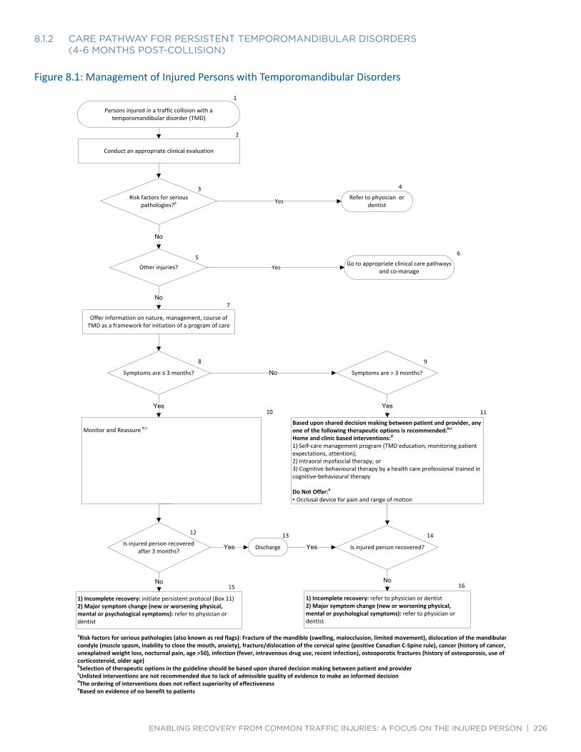
Management of Injured Persons with Temporomandibular Disorders
8.1.2 caRE PaThway foR PERsisTEnT TEmPoRomandibulaR disoRdERs (4-6 monThs PosT-collision)
Yes
No
Yes
No
No
Yes Yes
Yes Yes
No No
Enabling REcovERy fRom common TRaffic injuRiEs: a focus on ThE injuREd PERson | 226
Figure 8.1: Management of Injured Persons with Temporomandibular Disorders
1
Persons injured in a traffic collision with a temporomandibular disorder (TMD)
2
Conduct an appropriate clinical evaluation
3 Risk factors for serious
pathologies?a
4 Refer to physician or
dentist
5 Other injuries?
6 Go to appropriate clinical care pathways
and co‐manage
7
Offer information on nature, management, course of TMD as a framework for initiation of a program of care
8
Symptoms are ≤ 3 months?
9
Symptoms are > 3 months?
10
Monitor and Reassure b,c
11 Based upon shared decision making between patient and provider, any one of the following therapeutic options is recommended:b,c
Home and clinic based interventions:d
1) Self‐care management program (TMD education, monitoring patient expectations, attention); 2) Intraoral myofascial therapy; or 3) Cognitive‐behavioural therapy by a health care professional trained in cognitive‐behavioural therapy
Do Not Offer:e
• Occlusal device for pain and range of motion
12
Is injured person recovered after 3 months?
13
Discharge
14
Is injured person recovered?
15
1) Incomplete recovery: initiate persistent protocol (Box 11) 2) Major symptom change (new or worsening physical, mental or psychological symptoms): refer to physician or dentist
16
1) Incomplete recovery: refer to physician or dentist 2) Major symptom change (new or worsening physical, mental or psychological symptoms): refer to physician or dentist
a Risk factors for serious pathologies (also known as red flags): Fracture of the mandible (swelling, malocclusion, limited movement), dislocation of the mandibular condyle (muscle spasm, inability to close the mouth, anxiety), fracture/dislocation of the cervical spine (positive Canadian C‐Spine rule), cancer (history of cancer, unexplained weight loss, nocturnal pain, age >50), infection (fever, intravenous drug use, recent infection), osteoporotic fractures (history of osteoporosis, use of corticosteroid, older age) b Selection of therapeutic options in the guideline should be based upon shared decision making between patient and provider c Unlisted interventions are not recommended due to lack of admissible quality of evidence to make an informed decision d The ordering of interventions does not reflect superiority of effectiveness e Based on evidence of no benefit to patients

Enabling REcovERy fRom common TRaffic injuRiEs: a focus on ThE injuREd PERson | 227
SECTION 8.1.3
Key recOMMendatiOnS fOr the ManageMent Of recent OnSet teMPOrOMandibular diSOrderS
Most individuals with TMD recover on their own within a few weeks of the injury. However, it is recommended that the following be performed as a component of standard clinical care.
• Monitor the symptoms.
• Reassure patients about the benign and self-limited nature of their pain.
• Educate patients about the nature of their pain.
• Encourage patients to maintain their activities of daily living.
• Do not provide ineffective or experimental treatments.
SECTION 8.1.4
Key recOMMendatiOnS fOr the ManageMent Of PerSiStent teMPOrOMandibular diSOrderS
This section summarizes the key recommendations for the management of TMD for the period extending from 4 to 6 months post-collision. The wording of recommendations follows the guidance from the National Institute for Health and Care Excellence (NICE). Recommendations beginning with “offer” indicate that, according to the evidence, an intervention is associated with outcomes that were superior to other interventions, placebo/sham, or no intervention. The wording “consider” indicates that an intervention is as effective as another one. The wording “do not offer” indicates, according to the evidence, an intervention does not benefit patients. A detailed explanation of the wording of recommendations is presented in section 2.5.2.4 of this report.
• Provide care in partnership with the patient. Involve the patient in care planning and decision-making.
• Reassure patients about the benign and self-limited nature of their pain.
• Encourage patients to maintain their activities of daily living.
• Deliver time-limited care.
• Do not provide ineffective or experimental treatments.

Enabling REcovERy fRom common TRaffic injuRiEs: a focus on ThE injuREd PERson | 228
SECTION 8.1.4.1
Self-care ManageMent
Patient education aims to enable individuals to make informed decisions about their personal health-related behaviour. Structured education strategies refer to standardized interventions such as scripted discussion, pamphlets or videos. Educational interventions should begin with an assessment of the person’s knowledge of the injury and their health goals. The content of the structured education interventions may include (but is not limited to): reassurance about the favourable prognosis of TMD; advice on return to activities; maintenance of activities of daily living; discussion of expected pain and pain mechanism; discussion of prognosis; pain coping skills; and self-care strategies or general health.
Table 8.B: Self-care management for persistent temporomandibular disorders
Recommendation 8.1.4.1.1
Consider a maximum of 4 sessions over 8 weeks of structured self-care management program.*
References: • Decision Determinants and Evidence Table for TMD – Report 1 – Appendix 6
* Structured self-care management involving distribution of information about temporomandibular disorders and a patient manual on general health information (e.g. pain medications, communicating with health care providers, and making treatment decisions). Each session focused on reviewing main points of the manual and discussing the patient’s reactions and questions.
SECTION 8.1.4.2
SOft tiSSue theraPy
Soft tissue therapy is a mechanical therapy in which muscles, tendons, and ligaments are passively pressed or kneaded by hand or with mechanical devices. It includes relaxation massage, clinical massage, movement re-education and energy work.
Table 8.C: Soft tissue therapy for persistent temporomandibular disorders
Recommendation 8.1.4.2.1 Offer up to 10 sessions over 5 weeks of intraoral myofascial therapy.*
References: • Decision Determinants and Evidence Table for TMD – Report 1 – Appendix 6
* Intraoral myofascial therapy involving: a) intraoral temporalis release; b) intraoral medial and lateral pterygoid technique; and c) Intraoral spenopalatine ganglion technique.
Enabling REcovERy fRom common TRaffic injuRiEs: a focus on ThE injuREd PERson | 229
SECTION 8.1.4.3
PSychOlOgical interventiOn
A psychological intervention is a method used to treat psychological distress, consequences of musculoskeletal injuries (such as pain), or psychological disorders; primarily (but not exclusively) by verbal or non-verbal communication. Psychological interventions can be broadly subdivided into several theoretical orientations, including but not limited to psychodynamic, psychoanalytic, behavioural/cognitive behavioural, humanistic and existential, family/systems approaches and combinations of these approaches. Psychological interventions can include (but are not limited to) in-person psycho-education; booklet/written material that includes a psycho-educational component; cognitive-behavior interventions, or a guided psychological self-help intervention.
Table 8.D: Psychological interventions for persistent temporomandibular disorders
Recommendation 8.1.4.3.1
Consider a maximum of 4 sessions over 8 weeks of cognitive-behavioural therapy.*
References: • Decision Determinants and Evidence Table for TMD – Report 1 – Appendix 6
* Cognitive-behavioural pain management involves progressive relaxation and abdominal/diaphragmatic breathing techniques, a relaxation audiotape, discussion regarding fear-avoidance, the identification and challenging of negative thoughts in response to pain, relapse prevention, ways to maintain gains and how to deal with setbacks.
SECTION 8.1.4.4
PaSSive PhySical MOdalitieS
Passive physical modalities include two categories of interventions: physico-chemical and structural. Physico-chemical modalities use a thermal or electromagnetic agent to affect the body at or beneath the skin level. Structural modalities include functional or non-functional assistive devices. Functional assistive devices intend to align, support or otherwise indirectly facilitate function in the affected region. Non-functional devices intend to achieve a state of rest in specific anatomic positions or prevent movement.
Table 8.E: Passive physical modalities for persistent temporomandibular disorders
Recommendation 8.1.4.4.1
Do not offer an occlusal device.*
References: • Decision Determinants and Evidence Table for TMD – Report 1 – Appendix 6
* An occlusal device includes any removable artificial occlusal surface used to affect the relationship of the mandible to the maxillae. (http://www.academyofprosthodontics.org/_Library/ap_articles_download/GPT8.pdf)
Enabling REcovERy fRom common TRaffic injuRiEs: a focus on ThE injuREd PERson | 230
SECTION 9.0
RECOMMENDATION FOR THE CLINICAL MANAGEMENT OF MILD TRAUMATIC BRAIN INJURY (MTBI)
SECTION 9.0
recOMMendatiOn fOr the clinical ManageMent Of Mild trauMatic brain injury (Mtbi)
Enabling REcovERy fRom common TRaffic injuRiEs: a focus on ThE injuREd PERson | 231
SECTION 9.1
bacKgrOund
Mild traumatic brain injury (MTBI) is an acute brain injury resulting from mechanical energy to the head from external physical forces [1]. MTBI is common with 70-90% of all treated brain injuries being considered as mild [1]. In Ontario, Canada, the incidence of MTBI presenting to emergency departments or family physicians ranges from 493 per 100,000 to 653 per 100,000 [2]. The best available evidence suggests that most individuals with MTBI substantially improve or recover within a few months [3, 4].
MTBI presents clinically with physical (e.g., headache, nausea, dizziness), behavioural/emotional (e.g., fatigue, depression, sleep problems), and cognitive symptoms (e.g., feeling slowed down, concentration, memory difficulties) [5]. Although most individuals with MTBI recover within days to months, cognitive deficits may persist past six months [3]. These individuals may continue to experience decreased functional ability, emotional distress, and delayed return to work or school [6]. Therefore, effectively managing patients with MTBI is important to prevent chronic symptoms and disability.
The clinical management of MTBI remains controversial [7] and evidence-based clinical practice guidelines have been designed to assist with the management of MTBI. Clinical practice guidelines are systematically developed statements designed to help clinicians provide quality care to patients [8, 9]. However, the quality of commonly used guidelines varies greatly [10]. Therefore, the methodological quality of guidelines should be assessed prior to their use in practice for patients.
A recent systematic review of clinical practice guidelines found that only 50% of guidelines available to inform the management of traffic injuries meet accepted quality standards [10]. Of those, three addressed the early management of MTBI [6, 11, 12]. One of these guidelines entitled “Guidelines for Mild Traumatic Brain Injury and Persistent Symptoms, First Edition” was published in 2008 by the Ontario Neurotrauma Foundation [12]. In 2013, a revised version of the guideline (Guidelines for Concussion/Mild Traumatic Brain Injury and Persistent Symptoms, Second Edition) was published [13].
The availability of a high quality, Ontario-based and current clinical practice guideline for the management of MTBI is relevant to this project. Therefore, the guideline expert panel recommended that a detailed evaluation of the “Guidelines for Concussion/Mild Traumatic Brain Injury and Persistent Symptoms, Second Edition” [13] is conducted to determine its methodological quality and applicability.
This report summarizes the evaluation of the “Guidelines for Concussion/Mild Traumatic Brain Injury and Persistent Symptoms, Second Edition” [13] and recommends that the guideline be used for the management of MTBI resulting from traffic collisions in Ontario.
The summary report is available in appendix 7.

Enabling REcovERy fRom common TRaffic injuRiEs: a focus on ThE injuREd PERson | 232
SECTION 9.2
review Panel
Dr. Pierre Côté (Chair) formed a seven member multidisciplinary review panel to evaluate the quality of the “Guidelines for Concussion/Mild Traumatic Brain Injury and Persistent Symptoms, Second Edition” [13]. Dr. Jessica Wong chaired the review panel. Each member independently appraised the quality of the guidelines using the Appraisal of Guidelines for Research and Evaluation version II (AGREE II) instrument [14-16].
SECTION 9.3
critical aPPraiSal Of the Mtbi guidelineS
The AGREE II instrument was used to evaluate the guideline. The AGREE II instrument is used internationally to assess the development and reporting of guidelines. It was developed in 2003 by the AGREE Collaboration, which is an international team of guideline developers and researchers [14-16]. The AGREE II has been used to evaluate guidelines for the management of various conditions, including cancer, osteoarthritis, cardiac conditions, stroke, and chronic pain. This instrument has also been found to be valid and reliable [14-16]. The AGREE II instrument includes 23 items and six quality-related domains. Each domain assesses the methodological quality and reporting in the following areas: 1) scope and purpose; 2) stakeholder involvement; 3) rigour of development; 4) clarity of presentation; 5) applicability; and 6) editorial independence.
SECTION 9.4
reSultS Of the review
The review panel rated the overall quality of the guideline as high. The review panel stated that the clinical recommendations in the guideline were relevant to health care professionals who manage MTBI related to traffic collisions. Therefore, the review panel unanimously recommended that the guidelines should be used to guide the treatment and rehabilitation of MTBI related to traffic collisions. The recommendation was approved by the Guideline Expert Panel.
SECTION 9.5
ManageMent Of Mtbi
Health care professionals who care for patients with MTBI should follow the recommendations outlined in the “Guidelines for Concussion/Mild Traumatic Brain Injury and Persistent Symptoms, Second Edition” [13].

Enabling REcovERy fRom common TRaffic injuRiEs: a focus on ThE injuREd PERson | 233
SECTION 9.6
referenceS
1. Cancelliere C, Cassidy JD, Cote P, et al. Protocol for a systematic review of prognosis after mild traumatic brain injury: an update of the WHO Collaborating Centre Task Force findings. Syst Rev 2012;1:17.
2. Ryu WH, Feinstein A, Colantonio A, Streiner DL, Dawson DR. Early identification and incidence of mild TBI in Ontario. Can J Neurol Sci 2009;36:429-35.
3. Carroll LJ, Cassidy JD, Cancelliere C, et al. A systematic review of the prognosis after mild traumatic brain injury in adults: cognitive, psychiatric and mortality outcomes. Results of the International Collaboration on MTBI Prognosis (ICoMP). Archives of Physical Medicine and Rehabilitation 2013: in press.
4. Cassidy JD, Cancelliere C, Carroll LJ, et al. Systematic review of self-reported prognosis in adults after mild traumatic brain injury: results of the International Collaboration on Mild Traumatic Brain Injury Prognosis. Arch Phys Med Rehabil 2014;95:S132-51.
5. Willer B, Leddy JJ. Management of concussion and post-concussion syndrome. Curr Treat Options Neurol 2006;8:415-26.
6. Motor Accidents Authority of NSW (MAA NSW). Guidelines for Mild Traumatic Brain Injury following a Closed Head Injury. 2008.
7. Nygren-de Boussard C, Holm LW, Cancelliere C, et al. Nonsurgical interventions after mild traumatic brain injury: a systematic review. Results of the International Collaboration on Mild Traumatic Brain Injury Prognosis. Arch Phys Med Rehabil 2014;95:S257-64.
8. Shekelle PG, Woolf SH, Eccles M, Grimshaw J. Clinical guidelines: developing guidelines. BMJ 1999;318:593-6.
9. Shekelle P, Woolf S, Grimshaw JM, Schunemann HJ, Eccles MP. Developing clinical practice guidelines: reviewing, reporting, and publishing guidelines; updating guidelines; and the emerging issues of enhancing guideline implementability and accounting for comorbid conditions in guideline development. Implement Sci 2012;7:62.
10. Alonso-Coello P, Irfan A, Sola I, et al. The quality of clinical practice guidelines over the last two decades: a systematic review of guideline appraisal studies. Qual Saf Health Care 2010;19:e58.
11. Agency for Healthcare Research and Quality (AHRQ).Triage, assessment, investigation and early management of head injury in infants, children and adults. 2007.
12. Ontario Neurotrauma Foundation. Guidelines for Mild Traumatic Brain Injury and Persistent Symptoms. 2008.
13. Ontario Neurotrauma Foundation. Guidelines for Concussion/Mild Traumatic Brain Injury and Persistent Symptoms: Second Edition. 2013.
14. Brouwers MC, Kho ME, Browman GP, et al. AGREE II: advancing guideline development, reporting and evaluation in health care. CMAJ 2010;182:E839-42.
15. Brouwers MC, Kho ME, Browman GP, et al. Development of the AGREE II, part 1: performance, usefulness and areas for improvement. CMAJ 2010;182:1045-52.
16. Brouwers MC, Kho ME, Browman GP, et al. Development of the AGREE II, part 2: assessment of validity of items and tools to support application. CMAJ 2010;182:E472-8.

Enabling REcovERy fRom common TRaffic injuRiEs: a focus on ThE injuREd PERson | 234
SECTION 10.0
GUIDELINE FOR THE CLINICAL MANAGEMENT OF LOw BACk PAIN wITH AND wITHOUT RADICULOPATHY

SECTION 10.0
guideline fOr the clinical ManageMent Of lOw bacK Pain with and withOut radiculOPathy
Enabling REcovERy fRom common TRaffic injuRiEs: a focus on ThE injuREd PERson | 235
10.1 Management of non-specific low back pain 10.1.1 Care pathway for recent onset non-specific low back pain (0-3 months post-collision) 10.1.2 Care pathway for persistent non-specific low back pain (4-6 months post-collision) 10.1.3 Key recommendations for the management of recent onset non-specific low back pain 10.1.4 Key recommendations for the management of persistent non-specific low back pain 10.2 Management of lumbar disc herniation with radiculopathy 10.2.1 Care pathway for recent onset lumbar disc herniation with radiculopathy (0-3 months post-collision) 10.2.2 Care pathway for persistent lumbar disc herniation with radiculopathy (4-6 months post-collision) 10.2.3 Key recommendations for the management of recent onset lumbar disc herniation with radiculopathy 10.2.4 Key recommendations for the management of persistent lumbar disc herniation with radiculopathy
This evidence-based guideline establishes the best practice for the clinical management of non-specific low back pain that is caused or exacerbated by a motor vehicle collision. This guideline covers recent onset (0-3 months post-collision) and persistent (4-6 months post-collision) non-specific low back pain; it does not cover non-specific low back pain that persists for more than 6 months post-collision.
In this guideline, non-specific low back pain is defined as low back pain with or without radiculopathy in the absence of specific pathological entities (i.e., fracture, dislocation, neoplasm, infection, or systemic disease). The clinical management of low back pain with or without radiculopathy is outlined in sections 10.1 and 10.2.
This guideline is not indicated for conditions that include the presence of major structural or other pathological causes of low back pain.
In Canada, 60% of people with neck pain and associated disorders related to motor vehicle collisions experience low back pain. Most people recover from low back pain.
The clinical management recommended in this guideline aims to: 1) accelerate recovery; 2) reduce the intensity of symptoms; 3) promote early restoration of function; 4) prevent chronic pain and disability; 5) improve health-related quality of life; 6) reduce recurrences; and 7) promote active participation of patients in their care.
Patients with multiple injuries should be managed using all appropriate care pathways. For example, patients with low back pain commonly suffer from neck pain. Patients with low back pain and neck pain and its associated disorders (NAD) should also receive care as recommended in the NAD care pathways described in Chapter 4.
Patient-centered care is an internationally recognized principle that was fundamental to the development of this guideline. This guideline reinforces the importance of communication and partnership between patients and health care professionals.
All recommendations included in this guideline are derived from a synthesis of high quality clinical practice guidelines.
10.0 guidElinE foR ThE clinical managEmEnT of non-sPEcific low back Pain
Enabling REcovERy fRom common TRaffic injuRiEs: a focus on ThE injuREd PERson | 236
Interventions not described in this guideline are not recommended for the management of patients with non-specific low back pain because of a lack of evidence about their effectiveness and safety.
Health care professionals eligible to provide care under this guideline are those defined by the Statutory Accident Benefits Schedules (SABS).
This guideline is organized into two sections. Each section provides evidence-based recommendations for the clinical management of non-specific low back pain:
• Section 10.1 - Management of non-specific low back pain • Section 10.2 - Management of lumbar disc herniation with radiculopathy
All recommendations presented in this guideline integrate the: • Key decision determinants based upon the framework developed by Ontario Health Technology Advisory
Committee (OHTAC); • Best evidence obtained from a critical review of current scientific literature; and • Qualitative research exploring the experiences of persons treated for traffic injuries in Ontario
All background documents and references are available at http://www.fsco.gov.on.ca

Enabling REcovERy fRom common TRaffic injuRiEs: a focus on ThE injuREd PERson | 237
SECTION 10.1
ManageMent Of nOn-SPecific lOw bacK Pain
Quick Reference Guide – Management of Non-specific Low Back Pain
Symptoms ≤ 3 months post‐collision Symptoms > 3 months post‐collisionFor all injured persons with non‐specific low back pain: Rule out risk factors for serious pathologiesa
Offer information on nature, management, course of non‐ specific low back pain as a framework for initiation of a program of careConduct ongoing assessment for symptom improvement or worsening/progression during intervention and refer accordinglyReassess and Monitor for presence of depression, passive coping strategies, job dissatisfaction, higher disability levels, disputed compensation claims, or somatization.Discharge injured person as appropriate at any point during intervention and recovery
Based upon shared decision making between the patient and provider, any one of the following therapeutic interventions is recommended:b,c
Home and clinic based interventions: Structured education (advice to stay active), reassurance, and:
1. Manipulation2. Muscle Relaxantsd
Refer to specific recommendation for treatment details (Section 10.1.3)
Based upon shared decision making between the patient and provider, any one of the following therapeutic interventions is recommended:b,c
Home and clinic based interventions: Structured education (advice to stay active), reassurance, and:
1. Exercise2. Manipulation or mobilization3. Clinical or relaxation massage4. Non‐ steroidal anti‐ inflammatory drugsd
5. Needle acupuncture6. Multimodal care (that includes the combination of (for patients who have high levels of disability or significant distress):
a)Exerciseb) Cognitive/behavioural approaches
Refer to specific recommendation for treatment details (Section 10.1.4)
Do Not Offer:e
Passive physical modalities Botulinum toxin injections
Outcome: Recovered → Discharge Unrecovered: Incomplete recovery → Initiate persistent protocol
Signs of lumbar disc herniation with radiculopathy → lumbar disc herniation with radiculopathy care pathwaySigns progress to serious pathology (new or worsening physical, mental or psychological symptoms) → Refer to physician
Outcome: Recovered → Discharge Unrecovered: Incomplete recovery Refer to physician
Signs of lumbar disc herniation with radiculopathy → lumbar disc herniation with radiculopathy care pathwaySigns progress to serious pathology (new or worsening physical, mental or psychological symptoms) → Refer to physician
a Risk factors for serious pathologies (also known as red flags): Cancer (history of cancer, unexplained weight loss, nocturnal pain, age >50), vertebral infection (fever, intravenous drug use, recent infection), cauda equina syndrome (urinary retention, motor deficits at multiple levels, fecal incontinence, saddle anesthesia), osteoporotic fractures (history of osteoporosis, use of corticosteroid, older age), ankylosing spondylitis (morning stiffness, improvement with exercise, alternating buttock pain, awakening due to back pain during the second part of the night, younger age), inflammatory arthritis (morning stiffness, swelling in multiple joints)b This guideline does not include interventions for which there is a lack of evidence of effectivenessc The ordering of interventions does not reflect superiority of effectivenessd The evidence indicates that analgesia is the primary therapeutic benefit of the muscle relaxant and NSAID classes of medication. Pain reduction should be apparent during the initial period of usage; in the absence of therapeutic benefit, prolongation of usage is not warranted. There is no evidence of differential efficacy for the various drugs within each class. There is also no evidence that any combination of these medications provides added benefit. There are potentially significant adverse effects associated with use of these classes of medications. Finally, the non‐opioid first ‘step’ in the Analgesic Ladder includes NSAIDs, muscle relaxant and acetaminophen (Vargas‐Schaffer G. Is the WHO analgesic ladder still valid? Twenty‐f our years of experience. Vol 56: June 2010 Canadian Family Physician). However, the evidence does not indicate that acetaminophen is an effective analgesic for either NAD or low back pain; therefore, the use of acetaminophen is not recommended. e Based on evidence of no benefit to patients
SECTION 10.1.1
care Pathway fOr recent OnSet nOn-SPecific lOw bacK Pain (0 - 3 MOnthS POSt-cOlliSiOn)
The care pathway is presented in Figure 10.1.
At initial contact, health care professionals should educate and reassure the patient that non-specific low back pain will resolve within a few months of symptom onset. Patients greatly improve their recovery by actively participating in their care. Clinical care aims to accelerate recovery by reducing pain and improving function. The care pathway recommended for the first three months of care for non-specific low back pain is described below.

10.1.1 caRE PaThway foR REcEnT onsET non-sPEcific low back Pain (0 - 3 monThs PosT-collision)
Enabling REcovERy fRom common TRaffic injuRiEs: a focus on ThE injuREd PERson | 238
Assess the Patient
Conduct an appropriate clinical evaluation to rule out major structural or other pathologies as the cause of the symptoms. The presence of a risk factor for serious pathologies (also known as red flags) identified during the history and examination warrants further investigation and referral to the appropriate health care professional. However, once pathology has been ruled out, the patient should be treated according to the non-specific low back pain care pathway.
Table 10.A Risk factors of serious pathology (red flags) for low back pain
Possible Cause Risk factors of serious pathology identified during history or physical examination*
Cancer • History of cancer • Unexplained weight loss • Nocturnal pain • Age > 50
Vertebral infection • Fever • Intravenous drug use • Recent infection
Cauda equina syndrome • Urinary retention • Motor deficits at multiple levels • Fecal incontinence • Saddle anesthesia
Osteoporotic fractures • History of osteoporosis • Use of corticosteroid • Older age
Ankylosing spondylitis • Morning stiffness • Improvement with exercise • Alternating buttock pain • Awakening due to back pain during the second part of the night • Younger age
Inflammatory arthritis • Morning stiffness • Swelling in multiple joints
* Adapted from Chou et al. Diagnosis and Treatment of Low Back Pain: A Joint Clinical Practice Guideline from the American College of Physicians and the American Pain Society. Ann Intern Med. 2007;147:478-491.

10.1.1 caRE PaThway foR REcEnT onsET non-sPEcific low back Pain (0 - 3 monThs PosT-collision)
Enabling REcovERy fRom common TRaffic injuRiEs: a focus on ThE injuREd PERson | 239
If neurological signs related to a lumbar disc herniation with radiculopathy are present, the patient should be managed under the “Care Pathway for the Management of Lumbar Disc Herniation with Radiculopathy” (see section 10.2).
Assess the Prognostic Factors
Assess the prognostic factors for delayed recovery. Most patients recover from their injury. Patients with the following prognostic factors may have a higher risk for delayed recovery:*
* Chou et al. Diagnosis and Treatment of Low Back Pain: A Joint Clinical Practice Guideline from the American College of Physicians and the American Pain Society. Ann Intern Med. 2007;147:478-491
• Depression • Passive coping strategies • Job dissatisfaction • High disability levels • Disputed compensation claims • Somatization
Educate and Reassure the Patient
Develop a patient-centred care plan in partnership with the patient.
Health care professionals need to reassure patients that there are no major structural or progressive pathologies (such as a fracture) in their back.
Prognostic factors for poor recovery should be addressed when present. The care should start with education and reassurance about the benign and self-limited nature of low back pain and the importance of maintaining activity and movement. This is particularly important when the patient reports poor expectation of recovery.
It is also important to reassure patients that it is normal to feel some anxiety, distress or anger following a traffic collision. In the presence of such symptoms or emotions, the health care professional should listen to the patient’s concerns, discuss them and adjust the care plan accordingly.
Determine if Ongoing Clinical Care is Necessary
Health care professionals should first determine if the patient requires ongoing clinical care. Patients with mild low back pain may not require ongoing clinical care. Rather, patients can be managed with reassurance, education, returning to usual activities as tolerated, and staying active.
Deliver the Care Plan for Recent Onset Non-specific Low Back Pain (0-3 months post-collision)
Patients who require ongoing clinical care should be encouraged to actively participate in their care by returning to usual activities as tolerated and staying active on a regular basis. Based upon shared decision making between the patient and provider, any one of the following therapeutic interventions is recommended:
10.1.1 caRE PaThway foR REcEnT onsET non-sPEcific low back Pain (0 - 3 monThs PosT-collision)
Enabling REcovERy fRom common TRaffic injuRiEs: a focus on ThE injuREd PERson | 240
• Structured patient education • Manipulation • Muscle relaxants
Discuss the risks, benefits, and adverse events of selected interventions in the care plan with the patient.
Reassess and Take the Indicated Course of Action
Reassess the patient at every visit to determine if additional care is necessary, or if the condition is worsening.
Patients should be discharged as soon as they report significant improvement or recovery. It is recommended that health care professionals use the self-rated recovery question to measure patient recovery: “How well do you feel you are recovering from your injuries?” The response options include: 1) completely better, 2) much improved, 3) slightly improved, 4) no change, 5) slightly worse, 6) much worse, and 7) worse than ever. Patients reporting to be ‘completely better’ or ‘much improved’ should be considered recovered. Patients who have not recovered should follow the care pathway outlined in the guideline.*
* The use of a valid and reliable condition-specific instrument (e.g. Oswestry Disability Index) is encouraged but should not be used to measure overall recovery.
Patients who develop lumbar disc herniation with radiculopathy should be managed according to the care pathway for the management of lumbar disc herniation with radiculopathy (section 10.2).
Patients with worsening of symptoms and those who develop new physical, mental or psychological symptoms (other than lumbar disc herniation with radiculopathy) should be referred to their physician for further evaluation at any time point during their care.
Patients who have not significantly improved or recovered within the first 3 months after the injury should enter the care pathway for persistent non-specific low back pain described in section 10.1.2.
SECTION 10.1.2
care Pathway fOr PerSiStent nOn-SPecific lOw bacK Pain (4 - 6 MOnthS POSt-cOlliSiOn)
The care pathway is presented in Figure 10.1.
Patients who still experience symptoms and disability more than 3 months after the injury may benefit from receiving additional clinical care. The primary goals of the clinical care are to promote recovery by reducing symptoms and return patients to their normal activities of daily living. The care plan should focus on exercise and movement, but can be supplemented by a short course of passive care.

10.1.2 caRE PaThway foR PERsisTEnT non-sPEcific low back Pain (4 - 6 monThs PosT-collision)
Enabling REcovERy fRom common TRaffic injuRiEs: a focus on ThE injuREd PERson | 241
Assess the Patient
Conduct an appropriate clinical evaluation to rule out major structural or other pathologies as the cause of the symptoms. The presence of a risk factor for serious pathologies (also known as red flags) identified during the history and examination warrants further investigation and referral to the appropriate health care professional. However, once pathology has been ruled out, the patient should be treated according to the non-specific low back pain care pathway.
Table 10.A Risk factors of serious pathology (red flags) for low back pain
Possible Cause Risk factors of serious pathology identified during history or physical examination*
Cancer • History of cancer • Unexplained weight loss • Nocturnal pain • Age > 50
Vertebral infection • Fever • Intravenous drug use • Recent infection
Cauda equina syndrome • Urinary retention • Motor deficits at multiple levels • Fecal incontinence • Saddle anesthesia
Osteoporotic fractures • History of osteoporosis • Use of corticosteroid • Older age
Ankylosing spondylitis • Morning stiffness • Improvement with exercise • Alternating buttock pain • Awakening due to back pain during the second part of the night • Younger age
Inflammatory arthritis • Morning stiffness • Swelling in multiple joints
* Adapted from Chou et al. Diagnosis and Treatment of Low Back Pain: A Joint Clinical Practice Guideline from the American College of Physicians and the American Pain Society. Ann Intern Med. 2007;147:478-491.
If neurological signs related to a lumbar disc herniation with radiculopathy are present, the patient should be managed under the “Care Pathway for the Management of Lumbar Disc Herniation with Radiculopathy” (see section 10.2).

10.1.2 caRE PaThway foR PERsisTEnT non-sPEcific low back Pain (4 - 6 monThs PosT-collision)
Enabling REcovERy fRom common TRaffic injuRiEs: a focus on ThE injuREd PERson | 242
Assess the Prognostic Factors
Assess the prognostic factors for delayed recovery. Most patients recover from their injury. Patients with the following prognostic factors may have a higher risk for delayed recovery*:
* Chou et al. Diagnosis and Treatment of Low Back Pain: A Joint Clinical Practice Guideline from the American College of Physicians and the American Pain Society. Ann Intern Med. 2007;147:478-491
• Depression • Passive coping strategies • Job dissatisfaction • High disability levels • Disputed compensation claims • Somatization
Educate and Reassure the Patient
Develop a patient-centred care plan in partnership with the patient.
Health care professionals need to reassure patients that there are no major structural or progressive pathologies (such as a fracture) in their back.
Prognostic factors for poor recovery should be addressed when present. The care should start with education and reassurance about the benign and self-limited nature of low back pain and the importance of maintaining activity and movement. This is particularly important when the patient reports poor expectation of recovery.
It is also important to reassure patients that it is normal to feel some anxiety, distress or anger following a traffic collision. In the presence of such symptoms or emotions, the health care professional should listen to the patient’s concerns, discuss them and adjust the care plan accordingly.
Deliver the Care Plan for Persistent Non-specific Low Back Pain (4-6 months post-collision)
The goal of the care plan is to promote activity through exercise and clinical interventions that promote resolution of symptoms and restoration of function. Patients requiring clinical care should be encouraged to participate in their program of care by remaining active and returning to usual activities as tolerated.
Health care professionals should discuss treatment options with their patients and, through a process of shared decision making, determine which therapeutic option they wish to pursue. Based upon shared decision making between the patient and provider, any one of the following therapeutic interventions is recommended:
• Structured patient education • Exercise • Manipulation or mobilization • Non-steroidal anti-inflammatory drugs

10.1.2 caRE PaThway foR PERsisTEnT non-sPEcific low back Pain (4 - 6 monThs PosT-collision)
Enabling REcovERy fRom common TRaffic injuRiEs: a focus on ThE injuREd PERson | 243
• Massage • Acupuncture • Multimodal care that includes the combination of exercise and cognitive/behavioural approaches for
patients who have high levels of disability or significant distress
Interventions that are not recommended include:
• Passive physical modalities • Botulinum toxin injections
Discuss the risks, benefits, and adverse effects of selected interventions in the care plan with the patient.
Reassess and Take the Indicated Course of Action
Reassess the patient at every visit to determine if additional care is necessary, or if the condition is worsening.
Patients should be discharged as soon as they report significant improvement or recovery. It is recommended that health care professionals use the self-rated recovery question to measure patient recovery: “How well do you feel you are recovering from your injuries?” The response options include: 1) completely better, 2) much improved, 3) slightly improved, 4) no change, 5) slightly worse, 6) much worse, and 7) worse than ever. Patients reporting to be ‘completely better’ or ‘much improved’ should be considered recovered. Patients who have not recovered should follow the care pathway outlined in the guideline*.
* The use of a valid and reliable condition-specific instrument (e.g. Oswestry Disability Index) is encouraged but should not be used to measure overall recovery.
Patients who develop lumbar disc herniation with radiculopathy should be managed according to the care pathway for the management of lumbar disc herniation with radiculopathy (section 10.2).
Patients with worsening of symptoms and those who develop new physical, mental or psychological symptoms (including lumbar disc herniation with radiculopathy) should be referred to their physician for further evaluation. Patients who have not significantly improved or recovered should be referred to the physician for further evaluation at any time point during their care.

10.1.2 caRE PaThway foR PERsisTEnT non-sPEcific low back Pain (4 - 6 monThs PosT-collision)
Yes
No
No
Yes
Yes
No
No
Yes
No
YesYes
Yes Yes
No No
Enabling REcovERy fRom common TRaffic injuRiEs: a focus on ThE injuREd PERson | 244
Figure 10.1: Care Pathway for the Management of Non-specific Low Back Pain 1
Persons injured in a traffic collision with non-specific low
back pain
2
Conduct an appropriate clinical evaluation
3Risk factors for serious pathologies?a
4 Refer to physician
5
Non-specific low back pain
6
Refer to physician
7 Poor prognostic factors?b
8 Adjust
modifiable prognostic
factors
9
Offer information on nature, management, course of non-specific low back pain as a framework for initiation of a program of care.
10 Is treatment required?
11
Discharge
12 Are symptoms ≤3 months?
13
Symptoms are > 3 months.
14
Based upon shared decision making between the patient and provider, any one of the following therapeutic interventions is recommended:c,d
Home and clinic based interventions: Structured education (advice to stay active), reassurance, and: 1) Manipulation 2) Muscle relaxantsf
Refer to specific recommendation for treatment details (Section 10.1.3)
15
Based upon shared decision making between the patient and provider, any one of the following therapeutic interventions is recommended:c,d,e
Home and clinic based interventions: Structured education (advice to stay active), reassurance, and: 1) Exercise 2) Manipulation or mobilization 3) Clinical or relaxation massage 4) Non-steroidal anti-inflammatory drugs (NSAIDs)f
5) Needle acupuncture 6) Multimodal care that includes the combination of (for patients who have high levels
of disability or significant distress): a) Exercise b) Cognitive/behavioural approaches
Do not offere : 1) Passive physical modalities 2) Botulinum toxin injections Refer to specific recommendation for treatment details (Section 10.1.4)
16 Is injured person recovered
after 3 months?
17
Discharge
18
Is injured person recovered?
19 1) Incomplete recovery: Initiate persistent protocol (Box 15) 2) Signs of lumbar disc herniation with radiculopathy: proceed to lumbar disc herniation with radiculopathy care pathway 3) Signs progress to serious pathology (new or worsening physical, mental or psychological symptoms): Refer to physician
20 1) Incomplete recovery: refer to physician 2) Signs of lumbar disc herniation with radiculopathy: proceed to lumbar disc herniation with radiculopathy care pathway 3) Signs progress to serious pathology (new or worsening physical, mental or psychological symptoms): Refer to physician
a Risk factors for serious pathologies (also known as red flags): Cancer (history of cancer, unexplained weight loss, nocturnal pain, age >50), vertebral infection (fever, intravenous drug use, recent infection), cauda equina syndrome (urinary retention, motor deficits at multiple levels, fecal incontinence, saddle anesthesia), osteoporotic fractures (history of osteoporosis, use of corticosteroid, older age), ankylosing spondylitis (morning stiffness, improvement with exercise, alternating buttock pain, awakening due to back pain during the second part of the night, younger age), inflammatory arthritis (morning stiffness, swelling in multiple joints) b Factors delaying recovery: Depression, passive coping strategies, job dissatisfaction, high disability levels, disputed compensation claims, somatization c This guideline does not include interventions for which there is a lack of evidence of effectiveness d The ordering of interventions does not reflect superiority of effectiveness e Based on evidence of no benefit to patients f The evidence indicates that analgesia is the primary therapeutic benefit of the muscle relaxant and NSAID classes of medication. Pain reduction should be apparent during the initial period of usage; in the absence of therapeutic benefit, prolongation of usage is not warranted. There is no evidence of differential efficacy of the various drugs within each class. There is also no evidence that any combination of these medications provides added benefit. There are potentially significant adverse effects associated with use of these classes of medications. Finally, the non-opioid first ‘step’ in the Analgesic Ladder includes NSAIDs, muscle relaxant, and acetaminophen (Vargas-Schaffer G. Is the WHO analgesic ladder still valid? Twenty four years of experience. Vol 56: June 2010 Canadian Family Physician). However, the evidence does not indicate that acetaminophen is an effective analgesic for either NAD or low back pain; therefore, the use of acetaminophen is not recommended.

Enabling REcovERy fRom common TRaffic injuRiEs: a focus on ThE injuREd PERson | 245
SECTION 10.1.3
Key recOMMendatiOnS fOr the ManageMent Of recent OnSet nOn-SPecific lOw bacK Pain
This section summarizes the key recommendations for the management of non-specific low back pain for the first 3 months post-collision. The wording of recommendations follows the guidance from the National Institute for Health and Care Excellence (NICE). Recommendations beginning with “offer” indicate that, according to the evidence, an intervention is associated with outcomes that were superior to other interventions, placebo/sham, or no intervention. The wording “consider” indicates that an intervention is as effective as another one. The wording “do not offer” indicates that, according to the evidence, an intervention does not benefit patients. A detailed explanation of the wording of recommendations is presented in section 2.5.2.4 of this report.
• Provide care in partnership with the patient. Involve the patient in care planning and decision-making.
• Reassure patients about the benign and self-limited nature of their pain.
• Educate patients about the benefits of being actively engaged and participating in their care plan by remaining active and continuing movements.
• Emphasize active rather than passive treatments.
• Deliver time-limited care.
• Do not provide ineffective or experimental treatments.
SECTION 10.1.3.1
Structured Patient educatiOn
Structured patient education aims to enable individuals to make informed decisions about their personal health-related behaviour. Structured education strategies refer to standardized interventions such as scripted discussion, pamphlets or videos. Educational interventions should begin with an assessment of the person’s knowledge of the injury and their health goals. The content of the structured education interventions may include (but is not limited to): reassurance about the favourable prognosis of non-specific low back pain; advice on return to usual activities, including work; instruction of exercise; discussion of expected pain and pain mechanism; discussion of prognosis; pain coping skills; discussion of workplace ergonomics; and self-care strategies or general health.

10.1.3.1 sTRucTuREd PaTiEnT EducaTion
Enabling REcovERy fRom common TRaffic injuRiEs: a focus on ThE injuREd PERson | 246
Table 10.B: Structured patient education for recent onset non-specific low back pain
Recommendation 10.1.3.1.1
Provide information about the nature, management, and course of non-specific low back pain as a framework for the initiation of the program of care.
10.1.3.1.2 Consider a structured patient education program as an adjunct to an effec-tive program of care based on individual patient presentation.*
References: • Decision Determinants and Evidence Table for the Systematic Review of Low Back Pain Guidelines –
Report 1 - Appendix 8
* The structured education program should focus on providing advice to stay active, returning to activities as tolerated, avoiding prescribed bed rest, reassuring the patient by addressing the expectation of recovery, and instruction on effective self-care options for pain management.
SECTION 10.1.3.2
Manual theraPy
Manual therapy refers to techniques that involve the application of hands-on and/or mechanically assisted treatments, including manipulation, mobilization and traction.
Table 10.C: Manual therapy for recent onset non-specific low back pain
Recommendation 10.1.3.2.1 Consider a maximum of seven sessions over one month of manipulation.*
References: • Cherkin DC, Deyo RA, Battie M, Street J, Barlow W. A comparison of physical therapy, chiropractic
manipulation, and provision of an educational booklet for the treatment of patients with low back pain. New Engl J Med 1998; 339(15): 1021-1029.
• Decision Determinants and Evidence Table for Low Back Pain Guidelines – Report 1 - Appendix 8
* Manipulation includes techniques incorporating a high velocity, low amplitude impulse or thrust applied at or near the end of a joint’s passive range of motion.

Enabling REcovERy fRom common TRaffic injuRiEs: a focus on ThE injuREd PERson | 247
SECTION 10.1.3.3
MedicatiOn
Medications covered in this guideline include non-opioid analgesics (acetaminophen), non-steroidal anti-inflammatory drugs (NSAIDs), and muscle relaxants.
Table 10.D: Medication for recent onset non-specific low back pain
Recommendation 10.1.3.3.1 Consider muscle relaxants*
References: • Decision Determinants and Evidence Table for the Systematic Review on Muscle Relaxants for Neck
Pain and Low Back Pain – Report 4 – Appendix 8 • Decision Determinants and Evidence Table for the Systematic Review of Non-opioid Analgesic Drugs
for Neck and Low Back Pain – Report 2 - Appendix 8 • Decision Determinants and Evidence Table for the Systematic Review of Non-steroidal Anti-inflam-
matory Drugs for Neck and Low Back Pain – Report 3 - Appendix 8
* The evidence indicates that analgesia is the primary therapeutic benefit of the muscle relaxant and NSAID classes of medication. Pain reduction should be apparent during the initial period of usage; in the absence of therapeutic benefit, prolongation of usage is not warranted. There is no evidence of differential efficacy for the various drugs within each class. There is also no evidence that any combination of these medications provides added benefit. There are potentially significant adverse effects associated with use of these classes of medications. Finally, the non-opioid first ‘step’ in the Analgesic Ladder includes NSAIDs, muscle relaxant and acetaminophen (Vargas-Schaffer G. Is the WHO analgesic ladder still valid? Twenty-four years of experience. Vol 56: June 2010 Canadian Family Physician). However, the evidence does not indicate that acetaminophen is an effective analgesic for either NAD or low back pain; therefore, the use of acetaminophen is not recommended.
SECTION 10.1.4
Key recOMMendatiOnS fOr the ManageMent Of PerSiStent nOn-SPecific lOw bacK Pain
This section summarizes the key recommendations for the management of non-specific low back pain for the period extending from 4 to 6 months post-collision. The wording of recommendations follows the guidance from the National Institute for Health and Care Excellence (NICE). Recommendations beginning with “offer” indicate that, according to the evidence, an intervention is associated with outcomes that were superior to other interventions, placebo/sham, or no intervention. The wording “consider” indicates that an intervention is as effective as another one. The wording “do not offer” indicates, according to the evidence, an intervention does not benefit patients. A detailed explanation of the wording of recommendations is presented in section 2.5.2.4 of this report.

10.1.4 kEy REcommEndaTions foR ThE managEmEnT of PERsisTEnT non-sPEcific low back Pain
Enabling REcovERy fRom common TRaffic injuRiEs: a focus on ThE injuREd PERson | 248
• Provide care in partnership with the patient. Involve the patient in care planning and decision-making.
• Reassure patients about the benign and self-limited nature of their pain.
• Educate patients about the benefits of being actively engaged and participating in their care plan by remaining active and continuing movements.
• Emphasize active rather than passive treatments.
• Deliver time-limited care.
• Do not provide ineffective or experimental treatments.
SECTION 10.1.4.1
Structured Patient educatiOn
Structured patient education aims to enable individuals to make informed decisions about their personal health-related behaviour. Structured education strategies refer to standardized interventions such as scripted discussion, pamphlets, or videos. Educational interventions should begin with an assessment of the person’s knowledge of the injury and their health goals. The content of the structured education interventions may include (but is not limited to): reassurance about the favourable prognosis of non-specific low back pain; advice on return to usual activities, including work; instruction of exercise; discussion of expected pain and pain mechanism; discussion of prognosis; stress-coping skills; discussion of workplace ergonomics; and self-care strategies or general health.
Table 10.E: Structured patient education for persistent non-specific low back pain
Recommendation 10.1.4.1.1
Provide information about the nature, management, and course of non-specific low back pain as a framework for the initiation of the program of care.
10.1.4.1.2 Consider a structured patient education program as an adjunct to an effec-tive program of care based on individual patient presentation.*
References: • Decision Determinants and Evidence Table for the Systematic Review of Low Back Pain Guidelines –
Report 1 – Appendix 8
* The structured education program should focus on providing advice to stay active, returning to activities as tolerated, reassuring the patient by addressing the expectation of recovery, brief educational interventions for short-term improvement, and instruction on effective self-care options for pain management.

Enabling REcovERy fRom common TRaffic injuRiEs: a focus on ThE injuREd PERson | 249
SECTION 10.1.4.2
exerciSe
Exercise refers to any series of movements with the aim of training or developing the body by routine practice or as physical training to promote good physical health. Exercise therapy includes a wide variety of techniques common for the treatment and rehabilitation of non-specific low back pain.
Table 10.F: Exercise for recent onset non-specific low back pain
Recommendation 10.1.4.2.1
Consider a maximum of eight sessions over 12 weeks of exercise (aerobic activity, movement instruction, muscle strengthening, postural control, or stretching). Consider a group of supervised exercise program, in a group of up to 10 people. A one-to-one supervised exercise program may be considered if a group program is not suitable for a particular person.
References: • Decision Determinants and Evidence Table for the Systematic Review of Low Back Pain Guidelines –
Report 1 - Appendix 8
SECTION 10.1.4.3
Manual theraPy
Manual therapy refers to techniques that involve the application of hands-on and/or mechanically assisted treatments, including manipulation, mobilization and traction.
Table 10.G: Manual therapy for persistent non-specific low back pain
Recommendation 10.1.4.3.1
Consider a maximum of nine sessions over 12 weeks of manipulation* or mobilization**.
References: • Decision Determinants and Evidence Table for the Systematic Review of Low Back Pain Guidelines –
Report 1 – Appendix 8
* Manipulation includes techniques incorporating a high velocity, low amplitude impulse or thrust applied at or near the end of a joint’s passive range of motion. ** Mobilization refers to techniques incorporating a low velocity and small or large amplitude oscillatory movement, within a joint’s passive range of motion

Enabling REcovERy fRom common TRaffic injuRiEs: a focus on ThE injuREd PERson | 250
SECTION 10.1.4.4
SOft tiSSue theraPy
Soft tissue therapy is a mechanical therapy in which muscles, tendons, and ligaments are passively pressed or kneaded by hand or with mechanical devices. It includes relaxation massage, clinical massage, movement re-education, and energy work.
Table 10.H: Soft tissue therapy for persistent non-specific low back pain
Recommendation 10.1.4.4.1
Consider a maximum of ten sessions over ten weeks of clinical massage* or relaxation massage**.
References: • Decision Determinants and Evidence Table for the Systematic Review of Low Back Pain Guidelines –
Report 1 – Appendix 8
* Clinical massage refers to a group of soft tissue therapies that targets muscles with specific goals such as relieving pain, releasing muscle spasms or improving restricted motion. An example of clinical massage is myofascial trigger point therapy. ** Relaxation massage refers to a group of soft tissue therapies intended to relax muscles. Examples of relaxation massage techniques are effleurage, petrissage, and tapotement.
SECTION 10.1.4.5
MedicatiOn
Medications covered in this guideline include non-opioid analgesics (acetaminophen), non-steroidal anti-inflammatory drugs (NSAIDs), and muscle relaxants.

10.1.4.5 mEdicaTion
Enabling REcovERy fRom common TRaffic injuRiEs: a focus on ThE injuREd PERson | 251
Table 10.I: Medication for persistent non-specific low back pain
Recommendation 10.1.4.5.1 Consider non-steroidal anti-inflammatory drugs*
10.1.4.5.2 Do not offer botulinum toxin injections
References: • Decision Determinants and Evidence Table for the Systematic Review on Muscle Relaxants for Neck
Pain and Low Back Pain – Report 4 – Appendix 8 • Decision Determinants and Evidence Table for the Systematic Review on Non-opioid Analgesic
Drugs for Neck Pain and Low Back Pain – Report 2 – Appendix 8 • Decision Determinants and Evidence Table for the Systematic Review of Non-steroidal Anti-inflam-
matory Drugs for Neck and Low Back Pain Guidelines – Report 3 - Appendix 8
* The evidence indicates that analgesia is the primary therapeutic benefit of the muscle relaxant and NSAID classes of medication. Pain reduction should be apparent during the initial period of usage; in the absence of therapeutic benefit, prolongation of usage is not warranted. There is no evidence of differential efficacy for the various drugs within each class. There is also no evidence that any combination of these medications provides added benefit. There are potentially significant adverse effects associated with use of these classes of medications. Finally, the non-opioid first ‘step’ in the Analgesic Ladder includes NSAIDs, muscle relaxant and acetaminophen (Vargas-Schaffer G. Is the WHO analgesic ladder still valid? Twenty-four years of experience. Vol 56: June 2010 Canadian Family Physician). However, the evidence does not indicate that acetaminophen is an effective analgesic for either NAD or low back pain; therefore, the use of acetaminophen is not recommended.
SECTION 10.1.4.6
acuPuncture
Acupuncture is a therapeutic technique that utilizes a thin metal needle to puncture the skin and stimulate specific points. Various acupuncture techniques exist, as well as the use of other types of stimulation in combination with or instead of a needle. Acupuncture interventions include body needling, moxibustion, electroacupuncture, laser acupuncture, microsystem acupuncture and acupressure.
Table 10.J: Acupuncture for persistent non-specific low back pain
Recommendation 10.1.4.6.1 Consider a maximum of 10 sessions over 12 weeks of needle acupuncture.
References: • Decision Determinants and Evidence Table for the Systematic Review of Low Back Pain Guidelines –
Report 1 – Appendix 8

Enabling REcovERy fRom common TRaffic injuRiEs: a focus on ThE injuREd PERson | 252
SECTION 10.1.4.7
MultiMOdal care
Multimodal care includes at least two distinct therapeutic modalities, provided by one or more health care disciplines. The evidence suggests that two interventions should be included in multimodal care: exercise and cognitive/behavioural approaches.
Table 10.K: Multimodal care for persistent non-specific low back pain
Recommendation 10.1.4.7.1
For patients who have high levels of disability or significant distress, consider a maximum of 100 hours over a maximum of 8 weeks of multimodal care that combines exercise and cognitive/behavioural approaches.*
References: • Decision Determinants and Evidence Table for the Systematic Review of Low Back Pain Guidelines –
Report 1 - Appendix 8
* Patients with high levels of disability or significant distress include those who are on sick leave from work or cannot engage in normal activities of daily living. Exercises may include aerobic, stretching, and strengthening exercises.
SECTION 10.1.4.8
PaSSive PhySical MOdalitieS
Passive physical modalities include two categories of interventions: physico-chemical and structural. Physico-chemical modalities use a thermal or electromagnetic agent to affect the body at or beneath the skin level. Structural modalities include functional or non-functional assistive devices. Functional assistive devices intend to align, support or otherwise indirectly facilitate function in the affected region. Non-functional devices intend to achieve a state of rest in specific anatomic positions or prevent movement.
Table 10.L: Passive physical modalities for persistent non-specific low back pain
Recommendation 10.1.4.8.1 Do not offer passive physical modalities.*
References: • Decision Determinants and Evidence Table for the Systematic Review of Low Back Pain Guidelines –
Report 1 - Appendix 8
* Examples of passive physical modalities that are not recommended are transcutaneous electrical nerve stimulation (TENS), ultrasound, laser, and interferential therapy.

Enabling REcovERy fRom common TRaffic injuRiEs: a focus on ThE injuREd PERson | 253
SECTION 10.2
ManageMent Of luMbar diSc herniatiOn with radiculOPathy
Quick Reference Guide – Management of lumbar disc herniation with radiculopathy
Symptoms ≤ 3 months post‐collision Symptoms > 3 months post‐collision For all injured persons with lumbar disc herniation with radiculopathy: Rule out risk factors for serious pathologiesa
Offer information on nature, management, course of lumbar disc herniation with radiculopathy as a framework for initiation of a program of care Conduct ongoing assessment for symptom improvement or worsening/progression during intervention and refer accordingly Reassess and Monitor for presence of depression, passive coping strategies, job dissatisfaction, higher disability levels, disputed compensation claims, or somatization. Discharge injured person as appropriate at any point during intervention and recovery
Based upon shared decision making between the patient and provider, the following therapeutic interventions are recommended:b,c
Home and clinic based interventions: Structured education (advice to stay active), reassurance, and:
1. Manipulation for symptomatic relief
Refer to specific recommendation for treatment details (Section 10.2.3)
Refer to medical physician for consideration of further investigation of the neurological deficits.
Outcome: Recovered → Discharge
Improvement (neurological signs no longer present) → Refer to non‐ specific low back pain care pathway
Unrecovered: Incomplete recovery → Initiate persistent protocol Signs progress to serious pathology (new or worsening physical, mental or psychological symptoms) → Refer to physician
a Risk factors for serious pathologies (also known as red flags): Cancer (history of cancer, unexplained weight loss, nocturnal pain, age >50), vertebral infection (fever, intravenous drug use, recent infection), cauda equina syndrome (urinary retention, motor deficits at multiple levels, fecal incontinence, saddle anesthesia), osteoporotic fractures (history of osteoporosis, use of corticosteroid, older age), ankylosing spondylitis (morning stiffness, improvement with exercise, alternating buttock pain, awakening due to back pain during the second part of the night, younger age), inflammatory arthritis (morning stiffness, swelling in multiple joints) b This guideline does not include interventions for which there is a lack of evidence of effectiveness c The ordering of interventions does not reflect superiority of effectiveness
SECTION 10.2.1
care Pathway fOr recent OnSet luMbar diSc herniatiOn with radiculOPathy (0-3 MOnthS POSt-cOlliSiOn)
The care pathway is presented in Figure 10.2.
At initial contact, health care professionals should educate and reassure the patient that back and leg pain will resolve within a few months of symptom onset. Patients greatly improve their recovery by actively engaging in their care. Clinical care aims to accelerate recovery by reducing pain and improving function. The care pathway recommended for the first 3 months of care for lumbar disc herniation with radiculopathy is described below.
Patients who still suffer from neurological signs after 3 months of care should be referred to their physician for further evaluation.
Assess the Patient
Conduct an appropriate clinical evaluation to rule out major structural or other pathologies as the cause of the

10.2.1 caRE PaThway foR REcEnT onsET lumbaR disc hERniaTion wiTh RadiculoPaThy (0-3 monThs PosT-collision)
Enabling REcovERy fRom common TRaffic injuRiEs: a focus on ThE injuREd PERson | 254
symptoms. The presence of a risk factor for serious pathologies (also known as red flags) identified during the history and examination warrants further investigation and referral to the appropriate health care professional. However, once pathology has been ruled out, the patient should be treated according to the lumbar disc herniation with radiculopathy clinical pathway.
Table 10.A Risk factors of serious pathology (red flags) for low back pain
Possible Cause Risk factors of serious pathology identified during history or physical examination*
Cancer • History of cancer • Unexplained weight loss • Nocturnal Pain • Age > 50
Vertebral infection • Fever • Intravenous drug use • Recent infection
Cauda equina syndrome • Urinary retention • Motor deficits at multiple levels • Fecal incontinence • Saddle anesthesia
Osteoporotic fractures • History of osteoporosis • Use of corticosteroid • Older age
Ankylosing spondylitis • Morning stiffness • Improvement with exercise • Alternating buttock pain • Awakening due to back pain during the second part of the night • Younger age
Inflammatory arthritis • Morning stiffness • Swelling in multiple joints
* Adapted from Chou et al. Diagnosis and Treatment of Low Back Pain: A Joint Clinical Practice Guideline from the American College of Physicians and the American Pain Society. Ann Intern Med. 2007;147:478-491.

10.2.1 caRE PaThway foR REcEnT onsET lumbaR disc hERniaTion wiTh RadiculoPaThy (0-3 monThs PosT-collision)
Enabling REcovERy fRom common TRaffic injuRiEs: a focus on ThE injuREd PERson | 255
Assess neurological signs (decreased deep tendon reflexes, muscle weakness, or sensory deficits).
Patients without neurological signs should be managed under the care pathways for the management of non-specific low back pain (see section 10.1)
Educate and Reassure the Patient
Develop a patient-centred care plan in partnership with the patient.
Health care professionals need to reassure patients that there are no major structural or progressive pathologies (such as a fracture) in their back.
Prognostic factors for poor recovery should be addressed when present. The care should start with education and reassurance about the benign and self-limited nature of most lumbar disc herniation with radiculopathy and the importance of maintaining activity and movement. This is particularly important when the patient reports poor expectation of recovery.
It is also important to reassure patients that it is normal to feel some anxiety, distress or anger following a traffic collision. In the presence of such symptoms or emotions, the health care professional should listen to the patient’s concerns, discuss them and adjust the care plan accordingly.
Deliver the Clinical Care for Recent Onset Lumbar Disc Herniation with Radiculopathy (0-3 months post-collision)
The goal of the care plan is to promote activity and clinical interventions that promote resolution of symptoms and restoration of function. Based upon shared decision making between the patient and provider, the following therapeutic interventions are recommended:
• Structured patient education • Spinal manipulation
Reassess and Take the Indicated Course of Action
Reassess the patient at every visit to determine if additional care is necessary, or if the condition is worsening.
Patients should be discharged as soon as they report significant improvement or recovery. It is recommended that health care professionals use the self-rated recovery question to measure patient recovery: “How well do you feel you are recovering from your injuries?” The response options include: 1) completely better, 2) much improved, 3) slightly improved, 4) no change, 5) slightly worse, 6) much worse, and 7) worse than ever. Patients reporting to be ‘completely better’ or ‘much improved’ should be considered recovered. Patients who have not recovered should follow the care pathway outlined in the guideline*.
* The use of a valid and reliable condition-specific instrument (e.g. Oswestry Disability Index) is encouraged but should not be used to measure overall recovery.
Patients who improve and no longer report leg pain but still experience back pain should be managed according to the care pathways for the management of non-specific low back pain (section 10.1)

10.2.1 caRE PaThway foR REcEnT onsET lumbaR disc hERniaTion wiTh RadiculoPaThy (0-3 monThs PosT-collision)
Enabling REcovERy fRom common TRaffic injuRiEs: a focus on ThE injuREd PERson | 256
Patients with worsening of symptoms and those who develop new physical, mental or psychological symptoms should be referred to their physician for further evaluation at any time point during their care.
Patients who still suffer from neurological signs after the first 3 months of care should be referred to their physician for further evaluation.
SECTION 10.2.2
care Pathway fOr PerSiStent luMbar diSc herniatiOn with radiculOPathy (4-6 MOnthS POSt-cOlliSiOn)
The care pathway is presented in Figure 10.2.
Patients who still suffer from neurological signs after the first 3 months of care should be referred to their physician for further evaluation.

10.2.2 caRE PaThway foR PERsisTEnT lumbaR disc hERniaTion wiTh RadiculoPaThy (4-6 monThs PosT-collision)
Yes
No
Yes
No
Yes
No
No
Yes
No
Yes Yes
Yes
No
Enabling REcovERy fRom common TRaffic injuRiEs: a focus on ThE injuREd PERson | 257
Figure 10.2: Care Pathway for the Management of Lumbar Disc Herniation with Radiculopathy 1
Persons injured in a traffic collision with lumbar disc
herniation with radiculopathy
2
Conduct an appropriate clinical evaluation
3Risk factors for serious pathologies?a
4 Refer to physician
5 Other injuries?
6 Go to appropriate clinical care pathways
and co-manage
7 Poor prognostic factors?b
8 Adjust
modifiable prognostic
factors
9 Offer information on nature, management, course of lumbar
disc herniation with radiculopathy as a framework for initiation of a program of care.
10
Is treatment required?
11
Discharge
12
Are symptoms ≤3 months?
13
Symptoms are > 3 months.
14
Based upon shared decision making between the patient and provider, the following therapeutic interventions are recommended:b,c
Home and clinic based interventions: Structured education (advice to stay active), reassurance, and: 1) Manipulation for symptomatic relief
Refer to specific recommendation for treatment details (Section 10.2.3)
15
Refer to medical physician for consideration of further investigation of the neurological deficits.
16 Is injured person recovered
after 3 months?
17
Discharge
18
1) Incomplete recovery: Initiate persistent protocol (Box 15) 2) Major symptom change (new or worsening physical, mental or psychological symtoms): refer to physician
a Risk factors for serious pathologies (also known as red flags): Cancer (history of cancer, unexplained weight loss, nocturnal pain, age >50), vertebral infection (fever, intravenous drug use, recent infection), cauda equina syndrome (urinary retention, motor deficits at multiple levels, fecal incontinence, saddle anesthesia), osteoporotic fractures (history of osteoporosis, use of corticosteroid, older age), ankylosing spondylitis (morning stiffness, improvement with exercise, alternating buttock pain, awakening due to back pain during the second part of the night, younger age) b Unlisted interventions are not recommended due to lack of admissible quality of evidence to make an informed decision c The ordering of interventions does not reflect superiority of effectiveness
Enabling REcovERy fRom common TRaffic injuRiEs: a focus on ThE injuREd PERson | 258
SECTION 10.2.3
Key recOMMendatiOnS fOr the ManageMent Of recent OnSet luMbar diSc herniatiOn with radiculOPathy
This section summarizes the key recommendations for the management of recent onset lumbar disc herniation with radiculopathy for the first 3 months post-collision. The wording of recommendations follows the guidance from the National Institute for Health and Care Excellence (NICE). Recommendations beginning with “offer” indicate that, according to the evidence, an intervention is associated with outcomes that were superior to other interventions, placebo/sham, or no intervention. The wording “consider” indicates that an intervention is as effective as another one. The wording “do not offer” indicates that, according to the evidence, an intervention does not benefit patients. A detailed explanation of the wording of recommendations is presented in section 2.5.2.4 of this report.
• Provide care in partnership with the patient. Involve the patient in care planning and decision-making.
• Reassure patients about the benign and self-limited nature of their pain.
• Educate patients about the benefits of being actively engaged and participating in their care plan by remaining active and continuing movement.
• Emphasize active rather than passive treatments.
• Deliver time-limited care.
• Do not provide ineffective or experimental treatments.
SECTION 10.2.3.1
Structured Patient educatiOn
Structured patient education aims to enable individuals to make informed decisions about their personal health-related behaviour. Structured education strategies refer to standardized interventions such as scripted discussion, pamphlets or videos. Educational interventions should begin with an assessment of the person’s knowledge of the injury and their health goals. The content of the structured education interventions may include (but is not limited to): reassurance about the favourable prognosis of lumbar disc herniation with radiculopathy; advice on return to usual activities, including work; instruction of exercise; discussion of expected pain and pain mechanism; discussion of prognosis; stress-coping skills; discussion of workplace ergonomics; and self-care strategies or general health.

10.2.3.1 sTRucTuREd PaTiEnT EducaTion
Enabling REcovERy fRom common TRaffic injuRiEs: a focus on ThE injuREd PERson | 259
Table 10.M: Structured patient education for recent onset lumbar disc herniation with radiculopathy
Recommendation 10.2.3.1.1
Provide information about the nature, management, and course of lumbar disc herniation with radiculopathy as a framework for the initiation of the program of care.
10.2.3.1.2 Consider a structured patient education program as an adjunct to an effec-tive program of care based on individual patient presentation.*
References: • Decision Determinants and Evidence Table for the Systematic Review of Low Back Pain Guidelines –
Report 1 - Appendix 8
* The structured education program should focus on providing advice to stay active and reassuring the patient by addressing the expectation of recovery.
SECTION 10.2.3.2
Manual theraPy
Manual therapy refers to techniques that involve the application of hands-on and/or mechanically assisted treatments, including manipulation, mobilization and traction.
Table 10.N: Manual therapy for recent onset lumbar disc herniation with radiculopathy
Recommendation 10.2.3.2.1
Consider a maximum of 20 sessions over 6 weeks of manipulation for symptomatic relief.*
References: • Decision Determinants and Evidence Table for the Systematic Review of Low Back Pain Guidelines –
Report 1 - Appendix 8
* Manipulation includes techniques incorporating a high velocity, low amplitude impulse or thrust applied at or near the end of a joint’s passive range of motion.

Enabling REcovERy fRom common TRaffic injuRiEs: a focus on ThE injuREd PERson | 260
SECTION 10.2.4
Key recOMMendatiOnS fOr the ManageMent Of PerSiStent luMbar diSc herniatiOn with radiculOPathy
• Patients who still suffer from neurological deficits three months after their injury should be referred
to their physician for further evaluation.
Enabling REcovERy fRom common TRaffic injuRiEs: a focus on ThE injuREd PERson | 261
SECTION 11.0
INTERVENTIONS wITHOUT EVIDENCE OR INCONCLUSIVE EVIDENCE

SECTION 11.0
interventiOnS withOut evidence Or incOncluSive evidence
Enabling REcovERy fRom common TRaffic injuRiEs: a focus on ThE injuREd PERson | 262
11.1 Background 11.2 Interventions with inconclusive evidence 11.3 Evidence that could not be used to make recommendations 11.4 Interventions without evidence
SECTION 11.1
bacKgrOund
The research leading to the development of the clinical practice guidelines included in this report began in July 2012. Since then, 40 systematic reviews and 3 additional studies were conducted to inform the evidence-based management of common traffic injuries. All recommendations included in the guidelines are based on high quality scientific evidence (studies with a low risk of bias). The development of recommendations is described in section 2.5.
The conduct of systematic reviews has allowed the identification of interventions for which there is inconclusive evidence of effectiveness. The evidence was deemed inconclusive when the results of multiple high quality studies conflicted with each other, preventing the development of a coherent statement of effectiveness. Similarly, the reviews have allowed the identification of interventions for which no evidence exists to support or refute their effectiveness.
This chapter describes the interventions with inconclusive evidence of effectiveness and the interventions that lack evidence to support or refute their use. It is important to note that the statements presented below may need to be updated as new evidence is published.

Enabling REcovERy fRom common TRaffic injuRiEs: a focus on ThE injuREd PERson | 263
SECTION 11.2
interventiOnS with incOncluSive evidence
SECTION 11.2.1
PerSiStent nad i-ii
Table 11.A: Inconclusive Evidence for Persistent NAD I-II
11.2.1.1 Acupuncture Needle and multimodal (body needle acupuncture and ear acupressure) acupuncture
11.2.1.2 Psychological Interventions Cognitive behavioural therapy (CBT)
11.2.1.3 Psychological Interventions Biofeedback
References: • Decision Determinants and Evidence Table for NAD - Report 6 - Appendix 2 • Decision Determinants and Evidence Table for Psychological Interventions for NAD – Report 7 -
Appendix 2
SECTION 11.2.2
PerSiStent ePicOndylitiS
Table 11.B: Inconclusive Evidence for Persistent Epicondylitis
11.2.2.1 Passive Physical Modalities Shockwave therapy
References: • Decision Determinants and Evidence Table for Passive Physical Modalities for Upper Extremity
Injuries – Report 6 - Appendix 4

Enabling REcovERy fRom common TRaffic injuRiEs: a focus on ThE injuREd PERson | 264
SECTION 11.2.3
MuSculOSKeletal injurieS Of the ShOulder
Table 11.C: Inconclusive Evidence for Musculoskeletal Injuries of the Shoulder
11.2.3.1 Acupuncture Needle acupuncture
References: • Decision Determinants and Evidence Table for Acupuncture for Upper Extremity Injuries – Report
8 – Appendix 4
SECTION 11.2.4
PerSiStent achilleS tendinOPathy
Table 11.D: Inconclusive Evidence for Persistent Achilles Tendinopathy
11.2.4.1 Exercise Eccentric exercise
References: • Decision Determinants and Evidence Table for Exercise for Lower Extremity Injuries – Report 1 –
Appendix 5

Enabling REcovERy fRom common TRaffic injuRiEs: a focus on ThE injuREd PERson | 265
SECTION 11.2.5
PerSiStent PatellOfeMOral Pain
Table 11.E: Inconclusive Evidence for Persistent Patellofemoral Pain
11.2.5.1 Multimodal Multimodal care programs that include: 1) exercise, hot pack, cold pack; 2) taping, exercise, massage, mobilization, education; 3) foot orthoses, exercise; 4) flat insert, exercise; 5) mobilization, taping, exercise; or, 6) mobilization, taping, exercise, foot orthoses
11.2.5.2 Passive Physical Modalities Home-based electric muscle stimulation
References: • Decision Determinants and Evidence Table for Multimodal Care for Lower Extremity Injuries –
Report 4 – Appendix 5 • Decision Determinants and Evidence Table for Passive Physical Modalities for Lower Extremity
Injuries – Report 5 – Appendix 5
SECTION 11.2.6
PerSiStent Plantar faSciitiS
Table 11.F: Inconclusive Evidence for Persistent Plantar Fasciitis
11.2.6.1 Passive Physical Modalities Medium-level or graded shock-wave therapy
References: • Decision Determinants and Evidence Table for Passive Physical Modalities for Lower Extremity
Injuries – Report 5 – Appendix 5
Enabling REcovERy fRom common TRaffic injuRiEs: a focus on ThE injuREd PERson | 266
SECTION 11.2.7
PerSiStent teMPOrOMandibular diSOrderS
Table 11.G: Inconclusive Evidence for Persistent Temporomandibular Disorders
11.2.7.1 Psychological Interventions Biofeedback
References: • Decision Determinants and Evidence Table for Temporomandibular Disorders – Report 1 –
Appendix 6
SECTION 11.3
evidence that cOuld nOt be uSed tO MaKe recOMMendatiOnS
The following evidence could not be used to inform the management of persistent adductor-related groin pain because the study involved a highly specialized and intensive program of care developed for athletes. This evidence is not generalizable to individuals injured in traffic collisions.
Table 11.H: Evidence for Adductor-related Groin Pain
11.3.1 Exercise No recommendation can be made as this study applies to a specific subset of the population (male athletes 18-50 years old) undergoing an intensive return to play program
11.3.2 Multimodal Care No recommendation can be made as this study applies to a specific subset of the population (male athletes 18-50 years old) undergoing an intensive return to play program
References: • Decision Determinants and Evidence Table for Exercise for Lower Extremity Injuries – Report 2 –
Appendix 5 • Decision Determinants and Evidence Table for Multimodal Care for Lower Extremity Injuries – Re-
port 4 – Appendix 5

Enabling REcovERy fRom common TRaffic injuRiEs: a focus on ThE injuREd PERson | 267
SECTION 11.4
interventiOnS withOut evidence
SECTION 11.4.1
headache interventiOnS with nO evidence
Table 11.I: Headache Interventions with No Evidence
Category of Interventions with No Evidence
Soft tissue therapy
Passive physical modalities
Acupuncture
• Refer to Appendix 9 for full list of search terms
SECTION 11.4.2
uPPer extreMity injury interventiOnS with nO evidence
Table 11.J: Upper Extremity Injury Interventions with No Evidence
Category of Interventions with No Evidence
Education
Acupuncture
• Refer to Appendix 9 for full list of search terms

Enabling REcovERy fRom common TRaffic injuRiEs: a focus on ThE injuREd PERson | 268
SECTION 11.4.3
lOwer extreMity injury interventiOnS with nO evidence
Table 11.K: Lower Extremity Injury Interventions with No Evidence
Category of Interventions with No Evidence
Acupuncture
• Refer to Appendix 9 for full list of search terms
SECTION 11.4.4
teMPOrOMandibular diSOrder interventiOnS with nO evidence
Table 11.L: Temporomandibular Disorder Interventions with No Evidence
Category of Interventions with No Evidence
Exercise
Multimodal care
Acupuncture
Manual therapy
• Refer to Appendix 9 for full list of search terms

Enabling REcovERy fRom common TRaffic injuRiEs: a focus on ThE injuREd PERson | 269
GLOSSARY

GLOSSARY
Enabling REcovERy fRom common TRaffic injuRiEs: a focus on ThE injuREd PERson | 270
Acetaminophen A type of analgesic drug.
Acupuncture Acupuncture interventions are defined in accordance with the World Health Organization as body needling (traditional, medical, modern, dry needling, trigger point needling, etc.), moxibustion (burning of herbs), electroacupuncture, laser acupuncture, microsystem acupuncture (such as ear acupuncture), and acupressure (application of pressure at acupuncture points).
Adductor-related groin pain
Groin pain with pressure applied to the tendons of the muscles on the inside of the thigh (adductors), groin pain with resisted contraction of the adductors
Adequate methodological quality (for studies)
Studies with a low risk of bias as identified by the Scottish Intercollegiate Guidelines Network (SIGN) checklist. http://www.sign.ac.uk/methodology/checklists.html.
Adequate methodological quality (for guidelines)
Low risks of bias in the methodology of guideline development, as identified by the Appraisal of Guidelines for Research and Evaluation (AGREE) II instrument. http://www.agreetrust.org/resource-centre/agree-ii
Anti-inflammatory diet
A diet that would consist of fish, soybeans, cherries, berries, fruits, vegetables, nuts and whole grains, and a decrease of alcohol consumption.
Cervical collar A device worn by the patient and used to immobilize the neck.
Cervicogenic headache
Headache referred from the neck and felt in one or more regions of the head and/or face, as defined by the International Classification of Headache Disorders (ICHD) http://ihsclassification.org/en/02_klassifikation/03_teil2/11.02.01_cranial.html
Chronic tension-type headache
Headaches occurring on 15 or more days per month for more than 3 months and fulfilling other criteria from the International Classification of Headache Disorders (ICHD) http://ihsclassification.org/en/02_klassifikation/02_teil1/02.00.00_tension.html
Clinical massage Soft tissue therapies intended to target muscles with specific goals such as relieving pain, releasing muscle spasms or improving restricted motion, performed by a practitioner.

GLOSSARY
Enabling REcovERy fRom common TRaffic injuRiEs: a focus on ThE injuREd PERson | 271
Clinical practice guideline
A systematically developed statement that aims to assist clinicians in providing quality care to patients.
Cognitive behavioural therapy
A therapy that is used to help people think in a healthy way with a focus on thought (cognitive) and action (behavioral).
Corticosteroid Class of medications that reduce inflammation.
Cryotherapy The local use of low temperatures (e.g., ice).
Cupping massage A form of massage which utilizes cupping glasses being moved over the skin once suction (negative pressure) is created. The aim is to increase local blood circulation and relieve muscle tension.
Diacutaneous Fibrolysis
Application of a metal hook-based instrument as deeply as possible to the intermuscular septum between muscles with the aim of releasing tissue adherences to treat mechanical or inflammatory pain in the musculoskeletal system.
Dynamic muscle training
Exercises using dumbbells with the aim of activating large muscle groups in the neck and shoulder region.
Efficacy The ability of an intervention to produce a desired or intended result (exploratory study).
Effectiveness The degree to which an intervention is successful in producing a desired result.
Electric Muscle Stimulation (EMS)
A passive physical modality that stimulates muscle contraction by electrical impulses.
Electroacupuncture The stimulation of inserted acupuncture needles with an electrical current. The frequency and intensity of the electrical stimulation may vary.
Epicondylitis Epicondylitis is a painful condition affecting the elbow. It is commonly associated with pain or burning at the inner or outer part of the elbow, pain in the forearm muscles and weak grip strength.

GLOSSARY
Enabling REcovERy fRom common TRaffic injuRiEs: a focus on ThE injuREd PERson | 272
Episodic tension-type headache
Headaches with at least 10 episodes occurring on 1 to 15 days per month for at least 3 months and fulfilling other criteria from the International Classification of Headache Disorders (ICHD) http://ihsclassification.org/en/02_klassifikation/02_teil1/02.00.00_tension.html
Exercise Any series of movements with the aim of training or developing the body by routine practice or as physical training to promote good physical health.
General exercise program
An exercise program incorporating aerobic exercises, stretching, strengthening, endurance, co-ordination and functional activities for the whole body.
Guided imagery A technique used to induce relaxation. Recordings are designed to help individuals visualize themselves relaxing or engaging in positive changes or actions. State of awareness is similar to that of a meditative status.
Headache attributed to whiplash injury
Headache that develops after a whiplash injury to the neck, as defined by the International Classification of Headache Disorders (ICHD, http://ihs- classification.org/en/02_klassifikation/03_teil2/05.03.00_necktrauma.html, June 2013).
High-intensity strengthening
A strengthening program where load is gradually increased over the duration of the program, while repetitions are decreased.
Interferential current therapy
Interferential current therapy produces current to selectively excite large diameter nerve fibres and temporarily inhibit transmission of nociceptive signals in the spinal dorsal horn from pain mediating small diameter nerve fibres.
Inversion ankle sprain The most common type of ankle sprain involving tearing of the ligaments on the outside of the ankle.
Ischemic compression A soft tissue therapy that involves sustained pressure to a muscle that is applied with the hand or a device, performed by a health care professional.
Iyengar yoga Range of classical yoga poses adapted with the use of modified poses or supportive props for individuals with specific health issues.

GLOSSARY
Enabling REcovERy fRom common TRaffic injuRiEs: a focus on ThE injuREd PERson | 273
Kinesio tape A thin, pliable adhesive tape applied to the skin.
Lateral epicondylitis Pain at the outside of the elbow and in the upper forearm where the muscle tendon attaches to the bone (also known as tennis elbow).
Local microwave diathermy
Local microwave diathermy is a device which induces hyperthermia on deep tissue.
Low-grade nonspecific shoulder pain
Diffuse pain over the shoulder joint and/or upper arm that is exacerbated by shoulder movements. The pain intensity is less than 3/10 in intensity.
Lower extremity disorders
Involves grade I and II sprains or strains of the hip, thigh, knee, leg, ankle, and foot.
Low-Level Laser Therapy (LLLT)
Application of a coherent light beam (laser) to a region for the purpose of reducing local pain or promoting local healing.
Low load endurance exercise
Exercises intended to strengthen the muscles against resistance.
Manipulation Manual treatment applied to the spine or joints of the upper or lower extremity that incorporates a high velocity, low amplitude impulse or thrust applied at or near the end of a joint’s passive range of motion.
Manual or mechanically assisted traction
A manual or mechanically assisted application of an intermittent or continuous distractive force.
Manual therapy Techniques that involve the application of hands-on and/or mechanically assisted treatments, including manipulation, mobilization, and traction.
Massage A group of soft tissue therapies intended to target muscles for the purpose of specific goals and relax muscles
Mild Traumatic Brain Injury (MTBI)
An acute brain injury resulting from mechanical energy to the head from external physical forces**Cancelliere C, Cassidy JD, Côté P, et al. Protocol for a systematic review of prognosis after mild traumatic brain injury: an update of the WHO Collaborating Centre Task Force findings. Syst Rev 2012;1:17.

GLOSSARY
Enabling REcovERy fRom common TRaffic injuRiEs: a focus on ThE injuREd PERson | 274
Mobilization Manual treatment applied to the spine or joints of the upper or lower extremity that incorporates a low velocity and small or large amplitude oscillatory movement, within a joint’s passive range of motion.
Multimodal care Treatment involving at least two distinct therapeutic modalities, provided by one or more health care disciplines. The following were considered distinct therapeutic modalities: passive physical modalities; exercise; manual therapy which includes mobilization, manipulation or traction; acupuncture; education; psychological interventions; and soft tissue therapies.
Multimodal rehabilitation (combined physical and psychological treatment)
A treatment approach that combines physical (e.g., exercise) and psychological treatment (e.g., cognitive behavioural approaches)
Muscle energy technique
A soft tissue therapy performed by a health care professional that involves a stretch to the muscle after the muscle was contracted against resistance.
Muscle relaxants A broad range of drugs with different chemical structures and mechanisms of action, which fall into three groups according to their actions along the voluntary motor control – skeletal muscle axis: 1) muscle decoupler; 2) neuromuscular blockers; and 3) spasmolytics.
Musculoskeletal chest wall pain
Pain reported in the anterior and posterolateral chest wall (region bounded superiorly by the thoracic outlet, inferiorly by the diaphragmatic margin, and lateral to the most lateral margins of the erector spinae muscles).
Musculoskeletal thoracic spine pain
Pain reported within the region bounded superiorly by the first thoracic spinous process, inferiorly by the last thoracic spinous process, and by the most lateral margins of the erector spinae muscles.
Myofascial Release Therapy
A soft-tissue therapy aimed at relaxing contracted muscles and improving blood and lymph circulation in associated tissues. It uses slow and sometimes deep pressure applied directly to tissues.
Naprapathy A combination of manual techniques (such as massage, muscle stretching, spinal manipulation and spinal mobilization) used to increase physical function and decrease pain in the neuromusculoskeletal system.

GLOSSARY
Enabling REcovERy fRom common TRaffic injuRiEs: a focus on ThE injuREd PERson | 275
Needle acupuncture A medical technique that utilizes thin metal needles to puncture the skin at pre-specified local and distant points.
Non-invasive interventions
Non-invasive interventions include any form of treatment considered to be non- or minimally invasive and involve any non-surgical treatment options.
Nonspecific shoulder pain
Diffuse pain over the shoulder joint and/or upper arm that is exacerbated by shoulder movements.
Non-penetrating Does not puncture the skin.
Non-steroidal Anti-inflammatory Drugs (NSAIDs)
A class of drugs that helps to reduce inflammation and pain.
Occlusal device When used for temporomandibular disorders, this is a flat splint that covers all teeth, usually used to prevent clenching and/or bruxism.
Passive physical modalities
Physical modalities or devices that do not require the active participation of patients (including rest). These are divided into two categories: physico-chemical and structural. Physico-chemical modalities use thermal or electromagnetic effect, such as cold, heat or light application at the skin level, or light, ultrasonic or electromagnetic radiation affecting structures beneath the skin. Structural modalities include non-functional assistive devices that encourage rest in anatomic positions or actively inhibit or prevent movement and functional assistive devices that align, support or indirectly facilitate function in the affected region.
Patellofemoral pain syndrome
Anterior knee pain aggravated by walking up/down stairs, squatting, running, cycling or prolonged sitting.
Patient education A process to enable individuals to make informed decisions about their personal health-related behaviour.
Persistent Symptom duration of greater than three months.
Placebo A simulated or otherwise ineffective treatment intended to deceive the recipient. In double blinded experiments there is intention to deceive both recipient and treatment administrator.

GLOSSARY
Enabling REcovERy fRom common TRaffic injuRiEs: a focus on ThE injuREd PERson | 276
Pre-tensioned tape Tape is pre-tensioned prior to application on subjects and the subjects maintain the required postural changes while tape is applied.
Primary care Intended to meet the needs of most patients with regards to treatment, care, preventive measures and rehabilitation.
Physiotherapy The Ontario Regulated Health Professions Act, 1991 defines the practice of physiotherapy as described in the Physiotherapy Act, 1991: “The practice of physiotherapy is the assessment of neuromuscular, musculoskeletal and cardio respiratory systems, the diagnosis of diseases or disorders associated with physical dysfunction, injury or pain and the treatment, rehabilitation and prevention or relief of physical dysfunction, injury or pain to develop, maintain, rehabilitate or augment function and promote mobility.”
Progressive goal attainment program
A standardized intervention used to increase daily activities and address psychosocial issues considered barriers to recovery following a musculoskeletal injury.
Phlogenzym A proteolytic enzyme of 90 mg of bromeleain, 48 mg of trypsin and 100 mg of rutin.
Psychological interventions
Generic term for methods used to treat emotional disturbances or mental illness primarily by verbal or non-verbal communication. These interventions could either be led by a health care provider over one or more sessions, including in-person psychoeducation, or be delivered using a booklet/written material with a psychoeducation component, internet interventions or guided psychological self-help interventions.
Qigong Gentle, focused exercises for mind and body to increase and restore the flow of qi energy and encourage healing. http://oxfordmedicine.com/view/10.1093/med/9780199206773.001.0001/med-9780199206773-chapter-3#med-9780199206773-div1-39, August 2013)
Radiculopathy A condition involving the nerve root(s) with symptoms of pain, numbness, and/or weakness in the muscles.
Recent-onset Symptom duration of three months or less.
Relaxation massage A group of soft tissue therapies intended to relax muscles, performed by a practitioner.

GLOSSARY
Enabling REcovERy fRom common TRaffic injuRiEs: a focus on ThE injuREd PERson | 277
Relaxation training Used to guide individuals to relax muscles not needed for various daily Activities. This may include progressive relaxation training (different muscle groups are systematically tensed and relaxed) or autogenic relaxation training (self-control of the body’s physiological reactions).
Routine medical care Conventional medical treatments with the exception of acupuncture.
Self-care management
Structured self-care management involving distribution of information about temporomandibular disorders and a patient manual on general health information (e.g. pain medications, communicating with health care providers, and making treatment decisions).
Sham A procedure that is similar to the treatment under investigation, but omits the therapeutic element of that treatment.
Shock-wave therapy A passive physical modality that is placed onto the skin; it involves acoustic waves associated with a sudden rise in pressure and are generated by electrohydraulic, piezoelectric and electromagnetic devices to send sound waves into areas of soft tissue.
Short term Less than three months.
Shoulder Consists of the clavicle and the scapula that attach the upper limbs to the axial skeleton by a ligament system called the shoulder capsule, the rotator cuff muscles and the muscles of the upper back. The shoulder girdle forms an incomplete circle that allows for maximal flexibility of the upper limbs in all planes.
Soft tissue injuries Soft tissue injuries include but are not limited to grade I-II sprains/strains, tendonitis, tendinopathy, tendinosis, and non-specific diffuse pain.
Soft tissue therapy A mechanical therapy in which muscles, tendons, and ligaments are passively pressed and kneaded by hand or with mechanical devices.
Spinal manipulation Manual therapy applied to the spine that involves a high velocity, low amplitude impulse or thrust applied at or near the end of a joint’s passive range of motion.

GLOSSARY
Enabling REcovERy fRom common TRaffic injuRiEs: a focus on ThE injuREd PERson | 278
Strain-counterstrain A soft tissue therapy that involves applied pressure to a muscle with positioning of the neck to provide a small stretch a muscle, performed by a practitioner.
Structured patient education
A structured, standardized and condition-specific patient education intervention which can be differentiated from the usual clinical education routinely provided by clinicians in the course of clinical care by its structured nature (e.g. pamphlets, videos, structured consultation).
Subacromial decompression surgery
Removal of the bursa with partial resection of the antero-inferior part of the acromion and the coracoacromial ligament.
Subacromial impingement syndrome/ subacromial syndrome
A clinical syndrome that occurs when the tendons of the rotator cuff become irritated as they pass beneath the acromion (bone in the shoulder). This results in pain and weakness, particularly with overhead use of the shoulder.
Supervised exercise An exercise program supervised by practitioners.
Supervised graded neck strengthening
Graded activity exercises to strengthen the superficial and deep neck muscles
Surface electromyelogram (EMG) biofeedback
Used to improve muscle awareness and control during activities and assist with relaxation and coping skills. It provides instant feedback of myoelectric activity and can be implemented as a cognitive or educational tool to help control muscle responses.
Systematic review A review of a clearly formulated question that uses systematic and explicit methods to identify, select, and critically appraise relevant research, and to collect and analyze data from the studies that are included in the review (PRISMA, http://www.prisma-statement.org/statement.htm, May 2013)
Tension-type headache
Headache with most of the following characteristics: 1) felt on both sides of the head; 2) pressing, tightening, or non-pulsating quality; 3) mild or moderate intensity; and 4) not worsened with routine activities, as defined by the International Classification of Headache Disorders (ICHD, http://ihs-classification.org/en/02_klassifikation/02_teil1/02.00.00_tension.html, June 2013)

GLOSSARY
Enabling REcovERy fRom common TRaffic injuRiEs: a focus on ThE injuREd PERson | 279
Temporomandibular disorders
Also known as craniomandibular disorders, involve a group of pathologies that affect the masticatory muscles, the temporomandibular joint (TMJ), and surrounding structures and include sprain and strain injuries (Canadian Dental Association, http://www.cda-adc.ca/en/oral_health/complications/temporomandibular_disorder/, May 2013).
Traction Manual or mechanically assisted application of an intermittent or continuous distractive force.
Transcutaneous Electrical Nerve Stimulation (TENS)
A passive physical modality connected to the skin, using two or more electrodes to apply low level electrical current. Typically used with the intent to help pain management.
Trigger point therapy A form of clinical massage where pressure and/or longitudinal stroking is applied over a trigger point in a muscle.
Ultrasound Ultrasound is an oscillating sound pressure wave affecting structures beneath the skin surface.
Upper extremity disorders
Involves grade I and II sprains or strains of the shoulder, arm, elbow, forearm, wrist, and hand, as well as nerve entrapment syndromes such as carpal tunnel syndrome.
Variable duration Refers to the combination of recent-onset and persistent duration.
Yoga An ancient Indian practice involving postural exercises, breathing control, and meditation. http://oxfordmedicine.com/view/10.1093/med/9780199206773.001.0001/med-9780199206773-chapter-3#med-9780199206773-div1-47