Embed Size (px)
Citation preview
Enantiospecific Effects of Ketoconazole on ArylHydrocarbon ReceptorAneta Novotna1, Martina Korhonova1, Iveta Bartonkova1, Anatoly A. Soshilov2, Michael S. Denison2,
Katerina Bogdanova3, Milan Kolar3, Petr Bednar1, Zdenek Dvorak1*
1 Regional Centre of Advanced Technologies and Materials, Faculty of Science, Palacky University, Olomouc, Czech Republic, 2 Department of Environmental Toxicology,
University of California Davis, Davis, California, United States of America, 3 Department of Microbiology, Faculty of Medicine and Dentistry, Palacky University, Olomouc,
Czech Republic
Abstract
Azole antifungal ketoconazole (KET) was demonstrated to activate aryl hydrocarbon receptor (AhR). Since clinically used KETis a racemic mixture of two cis-enantiomers (2R,4S)-(+)-KET and (2S,4R)-(2)-KET, we examined the effects of KET enantiomerson AhR signaling pathway. (+)-KET dose-dependently activated AhR in human gene reporter cell line AZ-AHR, and displayed5–206 higher agonist activity (efficacy), as compared to (2)-KET; both enantiomers were AhR antagonists with equalpotency (IC50). Consistently, (+)-KET strongly induced CYP1A1 mRNA and protein in human HepG2 cells, while (2)-KETexerted less than 10% of (+)-KET activity. In primary human hepatocytes, both enantiomers preferentially induced CYP1A2over CYP1A1 mRNA and protein, and the potency of (+)-KET was slightly higher as compared to (2)-KET. Ligand bindingassay with guinea pig liver cytosols revealed that both (+)-KET and (2)-KET are weak ligands of AhR that displaced [3H]-TCDD with comparable potency. Similarly, both enantiomers weakly transformed AhR to DNA-binding form with similarpotency, as showed by EMSA, in guinea pig liver cytosolic extracts and nuclear extracts from mouse Hepa-1 cells. We alsoexamined effects of KET on glucocorticoid receptor (GR), a regulator of AhR activity. Both KET enantiomers antagonized GRwith similar potency, as revealed by gene reporter assay in AZ-GR cell line and down-regulation of tyrosine aminotransferasemRNA in human hepatocytes. Finally, we demonstrate enantiospecific antifungal activities of KET enantiomers in sixCandida spp. strains. In conclusion, the significance of current study is providing the first evidence of enatiospecific effects ofcis-enantiomers of ketoconazole on AhR-CYP1A pathway.
Citation: Novotna A, Korhonova M, Bartonkova I, Soshilov AA, Denison MS, et al. (2014) Enantiospecific Effects of Ketoconazole on Aryl HydrocarbonReceptor. PLoS ONE 9(7): e101832. doi:10.1371/journal.pone.0101832
Editor: Hitoshi Ashida, Kobe University, Japan
Received April 2, 2014; Accepted June 11, 2014; Published July 7, 2014
Copyright: � 2014 Novotna et al. This is an open-access article distributed under the terms of the Creative Commons Attribution License, which permitsunrestricted use, distribution, and reproduction in any medium, provided the original author and source are credited.
Data Availability: The authors confirm that all data underlying the findings are fully available without restriction. All data are included within the paper.
Funding: This research was supported by the Czech Science Agency [Grant GACR 13-01809S] (to ZD); a Superfund Research Grant from the U.S. National Instituteof Environmental Health Sciences (to MSD); and by the student project [PrF-2014-004] (to ZD). The funders had no role in study design, data collection andanalysis, decision to publish, or preparation of the manuscript.
Competing Interests: The authors have declared that no competing interests exist.
* Email: [email protected]
Introduction
Ketoconazole (KET), azole antifungal agent acts as an inhibitor
of enzymes involved in the synthesis of steroids. In particular,
KET inhibits lanosterol 14 a-demethylase (CYP 51), which is
necessary to convert lanosterol to ergosterol which leads to the
accumulation of 14-a-methylsterols and inhibition of fungal
growth. Furthermore, KET inhibits various enzymes involved in
adrenal cortisol synthesis, and has been effectively used in the
treatment of hypercortisolemia such as Cushing’s syndrome [1].
KET also inhibits the synthesis of testosterone in both testicular
and adrenal cells and therefore is used for treatment of androgen-
dependent diseases such as advanced prostate cancer [2]. Despite
the wide and frequent use of KET, there is much evidence of
hepatotoxicity and hepatic tumors associated with it [3–5]. KET is
extensively metabolized by the hepatic biotransformation enzymes
[6]. KET has two chiral centers, therefore it exists in four
enantiomers. The therapeutically active form of KET is a racemic
mixture consisting of two cis-enantiomers; (2R,4S)-(+)-KET and
(2S,4R)-(2)-KET. Two other trans-enantiomers were synthesized
by Rotstein et al. and evaluated for their selectivity in inhibiting a
number of cytochrome P-450 enzymes [7]. It was shown that cis-
enantiomers are more potent inhibitors of 14-a-demethylase than
trans-enantiomers. The largest difference in effects of individual
cis-enantiomers was found for inhibition of progesterone 17a,20-
lyase, when (+)-KET was a 40 times more potent inhibitor than (2)
-KET [7]. KET inhibits several P450 enzymes, including CYP2C9,
CYP2C19 and CYP3A4, resulting in potential drug-drug interac-
tions with other medications [8–10]. Stresser et al. demonstrated
that inhibition parameters of separate enantiomers for CYP3A4 are
substrate-dependent and the data should be interpreted with care
[11]. Nevertheless, enantioselective inhibition of CYP3A4 and
CYP3A5 was reported [8]. KET also antagonized human
glucocorticoid receptor (GR) [12] and displayed partial agonism
towards pregnane X receptor (PXR), hence its effects on drug-
metabolizing pathways are very complex [13].
It was described that KET and other antifungal drugs are
inducers of CYP1A genes in human and murine cancer cell lines,
tentatively through an AhR-dependent mechanism [14]. Since
KET is a mixture of two cis-enantiomers, we carried out the study
on stereospecific effects of KET cis-enantiomers, i.e. (2R,4S)-(+)-
PLOS ONE | www.plosone.org 1 July 2014 | Volume 9 | Issue 7 | e101832
KET and (2S,4R)-(2)-KET on AhR-CYP1A signaling pathway.
We measured transcriptional activity of AhR (and GR) using gene
reporter assay. The expression of CYP1A1/2 mRNA and protein
was evaluated in human hepatoma cell line HepG2 and in
primary human hepatocytes. In addition, ligand binding assay and
electromobility shift assay (EMSA) were performed to assess the
ability of KET enantiomers to bind to and transform AhR.
Overall, the novelty of current study is bringing the evidence on
enantiospecific effects of KET on the AhR signaling pathway.
Materials and Methods
Compounds and reagentsDimethylsulfoxide (DMSO), hygromycin B, dexamethasone
(DEX) and ketoconazole were purchased from Sigma-Aldrich
(Prague, Czech Republic). Cis-enantiomers of ketoconazole (2R,
4S)-(+)-KET and (2S, 4R)-(2)-KET were isolated from commer-
cial ketoconazole by preparative HPLC at Department of
Analytical Chemistry, Faculty of Science, Palacky University
Olomouc. 2,3,7,8-tetrachlorodibenzo-p-dioxin (TCDD) was from
Ultra Scientific (RI, USA). Luciferase lysis buffer was from
Promega (Hercules, CA). All other chemicals were of the highest
quality commercially available.
Cell culturesHuman Caucasian hepatocellular carcinoma cells HepG2
(ECACC No. 85011430) and Human Negroid cervix epitheloid
carcinoma cells HeLa (ECACC No. 93021013) were purchased
from European Collection of Cell Cultures (ECACC) and cultured in
Dulbecco’s modified Eagle’s medium (DMEM) supplemented with
10% of fetal bovine serum, 100 U/ml streptomycin, 100 mg/ml
penicillin, 4 mM L-glutamine, 1% non-essential amino acids, and
1 mM sodium pyruvate. Cells were maintained at 37uC and 5%
CO2 in a humidified incubator.
Primary human hepatocytes used in this study were obtained
from two sources: (i) from multiorgan donors LH52 (female; 60
years) and LH54 (male; 71 years), when: a) Tissue acquisition
protocol was in accordance with the requirements issued by
‘‘Ethical Committee of the Faculty Hospital Olomouc, Czech Republic’’ and
with Transplantation law #285/2002 Sb., b) ‘‘Ethical Committee of
the Faculty Hospital Olomouc, Czech Republic’’ approved the study and
the use of primary human hepatocytes for research presented in
the current paper., c) The consent from the next of kin of the
donor was waived by ‘‘Ethical Committee of the Faculty Hospital
Olomouc, Czech Republic’’; (ii) long-term human hepatocytes in
monolayer Batch HEP220770 (female; 35 years), Batch
HEP220774 (female; 66 years) (purchased from Biopredic
International, Rennes, France). Cells were cultured in serum-free
medium. Cultures were maintained at 37uC and 5% CO2 in a
humidified incubator.
mRNA determination and quantitative reversetranscriptase polymerase chain reaction
Total RNA was isolated using TRI Reagent (Molecular Research
Center, Cincinnati, OH, USA). cDNA was synthesized from
1000 ng of total RNA using M-MLV Reverse Transcriptase
(Finnzymes, Espoo, Finland) at 42uC for 60 min in the presence of
random hexamers (Takara, Shiga, Japan). qRT-PCR was carried
out using LightCycler FastStart DNA MasterPLUS SYBR Green I
(Roche Diagnostic Corporation, Prague, Czech Republic) on a
Light Cycler 480 II apparatus (Roche Diagnostic Corporation).
CYP1A1, CYP1A2, tyrosine aminotransferase (TAT) and
GAPDH mRNAs were determined as described previously
[15,16]. Measurements were performed in triplicates. Gene
expression was normalized to GAPDH as a housekeeping gene.
Protein detection and Western blottingTotal protein extracts were prepared from cells on 6-well plates.
Cells were washed twice with ice-cold PBS and scraped into 1 ml
of PBS. The suspension was centrifuged (4500 RPM/5 min/4uC)
and the pellet was resuspended in 150 ml of ice-cold lysis buffer
(150 mM NaCl; 10 mM Tris pH 7.2; 0.1% (w/v) SDS; anti-
protease cocktail, 1% (v/v) Triton X-100; anti-phosphatase
cocktail, 1% (v/v) sodium deoxycholate; 5 mM EDTA). The
mixture was vortexed and incubated for 10 min on ice and then
centrifuged (15000 RPM/13 min/4uC). Supernatant was collected
and the protein content was determined by the Bradford reagent.
SDS–PAGE gels (10%) were run on a BioRad apparatus
according to the general procedure followed by the protein
transfer onto PVDF membrane. The membrane was saturated
with 5% non-fat dried milk for 1 h at room temperature. Blots
were probed with primary antibodies against CYP1A1 (goat
polyclonal, sc-9828, G-18, dilution 1:500), CYP1A2 (mouse
monoclonal, sc-374228, G-4, dilution 1:2000), actin (goat poly-
clonal; sc-1616, 1–19, dilution 1:2000), all purchased from Santa
Cruz Biotechnology (Santa Cruz, CA, USA). Chemiluminescent
detection was performed using horseradish peroxidase-conjugated
secondary antibodies (Santa Cruz Biotechnology) and Western
blotting Luminol kit (Santa Cruz Biotechnology).
EROD assayHepG2 cells were plated on 96-well dishes at a density of 36104
cells per well in culture medium supplemented with 10% FBS and
stabilized for 24 h. Cells were treated for 24 h with tested
compounds. The catalytic activity of 7-ethoxyresorufin-O-deethy-
lase (EROD) in cell cultures was measured as described elsewhere
(Donato et al. 1993). Briefly, monolayers were washed with PBS
and the medium containing 8 mM 7-ethoxyresorufin and 10 mM
dicumarol was applied to the cells. After 30 min of incubation at
37uC, an aliquot of 75 ml of the medium was mixed with 125 ml of
methanol and fluorescence was measured in 96-well plate with
530 nm excitation and 590 nm emission filters. The formation of
resorufine was linear up to 60 min. The data were expressed as the
ratio of treated over control values (DMSO-treated cells).
Gene reporter assay and cytotoxicityA stably transfected gene reporter cell line AZ-AHR, derived
from human hepatoma HepG2 cells transfected with a construct
containing several AhR binding sites upstream of a luciferase
reporter gene, was used for assessment of AhR transcriptional
activity [17]. A stably transfected gene reporter cell line AZ-GR,
derived from human cervix carcinoma HeLa cells transfected with
a construct containing several GR response elements upstream of
a luciferase reporter gene, was used for assessment of GR
transcriptional activity [18]. Cells were incubated for 24 h with
tested compounds and/or vehicle (DMSO; 0.1% v/v), in the
presence or absence of TCDD (5 nM; AZ-AHR cells) or DEX
(100 nM; AZ-GR cells). After the treatments, cells were lysed and
luciferase activity was measured. In parallel, cell viability was
determined by conventional MTT test.
Electrophoretic mobility shift assayElectrophoretic mobility shift assay was performed in guinea pig
cytosol and nuclear extract from mouse hepatoma cells. Hepa-
1c1c7 cells were incubated for 2 h with tested compounds.
Nuclear extracts procedure and EMSA analysis was performed as
Ketoconazole Enantio-Specifically Affects AhR
PLOS ONE | www.plosone.org 2 July 2014 | Volume 9 | Issue 7 | e101832
previously described (Soshilov & Denison; 2014). Guinea pig
hepatic cytosol (8 mg/mL in HEDG) was incubated for 2 h in a
room temperature water bath with tested compounds. An aliquot
of the reaction was mixed with poly[dINdC] and [32P]-DRE
(100.000 cpm), and AHR:DRE:[32P]DRE complexes were re-
solved by gel retardation analysis, visualized by autoradiography
and quantified by phosphorimager analysis (FujiFilm) of the dried
gels [19].
Ligand binding assay[3H]-TCDD was kindly provided by Dr. Steven Safe (Texas
A&M University) and 2,3,7,8-tetrachlorodibenzofuran (TCDF)
was from Accustandard (New Haven, CT, USA). The competitive
displacement of [3H]-TCDD from guinea pig hepatic cytosol was
as previously described (Korashy et al., 2011). Briefly, hepatic
guinea pig cytosol diluted to 8 mg/mL protein in MEDG (25 mM
MOPS-NaOH, pH 7.5, 1 mM EDTA, 1 mM DTT, 10%, v/v
glycerol) was incubated with (+)-KET, (2)-KET and rac-KET at
concentrations 10 mM, 30 mM and 50 mM or 200 nM TCDF for
1 h at room temperature in the presence of 2 nM [3H]-TCDD.
The amount of [3H]-TCDD specific binding was determined by
hydroxyapatite protocol, and specific binding was determined as
the difference between the ‘no competitor’ and TCDF reaction
(Denison et al., 2002). [3H]-TCDD specific activity was 9.5 Ci/
mmol. Non-specific binding was approximately 30% of total
binding, i.e. specific binding was approximately 70% of total
binding.
MIC assessmentAntifungal activity was assessed using the standard microdilu-
tion method determining the minimum inhibitory concentrations
(MICs) of the tested samples leading to inhibition of yeast growth.
Disposable microtiter plates were used for the tests. The samples
were diluted in brain heart infusion broth (HiMedia). For
antifungal assays, the following Candida spp. strains were used: C.
albicans, C. krusei, C. tropicalis and C. parapsilosis. To prepare a yeast
suspension, 2–3 yeast colonies (following 12-hour incubation on
Sabouraud agar) were inoculated into 2 mL of Mueller Hinton
broth and incubated (1 h/37uC/5%CO2). The tested samples
were diluted with the medium to a concentration of 100 mM.
Subsequently, 100 mL were transferred into the first microplate
well and serially diluted by a factor of 2. The prepared microplates
were inoculated with a standard amount of the tested Candida spp.
– the inoculum density in a well was equal to 105 CFU/mL. The
MIC was read after 48 hours of incubation at 37uC as the
minimum inhibitory concentration of the tested substance that
inhibited the growth of the Candida spp. strains. The minimum
fungicidal concentration (MFC) is the minimum concentration of
the sample required to kill the yeast after a defined period of
incubation. The MFCs were determined by the inoculation
method. With an applicator, 10 mL were transferred from the
microplate wells with defined concentrations of the tested sample
and inoculated onto Sabouraud agar (Trios). The MFC was
determined as the lowest concentration that inhibited the visible
growth of the used yeast. For growth curve assessment, the samples
were serially diluted in 96-well microtiter plates. The plates were
then inoculated with suspensions of the Candida spp. strains,
covered with foil to prevent evaporation and moved to a
spectrophotometer with a built-in incubator. Absorbance
(630 nm) was measured every hour during 24 hours and the
obtained values were used to plot growth curves
StatisticsExperiments in cell cultures were performed at least four times,
with each experiment performed in triplicates. For measurement
of luminescence (luciferase activity) and absorbance (MTT),
triplicates from each sample were run. One-way analysis of
variance followed by Dunnett’s multiple comparison post hoc test
or Student’s t test was used for statistical analysis of data.
Results
Effects of ketoconazole enantiomers on transcriptionalactivity of aryl hydrocarbon receptor AhR in human genereporter cell line AZ-AHR
Prior to the agonist or antagonist experiments, the cytotoxicity
of tested compounds was assessed using the conventional MTT
assay. For this purpose, AZ-AHR cells were incubated for 24 h
with (+)-KET, (2)-KET and commercial rac-KET at concentra-
tion ranging from 100 pM to 100 mM. The vehicle was DMSO
(0.1% v/v). After the treatment, a MTT test was performed. The
values of IC50 were 77.3623.9 mM, 72.7627.29 mM and
67.2624.8 mM for (+)-KET, (2)-KET and rac-KET, respectively
(Figure 1A; showed representative plot from passage #4). There
were no significant differences between the cytotoxicity of (+)-
KET, (2)-KET and rac-KET.
Gene reporter assays were performed in two different experi-
mental layouts. In agonist mode, cells were treated with increasing
concentrations of tested compounds, and the half-maximal
effective concentrations (EC50) were calculated. In antagonist mode,
cells were incubated with increasing concentrations of tested
compounds in combination with model agonist (TCDD; 5 nM),
and half-maximal inhibitory concentrations (IC50) were calculated.
An induction of AhR-dependent luciferase activity by 5 nM
TCDD in four consequtive passages of AZ-AHR cells varied from
366-fold to 613-fold (average induction 435-fold), as compared to
vehicle-treated cells. Enantiomer (+)-KET strongly, dose-depen-
dently activated AhR in concentrations up to 20 mM, with
EC50 = 3.2 mM, and efficacy 10%–34% of TCDD at concentra-
tion 20 mM as compared to 5 nM TCDD (Figure 1B). At higher
concentrations, luciferase activity declined likely due to the
cytotoxicity (Figure 1A) and AhR-unrelated effects. On the other
hand, only weak induction of luciferase activity was observed for
(2)-KET, with EC50 = 2.2 mM, and efficacy 1%–8% at concen-
tration 20 mM as compared to 5 nM TCDD (Figure 1B). Racemic
commercial KET showed average effects of the combination of
both (+)-KET and (2)-KET. TCDD-inducible transcriptional
activity of AhR was dose-dependently inhibited by all forms of
KET, with IC50 values of 22.366.3 mM, 17.561.7 mM and
31.163.7 mM for (+)-KET, (2)-KET and rac-KET, respectively.
Unlike agonist activity, the inhibitory effects of KET on AhR were
not enantiospecific (Figure 1C). Overall, we observed significant
enantiospecific differences in agonistic, but not antagonistic effects
of (+)-KET and (2)-KET on AhR.
Effect of ketoconazole enantiomers on CYP1A1 mRNA,protein and EROD activity in human cancer cell lineHepG2
In next series of experiments, we tested the ability of KET
enantiomers to induce the expression of prototypical AhR-
responsive gene - CYP1A1. Human hepatoma HepG2 cells were
treated with TCDD (5 nM), vehicle (DMSO; 0.1% V/V), rac-
KET, (+)-KET and (2)-KET at concentrations 1 mM, 30 mM and
50 mM for 24 h (mRNA expression, EROD activity) and 48 h
(protein expression). Dioxin, a model activator of AhR and
Ketoconazole Enantio-Specifically Affects AhR
PLOS ONE | www.plosone.org 3 July 2014 | Volume 9 | Issue 7 | e101832
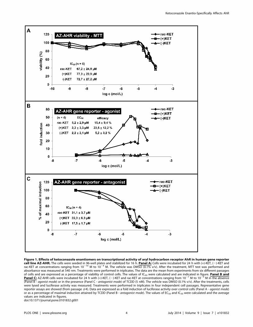
Figure 1. Effects of ketoconazole enantiomers on transcriptional activity of aryl hydrocarbon receptor AhR in human gene reportercell line AZ-AHR. The cells were seeded in 96-well plates and stabilized for 16 h. Panel A: Cells were incubated for 24 h with (+)-KET, (2)-KET andrac-KET at concentrations ranging from 10210 M to 1024 M. The vehicle was DMSO (0.1% v/v). After the treatment, MTT test was performed andabsorbance was measured at 540 nm. Treatments were performed in triplicates. The data are the mean from experiments from six different passagesof cells and are expressed as a percentage of viability of control cells. The values of IC50 were calculated and are indicated in figure. Panel B andPanel C: AZ-AHR cells were incubated for 24 h with (+)-KET, (2)-KET and rac-KET at concentrations ranging from 1027 M to 1024 M in the absence(Panel B - agonist mode) or in the presence (Panel C - antagonist mode) of TCDD (5 nM). The vehicle was DMSO (0.1% v/v). After the treatments, cellswere lysed and luciferase activity was measured. Treatments were performed in triplicates in four independent cell passages. Representative genereporter assays are showed (from passage #4). Data are expressed as a fold induction of luciferase activity over control cells (Panel A - agonist mode)or as a percentage of maximal induction attained by TCDD (Panel B - antagonist mode). The values of EC50 and IC50 were calculated and the averagevalues are indicated in figures.doi:10.1371/journal.pone.0101832.g001
Ketoconazole Enantio-Specifically Affects AhR
PLOS ONE | www.plosone.org 4 July 2014 | Volume 9 | Issue 7 | e101832
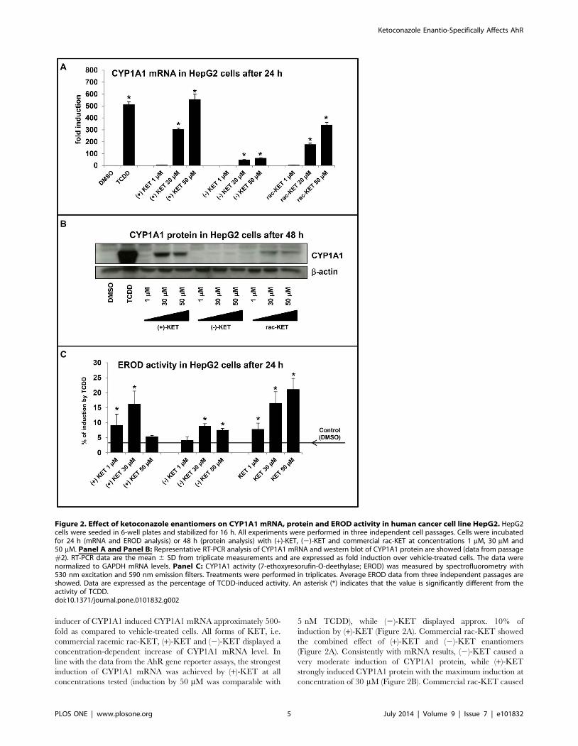
inducer of CYP1A1 induced CYP1A1 mRNA approximately 500-
fold as compared to vehicle-treated cells. All forms of KET, i.e.
commercial racemic rac-KET, (+)-KET and (2)-KET displayed a
concentration-dependent increase of CYP1A1 mRNA level. In
line with the data from the AhR gene reporter assays, the strongest
induction of CYP1A1 mRNA was achieved by (+)-KET at all
concentrations tested (induction by 50 mM was comparable with
5 nM TCDD), while (2)-KET displayed approx. 10% of
induction by (+)-KET (Figure 2A). Commercial rac-KET showed
the combined effect of (+)-KET and (2)-KET enantiomers
(Figure 2A). Consistently with mRNA results, (2)-KET caused a
very moderate induction of CYP1A1 protein, while (+)-KET
strongly induced CYP1A1 protein with the maximum induction at
concentration of 30 mM (Figure 2B). Commercial rac-KET caused
Figure 2. Effect of ketoconazole enantiomers on CYP1A1 mRNA, protein and EROD activity in human cancer cell line HepG2. HepG2cells were seeded in 6-well plates and stabilized for 16 h. All experiments were performed in three independent cell passages. Cells were incubatedfor 24 h (mRNA and EROD analysis) or 48 h (protein analysis) with (+)-KET, (2)-KET and commercial rac-KET at concentrations 1 mM, 30 mM and50 mM. Panel A and Panel B: Representative RT-PCR analysis of CYP1A1 mRNA and western blot of CYP1A1 protein are showed (data from passage#2). RT-PCR data are the mean 6 SD from triplicate measurements and are expressed as fold induction over vehicle-treated cells. The data werenormalized to GAPDH mRNA levels. Panel C: CYP1A1 activity (7-ethoxyresorufin-O-deethylase; EROD) was measured by spectrofluorometry with530 nm excitation and 590 nm emission filters. Treatments were performed in triplicates. Average EROD data from three independent passages areshowed. Data are expressed as the percentage of TCDD-induced activity. An asterisk (*) indicates that the value is significantly different from theactivity of TCDD.doi:10.1371/journal.pone.0101832.g002
Ketoconazole Enantio-Specifically Affects AhR
PLOS ONE | www.plosone.org 5 July 2014 | Volume 9 | Issue 7 | e101832
Ketoconazole Enantio-Specifically Affects AhR
PLOS ONE | www.plosone.org 6 July 2014 | Volume 9 | Issue 7 | e101832
higher induction of CYP1A1 protein than (2)-KET but signifi-
cantly weaker in comparison with (+)-KET (Figure 2B). We also
tested a capability of (+)-KET, (2)-KET and rac-KET to induce
catalytic activity of CYP1A1 in HepG2 cells (EROD assay). Cells
were treated for 24 h with tested compounds at concentrations
1 mM, 30 mM and 50 mM, with 5 nM TCDD or vehicle (DMSO;
0.1% v/v). TCDD induced EROD activity with the average
increase of 22–23-fold. Rac-KET increased the EROD activity in
a concentration-dependent manner with the maximum at 50 mM.
(+)-KET and (2)-KET reached the maximal EROD induction at
concentration 30 mM and the increase was 15% and 8% of TCDD
potency, respectively (Figure 2C). Since EROD activity measured
in cell culture comprise both the effect on catalytic activity and
induction/repression of CYP1A1 gene, the data obtained must be
interpreted with prudence. Collectively, the effects of KET on
CYP1A1 mRNA and protein expression in HepG2 cells were
enantiospecific.
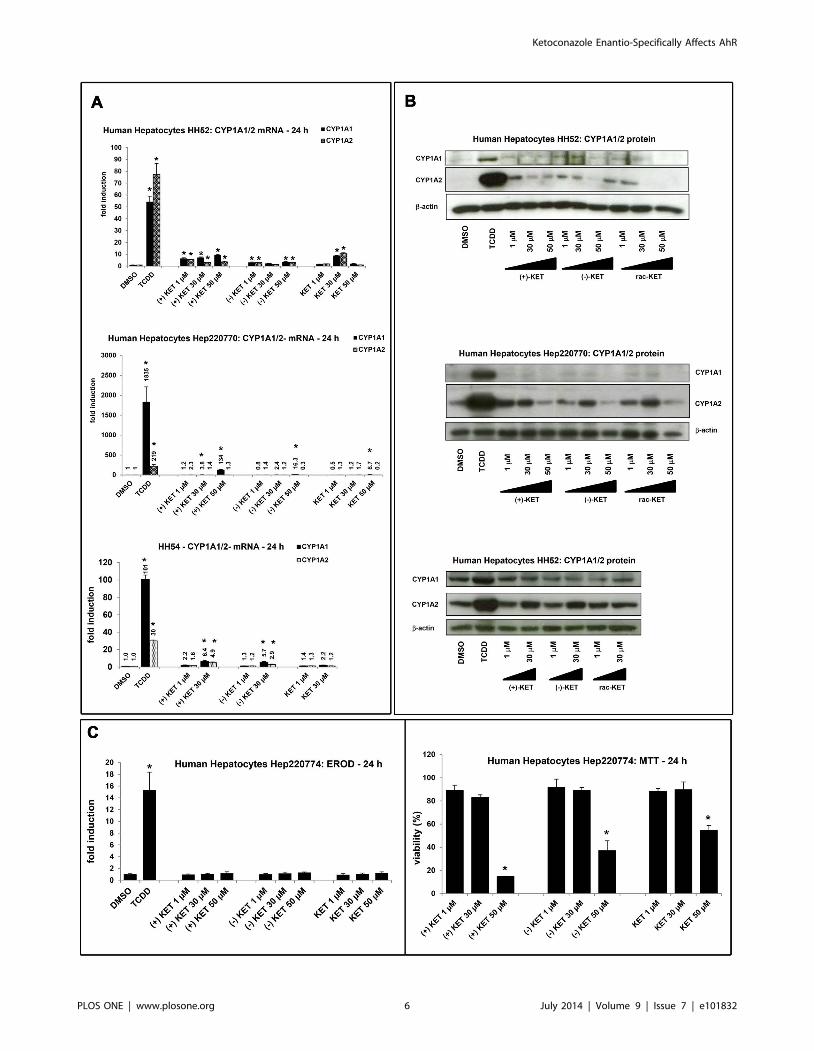
Figure 3. Effect of ketoconazole enantiomers on CYP1A mRNA, protein and EROD activity in primary human hepatocytes. Panel A:RT-PCR analyses CYP1A1 and CYP1A2 mRNA: Human hepatocytes were incubated for 24 h with (+)-KET, (2)-KET and commercial rac-KET atconcentrations 1 mM, 30 mM and 50 mM. Results from three different cultures (HH52, HH54, Hep220770) are shown. Data are the mean 6 SD fromtriplicate measurements and are expressed as fold induction over vehicle-treated cells. Data were normalized to GAPDH mRNA levels. Panel B:CYP1A1 and CYP1A2 protein analyses: Human hepatocytes were incubated for 48 h with (+)-KET, (2)-KET and commercial rac-KET at concentrations1 mM, 30 mM and 50 mM. Western blots from three different cultures (HH52, HH54, Hep220770) are shown. Panel C: EROD and cytotoxicity: Humanhepatocytes (culture Hep220774) were treated with (+)-KET, (2)-KET and commercial rac-KET at concentrations 1 mM, 30 mM and 50 mM. Upper bargraph: An activity of 7-ethoxyresorufin-O-deethylase (EROD) was measured by fluorescent spectrophotometry with 530 nm excitation and 590 nmemission filters. Treatments were performed in triplicates. The data are expressed as fold induction over the value from control cells. Lower bargraph: A conventional MTT test was performed and absorbance was measured at 540 nm. Treatments were performed in triplicates. The data areexpressed as percentage of viability of control cells.doi:10.1371/journal.pone.0101832.g003
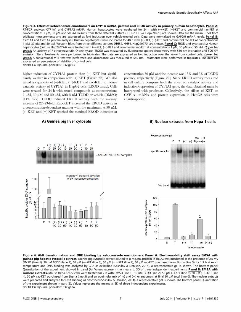
Figure 4. AhR transformation and DRE binding by ketoconazole enantiomers. Panel A: Electromobility shift assay EMSA withguinea pig hepatic cytosolic extract. Guinea pig cytosolic extract diluted to 8 mg/mL protein in HEDG was incubated in the presence of 2% v/vDMSO (lane 1), 20 nM TCDD (lane 2), 50 mM (+)-KET (line 3), 50 mM (2)- KET (line 4), 50 mM rac-KET purchased from Sigma (line 5) for 1.5 h at roomtemperature and DNA binding was analyzed by GRA as described (Soshilov & Denison, 2014). A representative gel is shown. The bottom panel:Quantitation of the experiment showed in panel (A). Values represent the means 6 SD of three independent experiments. Panel B: EMSA withnuclear extracts. Mouse Hepa-1c1c7 cells were treated for 2 h with DMSO (line 1), 10 nM TCDD (line 2), 50 mM (+)-KET (line 3), 50 mM (2)- KET (line4), 50 mM rac-KET purchased from Sigma (line 5) and an equimolar mix of (+) and (2) enantiomers at final 50 mM total (line 6). The nuclear extractswere prepared and analyzed for DNA binding as described (Soshilov & Denison, 2014). A representative gel is shown. The bottom panel: Quantitationof the experiment shown in part (B). Values represent the means 6 SD of three independent experiments.doi:10.1371/journal.pone.0101832.g004
Ketoconazole Enantio-Specifically Affects AhR
PLOS ONE | www.plosone.org 7 July 2014 | Volume 9 | Issue 7 | e101832
Effect of ketoconazole enantiomers on CYP1A1/2 mRNA,protein and EROD activity in primary human hepatocytes
Since KET induced CYP1A1 mRNA and protein in human
hepatoma cells HepG2, we examined a capability of KET to
induce CYP1A1 and CYP1A2 mRNA and protein in primary
human hepatocytes, a more physiological and metabolically
competent cell model. Human hepatocytes were treated for 24 h
or 48 h with TCDD (5 nM), vehicle (DMSO; 0.1% V/V), rac-
KET, (+)-KET or (2)-KET at concentrations 1 mM, 30 mM and
50 mM.
Induction of CYP1A1/CYP1A2 mRNAs by 5 nM TCDD in
three different human hepatocytes cultures was 53-fold/79-fold
(for HH52), 1835-fold/219-fold (for Hep220770) and 101-fold/30-
fold (for HH54). Induction profiles by KET enantiomers varied
between individual human hepatocytes cultures. In culture
Hep220770, CYP1A1 mRNA was strongly induced by (+)-KET
(134-fold; 50 mM), and weakly by (2)-KET (16-fold; 50 mM) and
rac-KET (9-fold; 50 mM), while there was no induction of
CYP1A2 mRNA by any form of KET. In culture HH54, both
CYP1A1 and CYP1A2 mRNAs were induced by (+)-KET and (2)
-KET, but not by rac-KET. The effects of KET enantiomers were
similar (3-fold to 6-fold), and the inductions of CYP1A1 mRNA
and CYP1A2 mRNA were comparable. In culture HH52, the
effects of (+)-KET on CYP1A1 mRNA were a bit stronger as
compared to (2)-KET or rac-KET. The induction of CYP1A2
mRNA by all forms of KET was equipotent (Figure 3A). Overall,
while all forms of KET induced CYP1A genes in primary human
hepatocytes, there were no significant enantiospecific effects, and
the induction profiles varied between human hepatocytes cultures.
We found very faint or no induction of CYP1A1 protein after
the treatment with any form of KET, while TCDD caused drastic
increase of CYP1A1 protein in all three primary human
hepatocytes cultures (HH52, HH54, Hep220770) (Figure 3B).
On the other hand, dose-dependent induction of CYP1A2 protein
was observed for all KET forms (Figure 3B): (i) at 1 mM
concentration, the magnitude of induction was highest for (+)-
KET as compared to (2)-KET and rac-KET; (ii) at 30 mM
concentration, the induction by all KET forms was equipotent; (iii)
at 50 mM concentration, the expression of CYP1A2 protein
dropped, probably due to the cytotoxic effects of KET in human
hepatocytes (vide infra).
Next, we tested capability of ketoconazole to induce catalytic
activity of CYP1A1/1A2 enzymes and cytotoxicity in primary
human hepatocytes. The cells were treated for 24 h with (+)-KET,
(2)-KET and commercial rac-KET (1 mM, 30 mM, 50 mM), and
with TCDD (5 nM) and vehicle (DMSO; 0.1% V/V). TCDD
caused the induction of EROD activity approximately 15-fold
while no significant induction of EROD activity was observed for
any tested form of KET (Figure 3C). In the cytotoxicity assay, we
found all forms of KET highly toxic against human hepatocytes at
the concentration 50 mM. (+)-KET, (2)- KET and rac-KET
decreased viability down to 15%, 37% and 46% of control value,
respectively (Figure 3C).
Collectively, CYP1A1 and CYP1A2 genes were induced by
KET in human hepatocytes. The induction profiles displayed
inter-individual variability, and inconsistency between mRNA and
protein expression was observed.
AhR transformation and DRE binding by ketoconazoleenantiomers
We carried out the electrophoretic mobility shift assay (EMSA)
to reveal whether effects of KET on AhR-CYP1A pathway involve
transformation of AhR to DNA binding form, and whether these
effects are enantiospecific. For this purpose, EMSA was performed
in two experimental systems: (i) Hepatic guinea pig cytosol was
treated for 2 hours with DMSO, 20 nM TCDD, (+)-KET, (2)-
KET and commercial rac-KET (from Sigma) at 10 mM, 30 mM
and 50 mM. All forms of KET transformed AhR to DNA binding
form, but the activation was very weak and not statistically above
background. There was a slightly stronger effect of (+)-KET and
rac-KET as compared to (2)-KET, but the difference was not
significant (Figure 4A). (ii) Since we obtained only weak signals in
guinea pig liver cytosols, we also analyzed nuclear extracts from
mouse hepatoma Hepa-1c1c7 cells treated for 2 hours with
DMSO, TCDD (10 nM), (+)-KET (50 mM), (2)-KET (50 mM),
commercial rac-KET (Sigma; 50 mM) or an equimolar mix of (+)/
(2) enantiomers (50 mM total). Similarly as in guinea pig liver
cytosol, (+)-KET and (2)-KET only weakly transformed AhR to
DNA binding form. The effects of (2)-KET were slightly stronger
as compared to (+)-KET. Surprisingly, commercial racemic KET
strongly induced formation of AhR-ARNT-DRE complex
(approx. 60% of TCDD effects), while racemic KET obtained
by mixing pure (+)-KET and pure (2)-KET in ratio 1:1, displayed
the effects much weaker and comparable with individual
enantiomers (Figure 4B). These results suggest the presence of
impurities in commercial KET that can activate the mouse AhR
more strongly than KET-enantiomers [20]. In addition, we
observed formation of the second band with lower molecular
weight than AhR-ARNT-DRE, by purified KET enantiomers but
not by TCDD and commercial KET. Overall, the capability of
ketoconazole enantiomers to induce a formation of AhR-ARNT-
DRE complex does not seem to be enantiospecific, and the effects
are much weaker than expected with regard to the magnitude of
CYP1A1 induction and AhR activation in HepG2 cells.
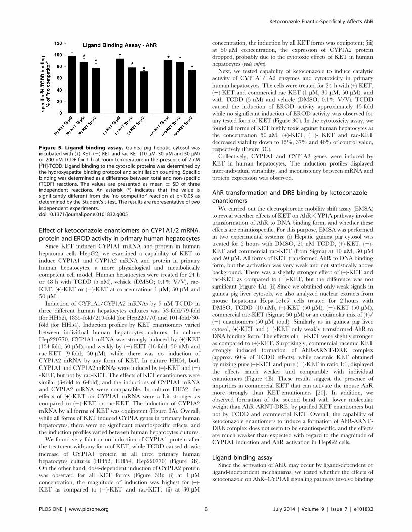
Ligand binding assaySince the activation of AhR may occur by ligand-dependent or
ligand-independent mechanisms, we tested whether the effects of
ketoconazole on AhR–CYP1A1 signaling pathway involve binding
Figure 5. Ligand binding assay. Guinea pig hepatic cytosol wasincubated with (+)-KET, (2)-KET and rac-KET (10 mM, 30 mM and 50 mM)or 200 nM TCDF for 1 h at room temperature in the presence of 2 nM[3H]-TCDD. Ligand binding to the cytosolic proteins was determined bythe hydroxyapatite binding protocol and scintillation counting. Specificbinding was determined as a difference between total and non-specific(TCDF) reactions. The values are presented as mean 6 SD of threeindependent reactions. An asterisk (*) indicates that the value issignificantly different from the ‘no competitor’ reaction at p,0.05 asdetermined by the Student’s t-test. The results are representative of twoindependent experiments.doi:10.1371/journal.pone.0101832.g005
Ketoconazole Enantio-Specifically Affects AhR
PLOS ONE | www.plosone.org 8 July 2014 | Volume 9 | Issue 7 | e101832
to the AhR. We performed AhR ligand binding assay using guinea
pig hepatic cytosol. All tested forms of KET competitively, dose-
dependently inhibited [3H]-TCDD binding to the AhR when
present in the binding incubation at 30 mM and 50 mM (Figure 5).
The differences between the effects of (+)-KET and (2)-KET
enantiomers were not significant. Overall, both KET cis-enantio-
mers are weak AhR ligands their effects on AhR-CYP1A1
signaling pathway occur via ligand-dependent mechanism. The
binding of KET to AhR was not enantiospecific.
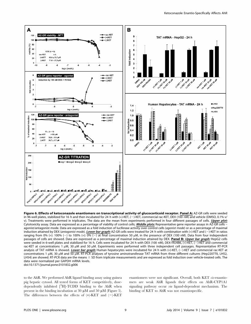
Figure 6. Effects of ketoconazole enantiomers on transcriptional activity of glucocorticoid receptor. Panel A: AZ-GR cells were seededin 96-well plates, stabilized for 16 h and then incubated for 24 h with (+)-KET, (2)-KET, commercial rac-KET, DEX (100 nM) and vehicle (DMSO, 0.1% v/v). Treatments were performed in triplicates. The data are the mean from experiments performed in four different passages of cells. Upper plot:Cytotoxicity assay. Data are expressed as a percentage of viability of control cells; Middle plots: Representative gene reporter assays in AZ-GR cells –agonist/antagonist mode. Data are expressed as a fold induction of luciferase activity over control cells (agonist mode) or as a percentage of maximalinduction attained by DEX (antagonist mode). Lower bar graph: AZ-GR cells were treated for 24 h with combination with (+)-KET and (2)-KET in ratiosranging from 0% (+): 100% (2) to 100% (+): 0% (2) at final concentration 50 mM, in the presence of DEX (100 nM). Data from four independentpassages of cells are showed. Data are expressed as a percentage of maximal induction attained by DEX. Panel B: Upper bar graph: HepG2 cellswere seeded in 6-well plates and stabilized for 16 h. Cells were incubated for 24 h with DEX (100 nM), DEX+RU486, (+)-KET, (2)-KET and commercialrac-KET at concentrations 1 mM, 30 mM and 50 mM. Experiments were performed with three independent cell passages. Representative RT-PCRanalysis of TAT mRNA is showed. Lower bar graph: Human hepatocytes were incubated for 24 h with (+)-KET, (2)-KET and commercial rac-KET atconcentrations 1 mM, 30 mM and 50 mM. RT-PCR analyses of tyrosine aminotransferase TAT mRNA from three different cultures (Hep220770, LH52,LH54) are showed. RT-PCR data are the means 6 SD from triplicate measurements and are expressed as fold induction over vehicle-treated cells. Thedata were normalized per GAPDH mRNA levels.doi:10.1371/journal.pone.0101832.g006
Ketoconazole Enantio-Specifically Affects AhR
PLOS ONE | www.plosone.org 9 July 2014 | Volume 9 | Issue 7 | e101832
Effects of ketoconazole enantiomers on transcriptionalactivity of glucocorticoid receptor
There is multiple evidence for the role of glucocorticoid
receptor (GR) in regulation of AhR activity [21]. Therefore, we
tested whether the effects of KET on AhR involve GR. For this
purpose, we incubated gene reporter cell line AZ-GR for 24 h (+)-
KET, (2)-KET and rac-KET [18]. All forms of KET displayed
cytotoxic effect in AZ-GR cell line without significant enantios-
pecificity, as assessed by MTT test. The values of IC50 were
82.6623.7 mM for (+)-KET and 71.6627.5 mM for (2)-KET.
The IC50 value for rac-KET was higher than 100 mM (Figure 6A).
Gene reporter assays were performed in agonist and antagonist mode
(similarly as described in section 3.1.). In agonist mode, cells were
incubated with increasing concentrations of (+)-KET, (2)-KET
and commercial rac-KET and with model GR agonist (DEX;
100 nM). An induction of GR-dependent luciferase activity by
100 nM DEX in four independent passages of AZ-GR cells varied
from 45-fold to 85-fold (average induction 70-fold), as compared to
vehicle-treated cells. Cis-enatiomers (+)-KET and (2)-KET did not
induce luciferase activity in AZ-GR cells up to maximal applied
concentration, i.e. 100 mM. Interestingly, approximately 5-fold
increase of luciferase activity was observed for commercial rac-
KET at concentration 100 mM (Figure 6A). In antagonist mode, (+)-
KET and (2)-KET showed strong antagonist effect on GR
transcription activity, and there was no significant difference
between the effects of individual enantiomers. The values of IC50
were 29.660.9 mM and 26.763.7 mM for (+)-KET and (2)-KET,
respectively. We observed only weak inhibition of DEX-induced
luciferase activity by commercial rac-KET (Figure 6A). To further
elucidate the discrepancy between the effects of pure enantiomers
as compared to commercial racemic KET, we performed titration
experiment in antagonist mode to examine the effect of (+)- and (2)-
KET on GR transcription activity. The AZ-GR cells were treated
for 24 h with DEX (100 nM) in combination with mixtures of
pure (+)-KET and pure (2)-KET, in ratios from 0% (+): 100% (2)
to 100% (+): 0% (2), at final concentration 50 mM. Antagonist
effect by mixture of 50% (+)-KET: 50% (2)-KET was much
stronger as compared to commercial rac-KET (Figure 6A). The
plausible explanation could be the influence of impurities
contained in commercial rac-KET, similarly as observed in
EMSA experiments.
We also analyzed the expression of tyrosine aminotransferase
(TAT), the GR-target gene, in cell line HepG2 and in primary
human hepatocytes. In HepG2 cells, we found strong, concentra-
tion-dependent induction of TAT mRNA by (+)-KET, (2)-KET
and rac-KET. The magnitude of induction by 30 mM and 50 mM
KET was comparable with effects of DEX, and the effects of KET
were not enantiospecific (Figure 6B). Primary human hepatocytes
are routinely cultured in the presence of DEX, therefore, the TAT
gene is induced under these conditions. All forms of KET strongly,
and dose-dependently down-regulated TAT mRNA in three
different primary hepatocytes cultures Hep220770, HH52 and
HH54. Again, the effects of KET were not enantiospecific.
Overall, KET displayed strong, but inconsistent and enantio non-
specific agonist and/or antagonist effects on GR in various in vitro
cell systems (Figure 6A, Figure 6B).
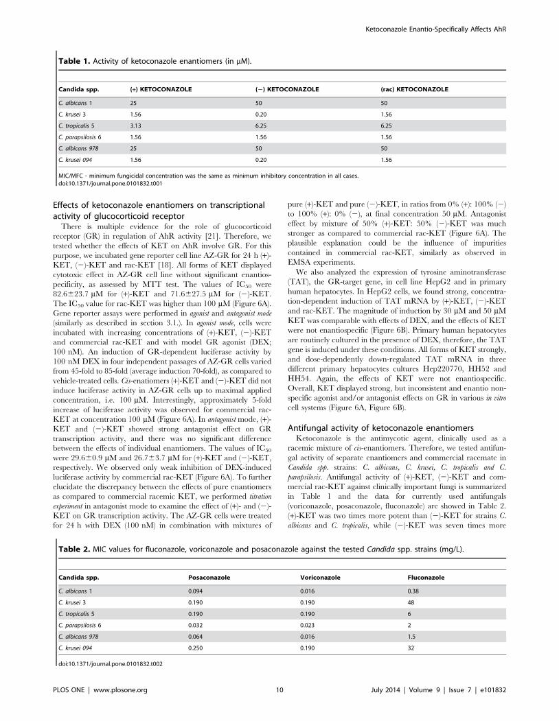
Antifungal activity of ketoconazole enantiomersKetoconazole is the antimycotic agent, clinically used as a
racemic mixture of cis-enantiomers. Therefore, we tested antifun-
gal activity of separate enantiomers and commercial racemate in
Candida spp. strains: C. albicans, C. krusei, C. tropicalis and C.
parapsilosis. Antifungal activity of (+)-KET, (2)-KET and com-
mercial rac-KET against clinically important fungi is summarized
in Table 1 and the data for currently used antifungals
(voriconazole, posaconazole, fluconazole) are showed in Table 2.
(+)-KET was two times more potent than (2)-KET for strains C.
albicans and C. tropicalis, while (2)-KET was seven times more
Table 1. Activity of ketoconazole enantiomers (in mM).
Candida spp. (+) KETOCONAZOLE (2) KETOCONAZOLE (rac) KETOCONAZOLE
C. albicans 1 25 50 50
C. krusei 3 1.56 0.20 1.56
C. tropicalis 5 3.13 6.25 6.25
C. parapsilosis 6 1.56 1.56 1.56
C. albicans 978 25 50 50
C. krusei 094 1.56 0.20 1.56
MIC/MFC - minimum fungicidal concentration was the same as minimum inhibitory concentration in all cases.doi:10.1371/journal.pone.0101832.t001
Table 2. MIC values for fluconazole, voriconazole and posaconazole against the tested Candida spp. strains (mg/L).
Candida spp. Posaconazole Voriconazole Fluconazole
C. albicans 1 0.094 0.016 0.38
C. krusei 3 0.190 0.190 48
C. tropicalis 5 0.190 0.190 6
C. parapsilosis 6 0.032 0.023 2
C. albicans 978 0.064 0.016 1.5
C. krusei 094 0.250 0.190 32
doi:10.1371/journal.pone.0101832.t002
Ketoconazole Enantio-Specifically Affects AhR
PLOS ONE | www.plosone.org 10 July 2014 | Volume 9 | Issue 7 | e101832
potent than (+)-KET for other tested microorganisms. Hence, we
demonstrate enantiospecific antifungal activity of ketoconazole,
which is in line with observations of other authors [7].
Discussion
The effects of KET on drug-metabolizing pathways are very
complex, and involve inhibition of cytochrome P450 catalytic
activities (e.g. CYP3A4, CYP3A5, CYP2C9), agonism and/or
antagonism of several receptors, including glucocorticoid receptor
(GR) [12], aryl hydrocarbon receptor (AhR) and pregnane X
receptor (PXR) [22], or interactions with drug transporters [13].
With regard to AhR, azole antifungal drugs were shown to
influence AhR-dependent genes in aquatic species [23,24], rodents
[25] and in murine and human cancer cell lines [14]. Clinically
used KET is a mixture of two cis-enatiomers (2R,4S)-(+)-KET and
(2S,4R)-(2)-KET. Enantiospecific effects of KET on catalytic
activities of CYP3A4/5 [8] and progesterone 17a, 20-lyase were
reported [7]. A phase II clinical study was conducted with
compound DIO-902 (which is ketoconazole enantiomer (2)-
KET), as a candidate drug for the treatment of Diabetes mellitus
Type II [26]. The mechanism of DIO-902 action was enantios-
pecific inhibition of cortisol synthesis. However, due to the side
effects, a study was interrupted and DIO-902 was suspended [27].
In the current paper, we investigated enantiospecific effects of
KET on AhR-CYP1A signaling pathway in cancer cell lines and
in human hepatocytes, and we provide the first evidence of
enantiospecific interactions of KET with AhR-signaling pathway
in vitro. In particular, we demonstrate that:
(i) KET activates AhR in gene reporter cell line and dose-
dependently induces CYP1A1 mRNA and CYP1A1
protein in HepG2 cells, with enantiospecific pattern, i.e.
(+)-KET was much more active as compared to (2)-KET.
(ii) KET enantiospecifically induces CYP1A1 and CYP1A2
mRNA and CYP1A2 protein in primary cultures of human
hepatocytes. The effects of enantiomer (+)-KET were
stronger than those of (2)-KET enantiomer.
(iii) Both KET enantiomers are weak ligands for AhR, as
revealed by ligand binding assay performed in guinea pig
liver cytosols. In addition, both enantiomers of KET
transformed AhR to DNA-binding form, as revealed by
EMSA assays in guinea pig liver cytosols and Hepa-1c1c7
murine cells. Neither binding to AhR nor transformation
of AhR to DNA-binding form was enantiospecific.
Interestingly, we observed that commercial racemic KET
strongly induced formation of AhR-ARNT-DRE complex
(approx. 60% of TCDD effects), while in mouse but not
guinea pig the racemic KET obtained by mixing pure (+)-
KET and pure (2)-KET in ratio 1:1, displayed the effects
much weaker and comparable with individual enantio-
mers. Strong effects of commercial racemic KET are
probably caused by the impurities present in commercial
product [20]. This implies that many in vitro effects of KET
described in scientific literature may be due to the presence
of impurities, and these data should be interpreted with
prudence.
(iv) KET is strong antagonist of human glucocorticoid receptor
GR, as revealed by gene reporter assays in transgenic AZ-
GR cells and by down-regulation of tyrosine aminotrans-
ferase TAT in primary human hepatocytes cultured in
dexamethasone-containing medium. We also observed
induction of TAT in HepG2 cells by KET, suggesting
partial agonist and antagonist effects of KET. The effects
of KET on GR were not enantiospecific, implying no role
for GR in enantiospecific induction of CYP1A1 and
activation of AhR. This is an important finding, taking in
account a pivotal role of GR in regulation of AhR
transcriptional activity [21]. Racemic commercial KET
showed irregular behavior, suggesting the influence of
impurities, similarly as in EMSA experiment.
(v) Enantiospecific antifungal activity of KET was observed in
several Candida spp. strains including C. albicans 1, C. albicans
978, C. krusei 3, C. krusei 094 and C. tropicalis 5, which is also
in line with observations of other authors [7].
In conclusion, current study provides the first evidence of
enantiospecific effects of ketoconazole on AhR signaling pathway.
The results might have clinical significance since (+)-KET activates
and induces CYP1A1, but (2)-KET has higher antifungal activity
in some Cancida spp. strains.
Author Contributions
Conceived and designed the experiments: AN AAS MSD ZD. Performed
the experiments: KB IB AAS M. Korhonova. Analyzed the data: AN MK
IB AAS MSD MK ZD. Contributed reagents/materials/analysis tools: ZD
MSD M. Kolar PB. Wrote the paper: AN ZD.
References
1. Sonino N, Boscaro M, Paoletta A, Mantero F, Ziliotto D (1991) Ketoconazole
treatment in Cushing’s syndrome: experience in 34 patients. Clin Endocrinol
(Oxf) 35: 347–352.
2. Pont A, Graybill JR, Craven PC, Galgiani JN, Dismukes WE, et al. (1984) High-
dose ketoconazole therapy and adrenal and testicular function in humans. Arch
Intern Med 144: 2150–2153.
3. Stricker BH, Blok AP, Bronkhorst FB, Van Parys GE, Desmet VJ (1986)
Ketoconazole-associated hepatic injury. A clinicopathological study of 55 cases.
J Hepatol 3: 399–406.
4. Bercoff E, Bernuau J, Degott C, Kalis B, Lemaire A, et al. (1985) Ketoconazole-
induced fulminant hepatitis. Gut 26: 636–638.
5. Benson GD, Anderson PK, Combes B, Ishak KG (1988) Prolonged jaundice
following ketoconazole-induced hepatic injury. Dig Dis Sci 33: 240–246.
6. Daneshmend TK, Warnock DW (1988) Clinical pharmacokinetics of ketoco-
nazole. Clin Pharmacokinet 14: 13–34.
7. Rotstein DM, Kertesz DJ, Walker KA, Swinney DC (1992) Stereoisomers of
ketoconazole: preparation and biological activity. J Med Chem 35: 2818–2825.
8. Dilmaghanian S, Gerber JG, Filler SG, Sanchez A, Gal J (2004) Enantioselec-
tivity of inhibition of cytochrome P450 3A4 (CYP3A4) by ketoconazole:
Testosterone and methadone as substrates. Chirality 16: 79–85.
9. Allqvist A, Miura J, Bertilsson L, Mirghani RA (2007) Inhibition of CYP3A4 and
CYP3A5 catalyzed metabolism of alprazolam and quinine by ketoconazole as
racemate and four different enantiomers. Eur J Clin Pharmacol 63: 173–179.
10. Boxenbaum H (1999) Cytochrome P450 3A4 in vivo ketoconazole competitive
inhibition: determination of Ki and dangers associated with high clearance drugs
in general. J Pharm Pharm Sci 2: 47–52.
11. Stresser DM, Blanchard AP, Turner SD, Erve JC, Dandeneau AA, et al. (2000)
Substrate-dependent modulation of CYP3A4 catalytic activity: analysis of 27 test
compounds with four fluorometric substrates. Drug Metab Dispos 28: 1440–
1448.
12. Duret C, Daujat-Chavanieu M, Pascussi JM, Pichard-Garcia L, Balaguer P, et
al. (2006) Ketoconazole and miconazole are antagonists of the human
glucocorticoid receptor: consequences on the expression and function of the
constitutive androstane receptor and the pregnane X receptor. Mol Pharmacol
70: 329–339.
13. Dvorak Z (2011) Drug-drug interactions by azole antifungals: Beyond a dogma
of CYP3A4 enzyme activity inhibition. Toxicol Lett 202: 129–132.
14. Korashy HM, Shayeganpour A, Brocks DR, El-Kadi AO (2007) Induction of
cytochrome P450 1A1 by ketoconazole and itraconazole but not fluconazole in
murine and human hepatoma cell lines. Toxicol Sci 97: 32–43.
15. Jack SM, Dobbins M, Sword W, Novotna G, Brooks S, et al. (2011) Evidence-
informed decision-making by professionals working in addiction agencies serving
women: a descriptive qualitative study. Subst Abuse Treat Prev Policy 6: 29.
16. Vrzal R, Daujat-Chavanieu M, Pascussi JM, Ulrichova J, Maurel P, et al. (2008)
Microtubules-interfering agents restrict aryl hydrocarbon receptor-mediated
Ketoconazole Enantio-Specifically Affects AhR
PLOS ONE | www.plosone.org 11 July 2014 | Volume 9 | Issue 7 | e101832
CYP1A2 induction in primary cultures of human hepatocytes via c-jun-N-
terminal kinase and glucocorticoid receptor. Eur J Pharmacol 581: 244–254.
17. Novotna A, Pavek P, Dvorak Z (2011) Novel stably transfected gene reporter
human hepatoma cell line for assessment of aryl hydrocarbon receptor
transcriptional activity: construction and characterization. Environ Sci Technol
45: 10133–10139.
18. Novotna A, Pavek P, Dvorak Z (2012) Construction and characterization of a
reporter gene cell line for assessment of human glucocorticoid receptor
activation. Eur J Pharm Sci 47: 842–847.
19. Denison MS, Rogers JM, Rushing SR, Jones CL, Tetangco SC, et al. (2002)
Analysis of the aryl hydrocarbon receptor (AhR) signal transduction pathway.
Curr Protoc Toxicol Chapter 4: Unit4 8.
20. Castro-Puyana M, Garcia-Ruiz C, Cifuentes A, Crego AL, Marina ML (2006)
Identification and quantitation of cis-ketoconazole impurity by capillary zone
electrophoresis-mass spectrometry. J Chromatogr A 1114: 170–177.
21. Monostory K, Dvorak Z (2011) Steroid regulation of drug-metabolizing
cytochromes P450. Curr Drug Metab 12: 154–172.
22. Svecova L, Vrzal R, Burysek L, Anzenbacherova E, Cerveny L, et al. (2008)
Azole antimycotics differentially affect rifampicin-induced pregnane X receptor-mediated CYP3A4 gene expression. Drug Metab Dispos 36: 339–348.
23. Hasselberg L, Grosvik BE, Goksoyr A, Celander MC (2005) Interactions
between xenoestrogens and ketoconazole on hepatic CYP1A and CYP3A, injuvenile Atlantic cod (Gadus morhua). Comp Hepatol 4: 2.
24. Babin M, Casado S, Chana A, Herradon B, Segner H, et al. (2005) CytochromeP4501A induction caused by the imidazole derivative Prochloraz in a rainbow
trout cell line. Toxicol In Vitro 19: 899–902.
25. Sun G, Thai SF, Tully DB, Lambert GR, Goetz AK, et al. (2005)Propiconazole-induced cytochrome P450 gene expression and enzymatic
activities in rat and mouse liver. Toxicol Lett 155: 277–287.26. Schwartz SL, Rendell M, Ahmann AJ, Thomas A, Arauz-Pacheco CJ, et al.
(2008) Safety profile and metabolic effects of 14 days of treatment with DIO-902:results of a phase IIa multicenter, randomized, double-blind, placebo-controlled,
parallel-group trial in patients with type 2 diabetes mellitus. Clin Ther 30: 1081–
1088.27. Arakaki R, Welles B (2010) Ketoconazole enantiomer for the treatment of
diabetes mellitus. Expert Opin Investig Drugs 19: 185–194.
Ketoconazole Enantio-Specifically Affects AhR
PLOS ONE | www.plosone.org 12 July 2014 | Volume 9 | Issue 7 | e101832