Embed Size (px)
Citation preview
Reactive & Functional Polymers 82 (2014) 103–110
Contents lists available at ScienceDirect
Reactive & Functional Polymers
journal homepage: www.elsevier .com/ locate/ react
Encapsulation of Congo Red in carboxymethyl guar gum–alginate gelmicrospheres
http://dx.doi.org/10.1016/j.reactfunctpolym.2014.06.0061381-5148/� 2014 Elsevier Ltd. All rights reserved.
⇑ Corresponding author. Tel.: +54 221 483 37 94x132(O).E-mail address: [email protected] (G.R. Castro).
Valeria E. Bosio a, Sreyasree Basu b, Fraqooue Abdullha b, M. Elizabeth Chacon Villalba c, Jorge A. Güida c,d,e,Arup Mukherjee b, Guillermo R. Castro a,⇑a Nanobiomaterials Laboratory, Institute of Applied Biotechnology (CINDEFI), School of Sciences, Universidad Nacional de La Plata – CONICET (CCT La Plata), 1900 La Plata, Argentinab Department of Chemical Technology, Calcutta University, 92 A.P.C. Road, Kolkata 700009, Indiac CEQUINOR, School of Sciences, Universidad Nacional de La Plata – CONICET (CCT La Plata), CC 962, 1900 La Plata, Argentinad School of Engineering, Universidad Nacional de La Plata, 1900 La Plata, Argentinae Department of Basic Sciences, Universidad Nacional de Luján, Luján, Argentina
a r t i c l e i n f o a b s t r a c t
Article history:Received 28 January 2014Received in revised form 17 June 2014Accepted 18 June 2014Available online 26 June 2014
Keywords:Congo RedGel microspheresGuar gumAlginateCoacervatesInfrared spectroscopy
Congo Red (CR) is a hydrophobic dye commonly used for diagnosis and potentially useful as therapeuticagent of beta amyloid plaques in neurodegenerative diseases. CR, as drug model, was encapsulated onAlginate–Carboxy Methyl Guar Gum (Alg–CMGG) blend microspheres. Guar gum 18% carboxymethylated(CMGG) derivative was synthesized in order to improve aqueous solubility, polymer blending and helpreduce surface tension. The derivative was confirmed by FTIR spectroscopy, and elemental analysis. Sur-face tension of the new CMGG is reduced in about 50% compared with the native polymer. Lowering ofGuar Gum (GG) aqueous solutions viscosity from 30,000 cps to 350–400 cps in case of CMGG is indicatingpseudoplastic fluid behavior modifications. Vibrational spectroscopy analysis confirmed interactionsamong CR molecules in alginate–CMGG matrices ascribed largely to the aromatic motif of the dye andthe biopolymer a polar regions. CR was encapsulated on 68/32% alginate/CMGG blend microspheres asthe best formulation tested. The release of CR from the microspheres was not detected at pH = 1.2 in25 min, but 62% of CR was found in the supernatant when the pH was raised to 7.4 at 37 �C after 8 hincubation.
� 2014 Elsevier Ltd. All rights reserved.
1. Introduction
Congo Red (CR) is a linear anionic secondary diazo dye used asan acid-base indicator. Additionally, CR was used to detect fibrilproteins enriched in b sheet conformation useful in histologicalstudies of some neurodegenerative pathologies such asAlzheimer’s, Creutzfeldt–Jacob’s, Huntington’s and Parkinson’s dis-eases [1]. Moreover, CR delays appearance of clinical signs onexperimental prion trials [2]. The compound could also be usedas a palliative in neurodegenerative disease therapies. CR is solublein many organic solvents, but yielding red colloidal fluorescentsolutions in aqueous media because of its hydrophobicity madeby the presence of biphenyl and naphthalene groups in the mole-cule [3]. The postulated mechanism for CR aggregation is by hydro-phobic interaction involving the p–p bonds of the aromatic ringsmaking planar structures [4]. Based on the CR physicochemicalproperties, the dye is an excellent candidate to test the potential
encapsulation of common hydrophobic drugs, e.g. anthracyclines,taxanes, fluoroquinolones, into biopolymeric gel matrices.
In the last decades, biopolymers have received increasing atten-tion both in the academia and in industry. Remarkable biopolymerproperties like structural diversity, biological specific propertiesover a range of molecular dimensions and favorable non-covalentphysiological interactions were unearthed. Additionally, tailorabil-ity, biodegradability, mild environmental synthesis, and conve-nient rheological modulations have made biopolymers veryattractive tools for a myriad of applications [5]. Ultrapure Alginates(Alg) produced under GMP/ISO 9000 guidelines are one of mostcommon biopolymer gels currently used in food and pharmaceuti-cal industries. Algs are linear biopolymers composed of b-mannu-ronic acid (M) and a-guluronic acid (G) linked by 1–4 bonds,purified from the seaweeds and some bacteria. Alg hydrogels areformed by ionic crosslinking in presence of divalent cations (e.g.calcium and zinc), which cooperatively interact with different car-boxylate ions forming ionic bridges between different polymerchains. Alg gel structure is commonly named as ‘‘egg box’’ becauseof the analogy with the egg containers. Alginate gels have been

104 V.E. Bosio et al. / Reactive & Functional Polymers 82 (2014) 103–110
used extensively as a matrix for entrapment of many moleculesand cells, and also in tissue engineering applications [6]. However,Alg gels are showing some drawbacks such as high hydrophilicityand erodability at alkaline pH, poor mechanical strength, and gelinstability in freeze–thaw cycles which are preventing extensiveapplications. Some alternative strategies were developed earlierin order to improve Alg gel quality for many applications. Priorattempts in similar applications relied upon polymer chemicalmodification and covalent linking with a wide range of moleculeslike polyoles, chitosan, to sorbitan esters, amongst others [7].Exhaustive chemical alterations have rendered Algs untenable inphysiological or biological applications. Biopolymeric blends innon-covalent interactions are immerging alternatives and are moreclearly understood in very recent years. Furthermore, inherent bio-molecular blend interactions are low energy processes that areindustrially more acceptable and easy to prepare. Novel propertiesof individual polymer blends can also add advantages in pin pointapplications [8].
A very attractive molecule to develop polymer blends is GuarGum (GG). GG is a biopolymer synthesized in the endosperm ofCyamopsis tetragonolobus, a seed legume commonly found in thenorth of India and Pakistan. Chemically, GG is a galactomannancomposed by a linear chain of b 1,4-D-mannopyranoses to whichD-galactopyranoses residues are a 1,6-linked at every second man-nosyl residue forming short side-branches (2:1 ratio) with a molec-ular weight of about 200 kDa. GG is also a robust engineeringbiopolymer that has found applications in concretes, cement set-tings and in petroleum oil drilling as a drilling mud. Relevant GGproperties include thixotropy (the decrease in viscosity over timeat a constant shear rate) above 1.0% concentration in water, theability to retard ice crystal growth non-specifically by slowingmass transfer across the solid/liquid interface, good stability dur-ing several freezing–thawing cycles, and water swelling activitiespH-dependently. Additionally, GG has approximately 8-times thewater-thickening potency of corn starch, and can be used inmulti-phase formulations as an emulsifier preventing oil dropletsfrom coalescing, and/or as a stabilizer in order to prevent molecu-lar aggregation and particles from settling, and it can form gels inpresence of calcium and other polyvalent cations [9]. GG is com-monly used in cosmetics and foods as a thickener in the USA andin the EU (E412 additive code). In foods, GG is an additive in thedairy industry (yogurts, kefir, and liquid cheese products), also toprevent ice crystal formation on sauces and dressing, meats, bakedgoods, ice creams, and in dry foods (e.g. soups and deserts). How-ever, GG standalone presents some problems to be handled, likelow hydrophilicity and concomitantly slightly solubility in aqueoussolutions. A typical strategy to increase the solubility and polymercompatibility of GG is by introducing polar groups in the mainstructure of polymer.
The GG trimethyl amine derivative is currently used in the hairconditioner Jaguar� under patent protection. Alternativelycarboxymethylation of GG, is possible where in, the reactive pri-mary hydroxyl groups of galactomannan could be substituted withcarboxylate functions. Consequently, the Carboxy Methyl GuarGum (CMGG) derivative can became more soluble in water andmaking clear and low viscosity solutions. Despite of many advan-tages, CMGG impede in standalone gel formation in presence ofmultivalent ions due to functional groups distance geometry andbiopolymer structural constrains. Similar galactomannan biopoly-mers however can help shape polymer blends and develop inter-penetrating smart materials networks for biological andenvironmental application [10].
In previous study, hundred percent encapsulation efficiency ofBSA (bovine serum albumin) in Alg–GG matrix crosslinked withglutaraldehyde was reported. Also, the BSA release from the gelmatrix was pH-dependent, and unsusceptible to freeze and
thawing procedures [7]. The same research group described theentrapment of crosslinked subtilisin crystal aggregates in Alg–GGgel matrix increasing the enzyme stability under harsh environ-mental conditions [11]. Furthermore, a successful purification ofthe Jacalin lectin using Alg–CMGG in a fluidized bed techniquewas described [12]. Later, the swelling and degradation of Alg–CMGG cross-linked with divalent barium ion at different pHs andpretreatments was also reported [8]. On the other side, both Algand GG biopolymers has been reported individually to havebeneficial effect of human health reducing serum cholesterol, andhaving positive effects on blood glucose [13,14].
The aim of the present work is to develop alginate–carboxymethyl guar gum hydrogel blend microspheres containing CR asmolecular cargo for drug delivery. That model could also be usedfurther as a therapeutic agent to reduce abnormal protein b foldingassociated to some neuronal disorders.
In order to develop the biopolymer blend, Carboxy Methylderivative of GG (CMGG) was synthesized and characterized bycentesimal composition, derivatization degree, viscosity, surfacetension studies, and FTIR. Alg–CMGG gel microsphere blendcomposition was optimized by controlled release kinetic andswelling studies in vitro under different experimental conditions.The studies were complemented by optical and scanning electronicmicroscopy (OM and SEM), and the interactions between thehydrogel components and cargo were analyzed by viscosimetryand correlated with FTIR and Raman spectroscopies.
2. Materials and methods
2.1. Materials
Congo Red (CR), the sodium salt of benzidinediazo-bis-1-naph-thylamine-4-sulfonic acid (purity > 99%) was purchased fromMerck (AG. Darmstadt, Germany). Low viscosity sodium Alginate(Alg) (average Mn 1.0 � 105 Da) was obtained from Biochem S.A.(Buenos Aires, Argentina). All other reagents used were of analyti-cal grade purchased from Sigma (St. Louis, MO) or Merck (Darms-tadt, Germany). Guar Gum (GG, average Mn = (2.20 ± 0.20) �105 Da) was kindly provided by Hindustan Gums & Chemicals(India).
CR stock solutions (1.0 g/L) and its dilutions were made in miliQwater and/or proper buffers. Calibration curve of CR was made atkmax 497 nm in a Beckman DU 640 UV–Vis spectrophotometer(Beckman-Coulter, CA, USA).
2.2. Synthesis of Carboxy Methyl Guar-gum (CMGG)
50.0 g of guar gum was taken in a three neck round bottom flaskfitted with a condenser, a nitrogen purging set up and a mechanicalstirrer. Isopropanol (100 mL) was added in the reaction mixtureand the biopolymer was allowed to soak under stir for 30 min.Afterward, 30.0 ml of 0.25% (w/v) aqueous NaOH solution wasadded through the condenser and the resulting mixture stirredfor additional 10 min at room temperature. A dropping funnelwas then attached through a fork followed by the addition of150 mL of 170 mg/mL aqueous chloroacetic acid solution to theflask. The solution pH was adjusted to 7.0 ± 0.2 with 1.0% (w/v)NaOH under nitrogen purging. The addition time altogether was1 h. The stirring was continued further for three hours and thereaction temperature was raised to 55 �C. The reaction mixturewas cooled and a light yellow solid separated by filtration througha Buckner funnel. The precipitate was washed twice with 80% (v/v)isopropanol in water, and 50% ethanol–water solution, and dried ina vacuum desiccator. The mass obtained was 43.0 g.

V.E. Bosio et al. / Reactive & Functional Polymers 82 (2014) 103–110 105
CMGG was further purified for analysis and applications. Typi-cally, 1.0 gm of CMGG was suspended in a solution containing5.0 mL of isopropanol and 15 mL of concentrated hydrochloric acidfor 10 min; followed by washing with water until free fromchloride.
2.3. Determination of degree of substitution in CMGG
The degree of surface functionalization on Guar Gum (GG)structure was confirmed by standard alkali titration. A weighedhalf from 1.0 g of CMGG sample was dissolved in excess of100 mM sodium hydroxide and stirred over a magnetic stirrer for20 min. The unreacted excess of sodium hydroxide was backtitrated against standard 100 mM hydrochloric acid with methylorange as indicator. A titration blank was simultaneously carriedout without a CMGG sample. The other half portion of CMGGwas dried in an oven at 102 �C to constant weight. This weightwas taken as equal to the sample weight in alkali titration. Thedegree of GG substitution was estimated as follows:
DS ¼ M1TðV0 � V1ÞW � ½M2 � TðV0 � V1Þ�
¼ 162TðV0 � V1ÞW � ½58TðV0 � V1Þ�
ð1Þ
where W is the weight of CMGG taken for test (calculated on drymaterial basis); V0, the volume of hydrochloric acid consumed inblank titration without CMGG sample; V1, the volume of the hydro-chloric acid in titration of the test sample; T, the titer of the 100 mMhydrochloric acid solution; M1, the molecular weight of the unsub-stituted monomer unit (162); M2, the molecular weight of thesubstituting unit (58).
2.4. Percentage composition (C, H, N, S) analysis
Percentage C, H, N, S analysis was carried out by combustiontechnique in the CHNS analyzer (model CHNS-932, M/s Leco Co.,Mi, USA). Samples weighted in semi-micro balance Paul Bunge(model 23, Hamburg, Germany) were taken in a tin cup and werecompletely combusted in a stream of oxygen from cylinder. Evolv-ing element oxides were monitored in instrument IR devise andcompared against standard supplied samples of acetophenon,and cystene from Leco Co. (Mi, USA).
2.5. Surface tension studies
Surface tension lowering in water due to guar-gum and itsderivative were studied as this center in one of the principle appli-cation area for guar gum compounds. GG and CMGG solutions(0.1%, w/v) were used for determination of surface tension atdefined pH in distilled water (carbon dioxide free) in Dynamic Con-tact Angle Meter and Tensiometer (DCAT, Data Physics, Germany).
2.6. Viscosity studies
Analysis of 1.0% and 2.0% (v/v) aqueous solutions of GG andCMG were prepared in HPLC grade water and were degassed undervacuum. Viscosities were measured using number LV-2 to LV-4spindle in a Brookfield viscometer (model LVT, Brookfield, USA).The results were compared against the standard viscosity samplesupplied by the manufacturer. The viscosity resultants wererecorded at 30 �C (Table 2).
The apparent viscosities of Alg–CMGG blends were determinedby an Ubbelobde viscosimeter (S-100) with or without CR at 37 �C.The results were expressed as relative viscosities of each blendwere estimated by apparent viscosities of the solutions dividedby the apparent viscosity of the solvent under the same experi-mental conditions.
2.7. Thermal analysis
The temperature effect on stability of the polymers was studiedby differential thermal (DTA) and thermogravimetry analyses(TGA). The studies were performed in Pyris-Diamond TG/DTAinstrument (Perkin–Elmer, USA).
Small amount of polymers were kept in a platinum crucibleunder continuous nitrogen flow. Samples were scanned in the tem-perature range 30–350 �C with a heating rate of 10 �C/min.
2.8. Molecular weight studies determined by Dynamic Light Scattering(DLS)
Extensive chemical reactions often affect the average molecularweight of polymers. In order to determine the average molecularweight of GG and CMGG, light scattering experiments were carriedout in Zetasizer (nano-ZS, Malvern Instruments, UK).
2.9. Biopolymer formulations, microsphere formation
Different Alg:CMGG hydrogel blends (from 100:0 to 68.5:31.5)were tested to determine optimal conditions for CR entrapmentand microsphere stability. Biopolymer blends containing percent-ages of CMGG higher than 31.5% were not able to form stablehydrogels in our experimental conditions, and disregarded for fur-ther experiments.
Hydrogel microspheres were made by extrusion of thebiopolymer mixture through a 100 lm syringe attached to a pump(Watson-Marlow, UK). Alg (3.5%) and CMGG (0.5%) solution with orwithout CR (0.4%) were dropped into a solution containing 50 mMCaCl2 (0 �C) under continuous stirring to avoid bead coalescence.Microspheres were aged in calcium chloride solution for 48 h, fol-lowed by filtration on paper (Whatman #1). Filtered microsphereswere kept in solution containing 50 mM CaCl2 and 10 lM NaN3 at5 �C. Alternatively, microspheres were lyophilized and/or air-driedat room temperature and then stored as mentioned before.
2.10. Vibrationals analysis: FTIR and Raman spectroscopies
The FTIR analysis of GG and CMGG were carried out in a FTIRJasco (model 670 Plus, Jasco, Japan). Each compound was pressedinto a pellet in FTIR grade Potassium Bromide (KBr, Pike technolo-gies, USA) and scanned with background correction at 256 scans,against a high energy ceramic source and DLATGS detector. FTIRscans corresponding to GG and CMGG are showed in Fig. 2.
Infrared determination of biopolymer formulations in KBrpellets were recorded in the 4000–400 cm�1 range on a BrukerEquinox 55 FTIR spectrophotometer (Billerica MA, USA). TheRaman spectra of the solids, run in the region between 3500 and100 cm�1, were obtained with a FRA 106 accessory mounted on aBruker IFS 66 FTIR instrument (Billerica MA, USA) with an excita-tion line from an Nd–YAG laser of 1064 nm. Infrared and Ramanspectra were scanned at 4 cm�1 resolution. The assignments werebased on data found in previous literature [15–19].
2.11. Scanning electron microscopy (SEM)
The surface morphology of freeze- and air-dried microsphereswas determined using scanning electron microscopes (PhilipsSEM 505, Netherlands; or FEI, Quanta 200 SEM equipped with aFalcon System running Genesis 1.1, USA). Microspheres were sput-tered with gold and scanned at an accelerating voltage of 25 kV.
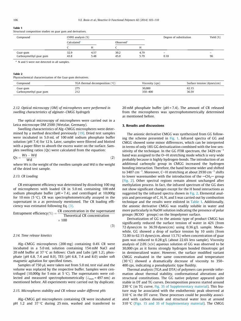
Table 1Structural composition studies on guar gum and derivatives.
Compound CHNS analysis (%) Degree of substitution Yield (%)
Calculateda Observeda
C H C H
Guar-gum 32.9 4.57 30.2 4.79 – –Carboxymethyl guar gum 43.8 5.48 45.0 5.79 0.18 96
a N and S were not detected in all samples.
Table 2Physicochemical characterization of the Guar-gum derivatives.
Compound TGA thermal decomposition (�C) Viscosity (cps) Surface tension (dynes/cm)
Guar-gum 275 30,000 62.15Carboxymethyl guar gum 212 350–400 36.59
106 V.E. Bosio et al. / Reactive & Functional Polymers 82 (2014) 103–110
2.12. Optical microscopy (OM) of microspheres were performed inswelling characteristics of alginate–CMGG hydrogels
The optical microscopy of microspheres were carried out in aLeica microscope DM 2500 (Wetzlar, Germany).
Swelling characteristics of Alg–CMGG microspheres were deter-mined by a method described previously [19]. Dried test sampleswere incubated in 5.0 mL of 100 mM sodium phosphate buffersolution (pH 7.4) for 12 h. Later, samples were filtered and blottedwith a paper filter to absorb the excess water on the surface. Sam-ples swelling ratios (Qs) were calculated from the equation:
Qs ¼Ws�WdWd
ð2Þ
where Ws is the weight of the swollen sample and Wd is the weightof the dried test sample.
2.13. CR Loading
CR entrapment efficiency was determined by dissolving 100 mgof microspheres with loaded CR in 5.0 mL containing 100 mMsodium phosphate buffer (pH = 7.4), and centrifuged at 10,000gfor 10 min (5 �C). CR was spectrophotometrically assayed in thesupernatant in a as previously mentioned. The CR loading effi-ciency was estimated following Eq. (3).
Entrapment efficiencyð%Þ ¼ CR concentration in the supernatantTheoretical CR concentration
� 100
ð3Þ
2.14. Time release kinetics
Alg–CMGG microspheres (200 mg) containing 0.4% CR wereincubated in a 5.0 mL solution containing 154 mM NaCl and20 mM buffer at 37 �C as follows: Clark and Lubs (pH 1.2), phos-phate (pH 6.8, 7.4 and 8.0), TES (pH 6.8, 7.4 and 8.0) under softmagnetic agitation for specified times.
Samples of 750 lL were taken out from 5.0 mL test vial and thevolume was replaced by the respective buffer. Samples were cen-trifuged (10,000g for 5 min at 5 �C). The supernatants were col-lected and measured spectrophotometrically (kmax = 497 nm) asmentioned before. All experiments were carried out by duplicate.
2.15. Microspheres stability and CR release under different pHs
Alg–CMGG gel microspheres containing CR were incubated atpH 1.2 and 37 �C during 25 min, washed and transferred to
20 mM phosphate buffer (pH = 7.4). The amount of CR releasedfrom the microspheres was spectrophotometrically determinedas mentioned before.
3. Results and discussions
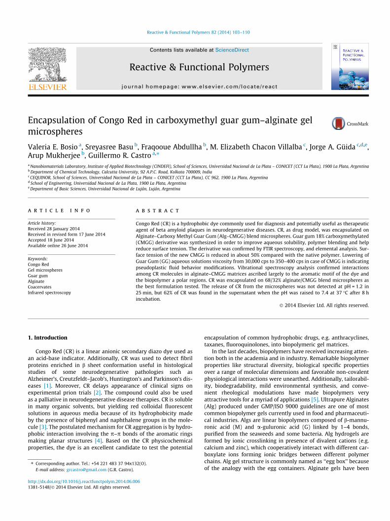
The anionic derivative CMGG was synthesized from GG follow-ing the scheme presented in Fig. 1. Infrared spectra of GG andCMGG showed some minor differences, which can be interpretedin terms of only 18% GG derivatization combined with the low sen-sitivity of the technique. In the GG FTIR spectrum, the 3429 cm�1
band was assigned to the OAH stretching mode which is very wideprobably because is highly hydrogen-bonds. The introduction of anadditional carboxylic group in CMGG increased the hydrogenbonding interaction. Therefore, the band become wider and shiftedto 3407 cm�1. Moreover, CAH stretching at about 2930 cm�1 shiftsto lower wavenumber with the introduction of the ACH2A group(Fig. 2). Other spectral regions remain almost unchanged aftermethylation process. In fact, the infrared spectrum of the GG doesnot show significant changes except for the H bond interactions assuggested by the infrared spectra shown in Fig. 2. Elemental anal-ysis and percentage of C, H, N, and S was carried out by combustiontechnique and the results were enlisted in Table 1. Additionally,the anionic derivative CMGG was readily soluble in water andmore particularly in NaOH solution indicating the presence of polargroups (RCOO� groups) on the biopolymer surface.
Derivatization of GG to the anionic type of product CMGG hassignificantly reduced the surface tension of water in 42% (from72 dynes/cm to 36.59 dynes/cm) using 0.36 g/L sample. Mean-while, GG showed a drop of surface tension by 10 units (from72.00 to 62.15 dynes/cm, about 13.7%) when concentration of guargum was reduced to 0.28 g/L (about 22.6% less sample). Viscosityanalysis of 2.0% (v/v) aqueous solution of GG was observed to be30,000 cps as it forms strongly hydrogen bonded thixotropic gelin demineralized water. However, the surface modified variantCMGG evaluated in the same concentration and temperature(30 �C) showed a dramatically decrease of viscosity to 350–400 cps, indicating a pseudoplastic type fluidity.
Thermal analysis (TGA and DTA) of polymers can provide infor-mation about thermal stability, conformational alterations andstructural constitutions. The GG native polymer appeared quitestable in DT and TG curves. Decomposition process started around230 �C (in TG curve, Fig. 3S of Supplementary material). This fea-ture may be associated with the endothermic peak observed at310 �C in the DT curve. These processes could be possibly associ-ated with carbon dioxide and structural water loss at around310 �C (Figs. 1S and 3S of Supplementary material). The CMGG
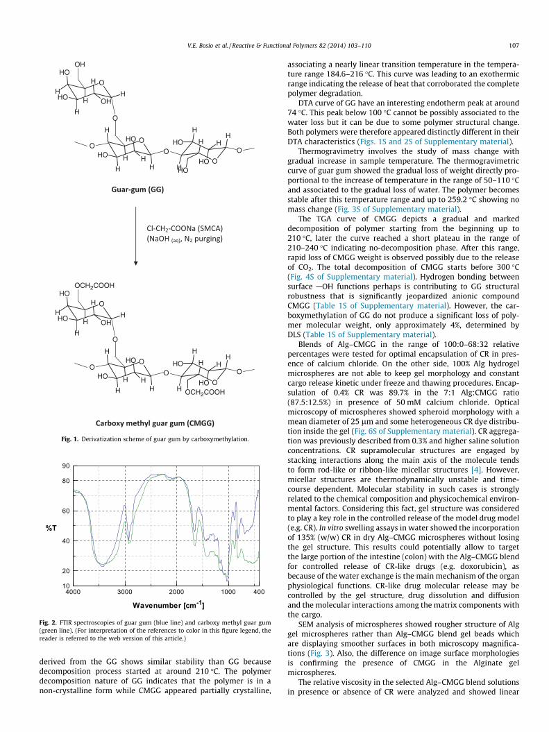
Fig. 1. Derivatization scheme of guar gum by carboxymethylation.
10
90
20
40
60
80
4000 400100020003000
%T
Wavenumber [cm-1]
Fig. 2. FTIR spectroscopies of guar gum (blue line) and carboxy methyl guar gum(green line). (For interpretation of the references to color in this figure legend, thereader is referred to the web version of this article.)
V.E. Bosio et al. / Reactive & Functional Polymers 82 (2014) 103–110 107
derived from the GG shows similar stability than GG becausedecomposition process started at around 210 �C. The polymerdecomposition nature of GG indicates that the polymer is in anon-crystalline form while CMGG appeared partially crystalline,
associating a nearly linear transition temperature in the tempera-ture range 184.6–216 �C. This curve was leading to an exothermicrange indicating the release of heat that corroborated the completepolymer degradation.
DTA curve of GG have an interesting endotherm peak at around74 �C. This peak below 100 �C cannot be possibly associated to thewater loss but it can be due to some polymer structural change.Both polymers were therefore appeared distinctly different in theirDTA characteristics (Figs. 1S and 2S of Supplementary material).
Thermogravimetry involves the study of mass change withgradual increase in sample temperature. The thermogravimetriccurve of guar gum showed the gradual loss of weight directly pro-portional to the increase of temperature in the range of 50–110 �Cand associated to the gradual loss of water. The polymer becomesstable after this temperature range and up to 259.2 �C showing nomass change (Fig. 3S of Supplementary material).
The TGA curve of CMGG depicts a gradual and markeddecomposition of polymer starting from the beginning up to210 �C, later the curve reached a short plateau in the range of210–240 �C indicating no-decomposition phase. After this range,rapid loss of CMGG weight is observed possibly due to the releaseof CO2. The total decomposition of CMGG starts before 300 �C(Fig. 4S of Supplementary material). Hydrogen bonding betweensurface AOH functions perhaps is contributing to GG structuralrobustness that is significantly jeopardized anionic compoundCMGG (Table 1S of Supplementary material). However, the car-boxymethylation of GG do not produce a significant loss of poly-mer molecular weight, only approximately 4%, determined byDLS (Table 1S of Supplementary material).
Blends of Alg–CMGG in the range of 100:0–68:32 relativepercentages were tested for optimal encapsulation of CR in pres-ence of calcium chloride. On the other side, 100% Alg hydrogelmicrospheres are not able to keep gel morphology and constantcargo release kinetic under freeze and thawing procedures. Encap-sulation of 0.4% CR was 89.7% in the 7:1 Alg:CMGG ratio(87.5:12.5%) in presence of 50 mM calcium chloride. Opticalmicroscopy of microspheres showed spheroid morphology with amean diameter of 25 lm and some heterogeneous CR dye distribu-tion inside the gel (Fig. 6S of Supplementary material). CR aggrega-tion was previously described from 0.3% and higher saline solutionconcentrations. CR supramolecular structures are engaged bystacking interactions along the main axis of the molecule tendsto form rod-like or ribbon-like micellar structures [4]. However,micellar structures are thermodynamically unstable and time-course dependent. Molecular stability in such cases is stronglyrelated to the chemical composition and physicochemical environ-mental factors. Considering this fact, gel structure was consideredto play a key role in the controlled release of the model drug model(e.g. CR). In vitro swelling assays in water showed the incorporationof 135% (w/w) CR in dry Alg–CMGG microspheres without losingthe gel structure. This results could potentially allow to targetthe large portion of the intestine (colon) with the Alg–CMGG blendfor controlled release of CR-like drugs (e.g. doxorubicin), asbecause of the water exchange is the main mechanism of the organphysiological functions. CR-like drug molecular release may becontrolled by the gel structure, drug dissolution and diffusionand the molecular interactions among the matrix components withthe cargo.
SEM analysis of microspheres showed rougher structure of Alggel microspheres rather than Alg–CMGG blend gel beads whichare displaying smoother surfaces in both microscopy magnifica-tions (Fig. 3). Also, the difference on image surface morphologiesis confirming the presence of CMGG in the Alginate gelmicrospheres.
The relative viscosity in the selected Alg–CMGG blend solutionsin presence or absence of CR were analyzed and showed linear

108 V.E. Bosio et al. / Reactive & Functional Polymers 82 (2014) 103–110
responses in both cases. However, incorporation of CR to the Alg–CMGG blend reduces the relative viscosity in about one third(Fig. 4). These results are suggesting strong interaction amongstCR and Alg–CMGG matrix solution. FTIR and Raman spectroscopieswere further tested the hypothesis of CR and Alg–CMGGinteraction.
FTIR and Raman spectroscopies were used to test the hypothe-sis of CR and Alg–CMGG interaction. The matrix formulation con-taining CR was analyzed simultaneously by infrared and Ramanspectroscopies because both techniques are providing complemen-tary information. CR bands are not observed in infrared spectrabecause it́s low concentration. Conversely, the strong dispersionobserved for CR in Raman provided good quality spectra even inthe low concentration level of CR. Consequently, infrared spectros-copy reveals interactions between matrix components whileRaman evidenced interactions between CR and the matrix.
Wavenumbers and assignment of Raman spectra for the com-mercial CR and the CR in formulation are collected in Table 3.The Raman spectra are shown in Fig. 5S (see Supplementary mate-rials). New bands were observed in the Raman spectra of the for-mulation containing CR at 1562, 1321, 1265 and 1176 cm�1. The1562 and 1176 cm�1 peaks corresponds to splitting bands ofm(phenyl ring) and m(/-N-azo), m(ASO2AOA)sym, respectively,while 1321 and 1265 cm�1 corresponds to GG bands contained inthe matrix.
The naphthyl ring band at 1353 cm�1 undergoes appreciableshift to upper wavelength (+24 cm�1) as consequence of CR-matrixinteraction, while CR bands for m(AN@NA)azo modes at 1453 and1407 cm�1 does not shift significantly when the dye is inside thematrix, therefore this group does not interact with the biopolymerblend. The conclusion is also supported by structural consider-ations of the CR molecule considering the lone pair on nitrogenof azo groups is restricted by steric hindrance made by phenylgroups and naphthyl groups [19].
The main interaction between CR and the matrix is based on thearomatic motifs of CR associated with non-polar sections of the
Fig. 3. SEM microscopy of Alg (A and B) and
polymer blend determined by spectroscopic analysis [20]. Consid-ering the ‘‘egg box’’ structure of alginate gels made by ionic gela-tion, and the low molecular weight and the planar structure ofCR, it can be possible to hypothesize that the CR molecule couldbe located in the free non-ionic pockets in between two moleculeslinked by calcium ions [21]. Additionally, CMGG is not allowed tofit because of its molecular weight and hydrophilicity. This conclu-sion is relevant since the CR molecule entrapped inside the hydro-phobic motif of alginate ‘‘egg-box’’ gel structure is not able todevelop molecular stacking, and consequently the amount of CRreleased from the microspheres can be determined preciselyincreasing the CR biodisponibility concomitantly with a toxicitydecrease.
Similarly, the adsorption of Direct blue 1 dye onto cellulosefibrils determined by Raman and UV–Vis spectroscopies wasattributed to hydrogen bonding and by hydrophobic interactionbetween biphenyl groups of the diazo dye containing planar p-electrons and a polar regions of cellulose [22].
CR kinetic release from Alg–CMGG gel microspheres wasstudied in vitro under very acid (pH 1.2) and blood physiological(phosphate buffer, pH 7.4) conditions. No changes on microspheresmorphology and CR release to the media were observed under acidconditions (pH 1.2) at 37 �C for 25 min. On the contrary, when thepH is increased above 6.0 a CR release from the microspheres to themedia was detected. The CR kinetic release is showing hyperbolicbehavior at pH 7.4 with 68.2% CR released in about 8 h (Fig. 5).
The changes in the CR release profile from the matrix can beexplained based on the effect of environmental pH on the structureand chemical composition of alginate gels. Alginates are composedby beta-mannuronic acid (M units) and alpha-guluronic acid (Gunits) linked by 1–4 bonds with pKa of 3.38 and 3.65 respectively.Alginate hydrogels are formed by stacking of the G units in pres-ence of multivalent ions, e.g. calcium, which cooperatively interactforming ionic bridges between different polymer chains making anstructure known as ‘‘egg box’’ by analogy with egg containers. Inbetween the crosslinking points, the mannuronic (M) units remain
Alg–CMGG (7:1, C and D) microspheres.

Relatives Viscosities1.0 1.5 2.0 2.5 3.0 3.5 4.0 4.5
%p/
v
0.05
0.10
0.15
0.20
0.25
0.30
0.35
0.40S1/S2 = 1.49
S1 = 0,1534r ² = 0,995
S2 = 0,1029r ² = 0,995
% p/v0.10 0.15 0.20 0.25 0.30 0.35
Rel
Vis
c
1.0
1.5
2.0
Fig. 4. Relative viscosity change of 1.0 and 2.0% Alg–CMGG blend solution in the absence (d) or presence (j) of CR, and the ratio between both curves (�) at 37 �C.
Table 3Raman assignments of CR molecule (solid state) pure and in the formulation(Alg–CMGG).
CR wavenumbers (cm�1) Assignments
Pure Formulation
1590 1592 Phenyl ring1453 1453 m(AN@NA)azo1407 1407 m(AN@NA)azo1353 1377 Naphthyl ring1332 1332 Naphthyl ring1279 1285 m(/-/)1155 1176 m(/-N-azo), m(ASO2AOA)sim
The idea was to highlight the major changes in the wavelengths betweeen the CRcompared to the formulation.
Time (min)0 100 200 300 400 500
Rem
aini
ng L
oad
(mg)
12
14
16
18
20
22
Fig. 5. Time release of CR from Alg (j) and Alg–CMGG (7:1, d) in 154 mM NaCl and20 mM phosphate buffer (pH 7.4) at 37 �C.
V.E. Bosio et al. / Reactive & Functional Polymers 82 (2014) 103–110 109
mostly free and the degree of ionization of carboxylate residuesdepends on external pH. At pH 1.2, the carboxylate of M unit isin the acid form (non-polar, pKa 3.38). Meanwhile at pH 6.8 to8.0, the M carboxylate residue remains as anionic form. Based onthe hydrophobic motifs of Congo Red and the interaction withthe mannuronic acid residues, the dye is fast released from thematrix at alkaline pH compared to the acid one. Similar environ-mental sensitive gel matrices made of synthetic polymers from
methacrylate derivatives containing different molecular cargoeswere previously reported in the literature [23–25].
Additionally, the effect of CMGG coating on alginate micro-spheres is forming an additional diffusional barrier delaying theCR release from the gels.
4. Conclusions
CMGG derivative bring more soluble biopolymer compared toGG, easy to handle because of the low viscosity and making clearsolutions, both are relevant properties for large scale use. CMGGstabilize Alg gels and let to perform freeze-dry procedures withoutlosing gel structure allowing to slow release of the CR cargo.
Viscosimetric and spectroscopic analysis are revealing theinteraction between the Alg–CMGG gel biopolymer blend and thearomatic rings of CR.
Encapsulation of CR in Alg–CMGG blend microspheres showedexcellent stability at acid pH in where can be preserved with cargorelease to the medium. The slow release of CR from themicrospheres at pH 7.4 allows to control cargo delivery and dem-onstrating the advantage of encapsulation in Alg–CMGG to reducethe risk of molecular stacking favoring CR release as model forhydrophobic drugs. The present results could be also extrapolatedto other molecules with pharmacological uses showing high toxic-ity and/or low biodisponibility.
Experiments in cell cultures and CR intracellular trackingstudies are now under study in our laboratory.
Acknowledgments
Support from CONICET, Universidad Nacional de La Plata, andANPCyT (PIP-0214, X/545, and PICT2011-2116 respectively) fromArgentina to GRC; and from CONICET (PIP-0327), and UniversidadNacional de Luján to JAG is gratefully acknowledged. MEChV ismember of Professional Staff of Comisión de InvestigacionesCientíficas, Provincia de Buenos Aires, Argentina (CICPBA). One ofthe author’s (FA) would like to thank UGC, Govt. of India for asenior research fellowship grant.
Appendix A. Supplementary material
Supplementary data associated with this article can be found, inthe online version, at http://dx.doi.org/10.1016/j.reactfunctpolym.2014.06.006.

110 V.E. Bosio et al. / Reactive & Functional Polymers 82 (2014) 103–110
References
[1] P. Frid, S.V. Anisimov, N. Popovic, Brain Res. Rev. 53 (2007) 135–160.[2] K.T. Adjou, M. Seman, Therapie 57 (2002) 123–127.[3] K. Mazeau, M. Wyszomirski, Cellulose 19 (2012) 1495–1506.[4] M. Skowronek, B. Stopa, L. Konieczny, J. Rybarska, B. Piekarska, E. Szneler, G.
Bakalarski, I. Roterman, Biopolymers 46 (1998) 267–281.[5] G.A. Islan, G.R. Castro, Tailoring of alginate–gelatin microspheres properties for
oral Ciprofloxacin-controlled release against Pseudomonas aeruginosa, DrugDeliv, 2014. http://dx.doi.org/10.3109/10717544.2013.870257 (In press).
[6] G.A. Islan, V.E. Bosio, G.R. Castro, Macromol. Biosci. 13 (2013) 1238–1248.[7] M. George, T.E. Abraham, Int. J. Pharm. 335 (2007) 123–129.[8] S.K. Bajpai, S.K. Saxena, S. Sharma, React. Funct. Polym. 66 (2006) 659–666.[9] D. Das, T. Ara, S. Dutta, A. Mukherjee, Bioresour. Technol. 102 (2011) 5878–
5883.[10] S. Kaity, A. Ghosh, Ind. Eng. Chem. Res. 52 (2013) 10033–10045.[11] K. Sangeetha, T.E. Abraham, Int. J. Biol. Macromol. 43 (2008) 314–319.[12] I. Roy, M. Sardar, M.N. Gupta, Biochem. Eng. J. 23 (2005) 193–198.[13] N. Shahzadi, M.S. Butt, M.K. Sharif, M. Nasir, LWT-Food Sci. Technol. 40 (2007)
1198–1205.
[14] J.R. Paxman, J.C. Richardson, P.W. Dettmar, B.M. Corfe, Nutr. Res. 28 (2008)501–505.
[15] D. Lin-Vien, N.B. Colthup, W.G. Fateley, J.G. Grasselli, The Handbook of Infraredand Raman Characteristic Frequencies of Organic Molecules, Academic Press,New York, 1991.
[16] J. Herranen, J. Kinnunen, B. Mattsson, H. Rinne, F. Sundholm, L. Torell, SolidState Ionics 80 (1995) 201–212.
[17] J. Sajid, A. Elhaddaoui, S. Turrell, J. Mol. Struct. 408–409 (1997) 181–184.[18] B. Smith, Infrared Spectral Interpretation, CRC Press, Boca Raton, 1999.[19] C.E. Bonancêa, G.M. do Nascimento, M.L. de Souza, M.L.A. Temperini, P. Corio,
Appl. Catal. B 69 (2006) 34–42.[20] S.-C. Chen, Y.-C. Wu, F.-L. Mi, Y.-H. Lin, L.-C. Yu, H.-W. Sung, J. Control. Release
96 (2004) 285–300.[21] W.H. Ojala, C.R. Ojala, W.B. Gleason, Antiviral Chem. Chemother. 6 (1995) 25–
33.[22] L.C. Abbott, S.N. Batchelor, L. Jansen, J. Oakes, J.R. Lindsay, J.N. Moore, New J.
Chem. 28 (2004) 815–821.[23] L. Brannon-Peppas, N.A. Peppas, J. Controlled Release 8 (1989) 267–274.[24] L.-C. Dong, A.S. Hoffman, J. Controlled Release 15 (1991) 141–152.[25] M.F.A. Taleb, S.E. Abdel-Aal, N.A. El-Kelesh, E.-S.A. Hegazy, Eur. Polym. J. 43
(2007) 468–477.