Embed Size (px)
Citation preview
EXPERIMENTAL AND THERAPEUTIC MEDICINE 14: 547-554, 2017
Abstract. The present study aimed to investigate the clinical and radiological characteristics in addition to the broncho-scopic appearance in patients with endobronchial aspergilloma (EBA). Clinical and radiological characteristics were analyzed alongside the bronchoscopic appearance in 17 patients with EBA diagnosed by bronchoscopy with histological examina-tion. The present study assessed the relevant literature and 13 males and 4 females were included in the comparison, with a median age of 59. Associated diseases included 8 previous diagnoses of pulmonary tuberculosis (47.6%), 4 previous diagnoses lung cancer (23.5%), 1 pulmonary resection (5.9%) and 1 bronchial foreign body (5.9%). The primary symptom was hemoptysis (9/17, 53%). Chest computed tomography (CT) indicated a markedly higher incidence of aspergillosis lesion in
the left lung (13/17; 76.5%) compared with the right lung (4/17; 23.5%). CT manifestation included space occupying disease in 10 patients (58.8%), aspergilloma in 3 patients (17.6%), pneu-monic consolidation in 2 patients (11.8%) and ground glass opacity in 1 patient (5.9%). Bronchoscopy examination identi-fied masses in all 17 patients' bronchial lumen and 15 patients had endobronchial obstruction by necrotic material. The case presented in the current study demonstrated the merits of combining bronchosopic intervention with voriconazole. The dominant symptom of EBA was hemoptysis. Chest CT demonstrated that aspergillosis lesions were more frequently identified in the left lung compared with the right. EBA often occurs in individuals with underlying lung diseases, which cause lumen structural change or bronchial obstruction. EBA may be clearly diagnosed by bronchoscopy biopsy, although the potential for a co-exististing tumor requires consideration. Bronchoscopic intervention and anti-fungal therapy may have an advantage in the effective treatment of patients with EBA.
Introduction
Aspergillus spp. are a ubiquitous fungus in the environment, which are easily identified in soil, water and various types of decomposing organic matter (1). Inhalation of Aspergillus spores causes pulmonary aspergillosis, which is the most common form of aspergillus filamentous fungal infection, and is often diagnosed in patients with immune deficiencies or systemic diseases (2). Aspergillosis is currently divided into the following categories (3): i) Invasive pulmonary aspergil-losis (IPA), a severe disease and major cause of mortality in severely immunocompromised patients; ii) chronic necrotizing aspergillosis (CNA), presents as a locally invasive disease and is observed primarily in patients who are mildly immuno-compromised or have chronic lung disease; and iii) allergic bronchopulmonary aspergillosis (ABPA), a non-invasive hypersensitivity pulmonary disease caused by Aspergillus. Aspergilloma is a fungus ball that develops in a pre-existing cavity in lung parenchyma, which may be easily detected by X-ray and computed tomography (CT) scans (3). In addition, a novel category of aspergillosis with semi-invasive form (SIA) has been described (4).
Endobronchial aspergilloma: A case report and literature reviewDONGDONG HUANG1*, BING LI2*, HAIQING CHU2, ZHEMIN ZHANG2, QIUHONG SUN2,
LAN ZHAO2, LIYUN XU2, LI SHEN2, TAO GUI2, HUIKANG XIE3 and JUN ZHANG4
1Department of Clinical Medicine, Tongji University School of Medicine, Shanghai 200092; Departments of 2Respiratory Medicine and 3Pathology, Shanghai Pulmonary Hospital, Tongji University School of Medicine,
Shanghai 200433, P.R. China; 4Division of Hematology, Oncology and Blood & Marrow Transplantation, Department of Internal Medicine, Holden Comprehensive Cancer Center,
University of Iowa Carver College of Medicine, Iowa, IA 52242, USA
Received March 6, 2016; Accepted March 23, 2017
DOI: 10.3892/etm.2017.4540
Correspondence to: Professor Haiqing Chu or Professor Zhemin Zhang, Department of Respiratory Medicine, Shanghai Pulmonary Hospital, Tongji University School of Medicine, 507 Zhengmin Road, Shanghai 200433, P.R. ChinaE-mail: [email protected]: [email protected]
*Contributed equally
Abbreviations: EBA, endobronchial aspergilloma; CT, computer tomography; IPA, invasive pulmonary aspergillosis; CAN, chronic necrotizing aspergillosis; ABPA, allergic bronchopulmonary aspergillosis; SIA, semi-invasive aspergillosis; FDG, 18F- deoxyglucose; FOB, fiberoptic bronchoscopy; DCMP, dilated cardiomyopathy; HT, hypertension; NSCLC, non-small cell lung cancer; ET, essential thrombocytosis; SDH, subarachnoid hemorrhage; ICH, intracranial hemorrhage; TB, tuberculosis; BW, bronchial washing; MTB, Mycobacterium tuberculosis; IV, intravenous; GGO, ground glass opacity; LLL, left lower lobe; LUL, left upper lobe; RML, right middle lobe; RUL, right upper lobe; RBI, right bronchus intermedius
Key words: fungi, endobronchial aspergilloma, bronchoscopy
HUANG et al: ENDOBRONCHIAL ASPERGILLOMA548
Endobronchial aspergilloma (EBA) is a rare disease, described as a massive non-invasive overgrowth of the asper-gillus species in bronchial lumen, causing space-occupying lesions (5,6). It is often incidentally diagnosed during bron-choscopy to evaluate the cause of hemoptysis (5,6), and may be considered as another form of pulmonary aspergillosis. At present, there are very few reports regarding the charac-teristics of EBA. The present study aimed to investigate the clinical and radiological characteristics in addition to the bronchoscopic appearance in patients with EBA through a representative case from Shanghai Pulmonary Hospital affili-ated to Tongji University (Shanghai, China) and a review of the current literature.
Case report
Study design. The present study reports a case of EBA with no prior treatment that was diagnosed at Shanghai Pulmonary Hospital affiliated to Tongji University, informed consent was also obtained from the patient. A retrospective analysis of the clinical and radiological characteristics and bronchoscopic results was also completed by combining this case with 16 other previously reported EBA cases (5-10). All patients we subjected to a chest CT, bronchoscopy and histological examination. The tissue used for histological examination was obtained via bronchoscopy biopsy of the mass in the trachea, avoiding necrotic tissue or secondary infection areas, with a volume of at least 0.5x0.5x0.2 cm and had a fresh non-necrotic color. The tissue was fixed using 10% neutral formaldehyde for 24 h at room temperature, routinely selected, embedded, sliced in continuous sections with a thickness of 4 µm and hematox-ylin-eosin (HE) stained. The test results and basic clinical data were collected. Primary evidence to confirm EBA were as follows: i) Presence of bronchoscopy-visible intraluminal mass and necrotic tissue; ii) characteristic appearance of Aspergillus species under microscopic examination, such as septate fun gal hyphae bearing acute branching angles; iii) positive result of Aspergillus culture; and iv) following the elimination of other possibilities, such as tumors, foreign bodies, parasites or mucus by evaluating the patient's personal history, performing bronchoscopy and pathology examinations.
Representative case. A 48-year-old male was admitted to Shanghai Pulmonary Hospital affiliated to Tongji University due to a recurrent fever, cough, expectoration and bloody sputum lasting 3 months. The patient had a 20-year history of smoking, and a 4-year history of diabetes and was undergoing diet therapy. On admission, he presented with normal vital signs. Physical examination of the chest revealed no evident crackles or wheezing. Complete blood count results were normal. Routine blood chemistry and tumor markers were all within normal limits. Mycodextranase and latex agglutination tests from the blood were negative. Human immunodeficiency virus antibody test was negative. Chemical luminescence of syphilis test was negative.
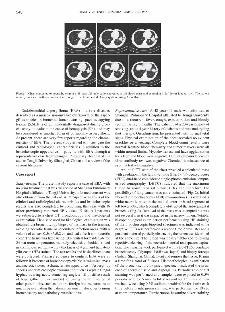
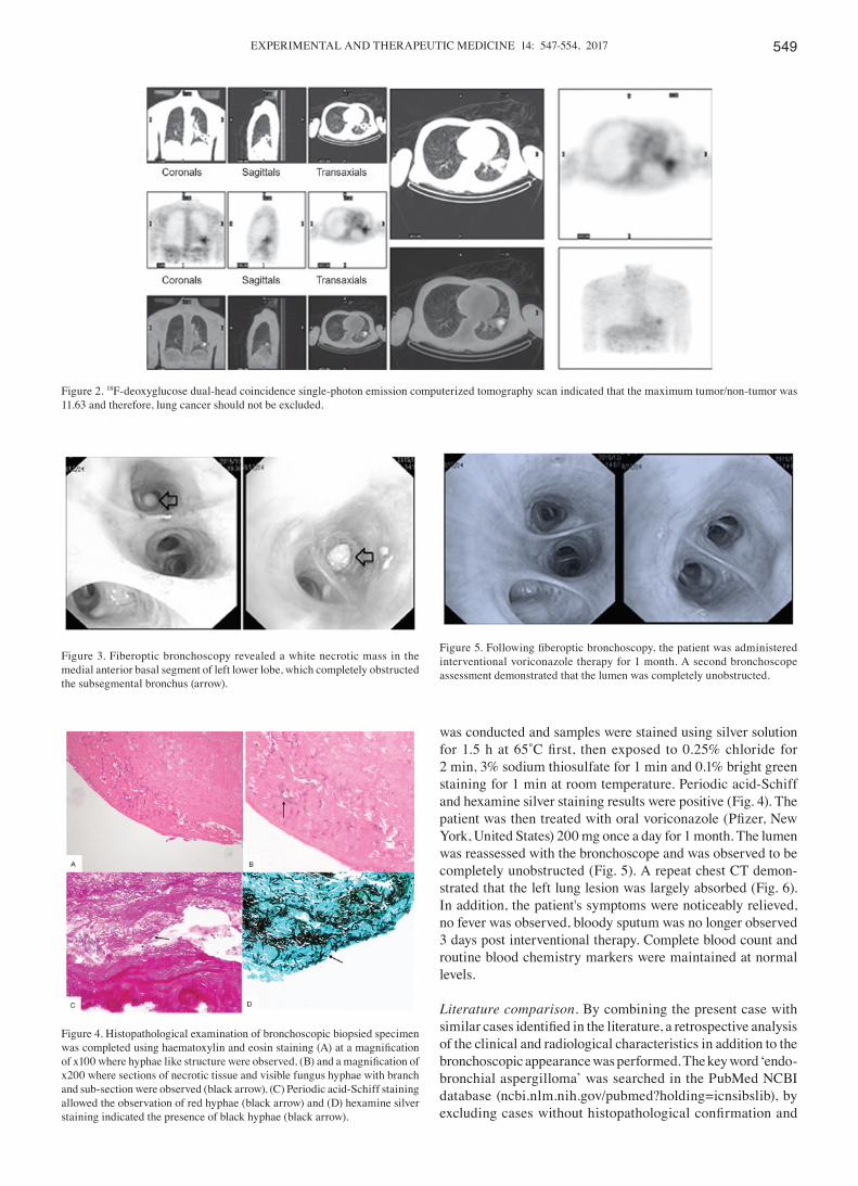
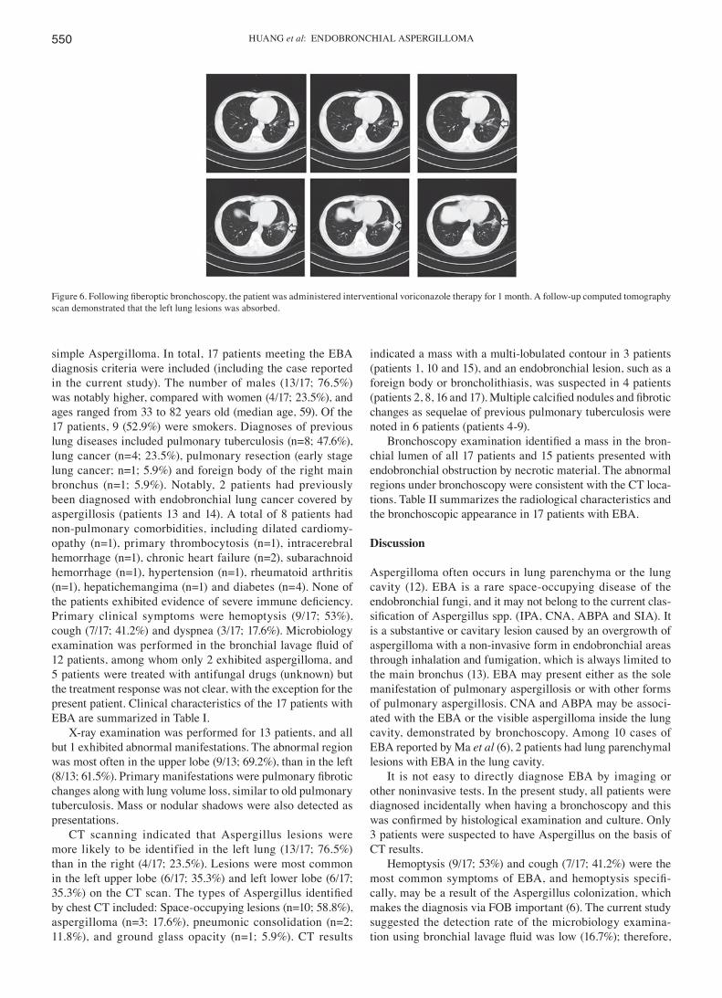
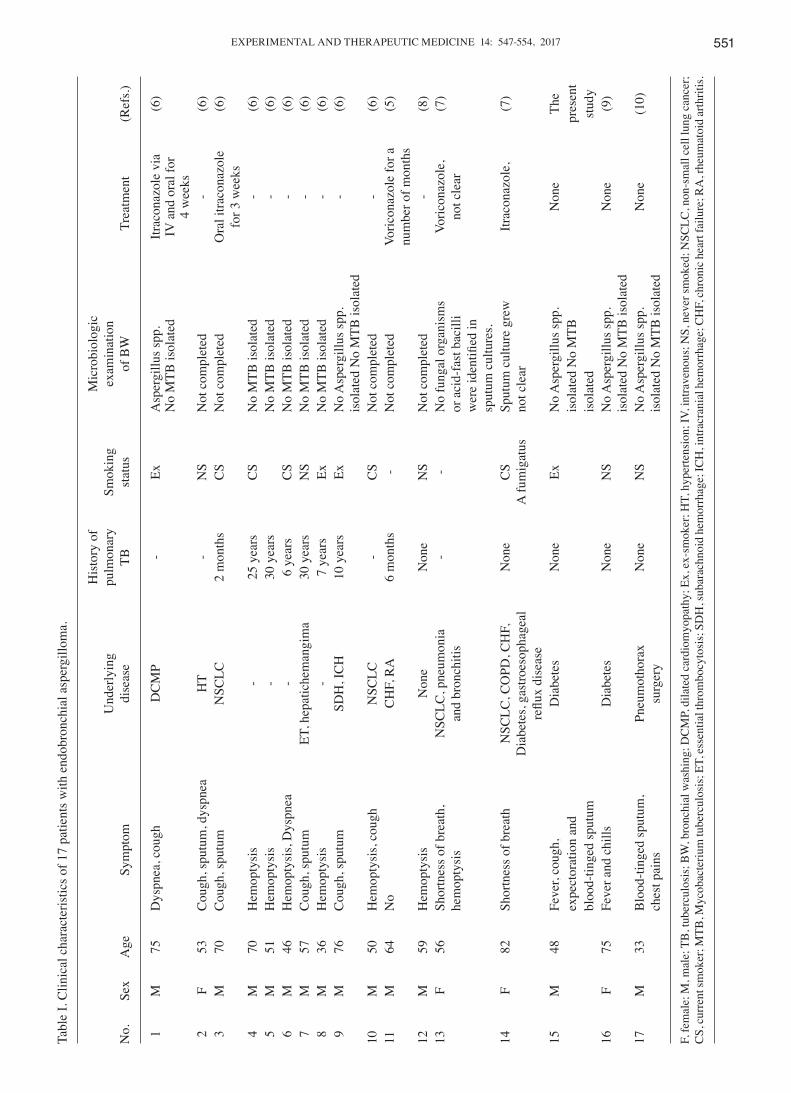
An intial CT scan of the chest revealed a spiculated mass with exudation in the left lower lobe (Fig. 1). 18F- deoxyglucose (FDG) dual-head coincidence single-photon emission comput-erized tomography (DHTC) indicated that the maximum tumor to non-tumor ratio was 11.63 and therefore, the possibility of lung cancer was not eliminated (Fig. 2). Initial fiberoptic bronchoscopy (FOB) examination (11) revealed a white necrotic mass in the medial anterior basal segment of left lower lobe, which completely obstructed the subsegmental bronchus (Fig. 3). Removal of the mass was attempted but was not successful as it was impacted in the narrow lumen. Notably, histopathological examination performed using HE staining of the bronchoscopic biopsied specimen was indicated to be negative. FOB was performed a second time 2 days later and a purulent material partially obstructing the lumen was identified at the same site. The lumen was finally unblocked following repetitive clearing of the necrotic material and sputum aspira-tion. The clearing work performed with a BF-1T260 bendable bronchoscopy (Olympus, Ishikawa, Japan) and biopsy forceps (Aohua, Shanghai, China), to cut and remove the tissue, 10 min a time for a total of 3 times. Histopathological examination of the bronchoscopic biopsied specimen indicated the pres-ence of necrotic tissue and Aspergillus. Periodic acid-Schiff staining was performed and samples were exposed to 0.5% periodic acid for 5 min, Schiffs' reagent for 15 min and then washed twice using 0.5% sodium metabisulfite for 1 min each time before bright green staining was performed for 10 sec at room temperature. Furthermore, hexamine silver staining
Figure 1. Chest computed tomography scan of a 48-year-old male patient revealed a spiculated mass and exudation in left lower lobe (arrow). The patient initially presented with a recurrent fever, cough, expectoration and bloody sputum lasting 3 months.
EXPERIMENTAL AND THERAPEUTIC MEDICINE 14: 547-554, 2017 549
was conducted and samples were stained using silver solution for 1.5 h at 65˚C first, then exposed to 0.25% chloride for 2 min, 3% sodium thiosulfate for 1 min and 0.1% bright green staining for 1 min at room temperature. Periodic acid-Schiff and hexamine silver staining results were positive (Fig. 4). The patient was then treated with oral voriconazole (Pfizer, New York, United States) 200 mg once a day for 1 month. The lumen was reassessed with the bronchoscope and was observed to be completely unobstructed (Fig. 5). A repeat chest CT demon-strated that the left lung lesion was largely absorbed (Fig. 6). In addition, the patient's symptoms were noticeably relieved, no fever was observed, bloody sputum was no longer observed 3 days post interventional therapy. Complete blood count and routine blood chemistry markers were maintained at normal levels.
Literature comparison. By combining the present case with similar cases identified in the literature, a retrospective analysis of the clinical and radiological characteristics in addition to the bronchoscopic appearance was performed. The key word ‘endo-bronchial aspergilloma’ was searched in the PubMed NCBI database (ncbi.nlm.nih.gov/pubmed?holding=icnsibslib), by excluding cases without histopathological confirmation and
Figure 2. 18F-deoxyglucose dual-head coincidence single-photon emission computerized tomography scan indicated that the maximum tumor/non-tumor was 11.63 and therefore, lung cancer should not be excluded.
Figure 3. Fiberoptic bronchoscopy revealed a white necrotic mass in the medial anterior basal segment of left lower lobe, which completely obstructed the subsegmental bronchus (arrow).
Figure 4. Histopathological examination of bronchoscopic biopsied specimen was completed using haematoxylin and eosin staining (A) at a magnification of x100 where hyphae like structure were observed, (B) and a magnification of x200 where sections of necrotic tissue and visible fungus hyphae with branch and sub-section were observed (black arrow). (C) Periodic acid-Schiff staining allowed the observation of red hyphae (black arrow) and (D) hexamine silver staining indicated the presence of black hyphae (black arrow).
Figure 5. Following fiberoptic bronchoscopy, the patient was administered interventional voriconazole therapy for 1 month. A second bronchoscope assessment demonstrated that the lumen was completely unobstructed.
HUANG et al: ENDOBRONCHIAL ASPERGILLOMA550
simple Aspergilloma. In total, 17 patients meeting the EBA diagnosis criteria were included (including the case reported in the current study). The number of males (13/17; 76.5%) was notably higher, compared with women (4/17; 23.5%), and ages ranged from 33 to 82 years old (median age, 59). Of the 17 patients, 9 (52.9%) were smokers. Diagnoses of previous lung diseases included pulmonary tuberculosis (n=8; 47.6%), lung cancer (n=4; 23.5%), pulmonary resection (early stage lung cancer; n=1; 5.9%) and foreign body of the right main bronchus (n=1; 5.9%). Notably, 2 patients had previously been diagnosed with endobronchial lung cancer covered by aspergillosis (patients 13 and 14). A total of 8 patients had non-pulmonary comorbidities, including dilated cardiomy-opathy (n=1), primary thrombocytosis (n=1), intracerebral hemorrhage (n=1), chronic heart failure (n=2), subarachnoid hemorrhage (n=1), hypertension (n=1), rheumatoid arthritis (n=1), hepatichemangima (n=1) and diabetes (n=4). None of the patients exhibited evidence of severe immune deficiency. Primary clinical symptoms were hemoptysis (9/17; 53%), cough (7/17; 41.2%) and dyspnea (3/17; 17.6%). Microbiology examination was performed in the bronchial lavage fluid of 12 patients, among whom only 2 exhibited aspergilloma, and 5 patients were treated with antifungal drugs (unknown) but the treatment response was not clear, with the exception for the present patient. Clinical characteristics of the 17 patients with EBA are summarized in Table I.
X-ray examination was performed for 13 patients, and all but 1 exhibited abnormal manifestations. The abnormal region was most often in the upper lobe (9/13; 69.2%), than in the left (8/13; 61.5%). Primary manifestations were pulmonary fibrotic changes along with lung volume loss, similar to old pulmonary tuberculosis. Mass or nodular shadows were also detected as presentations.
CT scanning indicated that Aspergillus lesions were more likely to be identified in the left lung (13/17; 76.5%) than in the right (4/17; 23.5%). Lesions were most common in the left upper lobe (6/17; 35.3%) and left lower lobe (6/17; 35.3%) on the CT scan. The types of Aspergillus identified by chest CT included: Space-occupying lesions (n=10; 58.8%), aspergilloma (n=3; 17.6%), pneumonic consolidation (n=2; 11.8%), and ground glass opacity (n=1; 5.9%). CT results
indicated a mass with a multi-lobulated contour in 3 patients (patients 1, 10 and 15), and an endobronchial lesion, such as a foreign body or broncholithiasis, was suspected in 4 patients (patients 2, 8, 16 and 17). Multiple calcified nodules and fibrotic changes as sequelae of previous pulmonary tuberculosis were noted in 6 patients (patients 4-9).
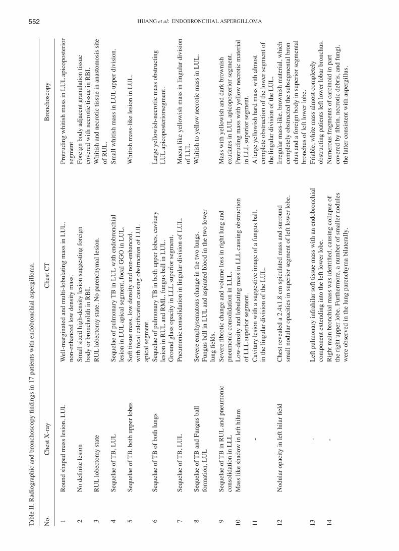
Bronchoscopy examination identified a mass in the bron-chial lumen of all 17 patients and 15 patients presented with endobronchial obstruction by necrotic material. The abnormal regions under bronchoscopy were consistent with the CT loca-tions. Table II summarizes the radiological characteristics and the bronchoscopic appearance in 17 patients with EBA.
Discussion
Aspergilloma often occurs in lung parenchyma or the lung cavity (12). EBA is a rare space-occupying disease of the endobronchial fungi, and it may not belong to the current clas-sification of Aspergillus spp. (IPA, CNA, ABPA and SIA). It is a substantive or cavitary lesion caused by an overgrowth of aspergilloma with a non-invasive form in endobronchial areas through inhalation and fumigation, which is always limited to the main bronchus (13). EBA may present either as the sole manifestation of pulmonary aspergillosis or with other forms of pulmonary aspergillosis. CNA and ABPA may be associ-ated with the EBA or the visible aspergilloma inside the lung cavity, demonstrated by bronchoscopy. Among 10 cases of EBA reported by Ma et al (6), 2 patients had lung parenchymal lesions with EBA in the lung cavity.
It is not easy to directly diagnose EBA by imaging or other noninvasive tests. In the present study, all patients were diagnosed incidentally when having a bronchoscopy and this was confirmed by histological examination and culture. Only 3 patients were suspected to have Aspergillus on the basis of CT results.
Hemoptysis (9/17; 53%) and cough (7/17; 41.2%) were the most common symptoms of EBA, and hemoptysis specifi-cally, may be a result of the Aspergillus colonization, which makes the diagnosis via FOB important (6). The current study suggested the detection rate of the microbiology examina-tion using bronchial lavage fluid was low (16.7%); therefore,
Figure 6. Following fiberoptic bronchoscopy, the patient was administered interventional voriconazole therapy for 1 month. A follow‑up computed tomography scan demonstrated that the left lung lesions was absorbed.
EXPERIMENTAL AND THERAPEUTIC MEDICINE 14: 547-554, 2017 551Ta
ble
I. C
linic
al c
hara
cter
istic
s of 1
7 pa
tient
s with
end
obro
nchi
al a
sper
gillo
ma.
H
isto
ry o
f
Mic
robi
olog
ic
U
nder
lyin
g pu
lmon
ary
Smok
ing
exam
inat
ion
N
o.
Sex
Age
Sy
mpt
om
dise
ase
TB
stat
us
of B
W
Trea
tmen
t (R
efs.)
1
M
75
Dys
pnea
, cou
gh
DC
MP
- Ex
A
sper
gillu
s spp
. Itr
acon
azol
e vi
a (6
)
No
MTB
isol
ated
IV
and
ora
l for
4 w
eeks
2
F 53
C
ough
, spu
tum
, dys
pnea
H
T -
NS
Not
com
plet
ed
- (6
) 3
M
70
C
ough
, spu
tum
N
SCLC
2
mon
ths
CS
Not
com
plet
ed
Ora
l itra
cona
zole
(6
)
fo
r 3 w
eeks
4
M
70
Hem
opty
sis
- 25
yea
rs
CS
No
MTB
isol
ated
-
(6)
5
M
51
Hem
opty
sis
- 30
yea
rs
N
o M
TB is
olat
ed
- (6
) 6
M
46
H
emop
tysi
s, D
yspn
ea
- 6
year
s C
S N
o M
TB is
olat
ed
- (6
) 7
M
57
C
ough
, spu
tum
ET
, hep
atic
hem
angi
ma
30 y
ears
N
S N
o M
TB is
olat
ed
- (6
) 8
M
36
H
emop
tysi
s -
7 ye
ars
Ex
No
MTB
isol
ated
-
(6)
9
M
76
Cou
gh, s
putu
m
SDH
, IC
H
10 y
ears
Ex
N
o A
sper
gillu
s spp
. -
(6)
is
olat
ed N
o M
TB is
olat
ed10
M
50
H
emop
tysi
s, co
ugh
NSC
LC
- C
S N
ot c
ompl
eted
-
(6)
11
M
64
No
CH
F, R
A
6 m
onth
s -
Not
com
plet
ed
Voric
onaz
ole
for a
(5
)
nu
mbe
r of m
onth
s12
M
59
H
emop
tysi
s N
one
Non
e N
S N
ot c
ompl
eted
-
(8)
13
F 56
Sh
ortn
ess o
f bre
ath,
N
SCLC
, pne
umon
ia
- -
No
fung
al o
rgan
ism
s Vo
ricon
azol
e,
(7)
he
mop
tysi
s an
d br
onch
itis
or a
cid-
fast
bac
illi
not c
lear
w
ere
iden
tified
in
sput
um c
ultu
res.
14
F 82
Sh
ortn
ess o
f bre
ath
NSC
LC, C
OPD
, CH
F,
Non
e C
S Sp
utum
cul
ture
gre
w
Itrac
onaz
ole,
(7
)
D
iabe
tes,
gast
roes
opha
geal
A fu
mig
atus
no
t cle
ar
re
flux
dise
ase
15
M
48
Feve
r, co
ugh,
D
iabe
tes
Non
e Ex
N
o A
sper
gillu
s spp
. N
one
The
ex
pect
orat
ion
and
is
olat
ed N
o M
TB
pr
esen
t
bloo
d-tin
ged
sput
um
is
olat
ed
st
udy
16
F 75
Fe
ver a
nd c
hills
D
iabe
tes
Non
e N
S
No
Asp
ergi
llus s
pp.
Non
e (9
)
isol
ated
No
MTB
isol
ated
17
M
33
Blo
od-ti
nged
sput
um,
Pneu
mot
hora
x N
one
NS
N
o A
sper
gillu
s spp
. N
one
(10)
ch
est p
ains
su
rger
y
is
olat
ed N
o M
TB is
olat
ed
F, fe
mal
e; M
, mal
e; T
B, t
uber
culo
sis;
BW
, bro
nchi
al w
ashi
ng; D
CM
P, d
ilate
d ca
rdio
myo
path
y; E
x, e
x-sm
oker
; HT,
hyp
erte
nsio
n; IV
, int
rave
nous
; NS,
nev
er sm
oked
; NSC
LC, n
on-s
mal
l cel
l lun
g ca
ncer
; C
S, cu
rren
t sm
oker
; MTB
, Myc
obac
teriu
m tu
berc
ulos
is; E
T, es
sent
ial t
hrom
bocy
tosi
s; S
DH
, sub
arac
hnoi
d he
mor
rhag
e; IC
H, i
ntra
cran
ial h
emor
rhag
e; C
HF,
chro
nic h
eart
failu
re; R
A, r
heum
atoi
d ar
thrit
is.
HUANG et al: ENDOBRONCHIAL ASPERGILLOMA552Ta
ble
II. R
adio
grap
hic
and
bron
chos
copy
find
ings
in 1
7 pa
tient
s with
end
obro
nchi
al a
sper
gillo
ma.
No.
C
hest
X-r
ay
Che
st C
T B
ronc
hosc
opy
1
Rou
nd sh
aped
mas
s les
ion,
LU
L W
ell-m
argi
nate
d an
d m
ulti-
lobu
latin
g m
ass i
n LU
L,
Prot
rudi
ng w
hitis
h m
ass i
n LU
L ap
icop
oste
rior
non-
enha
nced
low
den
sity
mas
s. se
gmen
t 2
N
o de
finite
lesi
on
Smal
l siz
ed h
igh‑
dens
ity le
sion
sugg
estin
g fo
reig
n Fo
reig
n bo
dy a
djac
ent g
ranu
latio
n tis
sue
body
or b
ronc
holit
h in
RB
I. co
vere
d w
ith n
ecro
tic ti
ssue
in R
BI.
3
RU
L lo
bect
omy
stat
e R
UL
lobe
ctom
y st
ate.
No
pare
nchy
mal
lesi
on.
Whi
tish
and
necr
otic
tiss
ue in
ana
stom
osis
site
of R
UL.
4
Sequ
elae
of T
B, L
UL
Sequ
elae
of p
ulm
onar
y TB
in L
UL
with
end
obro
nchi
al
Smal
l whi
tish
mas
s in
LUL
uppe
r div
isio
n.
le
sion
in L
UL
apic
al se
gmen
t, fo
cal G
GO
in L
UL.
5
Sequ
elae
of T
B, b
oth
uppe
r lob
es
Soft
tissu
e m
ass,
low
den
sity
and
non
-enh
ance
d,
Whi
tish
mas
s-lik
e le
sion
in L
UL.
with
foca
l cal
cific
atio
n ca
usin
g ob
stru
ctio
n of
LU
L
ap
ical
segm
ent.
6
Sequ
elae
of T
B o
f bot
h lu
ngs
Sequ
elae
of p
ulm
onar
y TB
in b
oth
uppe
r lob
es, c
avita
ry
Larg
e ye
llow
ish-
necr
otic
mas
s obs
truct
ing
lesi
on in
RU
L an
d R
ML,
fung
us b
all i
n LU
L.
LUL
apic
opos
terio
rseg
men
t.
G
roun
d gl
ass o
paci
ty in
LLL
supe
rior s
egm
ent.
7
Sequ
elae
of T
B, L
UL
Pneu
mon
ic c
onso
lidat
ion
in li
ngul
ar d
ivis
ion
of L
UL.
M
ucus
like
yel
low
ish
mas
s in
lingu
lar d
ivis
ion
of
LU
L 8
Se
quel
ae o
f TB
and
Fun
gus b
all
Seve
re e
mph
ysem
atou
s cha
nge
in th
e tw
o lu
ngs.
W
hitis
h to
yel
low
nec
rotic
mas
s in
LUL.
fo
rmat
ion,
LU
L Fu
ngus
bal
l in
LUL
and
aspi
rate
d bl
ood
in th
e tw
o lo
wer
lung
fiel
ds.
9
Sequ
elae
of T
B in
RU
L an
d pn
eum
onic
Se
vere
fibr
otic
cha
nge
and
volu
me
loss
in ri
ght l
ung
and
Mas
s with
yel
low
ish
and
dark
bro
wni
sh
cons
olid
atio
n in
LLL
pn
eum
onic
con
solid
atio
n in
LLL
. ex
udat
es in
LU
L ap
icop
oste
rior s
egm
ent.
10
Mas
s lik
e sh
adow
in le
ft hi
lum
Lo
w-d
ensi
ty a
nd lo
bula
ting
mas
s in
LLL
caus
ing
obst
ruct
ion
Prot
rudi
ng m
ass w
ith y
ello
w n
ecro
tic m
ater
ial
of L
LL su
perio
r seg
men
t. in
LLL
supe
rior s
egm
ent.
11
- C
avita
ry le
sion
with
a su
gges
tive
imag
e of
a fu
ngus
bal
l,
A la
rge
yello
wis
h ha
rd m
ass w
ith a
lmos
t
in
the
lingu
lar d
ivis
ion
of th
e LU
L.
com
plet
e ob
stru
ctio
n of
the
low
er se
gmen
t of
th
e lin
gula
r div
isio
n of
the
LUL,
12
Nod
ular
opa
city
in le
ft hi
lar fi
eld
Che
st re
veal
ed a
2.4
x1.8
cm
spic
ulat
ed m
ass a
nd su
rrou
nd
Irre
gula
r mas
s‑lik
e, b
row
nish
mat
eria
l, w
hich
sm
all n
odul
ar o
paci
ties i
n su
perio
r seg
men
t of l
eft l
ower
lobe
. co
mpl
etel
y ob
stru
cted
the
subs
egm
enta
l bro
n
chus
and
a fo
reig
n bo
dy in
supe
rior s
egm
enta
l
bron
chus
of l
eft l
ower
lobe
.13
-
Left
pulm
onar
y in
frah
ilar s
oft t
issu
e m
ass w
ith a
n en
dobr
onch
ial
Fria
ble,
whi
te m
ass a
lmos
t com
plet
ely
com
pone
nt e
xten
ding
into
the
left
low
er lo
be.
obst
ruct
ing
patie
nts l
eft l
ower
loba
r bro
nchu
s.14
‑
Rig
ht m
ain
bron
chia
l mas
s was
iden
tified
, cau
sing
col
laps
e of
N
umer
ous f
ragm
ents
of c
arci
noid
in p
art
the
right
upp
er lo
be. F
urth
erm
ore,
a n
umbe
r of s
mal
ler n
odul
es
cove
red
by fi
brin
, nec
rotic
deb
ris, a
nd fu
ngi,
wer
e ob
serv
ed in
the
lung
par
ench
yma
bila
tera
lly.
the
latte
r con
sist
ent w
ith a
sper
gillu
s.
EXPERIMENTAL AND THERAPEUTIC MEDICINE 14: 547-554, 2017 553
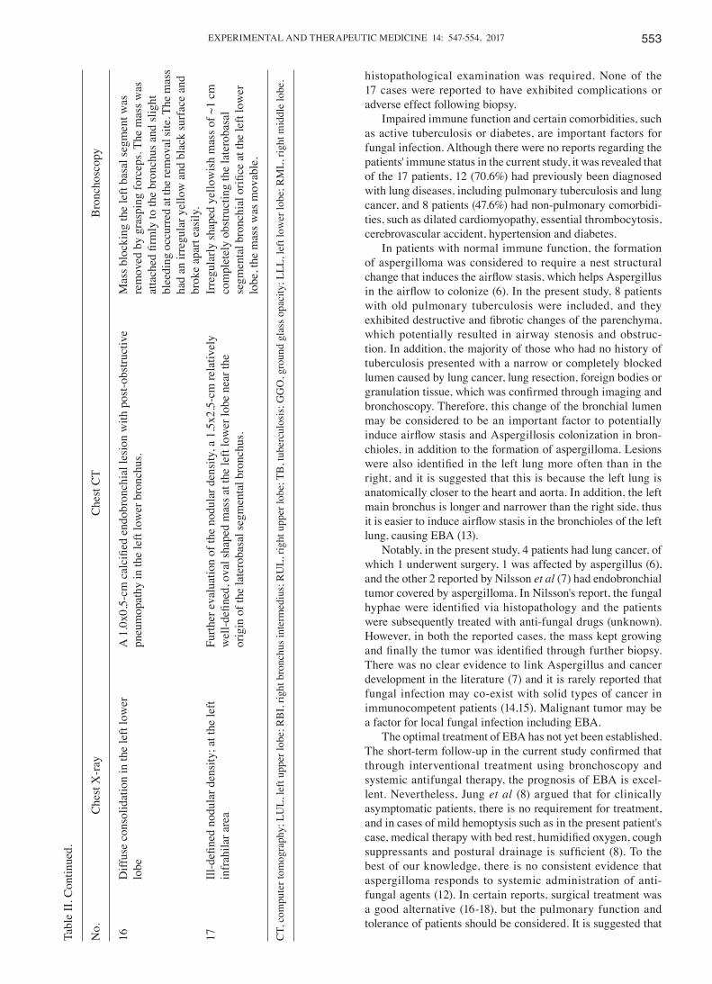
histopathological examination was required. None of the 17 cases were reported to have exhibited complications or adverse effect following biopsy.
Impaired immune function and certain comorbidities, such as active tuberculosis or diabetes, are important factors for fungal infection. Although there were no reports regarding the patients' immune status in the current study, it was revealed that of the 17 patients, 12 (70.6%) had previously been diagnosed with lung diseases, including pulmonary tuberculosis and lung cancer, and 8 patients (47.6%) had non-pulmonary comorbidi-ties, such as dilated cardiomyopathy, essential thrombocytosis, cerebro vascular accident, hypertension and diabetes.
In patients with normal immune function, the formation of aspergilloma was considered to require a nest structural change that induces the airflow stasis, which helps Aspergillus in the airflow to colo nize (6). In the present study, 8 patients with old pulmonary tuberculosis were included, and they exhibited destructive and fibrotic changes of the parenchyma, which potentially resulted in airway stenosis and obstruc-tion. In addition, the majority of those who had no history of tuberculosis presented with a narrow or completely blocked lumen caused by lung cancer, lung resection, foreign bodies or granulation tissue, which was confirmed through imaging and bronchoscopy. Therefore, this change of the bronchial lumen may be considered to be an important factor to potentially induce airflow stasis and Aspergillosis colo nization in bron-chioles, in addition to the formation of aspergilloma. Lesions were also identified in the left lung more often than in the right, and it is suggested that this is because the left lung is anatomically closer to the heart and aorta. In addition, the left main bronchus is longer and narrower than the right side, thus it is easier to induce airflow stasis in the bronchioles of the left lung, causing EBA (13).
Notably, in the present study, 4 patients had lung cancer, of which 1 underwent surgery, 1 was affected by aspergillus (6), and the other 2 reported by Nilsson et al (7) had endobronchial tumor covered by aspergilloma. In Nilsson's report, the fungal hyphae were identified via histopathology and the patients were subsequently treated with anti-fungal drugs (unknown). However, in both the reported cases, the mass kept growing and finally the tumor was identified through further biopsy. There was no clear evidence to link Aspergillus and cancer development in the literature (7) and it is rarely reported that fungal infection may co-exist with solid types of cancer in immunocompetent patients (14,15). Malignant tumor may be a factor for local fungal infection including EBA.
The optimal treatment of EBA has not yet been established. The short‑term follow‑up in the current study confirmed that through interventional treatment using bronchoscopy and systemic antifungal therapy, the prognosis of EBA is excel-lent. Nevertheless, Jung et al (8) argued that for clinically asymptomatic patients, there is no requirement for treatment, and in cases of mild hemoptysis such as in the present patient's case, medical therapy with bed rest, humidified oxygen, cough suppressants and postural drainage is sufficient (8). To the best of our knowledge, there is no consistent evidence that aspergilloma responds to systemic administration of anti-fungal agents (12). In certain reports, surgical treatment was a good alternative (16-18), but the pulmonary function and tolerance of patients should be considered. It is suggested that
Tabl
e II
. Con
tinue
d.
No.
C
hest
X-r
ay
Che
st C
T B
ronc
hosc
opy
16
Diff
use
cons
olid
atio
n in
the
left
low
er
A 1
.0x0
.5‑c
m c
alci
fied
endo
bron
chia
l les
ion
with
pos
t‑obs
truct
ive
Mas
s blo
ckin
g th
e le
ft ba
sal s
egm
ent w
as
lo
be
pneu
mop
athy
in th
e le
ft lo
wer
bro
nchu
s. re
mov
ed b
y gr
aspi
ng fo
rcep
s. Th
e m
ass w
as
at
tach
ed fi
rmly
to th
e br
onch
us a
nd sl
ight
blee
ding
occ
urre
d at
the r
emov
al si
te. T
he m
ass
ha
d an
irre
gula
r yel
low
and
bla
ck su
rfac
e an
d
brok
e ap
art e
asily
.17
Ill
‑defi
ned
nodu
lar d
ensi
ty; a
t the
left
Furth
er e
valu
atio
n of
the
nodu
lar d
ensi
ty, a
1.5
x2.5
‑cm
rela
tivel
y Ir
regu
larly
shap
ed y
ello
wis
h m
ass o
f ~1
cm
in
frah
ilar a
rea
wel
l‑defi
ned,
ova
l sha
ped
mas
s at t
he le
ft lo
wer
lobe
nea
r the
co
mpl
etel
y ob
stru
ctin
g th
e la
tero
basa
l
or
igin
of t
he la
tero
basa
l seg
men
tal b
ronc
hus.
segm
enta
l bro
nchi
al o
rifice
at t
he le
ft lo
wer
lobe
, the
mas
s was
mov
able
.
CT,
com
pute
r tom
ogra
phy;
LU
L, le
ft up
per l
obe;
RB
I, rig
ht b
ronc
hus i
nter
med
ius;
RU
L, ri
ght u
pper
lobe
; TB
, tub
ercu
losi
s; G
GO
, gro
und
glas
s opa
city
; LLL
, lef
t low
er lo
be; R
ML,
righ
t mid
dle
lobe
.
HUANG et al: ENDOBRONCHIAL ASPERGILLOMA554
selecting appropriate treatments according to different condi-tions of patients is reasonable, and the combination of direct bronchoscopic intervention with antifungal therapy may be advantageous.
In conclusion, the dominant symptom for EBA was identified to be hemoptysis in the present analysis. Chest CT demonstrated that aspergillosis lesions were more frequently identified in the left lung compared with the right. It was also revealed that endobronchial aspergilloma often occurs in individuals with comorbidities that may potentially impair immune function and underlying lung diseases that cause lumen structural change or bronchial obstruction. Previous studies have provided sufficient evidence that endobronchial aspergilloma may be clearly diagnosed by bronchoscopy biopsy, but the potential for the co-exististing of tumors requires consideration (5-10). Finally, based on clinical expe-rience, the combination of bronchosopic intervention and anti-fungal therapy may be an effective treatment for patients with EBA.
Acknowledgements
The abstract of this manuscript was presented at the ERS International Congress 3rd-7th September 2016 in London, UK and published as abstract no. PA3715 in the European Respiratory Journal 48 (suppl 60): 2016.
References
1. Silva EF, Barbosa Mde P, Oliveira MA, Martins RR and Fonti-nele e Silva J: Chronic necrotizing pulmonary aspergillosis. J Bras Pneumol 35: 95-98, 2009 (In English, Portuguese).
2. Hospenthal DR, Kwon-Chung KJ and Bennett JE: Concentrations of airborne aspergillus compared to the incidence of invasive aspergillosis: Lack of correlation. Med Mycol 36: 165-168, 1998.
3. Zmeili OS and Soubani AO: Pulmonary aspergillosis: A clinical update. QJM 100: 317-334, 2007.
4. Chabi ML, Goracci A, Roche N, Paugam A, Lupo A and Revel MP: Pulmonary aspergillosis. Diagn Interv Imaging 96: 435-442, 2015.
5. Araújo D, Figueiredo M and Monteiro P: Endobronchial asper-gilloma: An unusual presentation of pulmonary aspergillosis. Rev Port Pneumol (2006) 22: 61-62, 2016.
6. Ma JE, Yun EY, Kim YE, Lee GD, Cho YJ, Jeong YY, Jeon KN, Jang IS, Kim HC, Lee JD and Hwang YS: Endobronchial asper-gilloma: Report of 10 cases and literature review. Yonsei Med J 52: 787-792, 2011.
7. Nilsson JR, Restrepo CS and Jagirdar J: Two cases of endo-bronchial carcinoid masked by superimposed aspergillosis: A review of the literature of primary lung cancers associated with aspergillus. Ann Diagn Pathol 17: 131-136, 2013.
8. Jung SW, Kim MW, Cho SK, Kim HU, Lee DC, Yoon BK, Jeong JP and Ko YC: A case of Endobronchial aspergilloma associated with foreign body in immunocompetent patient without underlying lung disease. Tuberc Respir Dis (Seoul) 74: 231-234, 2013.
9. Yeo CD, Baeg MK and Kim JW: A case of endobronchial asper-gilloma presenting as a broncholith. Am J Med Sci 343: 501-503, 2012.
10. Kim JS, Rhee Y, Kang SM, Ko WK, Kim YS, Lee JG, Park JM, Kim SK, Kim SK, Lee WY and Chang J: A case of endobron-chial aspergilloma. Yonsei Med J 41: 422-425, 2000.
11. British Thoracic Society Bronchoscopy Guidelines Committee, a Subcommittee of the Standards of Crae Committee of the British Thoracic Society: British thoracic society guidelines on diag-nostic flexible bronchoscopy. Thorax 56 (Suppl I): i1‑i21, 2001.
12. Patterson KC and Strek ME: Diagnosis and treatment of pulmo-nary aspergillosis syndromes. Chest 146: 1358-1368, 2014.
13. Soubani AO and Chandrasekar PH: The clinical spectrum of pulmonary aspergillosis. Chest 121: 1988-1999, 2002.
14. Tashiro T, Izumikawa K, Tashiro M, Takazono T, Morinaga Y, Yamamoto K, Imamura Y, Miyazaki T, Seki M, Kakeya H, et al: Diagnostic significance of aspergillus species isolated from respiratory samples in an adult pneumology ward. Med Mycol 49: 581-587, 2011.
15. Ohmagari N, Raad II, Hachem R and Kontoyiannis DP: Inva-sive aspergillosis in patients with solid tumors. Cancer 101: 2300-2302, 2004.
16. Stevens DA, Kan VL, Judson MA, Morrison VA, Dummer S, Denning DW, Bennett JE, Walsh TJ, Patterson TF and Pankey GA: Practice guidelines for diseases caused by Asper-gillus. Infectious diseases society of america. Clin Infect Dis 30: 696-709, 2000.
17. Sagan D and Goździuk K: Surgery for pulmonary aspergilloma in immunocompetent patients: No benefit from adjuvant anti-fungal pharmacotherapy. Ann Thorac Surg 89: 1603-1610, 2010.
18. Lee JG, Lee CY, Park IK, Kim DJ, Chang J, Kim SK and Chung KY: Pulmonary aspergilloma: Analysis of prognosis in relation to symptoms and treatment. J Thorac Cardiovasc Surg 138: 820-825, 2009.