Embed Size (px)
Citation preview

Endocrine evaluation of patientswith critical illness
Greet Van den Berghe, MD, PhD*Department of Intensive Care Medicine,
University Hospital Gasthuisberg Catholic University of Leuven, B-3000 Leuven, Belgium
Critical illness is any condition requiring support of failing vital organsystems without which survival would not be possible. This life-threateningcondition, which may be evoked by trauma, extensive surgery or severemedical illnesses, is an ultimate example of acute, severe physical stress. Ifonset of recovery does not follow within a few days of intensive care, criticalillness often becomes prolonged and vital organ support is frequently neededfor weeks. Feeding does not reverse ongoing wasting of protein from skeletalmuscle and solid organs, which causes impairment of vital functions,weakness, and delayed or hampered recovery [1,2]. This is a frustratingclinical problem because despite adequate and successful treatment of theunderlying disease, dependency on intensive care persists and susceptibilityfor potentially lethal (septic) complications increases. Indeed, mortality fromprolonged critical illness is high: almost 3 out of 10 adult patients with anintensive care stay of more than three weeks do not survive [3]. Male patientsseem to have a higher risk for adverse outcome of prolonged critical illnessthan female patients do, an observation, which remains unexplained [3]. Inline with the foregoing is the inability of the classical scoring systems forseverity of illness, such as APACHE II [4], to predict mortality in anindividual chronic critically ill patient. This enigma reflects lack ofunderstanding of the pathophysiologic mechanisms underlying onset ofrecovery or, conversely, the failure to recover fromprolonged critically illness.
The acute and chronic phases of critical illness are associated with distinctendocrine alterations [5,6]. It remains a matter of debate whether or to whatextent these changes are adaptive or contributing to the metabolic
Endocrinol Metab Clin N Am
32 (2003) 385–410
* E-mail address: [email protected]
Supported by research grants from the Belgian Fund for Scientific Research (G. 0144.00),
the Research Council of the University of Leuven (OT 99/33), and the Belgian Foundation
for Research in Congenital Heart Diseases.
0889-8529/03/$ - see front matter � 2003, Elsevier Inc. All rights reserved.
doi:10.1016/S0889-8529(03)00005-7
disturbances present in the critically ill. The endocrine stress responses arepartially central and partially peripheral in origin. In addition, patients mayhave pre-existing central or peripheral endocrine diseases, either previouslydiagnosed or unknown. Hence, the puzzle is complex and endocrine func-tion testing in a critically ill patient a major challenge. Furthermore, the in-ability to define the endocrine changes as either adaptation or pathologyrenders the issue of treatment even more controversial.
This article reviews the novel insights in the dynamic neuroendocrinealterations as they occur during the course of critical illness. It also high-lights the complexity of differential diagnosis with pre-existing endocrinediseases and the available evidence of benefit or harm of certain endocrineinterventions.
Somatotropic axis
In normal physiology, growth hormone (GH) is released from thepituitary somatotropes in a pulsatile fashion, under the interactive controlof the hypothalamic GH-releasing hormone (GHRH), which is stimulatory,and somatostatin, which exerts an inhibitory effect [7]. Since the 1980s,a series of synthetic GH-releasing peptides (GHRPs) and nonpeptideanalogs have been developed with potent GH-releasing capacities actingthrough a specific G-protein coupled receptor located in the hypothalamusand the pituitary [8,9]. The conserved endogenous ligand for this receptorhas recently been discovered and named ‘‘ghrelin’’ [10]. Ghrelin originates inperipheral tissues, such as the stomach and in the hypothalamic arcuatenucleus, and there seems to be a third key factor in the complex physiologicregulation of pulsatile GH secretion. As shown in rodents [11], there is nowevidence that in the humans [12], the pulsatile nature of GH secretion isimportant for its metabolic effects [3,13].
Alterations within the somatotropic axis in the acute phase of critical illness
During the first hours or days after an acute insult, such as surgery,trauma or infection, circulating GH levels become elevated and the normalGH profile, consisting of peaks alternating with virtually undetectabletroughs, is altered: peak GH and interpulse concentrations are high and theGH pulse frequency is elevated (Fig. 1) [5,14,15]. It is still unclear whichfactor ultimately controls the stimulation of GH release in response tostress. As in starvation [16], more frequent withdrawal of the inhibitorysomatostatin or an increased availability of stimulatory (hypothalamic orperipheral) GH-releasing factors could hypothetically be involved. Serumconcentrations of insulin-like growth factor-1 (IGF-1) and the GH-de-pendent binding protein, IGF-binding protein-3 (IGFBP-3) and its acid-labile subunit (ALS) decrease, which is preceded by a drop in serum levels ofGH-binding protein (GHBP) [17]. The latter was found to reflect reduced
386 G. Van den Berghe / Endocrinol Metab Clin N Am 32 (2003) 385–410
GH-receptor expression in peripheral tissues [17]. Circulating levels of thesmall IGF-binding proteins, such as IGFBP-1, IGFBP-2 and IGFBP-6 areelevated [18,19]. This constellation, which has been confirmed in experi-mental human and animal models of acute stress and in acutely ill patients,has been interpreted as acquired peripheral resistance to GH [14,18]. It hasbeen suggested that these changes are brought about by the effects ofcytokines, such as tumor necrosis factor-a (TNFa), interleukin 1 (IL-1), andIL-6, the hypothesis being that reduced GH receptor expression and thuslow IGF-1 levels are the primary events (cytokine-induced) which, in turn,through reduced negative feedback inhibition, induces the abundant releaseof GH during acute stress, exerting direct lipolytic, insulin antagonizing andimmune-stimulating actions, while the indirect IGF-1 mediated effects ofGH are attenuated [20,21]. This explanation is plausible in that such changeswould prioritize essential substrates, such as glucose, FFA and amino acids(glutamine) toward survival rather than anabolism. Increased IGFBP-3protease activity in plasma has also been reported, however, and is believedto result in increased dissociation of IGF-1 from the ternary complex,thereby shortening the IGF-1 half-life in the circulation. The latter couldtheoretically be an adaptive escape mechanism to secure availability of freeIGF-1 at the tissue level [22].
Distinct alterations within the somatotropic axis during chroniccritical illness
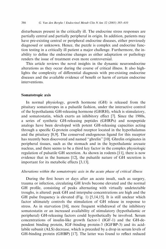
In chronic critical illness, the changes observed within the somatotropicaxis are different. First, the pattern of GH secretion is chaotic and theamount of GH, which is released in pulses, is reduced compared with theacute phase (Fig. 1) [6,23–25]. Moreover, although the nonpulsatile fraction
Fig. 1. Nocturnal serum concentration profiles of GH illustrating the differences between the
acute phase and the chronic phase of critical illness within an intensive care setting. (Adapted
from Van den Berghe G, de Zegher F, Bouillon R. Acute and prolonged critical illness
as different neuroendocrine paradigms. J Clin Endocrinol Metab 1998;83:1827–34; with
permission.)
387G. Van den Berghe / Endocrinol Metab Clin N Am 32 (2003) 385–410

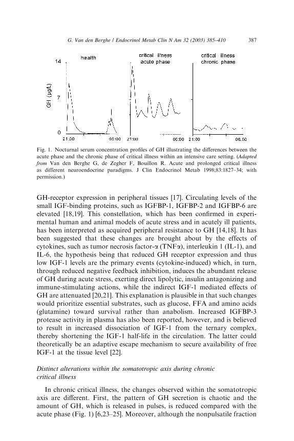
is still somewhat elevated and the number of pulses is still high, meannocturnal GH serum concentrations are scarcely elevated, if at all [23], whencompared with the healthy, nonstressed condition, and substantially lowerthan in the acute phase of stress [5]. The authors observed that, whenintensive care patients are studied from 7 to 10 days illness onward, in theabsence of drugs known to exert profound effects on GH secretion such asdopamine [26,27] and calcium channel blockers or glucocorticoids, meannocturnal GH levels are uniformly around 1 lg/L [23], trough levels areeasily detectable (and thus still elevated) and peak GH levels hardly everexceed 2 lg/L [6,23–25]. These results are surprisingly independent of thepatient’s age, gender, body composition and type of underlying disease [3,5].Second, only the pulsatile fraction of GH secretion—which is substantiallyreduced—correlates positively with circulating levels of IGF-1, IGFBP-3,and ALS, all of which are low [6,24,25]. Thus the more pulsatile GHsecretion is suppressed, the lower the circulating levels of the GH dependentIGF-1 and ternary complex binding proteins become, and this no longerrepresents a state of GH resistance. Elevated serum levels of GHBP [3], as-sumed to reflect GH receptor expression in peripheral tissues, in prolongedcritically ill patients compared with those measured in a matched controlgroup are in line with recovery of GH responsiveness with time duringsevere illness [3,6]. Moreover, low serum levels of GH-dependent IGF-1 andbinding proteins (IGFBP-3, ALS, and IGFBP-5) are tightly related tobiochemical markers of impaired anabolism such as low serum osteocalcinand leptin concentrations during prolonged critical illness [6]. These findingssuggest that relative GH deficiency, epitomized by reduced pulsatile GHsecretion, participates in the pathogenesis of the ‘‘wasting syndrome’’especially in the chronic phase of critical illness. Furthermore there isa gender dissociation in that men show a greater loss of pulsatility andregularity within the GH secretory pattern than women (despite in-distinguishable total GH output) and concomitantly lower IGF-1 andALS levels (Fig. 2) [3]. It remains unknown if the (paradoxical) sexualdimorphism within the GH/IGF-1 axis and the fact that men seem to be athigher risk for an adverse outcome from chronic critical illness than women[3] is a casual or causal association.
Pathophysiology of chronic changes within the somatotropic axis
The pathogenesis of the secretory pattern of GH in prolonged criticalillness is probably complex. One of the possibilities is that the pituitary istaking part in the ‘‘multiple organ failure syndrome’’ becoming unable tosynthesize and secrete GH. An alternative explanation could be that the lackof pulsatile GH secretion is due to increased somatostatin tone or toa reduced stimulation by endogenous releasing factors, such as GHRH orghrelin. Studying GH responses to administration of GH-secretagogues(GHRH and GHRP), in a saturating dose, enables to differentiate between
388 G. Van den Berghe / Endocrinol Metab Clin N Am 32 (2003) 385–410
a primarily pituitary and a hypothalamic origin of the impaired GH releasein prolonged critically ill patients. Indeed, the combined administration ofGHRH and GHRP seems to be a most powerful stimulus for pituitary GHrelease in humans [28]. A low GH response in critical illness would thus fitwith a pituitary dysfunction or a high somatostatin tone and a high GHresponse would be compatible with reduced (hypothalamic) stimulation ofthe somatotropes.
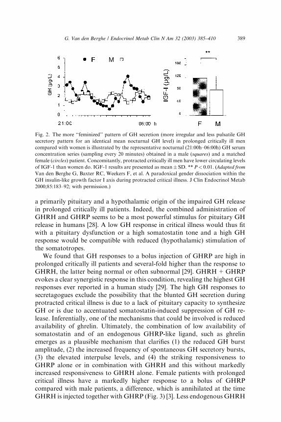
We found that GH responses to a bolus injection of GHRP are high inprolonged critically ill patients and several-fold higher than the response toGHRH, the latter being normal or often subnormal [29]. GHRH+GHRPevokes a clear synergistic response in this condition, revealing the highest GHresponses ever reported in a human study [29]. The high GH responses tosecretagogues exclude the possibility that the blunted GH secretion duringprotracted critical illness is due to a lack of pituitary capacity to synthesizeGH or is due to accentuated somatostatin-induced suppression of GH re-lease. Inferentially, one of the mechanisms that could be involved is reducedavailability of ghrelin. Ultimately, the combination of low availability ofsomatostatin and of an endogenous GHRP-like ligand, such as ghrelinemerges as a plausible mechanism that clarifies (1) the reduced GH burstamplitude, (2) the increased frequency of spontaneous GH secretory bursts,(3) the elevated interpulse levels, and (4) the striking responsiveness toGHRP alone or in combination with GHRH and this without markedlyincreased responsiveness to GHRH alone. Female patients with prolongedcritical illness have a markedly higher response to a bolus of GHRPcompared with male patients, a difference, which is annihilated at the timeGHRH is injected together with GHRP (Fig. 3) [3]. Less endogenous GHRH
Fig. 2. The more ‘‘feminized’’ pattern of GH secretion (more irregular and less pulsatile GH
secretory pattern for an identical mean nocturnal GH level) in prolonged critically ill men
compared with women is illustrated by the representative nocturnal (21:00h–06:00h) GH serum
concentration series (sampling every 20 minutes) obtained in a male (squares) and a matched
female (circles) patient. Concomitantly, protracted critically ill men have lower circulating levels
of IGF-1 than women do. IGF-1 results are presented as mean� SD. ** P< 0.01. (Adapted from
Van den Berghe G, Baxter RC, Weekers F, et al. A paradoxical gender dissociation within the
GH insulin-like growth factor I axis during protracted critical illness. J Clin Endocrinol Metab
2000;85:183–92; with permission.)
389G. Van den Berghe / Endocrinol Metab Clin N Am 32 (2003) 385–410
action in prolonged critically ill men, possibly due to the concomitant pro-found hypoandrogenism [3], accompanying loss of action of an endogenousGHRP-like ligand with prolonged stress in both genders, may explainthis finding.
Effects of GH-releasing factors in the chronic phase of critical illness
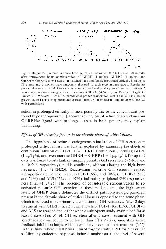
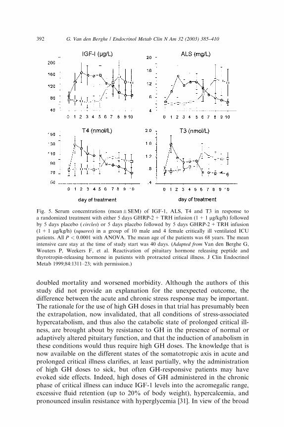
The hypothesis of reduced endogenous stimulation of GH secretion inprolonged critical illness was further explored by examining the effects ofcontinuous infusion of GHRP +/� GHRH. Continuously infusing GHRP(1 lg/kg/h), and even more so GHRH+GHRP (1+1 lg/kg/h), for up to 2days was found to substantially amplify pulsatile GH secretion (> 6-fold and> 10-fold respectively) in this condition, without altering the high burstfrequency (Fig. 4) [24,25]. Reactivating pulsatile GH secretion evokeda proportionate increase in serum IGF-1 (66% and 106%), IGFBP-3 (50%and 56%) and ALS (65% and 97%), indicating peripheral GH-responsive-ness (Fig. 4) [24,25]. The presence of considerable responsiveness to re-activated pulsatile GH secretion in these patients and the high serumlevels of GHBP clearly delineates the distinct pathophysiologic paradigmpresent in the chronic phase of critical illness as opposed to the acute phase,which is believed to be primarily a condition of GH-resistance. After 2 daystreatment with GHRP, (near) normal levels of IGF-1, IGFBP-3, IGFBP-5,and ALS are reached and, as shown in a subsequent study, maintained for atleast 5 days (Fig. 5) [6]. GH secretion after 5 days treatment with GH-secretagogues was found to be lower than after 2 days, suggesting activefeedback inhibition loops, which most likely prevented overtreatment [6,25].In this study, where GHRP was infused together with TRH for 5 days, theself-limiting endocrine responses induced anabolism at the level of several
Fig. 3. Responses (increments above baseline) of GH obtained 20, 40, 60, and 120 minutes
after intravenous bolus administration of GHRH (1 lg/kg), GHRP-2 (1 lg/kg), and
GHRH+GHRP-2 (1+1 lg/kg) in matched male and female protracted critically ill patients.
Five men and 5 women were randomly allocated to each secretagogue group. Results are
presented as mean� SEM. Circles depict results from female and squares from male patients. P
values were obtained using repeated measures ANOVA. (Adapted from Van den Berghe G,
Baxter RC, Weekers F, et al. A paradoxical gender dissociation within the GH insulin-like
growth factor I axis during protracted critical illness. J Clin Endocrinol Metab 2000;85:183–92;
with permission.)
390 G. Van den Berghe / Endocrinol Metab Clin N Am 32 (2003) 385–410
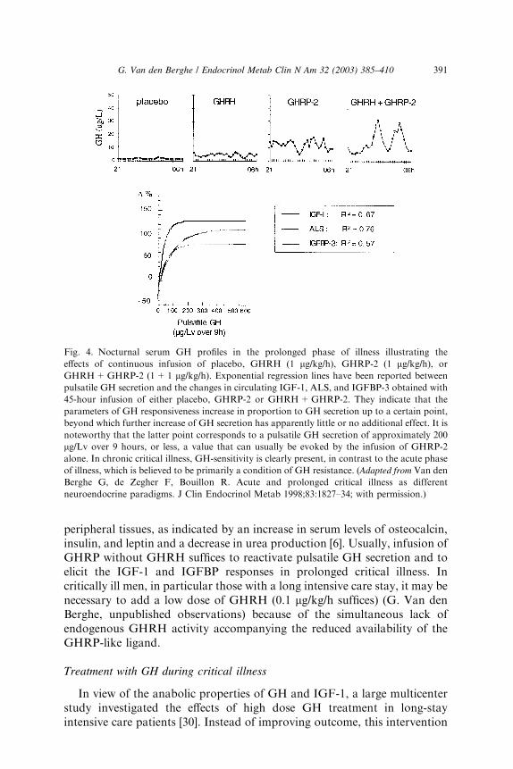
peripheral tissues, as indicated by an increase in serum levels of osteocalcin,insulin, and leptin and a decrease in urea production [6]. Usually, infusion ofGHRP without GHRH suffices to reactivate pulsatile GH secretion and toelicit the IGF-1 and IGFBP responses in prolonged critical illness. Incritically ill men, in particular those with a long intensive care stay, it may benecessary to add a low dose of GHRH (0.1 lg/kg/h suffices) (G. Van denBerghe, unpublished observations) because of the simultaneous lack ofendogenous GHRH activity accompanying the reduced availability of theGHRP-like ligand.
Treatment with GH during critical illness
In view of the anabolic properties of GH and IGF-1, a large multicenterstudy investigated the effects of high dose GH treatment in long-stayintensive care patients [30]. Instead of improving outcome, this intervention
Fig. 4. Nocturnal serum GH profiles in the prolonged phase of illness illustrating the
effects of continuous infusion of placebo, GHRH (1 lg/kg/h), GHRP-2 (1 lg/kg/h), or
GHRH+GHRP-2 (1+1 lg/kg/h). Exponential regression lines have been reported between
pulsatile GH secretion and the changes in circulating IGF-1, ALS, and IGFBP-3 obtained with
45-hour infusion of either placebo, GHRP-2 or GHRH+GHRP-2. They indicate that the
parameters of GH responsiveness increase in proportion to GH secretion up to a certain point,
beyond which further increase of GH secretion has apparently little or no additional effect. It is
noteworthy that the latter point corresponds to a pulsatile GH secretion of approximately 200
lg/Lv over 9 hours, or less, a value that can usually be evoked by the infusion of GHRP-2
alone. In chronic critical illness, GH-sensitivity is clearly present, in contrast to the acute phase
of illness, which is believed to be primarily a condition of GH resistance. (Adapted from Van den
Berghe G, de Zegher F, Bouillon R. Acute and prolonged critical illness as different
neuroendocrine paradigms. J Clin Endocrinol Metab 1998;83:1827–34; with permission.)
391G. Van den Berghe / Endocrinol Metab Clin N Am 32 (2003) 385–410
doubled mortality and worsened morbidity. Although the authors of thisstudy did not provide an explanation for the unexpected outcome, thedifference between the acute and chronic stress response may be important.The rationale for the use of high GH doses in that trial has presumably beenthe extrapolation, now invalidated, that all conditions of stress-associatedhypercatabolism, and thus also the catabolic state of prolonged critical ill-ness, are brought about by resistance to GH in the presence of normal oradaptively altered pituitary function, and that the induction of anabolism inthese conditions would thus require high GH doses. The knowledge that isnow available on the different states of the somatotropic axis in acute andprolonged critical illness clarifies, at least partially, why the administrationof high GH doses to sick, but often GH-responsive patients may haveevoked side effects. Indeed, high doses of GH administered in the chronicphase of critical illness can induce IGF-1 levels into the acromegalic range,excessive fluid retention (up to 20% of body weight), hypercalcemia, andpronounced insulin resistance with hyperglycemia [31]. In view of the broad
Fig. 5. Serum concentrations (mean�SEM) of IGF-1, ALS, T4 and T3 in response to
a randomized treatment with either 5 days GHRP-2+TRH infusion (1+1 lg/kg/h) followedby 5 days placebo ( circles) or 5 days placebo followed by 5 days GHRP-2+TRH infusion
(1+1 lg/kg/h) (squares) in a group of 10 male and 4 female critically ill ventilated ICU
patients. All P < 0.0001 with ANOVA. The mean age of the patients was 68 years. The mean
intensive care stay at the time of study start was 40 days. (Adapted from Van den Berghe G,
Wouters P, Weekers F, et al. Reactivation of pituitary hormone releasing peptide and
thyrotropin-releasing hormone in patients with protracted critical illness. J Clin Endocrinol
Metab 1999;84:1311–23; with permission.)
392 G. Van den Berghe / Endocrinol Metab Clin N Am 32 (2003) 385–410

spectrum of target tissues for GH, and taking into account the pre-existingimpairment of vital organ functions in the critically ill, the excessive doses ofGH may have further deteriorated the function of multiple organs.
A question that arises from the results of this trial is what intensive carephysicians should do at the time patients, who are GH deficient and are onGH treatment, become critically ill and admitted to the ICU. Should GHsubstitution therapy be discontinued at that occasion? A consensus state-ment from the GH-Research Society [32] advises not to discontinue in viewof the lack of evidence that the low GH doses used for substitution therapyare harmful.
Thyrotropic axis
Changes in the acute phase of critical illness
Within 2 hours after surgery or trauma, serum levels of T3 decreasewhereas T4 and TSH briefly increase (Fig. 6) [33]. Apparently, low T3 levelsat that stage are mainly caused by a decreased peripheral conversion ofT4 to T3 [34]. Subsequently, circulating TSH and T4 levels often returnto ‘‘normal’’ whereas T3 levels remain low. Although mean serum TSHconcentrations are indistinguishable from normal at that point, the normal
Fig. 6. Simplified overview of the major changes occurring within the thyroid axis during
the acute and the chronic phase of critical illness. (Adapted from Van den Berghe G. Novel
insights into the neuroendocrinology of critical illness. Eur J Endocrinol 2000;143:1–13; with
permission).
393G. Van den Berghe / Endocrinol Metab Clin N Am 32 (2003) 385–410

nocturnal TSH surge is absent [35,36]. The magnitude of the T3 drop within24 hours has been found to reflect the severity of illness [37,38]. Thecytokines TNFa, IL-1, and IL-6 have been investigated as putative medi-ators of the acute low T3 syndrome. Although these cytokines are capableof mimicking the acute stress-induced alterations in thyroid status, cytokineantagonism in a human model failed to restore normal thyroid function [39].Low concentrations of binding proteins and inhibition of hormone binding,transport and metabolism by elevated levels of free fatty acids, and bilirubinhave been proposed as factors contributing to the low T3 syndrome at tissuelevel [40]. Teleologically, the acute changes in the thyroid axis may reflect anattempt to reduce energy expenditure, as happens during starvation [41],and thus as an appropriate response that does not warrant intervention.This, however, remains a controversial issue because valid data to supportor refute this statement are lacking [42]. Although short-term intravenousadministration of T3 to patients after cross clamp removal during electivecoronary bypass grafting has shown to improve postoperative cardiacfunction [43,44], the pharmacologic doses of T3 that resulted in supra-normal serum T3 levels and the absence of an effect on mortality do notrefute an adaptive nature of the ‘‘acute’’ low T3 syndrome.
Changes in prolonged critical illness
Patients treated in intensive care units for several weeks present witha somewhat different set of changes within the thyroid axis. A single sampleusually reveals low or low-normal TSH values and low T4 and T3 serumconcentrations [45]. Overnight repeated sampling, however, revealed thatessentially the pulsatility in the TSH secretory pattern is dramaticallydiminished and that, as for the GH axis, it is the loss of TSH pulse amplitudewhich is related to low serum levels of thyroid hormone [45]. Moreover,Fliers and coworkers have elegantly demonstrated by post-mortem exam-ination of human brain specimen that at the time death follows chronicsevere illness, the expression of TRH gene in hypothalamic paraventricularnuclei is reduced whereas this is not the case after death from acute insultssuch as lethal trauma due to a road accident [46]. These researchers observeda positive correlation between TRH mRNA in the paraventricular nucleiand blood levels of TSH and T3. Together, these findings indicate thatproduction and or release of thyroid hormones is reduced in the chronicphase of critical illness, due to reduced hypothalamic stimulation of thethyrotropes, in turn leading to reduced stimulation of the thyroid gland.In line with this concept is the increase of TSH marking onset of recoveryfrom severe illness [47]. The exact mechanisms underlying the neuroendo-crine pathogenesis of the low thyroid hormone levels in prolonged critical ill-ness are unknown. As circulating cytokine levels are usually much lower atthat stage [48], other mechanisms operational within the central nervoussystem are presumably involved. Endogenous dopamine and prolonged
394 G. Van den Berghe / Endocrinol Metab Clin N Am 32 (2003) 385–410
hypercortisolism may each play a role because exogenous dopamineand glucocorticoids are known to provoke or aggravate hypothyroidismin critical illness [49,50].
Low thyroid hormone levels in protracted critical illness correlateinversely with urea production and bone degradation which could reflecteither an adaptive, protective mechanism against hypercatabolism ora causal relation [6]. Restoring physiologic levels of thyroid hormonesby continuously infusing TRH (together with a GH-secretagogue) (Fig. 5)was found to reduce rather than increase hypercatabolism [6], an effectthat was related only to thyroid hormone changes. During TRH infu-sion in prolonged critical illness, the negative feedback exerted by thyroidhormones on the thyrotropes was found to be maintained, thus precludingoverstimulation of the thyroid axis [23,25]. This self-limitation may beextremely important during critical illness to avoid hyperthyroidism, whichwould inadvertently aggravate catabolism. The coinfusion of TRH andGH-releasing factors seems a better strategy than the infusion of TRHalone because the combination, but not TRH alone, avoids an increasein circulating reverse T3 [6,23]. The latter may point to the effect of GH onthe activity of type I deiodinase and eventually to other important interac-tions among different anterior pituitary axes for optimal peripheral re-sponses [51].
Treatment with thyroid hormone or releasing factors duringprolonged critical illness
It remains controversial whether correction of the illness-associated lowserum and tissue concentrations of T3 by either T4 or T3 administration isrequired to improve clinical problems distinctively associated with prolongedcritical illness [52,53]. Pioneering studies with T4 administration so far havefailed to demonstrate clinical benefit within an intensive care setting, but inview of the impaired conversion of T4 to T3, this is not really surprising[54,55]. A recent report on thyroid hormone treatment using substitutiondoses of T3 in dopamine-treated pediatric patients after correction ofcongenital anomaly revealed improvement of postoperative cardiac function[56]. In contrast to treatment with thyroid hormones, infusing TRH allowsfor peripheral shifts in thyroid hormone metabolism during intercurrentevents and, accordingly, permits the body to elaborate appropriateconcentrations of thyroid hormones in the circulation and at tissue level,thus setting the scene for a safer treatment than the administration of T3 [25].The peripheral tissue responses to the normalization of serum concentrationsof IGF-1 and binding proteins as evoked by GHRP infusion seem to dependon the coinfusion of TRH and the concomitant normalization of the thyroidaxis. Indeed, GHRP-2 infused alone evokes identical increments in serumconcentrations of IGF-1, IGFBP-3, and ALS, but is devoid of the anabolictissue responses that are present with the combined infusion of GHRP and
395G. Van den Berghe / Endocrinol Metab Clin N Am 32 (2003) 385–410

TRH [23]. Outcome benefit of TRH infusion alone or in combination withGH-secretagogues in prolonged critical illness is yet to be studied.
The diagnosis of pre-existing thyroid disease and its management duringcritical illness can be extremely difficult and in view of the controversy,recommendations for clinical practice is often not evidence-based. In view ofthe hypothalamic-pituitary suppression occurring in the chronic phase ofcritical illness in patients without previous endocrine disease, it is virtuallyimpossible to diagnose pre-existing central hypothyroidism during the timea patient is treated within the ICU. Patients with pre-existing primaryhypothyroidism, myxedema coma being the extreme presentation, are ex-pected to show low serum levels of T4 and T3 in combination with high TSHconcentrations. A complicating factor, however, is the simultaneous presenceof primary hypothyroidism and severe nonthyroidal critical illness. Indeed,the nonthyroidal critical illness evokes the sum of changes within thehypothalamus–pituitary–thyroid axis occurring in the framework of disease,as described earlier. A decrease in serum T3 and an increase in serum reverseT3 (rT3) are the most common changes in acute nonthyroidal critical illness,but serum T3 may be undetectable and serum T4 also dramatically reducedin patients with protracted nonthyroidal critical illness. Therefore, in patientswith myxedema coma with severe comorbidity (pneumonia, sepsis) serum T3and T4 will be low, but could be indistinguishable from those values observedin prolonged nonthyroidal critical illness. Whereas serum TSH is markedlyincreased in uncomplicated primary hypothyroidism, it is paradoxicallynormal or even decreased in severely ill patients. Therefore, serum TSH maybe lower than anticipated, from the severe hypothyroid condition of thepatient with myxedema coma and concomitant illness, or even frankly low.Thus, a high serum TSH concentration, when observed, is in agreement withprimary hypothyroidism but a normal or a low TSH does not exclude itduring intercurrent critical illness. Indeed, serum TSH may be paradoxicallylow in this setting because of concomitant nonthyroidal critical illness,especially in patients given high-dose corticosteroids or dopamine. Otheriatrogenic factors causing hypothyroidism, particularly in a surgical ICU, areiodine wound dressings, iodine containing contrast agents used for radiologicimaging, and drugs such as somatostatin and amiodarone. The finding ofa high ratio of T3 to T4 in serum, a low thyroid hormone-binding ratio, anda low serum rT3 may favor the presence of primary hypothyroidism, whereasopposite changes occur in nonthyroidal critical illness. The diagnosticaccuracy of any of these measurements is limited and in many patients nodefinite laboratory diagnosis can be established. In these patients, history,physical examination, and the possible presence of thyroid autoantibodiesmay give further clues to the presence or absence of thyroid disease.Repeated thyroid function tests after improvement of the nonthyroidalillness will provide a final answer.
When and how to treat primary hypothyroidism during the course ofan intercurrent nonthyroidal critical illness remains controversial. One
396 G. Van den Berghe / Endocrinol Metab Clin N Am 32 (2003) 385–410

exception, however, is a presumed diagnosis of myxedema coma, for whichthere is general agreement that patients should be treated with a parenteralform of thyroid hormone. The proper initiation of thyroid hormone replace-ment therapy, however, in this case, still remains controversial becausecontrolled studies on the optimal treatment regimen are lacking. The firstuncertainty relates to the type of thyroid hormone to be given: should it be T4alone, T3 alone or the combination of both. The second uncertainty is theoptimal initial dosage of any thyroid hormone replacement regimen. Manyclinicians prefer a loading dose of up to 300 to 500 lg intravenous T4 toquickly restore circulating levels of T4 to approximately 50% of theeuthyroid value [57,58], followed by 50 to 100 lg of intravenous T4 dailyuntil oral medication can be given. Higher doses do not seem to be beneficial,although Kaptein et al [59] found no increased cardiovascular risk in severelyill hypothyroid patients treated with larger doses of T4.
Some authors have advocated the use of T3 in addition to T4 because T3does not require conversion by 50-deiodinase enzymes to a biologicallyactive form. In an animal experimental study by Morreale de Escobar et al[60] replacement therapy for hypothyroidism with T4 alone did not ensureeuthyroidism in all tissues, and a subsequent study showed that only thecombined treatment with T4 and T3 induces euthyroidism in all tissues.Tissue-specific deiodinase activities acting as local regulatory mechanismsare the presumed explanation for these findings. In a recent study inhypothyroid patients it appeared that partial substitution of T3 for T4 mightimprove mood and neuropsychologic function in hypothyroid patients,possibly by increased bioavailability of T3 in the CNS [61]. Whereas thesefascinating results await confirmation by others, replacement therapy witha combination of T4 and T3 in compensated hypothyroidism remains anexperimental modality [62].
The author’s experimental protocol for thyroid hormone therapy duringintensive care of presumed hypothyroidism, either pre-existing or iatrogen-ically induced at the time reversal of the iatrogenic cause seems impossible,advises a dose of 100 to 200 lg T4 IV bolus per 24h combined with T30.6 lg/kg ideal body weight per 24h in continuous IV infusion, targetingthyroid hormone levels in the low normal range (G. Van den Berghe et al,unpublished data, 2002).
Lactotropic axis
Prolactin responses to acute and prolonged critical illness
It has been suggested that the changes in prolactin secretion in responseto stress may contribute to altered immune function during the course ofcritical illness. The evidence for this includes the presence of prolactinreceptors on human T- and B-lymphocytes [63] and the prolactindependency of T-lymphocytes for maintaining immune competence [64].
397G. Van den Berghe / Endocrinol Metab Clin N Am 32 (2003) 385–410
In mice, inhibition of prolactin release results in impaired lymphocytefunction, in depressed lymphokine-dependent macrophage activation, andin death from a normally nonlethal exposure to bacteria [65]. The immunesuppressive drug, Cyclosporine, is known to compete with prolactin fora common binding site on T-cells which may explain part of its effects[66,67]. The prolactin-suppressing drug, bromocriptine, has been shown tobe an adjuvant immunosuppressant in humans after heart transplantation[67]. Prolactin was among the first hormones known to have increasedserum concentrations in response to acute physical or psychologic stress[68], an increase that may be mediated by VIP, oxytocin, dopaminergicpathways or other still uncharacterized factors. Cytokines may again playa signaling role. Whether hyperprolactinemia during the initial phase ofcritical illness contributes to the vital initial activation of the immunecascade remains speculative.
In chronic critical illness, serum prolactin levels are no longer as high asin the acute phase and the secretory pattern is characterized by a reducedpulsatile fraction [25,41]. A role for endogenous dopamine has beensuggested [69]. It is unknown whether the blunted prolactin secretion in thechronic phase plays a role in the anergic immune dysfunction or in theincreased susceptibility for infections characterizing the chronically ill [70].Exogenous dopamine, often infused as an inotropic drug in intensive care-dependent patients, has been shown to further suppress prolactin secretionand was found to aggravate concomitantly T-lymphocyte dysfunction andimpaired neutrophyl chemotaxis [69,71].
Prolactin as a therapeutic target?
Prolactin is currently not available for therapy. Future studies are neededto evaluate the therapeutic potential of thyrotropin releasing hormone-induced prolactin release for optimizing immune function during criticalillness [50]. Also, it remains enigmatic whether patients on treatment forprolactinoma should interrupt or continue this treatment during an inter-current critical illness.
Luteinizing hormone–testosterone axis
Changes in LH–testosterone axis in acute and prolonged critical illness
Also for LH, the pulsatility in the secretory pattern is important for itsbioactivity [72,73]. Because testosterone is the most important endogenousanabolic steroid, changes within the LH–testosterone axis in the male couldbe relevant for the catabolic state of critical illness. Low serum testosteronelevels in men accompany a variety of catabolic states. These conditionsinclude starvation [74,75], the postoperative phase [76], myocardial
398 G. Van den Berghe / Endocrinol Metab Clin N Am 32 (2003) 385–410
infarction [77], burn injury [78,79], psychologic and physical stress [80,81],and chronic critical illness [82].
Low serum testosterone concentrations and elevated LH levels observedduring the acute stress of surgery or myocardial infarction [76,77,83] suggestan immediate Leydig-cell suppression, of which the exact cause remainsobscure. Inflammatory cytokines (IL-1 and IL-2) may play a role, assuggested by experimental studies [84,85]. It may be considered as appro-priate that the secretion of anabolic androgens be switched off in circum-stances of acute stress, to conserve energy and metabolic substrates for, atthat time at least, less vital functions.
When critical illness becomes prolonged, hypogonadotropism develops[78,86]. Concomitantly, circulating levels of testosterone become extremelylow (often undetectable) in men whereas estimated free estradiol concen-trations remain normal suggesting increased aromatization of adrenalandrogens [3]. The progressive decrease of serum gonadotropin levels,however, seems to lag behind the rapid decline in serum testosterone[77,83,87]. In prolonged critically ill men, a high LH pulse frequency with anabnormally low LH pulse amplitude has been observed [82], which wasinterpreted as an impaired compensatory LH hypersecretion in response tothe low serum testosterone levels. Thus, again, it seems to be mainly animpairment of the pulsatile component of LH secretion, which occurs inresponse to the sustained stress of prolonged critical illness [82]. Endogenousdopamine, opiates and the preserved estradiol levels [3] may be involved inthe pathogenesis of hypogonadotropism, as exogenous dopamine, opioids,and estrogens may further diminish blunted LH secretion [82,88].
Animal data suggest that prolonged exposure of the brain to IL-1 mayalso play a role through the suppression of LHRH synthesis [84]. Thepioneering studies evaluating androgen treatment in prolonged critical illnessfailed to demonstrate conclusive clinical benefit [89]. In view of the secretorycharacteristics of the other anterior pituitary hormones, we recentlyinvestigated the therapeutic potential of LHRH pulses in prolonged criticallyill men, alone and together with GHRP-2 and TRH. LHRH alone seemsonly partially and transiently effective [90]. When LHRH pulses were giventogether with GHRP-2 and TRH infusion however, target organ responsesand anabolic effects followed [23]. These data underline the importance ofcorrecting all the hypothalamic/pituitary defects instead of applying a singlehormone treatment.
Sex steroid substitution therapy during critical illness?
Because critical illness in itself induces profound hypoandrogenism inmale patients, of which it remains unknown whether this reflects adaptationor pathology, it is not clear if androgen substitution therapy for pre-existinghypogonadism should be interrupted or continued during the course of anintercurrent critical illness. Sex steroids in women are usually not continuedduring critical illness.
399G. Van den Berghe / Endocrinol Metab Clin N Am 32 (2003) 385–410
Pituitary–adrenal axis
Pituitary–adrenal responses to acute and prolonged critical illness
The pituitary–adrenal axis also responds differently to acute andprolonged critical illness. It has been long known that the vital stress-induced hypercortisolism induced by surgery, trauma or sepsis, is associatedwith augmented ACTH release, which, in turn, is presumably driven bycorticotrophin-releasing hormone (CRH), cytokines and the noradrenergicsystem. Concomitantly, circulating aldosterone increases markedly, mostlikely under the control of an activated renin-angiotensin system [91].Hypercortisolism acutely shifts carbohydrate, fat, and protein metabolism,so that energy is instantly and selectively available to vital organs such as thebrain and anabolism is delayed. Intravascular fluid retention and theenhanced inotropic and vasopressor response to respectively catecholaminesand angiotensin II offer hemodynamic advantages in the ‘‘fight and flight’’response. In addition, hypercortisolism elicited by acute disease or traumacan be interpreted as an attempt of the organism to mute its owninflammatory cascade, thus protecting itself against overresponses [92–94].
In chronic critical illness, serum ACTH was found to be low while cortisolconcentrations remained elevated, indicating that cortisol release may in thisphase be driven through an alternative pathway, possibly involvingendothelin [95]. Why ACTH levels are low in chronic critical illness isunclear; a role for atrial natriuretic peptide or substance P has been suggested[95]. In contrast to serum cortisol, circulating levels of adrenal androgenssuch as dehydroepiandrosterone sulfate (DHEAS), which has immunostim-ulatory properties on Th1–helper-cells, are low during chronic critical illness[96–98]. Moreover, despite increased plasma renin activity, paradoxicallydecreased concentrations of aldosterone are found in protracted criticalillness [99]. This constellation suggests a shift of pregnenolone metabolismaway from mineralocorticoid and adrenal androgen pathways toward theglucocorticoid pathway, orchestrated by an unknown peripheral drive.Ultimately, the latter mechanism may also fail, as indicated by a 20-foldhigher incidence of adrenal insufficiency present in critically ill patients overage 50 years and being treated at the intensive care unit for more than 14 days[100]. This type of relative adrenal failure coincides with adverse outcome andsuggests that high levels of glucocorticoids remain essential for hemodynamicstability. Whether hypercortisolism in the chronic phase of critical illness isexclusively beneficial remains uncertain. Sustained hypercortisolism in thepresence of low levels of DHEAS and prolactin could theoretically evokeimbalance between immunosuppressive and immunostimulatory pathwaysand thus could be seen as participating in the increased susceptibility forinfectious complications. Other conceivable though unproven drawbacks ofprolonged hypercortisolism include impaired wound healing and myopathy,complications that are often observed during protracted critical illness.
400 G. Van den Berghe / Endocrinol Metab Clin N Am 32 (2003) 385–410

Treatment of adrenal failure during critical illness
In a patient with previously diagnosed primary or central adrenalinsufficiency and in patients previously treated with systemic glucocorti-coids, treatment should be continued and additional coverage for the stressof critical illness should be provided. For more details, we refer to previouschapters in this issue. Also, a true Addisonian crisis needs treatment insevere stress conditions. Hydrocortisone 100 mg followed by 50 to 100 mgevery 6 hours on the first day, 50 mg every 6 hours on the second day, and 25mg every 6 hours on the third day, tapering to a maintenance dose by thefourth to fifth day. In prolonged critical conditions, the maintenance doseshould be kept two to three times the basal need. Special attention should begiven to patients with concomitant diabetes insipidus, as lack of cortisolmay prevent polyuria because cortisol is needed for free-water clearance.Inversely, in these patients glucocorticoid therapy may induce or aggravatediabetes insipidus. Another specific condition is the post-hypophysectomy-phase for Cushing’s disease, characterized by a high vulnerability forAddisonian-like crisis. Drugs such as phenytoin, barbiturates, rifampicinand thyroid hormone can accelerate glucocorticoid metabolism by inductionof microsomal enzyme activity and can increase the glucocorticoid replace-ment dose requirements. If this increased requirement is not met, adrenalcrisis may occur.
Recently, however, the concept of relative hyopthalamic–pituitary–adrenal insufficiency in acute sepsis has been launched [101–103], whichadvocates short-term treatment with stress doses of glucocortioids asbeneficial in patients without a full blown adrenal failure [104,105]. Thecontroversy about this concept of relative adrenal axis failure in acute stressconditions is explained by the problems regarding diagnosis. Indeed,accurate ‘‘normal’’ values for baseline cortisol levels in this type of stress,normal reference values for cortisol responses to a short ACTH test, anddata to support the option to use a low dose or high dose ACTH test[103,106], are still unavailable. Another issue of controversy regarding theconcept of relative adrenal failure in acute sepsis is the dose and the durationof treatment once it has been initiated. Treating septic patients withglucocorticoids at too high a dose and for too long a time will conceivablyaggravate the loss of lean tissue, prolong the ICU-dependency, and increasethe susceptibility for potentially lethal complications.
Endocrine predictors of adverse outcome of critical illness
In the acute phase of critical illness, a high serum cortisol or low T3concentrations [38], indicate poor prognosis. In the prolonged critically illpatient, however, these markers lack sensitivity. Recently, preliminary datawere published showing that another parameter—serum concentration of
401G. Van den Berghe / Endocrinol Metab Clin N Am 32 (2003) 385–410
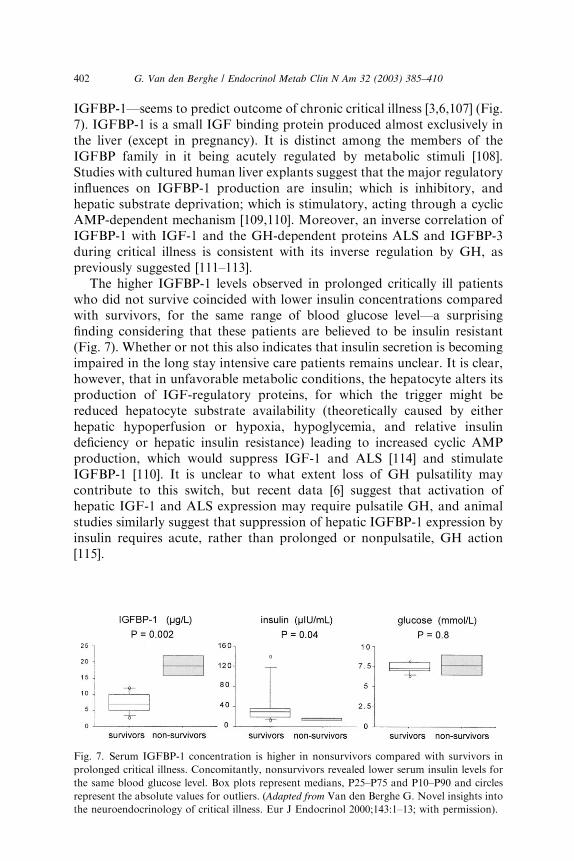
IGFBP-1—seems to predict outcome of chronic critical illness [3,6,107] (Fig.7). IGFBP-1 is a small IGF binding protein produced almost exclusively inthe liver (except in pregnancy). It is distinct among the members of theIGFBP family in it being acutely regulated by metabolic stimuli [108].Studies with cultured human liver explants suggest that the major regulatoryinfluences on IGFBP-1 production are insulin; which is inhibitory, andhepatic substrate deprivation; which is stimulatory, acting through a cyclicAMP-dependent mechanism [109,110]. Moreover, an inverse correlation ofIGFBP-1 with IGF-1 and the GH-dependent proteins ALS and IGFBP-3during critical illness is consistent with its inverse regulation by GH, aspreviously suggested [111–113].
The higher IGFBP-1 levels observed in prolonged critically ill patientswho did not survive coincided with lower insulin concentrations comparedwith survivors, for the same range of blood glucose level—a surprisingfinding considering that these patients are believed to be insulin resistant(Fig. 7). Whether or not this also indicates that insulin secretion is becomingimpaired in the long stay intensive care patients remains unclear. It is clear,however, that in unfavorable metabolic conditions, the hepatocyte alters itsproduction of IGF-regulatory proteins, for which the trigger might bereduced hepatocyte substrate availability (theoretically caused by eitherhepatic hypoperfusion or hypoxia, hypoglycemia, and relative insulindeficiency or hepatic insulin resistance) leading to increased cyclic AMPproduction, which would suppress IGF-1 and ALS [114] and stimulateIGFBP-1 [110]. It is unclear to what extent loss of GH pulsatility maycontribute to this switch, but recent data [6] suggest that activation ofhepatic IGF-1 and ALS expression may require pulsatile GH, and animalstudies similarly suggest that suppression of hepatic IGFBP-1 expression byinsulin requires acute, rather than prolonged or nonpulsatile, GH action[115].
Fig. 7. Serum IGFBP-1 concentration is higher in nonsurvivors compared with survivors in
prolonged critical illness. Concomitantly, nonsurvivors revealed lower serum insulin levels for
the same blood glucose level. Box plots represent medians, P25–P75 and P10–P90 and circles
represent the absolute values for outliers. (Adapted from Van den Berghe G. Novel insights into
the neuroendocrinology of critical illness. Eur J Endocrinol 2000;143:1–13; with permission).
402 G. Van den Berghe / Endocrinol Metab Clin N Am 32 (2003) 385–410
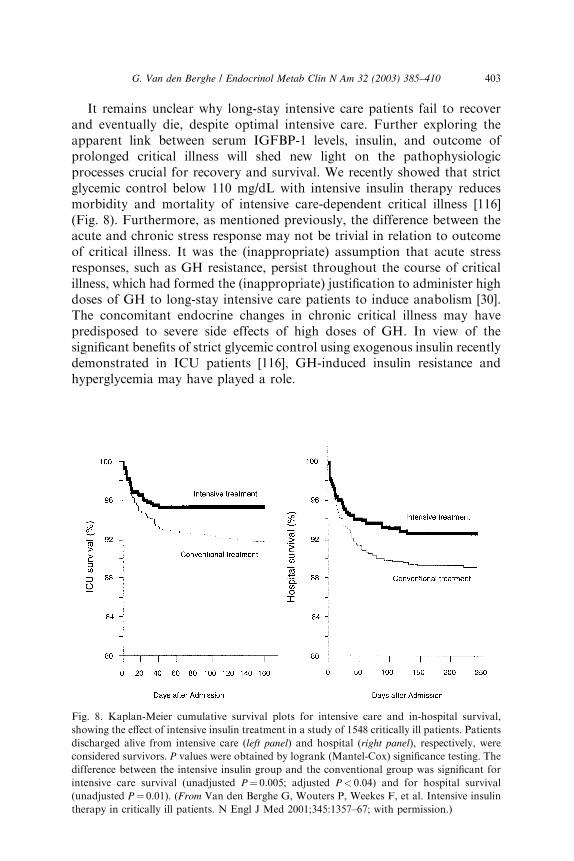
It remains unclear why long-stay intensive care patients fail to recoverand eventually die, despite optimal intensive care. Further exploring theapparent link between serum IGFBP-1 levels, insulin, and outcome ofprolonged critical illness will shed new light on the pathophysiologicprocesses crucial for recovery and survival. We recently showed that strictglycemic control below 110 mg/dL with intensive insulin therapy reducesmorbidity and mortality of intensive care-dependent critical illness [116](Fig. 8). Furthermore, as mentioned previously, the difference between theacute and chronic stress response may not be trivial in relation to outcomeof critical illness. It was the (inappropriate) assumption that acute stressresponses, such as GH resistance, persist throughout the course of criticalillness, which had formed the (inappropriate) justification to administer highdoses of GH to long-stay intensive care patients to induce anabolism [30].The concomitant endocrine changes in chronic critical illness may havepredisposed to severe side effects of high doses of GH. In view of thesignificant benefits of strict glycemic control using exogenous insulin recentlydemonstrated in ICU patients [116], GH-induced insulin resistance andhyperglycemia may have played a role.
Fig. 8. Kaplan-Meier cumulative survival plots for intensive care and in-hospital survival,
showing the effect of intensive insulin treatment in a study of 1548 critically ill patients. Patients
discharged alive from intensive care (left panel) and hospital (right panel), respectively, were
considered survivors. P values were obtained by logrank (Mantel-Cox) significance testing. The
difference between the intensive insulin group and the conventional group was significant for
intensive care survival (unadjusted P¼ 0.005; adjusted P< 0.04) and for hospital survival
(unadjusted P¼ 0.01). (From Van den Berghe G, Wouters P, Weekes F, et al. Intensive insulin
therapy in critically ill patients. N Engl J Med 2001;345:1357–67; with permission.)
403G. Van den Berghe / Endocrinol Metab Clin N Am 32 (2003) 385–410
Summary
Prolonged critical illness has a high morbidity and mortality. The acuteand chronic phases of critical illness are associated with distinct endocrinealterations. The acute neuroendocrine response to critical illness involves anactivated anterior pituitary function. In prolonged critical illness, however,a reduced pulsatile secretion of anterior pituitary hormones and the so-called‘‘wasting syndrome’’ occur. The impaired pulsatile secretion of GH,thyrotropin and gonadotropin can be re-amplified by relevant combinationsof releasing factors, which also substantially increase circulating levels ofIGF-1, GH-dependent IGFBPs, thyroxin, tri-iodothyronine and testoster-one. Anabolism is clearly re-initiated at the time GH secretagogues,thyrotropin-releasing hormone and gonadotropin-releasing hormone arecoadministered but the effect on survival remains unknown. A lethaloutcome of critical illness is predicted by a high serum concentration ofIGFBP-1, pointing to impaired insulin effect rather than pituitary function,and survival was recently shown to be dramatically improved by strictnormalization of glycemia with exogenous insulin.
In addition to the illness-induced endocrine alterations, patients mayhave pre-existing central or peripheral endocrine diseases, either previouslydiagnosed or unknown. Hence, endocrine function testing in a critically illpatient represents a major challenge and the issue of treatment remainscontroversial.
The recent progress in knowledge of the neuroendocrine response tocritical illness and its interrelation with peripheral hormonal and metabolicalterations during stress, allows for potential new therapeutic perspectives tosafely reverse the wasting syndrome and improve survival.
References
[1] Streat SJ, Beddoe AH, Hill GL. Aggressive nutritional support does not prevent protein
loss despite fat gain in septic intensive care patients. J Trauma 1987;27:262–6.
[2] Carroll P. Protein metabolism and the use of growth hormone and insulin-like growth
factor-I in the critically ill patient. Growth Horm IGF Res 1999;9:400–13.
[3] Van den Berghe G, Baxter RC, Weekers F, et al. A paradoxical gender dissociation within
the growth hormone. insulin-like growth factor I axis during protracted critical illness.
J Clin Endocrinol Metab 2000;85:183–92.
[4] Knaus WA, Draper EA, Wagner DP, et al. APACHE II: A severity of disease clas-
sification system. Crit Care Med 1985;13:818–29.
[5] Van den Berghe G, de Zegher F, Bouillon R. Acute and prolonged critical illness as
different neuroendocrine paradigms. J Clin Endocrinol Metab 1998;83:1827–34.
[6] Van den Berghe G, Wouters P, Weekers F, et al. Reactivation of pituitary hormone
release and metabolic improvement by infusion of growth hormone releasing peptide
and thyrotropin-releasing hormone in patients with protracted critical illness. J Clin
Endocrinol Metab 1999;84:1311–23.
404 G. Van den Berghe / Endocrinol Metab Clin N Am 32 (2003) 385–410
[7] Thorner MO, Vance ML, Laws ER, Horvath E, Kovacs K. The anterior pituitary. In:
Wilson JD, Foster DW, Kronenberg HM, Larsen PR, editors. Williams Textbook of
Endocrinology. 9th edition. Philadelphia: W.B. Saunders; 1998. p. 249–340.
[8] Bowers CY, Momany FA, Reynolds GA, et al. On the in vitro and in vivo activity of
a new synthetic hexapeptide that acts on the pituitary to specifically release growth
hormone. Endocrinology 1984;114:1537–45.
[9] Howard AD, Feighner SD, Cully DF, et al. A receptor in pituitary and hypothalamus
that functions in growth hormone release. Science 1996;273:974–7.
[10] Masayasu Kojima, Hiroshi Hosoda, Yukari Date, et al. Ghrelin is a growth-hormone-
releasing acylated peptide from stomach. Nature 1999;402:656–60.
[11] Gevers EF, Wit JM, Robinson IC. Growth, growth hormone (GH) binding protein, and
GH receptors are differentially regulated by peak and trough components of GH
secretory pattern in the rat. Endocrinology 1996;137:1013–8.
[12] Giustina A, Veldhuis JD. Pathophysiology of the neuroregulation of growth hormone
secretion in experimental animals and the human. Endocr Rev 1998;19:717–97.
[13] Hindmarsh PC, Dennison E, Pincus SM, et al. A sexually dimorphic pattern of
growth hormone secretion in the elderly illness. J Clin Endocrinol Metab 1999;84:
2679–85.
[14] Ross R, Miell J, Freeman E, Jones J, et al. Critically ill patients have high basal growth
hormone levels with attenuated oscillatory activity associated with low levels of insulin-
like growth factor-1. Clin Endocrinol 1991;35:47–54.
[15] Voerman HJ, Strack van Schijndel RJM, de Boer H, et al. Growth hormone: secretion
and administration in catabolic adult patients, with emphasis on the critically ill patient.
Neth J Med 1992;41:229–44.
[16] Hartman ML, Veldhuis JD, Johnson ML, et al. Augmented growth hormone secretory
burst frequency and amplitude mediate enhanced GH secretion during a two day fast in
normal men. J Clin Endocrinol Metab 1992;74:757–65.
[17] Hermansson M, Wickelgren RB, Hammerqvist F, et al. Measurement of human growth
hormone receptor messenger ribonucleic acid by a quantitative polymerase chain
reaction-based assay: demonstration of reduced expression after elective surgery. J Clin
Endocrinol Metab 1997;82:421–8.
[18] Baxter RC, Hawker FH, To C, et al. Thirty day monitoring of insulin-like growth factors
and their binding proteins in intensive care unit patients. Growth Horm IGF Res
1998;8:455–63.
[19] Rodriguez-Arnao J, Yarwood Y, Ferguson C, et al. Reduction in circulating IGF-I and
hepatic IGF-I mRNA levels after ceacal ligation and puncture are associated with dif-
ferential regulation of hepatic IGF-binding protein-1, -2 and -3 mRNA levels. J Endo
crinol 1996;151:287–92.
[20] Bentham J, Rodriguez-Arnao-J, Ross RJ. Acquired growth hormone resistance in
patients with hypercatabolism. Horm Res 1993;40:87–91.
[21] Timmins AC, Cotterill AM, Cwyfan Hughes SC, et al. Critical illness is associated with
low circulating concentrations of insulin-like growth factors-I and -II, alterations in
insulin-like growth factor binding proteins, and induction of an insulin-like growth factor
binding protein-3 protease. Crit Care Med 1996;24:1460–6.
[22] Gibson FA, Hinds CJ. Growth Hormone and insulin-like growth factors in critical illness.
Intens Care Med 1997;23:369–78.
[23] Van den Berghe G, Baxter RC, Weekers F, et al. The combined administration of GH-
releasing peptide-2 (GHRP-2), TRH and GnRH to men with prolonged critical illness
evokes superior endocrine and metabolic effects than treatment with GHRP-2 alone. Clin
Endocrinol 2002;56:655–69.
[24] Van den Berghe G, de Zegher F, Veldhuis JD, et al. The somatotropic axis in critical
illness: effect of continuous GHRH and GHRP-2 infusion. J Clin Endocrinol Metab
1997;82:590–9.
405G. Van den Berghe / Endocrinol Metab Clin N Am 32 (2003) 385–410

[25] Van denBergheG, deZegher F, BaxterRC, et al.Neuroendocrinology of prolonged critical
illness: effect of continuous thyrotropin-releasing hormone infusion and its combination
with growth hormone-secretagogues. J Clin Endocrinol Metab 1998;83:309–19.
[26] Van den Berghe G, de Zegher F, Lauwers P, et al. Growth hormone secretion in critical
illness: effect of dopamine. J Clin Endocrinol Metab 1994;79:1141–6.
[27] Van den Berghe G, de Zegher F. Anterior pituitary function during critical illness and
dopamine treatment. Crit Care Med 1996;24:1580–90.
[28] Micic D, Popovic V, Doknic M, et al. Preserved growth hormone (GH) secretion in aged
and very old subjects after testing with the combined stimulus GH-releasing hormone plus
GH-releasing hexapeptide-6. J Clin Endocrinol Metab 1998;83:2569–72.
[29] Van den Berghe G, de Zegher F, Bowers CY, et al. Pituitary responsiveness to growth
hormone (GH) releasing hormone, GH-releasing peptide-2 and thyrotropin releasing
hormone in critical illness. Clin Endocrinol 1996;45:341–51.
[30] Takala J, Ruokonen E, Webster NR, et al. Increased mortality associated with growth
hormone treatment in critically ill adults. N Engl J Med 1999;341:785–92.
[31] Van den Berghe G. Whither growth hormone in the catabolic state of critical illness?
Growth Horm IGF Res 1999;9:397–9.
[32] Christiansen JS, Bengtsson BA, Thorner MO, et al. Critical Evaluation of the safety of
recombinant human growth hormone administration: statement from the growth
hormone research society. J Clin Endocrinol Metab 2001;86:1868–70.
[33] Michalaki M, Vagenakis A, Makri M, et al. Dissociation of the early decline in serum T3
concentration and serum IL-6 rise and TNF alfa in non-thyroidal illness syndrome
induced by abdominal surgery. J Clin Endocrinol Metab 2001;86:4198–205.
[34] Chopra IJ, Huang TS, Beredo A, et al. Evidence for an inhibitor of extrathyroidal
conversion of thyroxine to 3,5,30-triiodothyronine in sera of patients with non-thyroidal
illness. J Clin Endocrinol Metab 1985;60:666–72.
[35] Bartalena L, Martino E, Brandi LS, et al. Lack of nocturnal serum thyrotropin surge after
surgery. J Clin Endocrinol Metab 1990;70:293–6.
[36] Romijn JA, Wiersinga WM. Decreased nocturnal surge of thyrotropin in nonthyroidal
illness. J Clin Endocrinol Metab 1990;70:35–42.
[37] Rothwell PM, Lawler PG. Prediction of outcome in intensive care patients using
endocrine parameters. Crit Care Med 1995;23:78–83.
[38] Schlienger JL, Sapin R, Capgras T, et al. Evaluation of thyroid function after myocardial
infarction. Ann Endocrinol (Paris) 1991;52:283–8.
[39] van der Poll T, van Zee K, Endert E, et al. Interleukin-1 receptor blockade does not affect
endotoxin-induced changes in plasma thyroid hormone and thyrotropin concentration in
man. J Clin Endocrinol Metab 1995;80:1341–6.
[40] Lim CF, Doctor R, Visser TJ, et al. Inhibition of thyroxine transport into cultured rat
hepatocytes by serum of non-uremic critically ill patients: effects of bilirubin and non-
esterified fatty acids. J Clin Endocrinol Metab 1993;76:1165–72.
[41] Gardner DF, Kaplan MM, Stanley CA, et al. Effect of triiodothyronine replacement
on the metabolic and pituitary responses to starvation. N Engl J Med 1979;300:
579–584.
[42] De Groot LJ. Dangerous dogmas in medicine: the non-thyroidal illness syndrome. J Clin
Endocrinol Metab 1999;84:151–64.
[43] Kemperer JD, Klein I, Gomez M, et al. Thyroid hormone treatment after coronary
bypass surgery. N Engl J Med 1995;333:1522–7.
[44] Mullis-Jansson SL, Argenziano M, Corwin S, et al. A randomized double blind study on
the effect of triiodthyronine on cardiac function and morbidity after coronary bypass
surgery. J Thorac Cardiov Sur 1999;117:1128–34.
[45] Van den Berghe G, de Zegher F, Veldhuis JD, et al. Thyrotropin and prolactin release in
prolonged critical illness: dynamics of spontaneous secretion and effects of growth
hormone secretagogues. Clin Endocrinol 1997;47:599–612.
406 G. Van den Berghe / Endocrinol Metab Clin N Am 32 (2003) 385–410
[46] Fliers E, Guldenaar SEF, Wiersinga WM, et al. Decreased hypothalamic thyrotropin-
releasing hormone gene expression in patients with non-thyroidal illness. J Clin Endo-
crinol Metab 1997;82:4032–6.
[47] Bacci V, Schussler GC, Kaplan TC. The relationship between serum triiodothyronine and
thyrotropin during systemic illness. J Clin Endocrinol Metab 1982;54:1229–35.
[48] Damas P, Reuter A, Gysen P, et al. Tumor necrosis factor and interleukin-1 serum levels
during severe sepsis in humans. Crit Care Med 1989;17:975–8.
[49] Faglia G, Ferrari C, Beck-Peccoz P, et al. Reduced plasma thyrotropin response to
thyrotropin releasing hormone after dexamethasone administration in normal humans.
Horm Metab Res 1973;5:289–91.
[50] Van den Berghe G, de Zegher F, Vlasselaers D, et al. Thyrotropin Releasing Hormone in
critical illness: from a dopamine-dependent test to a strategy for increasing low serum
triiodothyronine, prolactin and growth hormone concentrations. Crit Care Med 1996;
24:590–5.
[51] Van den Berghe G, Wouters P, Bowers CY, et al. Growth hormone releasing peptide-2
infusion synchronizes growth hormone, thyrotropin and prolactin secretion in prolonged
critical illness. Eur J Endocrinol 1999;140:17–22.
[52] Arem R, Wiener GJ, Kaplan SG, et al. Reduced tissue thyroid hormone levels in fatal
illness. Metabolism 1993;42:1102–8.
[53] Vaughan GM, Mason AD, McManus WF, et al. Alterations of mental status and thyroid
hormones after thermal injury. J Clin Endocrinol Metab 1985;60:1221–5.
[54] Brent GA, Hershman JM. Thyroxine therapy in patients with severe non-thyroidal
illnesses and low serum thyroxine concentrations. J Clin Endocrinol Metab 1986;63:
1–7.
[55] Becker RA, Vaughan GM, Ziegler MG, et al. Hypermetabolic low triiodothyronine
syndrome in burn injury. Crit Care Med 1982;10:870–5.
[56] Bettendorf M, Schmidt KG, Grulich-Henn J, et al. Triiodothyronine treatment in children
after cardiac surgery: a double-blind, randomized, placebo-controlled study. Lancet
2000;356:529–34.
[57] Nicoloff JT. Thyroid storm and myxedema coma. Med Clin North Am 1985;69:1005–17.
[58] Ringel MD. Management of hypothyroidism and hyperthyroidism in the intensive care
unit. Crit Care Clin 2001;17:59–74.
[59] Kaptein EM, Quion-Verde H, Swinney RS, Egodage PM, Massry SG. Acute
hemodynamic effects of levothyroxine loading in critically ill hypothyroid patients. Arch
Intern Med 1986;146:662–6.
[60] Escobar-Morreale HF, Obregon MJ, Escobar del Rey F, Morreale de Escobar G.
Replacement therapy for hypothyroidism with thyroxine alone does not ensure
euthyroidism in all tissues, as studied in thyroidectomized rats. J Clin Invest 1995;
96:2828–38.
[61] Bunevicius R, Kazanavicius G, Zalinkevicius R, Prange AJ. Effects of thyroxine plus
triiodothyronine in patients with hypothyroidism. N Engl J Med 1999;340:424–9.
[62] Wiersinga WM. Thyroid hormone replacement therapy. Horm Res 2001;56:74–81.
[63] Russell DH. New aspects of prolactin and immunity: a lymphocyte-derived prolactin-like
product and nuclear protein kinase C activation. Trends Pharmacol Sci 1989;10:40–4.
[64] Bernton EW, Meltzer MS, Holaday JW. Suppression of macrophage activation and T-
lymphocyte function in hypoprolactinemic mice. Science 1988;239:401–4.
[65] Russell DH, Larson DF, Cardon SB, et al. Cyclosporin inhibits prolactin induction of
ornithinedecarboxilase in rat tissue. Mol Cell Endocrinol 1984;35:159–66.
[66] Russell DH, Kibler R, Matrisian L, et al. Prolactin receptors on human T- and B-
lymphocytes: antagonism of prolactin-binding by cyclosporin. J Immunol 1985;134:
3027–3031.
[67] Carrier M, Wild J, Pelletier C, et al. Bromocriptine as an adjuvant to cyclosporine
immunosuppression after heart transplantation. Ann Thorac Surg 1990;49:129–32.
407G. Van den Berghe / Endocrinol Metab Clin N Am 32 (2003) 385–410
[68] Noel GL, Suh HK, Stone SJG, et al. Human prolactin and growth hormone release
during surgery and other conditions of stress. J Clin Endocrinol Metab 1972;35:840–51.
[69] Van den Berghe G, de Zegher F, Lauwers P. Dopamine and the euthyroid sick syndrome
in critical illness. Clin Endocrinol 1994;41:731–7.
[70] Meakins JL, Pietsch JB, Bubenick O, et al. Delayed hypersensitivity: indicator of acquired
failure of host defenses in sepsis and trauma. Ann Surg 1977;188:241–50.
[71] Devins SS, Miller A, Herndon BL, et al. Effects of dopamine on T-lymphocyte
proliferative responses and serum prolactin concentrations in critically ill patients. Crit
Care Med 1992;263:9682–5.
[72] Belchetz PE, Plant TM, Nakai Y, et al. Hypophyseal responses to continuous and
intermittent delivery of hypothalamic gonadotropin-releasing hormone. Science 1978;
202:631–3.
[73] Santoro N, Filicori M, Crowley WF Jr. Hypogonadotropic disorders in men and women:
diagnosis and therapy with pulsatile gonadotropin releasing hormone. Endocr Rev 1986;
7:11–23.
[74] Klibanski A, Beitens IZ, Badger TM, et al. Reproductive function during fasting in man.
J Clin Endocrinol Metab 1981;53:258–66.
[75] Veldhuis JD, Iranmanesh A, Evans WS, et al. Amplitude suppression of the pulsatile
mode of immunoradiometric LH release in fasting-induced hypoandrogenemia in normal
men. J Clin Endocrinol Metab 1993;76:587–93.
[76] Wang C, Chan V, Yeung RTT. Effect of surgical stress on pituitary-testicular function.
Clin Endocrinol 1978;9:255–66.
[77] Wang C, Chan V, Tse TF, et al. Effect of acute myocardial infarction on pituitary
testicular function. Clin Endocrinol 1978;9:249–53.
[78] Vogel AV, Peake GT, Rada RT. Pituitary-testicular axis dysfunction in burned men.
J Clin Endocrinol Metab 1985;60:658–65.
[79] Lephart ED, Baxter CR, Parker Jr CR. Effect of burn trauma on adrenal and testicular
steroid hormone production. J Clin Endocrinol Metab 1987;64:842–8.
[80] Kreutz LD, Rose RM, Jennings JR. Suppression of plasma testosterone levels and
psychological stress: a longitudinal study of young men in officer candidate school. Arch
Gen Psychiat 1972;26:479–82.
[81] Aakvaag A, Bentdal O, Quigstad K, et al. Testosterone and testosterone binding globulin
in young men during prolonged stress. Int J Androl 1978;1:22–31.
[82] Van den Berghe G, de Zegher F, Lauwers P, et al. Luteinizing hormone secretion and
hypoandrogenemia in critically ill men: effect of dopamine. Clin Endocrinol 1994;41:563–9.
[83] Dong Q, Hawker F, McWilliam D, et al. Circulating immunoreactive inhibin and
testosterone levels in patients with critical illness. Clin Endocrinol 1992;36:399–404.
[84] Rivier C, Vale W. In the rat, interleukin 1-a acts at the level of the brain and the gonads
to interfere with gonadotropin and sex steroid secretion. Endocrinology 1989;124:2105–9.
[85] Guo H, Calkins JH, Sigel MM, et al. Interleukin-2 is a potent inhibitor of Leydig cell
steroidogenesis. Endocrinology 1990;127:1234–9.
[86] Woolf PD, Hamill RW, McDonald JV, et al. Transient hypogonadotropic hypogonadism
caused by critical illness. J Clin Endocrinol Metab 1985;60:444–50.
[87] Spratt DI, Cox P, Orav J, et al. Reproductive axis suppression in acute illness is related to
disease severity. J Clin Endocrinol Metab 1993;76:1548–54.
[88] Cicero TJ, Bell RD, Wiest WG, et al. Function of the male sex organs in heroin and
methadone users. N Engl J Med 1975;292:882–7.
[89] Tweedle D, Walton C, Johnston IDA. The effect of an anabolic steroid on postoperative
nitrogen balance. Br J Clin Pract 1972;27:130–2.
[90] Van den Berghe G, Weekers F, Baxter RC, et al. Five days pulsatile GnRH
administration unveils combined hypothalamic-pituitary-gonadal defects underlying
profound hypoandrogenism in men with prolonged critical illness. J Clin Endocrinol
Metab 2001;86:3217–26.
408 G. Van den Berghe / Endocrinol Metab Clin N Am 32 (2003) 385–410
[91] O’Leary E, Hubbard K, Tormey W, et al. Laparoscopic cholecystectomy: hemodynamic
and neuroendocrine responses after pneumoperitoneum and changes in position. Br J
Anaesth 1996;76:640–4.
[92] Munck A, Guyre P, Holbrook N. Physiological functions of glucocorticoids during stress
and their relation to pharmacological actions. Endocr Rev 1984;5:25–44.
[93] Starling EH. The wisdom of the body. The Harveian Oration Delivered to the Royal
College of Physicians. London: H.K. Lewis; 1923.
[94] Cannon WB. The wisdom of the body. New York: Norton; 1932.
[95] Vermes I, Bieshuizen A, Hampsink RM, et al. Dissociation of plasma adrenocorticotropin
and cortisol levels in critically ill patients: possible role of endothelin and atrial natriuretic
hormone. J Clin Endocrinol Metab 1995;80:1238–42.
[96] Suzuki T, Suzuki N, Daynes RA, et al. Dehydroepiandrosterone enhances IL2 production
and cytotoxic effector function of human T-cells. Clin Immunol Immunopathol 1991;
61:202–11.
[97] Parker LN, Levin ER, Lifrak E. Evidence for adrenocortical adaptation to severe illness.
J Clin Endocrinol Metab 1985;60:947–52.
[98] Van den Berghe G, de Zegher F, Schetz M, et al. Dehydroepiandrosterone sulphate in
critical illness: effect of dopamine. Clin Endocrinol 1995;43:457–63.
[99] Zipser RD, Davenport MW, Martin KL, et al. Hyperreninemic hypoaldosteronism in the
critically ill: a new entity. J Clin Endocrinol Metab 1981;53:867–73.
[100] Barquist E, Kirton O. Adrenal insufficiency in the surgical intensive care unit patient.
J Trauma 1997;42:27–31.
[101] Annane D, Sebille V, Troche G, Raphael J-C, Gajdos P, Bellissant E. A 3-level prognostic
classification in septic shock based on cortisol levels and cortisol response to corti-
cotropin. JAMA 2000;283:1038–45.
[102] Beishuizen A, Vermes I, Hylkema BS, Haanen C. Relative eosinophilia and functional
adrenal insufficiency in critically ill patients [letter]. Lancet 1999;353:1675–6.
[103] Richards ML, Caplan RH, Wickus GG, Lambert PJ, Kisken WA. The rapid low-dose
(1 microgram) cosyntropin test in the immediate postoperative period: results in elderly
subjects after major abdominal surgery. Surgery 1999;125:431–40.
[104] Bollaert PE, Charpentier C, Levy B, et al. Reversal of late septic shock with supra-
physiological doses of hydrocortisone. Crit Care Med 1998;26:645–50.
[105] Briegel J, Forst H, Haller M, et al. Stress doses of hydrocortisone reverse hyperdynamic
septic shock: a prospective, randomized, double-blind, single-center study. Crit Care Med
1999;27:723–73.
[106] Streeten DHP. What test for hypothalamic-pituitary adrenocortical insufficiency? Lancet
1999;354:179–80.
[107] Van den Berghe G. Novel insights into the neuroendocrinology of critical illness. Eur J
Endocrinol 2000;143:1–13.
[108] Yeoh SI, Baxter RC. Metabolic regulation of the growth hormone independent insulin-
like growth factor binding protein in human plasma. Acta Endocrinol (Copenh) 1988;119:
465–73.
[109] Lewitt MS, Baxter RC. Regulation of growth hormone-independent insulin-like growth
factor-binding protein (BP-28) in cultured human fetal liver explants. J Clin Endocrinol
Metab 1989;69:246–52.
[110] Lewitt MS, Baxter RC. Inhibitors of glucose uptake stimulate the production of insulin-
like growth factor binding protein (IGFBP-1) by human fetal liver. Endocrinology
1990;126:1527–33.
[111] Baxter RC. Circulating binding proteins for the insulin-like growth factors. Trends
Endocrin Met 1993;4:91–6.
[112] Norrelund H, Fisker S, Vahl N, et al. Evidence supporting a direct suppressive effect of
growth hormone on serum IGFBP-1 levels, experimental studies in normal, obese and
GH-deficient adults. Growth Horm IGF Res 1999;9:52–60.
409G. Van den Berghe / Endocrinol Metab Clin N Am 32 (2003) 385–410

[113] Olivecrona H, Hilding A, Ekstrom C, et al. Acute and short-term effects of growth
hormone on insulin-like growth factors and their binding proteins: serum levels and
hepatic messenger ribonucleic acid responses in humans. J Clin Endocrinol Metab 1999;
84:553–60.
[114] Delhanty PJD, Baxter RC. The regulation of acid-labile subunit gene expression and
secretion by cyclic adenosine 30,50-monophosphate. Endocrinology 1998;139:260–5.
[115] Hu M, Robertson DG, Murphy LJ. Growth hormone modulates insulin regulation of
hepatic insulin-like growth factor binding protein-1 transcription. Endocrinology 1996;
137:3702–9.
[116] Van den Berghe G, Wouters P, Weekers F, et al. Intensive insulin therapy in critically ill
patients. N Engl J Med 2001;345:1357–67.
410 G. Van den Berghe / Endocrinol Metab Clin N Am 32 (2003) 385–410