Embed Size (px)
Citation preview

Endocrine-Related Cancer (1998) 5 253-270
Endocrine-Related Cancer (1998) 5 253-270 © 1998 Society for Endocrinology Printed in Great Britain1351-0088/98/005-253 $08.00/0 253
Introduction
Cancers of the prostate, lung and colon are among the mostcommon malignancies diagnosed in men living in theWestern world. In fact, recent estimates have placedprostate cancer as the most commonly diagnosedmalignancy, and the second leading cause of cancer-related death among men in the United States (Landis etal. 1998). Prostate cancer is comparatively less commonin other parts of the world, where the incidence and/ormortality rates of other tumors, such as those that arisefrom the mouth, pharynx and stomach exceed those ofprostate cancer.
Prostate cancer is unique among carcinomas in that itis a slow growing malignancy that is diagnosed almostexclusively in men over 50 years of age. Consequently,when determining survival benefits associated with any ofthe currently available forms of local treatment, at least tento fifteen years of follow-up is required before a truesurvival benefit can be confirmed. Thus, for an individualpatient with competing co-morbidities, overall lifeexpectancy must be taken into consideration before
definitive local treatment can be recommended. It is forthis reason that some patients may require no treatmentwhatsoever. This group may include patients who areelderly with significant co-morbid conditions as well asthose patients with low grade, low volume disease. In thisregard, ‘watchful waiting’ has become a treatment optionthat is unique to prostate cancer (Johansson et al. 1992,Chodak et al. 1994).
For most men diagnosed with prostate cancer,however, there is a high risk of disease progression if notreatment is delivered. In these patients, the benefitsderived from local treatment with respect to disease-freeand overall survival must be balanced against potentialside-effects associated with such treatment. For patientswith favorable disease characteristics, such as a low pre-treatment serum prostate specific antigen (PSA), a lowclinical stage, and a well or moderately differentiatedtumor, disease-free and overall survival five yearsfollowing treatment is excellent utilizing either radicalprostatectomy or radiotherapy as definitive localtreatment. However, these results have not been asencouraging for patients with higher risk disease
The role of stroma in prostatic carcinogenesis
G D Grossfeld1, S W Hayward1,2, T D Tlsty3 and G R Cunha1,2
Departments of 1Urology, 2Anatomy and 3Pathology, University of California, San Francisco, California 94143, USA
(Requests for offprints should be addressed to G D Grossfeld, Department of Urology, U-575, University of California, San Francisco, California 94143-0738, USA)
AbstractMost human prostate tumors are adenocarcinomas which arise from the epithelial cells that line theglands and ducts of the prostate. Consequently, the malignant epithelial cell, or more specificallygenetic damage suffered by that malignant epithelial cell, has been the major focus of prostate cancerresearch to date. There is, however, increasing evidence to suggest that alterations in the stromalmicroenvironment associated with a malignant epithelium may be necessary for progression ofcarcinogenesis.
We have recently hypothesized that interactions between the stroma and epithelium becomealtered as a result of genetic damage to the prostatic epithelial cell. During prostatic carcinogenesis,this abnormal signaling may lead to changes in both the prostatic epithelium and smooth muscle withconcomitant loss of growth control. In this way, both a malignant epithelium and an abnormal or ‘tumorstroma’ evolve.
The purpose of this article is to describe interactions between the stroma and epithelium of thenormal prostate, and then to summarize evidence suggesting that stromal cells derived from benign
Endocrine-Related Cancer (1998) 5 253-270
versus malignant sources may exert differential effects on epithelial cell growth and differentiation.
Downloaded from Bioscientifica.com at 09/05/2018 10:51:11PMvia free access

Grossfeld et al.: Stroma in prostatic carcinogenesis
254
characteristics (Catalona & Smith 1994, Walsh et al. 1994,Zietman et al. 1994, Hanks et al. 1995, Zagars & Pollack1995). These results, in combination with the potentialside-effects that have been associated with both surgeryand conventional external beam radiotherapy (includingurinary incontinence, rectal complaints and impotence),have led to several newer treatment options aimed atproviding better results with fewer side-effects. However,any benefit of these newer treatment modalities withrespect to disease-free survival or improved quality of liferemains unproven.
Our laboratory has begun to examine some of theunique biological characteristics associated with prostatecancer with the ultimate goal of formulating novel, non-invasive treatmentment strategies. More than 95% of
human prostatic cancers are adenocarcinomas which arisefrom the epithelial cells that line the glands and ducts ofthe prostate (Stamey & McNeal 1992). Consequently,most research to date on prostate cancer has examinedchanges occurring in the prostatic epithelial cell as itprogresses from a normal to a frankly malignantcarcinoma cell. There is, however, a growing body ofevidence to suggest that, as a carcinoma evolves, changesalso occur in the stromal compartment associated with thetumor. In many instances these changes may serve toenhance the invasive and/or malignant potential of thenascent epithelial tumor. With this in mind, we havehypothesized that epigenetic influences originating fromstromal cells in the immediate vicinity of a prostatic tumormay be critical in determining whether a particular tumor
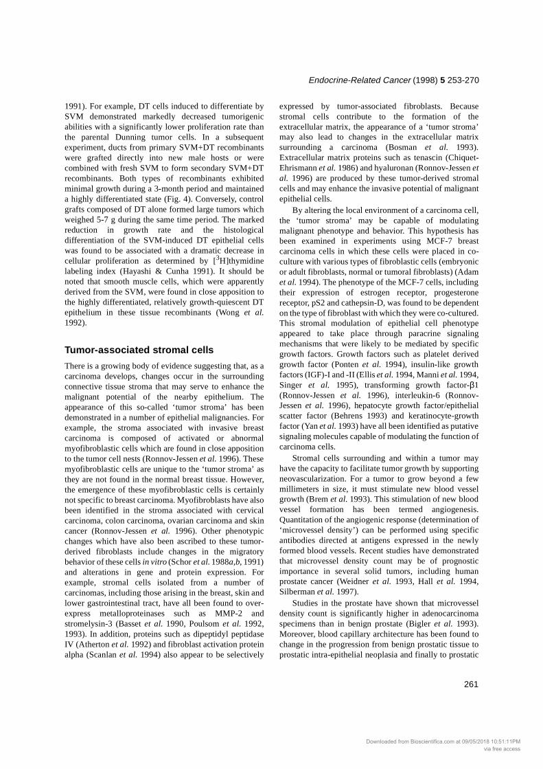
Figure 1 Schematic representation of interactions between the prostatic stroma and prostatic epithelium during normal development, adulthood and carcinogenesis. (a) Testosterone acts through androgen receptors (AR) in the urogenital sinus mesenchyme to induce epithelial development and differentiation. In a reciprocal fashion, epithelium signals to mesenchyme to induce smooth muscle differentiation. (b) In the growth-quiescent adult prostate, testosterone acts through both smooth muscle and epithelial AR. Androgens act through prostatic smooth muscle AR to maintain prostatic epithelium in a fully differentiated, growth-quiescent state and through epithelial AR to stimulate secretory function of the fully differentiated epithelium. It is hypothesized that the epithelium also acts to maintain smooth muscle differentiation through paracrine acting factors. (c) Prostatic carcinogenesis appears to be initiated by genetic insult to the epithelium. This leads to a change in epithelial phenotype and aberrant local signaling between the epithelium and smooth muscle. (d) The aberrant local signaling described in (c) leads to phenotypic changes in both the stromal and epithelial compartments of the tumor. The stroma becomes a fibroblastic 'tumor stroma' which promotes epithelial mitogenesis. This would predictably lead to increased epithelial proliferation, migration and, ultimately, an invasive epithelial phenotype.
Downloaded from Bioscientifica.com at 09/05/2018 10:51:11PMvia free access

Endocrine-Related Cancer (1998) 5 253-270
255
assumes a slowly growing or an invasive phenotype(Cunha et al. 1996, Hayward et al. 1996b, 1997b). It ispossible that, following genetic alteration to the prostaticepithelium, signaling from the epithelium to the sur-rounding smooth muscle becomes aberrant. This mayresult in stromal dedifferentiation towards a fibroblasticphenotype. One of the consequences of such a trans-formation may be that the local microenvironmentchanges from promoting epithelial homeostasis topromoting epithelial mitogenesis. These changes wouldbe predicted to lead to increased epithelial prolif-eration,migration and, ultimately, could enhance the in-vasivepotential of the genetically altered epithelial cell (Figs 1and 2).
The purpose of this article will be (1) to describe thestroma of the normal (non-malignant) prostate, (2) tosummarize genetic changes that are known to occur in the
prostatic epithelium during carcinogenesis, (3) to presentevidence suggesting that stroma derived from non-malignant sources may be able to alter the malignantphenotype of prostatic carcinoma cells, (4) to summarizethe evidence in support of a ‘tumor stroma’, and (5) tosummarize data describing the role of tumor stroma inprostatic carcinogenesis.
The stroma of the normal prostateThe human prostate is composed of two compartments:(1) an epithelial compartment, which includes theexocrine glands with their associated ductal structures,and (2) a surrounding connective tissue stroma. Thestroma of the human prostate consists of a number ofdifferent cell types. The most abundant cell type in thiscompartment is the smooth muscle cell, which is derived
Figure 2 Immunohistochemical expression of smooth muscle α-actin and vimentin in the stroma of normal prostate and prostate cancer. (a) Smooth muscle α-actin expression in normal prostate. Note that the stroma is comprised predominantly of smooth muscle. (b) Smooth muscle α-actin expression in the stroma associated with prostate cancer. Note the paucity of immunostaining in this high power field. (c) Vimentin expression in normal prostate. Other than in blood vessels (which serve as positive internal controls), there is virtually no vimentin staining in the stroma of the normal prostate. (d) Vimentin expression in prostate cancer. Note the increased expression of this fibroblastic marker in the stroma associated with prostate cancer compared with the stroma of the normal prostate. The findings summarized in panels a-d suggest that the predominantly smooth muscle stroma of the normal prostate is altered in prostatic carcinogenesis to include vimentin-positive fibroblastic cells.
Downloaded from Bioscientifica.com at 09/05/2018 10:51:11PMvia free access

Grossfeld et al.: Stroma in prostatic carcinogenesis
256
from the mesenchyme of the embryonic urogenital sinus(UGS). Other cell types located in the stroma of the normaladult prostate include fibroblasts, nerves, endothelial cellsand vascular smooth muscle cells. In addition to being themost abundant stromal cell type, the smooth muscle cellappears to be the most important cell type with respect toprostatic development and maintenance of homeostasis. Inthis regard, changes in the smooth muscle cell may beimportant in the evolution of prostatic carcinogenesis (seebelow).
Tissue recombination experiments utilizing theandrogen insensitive testicular feminized (tfm) mousehave established that an androgen responsive stroma isnecessary for the development of normal prostaticepithelial architecture in the rodent (Cunha et al. 1987). Inadulthood, prostatic smooth muscle cells, which areknown to express androgen receptors (AR), interact withepithelial cells and under androgenic conditions maintainthe epithelium in a fully differentiated, growth-quiescentstate (Cunha et al. 1996). This occurs both in the presenceand in the absence of epithelial AR expression whichemphasizes the paracrine nature of smooth muscle-epithelial interactions. Human prostatic smooth musclecells, which also express AR, are believed to play a similarrole in maintaining prostatic homeostasis. However,definitive experiments to confirm this role for humanprostatic smooth muscle cells are not possible given ourinability to access AR-deficient human fetal urogenitalsinuses.
Fibroblasts, which make up a large proportion of thestroma of the rodent prostate, are found sporadically in thenormal human prostate. It has been suggested that thesecells are important in mediating epithelial proliferation inthe rodent prostate (Nemeth & Lee 1996). Their functionin the human prostate remains unclear; they may serve asa scaffolding to keep smooth muscle bundles together, orthey may play an active role in organ homeostasis.
Prostatic development occurs as a direct result ofandrogenic stimulation of the fetal UGS. Analysis oftissue recombinants composed of AR-positive wild typeurogenital sinus mesenchyme (UGM) plus AR-negativetfm epithelium indicates that androgens act through AR inthe mesenchymal cells of the UGS to stimulate epithelialproliferation, ductal branching morphogenesis, andcolumnar cytodifferentiation. In rats and mice, prostatictissue can develop from the UGS derived from either amale or a female embryo if appropriately stimulated byandrogens at critical developmental stages. In laboratoryrodents the initial phases of prostatic budding requireexposure to androgens prenatally. In contrast, ductalbranching morphogenesis, canalization and epithelialcytodifferentiation all require postnatal androgenicstimulation. These early developmental events occurbetween birth and the onset of puberty, during which time
circulating androgen levels are very low. Theseobservations indicate that the developing prostate isextremely sensitive to low levels of circulating androgen(Donjacour & Cunha 1988).
Interactions between stroma and epithelium appear tobe reciprocal in nature. Not only does the developingstroma induce epithelial development, but the developingepithelium also induces primitive mesenchymal cells todifferentiate into smooth muscle (Cunha et al. 1992b).Urogenital sinus mesenchyme grown in the absence ofepithelium will not form smooth muscle. Thus, thedevelopment of prostatic smooth muscle in vivo requiresthe presence of both epithelium and appropriateandrogenic stimulation. This ability of epithelium toinduce the formation of visceral smooth muscle is notrestricted to the prostate, but it appears to be a commonfeature of many organs including the gut, uterus andbladder (Haffen et al. 1982, Cunha et al. 1989, Baskin etal. 1996).
The differentiation of prostatic smooth muscle occursin an orderly manner with the sequential expression of anumber of characteristic markers, including vimentin,actin, myosin, desmin and vinculin. The adult prostate, inwhich the stroma contains fully differentiated smoothmuscle cells, is essentially growth-quiescent andmaintains very low and balanced levels of cellularproliferation and cell death. It should be emphasized thatthis growth-quiescent, homeostatic state exists in thepresence of high levels of circulating androgens. In theadult rodent, androgens act directly on the prostaticsmooth muscle cells to maintain this fully differentiatedgrowth-quiescent state (Hayward et al. 1996b). We havepostulated that androgens act in a similar fashion in theadult human prostate to maintain growth-quiescence(Hayward et al. 1996b). Evidence supporting thishypothesis is as follows. After castration, the wellrecognized rapid regression of prostatic epithelium isassociated with an ordered loss of expression of thevarious smooth muscle differentiation markers. Thisappears to reflect a ‘dedifferentiation’ of the smoothmuscle cells. The loss of smooth muscle markersfollowing castration occurs in the order opposite to that towhich these markers were expressed during normaldevelopment (Hayward et al. 1996a). In its final form, theprostatic stroma of a long-term castrated animal reverts toa stroma which contains fibroblasts or mesenchymal cellsthat coexpress AR and vimentin. Very little expression ofthe characteristic smooth muscle markers is evident in thefully regressed prostate.
If exogenous androgens are subsequently admini-stered to a long-term castrated animal, prostatic tissue willrespond in a highly coordinated manner, with thedifferentiation of both stromal smooth muscle (againexpressing its characteristic markers) and a secretory
Downloaded from Bioscientifica.com at 09/05/2018 10:51:11PMvia free access

Endocrine-Related Cancer (1998) 5 253-270
257
epithelium (Bruchovsky et al. 1975). Thus, the relativelyundifferentiated fibroblastic cells in the prostatic stroma ofa castrated animal can respond to androgens by inducingepithelial proliferation and columnar cyto-differentiationwhile they revert back to highly differen-tiated smoothmuscle cells.
In summary, low levels of circulating androgens actupon the mesenchymal cells of the developing prostate toinduce prostatic epithelial proliferation and differen-tiation. In contrast, high circulating levels of androgen inthe adult act through the prostatic smooth muscle tomaintain a fully differentiated, growth-quiescent epithe-lium. Proliferative effects of stroma on epithelium aremediated through the stromal AR, while the epithelial ARappears to be required only for the expression of prostaticsecretory proteins (Cunha & Young 1991, Donjacour &Cunha 1993). In long-term castrated animals, exogenousandrogens initially promote prostatic epithelialproliferation and cytodifferentiation as well as the re-emergence of a smooth muscle stroma. Ultimately,androgen replacement leads to regeneration of a fullydifferentiated, growth-quiescent gland. These data suggestthat the local control of prostatic epithelial proliferationand differentiation occurs through androgenic stimulationof the prostatic stroma, and that the nature of the epithelialresponse to such a stimulation is predominantlydetermined by the nature of the stromal cells which arestimulated. Thus, AR-expressing prostatic smooth musclecells appear to respond to androgenic stimulation byinhibiting epithelial proliferation and maintainingepithelial differentiation, while an AR-expressingfibroblastic stroma (either the urogenital sinusmesenchyme or the stroma from an androgen-deprivedadult prostate) may respond to androgens by stimulatingepithelial proliferation and eliciting columnar cyto-differentiation.
Genetic changes in human prostatic cancerIt appears that human prostate cancer begins with geneticalteration to the prostatic epithelium. In this regard, avariety of techniques have been useful in demonstratinggenetic abnormalities in the prostatic epithelium, both inthe primary tumor and at metastatic sites. However, noneof these techniques has successfully defined a specificmutation that is characteristic for human prostate cancer.Although no study to date has specifically addressedgenetic changes to the prostatic stroma duringcarcinogenesis, preliminary data from our laboratoryusing karyotypic analysis and comparative genomichybridization demonstrate the absence of gross geneticalterations in the stromal cells surrounding a prostatecancer.
The familial concentration of some prostate cancerssuggests that risk for this tumor may be inherited in anautosomal dominant fashion. Risk factors for the inheritedform of this disease, including early age of onset andmultiple affected family members, has allowed for theidentification of some putative genetic abnormalities inthese patients (Carter et al. 1990, 1991, 1992, Smith et al.1996). Loss of heterozygosity has been reportedfrequently on chromosomes 10q, 7q and 16q in prostatecancer specimens (Isaacs et al. 1995). This has resulted inthe recent description of a candidate tumor suppressorgene, PTEN/MMAC1, for patients with inherited prostatecancer (Suzuki et al. 1998). It must be emphasized,however, that patients with hereditary prostate cancerrepresent only a minority of prostate cancer patients.Aside from these rare families, prostate cancer appears tobe a common but spontaneously arising disease with noconsistent pattern of genetic alteration.
Previous studies have described aberrant expression ofoncogenes, metastasis suppressor genes and tumorsuppressor genes in prostate tumor specimens. Whileoncogenes such as ras, c-myc, c-sis and neu have beenshown to be overexpressed in prostate cancer samples, nosingle oncogene is consistently overexpressed across awide range of prostate tumors (Peehl 1993, Wang & Wong1997). As a result, neither the initiation nor progression ofprostate cancer can be linked to a specific oncogene.While experimental overexpression of various oncogeneshas demonstrated the potential to initiate and/or promotethe disease (Thompson et al. 1989, 1993a,b, Bello et al.1997, Webber et al. 1997), the relevance of theseinteresting findings to clinical disease remains to beestablished.
A second class of genes which may prove to beimportant in human prostate cancer progression are the socalled ‘metastasis suppressor’ genes. Examples of suchgenes include the KAI1 gene and thymosin-beta 15 (agene which appears to be related to cell motility). KAI1has been mapped to human chromosome 11p11.2.Introduction of this gene into the metastatic rat AT6.1prostate cancer cells was shown to suppress prostatecancer metastases (Dong et al. 1995). In the human,expression of this gene appears to be reduced in cell linesderived from metastatic prostate tumors (Dong et al. 1995,1996a,b). Thymosin-beta 15, a 5.3 kDa protein, bindsactin monomers, thereby inhibiting actin polymerization(Gold et al. 1997). In the rat Dunning tumor model, cellmotility has been shown to correlate well with metastaticphenotype. Transfection of these tumor cells with anti-sense thymosin-beta 15 has been shown to cause adecrease in cell motility (Bao et al. 1996). Thymosin-beta15 levels appear to be elevated in human prostate cancer,and levels of this protein have been correlated withGleason grade (Bao et al. 1996). Additional studies have
Downloaded from Bioscientifica.com at 09/05/2018 10:51:11PMvia free access

Grossfeld et al.: Stroma in prostatic carcinogenesis
258
suggested that there may be other metastasis suppressorgenes important in human prostate cancer located onchromosomes 8 and 10 (Ichikawa et al. 1996).
The tumor suppressor genes represent a third class ofgenes that may demonstrate alterations in human prostatecancer specimens. Mutations in the retinoblastoma (Rb)gene have been reported to occur in prostate tumorsamples; however, such mutations are present in only aminority of cases (Isaacs 1995, Kubota et al. 1995). p53alterations are also inconsistently found in prostate cancer,appearing in only 20% of cases. In contrast, alterations inE-cadherin may be the most commonly detected defect inhuman prostate cancers, with up to 50% of casesdemonstrating altered expression of this protein.
E-cadherin is a 120 kDa transmembrane glycoproteinwhich is involved in epithelial cell adhesion (Birchmeier& Behrens 1994, Birchmeier et al. 1995, Jiang 1996,Shiozaki et al. 1996). The extracellular domain of E-cadherin undergoes calcium-dependent homophilic inter-actions in adherent junctions and along lateral epithelialmembranes. The cytoplasmic domain of E-cadherin isassociated with a group of proteins known as catenins thatlink the intracellular domain of E-cadherin to the actinmicrofilament network (Jiang 1996). A functional E-cadherin system is required to maintain normal epithelialmorphology.
Regulation of E-cadherin is poorly understood. Themolecule is developmentally regulated in the embryowhere it is initially expressed coincident with theappearance of epithelial tissue at the time of compactionof the embryo. Epithelial to mesenchymal transformation(i.e. the generation of mesoderm through the primitivestreak) is associated with loss of E-cadherin, whilemesenchymal to epithelial transformation (i.e. formationof the nephron) is associated with increased expression ofE-cadherin (Hay & Zuk 1995). A critical role for E-cadherin in development (Takeichi 1988) is underscoredby the observation that 'knockout' of the E-cadherin genein transgenic mice is embryonic lethal (Larue et al. 1994).While regulatory elements have been identified in the E-cadherin promoter (Behrens et al. 1992), the relevance ofthese regulatory elements to the biology of E-cadherinremains unclear.
An important role for E-cadherin expression in cellularadhesion is demonstrated by the fact that cells lacking E-cadherin are unable to aggregate or adhere. Furthermore,neutralizing antibodies to E-cadherin cause adherent cellsto dissociate. Mutation or deletion of genes encoding E-cadherin or the cadherin-associated proteins (catenins)also render cells non-adherent. Transfection of E-cadherin-negative cells with E-cadherin cDNA leads toreversal of the non-adherent phenotype.
These concepts are of obvious importance to tumorcell invasion and metastasis in the prostate and many other
organs. Experimental impairment of E-cadherin witheither antibodies or anti-sense cDNA converts non-invasive cells into invasive ones (Vleminckx et al. 1991).Moreover, loss of E-cadherin expression has been shownto coincide with the transformation of a well differentiatedadenoma into a carcinoma using a transgenic mousemodel of pancreatic carcinogenesis (Perl et al. 1998). Inthe prostate, decreased expression of E-cadherin ininvasive and metastatic cancers is associated with poorprognosis (Giroldi et al. 1994, Umbas et al. 1994,Shiozaki et al. 1996). While E-cadherin is consistentlyexpressed in normal epithelial cells (including prostaticepithelium) and in various highly differentiated ‘non-invasive’ carcinoma cells (including prostatic carcin-omas), E-cadherin expression is considerably lower (orentirely lacking) in poorly differentiated invasivecarcinoma cells (including prostatic carcinomas) (Umbaset al. 1992, Giroldi & Schalken 1993, Birchmeier &Behrens 1994, Giroldi et al. 1994, Jiang 1996).Bussemakers et al. (1993) demonstrated that coincidentwith the loss of E-cadherin expression, a variant of the ratDunning prostatic tumor spontaneously changed from awell differentiated, androgen-responsive, slow growingnon-metastatic line into an anaplastic, androgen-independent, rapidly growing metastatic line. Similarly,patients with prostatic tumors that do not express E-cadherin, or that have abnormal patterns of E-cadherinexpression, appear to have a poorer prognosis thanpatients with prostatic tumors that express E-cadherinnormally (Umbas et al. 1994). The relatively common lossof portions of chromosome 16 in human prostaticcarcinoma may result in deletion of all or part of the E-cadherin gene which maps to 16q21 (Joos et al. 1995).Other mechanisms that may cause dysfunction ofcadherin-mediated interactions include increased tyrosinephosphorylation of β-catenin and mutational inactivationof either the extracellular or intracellular domains of theE-cadherin gene (Behrens 1993, 1994). This type ofmutational inactivation of the E-cadherin gene has notbeen described in human prostatic carcinoma but has beenreported in endometrial and gastric cancers (Becker et al.1993, Risinger et al. 1994). In some high grade prostaticcarcinomas, E-cadherin levels may remain relativelynormal while α-catenin may be absent (Isaacs et al. 1994).The absence of α-catenin also renders the E-cadherinsystem non-functional (Hulsken et al. 1994). For example,impaired E-cadherin function in the PC3 prostaticcarcinoma cell line appears to be caused by homozygousdeletion of α-catenin (Morton et al. 1993)
Previous studies have suggested that ‘host factors’may down-regulate E-cadherin expression in carcinomacells. This observation was made from experiments inwhich virus-transformed MDCK cells (expressing highlevels of E-cadherin and exhibiting an adhesive
Downloaded from Bioscientifica.com at 09/05/2018 10:51:11PMvia free access

Endocrine-Related Cancer (1998) 5 253-270
259
phenotype) were transplanted into athymic mice. Thesecells, which were originally shown to be non-invasivebased upon in vitro assays, produced invasive metastatictumors in vivo which contained both E-cadherin-positiveand E-cadherin-negative cells. When these metastatic E-cadherin-negative cells were subsequently isolated andexamined in vitro, adherent E-cadherin-positive cells werere-derived (Mareel et al. 1991). Although their precisenature was not specified, unknown ‘host factors’ weresuggested to be involved in this down-regulation of E-cadherin. Further studies, in which micro-encapsulatedvirus-transformed MDCK cells were transplanted intoathymic rodent hosts, demonstrated that the loss of E-cadherin expression in vivo required ‘immediate contactsbetween tumor cells and host cells or upon host factors thatcould not cross the capsule membrane’ (Vandenbossche etal. 1994).
In order to determine if stromal cells are capable ofregulating E-cadherin expression, we have developed anin vitro model system in which epithelial cells are platedonto a confluent lawn of fibroblastic cells which have beenderived from either a tumor or a non-tumor environment.In addition to human prostatic stromal cells, we have alsoused a rat Dunning-tumor derived stromal cell line(DT3RPF; a generous gift from Dr Wallace McKeehan,Texas A&M University, USA) and a benign mouseembryonic fibroblastic cell line in our in vitro system(Hayward et al. 1997a). Stromal cells were grown toconfluence on positively charged microscope slides, afterwhich 5000/cm2 cells of the non-tumorigenic SV40Timmortalized human prostatic epithelial cell line BPH-1were plated onto the confluent stromal lawn. Co-cultureswere then grown for 48 h. Control cultures composed ofstromal cells alone, or BPH-1 cells alone were also grownon microscope slides for 48 h. Colony morphology(assessed by cytokeratin expression) and E-cadherinexpression were examined using immunohisto-chemistry.
BPH-1 cells cultured for 48 h on a confluent lawn ofbenign fibroblasts (of either human or rodent origin) grewas discreet coherent colonies. Epithelial cells appeared tobe contact inhibited. Epithelial colonies had smoothregular borders and epithelial cells appropriatelyexpressed E-cadherin along cell membranes. Few singlecells were observed. In contrast, BPH-1 cells cultured for48 h on a confluent lawn of tumor-derived fibroblasts(either from human prostate tumors or from the DT3RPFcell line) grew as single cells or as small colonies withirregular borders. Many cells appeared to overlap,suggesting a loss of contact inhibition. E-cadherinexpression was either markedly decreased or undetectablein epithelial cells grown in co-culture with the tumor-derived fibroblasts (Fig. 3).
‘Benign’ stroma may induce differentiation of prostatic carcinoma cellsIn the adult prostate, the stromal smooth muscleapparently functions to maintain the surroundingepithelium in a homeostatic, growth-quiescent state. Wehave hypothesized that the undifferentiated stroma
Figure 3 Colony morphology and E-cadherin expression in BPH-1 cells grown on a confluent layer of 'benign' and carcinoma-associated fibroblasts. (a) BPH-1 cells grown on a confluent lawn of stromal cells derived from a normal (non-malignant) prostate. Note the large epithelial colonies. (b) BPH-1 cells grown on fibroblasts derived from a prostatic tumor. Note the presence of smaller colonies and many single cells. Colonies have ragged, irregular edges. (c) E-cadherin expression along the cell membranes of coherent BPH-1 cells grown on fibroblasts derived from a normal (non-malignant) prostate. In contrast E-cadherin expression was absent from BPH-1 cells grown on fibroblasts derived from prostatic tumors.
Downloaded from Bioscientifica.com at 09/05/2018 10:51:11PMvia free access
Grossfeld et al.: Stroma in prostatic carcinogenesis
260
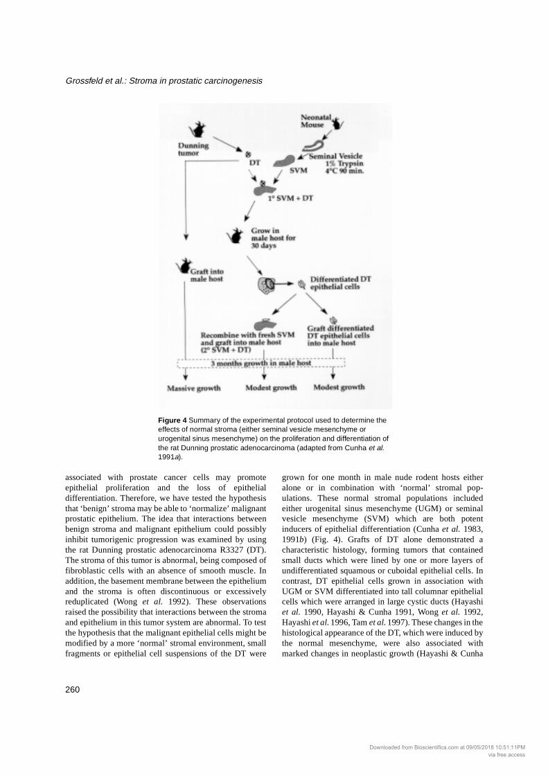
associated with prostate cancer cells may promoteepithelial proliferation and the loss of epithelialdifferentiation. Therefore, we have tested the hypothesisthat ‘benign’ stroma may be able to ‘normalize’ malignantprostatic epithelium. The idea that interactions betweenbenign stroma and malignant epithelium could possiblyinhibit tumorigenic progression was examined by usingthe rat Dunning prostatic adenocarcinoma R3327 (DT).The stroma of this tumor is abnormal, being composed offibroblastic cells with an absence of smooth muscle. Inaddition, the basement membrane between the epitheliumand the stroma is often discontinuous or excessivelyreduplicated (Wong et al. 1992). These observationsraised the possibility that interactions between the stromaand epithelium in this tumor system are abnormal. To testthe hypothesis that the malignant epithelial cells might bemodified by a more ‘normal’ stromal environment, smallfragments or epithelial cell suspensions of the DT were
grown for one month in male nude rodent hosts eitheralone or in combination with ‘normal’ stromal pop-ulations. These normal stromal populations includedeither urogenital sinus mesenchyme (UGM) or seminalvesicle mesenchyme (SVM) which are both potentinducers of epithelial differentiation (Cunha et al. 1983,1991b) (Fig. 4). Grafts of DT alone demonstrated acharacteristic histology, forming tumors that containedsmall ducts which were lined by one or more layers ofundifferentiated squamous or cuboidal epithelial cells. Incontrast, DT epithelial cells grown in association withUGM or SVM differentiated into tall columnar epithelialcells which were arranged in large cystic ducts (Hayashiet al. 1990, Hayashi & Cunha 1991, Wong et al. 1992,Hayashi et al. 1996, Tam et al. 1997). These changes in thehistological appearance of the DT, which were induced bythe normal mesenchyme, were also associated withmarked changes in neoplastic growth (Hayashi & Cunha
Figure 4 Summary of the experimental protocol used to determine the effects of normal stroma (either seminal vesicle mesenchyme or urogenital sinus mesenchyme) on the proliferation and differentiation of the rat Dunning prostatic adenocarcinoma (adapted from Cunha et al. 1991a).
Downloaded from Bioscientifica.com at 09/05/2018 10:51:11PMvia free access
Endocrine-Related Cancer (1998) 5 253-270
261
1991). For example, DT cells induced to differentiate bySVM demonstrated markedly decreased tumorigenicabilities with a significantly lower proliferation rate thanthe parental Dunning tumor cells. In a subsequentexperiment, ducts from primary SVM+DT recombinantswere grafted directly into new male hosts or werecombined with fresh SVM to form secondary SVM+DTrecombinants. Both types of recombinants exhibitedminimal growth during a 3-month period and maintaineda highly differentiated state (Fig. 4). Conversely, controlgrafts composed of DT alone formed large tumors whichweighed 5-7 g during the same time period. The markedreduction in growth rate and the histologicaldifferentiation of the SVM-induced DT epithelial cellswas found to be associated with a dramatic decrease incellular proliferation as determined by [3H]thymidinelabeling index (Hayashi & Cunha 1991). It should benoted that smooth muscle cells, which were apparentlyderived from the SVM, were found in close apposition tothe highly differentiated, relatively growth-quiescent DTepithelium in these tissue recombinants (Wong et al.1992).
Tumor-associated stromal cells
There is a growing body of evidence suggesting that, as acarcinoma develops, changes occur in the surroundingconnective tissue stroma that may serve to enhance themalignant potential of the nearby epithelium. Theappearance of this so-called ‘tumor stroma’ has beendemonstrated in a number of epithelial malignancies. Forexample, the stroma associated with invasive breastcarcinoma is composed of activated or abnormalmyofibroblastic cells which are found in close appositionto the tumor cell nests (Ronnov-Jessen et al. 1996). Thesemyofibroblastic cells are unique to the ‘tumor stroma’ asthey are not found in the normal breast tissue. However,the emergence of these myofibroblastic cells is certainlynot specific to breast carcinoma. Myofibroblasts have alsobeen identified in the stroma associated with cervicalcarcinoma, colon carcinoma, ovarian carcinoma and skincancer (Ronnov-Jessen et al. 1996). Other phenotypicchanges which have also been ascribed to these tumor-derived fibroblasts include changes in the migratorybehavior of these cells in vitro (Schor et al. 1988a,b, 1991)and alterations in gene and protein expression. Forexample, stromal cells isolated from a number ofcarcinomas, including those arising in the breast, skin andlower gastrointestinal tract, have all been found to over-express metalloproteinases such as MMP-2 andstromelysin-3 (Basset et al. 1990, Poulsom et al. 1992,1993). In addition, proteins such as dipeptidyl peptidaseIV (Atherton et al. 1992) and fibroblast activation proteinalpha (Scanlan et al. 1994) also appear to be selectively
expressed by tumor-associated fibroblasts. Becausestromal cells contribute to the formation of theextracellular matrix, the appearance of a ‘tumor stroma’may also lead to changes in the extracellular matrixsurrounding a carcinoma (Bosman et al. 1993).Extracellular matrix proteins such as tenascin (Chiquet-Ehrismann et al. 1986) and hyaluronan (Ronnov-Jessen etal. 1996) are produced by these tumor-derived stromalcells and may enhance the invasive potential of malignantepithelial cells.
By altering the local environment of a carcinoma cell,the ‘tumor stroma’ may be capable of modulatingmalignant phenotype and behavior. This hypothesis hasbeen examined in experiments using MCF-7 breastcarcinoma cells in which these cells were placed in co-culture with various types of fibroblastic cells (embryonicor adult fibroblasts, normal or tumoral fibroblasts) (Adamet al. 1994). The phenotype of the MCF-7 cells, includingtheir expression of estrogen receptor, progesteronereceptor, pS2 and cathepsin-D, was found to be dependenton the type of fibroblast with which they were co-cultured.This stromal modulation of epithelial cell phenotypeappeared to take place through paracrine signalingmechanisms that were likely to be mediated by specificgrowth factors. Growth factors such as platelet derivedgrowth factor (Ponten et al. 1994), insulin-like growthfactors (IGF)-I and -II (Ellis et al. 1994, Manni et al. 1994,Singer et al. 1995), transforming growth factor-β1(Ronnov-Jessen et al. 1996), interleukin-6 (Ronnov-Jessen et al. 1996), hepatocyte growth factor/epithelialscatter factor (Behrens 1993) and keratinocyte-growthfactor (Yan et al. 1993) have all been identified as putativesignaling molecules capable of modulating the function ofcarcinoma cells.
Stromal cells surrounding and within a tumor mayhave the capacity to facilitate tumor growth by supportingneovascularization. For a tumor to grow beyond a fewmillimeters in size, it must stimulate new blood vesselgrowth (Brem et al. 1993). This stimulation of new bloodvessel formation has been termed angiogenesis.Quantitation of the angiogenic response (determination of‘microvessel density’) can be performed using specificantibodies directed at antigens expressed in the newlyformed blood vessels. Recent studies have demonstratedthat microvessel density count may be of prognosticimportance in several solid tumors, including humanprostate cancer (Weidner et al. 1993, Hall et al. 1994,Silberman et al. 1997).
Studies in the prostate have shown that microvesseldensity count is significantly higher in adenocarcinomaspecimens than in benign prostate (Bigler et al. 1993).Moreover, blood capillary architecture has been found tochange in the progression from benign prostatic tissue toprostatic intra-epithelial neoplasia and finally to prostatic
Downloaded from Bioscientifica.com at 09/05/2018 10:51:11PMvia free access
Grossfeld et al.: Stroma in prostatic carcinogenesis
262
carcinoma (Montironi et al. 1993). With respect tolocalization within a tumor, a higher degree ofvascularization is found at the center of a prostatic tumorcompared with the tumor edge (Siegal et al. 1995).Microvessel density has been associated with other wellrecognized prognostic indicators in human prostate cancerincluding tumor Gleason grade (Wakui et al. 1992) andpathological stage (Fregene et al. 1993, Brawer et al.1994).
The angiogenic process in human prostate cancerappears to require the presence of stromal cells. This hasbeen demonstrated in a co-culture system using the humanprostatic tumor cell line, PC-3, where the induction ofangiogenesis required the presence of stromal fibroblasts(Janvier et al. 1997).
The above data suggest that the degree of tumorneovascularization (which is dependent upon stromalcells) may be related to tumor cell invasion and metastasisin prostate cancer. Taken together, these suggest thatalterations in the tumor-associated stromal cells may havean important role in prostate cancer progression. It is nosurprise, therefore, that some investigators have suggestedthat without a ‘tumor stroma’ there would be no tumors(Bosman et al. 1993, Ronnov-Jessen et al. 1996).
Abnormal stroma as a mediator of tumorigenesis in the prostate
The idea that stromal cells may facilitate prostaticcarcinogenesis has been investigated previously. Using an
in vivo mouse prostatic reconstitution system, Thompsonand colleagues infected either the urogenital sinus(prostatic anlagen) or its individual mesenchymal (UGM)or epithelial (UGE) components with a virus containingthe myc and ras oncogenes. In prostatic reconstitutionscontaining uninfected UGM+infected UGE, epithelialhyperplasias were detected. Similarly, in prostaticreconstitutions composed of infected UGM+uninfectedUGE, stromal desmoplasias were observed. Carcinomaswere found only in prostatic reconstitutions in which bothUGM and UGE were infected (Thompson 1990,Thompson et al. 1989, 1993a). These findingsdemonstrated that changes were required both in theepithelium and in the stromal microenvironment forprostatic carcinogenesis to occur.
Several studies examining the role of stromal cells inprostatic tumorigenesis have originated from thelaboratory of Leland Chung. Camps et al. (1990) reportedthat co-inoculation of tumorigenic NbF-1 fibroblasts withhuman PC-3 prostatic carcinoma cells accelerated tumorgrowth and shortened tumor latency period. Theinteraction between fibroblasts and epithelial cells in thissystem was bi-directional – PC-3 cells reciprocally en-hanced the tumorigenesis of sarcomatous NbF-1 fibro-blasts. Similar studies have been extended to the humanLnCaP prostatic carcinoma cell line (Chung 1991, Chunget al. 1991, Gleave et al. 1991, 1992, Wu et al. 1994). Co-inoculation of LnCaP cells with various non-tumorigenicfibroblasts demonstrated that fibroblasts differed in theirability to promote prostatic carcino-genesis (Gleave et al.
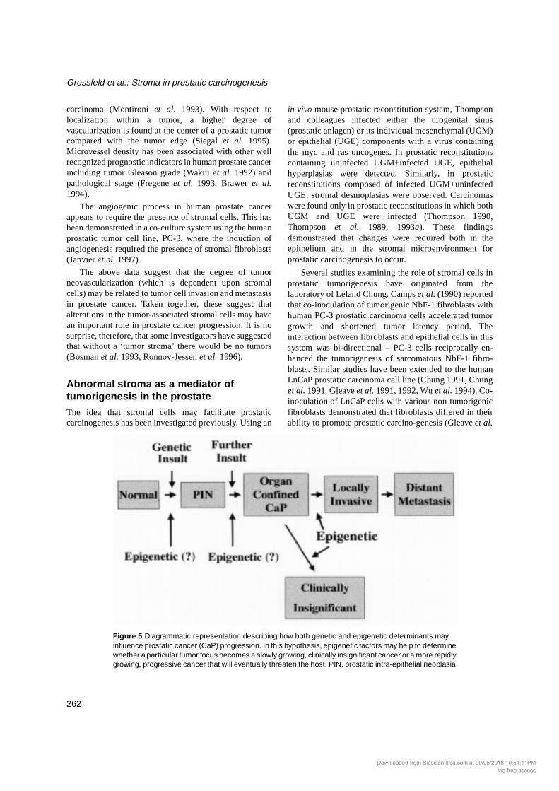
Figure 5 Diagrammatic representation describing how both genetic and epigenetic determinants may influence prostatic cancer (CaP) progression. In this hypothesis, epigenetic factors may help to determine whether a particular tumor focus becomes a slowly growing, clinically insignificant cancer or a more rapidly growing, progressive cancer that will eventually threaten the host. PIN, prostatic intra-epithelial neoplasia.
Downloaded from Bioscientifica.com at 09/05/2018 10:51:11PMvia free access

Endocrine-Related Cancer (1998) 5 253-270
263
1991, 1992). Fibroblasts derived from rat urogenital sinusmesenchyme and human bone, but not NIH 3T3 cells,normal rat kidney fibroblasts or normal human lungfibroblasts, enhanced tumorigenesis of ‘non-tumorigenic’(as described by Gleave and colleagues) human LnCaPcells in vivo. Conditioned media from bone fibroblasts, raturogenital sinus mesenchyme cells or NbF-1 fibroblastswere also effective in stimulating growth of LnCaP cellsin vitro, while conditioned media from 3T3 cells, normalrat kidney and normal human lung were ineffective in thisregard. Once again, this effect was bi-directional, asLnCaP conditioned medium stimulated rat urogenitalsinus mesenchyme growth by up to 275%. These datasupport the concept that fibroblasts can enhancetumorigenesis in the human prostate, and that the trophiceffect of fibroblasts on prostatic carcinogenesis is not aproperty of fibroblasts in general but is restricted toselected fibroblastic cells.
As described above, we have hypothesized thatinitiation of prostatic carcinogenesis involves geneticalteration of the prostatic epithelium, after which there isa sequential disruption in the reciprocal homeostaticinteractions between the prostatic smooth muscle and theassociated epithelium (Cunha et al. 1996, Hayward et al.1996b, 1997a). This altered signaling leads to the de-differentiation of both the emerging prostatic carcinomacells and the surrounding smooth muscle. Even though thephenotype of the smooth muscle changes, the tumor-
associated stromal cells are postulated to remaingenetically normal (Fig. 5).
Using an in vivo model system, we have examined theeffects of carcinoma-associated fibroblasts on adjacentepithelial cells. If our above hypothesis is correct, humancarcinoma-associated fibroblasts (CAF) should havedifferent effects on epithelial proliferation anddifferentiation than ‘normal’ fibroblasts derived frombenign human prostatic tissue. We tested this hypothesisusing a tissue recombination model in which benign ortumor-derived stromal cells isolated from human prostatictissue were recombined with either BPH-1 cells (a non-tumorigenic, SV40T immortalized human prostaticepithelial cell line) (Hayward et al. 1995) orphenotypically normal human prostatic ductal organoids(fragments of acini and ducts derived from benign prostatewhich retain in vivo architecture). The resultant tissuerecombinants were grown beneath the renal capsule ofadult athymic nude rodent hosts. Control grafts werecomposed of either epithelial or stromal cells alone andwere grown in conditions identical to the experimentaltissue recombinants. The amount of tissue recombinantgrowth was determined by measuring wet weights.Histopathological features of tissue recombinants weredetermined in hematoxylin and eosin stained sections.
Control grafts containing either benign or tumor-derived stromal cells alone or epithelial cells alonedemonstrated minimal growth during the experimentalperiod. Tissue recombinants composed of normal prostaticfibroblasts+BPH-1 cells also exhibited minimal growthafter sub-renal capsular grafting. In contrast, tissuerecombinants composed of carcinoma-associatedfibroblasts+BPH-1 cells exhibited striking growth aftersub-renal capsular grafting, reaching wet weights as highas 5 g after 41 days (from an initial wet weight ofapproximately 10 mg). Morphometric analysis demon-strated that these tumors were predominantly epithelial(80%), with a histological appearance that was consistentwith poorly-differentiated prostatic adenocarcinoma (Fig.6).
Tumors were composed of poorly differentiated,irregular epithelial cords. In some areas, epitheliumformed small glandular nests while in other areasepithelium appeared as single cells that were intermingledwithin a fibrous stroma. The epithelial nature of these cellswas confirmed immunohistochemically using a widespectrum anti-cytokeratin antibody. Furthermore,epithelial cells within these tumors exhibited nuclearstaining with an antibody to SV40T antigen, confirmingtheir BPH-1 origin. The majority of epithelial cellscontained large, pleomorphic nuclei with large nucleoli.Stromal cells were intermingled between the epithelialcell nests throughout the tumor. In contrast to what wasseen with CAF+BPH-1 tissue recombinants, none of the
Figure 6 Photomicrograph of a tissue recombinant composed of human prostate carcinoma-associated fibroblasts and BPH-1 cells. This tissue recombinant was grown beneath the renal capsule of a male athymic mouse host for 21 days. This hematoxylin and eosin stained tissue section demonstrates the characteristic appearance of a poorly differentiated adeno-carcinoma. Note that in some areas epithelial cells form nests that resemble glandular-like structures (arrows).
Downloaded from Bioscientifica.com at 09/05/2018 10:51:11PMvia free access

Grossfeld et al.: Stroma in prostatic carcinogenesis
264
grafts containing normal human ductal prostaticorganoids, including those prepared with carcinoma-associated fibroblasts, exhibited significant growth.
These studies demonstrated that stromal cells derivedfrom benign and malignant sources were able to exertdifferential effects on non-tumorigenic human prostaticepithelial cells in vivo. Carcinoma-associated fibroblasts,unlike normal fibroblasts, were capable of inducing aninvasive phenotype in an immortalized but non-tumorigenic human prostatic epithelial cell line. The samefibroblasts were unable to induce this phenotype ingenetically normal human prostatic epithelial cells, eventhough effects on epithelial differentiation were observed.These data suggest (1) that both genetic and epigeneticchanges may be important in human prostaticcarcinogenesis, (2) that carcinoma-associated fibroblastswere able to stimulate progression of an initiatedepithelium while normal fibroblasts were incapable ofstimulating progression and (3) that carcinoma-associatedfibroblasts were incapable of causing initiation in agenetically normal epithelium. Recent data obtained usingan in vitro co-culture system suggest that the carcinoma-associated fibroblasts may be capable of increasingproliferation and decreasing death rates of the BPH-1human prostatic epithelial cells.
It should be noted that some of Chung’s initial studiesattempted to examine the ability of prostatic stromal cellsto stimulate carcinogenic progression in vivo by co-inoculating prostatic stromal cells with prostatic epithelialcells in a rodent host. In these studies, a non-tumorigenicrat prostatic epithelial cell line (NbE-1 cells) was co-inoculated with a tumorigenic cell line of rat prostaticfibroblasts which formed sarcomas when grown alone(NbF-1 cells) (Chung et al. 1989). As early as nine daysafter transplantation, large ‘carcinosarcomas’ formed inwhich epithelium comprised only 2-5% of the tumor mass.The precise volume of epithelial growth was notcalculated in these studies, and any histopathologicalchanges that may have occurred in the epithelium as aresult of co-inoculation with the tumorigenic NbF-1 cellswere not specified. Furthermore, control experimentsexamining the in vivo characteristics of epithelial cellsgrown alone, or epithelial cells grown with a normal, non-tumorigenic stroma were not described. Moreover, thetumor growth period was not varied for any of theexperiments. Given the rapidity with which the tumorsformed, as well as the primarily sarcomatous appearanceof the tumors, it is likely that, instead of forming tumorsthemselves, the benign epithelial cells in these graftsremained benign epithelial cells ‘trapped’ within a NbF-1-derived sarcoma.
It is important to note that our experiments differ fromthose of Chung and colleagues in several ways. First,Chung’s studies utilized tumorigenic fibroblasts that
formed sarcomas when grown alone. Our stromal cellpopulations were genetically normal (by karyotypicanalysis and comparative genomic hybridization) andwere non-tumorigenic when grown alone. In fact, ourcarcinoma-associated fibroblasts exhibited virtually nogrowth during 6 weeks in vivo beneath the renal capsule.Secondly, the histological appearance of the tumors in ourstudy was consistent with that of a poorly differentiatedadenocarcinoma. Sarcomatous elements were notidentified. This is in marked contrast to the primarilysarcomatous tumors (carcinosarcomas) described byChung and colleagues. These important differences willenable us to utilize our model system to examine the roleof stromal-epithelial interactions in the initiation andprogression of human prostatic cancer.
Mechanisms of stromal-epithelial signaling in the normal and neo- plastic prostateThe data summarized above lead to two opposite butrelated ideas: (1) that carcinoma-associated stromal cellscan promote prostatic carcinogenesis, and (2) that normalstromal cells may be able to inhibit prostatic carcino-genesis by inducing differentiation and decreasing pro-liferation of epithelium. These data indicate that a detailedunderstanding of the signaling mechanisms betweenstroma and epithelium could allow for the rational designof therapies aimed at inhibiting prostate tumor growth.
Many studies have attempted to identify the signalingmolecules acting as paracrine mediators of stromal andepithelial interactions in the developing and adult prostate.In most studies to date, growth-quiescent adult prostatictissues have been utilized, and therefore the signalingmolecules that have been identified may be involved inhomeostasis rather than in carcinogenesis (Cunha et al.1992a, Cunha 1994). There is currently no detailed pictureof the spatial and temporal expression patterns of many ofthe growth factors found in the prostate. Considerationssuch as the cell of origin of a particular growth factor(epithelium, smooth muscle or fibroblast), the preciselocalization of a growth factor or its receptor in relation toproximal, intermediate or distal ductal architecture, andthe specific developmental stages when expression of aparticular growth factor occurs have not been adequatelyexamined.
In order to define better growth factor expression inthe prostate, we have recently developed species-specificreverse transcription-PCR methods which examine theexpression of growth factors in heterospecific tissuerecombinants (i.e. rodent mesenchyme with humanepithelium). Using this method, it has been possible todetermine whether epithelial or stromal cells areexpressing specific growth factors (Horseman et al. 1997,
Downloaded from Bioscientifica.com at 09/05/2018 10:51:11PMvia free access

Endocrine-Related Cancer (1998) 5 253-270
265
Hayward et al. 1998). This technique does not requiredisruption of organ architecture with potential adversemodification of RNA expression. Our results to date havedemonstrated that growth factors and their receptors areoften expressed in multiple compartments of the growingprostate gland.
For a limited number of growth factors, the expressionof ligand and receptor molecules is restricted to specificand separate cell types. For example, expression ofhepatocyte growth factor and its receptor (c-met) islocalized to stromal and epithelial cells respectively. Asimilar situation also exists for some members of thefibroblastic growth factor (FGF) family, including FGF-7.This growth factor is expressed by mesodermally derivedstromal cells, while its receptor (the FGF-R2 IIIb splicevariant) is exclusively expressed by epithelium.
Previous studies have demonstrated that specificgrowth factors may play an important role in stromal-epithelial signaling in the normal developing prostate.Thus, it is possible that altered expression of these growthfactors may contribute to the development of prostaticcancer. One such growth factor is FGF-7. During prostaticdevelopment, FGF-7 is capable of imitating some of theeffects of testosterone. For example, the addition ofexogenous FGF-7 in the absence of testosterone caninduce ductal branching morphogenesis in organ culturesof neonatal rat ventral prostate and mouse seminal vesicle(Alarid et al. 1994, Sugimura et al. 1996). In addition, aneutralizing antibody directed against FGF-7 was able toinhibit androgen-induced ductal branching morpho-genesis using this same rat ventral prostate model.Although stromal cell expression of FGF-7 may beregulated by androgens in vitro (Yan et al. 1992), RNAseprotection assays of FGF-7 and its receptor (FGF-R2 IIIb)failed to show regulation of these genes by androgens invivo (Thomson et al. 1997). In prostate cancer, the switchof FGF-7 receptor expression from the FGF-R2-IIIb to the-IIIc splice variant in rat Dunning tumor cells may resultin altered FGF ligand-specific responsiveness of theepithelium (Yan et al. 1993).
A second family of growth factors which maypotentially influence prostatic growth and carcinogenesisare the IGFs. The exact role of the IGFs in prostaticdevelopment and disease remains unclear. However,recent experiments from our laboratory have shown thatIGF-I is required for normal prostatic growth. Fetalprostatic rudiments harvested from both IGF-I and IGFtype 1 receptor knockout mice were grafted beneath therenal capsule of male nude mouse hosts. Grafts wereallowed to grow for one month. After this time period, therecovered tissue grafts were very small, with wet weightsof approximately 1-2 mg. This is compared with wetweights of 30-50 mg for wild-type fetal prostatic grafts.Histological examination of the IGF-I knockout and IGF
type 1 receptor knockout grafts demonstrated thatprostatic differentiation had occurred during this timeperiod, with the appearance of a few small ducts whichwere lined by tall columnar secretory epithelium (ADonjacour, unpublished observations). These datasuggested two important points: (1) that IGF-I wasrequired for normal prostatic growth, and (2) that localproduction (or activation) of the ligand appears to beimportant (normal levels of circulating IGF-I in the nudemouse host did not induce normal prostatic growth ingrafts of IGF-I knockout prostates). Previous studies havedemonstrated inappropriately increased expression ofIGF-II both in benign prostatic disease and in othercarcinomas such as those arising in the breast (Ellis et al.1994, Manni et al. 1994, Peehl et al. 1995, Singer et al.1995, Tennant et al. 1996, Dong et al. 1997). In addition,IGF-I appears to be down-regulated in human prostatetumors (Peehl et al. 1995, Tennant et al. 1996, Dong et al.1997). When taken together, these data support furtherinvestigation into the regulation of IGF-I, IGF-II and thetype 1 IGF receptor in benign and malignant prostaticdisease.
Conclusions
The data summarized above suggest that both genetic andepigenetic factors are important in the progression ofprostate cancer. Our recent in vivo and in vitro studies havedemonstrated that: (1) carcinoma-associated fibroblastsdo not form tumors when grown in the absence ofepithelium, (2) carcinoma-associated fibroblasts stimulateprogression of a genetically altered, non-tumorigenichuman prostatic epithelium towards a carcinomatousphenotype, (3) the same fibroblasts are unable to stimulateinitiation of a genetically normal human prostaticepithelium, and (4) stromal cells from non-malignantsources do not promote tumorigenesis in a geneticallyaltered human prostatic epithelium under identicalconditions. Our in vitro studies have also demonstratedthat the stromal microenvironment may be capable ofregulating epithelial E-cadherin expression. These data donot discount the possibility that epithelial cells with severegenetic alterations can grow autonomously as malignanttumors. Instead, they imply that interactions with thestromal microenvironment are important determinants inthe progression from a normal prostatic epithelium to aninvasive carcinoma.
Prostate cancer can exist in two forms: either as alatent, clinically insignificant tumor or as an aggressivetumor which will progress without treatment. At thepresent time, we have been unable to identify the specificfactors that determine the behavior of any given tumorfocus. The evidence presented above strongly suggeststhat the stromal microenvironment may be capable of
Downloaded from Bioscientifica.com at 09/05/2018 10:51:11PMvia free access

Grossfeld et al.: Stroma in prostatic carcinogenesis
266
modulating the biological potential of a particular tumor.This may be accomplished through regulation of tumorneovascularity or through specific phenotypic changes inthe surrounding stromal cells that result in alteredexpression of certain enzymes, cellular adhesionmolecules or growth factors.
The implications of our observations regarding tumorgrowth are potentially very important from both adiagnostic and a therapeutic perspective. For example, theability to identify tumor-associated stromal populationswhich promote down-regulation of E-cadherin could beextremely important for identifying patients at risk formalignant progression of their prostatic adenocarcinoma.From a therapeutic viewpoint, the prospect of targetingtherapy through the tumor-associated stroma to increaseepithelial E-cadherin expression could be used to maintainprostatic tumors in a highly differentiated, non-invasivestate. This is not a far-fetched possibility as studies inbreast cancer patients have already demonstrated thatagents such as 9-cis-retinoic acid, tamoxifen, IGF-I andtangeretin may up-regulate E-cadherin expression(Anzano et al. 1994, Bracke et al. 1994, Morton et al.1995).
Traditional therapy for all epithelial malignancies,including prostate cancer, has been targeted at themalignant epithelial cell. Due to its genetic instability, thiscell represents a ‘moving target’ for treatment. Althoughcarcinoma-associated fibroblasts are abnormal, pre-liminary studies using karyotypic analysis and com-parative genomic hybridization have demonstrated thatthese cells do not possess gross genetic alterations. Thus,they may provide a more stationary target at which todirect treatment.
Acknowledgements This work was supported by NIH grants DK52721, CA59831, DK 45861, CA 64872, DK 52708 and DK 47517,and by an AFUD/Pfizer USPG Research Scholarship toGDG.
ReferencesAdam L, Crepin M, Lelong JC, Spanakis E & Israel L 1994
Selective interactions between mammary epithelial cells and fibroblasts in co-culture. International Journal of Cancer 59 262-268.
Alarid ET, Rubin JS, Young P, Chedid M, Ron D, Aaronson SA & Cunha GR 1994 Keratinocyte growth factor functions in epithelial induction during seminal vesicle development. Proceedings of the National Academy of Sciences of the USA 91 1074-1078.
Anzano MA, Byers SW, Smith JM, Peer CW, Mullen LT, Brown CC, Roberts AB & Sporn MB 1994 Prevention of breast cancer in the rat with 9-cis-retinoic acid as a single agent and
in combination with tamoxifen. Cancer Research 54 4614-4617.
Atherton AJ, Monaghan P, Warburton MJ, Robertson D, Kenny AJ & Gusterson BA 1992 Dipeptidyl peptidase IV expression identifies a functional sub-population of breast fibroblasts. International Journal of Cancer 50 15-19.
Bao L, Loda M, Janmey PA, Stewart R, Anand-Apte B & Zetter BR 1996 Thymosin beta 15: a novel regulator of tumor cell motility upregulated in metastatic prostate cancer. Nature Medicine 12 1322-1328.
Baskin LS, Hayward SW, Young P & Cunha G 1996 Role of mesenchymal-epithelial interactions in bladder development. Journal of Urology 156 1820-1827.
Basset P, Bellocq JP, Wolf C, Stoll I, Hutin P, Limacher JM, Podhajcer OL, Chenard MP, Rio MC & Chambon P 1990 A novel metalloproteinase gene specifically expressed in stromal cells of breast carcinomas. Nature 348 699-704.
Becker KF, Atkinson MJ, Reich U, Huang HH, Nekarda H, Siewert JR & Hofler H 1993 Exon skipping in the E-cadherin gene transcript in metastatic human gastric carcinomas. Human Molecular Genetics 2 803-804.
Behrens J 1993 The role of cell adhesion molecules in cancer invasion and metastasis. Breast Cancer Research and Treatment 24 175-184.
Behrens J 1994 Cell contacts, differentiation, and invasiveness of epithelial cells. Invasion Metastasis 14 61-70.
Behrens J, Frixen U, Schipper J, Weidner M & Birchmeier W 1992 Cell adhesion in invasion and metastasis. Seminars in Cell Biology 3 169-178.
Bello D, Webber MM, Kleinman HK, Wartinger DD & Rhim JS 1997 Androgen responsive adult human prostatic epithelial cell lines immortalized by human papillomavirus 18. Carcinogenesis 18 1215-1223.
Bigler SA, Deering RE & Brawer MK 1993 Comparison of microscopic vascularity in benign and malignant prostate tissue. Human Pathology 24 220-226.
Birchmeier W & Behrens J 1994 Cadherin expression in carcinomas: role in the formation of cell junctions and the prevention of invasiveness. Biochimica et Biophysica Acta 1198 11-26.
Birchmeier W, Hulsken J & Behrens J 1995 E-cadherin as an invasion suppressor. Ciba Foundation Symposium 189 124-136.
Bosman FT, de Bruine A, Flohil C, van der Wurff A, ten Kate J & Dinjens WW 1993 Epithelial-stromal interactions in colon cancer. International Journal of Developmental Biology 37 203-211.
Bracke ME, Charlier C, Bruyneel EA, Labit C, Mareel MM & Castronovo V 1994 Tamoxifen restores the E-cadherin function in human breast cancer MCF-7/6 cells and suppresses their invasive phenotype. Cancer Research 54 4607-4609.
Brawer MK, Deering RE, Brown M, Preston SD & Bigler SA 1994 Predictors of pathologic stage in prostatic cacinoma. Cancer 73 678-687.
Brem H, Gresser I, Grosfeld J & Folkman J 1993 The combination of antiangiogenic agents to inhibit primary tumor growth and metastasis. Journal of Pediatric Surgery 28 1253-1257.
Downloaded from Bioscientifica.com at 09/05/2018 10:51:11PMvia free access

Endocrine-Related Cancer (1998) 5 253-270
267
Bruchovsky N, Lesser B, van Doorn EV & Craven S 1975 Hormonal effects on cell proliferation in rat prostate. Vitamins and Hormones 33 61-102.
Bussemakers MJ, van Bokhoven A, Mees SG, Kemler R & Schalken JA 1993 Molecular cloning and characterization of the human E-cadherin cDNA. Molecular Biology Reports 17 123-128.
Camps JL, Chang S-M, Hsu TC, Freeman MR, Hong S-J, Zhau HE, von Eschenbach AC & Chung LWK 1990 Fibroblast-mediated acceleration of human epithelial tumor growth in vivo. Proceedings of the National Academy of Sciences of the USA 87 75-79.
Carter BS, Carter HB & Isaacs JT 1990 Epidemiologic evidence regarding predisposing factors to prostatic cancer. Prostate 16 187-197.
Carter BS, Steinberg GD, Beaty TH, Childs B & Walsh PC 1991 Familial risk factors for prostate cancer. In Prostate Cancer: Cell and Molecular Mechanisms in Diagnosis and Treatment, pp 5-13. Ed JT Issacs. Cold Spring Harbor: Cold Spring Harbor Laboratory Press.
Carter BS, Beaty TH, Steinberg GD, Childs B & Walsh PC 1992 Mendelian inheritance of familial prostate cancer. Proceedings of the National Academy of Sciences of the USA 89 3367-3371.
Catalona WJ & Smith DS 1994 5-Year tumor recurrence rates after anatomical radical retropubic prostatectomy for prostate cancer. Journal of Urology 152 1837-1842.
Chiquet-Ehrismann R, Mackie EJ, Pearson CA & Sakakura T 1986 Tenascin: an extracellular matrix protein involved in tissue interactions during fetal development and oncogenesis. Cell 47 131-139.
Chodak GW, Thisted RA, Gerber GS, Johansson J-E, Adolfsson J, Jones GW, Chisholm GD, Moskovitz B, Livne PM & Warner J 1994 Results of conservative management of clinically localized prostate cancer. New England Journal of Medicine 330 242-248.
Chung LW 1991 Fibroblasts are critical determinants in prostatic cancer growth and dissemination. Cancer Metastasis Review 10 263-274.
Chung LWK, Chang S, Bell C, Zhau HE, Ro JY & Von Eschenbach AC 1989 Co-inoculation of tumorigenetic rat prostate mesenchymal cells with non-tumorigenic epithelial cells results in the development of carcinosarcoma in syngeneic and athymic animals. International Journal of Cancer 43 1179-1187.
Chung LWK, Gleave ME, Hsieh J-T, Hong S-J & Zhau HE 1991 Reciprocal mesenchymal-epithelial interaction affecting prostate tumour growth and hormonal responsiveness. In Prostate Cancer: Cell and Molecular Mechanisms in Diagnosis and Treatment, pp 91-122. Ed JT Isaacs. Cold Spring Harbor: Cold Spring Harbor Laboratory Press.
Cunha GR 1994 Role of mesenchymal-epithelial interactions in normal and abnormal development of male urogenital glands. In Ultrastructure of Male Urogenital Glands: Prostate, Seminal Vesicles, Urethral and Bulbourethral Glands, pp 15-34. Eds A Riva, F Testa-Riva & PM Motta. Boston, MA: Kluwer Academic Publishers.
Cunha GR & Young P 1991 Inability of Tfm (testicular feminization) epithelial cells to express androgen-dependent seminal vesicle secretory proteins in chimeric tissue recombinants. Endocrinology 128 3293-3298.
Cunha GR, Fujii H, Neubauer BL, Shannon JM, Sawyer LM & Reese BA 1983 Epithelial-mesenchymal interactions in prostatic development. I. Morphological observations of prostatic induction by urogenital sinus mesenchyme in epithelium of the adult rodent urinary bladder. Journal of Cell Biology 96 1662-1670.
Cunha GR, Donjacour AA, Cooke PS, Mee S, Bigsby RM, Higgins SJ & Sugimura Y 1987 The endocrinology and developmental biology of the prostate. Endocrine Reviews 8 338-363.
Cunha GR, Young P & Brody JR 1989 Role of uterine epithelium in the development of myometrial smooth muscle cells. Biology of Reproduction 40 861-871.
Cunha GR, Hayashi N & Wong YC 1991a Regulation of differentiation and growth of normal adult and neoplastic epithelia by inductive mesenchyme. In Prostate Cancer: Cell and Molecular Mechanisms in Diagnosis and Treatment, pp 73-90. Ed JT Issacs. Cold Spring Harbor: Cold Spring Harbor Laboratory Press.
Cunha GR, Young P, Higgins SJ & Cooke PS 1991b Neonatal seminal vesicle mesenchyme induces a new morphological and functional phenotype in the epithelia of adult ureter and ductus deferens. Development 111 145-158.
Cunha GR, Alarid ET, Turner T, Donjacour AA, Boutin EL & Foster BA 1992a Normal and abnormal development of the male urogenital tract: role of androgens, mesenchymal-epithelial interactions and growth factors. Journal of Andrology 13 465-475.
Cunha GR, Battle E, Young P, Brody J, Donjacour A, Hayashi N & Kinbara H 1992b Role of epithelial-mesenchymal interactions in the differentiation and spatial organization of visceral smooth muscle. Epithelial Cell Biology 1 76-83.
Cunha GR, Hayward SW, Dahiya R & Foster BA 1996 Smooth muscle-epithelial interactions in normal and neoplastic prostatic development. Acta Anatomica 155 63-72.
Dong G, Rajah R, Vu T, Hoffman AR, Rosenfeld RG, Roberts CT J, Peehl DM & Cohen P 1997 Decreased expression of Wilms' tumor gene WT-1 and elevated expression of insulin growth factor-II (IGF-II) and type 1 IGF receptor genes in prostatic stromal cells from patients with benign prostatic hyperplasia. Journal of Clinical Endocrinology and Metabolism 82 2198-2203.
Dong JT, Lamb PW, Rinker-Schaeffer CW, Vukanovic J, Ichikawa T, Isaacs JT & Barrett JC 1995 KAI1, a metastasis suppressor gene for prostate cancer on human chromosome 11p11.2. Science 268 884-886.
Dong JT, Rinker-Schaeffer CW, Ichikawa T, Barrett JC & Isaacs JT 1996a Prostate cancer - biology of metastasis and its clinical implications. World Journal of Urology 14 182-189.
Dong JT, Suzuki H, Pin SS, Bova GS, Schalken JA, Isaacs WB, Barrett JC & Isaacs JT 1996b Down-regulation of the KAI1 metastasis suppressor gene during the progression of human prostatic cancer infrequently involves gene mutation or allelic loss. Cancer Research 56 4387-4390.
Downloaded from Bioscientifica.com at 09/05/2018 10:51:11PMvia free access

Grossfeld et al.: Stroma in prostatic carcinogenesis
268
Donjacour AA & Cunha GR 1988 The effect of androgen deprivation on branching morphogenesis in the mouse prostate. Developmental Biology 128 1-14.
Donjacour AA & Cunha GR 1993 Assessment of prostatic protein secretion in tissue recombinants made of urogenital sinus mesenchyme and urothelium from normal or androgen-insensitive mice. Endocrinology 131 2342-2350.
Ellis MJ, Singer C, Hornby A, Rasmussen A & Cullen KJ 1994 Insulin-like growth factor mediated stromal-epithelial interactions in human breast cancer. Breast Cancer Research and Treatment 31 249-261.
Fregene TA, Khanuja PS, Noto AC, Gehani SK, Van Egmont EM, Luz DA & Pienta KJ 1993 Tumor-associated angiogenesis in prostate cancer. Anticancer Research 13 2377-2382.
Giroldi LA & Schalken JA 1993 Decreased expression of the intercellular adhesion molecule E-cadherin in prostate cancer: biological significance and clinical implications. Cancer Metastasis Review 12 29-37.
Giroldi LA, Bringuier PP & Schalken JA 1994 Defective E-cadherin function in urological cancers: clinical implications and molecular mechanisms. Invasion Metastasis 14 71-81.
Gleave M, Hsieh JT, Gao CA, von Eschenbach AC & Chung LW 1991 Acceleration of human prostate cancer growth in vivo by factors produced by prostate and bone fibroblasts. Cancer Research 51 3753-3761.
Gleave ME, Hsieh JT, von Eschenbach AC & Chung LW 1992 Prostate and bone fibroblasts induce human prostate cancer growth in vivo: implications for bidirectional tumor-stromal cell interaction in prostate carcinoma growth and metastasis. Journal of Urology 147 1151-1159.
Gold JS, Bao L, Ghoussoub RA, Zetter BR & Rimm DL 1997 Localization and quantitation of expression of the cell motility-related protein thymosin beta 15 in human breast milk tissue. Modern Pathology 10 1106-1112.
Haffen K, Kedinger M, Simon-Assmann PM & Lacroix B 1982 Mesenchyme-dependent differentiation of intestinal brush-border enzymes in the gizzard endoderm of the chick embryo. In Embryonic Development, Part B: Cellular Aspects, pp 261-270. Eds M Weber & M Burger. New York: A R Liss.
Hall MC, Troncoso P, Pollack A, Zhau HYE, Zagars GK, Chung LWK & von Eschenbach AC 1994 Significance of tumor angiogenesis in clinically localized prostate carcinoma treated with external beam radiotherapy. Urology 44 869-875.
Hanks GE, Lee WR & Schultheiss TE 1995 Clinical and biochemical evidence of control of prostate cancer at 5 years after external beam radiation. Journal of Urology 154 456-459.
Hay ED & Zuk A 1995 Transformations between epithelium and mesenchyme: normal, pathological, and experimentally induced. American Journal of Kidney Disease 26 678-690.
Hayashi N & Cunha GR 1991 Mesenchyme-induced changes in neoplastic characteristics of the Dunning prostatic adeno-carcinoma. Cancer Research 51 4924-4930
Hayashi N, Cunha GR & Wong YC 1990 Influence of male genital tract mesenchymes on differentiation of Dunning prostatic adenocarcinoma. Cancer Research 50 4747-4754.
Hayashi N, Tsuji M, Sugimura Y, Kawamura J & Cunha GR 1996 Change in the morphological and functional cyto-differentiation induced by seminal vesicle mesenchyme in cell suspensions of rat Dunning prostatic adenocarcinoma cells. International Journal of Cancer 68 788-794.
Hayward SW, Dahiya R, Cunha GR, Bartek J, Despande N & Narayan P 1995 Establishment and characterization of an immortalized but non-tumorigenic human prostate epithelial cell line: BPH-1. In Vitro 31A 14-24.
Hayward SW, Baskin LS, Haughney PC, Foster BA, Cunha AR, Dahiya R, Prins GS & Cunha GR 1996a Stromal development in the ventral prostate, anterior prostate and seminal vesicle of the rat. Acta Anatomica 155 94-103.
Hayward SW, Cunha GR & Dahiya R 1996b Normal development and carcinogenesis of the prostate: a unifying hypothesis. Annals of the New York Academy of Sciences 784 50-62.
Hayward SW, Grossfeld GD, Haughney PC, Carroll PR & Cunha GR 1997a Differential effects of benign and tumor stromal cells on human prostate epithelium. Journal of Urology 157 (Suppl 4) 268.
Hayward SW, Rosen MA & Cunha GR 1997b Stromal-epithelial interactions in normal and neoplastic prostate. British Journal of Urology 79 (Suppl 2) 18-26.
Hayward SW, Haughney PC, Rosen MA, Gruelich KM, Weier H-U, Dahiya R & Cunha GR 1998 Interactions between adult human prostatic epithelium and rat urogenital sinus mesen-chyme in a tissue recombination model. Differentiation 63 131-140.
Horseman ND, Zhao W, Montecino-Rodriguez E, Tanaka M, Nakashima K, Engle SJ, Smith F, Markoff E & Dorshkind K 1997 Defective mammopoiesis, but normal hematopoiesis, in mice with a targeted disruption of the prolactin gene. EMBO Journal 16 6926-6935.
Hulsken J, Behrens J & Birchmeier W 1994 Tumor-suppressor gene products in cell contacts: the cadherin-APC-armadillo connection. Current Opinion in Cell Biology 6 711-716.
Ichikawa T, Nihei N, Kuramochi H, Kawana Y, Killary AM, Rinker Schaeffer CW, Barrett JC, Isaacs JT, Kugoh H, Oshimura M & Shimazaki J 1996 Metastasis suppressor genes for prostate cancer. Prostate (Suppl) 6 31-35.
Isaacs WB 1995 Molecular genetics of prostate cancer. Cancer Surveys 25 357-379.
Isaacs WB, Bova GS, Morton RA, Bussemakers MJ, Brooks JD and Ewing CM 1994 Molecular biology of prostate cancer. Seminars in Oncology 21 514-521.
Isaacs WB, Bova GS, Morton RA, Bussemakers MJ, Brooks JD & Ewing CM 1995 Molecular biology of prostate cancer progression. Cancer Surveys 23 19-32.
Janvier R, Sourla A, Koutsilieris M & Doillon CJ 1997 Stromal fibroblasts are required for PC-3 human prostate cancer cells to produce capillary-like formation of endothelial cells in a three-dimensional co-culture system. Anticancer Research 17 1551-1558.
Jiang WG 1996 E-cadherin and its associated protein catenins, cancer invasion and metastasis. British Journal of Surgery 83 437-446.
Downloaded from Bioscientifica.com at 09/05/2018 10:51:11PMvia free access

Endocrine-Related Cancer (1998) 5 253-270
269
Johansson J-E, Adami H-O, Andersson S-O, Bergström R, Holmberg L & Krusemo UB 1992 High 10-year survival rate in patients with early, untreated prostatic cancer. Journal of the American Medical Association 267 2191-2196.
Joos S, Bergerheim US, Pan Y, Matsuyama H, Bentz M, du Manoir S & Lichter P 1995 Mapping of chromosomal gains and losses in prostate cancer by comparative genomic hybridization. Genes Chromosomes Cancer 14 267-276.
Kubota Y, Fujinami K, Uemura H, Dobashi Y, Miyamoto H, Iwasaki Y, Kitamura H & Shuin T 1995 Retinoblastoma gene mutations in primary human prostate cancer. Prostate 27 314-320.
Landis SH, Murray T, Bolden S & Wingo PA 1998 Cancer statistics 1998. CA-A Cancer Journal for Clinicians 48 6-7.
Larue L, Ohsugi M, Hirchenhain J & Kemler R 1994 E-cadherin null mutant embryos fail to form a trophectoderm epithelium. Proceedings of the National Academy of Sciences of the USA 91 8263-8267.
Manni A, Badger B, Wei L, Zaenglein A, Grove R, Khin S, Heitjan D, Shimasaki S & Ling N 1994 Hormonal regulation of insulin-like growth factor II and insulin-like growth factor binding protein expression by breast cancer cells in vivo: evidence for stromal epithelial interactions. Cancer Research 54 2934-2942.
Mareel M, Behrens J, Birchmeier W, De Bruyne GK, Vleminckx K, Hoogewus A, Fiers WC & Van Roy F 1991 Down-regulation of E-cadherin expression in Madin Darby canine kidney (MDCK) cells inside tumors of nude mice. International Journal of Cancer 47 922-928.
Montironi R, Galluzzi CM, Diamanti L, Taborro R, Scarpelli M & Pisani E 1993 Prostatic intra-epithelial neoplasia. Qualitative and quantitative analyses of the blood capillary architecture on thin tissue sections. Pathology Research and Practice 189 542-548.
Morton RA, Ewing CM, Nagafuchi A, Tsukita S & Isaacs WB 1993 Reduction of E-cadherin levels and deletion of the alpha-catenin gene in human prostate cancer cells. Cancer Research 53 3585-3590.
Morton RA, Ewing CM, Watkins JJ & Isaacs WB 1995 The E-cadherin cell--cell adhesion pathway in urologic malignancies. World Journal of Urology 13 364-368.
Nemeth JA & Lee C 1996 Prostatic ductal system in rats: regional variation in stromal organization. Prostate 28 124-128.
Peehl DM 1993 Oncogenes in prostate cancer. Cancer 71 1159-1164.
Peehl DM, Cohen P & Rosenfeld RG 1995 The insulin-like growth factor system in the prostate. World Journal of Urology 13 306-311.
Perl A-K, Wilgenbus P, Dahl U, Semb H & Christofori G 1998 A causal role for E-cadherin in the transition from adenoma to carcinoma. Nature 392 190-193.
Ponten F, Ren Z, Nister M, Westermark B & Ponten J 1994 Epithelial--stromal interactions in basal cell cancer: the PDGF system. Journal of Investigative Dermatology 102 304-309.
Poulsom R, Pignatelli M, Stetler-Stevenson W, Liotta LA, Wright PA, Jeffery RE, Longcroft JM, Rogers L & Stamp GWH 1992 Stromal expression of 72 kDa type IV collagenase
(MMP-2) and TIMP-2 mRNAs in colorectal neoplasia. American Journal of Pathology 141 389-396.
Poulsom R, Hanby AM, Pignatelli M, Jeffery RE, Longcroft JM, Rogers L & Stamp GWH 1993 Expression of gelatinase A and TIMP-2 mRNAs in desmoplastic fibroblasts in both mammary carcinomas and basal cell carcinomas of the skin. Journal of Clinical Pathology 46 429-436.
Risinger JI, Berchuck A, Kohler MF & Boyd J 1994 Mutations of the E-cadherin gene in human gynecologic cancers. Nature Genetics 7 98-102.
Ronnov-Jessen L, Peterson O & Bissell M 1996 Cellular changes involved in conversion of normal to malignant breast: importance of the stromal reaction. Physiological Reviews 76 69-125.
Scanlan MJ, Raj BK, Calvo B, Garin-Chesa P, Sanz-Moncasi MP, Healey JH, Old LJ & Rettig WJ 1994 Molecular cloning of fibroblast activation protein alpha, a member of the serine protease family selectively expressed in stromal fibroblasts of epithelial cancers. Proceedings of the National Academy of Sciences of the USA 91 5657-5661.
Schor SL, Schor AM, Grey AM & Rushton G 1988a Foetal and cancer patient fibroblasts produce an autocrine migration-stimulating factor not made by normal adult cells. Journal of Cell Science 90 391-399.
Schor SL, Schor AM & Rushton G 1988b Fibroblasts from cancer patients display a mixture of both foetal and adult-like phenotypic characteristics. Journal of Cell Science 90 401-407.
Schor SL, Grey AM, Picardo M, Schor AM, Howell A, Ellis I & Rushton G 1991 Heterogeneity amongst fibroblasts in the production of migration stimulating factor (MSF): implications for cancer pathogenesis. In Cell Motility Factors, pp 127-146. Ed ID Goldberg. Basel: Berkhäuser Verlag.
Shiozaki H, Oka H, Inoue M, Tamura S & Monden M 1996 E-cadherin mediated adhesion system in cancer cells. Cancer 77 1605-1613.
Siegal JA, Yu E & Brawer MK 1995 Topography of neovascularity in human prostate carcinoma. Cancer 75 2545-2551.
Silberman MA, Partin AW, Veltri RW & Epstein JI 1997 Tumor angiogenesis correlates wtih progression after radical prostatectomy but not with pathologic stage in Gleason sum 5 to 7 adenocarcinoma of the prostate. Cancer 79 772-779.
Singer C, Rasmussen A, Smith HS, Lippman ME, Lynch HT & Cullen KJ 1995 Malignant breast epithelium selects for insulin-like growth factor II expression in breast stroma: evidence for paracrine function. Cancer Research 55 2448-2454.
Smith JR, Freije D, Carpten JD, Gronberg H, Xu J, Isaacs SD, Brownstein MJ, Bova GS, Guo H, Bujnovszky P, Nusskern DR, Damber JE, Bergh A, Emanuelsson M, Kallioniemi OP, Walker-Daniels J, Bailey-Wilson JE, Beaty TH, Meyers DA, Walsh PC, Collins FS, Trent JM & Isaacs WB 1996 Major susceptibility locus for prostate cancer on chromosome 1 suggested by a genome-wide search. Science 274 1371-1374.
Stamey T & McNeal J 1992 Adenocarcinoma of the prostate. In Campbell's Urology, pp 1159-1221. Eds P Walsh, A Retik, T Stamey & JE Vaughn. Philadelphia: WB Saunders Co.
Downloaded from Bioscientifica.com at 09/05/2018 10:51:11PMvia free access

Grossfeld et al.: Stroma in prostatic carcinogenesis
270
Sugimura Y, Foster BA, Hom YK, Rubin JS, Finch PW, Aaronson SA, Hayashi N, Kawamura J & Cunha GR 1996 Keratinocyte growth factor (KGF) can replace testosterone in the ductal branching morphogenesis of the rat ventral prostate. International Journal of Developmental Biology 40 941-951.
Suzuki H, Freije D, Nusskern DR, Okami K, Cairns P, Sidransky D, Isaacs WB & Bova GS 1998 Interfocal heterogeneity of PTEN/MMAC1 gene alterations in multiple metastatic prostate cancer tissues. Cancer Research 58 204-209.
Takeichi M 1988 The cadherins: cell-cell adhesion molecules controlling animal morphogenesis. Development 102 639-655.
Tam NNC, Phil M, Wang YZ & Wong YC 1997 The influence of mesenchyme of neonatal seminal vesicle and embryonic urogenital sinus on the morphologic and functional cytodifferentiation of Dunning prostatic adenocarcinoma: roles of growth factors and proto-oncogenes. Urologic Oncology 3 85-93.
Tennant MK, Thrasher JB, Twomey PA, Drivdahl RH, Birnbaum RS & Plymate SR 1996 Protein and messenger ribonucleic acid (mRNA) for the type 1 insulin-like growth factor (IGF) receptor is decreased and IGF-II mRNA is increased in human prostate carcinoma compared with benign prostate epithelium. Journal of Clinical Endocrinology and Metabolism 81 3774-3782.
Thompson TC 1990 Growth factors and oncogenes in prostate cancer. Cancer Cells 11 345-354.
Thompson TC, Southgate J, Kitchener G & Land H 1989 Multistage carcinogenesis induced by ras and myc oncogenes in a reconstituted organ. Cell 56 917-930.
Thompson TC, Timme TL, Kadmon D, Park SH, Egawa S & Yoshida K 1993a Genetic predisposition and mesenchymal-epithelial interactions in ras+myc-induced carcinogenesis in reconstituted mouse prostate. Molecular Carcinogenesis 7 165-179.
Thompson TC, Truong LD, Timme TL, Kadmon D, McCune BK, Flanders KC, Scardino PT & Park SH 1993b Transgenic models for the study of prostate cancer. Cancer 71 1165-1171.
Thomson AA, Foster BA & Cunha GR 1997 Analysis of growth factor and receptor mRNAs during development of the rat seminal vesicle and prostate. Development 124 2431-2439.
Umbas R, Schalken JA, Aalders TW, Carter BS, Karthaus HF, Schaafsma HE, Debruyne FM & Isaacs WB 1992 Expression of the cellular adhesion molecule E-cadherin is reduced or absent in high-grade prostate cancer. Cancer Research 52 5104-5109.
Umbas R, Isaacs WB, Bringuier PP, Schaafsma HE, Karthaus HF, Oosterhof GO, Debruyne FM & Schalken JA 1994 Decreased E-cadherin expression is associated with poor prognosis in patients with prostate cancer. Cancer Research 54 3929-3933.
Vandenbossche GM, De Bruyne GK, Bruyneel EA, Clemminck G, Vleminckx K, Van Roy FM & Mareel MM 1994 Micro-encapsulation of MDCK-ras-e cells prevents loss of E-
cadherin invasion-suppressor function in vivo. International Journal of Cancer 57 73-80.
Vleminckx K, Vakaet L, Mareel M, Fiers W & Van Roy F 1991 Genetic manipulation of E-cadherin expression by epithelial tumor cells reveals an invasion suppressor role. Cell 66 107-119.
Wakui S, Furusato M, Itoh T, Sasaki H, Akiyama A, Kinoshita I, Asano K, Tokuda T, Aizawa S & Ushigome S 1992 Tumor angiogenesis in prostatic carcinoma with and without bone marrow metastatis: a morphometric study. Journal of Pathology 168 257-262.
Walsh PC, Partin AW & Epstein JI 1994 Cancer control and quality of life following anatomical radical retropubic prostatectomy: results at 10 years. Journal of Urology 152 1831-1836.
Wang YZ & Wong YC 1997 Oncogenes and tumor suppressor genes in prostate cancer: a review. Urologic Oncology 3 41-46.
Webber MM, Bello D, Kleinman HK & Hoffman MP 1997 Acinar differentiation by non-malignant immortalized human prostatic epithelial cells and its loss by malignant cells. Carcinogenesis 18 1225-1231.
Weidner N, Carroll PR, Flax J, Blumenfeld W & Folkman J 1993 Tumor angiogenesis correlates with metastasis in invasive prostate carcinoma. American Journal of Pathology 143 401-409.
Wong YC, Cunha GR & Hayashi N 1992 Effects of mesenchyme of embryonic urogenital sinus and neonatal seminal vesicle on the cytodifferentiation of the Dunning tumor: ultrastructural study. Acta Anatomica 143 139-150.
Wu HC, Hsieh JT, Gleave ME, Brown NM, Pathak S & Chung LW 1994 Derivation of androgen-independent human LNCaP prostatic cancer cell sublines: role of bone stromal cells. International Journal of Cancer 57 406-412.
Yan G, Fukabori Y, Nikolaropoulos S, Wang F & McKeehan WL 1992 Heparin-binding keratinocyte growth factor is a candidate stromal to epithelial cell andromedin. Molecular Endocrinology 6 2123-2128.
Yan G, Fukabori Y, McBride G, Nikolaropolous S & McKeehan W 1993 Exon switching and activation of stromal and embryonic FGF/FGF receptor genes in prostate epithelial cells accompanies stromal independence and malignancy. Molecular and Cellular Biology 13 4513-4522.
Zagars GK & Pollack A 1995 Radiation therapy for T1 and T2 prostate cancer: prostate-specific antigen and disease outcome. Urology 45 476-483.
Zietman AL, Coen JJ, Shipley WU, Willett CG & Efird JT 1994 Radical radiation therapy in the management of prostatic adenocarcinoma: the initial prostate specific antigen value as a predictor of treatment outcome. Journal of Urology 151 640-650.
Downloaded from Bioscientifica.com at 09/05/2018 10:51:11PMvia free access