Embed Size (px)
Citation preview
BRIEF ARTICLE
Endoscopic management of foreign bodies in the upper gastrointestinal tract: A review
Choichi Sugawa, Hiromi Ono, Mona Taleb, Charles E Lucas
Choichi Sugawa, Hiromi Ono, Mona Taleb, Charles E Lucas,
The Michael and Marian Ilitch, Department of Surgery, Wayne State University, Detroit, MI 48201, United StatesHiromi Ono, Department of Internal Medicine, Seiwa Memorial Hospital, Sapporo 063-0811, JapanAuthor contributions: All authors contributed to the literature search, study design, data collection, data analysis, data interpre-tation, writing, tables, and figures.Correspondence to: Choichi Sugawa, MD, The Michael and Marian Ilitch, Department of Surgery, Wayne State University, 4201 St Antoine, 6C-UHC, Detroit, MI 48201, United States. [email protected]: +1-313-5775013 Fax: +1-313-5775310Received: July 1, 2014 Revised: August 7, 2014 Accepted: September 6, 2014Published online: October 16, 2014
AbstractForeign body ingestion is a common condition, es-pecially among children who represent 80% of these emergencies. The most frequently ingested foreign bodies in children are coins, toys, magnets and batter-ies. Most foreign body ingestions in adults occur while eating, leading to either bone or meat bolus impaction. Flexible endoscopy is the therapeutic method of choice for relieving food impaction and removing true foreign bodies with a success rate of over 95% and with mini-mal complications. This review describes a comprehen-sive approach towards patients presenting with foreign body ingestion. Recommendations are based on a review of the literature and extensive personal experi-ence.
© 2014 Baishideng Publishing Group Inc. All rights reserved.
Key words: Foreign body; Endoscopic management; Esophageal stricture; Food bolus impaction; True for-eign body
Core tip: It is vitally important for physicians to rec-
ognize the current and most common types of upper gastrointestinal foreign bodies presented today. Knowl-edge regarding the modern advanced methods and techniques available when treating patients with foreign bodies will keep the success rate of recovery above 96%.
Sugawa C, Ono H, Taleb M, Lucas CE. Endoscopic management of foreign bodies in the upper gastrointestinal tract: A review. World J Gastrointest Endosc 2014; 6(10): 475-481 Available from: URL: http://www.wjgnet.com/1948-5190/full/v6/i10/475.htm DOI: http://dx.doi.org/10.4253/wjge.v6.i10.475
INTRODUCTIONAn estimated 1500 people in the United States die annu-ally from foreign bodies in the upper-gastrointestinal (GI) tract[1]. Ingestion of foreign bodies is common, especially, among children who represent 80% of these emergen-cies. Most foreign body ingestions in children, are coins, toys, magnets and batteries[2-4]. Most foreign body in-gestions in adults are related to eating, leading to either bone or meat bolus impaction[5]. Patients who purposely swallow a true foreign body (nonfood object) typically are younger and more often male; associated psychiatric illness and/or drug abuse are common[1,6]. Most ingested foreign bodies (80%-90%) pass spontaneously. However, approximately 10%-20% of foreign bodies necessitate an endoscopic procedure, whereas, less than 1% require operation[6-10]. This review emphasizes etiology, diagnosis, therapy and prognosis of upper GI foreign bodies based on a literature review and personal observations.
EPIDEMIOLOGYThe types of ingested objects vary with patient age[2-4,11]. Coins accounted for 66% of the upper GI foreign bodies found in patients less than 10 years of age; in contrast,
MINIREVIEWS
475 October 16, 2014|Volume 6|Issue 10|WJGE|www.wjgnet.com
Submit a Manuscript: http://www.wjgnet.com/esps/Help Desk: http://www.wjgnet.com/esps/helpdesk.aspxDOI: 10.4253/wjge.v6.i10.475
World J Gastrointest Endosc 2014 October 16; 6(10): 475-481ISSN 1948-5190 (online)
© 2014 Baishideng Publishing Group Inc. All rights reserved.
food boluses account for 60% of upper GI foreign bod-ies in those over 11 years old[5] (Table 1), A food bolus impaction, in the adult patients, if often due to an un-derlying structural abnormality, such as an esophageal web, ring, a benign or malignant stricture or eosinophilic esophagitis (Table 1)[8,9,12,13]. Roura et al[5] noted that 99% of ingested foreign bodies, in their series of 242 patients, become lodged in the upper GI tract; these foreign bodies were found in the pharynx in 39 patients, in the esophagus in 181 patients, in the stomach in 19 patients and in the small intestine in 3 patients.
PATHOPHYSIOLOGYThe majority (80%-90%) of foreign bodies and food im-pactions will pass spontaneously. Ten to twenty percent of gastrointestinal foreign bodies will require endoscopic intervention. Few patients who ingest foreign bodies re-quire surgery[6-10]. Impaction, perforation, or obstruction most often occurs at areas of acute angulations or physi-ologic narrowing. Potential sites for blocking include the cricopharyngeus muscle or upper sphincter, aortic arch, left main stem bronchus, gastroesophageal junction or lower sphincter, pylorus, duodenal sweep, ileocecal valve, and anus. Foreign bodies and food impactions in the esophagus have the highest incidence of complications with the complication rate directly proportional to the dwell time in the esophagus[14]. Perforation is most com-mon with sharp objects, and ranges from 15%-35%[6,15].
Materials retained in the upper GI tract generally fall into two categories, namely, a food bolus impaction and a true foreign body[15,16]. Classifications for foreign bodies, which define anatomic region and shape, are important for defining optimal therapy (Table 1). Sharp-pointed objects, food bolus impaction, and button batteries may lead to upper GI tract perforation, obstruction or bleed-ing, thereby necessitating earlier intervention (Table 2).
DIAGNOSISThe diagnosis is often apparent from the patient’s his-tory. The patient may report a sudden onset of dysphagia while eating, often accompanied by chest pain or odyno-phagia and an inability to handle secretions. When chil-dren are unable to provide a history, a sudden refusal to eat, drooling, or respiratory symptoms such as coughing or wheezing due to aspiration should alert the physician to suspect foreign body ingestion. A careful physical ex-amination should assess for signs of perforation such as subcutaneous emphysema or peritoneal signs. Drooling suggests complete esophageal obstruction.
Plain radiography may show the foreign body; per-foration is suggested by subcutaneous air, pneumomedi-astinum, or pleural effusion. Barium studies also have a very low yield; gastrografin is not recommended in the obstructed esophagus because it is hypertonic and can lead to pulmonary edema if aspirated[14]. CT scanning is superior to plain radiography and identifies the foreign
bodies in 70%-100% of patients[17-19].Urgent endoscopy is indicated when there is respi-
ratory distress, airway compromise, or when complete obstruction is suspected because a patient cannot handle internal secretions. Endoscopic diagnosis and therapeutic removal can be performed at the same time[16].
TREATMENTTimingOnce foreign body ingestion is diagnosed, the physician must decide whether intervention is necessary, what de-gree of urgency is merited, and what the optimal modal-ity of intervention might be. The timing of endoscopic intervention is dictated by the perceived risks of aspira-tion and/or perforation. Patients with sharp objects and disk batteries lodged in the esophagus require urgent endoscopic intervention. Urgent intervention is likewise needed for foreign bodies, such as food impactions, caus-ing obstruction and the inability to manage secretions (Table 2). Those without evidence of high-grade ob-struction, or acute distress, can be handled less urgently as spontaneous passage may occur. However, no foreign object or food bolus impaction should be allowed to re-main in the esophagus beyond 24-h after presentation[20].
SEDATIONConscious sedation is adequate for the majority of adult patients. General anesthesia with endotracheal intubation
Sugawa C et al . Endoscopic management of foreign bodies
476 October 16, 2014|Volume 6|Issue 10|WJGE|www.wjgnet.com
Table 1 Classifications of foreign bodies
Blunt objects Round objects: coin, button, toy BatterySharp-pointed objects Fine objects: needle, toothpick, bone, safety-pin Sharp irregular objects: partial denture, razor bladeLong objects Soft objects: string, cord Hard objects: toothbrush, spoon, screwdriver, ballpoint penFood bolus impactionBezoarObjects containing poisons Button battery Narcotic body packet
Table 2 Indications for foreign body removal
Emergent indications Sharp-pointed objects Needle, toothpick, bone, safety-pin, partial denture, razor blade, medication blister packs Object inducing esophageal obstruction Food bolus Object including poisons Button batteryNon Emergent Indications (blunt rounded objects) Coin, button, small toy
will give full protection of the airway and is ideal in most pediatric patients. Furthermore, general anesthesia with endotracheal intubation is best for the uncooperative psychotic patient and those who have ingested multiple objects, thereby, prolonging extrication time.
EQUIPMENTEndoscopesEndoscopists should be available and familiar with a wide range of tools for removing foreign bodies. A flexible endoscope is the diagnostic as well as therapeutic method of choice for food impaction and true foreign bodies with success rates of greater than 95% and complication rates of 0%-5%[7,8,16]. The push-and-pull double-balloon enteroscopy may be successful for removal of entrapped capsules from the small intestine[21-25].
Retrieval devicesRetrieval tools include grasping forceps, polypectomy snares, Dormier-type stone retrieval baskets, retrieval snare net, transparent cap-fitting device (used for endo-scopic mucosal resection)[26] and overtube[27,28]. A retract-able latex-rubber condom-typed hood is effective for delivering objects across the sphincter and for preventing mural injury from sharp or pointed edged objects[29]. An overtube protects the airway and facilitates passage of the endoscope during removal of multiple objects or piece-meal removal of a food impaction[27,28]. An overtube also protects the esophageal or gastroesophageal junction mu-
cosa from laceration during retrieval of sharp objects[28].
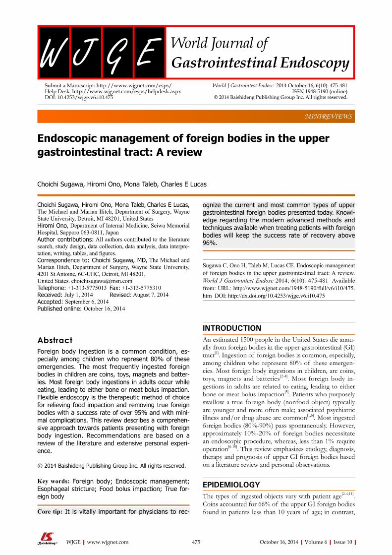
Food bolus impaction A food bolus impaction is usually the result of an un-derlying structural abnormality, such as a web, ring or stricture of the esophagus (Figure 1, Tables 1 and 2)[30]. An esophageal food bolus impaction often contains chewed meat lodged at one of these narrowed sites. Adult patients who develop food impaction have under-lying esophageal pathology in 88% to 97% of patients[31]. Esophageal obstruction by a food bolus is the most common type of foreign-body ingestion complication in adults[32]. The obstruction is often complete and may be associated with increased salivation, the inability to swal-low liquids, substernal pain, and aspiration[30]. Thus, suc-cessful endoscopic treatment of food impaction as well as the underlying pathologic lesion is essential.
Using a snare or snare basket, a food bolus can be re-trieved in one piece or by piecemeal extraction (Figure 1) or reduced in volume allowing it to pass spontaneously. The food may be successfully pushed into the stomach after it is cut into small pieces by a snare[31]. This tech-nique involves bypassing the esophageal narrowing with the endoscope, while assessing the cause of the obstruc-tion. After the endoscope is passed into the stomach, the food may be gently pushed distally. Forceful blind push-ing with the endoscope is dangerous. Similarly, advancing retrieval devices or dilators blindly beyond the impaction invites complications. If food is extracted through the mouth either in one piece or piecemeal (Figure 1), use of
477 October 16, 2014|Volume 6|Issue 10|WJGE|www.wjgnet.com
DC
BA
Figure 1 The photographs (A) show a piece of meat lodged at a narrowed gastroesophageal ring; the meat was removed with the snare (B and C). The photo (D) shows the ring after extraction with esophagitis; the narrowing was successfully dilated.
Sugawa C et al . Endoscopic management of foreign bodies
the battery fails to pass in 72 h[34,35].
Sharp-pointed object Common sharp pointed foreign bodies include bones, toothpicks, needles, safety pins, nails, dental appliances and medication blister packs (Tables 1 and 2, Figure 3). They should be removed, if possible, before they pass through the stomach, as 15%-35% of these objects will perforate the intestine, usually, near the ileocecal valve[6,15]. Budnick et al[36] reported 8176 toothpick-related injuries in the United States from 1979 to 1982; this is a rate of 3.6 per 100000 person-years. Patients often do not remember swallowing a toothpick and imaging studies demonstrate the presence of a toothpick in only 14% of patients[36-40]. Sharp foreign body ingestion, such as bones and tooth-picks, can be dangerous by causing airway compromise, bowel perforation or penetration[41,42], aortic or tracheal fistulae[43,44], or cardiac tamponade (Tables 1 and 2)[45,46]. Ingested sharp-pointed objects have the highest rates of perforation, which may be 35%[2,46]. Sharp objects within the esophagus should be urgently removed endoscopi-cally. Surgical intervention is indicated if the patient de-velops symptoms of perforation or if the ingested sharp object fails to progress within 72 h after ingestion[6]. Medication blister packs can cause bleeding or perfora-tion of the esophagus[47]. They can be removed by a snare net. For removal of sharp and pointed objects, use of an overtube or a retractable latex-rubber condom-type hood is recommended. One should always remember that ad-vancing points puncture, whereas, trailing ones do not.
an overtube to protect the airway against aspiration may be employed[27,31]. A stricture can be treated with a bal-loon dilator after successful extraction or passage of an impacted food bolus distally.
Blunt object The most common blunt foreign bodies are coins in-gested by children (Tables 1 and 2). Approximately 30% of coins will pass from the esophagus into the stomach within 24 h[33]. If the object has passed into the stomach and is less than 2 cm in size, it will usually pass through the entire gastrointestinal tract without difficulty. These can be retrieved using a retrieval snare net if objects fail to pass beyond the stomach by 3 to 4 wk[7].
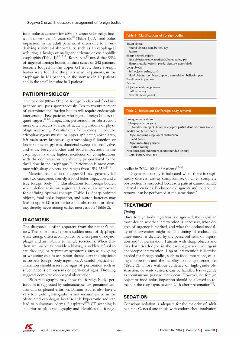
Button or small disk batteries Button or small disk batteries are found in watches, hear-ing aids, calculators and other electronic devices. If both poles of the battery come into contact with the mucosa, electrical conduction may result in corrosive injury, ne-crosis and perforation (Tables 1 and 2, Figure 2). Further-more, these agents contain either metallic salts (mercuric oxide, silver oxide, zinc oxide, or lithium oxide) or alka-line fluids (sodium or potassium hydroxide), which may leak into the gastrointestinal lumen and cause necrosis. After radiographic documentation, batteries lodged in the esophagus or stomach should be emergently removed. Use of a retrieval snare net or a stone retrieval basket is most often successful (Figure 2)[26]. Surgical management is recommended if severe abdominal pain develops or if
478 October 16, 2014|Volume 6|Issue 10|WJGE|www.wjgnet.com
DC
BA
Figure 2 A 54-year-old woman with history of psychiatric illness swallowed four AA batteries and two button batteries (A). All these batteries in the stomach were removed using a snare net (B) one by one (C and D). Note the multiple erosions and shallow ulcers caused by button batteries (A, B and C).
Sugawa C et al . Endoscopic management of foreign bodies
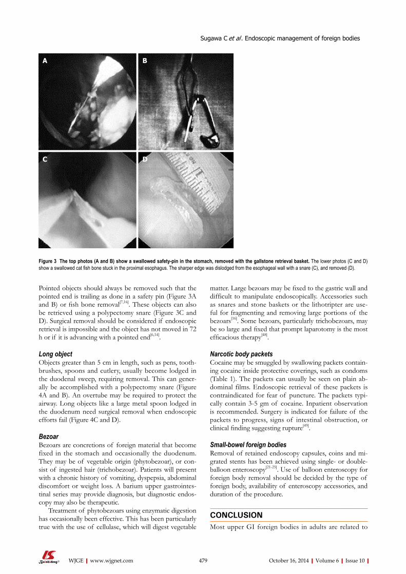
Pointed objects should always be removed such that the pointed end is trailing as done in a safety pin (Figure 3A and B) or fish bone removal[7,16]. These objects can also be retrieved using a polypectomy snare (Figure 3C and D). Surgical removal should be considered if endoscopic retrieval is impossible and the object has not moved in 72 h or if it is advancing with a pointed end[6,14].
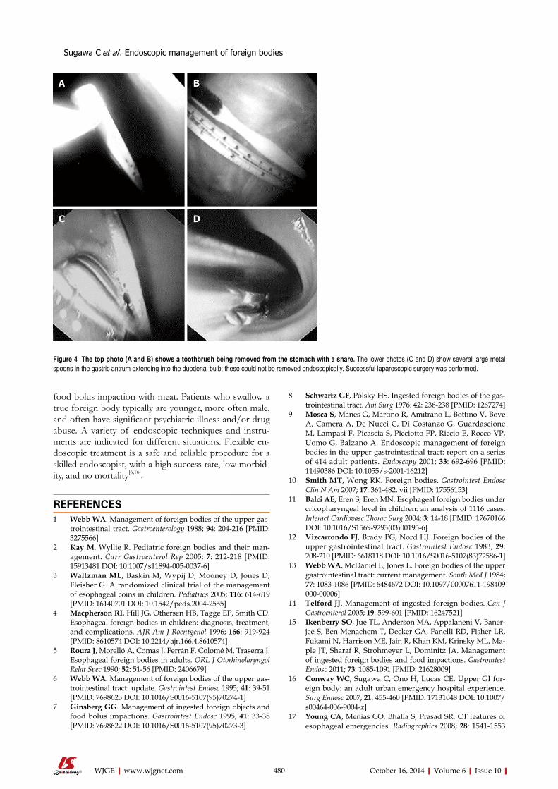
Long objectObjects greater than 5 cm in length, such as pens, tooth-brushes, spoons and cutlery, usually become lodged in the duodenal sweep, requiring removal. This can gener-ally be accomplished with a polypectomy snare (Figure 4A and B). An overtube may be required to protect the airway. Long objects like a large metal spoon lodged in the duodenum need surgical removal when endoscopic efforts fail (Figure 4C and D).
Bezoar Bezoars are concretions of foreign material that become fixed in the stomach and occasionally the duodenum. They may be of vegetable origin (phytobezoar), or con-sist of ingested hair (trichobezoar). Patients will present with a chronic history of vomiting, dyspepsia, abdominal discomfort or weight loss. A barium upper gastrointes-tinal series may provide diagnosis, but diagnostic endos-copy may also be therapeutic.
Treatment of phytobezoars using enzymatic digestion has occasionally been effective. This has been particularly true with the use of cellulase, which will digest vegetable
matter. Large bezoars may be fixed to the gastric wall and difficult to manipulate endoscopically. Accessories such as snares and stone baskets or the lithotripter are use-ful for fragmenting and removing large portions of the bezoars[16]. Some bezoars, particularly trichobezoars, may be so large and fixed that prompt laparotomy is the most efficacious therapy[48].
Narcotic body packets Cocaine may be smuggled by swallowing packets contain-ing cocaine inside protective coverings, such as condoms (Table 1). The packets can usually be seen on plain ab-dominal films. Endoscopic retrieval of these packets is contraindicated for fear of puncture. The packets typi-cally contain 3-5 gm of cocaine. Inpatient observation is recommended. Surgery is indicated for failure of the packets to progress, signs of intestinal obstruction, or clinical finding suggesting rupture[49].
Small-bowel foreign bodiesRemoval of retained endoscopy capsules, coins and mi-grated stents has been achieved using single- or double-balloon enteroscopy[21-25]. Use of balloon enteroscopy for foreign body removal should be decided by the type of foreign body, availability of enteroscopy accessories, and duration of the procedure.
CONCLUSIONMost upper GI foreign bodies in adults are related to
479 October 16, 2014|Volume 6|Issue 10|WJGE|www.wjgnet.com
DC
BA
Figure 3 The top photos (A and B) show a swallowed safety-pin in the stomach, removed with the gallstone retrieval basket. The lower photos (C and D) show a swallowed cat fish bone stuck in the proximal esophagus. The sharper edge was dislodged from the esophageal wall with a snare (C), and removed (D).
Sugawa C et al . Endoscopic management of foreign bodies
food bolus impaction with meat. Patients who swallow a true foreign body typically are younger, more often male, and often have significant psychiatric illness and/or drug abuse. A variety of endoscopic techniques and instru-ments are indicated for different situations. Flexible en-doscopic treatment is a safe and reliable procedure for a skilled endoscopist, with a high success rate, low morbid-ity, and no mortality[6,16].
REFERENCES1 Webb WA. Management of foreign bodies of the upper gas-
trointestinal tract. Gastroenterology 1988; 94: 204-216 [PMID: 3275566]
2 Kay M, Wyllie R. Pediatric foreign bodies and their man-agement. Curr Gastroenterol Rep 2005; 7: 212-218 [PMID: 15913481 DOI: 10.1007/s11894-005-0037-6]
3 Waltzman ML, Baskin M, Wypij D, Mooney D, Jones D, Fleisher G. A randomized clinical trial of the management of esophageal coins in children. Pediatrics 2005; 116: 614-619 [PMID: 16140701 DOI: 10.1542/peds.2004-2555]
4 Macpherson RI, Hill JG, Othersen HB, Tagge EP, Smith CD. Esophageal foreign bodies in children: diagnosis, treatment, and complications. AJR Am J Roentgenol 1996; 166: 919-924 [PMID: 8610574 DOI: 10.2214/ajr.166.4.8610574]
5 Roura J, Morelló A, Comas J, Ferrán F, Colomé M, Traserra J. Esophageal foreign bodies in adults. ORL J Otorhinolaryngol Relat Spec 1990; 52: 51-56 [PMID: 2406679]
6 Webb WA. Management of foreign bodies of the upper gas-trointestinal tract: update. Gastrointest Endosc 1995; 41: 39-51 [PMID: 7698623 DOI: 10.1016/S0016-5107(95)70274-1]
7 Ginsberg GG. Management of ingested foreign objects and food bolus impactions. Gastrointest Endosc 1995; 41: 33-38 [PMID: 7698622 DOI: 10.1016/S0016-5107(95)70273-3]
8 Schwartz GF, Polsky HS. Ingested foreign bodies of the gas-trointestinal tract. Am Surg 1976; 42: 236-238 [PMID: 1267274]
9 Mosca S, Manes G, Martino R, Amitrano L, Bottino V, Bove A, Camera A, De Nucci C, Di Costanzo G, Guardascione M, Lampasi F, Picascia S, Picciotto FP, Riccio E, Rocco VP, Uomo G, Balzano A. Endoscopic management of foreign bodies in the upper gastrointestinal tract: report on a series of 414 adult patients. Endoscopy 2001; 33: 692-696 [PMID: 11490386 DOI: 10.1055/s-2001-16212]
10 Smith MT, Wong RK. Foreign bodies. Gastrointest Endosc Clin N Am 2007; 17: 361-482, vii [PMID: 17556153]
11 Balci AE, Eren S, Eren MN. Esophageal foreign bodies under cricopharyngeal level in children: an analysis of 1116 cases. Interact Cardiovasc Thorac Surg 2004; 3: 14-18 [PMID: 17670166 DOI: 10.1016/S1569-9293(03)00195-6]
12 Vizcarrondo FJ, Brady PG, Nord HJ. Foreign bodies of the upper gastrointestinal tract. Gastrointest Endosc 1983; 29: 208-210 [PMID: 6618118 DOI: 10.1016/S0016-5107(83)72586-1]
13 Webb WA, McDaniel L, Jones L. Foreign bodies of the upper gastrointestinal tract: current management. South Med J 1984; 77: 1083-1086 [PMID: 6484672 DOI: 10.1097/00007611-198409000-00006]
14 Telford JJ. Management of ingested foreign bodies. Can J Gastroenterol 2005; 19: 599-601 [PMID: 16247521]
15 Ikenberry SO, Jue TL, Anderson MA, Appalaneni V, Baner-jee S, Ben-Menachem T, Decker GA, Fanelli RD, Fisher LR, Fukami N, Harrison ME, Jain R, Khan KM, Krinsky ML, Ma-ple JT, Sharaf R, Strohmeyer L, Dominitz JA. Management of ingested foreign bodies and food impactions. Gastrointest Endosc 2011; 73: 1085-1091 [PMID: 21628009]
16 Conway WC, Sugawa C, Ono H, Lucas CE. Upper GI for-eign body: an adult urban emergency hospital experience. Surg Endosc 2007; 21: 455-460 [PMID: 17131048 DOI: 10.1007/s00464-006-9004-z]
17 Young CA, Menias CO, Bhalla S, Prasad SR. CT features of esophageal emergencies. Radiographics 2008; 28: 1541-1553
480 October 16, 2014|Volume 6|Issue 10|WJGE|www.wjgnet.com
DC
BA
Figure 4 The top photo (A and B) shows a toothbrush being removed from the stomach with a snare. The lower photos (C and D) show several large metal spoons in the gastric antrum extending into the duodenal bulb; these could not be removed endoscopically. Successful laparoscopic surgery was performed.
Sugawa C et al . Endoscopic management of foreign bodies
[PMID: 18936020 DOI: 10.1148/rg.286085520]18 Marco De Lucas E, Sádaba P, Lastra García-Barón P, Ruiz-
Delgado ML, González Sánchez F, Ortiz A, Pagola MA. Value of helical computed tomography in the management of upper esophageal foreign bodies. Acta Radiol 2004; 45: 369-374 [PMID: 15323387 DOI: 10.1080/02841850410005516]
19 Goh BK, Tan YM, Lin SE, Chow PK, Cheah FK, Ooi LL, Wong WK. CT in the preoperative diagnosis of fish bone perforation of the gastrointestinal tract. AJR Am J Roent-genol 2006; 187: 710-714 [PMID: 16928935 DOI: 10.2214/AJR.05.0178]
20 Loh KS, Tan LK, Smith JD, Yeoh KH, Dong F. Complica-tions of foreign bodies in the esophagus. Otolaryngol Head Neck Surg 2000; 123: 613-616 [PMID: 11077351 DOI: 10.1067/mhn.2000.110616]
21 May A, Nachbar L, Ell C. Extraction of entrapped capsules from the small bowel by means of push-and-pull enteros-copy with the double-balloon technique. Endoscopy 2005; 37: 591-593 [PMID: 15933937 DOI: 10.1055/s-2005-861320]
22 Neumann H, Fry LC, Rickes S, Jurczok C, Malfertheiner P, Mönkemüller K. A ‘double-balloon enteroscopy worth the money’: endoscopic removal of a coin lodged in the small bowel. Dig Dis 2008; 26: 388-389 [PMID: 19188734 DOI: 10.1159/000177029]
23 Shibuya T, Osada T, Asaoka D, Mori H, Beppu K, Sakamoto N, Suzuki S, Sai JK, Nagahara A, Otaka M, Ohkusa T, Ogi-hara T, Takada Y, Watanabe S. Double-balloon endoscopy for treatment of long-term abdominal discomfort due to small bowel penetration by an eel bone. Med Sci Monit 2008; 14: CS107-CS109 [PMID: 18830197]
24 Kato S, Kani K, Takabayashi H, Yamamoto R, Yakabi K. Double balloon enteroscopy to retrieve an accidentally swallowed dental reamer deep in the jejunum. World J Gas-trointest Endosc 2011; 3: 78-80 [PMID: 21603036 DOI: 10.4253/wjge.v3.i4.78]
25 Chu YC, Yeh YH, Yang CC, Chen CH, Yueh SK, Mo LR. A new indication for double-balloon enteroscopy: removal of migrated metal stents through a Roux-en-Y anastomosis. Endoscopy 2007; 39 Suppl 1: E148 [PMID: 17611895 DOI: 10.1055/s-2006-944921]
26 Faigel DO, Stotland BR, Kochman ML, Hoops T, Judge T, Kroser J, Lewis J, Long WB, Metz DC, O’Brien C, Smith DB, Ginsberg GG. Device choice and experience level in endo-scopic foreign object retrieval: an in vivo study. Gastrointest Endosc 1997; 45: 490-492 [PMID: 9199906 DOI: 10.1016/S0016-5107(97)70179-2]
27 Spurling TJ, Zaloga GP, Richter JE. Fiberendoscopic re-moval of a gastric foreign body with overtube technique. Gastrointest Endosc 1983; 29: 226-227 [PMID: 6618122 DOI: 10.1016/S0016-5107(83)72591-5]
28 Tierney WM, Adler DG, Conway JD, Diehl DL, Farraye FA, Kantsevoy SV, Kaul V, Kethu SR, Kwon RS, Mamula P, Pedrosa MC, Rodriguez SA. Overtube use in gastrointesti-nal endoscopy. Gastrointest Endosc 2009; 70: 828-834 [PMID: 19703691]
29 Bertoni G, Sassatelli R, Conigliaro R, Bedogni G. A simple latex protector hood for safe endoscopic removal of sharp-pointed gastroesophageal foreign bodies. Gastrointest Endosc 1996; 44: 458-461 [PMID: 8905368 DOI: 10.1016/S0016-5107(96)70099-8]
30 Ko HH, Enns R. Review of food bolus management. Can J Gastroenterol 2008; 22: 805-808 [PMID: 18925301]
31 Longstreth GF, Longstreth KJ, Yao JF. Esophageal food impaction: epidemiology and therapy. A retrospective, ob-servational study. Gastrointest Endosc 2001; 53: 193-198 [DOI:
10.1067/mge.2001.112709]32 Vicari JJ, Johanson JF, Frakes JT. Outcomes of acute esopha-
geal food impaction: success of the push technique. Gastroin-test Endosc 2001; 53: 178-181 [PMID: 11174288 DOI: 10.1067/mge.2001.111039]
33 Soprano JV, Mandl KD. Four strategies for the management of esophageal coins in children. Pediatrics 2000; 105: e5 [PMID: 10617742 DOI: 10.1542/peds.105.1.e5]
34 David TJ, Ferguson AP. Management of children who have swallowed button batteries. Arch Dis Child 1986; 61: 321-322 [PMID: 3707180 DOI: 10.1136/adc.61.4.321]
35 Litovitz TL. Battery ingestions: product accessibility and clinical course. Pediatrics 1985; 75: 469-476 [PMID: 3883304]
36 Budnick LD. Toothpick-related injuries in the United States, 1979 through 1982. JAMA 1984; 252: 796-797 [PMID: 6748180]
37 Saccà N, Rodino’ S, D’Amico T, Fragomeni A, Sebkova L, Giglio A. An unintentional ingestion of a toothpick: a case report. Dig Liver Dis 2005; 37: 983-984 [PMID: 16202674 DOI: 10.1016/j.dld.2005.08.006]
38 Rioux M, Langis P. Sonographic detection of clinically un-suspected swallowed toothpicks and their gastrointestinal complications. J Clin Ultrasound 1994; 22: 483-490 [PMID: 7814653 DOI: 10.1002/jcu.1870220805]
39 Lacroix S, Ferland A, Gilbert P, Lemieux M, Bilodeau L, Po-irier P. Cardiac hazard associated with eating habits. A case of infected intrapericardial foreign body due to an ingested toothpick. Can J Cardiol 2009; 25: e263-e264 [PMID: 19584985 DOI: 10.1016/S0828-282X(09)70518-5]
40 Liu YY, Tseng JH, Yeh CN, Fang JT, Lee HL, Jan YY. Correct diagnosis and successful treatment for pericardial effusion due to toothpick injury: a case report and literature review. World J Gastroenterol 2007; 13: 4278-4281 [PMID: 17696263]
41 Schwartz JT, Graham DY. Toothpick perforation of the intestines. Ann Surg 1977; 185: 64-66 [PMID: 318821 DOI: 10.1097/00000658-197701000-00010]
42 Matsubara M, Hirasaki S, Suzuki S. Gastric penetration by an ingested toothpick successfully managed with computed tomography and endoscopy. Intern Med 2007; 46: 971-974 [PMID: 17603235 DOI: 10.2169/internalmedicine.46.0037]
43 D’Costa H, Bailey F, McGavigan B, George G, Todd B. Per-foration of the oesophagus and aorta after eating fish: an unusual cause of chest pain. Emerg Med J 2003; 20: 385-386 [PMID: 12835368 DOI: 10.1136/emj.20.4.385]
44 Sica GS, Djapardy V, Westaby S, Maynard ND. Diagnosis and management of aortoesophageal fistula caused by a foreign body. Ann Thorac Surg 2004; 77: 2217-2218 [PMID: 15172312 DOI: 10.1016/j.athoracsur.2003.06.031]
45 Vesna D, Tatjana A, Slobodan S, Slobodan N. Cardiac tam-ponade caused by migration of a swallowed sewing needle. Forensic Sci Int 2004; 139: 237-239 [PMID: 15040923 DOI: 10.1016/j.forsciint.2003.10.013]
46 Sharland MG, McCaughan BC. Perforation of the esopha-gus by a fish bone leading to cardiac tamponade. Ann Thorac Surg 1993; 56: 969-971 [PMID: 8215678 DOI: 10.1016/0003-4975(93)90368-R]
47 Chan FK, Sung JJ, Tam PY, Kwong KH, Lau JW. “Blister pack”-induced gastrointestinal hemorrhage. Am J Gastroen-terol 1997; 92: 172-173 [PMID: 8995968]
48 Andrus CH, Ponsky JL. Bezoars: classification, pathophysi-ology, and treatment. Am J Gastroenterol 1988; 83: 476-478 [PMID: 3284334]
49 June R, Aks SE, Keys N, Wahl M. Medical outcome of co-caine bodystuffers. J Emerg Med 2000; 18: 221-224 [PMID: 10699526 DOI: 10.1016/S0736-4679(99)00198-5]
P- Reviewer: Ciaccio E, Parsi MA S- Editor: Ji FF L- Editor: A E- Editor: Zhang DN
481 October 16, 2014|Volume 6|Issue 10|WJGE|www.wjgnet.com
Sugawa C et al . Endoscopic management of foreign bodies

© 2014 Baishideng Publishing Group Inc. All rights reserved.
Published by Baishideng Publishing Group Inc8226 Regency Drive, Pleasanton, CA 94588, USA
Telephone: +1-925-223-8242Fax: +1-925-223-8243
E-mail: [email protected] Desk: http://www.wjgnet.com/esps/helpdesk.aspx
http://www.wjgnet.com