Embed Size (px)
Citation preview
Chapter 29 & 30
Endosymbiotic Theory
“No great discovery was ever made without a bold guess.”
--Isaac Newton
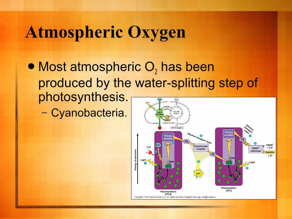
Atmospheric Oxygen
Most atmospheric O2 has been produced by the water-splitting step of photosynthesis.– Cyanobacteria.
Atmospheric Oxygen
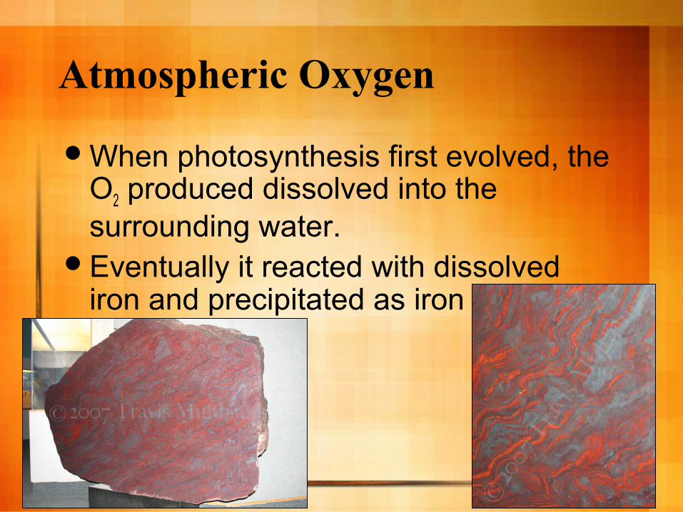
When photosynthesis first evolved, the O2 produced dissolved into the surrounding water.
Eventually it reacted with dissolved iron and precipitated as iron ore.
Atmospheric Oxygen
After the iron had precipitated out, O2 continued to accumulate until the waterways became saturated and the remaining O2 then entered the atmosphere.

Atmospheric Oxygen
Atmospheric oxygen continued to accumulate gradually from about 2.7 bya until about 2.3 bya and then dramatically increased.
The increase was likely due to the evolution of more oxygen producing organisms.
http://www.nature.com/nature/journal/v451/n7176/fig_tab/nature06587_F2.html
Atmospheric Oxygen
The increasing O2 levels on the planet likely led to the extinction of numerous prokaryotic groups.
Oxygen is a highly reactive compound that damages cells and disrupts chemical bonds.
Atmospheric Oxygen
Some species of bacteria survived in habitats that remained anaerobic, and others adapted to the changing atmosphere.

The First Eukaryotes
About 2.1 bya, the first eukaryotic fossils began forming.
Eukaryotic cells have a number of complex features.
Three such evolutionary novelties came to define the early eukaryotes.

A Change in Cell Structure and Function Three evolutionary novelties:
– 1. The formation of ribosome studded internal membranes.
– 2. The appearance of a cytoskeleton.– 3. The evolution of digestive vesicles.
1. A Ribosome Studded Membrane
The ribosome-studded membrane assisted in the movement of protein products throughout the internal portion of the cell without harm to other cytoplasmic factors.

2. The Appearance of a Cytoskeleton
The cytoskeleton is comprised of actin fibers and microtubules.– Allows form movement of the cell and
movement of the internal contents.The development allows for
phagocytosis.
3. Digestive Vesicles
The formation of digestive vesicles allowed for membrane bound enzymes to form.– If unbound, these enzymes would destroy
the cell.

Endosymbiotic Theory
Where did the features of eukaryotic cells come from?
Endosymbiotic Theory
A wide variety of evidence supports the theory that small prokaryotes began living in larger (host) cells.
These cells likely gained entry to the host as undigested prey, or internal parasites.– This process has been observed by
scientists in as little as 5 years.

Endosymbiotic Theory
The benefits of the relationship are easy to see.– A photosynthetic endosymbiont would
provide nutrients to the heterotrophic host.– The host would provide shelter for the
anaerobic prokaryote from the increasingly aerobic environment.
Endosymbiotic Theory
Over time, this relationship would result in a situation where to two parts would become inseperable giving rise to a single organism.
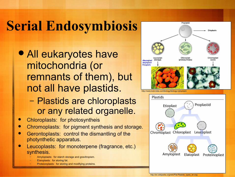
Serial Endosymbiosis
All eukaryotes have mitochondria (or remnants of them), but not all have plastids.– Plastids are chloroplasts
or any related organelle. Chloroplasts: for photosyntheis Chromoplasts: for pigment synthesis and storage. Gerontoplasts: control the dismantling of the
photynthetic apparatus. Leucoplasts: for monoterpene (fragrance, etc.)
synthesis.– Amyloplasts: for starch storage and gravitropism.– Elaioplasts: for storing fat.– Proteionplasts: for storing and modifying proteins.
http://en.wikipedia.org/wiki/File:Plastids_types_en.svg
http://www.tutorvista.com/biology/biology-cytoplasm
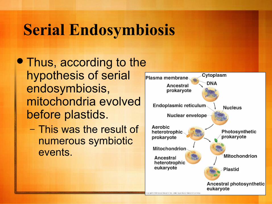
Serial Endosymbiosis
Thus, according to the hypothesis of serial endosymbiosis, mitochondria evolved before plastids.– This was the result of
numerous symbiotic events.

Evidence for Endosymbiosis
The evidence is overwhelming:– Both organelles have circular chromosomes.
These chromosomes lack histones.– Both organelles have their own DNA.
Both organelles can perform transcription and translation of their own DNA.
– Both organelles can self-replicate—via binary fission—just like prokaryotes.
Evidence for Endosymbiosis
The evidence is overwhelming:– The inner membranes of both organelles
have enzymes and transport systems that are homologous to those found in the plasma membranes of living prokaryotes.
– Both organelles are approximately the same size as typical bacterium.
– Both organelles use many bacteria-like enzymes.

Evidence for Endosymbiosis
The evidence is overwhelming:– Both organelles are sensitive to certain
antibiotics.– Some antibiotics interfere with mitochondrial
protein synthesis.Rifampicin-binds to bacterial RNA polymerase
preventing transcription.Can prevent mitochondrial RNA synthesis, but
only at a very high concentration.
Evidence for Endosymbiosis
The evidence is overwhelming:– Both organelles contain ribosomes.
These ribosomes are very similar to bacterial ribosomes.
The ribosomes are nearly the same size, have very similar RNA sequences, and are sensitive to the same antibiotics as bacterial ribosomes.
The ribosomes are more similar to bacterial ribosomes than they are to eukaryotic ribosomes.

Secondary Endosymbiosis
Secondary endosymbiosis is another step in eukaryotic evolution.
In this process, a heterotrophic eukaryote engulfed an unrelated photosynthetic eukaryote (plastid).– The plastids were likely ingested into the
food vacuole, and over time formed a symbiotic relationship with the host.
Secondary Endosymbiosis
Studies of plastid bearing eukaryotes demonstrate how this process has taken place.– Red and green algae, produced from
primary endosymbiosis, provide a nice example of this process.Chlorarachinophytes are a specific example.
– Green algae engulfed by a heterotrophic eukaryote.

Secondary Endosymbiosis
Within the engulfed cell, we see lines of evidence for this process having taken place.– Within the cell is remnants of an engulfed
cell with a vestigal nucleus—called a nucleomorph.Nucleomorphic genes are still transcribed. Their DNA sequences are very similar to
those of green algae—further supporting the hypothesis that an ancestral eukaryote engulfed a green algae.

Secondary Endosymbiosis
The plastids are surrounded by four membranes.– The inner two membranes originated as
an inner and an outer membrane of an ancient cyanobacterium.
– The third membrane is derived from the engulfed alga’s plasma membrane.
– The outermost membrane is derived from the heterotrophic eukaryote’s food vacuole.


Click here for a video summary.
Secondary Endosymbiosis Summary
Could it Really Occur?
It is now…Some eukaryotes live in low O2
environments and lack mitochondria.– They have endosymbionts that live within
them and generate energy for them.
Could it Really Occur?
Protists live symbiotically in the hindgut of termites. – The protists, in turn, are colonized by
symbiotic bacteria similar in size and distribution to mitochondria.
– These bacteria function well in low O2 environments--unlike mitochondria.They oxidize food and create ATP for the
protist.
Could it Really Occur?
A study of Pelomyxa palustris provides some interesting insight:– This ameoba lacks mitochondria.– It contains at least 2 kinds of
endosymbiotic bacteria.– Killing the bacteria with antibiotics causes
an increase in lactic acid.– This suggests that the bacteria oxidize the
end products of glucose fermentation—something mitochondria normally do.