Embed Size (px)
Citation preview

ENDOTHELIAL COLONY FORMING CELLS (ECFCS): IDENTIFICATION,
SPECIFICATION AND MODULATION IN CARDIOVASCULAR DISEASES
Lan Huang
Submitted to the faculty of the University Graduate School in partial fulfillment of the requirements
for the degree Doctor of Philosophy
in the Department of Biochemistry and Molecular Biology, Indiana University
December 2009

ii
Accepted by the Faculty of Indiana University, in partial
fulfillment of the requirements for the degree of Doctor of Philosophy.
Mervin C. Yoder, Jr., MD, Chair
David A. Ingram, Jr., MD
Doctoral Committee
Lawrence A. Quilliam, PhD
November 3rd, 2009
Mark D. Pescovitz, MD

iii
DEDICATION
I owe a tremendous debt of gratitude to my parents, Zhao Huang and Jianying
Chen, for their love, encouragement and support for my ongoing academic
pursuits.
I would also like to express my deepest gratitude to countless teachers and
mentors who have impressed upon me the beauty of science and helped to
explore my potential in the field of scientific research.

iv
ACKNOWLEDGEMENTS
Keith L. March, MD, PhD and Dongming Hou, MD, PhD
Indiana Center for Vascular Biology & Medicine
Indiana University School of Medicine
Indianapolis, IN
Co-authors of the paper that provided a pig model with acute myocardial
infarction
Momoko Yoshimoto, PhD, Michael J. Ferkowicz, PhD, Scott A. Johnson, MS and
Paul J. Critser
Department of Pediatrics
Wells Center for Pediatric Research
Indiana University School of Medicine
Indianapolis, IN
Provided intellectual feedback and technical assistance
Mervin C. Yoder, Jr., MD
Department of Pediatrics
Wells Center for Pediatric Research
Indiana University School of Medicine
Indianapolis, IN
Mentored and guided me to be a scientific investigator
Funding for this work was provided by:
The American Heart Association & Indiana University School of Medicine

v
ABSTRACT
Lan Huang
ENDOTHELIAL COLONY FORMING CELLS (ECFCS): IDENTIFICATION,
SPECIFICATION AND MODULATION IN CARDIOVASCULAR DISEASES
A hierarchy of endothelial colony forming cells (ECFCs) with different levels of
proliferative potential has been identified in human circulating blood and blood
vessels. High proliferative potential ECFCs (HPP-ECFCs) display properties
(robust proliferative potential in vitro and vessel-forming ability in vivo) consistent
with stem/progenitor cells for the endothelial lineage. Corneal endothelial cells
(CECs) are different from circulating and resident vascular endothelial cells
(ECs). Whereas systemic vascular endothelium slowly proliferates throughout life,
CECs fail to proliferate in situ and merely expand in size to accommodate areas
of CEC loss due to injury or senescence. However, we have identified an entire
hierarchy of ECFC resident in bovine CECs. Thus, this study provides a new
conceptual framework for defining corneal endothelial progenitor cell potential.
The identification of persistent corneal HPP-ECFCs in adult subjects might
contribute to regenerative medicine in corneal transplantation. While human cord
blood derived ECFCs are able to form vessels in vivo, it is unknown whether they
are committed to an arterial or venous fate. We have demonstrated that human
cord blood derived ECFCs heterogeneously express gene transcripts normally
restricted to arterial or venous endothelium. They can be induced to display an

vi
arterial gene expression pattern after vascular endothelial growth factor 165
(VEGF165) or Notch ligand Dll1 (Delta1ext-IgG) stimulation in vitro. However, the in
vitro Dll1 primed ECFCs fail to display significant skewing toward arterial EC
phenotype and function in vivo upon implantation, suggesting that in vitro priming
is not sufficient for in vivo specification. Future studies will determine whether
ECFCs are amenable to specification in vivo by altering the properties of the
implantation microenvironment. There is emerging evidence suggesting that the
concentration of circulating ECFCs is closely related to the adverse progression
of cardiovascular disorders. In a pig model of acute myocardial ischemia (AMI),
we have demonstrated that AMI rapidly mobilizes ECFCs into the circulation, with
a significant shift toward HPP-ECFCs. The exact role of the mobilized HPP-
ECFCs in homing and participation in repair of the ischemic tissue remains
unknown. In summary, these studies contribute to an improved understanding of
ECFCs and suggest several possible therapeutic applications of ECFCs.
Mervin C. Yoder, Jr., MD, Chair

vii
TABLE OF CONTENTS
List of Tables .......................................................................................................... x
List of Figures ......................................................................................................... xi
List of Abbreviations ............................................................................................... xiv
Chapter I
Introduction
A. The Formation of Functional Blood Vessels ............................................ 1
Vasculogenesis and Regulation ........................................................ 3
Angiogenesis and Regulation ........................................................... 6
Intussusceptive Angiogenesis (IA) and Regulation ........................... 19
Arteriogenesis and Regulation .......................................................... 20
B. Arteriovenous (AV) Differentiation ........................................................... 23
Regulation of Arteriovenous (AV) Specification ................................ 27
Plasticity of Arteriovenous (AV) Differentiation .................................. 35
C. Endothelial Colony Forming Cells (ECFCs) ............................................ 37
Chapter II
A Hierarchy of Endothelial Colony Forming Cell (ECFCs) Activity is Displayed by
Bovine Corneal Endothelial Cells (BCECs) ............................................................ 44
Introduction .................................................................................................. 44
Materials and Methods ................................................................................. 47
Results ......................................................................................................... 54
Discussion ................................................................................................... 69

viii
Chapter III
Human Cord Blood Plasma Can Replace Fetal Bovine Serum (FBS) for in vitro
Expansion of Functional Human Endothelial Colony Forming Cells (ECFCs) ........ 76
Introduction .................................................................................................. 76
Materials and Methods ................................................................................. 80
Results ......................................................................................................... 86
Discussion ................................................................................................... 97
Chapter IV
Dose-dependent Effects of Vascular Endothelial Growth Factor165 (VEGF165) and
Notch Ligand Delta Like 1 (Dll1) on in vitro Differentiation of Human Cord Blood
Derived Endothelial Colony Forming Cells (ECFCs) .............................................. 103
Introduction .................................................................................................. 103
Materials and Methods ................................................................................. 105
Results ......................................................................................................... 114
Discussion ................................................................................................... 130
Chapter V
Acute Myocardial Infarction in Swine Rapidly and Selectively Releases Highly
Proliferative Endothelial Colony Forming Cells (ECFCs) into Circulation (Cell
Transplantation paper) ........................................................................................... 135
Abstract ........................................................................................................ 136
Introduction .................................................................................................. 137
Materials and Methods ................................................................................. 140
Results ......................................................................................................... 148

ix
Discussion ................................................................................................... 161
Chapter VI
Summary and Perspectives .................................................................................... 166
References ............................................................................................................. 171
Curriculum Vitae

x
LIST OF TABLES
Table I.1 Molecules expressed preferentially in arterial and venous ECs ............... 24
Table II.1 Primers used for conventional RT-PCR .................................................. 51
Table IV.1 Primers used for conventional RT-PCR ................................................ 108
Table IV.2 Primers used for quantitative RT-PCR .................................................. 110
Table V.1 The number of MNC and ECFC colony in 100cc porcine blood ............. 150
xi
LIST OF FIGURES
Figure I.1 Formation of a functional vascular network from endothelial precursor
cells during murine embryonic development .......................................................... 2
Figure I.2 Model of AV specification ....................................................................... 26
Figure I.3 Common methods of ―EPC‖ culture ....................................................... 39
Figure II.1 The morphology of cultured bovine vessel wall derived ECs and
bovine corneal ECs ................................................................................................ 56
Figure II.2 Phenotypic and functional characterization of bovine vessel wall
derived ECs and bovine corneal ECs ..................................................................... 57
Figure II.3 RT-PCR analysis of gene expression in bovine vessel wall derived
ECs and bovine corneal ECs .................................................................................. 61
Figure II.4 Quantitation of the clonogenic and proliferative potential of single
endothelial cells derived from bovine vascular endothelium and bovine corneal
endothelium ............................................................................................................ 64
Figure II.5 Telomerase activity of HPP-ECFCs derived from bovine vessel wall
derived ECs and BCECs ........................................................................................ 66
Figure II.6 The formation of sphere colony by bovine corneal endothelial cells ...... 68
Figure III.1 The expression of arterial and venous specific genes in human
umbilical artery an d vein endothelial cells (HUAEC and HUVEC). ........................ 79
Figure III.2 Isolation of human cord blood ECFC-derived EC colonies from UCB
MNCs by using SRM .............................................................................................. 87
Figure III.3 Phenotypic analysis of human cord blood ECFC-derived ECs cultured
in SRM .................................................................................................................... 89

xii
Figure III.4 Quantitation of the clonogenic and proliferative potential of single ECs
derived from human cord blood cultured in SRM .................................................... 92
Figure III.5 Genomic stability in human cord blood ECFCs cultured in SRM .......... 93
Figure III.6 Human cord-blood-derived ECFC cultured in SRM demonstrate the
potential to form functional microvessels in immunodeficient mice ......................... 96
Figure III.7 The expression of arterial and venous specific genes in HUAECs and
HUVECs is measured by quantitative PCR ............................................................ 102
Figure IV.1 The expression of arterial and venous endothelial cell genes in
freshly isolated HUAEC, HUVEC and human cord blood derived ECFCs. ............. 116
Figure IV.2 The alteration of arterial and venous endothelial cell gene expression
in human cord blood derived ECFCs’ response to 14 days of rhVEGF165
stimulation .............................................................................................................. 120
Figure IV.3 The alteration of arterial and venous endothelial cell gene expression
in human cord blood derived ECFCs response to 3 days of Delta1ext-IgG
induction ................................................................................................................. 124
Figure IV.4 in vitro Dll1 primed ECFCs fail to display significant skewing toward
arterial EC phenotype and function in vivo upon implantation ................................ 128
Figure V.1 Overview of experimental design .......................................................... 142
Figure V.2 The formation of porcine aortic and PB ECFC-derived endothelial
colony. .................................................................................................................... 151
Figure V.3 Phenotypic and functional analysis of both aortic and PB ECFC-
derived ECs ............................................................................................................ 152

xiii
Figure V.4 Quantitation of the clonogenic and proliferative potential of single
endothelial cells derived from aortic and PB ECFCs .............................................. 155
Figure V.5 Quantitation of the clonogenic and proliferative potential of single
endothelial cells derived from PB ECFCs in the swine undergoing experimental
AMI ......................................................................................................................... 159

xiv
LIST OF ABBREVIATIONS
AcLDL ................................................................. acetylated low-density lipoprotein
AMI ............................................................................... acute myocardial infarction
Ang ...................................................................................................... Angiopoietin
APC ............................................................................................... allophycocyanin
AV ...................................................................................................... arteriovenous
BAEC ......................................................................... bovine aortic endothelial cell
BCAEC ....................................................... bovine coronary artery endothelial cell
BCEC ..................................................................... bovine corneal endothelial cell
BMP .......................................................................... bone morphogenetic proteins
BPAEC .................................................... bovine pulmonary artery endothelial cell
BSA ..................................................................................... bovine serum albumin
CAC ................................................................................ circulating angiogenic cell
CEC ................................................................................ circulating endothelial cell
CEC .................................................................................... corneal endothelial cell
CFU-Hill .............................................................................. colony forming unit-Hill
CNS ................................................................................... central nervous system
COUP-TFII................ chicken ovalbumin upstream promoter transcription factor II
DAB ...................................................................................... 3,3-diaminobenzidine
DAG ................................................................................................... diacylglycerol
DAPI .............................................. 4’, 6-diamidino-2-phenylindole dihydrochloride
Dll1 ........................................................................................................ Delta-like1
Dll4 ........................................................................................................ Delta-like4

xv
DPBS ......................................................... Dulbecco’s Phosphate Buffered Saline
ECFC ...................................................................... endothelial colony forming cell
ECM ......................................................................................... extracellular matrix
ECs ................................................................................................ endothelial cells
EDTA .................................................................. ethylene diamine tetraacetic acid
EGF ................................................................................... epidermal growth factor
EPC ................................................................................ endothelial progenitor cell
EPO ................................................................................................... erythropoietin
ER ....................................................................................... endoplasmic reticulum
FACS ................................................................. fluorescence activated cell sorting
FBS ........................................................................................... fetal bovine serum
FCS ................................................................................................ fetal calf serum
FE ...................................................................................................... phycoerythrin
FGF .................................................................................... fibroblast growth factor
FISH .................................................................... fluorescence in situ hybridization
FITC .............................................................................. fluorescein isothiocyanate
Flk-1 ........................................................................................... fetal liver kinase 1
Flt-1 .............................................................................. FMS-like tyrosine kinase 1
Flt-4 .............................................................................. FMS-like tyrosine kinase 4
Fn ........................................................................................................... fibronectin
Foxc ................................................................................................ Forkhead box c
FSS ............................................................................................. fluid shear stress
Grl ............................................................................................................... gridlock

xvi
GSL I .......................................................................... Griffonia simplicifolia I lectin
H&E ..................................................................................... hematoxylin and eosin
HCP .......................................................................... human umbilical cord plasma
HIF ..................................................................................... hypoxia-inducible factor
HPP-ECFC ................... high proliferative potential-endothelial colony forming cell
HRP ................................................................................... horseradish peroxidase
HUAEC ....................................................... human umbilical artery endothelial cell
HUVEC ......................................................... human umbilical vein endothelial cell
IA .............................................................................. intussusceptive angiogenesis
IL6 ....................................................................................................... interleukin 6
IP3 ........................................................................................ inositol-trisphosphate
ISV ...................................................................................... intersegmental vessels
KDR ......................................................................... kinase insert domain receptor
LEL ........................................................ Lycopersicon Esculentum (Tomato) lectin
LPP-ECFC ...................... low proliferative potential-endohtelial colony forming cell
MCP-1 ........................................................... monocyte chemoattractant protein-1
Mib .......................................................................................................... mindbomb
MMPs ............................................................................. matrix metalloproteinases
MNC ............................................................................................ mononuclear cell
MSC ..................................................................... mesenchymal stromal stem cell
NGF .......................................................................................... nerve growth factor
NIPs .......................................................................... neuropilin interacting proteins
NK ....................................................................................................... natural killer

xvii
NO ......................................................................................................... nitric oxide
NOS ........................................................................................ nitric oxide synthase
Nrp1 ....................................................................................................... neuropilin1
Nrp2 ....................................................................................................... neuropilin2
PDGFB .................................................................. platelet-derived growth factor B
PDGFR β .............................................. platelet-derived growth factor B receptor β
PDS ..................................................................................... plasma derived serum
PECAM1 ........................................... platelet/endothelial cell adhesion molecule-1
PHDs .............................................. prolyl hydroxylase domain-containing proteins
PKC .............................................................................................. protein kinase C
PLCγ ........................................................................................... phospholipase Cγ
PlGF ................................................................................... placental growth factor
PtdIns(4,5)P2 ............................................. phosphatidylinositol-4, 5-bisphosphate
Rbpj ....... recombination signal binding protein for immunoglobulin kappa J region
S1P ................................................................................. sphingosine-1-phosphate
SCF ................................................................................................ stem cell factor
SDF1 α ........................................................................ stromal cell derived 1 alpha
SDS-PAGE ................. sodium dodecyl sulfate-polyacrylamide gel electrophoresis
SEMA ...................................................................................... class 3 semaphorin
sFlt-1 ................................................................ soluble FMS-like tyrosine kinase 1
Shh ................................................................................................ sonic hedgehog
Sox ........................................................................................ Sry-related HMG box
TGFβ ..................................................................... transforming growth factor beta

xviii
TM ......................................................................................... trabecular meshwork
TNFα .......................................................................... tumor necrosis factor- alpha
TTC ...................................................................................... tetrazolium trichloride
UEA I ........................................................................... Ulex europaeus agglutinin I
VE-Cad ..................................................................... vascular endothelial cadherin
VEGF ................................................................ vascular endothelial growth factor
VEGFR ................................................ vascular endothelial growth factor receptor
vSMC ........................................................................ vascular smooth muscle cells
vWF ....................................................................................... von Willebrand factor
Wnt ....................................................................................................... wingless-int
αSMA ............................................................................ smooth muscle alpha actin

1
CHAPTER I
Introduction
A. The Formation of Functional Blood Vessels
Blood vessels deliver oxygen and nutrients while removing waste from all tissues
in the body. The vascular system is hierarchically organized and is composed of
functional arteries, capillaries and veins. Capillaries are composed solely of
endothelial cells (ECs), and occasionally are ensheathed with pericytes. Arteries
and veins have an inner layer of ECs and an outer layer of vessel wall, which
consists of the tunica intima, media and adventitia. The formation of blood
vessels is an elaborate process including vasculogenesis, angiogenesis,
arteriogenesis, vascular remodeling and maturation, all of which involves a wide
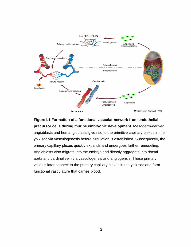
variety of biochemical and biomechanical factors (Figure I.1).
2
Figure I.1 Formation of a functional vascular network from endothelial
precursor cells during murine embryonic development. Mesoderm-derived
angioblasts and hemangioblasts give rise to the primitive capillary plexus in the
yolk sac via vasculogenesis before circulation is established. Subsequently, the
primary capillary plexus quickly expands and undergoes further remodeling.
Angioblasts also migrate into the embryo and directly aggregate into dorsal
aorta and cardinal vein via vasculogensis and angiogensis. These primary
vessels later connect to the primary capillary plexus in the yolk sac and form
functional vasculature that carries blood.

3
Vasculogenesis and Regulation
The development of the vascular system is one of the earliest events in
embryogenesis. Gastrulation begins at embryonic day (E) 6.5, as evidenced by
the formation of the primitive streak and leads to the formation of three principle
germ layers: the ectoderm, mesoderm and endoderm (Wells & Melton 1999).
The emergence of endothelial cells (ECs) is first observed in the proximal lateral
mesoderm of the extraembryonic yolk sac. The subpopulation of mesoderm cells
that expresses vascular endothelial growth factor receptor 2 (VEGFR2, also
known as kinase insert domain receptor, KDR in humans; or fetal liver kinase 1
Flk-1 in mice) gives rise to both angioblasts (endothelial progenitors) and
hemangioblastic cells (progenitors of both hematopoietic and endothelial
lineages) in the yolk sac (Drake & Fleming 2000, Kabrun et al 1997, Kataoka et
al 1997, Nishikawa 1997). During the migration of these mesodermal cells, the
primary capillary plexus (the earliest blood vessels) begin to form by angioblasts
as a honeycomb-like network that separates the distal embryo proper from the
proximal blood island region (Ferkowicz & Yoder 2005). At the same time, the
earliest hematopoietic progenitor cells (primitive erythrocytes) first appear near
the primitive streak and migrate past the developing primitive capillary plexus into
the blood island region of the proximal yolk sac, where they rapidly divide and
form a circumferential blood cell band at E7.75. The primitive erythrocytes in this
region are not surrounded by ECs initially. However, a sheet of ECs exists on the
visceral endodermal side of the blood band. ECs appear to invade the blood
band, subdividing it into primitive erythrocyte-filled channels that eventually form

4
a blood-filled vascular bed, which connects to the primitive capillary plexus right
before the onset of circulation around E8.25 (Ferkowicz et al 2003, Ferkowicz &
Yoder 2005). Extraembryonic EC differentiation initially occurs without
concomitant hematopoiesis. Primitive blood vessels are formed de novo by the
patterned assembly of angioblasts in a process termed vasculogenesis (Risau &
Flamme 1995).
Vasculogenesis in the embryo proper appears slightly later and is initiated
around E7-7.5 by mesoderm fated to give rise to the endocardium and cranial
regions (Drake & Fleming 2000, Tam & Behringer 1997). Subsequently, primary
vascular networks appear serially, lateral to the midline, as the paired dorsal
aortae, then the head and cardinal vessels (Argraves & Drake 2005, Flamme et
al 1997, Lawson et al 2002). Later, neovascularization of heart, lung, liver, spleen
and kidney occurs also primarily by vasculogenesis (Aird 2007a, Aird 2007b,
Coultas et al 2005, deMello et al 1997, Ferguson et al 2005, Robert et al 1998).
In addition, allantoic vasculogenesis occurs by the assembly of mesoderm-
derived precursor cells (Downs et al 1998, Drake & Fleming 2000), giving rise to
the umbilical vessels (Downs & Harmann 1997). Intraembryonic EC
differentiation, similar to that in the yolk sac, initiates in the absence of
hematopoiesis.
After the onset of circulation, the primary vascular networks are rapidly expanded
and remodeled into arteries, capillaries and veins; ultimately, a functional

5
circulatory loop is established. As development proceeds, the primitive
erythrocytes in circulation are replaced by definitive hematopoietic cells
(Brotherton et al 1979, McGrath & Palis 2005, Steiner & Vogel 1973). Thus,
vasculogenesis collectively results in the formation of the primary capillary plexus
in the yolk sac and the major embryonic vessels.
Mouse knockout studies have identified many genes that contribute to
vasculogenesis (Argraves & Drake 2005). While several signaling pathways
participate into this process, including FGF signaling (Poole et al 2001, Smith
1989), Wnt signaling (Wang & Wynshaw-Boris 2004, Zerlin et al 2008), BMP
signaling (Winnier et al 1995) TGFβ signaling (Sirard et al 1998, Yang et al 1998)
and Indian hedgehog signaling (Dyer et al 2001), none of these signaling
cascades is specific for developing the endothelial lineage.
In sharp contrast, Flk-1 plays a vital role in differentiating mesoderm exclusively
to endothelial and hematopoietic lineages in the early gastrulating embryo
(Dumont et al 1995, Roman & Weinstein 2000). Flk-1 deficiency (Flk-1-/-)
severely impairs the development of ECs and hematopoietic cells, and causes
embryonic death at E8.5 (Shalaby et al 1995). Similarly, vascular endothelial
growth factor (VEGF, Flk-1 ligand) is indispensable for vasculogenesis. Targeted
deletion of VEGF leads to embryonic lethality. VEGF-/- embryos display a
phenotype similar to that observed in Flk-1 knockout mice, but considerably less
severe (Carmeliet et al 1996, Ferrara et al 1996). Interestingly, further studies

6
demonstrate that VEGF is haploid-insufficient — loss of a single VEGF allele is
lethal in the mouse embryo, causing abnormal blood vessel development. This
indicates a tight dose-dependent regulation of embryonic vessel development by
VEGF.
Vascular endothelial growth factor receptor 1 (VEGFR1 or FMS-like tyrosine
kinase 1, Flt-1) is another VEGF receptor and is also primarily expressed in ECs.
Flt-1 knockout mice exhibit normal EC differentiation but abnormal vascular
organization in early embryogenesis (Fong et al 1995, Fong et al 1999). Flt-1 has
a higher affinity for VEGF binding but lower kinase activity compared to Flk-1.
Thus, Flt-1 might serve in part to modulate Flk-1 activity by sequestering excess
VEGF stimulation.
Angiogenesis and Regulation
Once the basic pattern of primitive extraembryonic and intraembryonic
vasculature is formed by vasculogenesis, blood vessels are expanded and
remodeled rapidly. This process, referred to angiogenesis, is defined as the
expansion of blood vessels from preexisting vessels. Angiogenesis involves
many morphological events including sprouting EC growth, intussuseptive
growth, stabilization, remodeling, pruning and specialization (Carmeliet 2000,
Flamme et al 1997, Jain 2003, Patan 2004, Risau & Flamme 1995).

7
Sprouting angiognesis happens in many regions such as the yolk sac, central
nervous system (CNS) and retina (Breier & Risau 1996, Gerhardt et al 2003,
Kurz et al 1996, Noguera-Troise et al 2006, Ridgway et al 2006). Angiogenic
sprouting is characterized by leading tip cells and trailing stalk cells (Gerhardt et
al 2003). This process could be prompted by an insufficient supply of oxygen and
guided by VEGF via Notch signaling (comprehensively reviewed in Gerhardt
2008 and Phng 2009) (Gerhardt 2008, Phng & Gerhardt 2009). Hypoxia
upregulates VEGF expression and activates nitric oxide synthase (NOS) which
leads to the bulk production of nitric oxide (NO) (Fraisl et al 2009). Existing
vessels then dilate in response to NO and become leaky in response to VEGF.
With the involvement of active proteases, the basement membrane and
extracellular matrix (ECM) are degraded. These events make ECs acquire
invasive and migratory ability.
ECs stimulated by VEGF compete for the tip cell position via Dll4/Notch1
signaling. The cell that produces more Dll4 than its neighbors eventually will
remain as a tip cell because it can sufficiently suppress the same response in
adjacent ECs via activated Notch1 signaling (Phng & Gerhardt 2009). VEGF also
induces the expression of its receptor Flk-1 and the formation of long, dynamic
filopodia in the tip cells. Thus, tip cells are able to migrate along VEGF gradients.
The VEGF gradient on EC surface is tightly regulated by mRNA levels, isoform
splicing, cell membrane retention and probably protein degradation. A spatial
concentration gradient of membrane-bound VEGF (VEGF188 and VEGF164 in

8
mice, and VEGF189 and VEGF165 in humans) functions as a chemoattractant
signal that promotes the polarized extension of tip cells. In contrast, diffused
VEGF (VEGF120 in mice; VEGF121 in humans) promotes EC proliferation but
does not guide tip cells (Gerhardt 2008).
While sprouting, the vascular lumen can form by ECs in the absence of mural
cells. Lumenization requires the coordinated participation of multiple molecules
including small GTPases and cell-cell, cell-matrix adhesion proteins (Avraamides
et al 2008, Dejana et al 2009, Horowitz & Simons 2008, Iruela-Arispe & Davis
2009, Koh et al 2008). Furthermore, growing vascular sprouts can generate
gradients of platelet-derived growth factor B (PDGFB), which promote the
recruitment of pericytes via PDGFB receptor β (PDGFR β). This ensures that the
growing endothelium is efficiently stabilized by mural cells, which enable ECs to
withstand the physical forces of blood flow (Gerhardt et al 2003).
Each new spout ultimately needs to suppress its motile behavior and connect
with adjacent sprouts or existing capillaries. Flow-dependent tissue oxygenation
downregulates paracrine VEGF production and thus helps establish a quiescent
state for these new vessels. The investment of mural cells and the deposition of
ECM proteins into the subendothelial basement further contribute to vessel
quiescence and maturation (Jain 2003, Jones et al 2006, Lucitti et al 2007). The
basement membrane provides critical support for the endothelium and
fundamentally affects its status, mainly through adhesive interaction with

9
integrins on the surface of ECs. For example, laminin-binding integrins such as
α3β1 and α6β1, as well as collagen IV and perlecan, are important for regulating
tube stabilization and EC quiescence (Davis & Senger 2005, Iruela-Arispe &
Davis 2009, Stratman et al 2009). Moreover, plasmin and Matrix
metalloproteinases (MMPs) (such as MMP3) are reported to be able to release
VEGF165 from the ECM by converting it into a soluble isoform VEGF121-like
component and sequester VEGF165 signaling, restoring EC quiescence
(Mancuso et al 2006). Furthermore, vascular architecture specializes according
to local tissue and organ growth, such as the formation of heart valves and
fenestration. Lastly, arteries and veins form and expand, acquiring additional
layers of mural cells, ECM and elastic laminae coverage, giving the vessels full
ability to sense pressure and respond accordingly.
Cumulative evidence indicates that many elements are capable of influencing
angiogenesis, including oxygen, metabolic intermediates, blood flow, cytokines
and growth factors (Adams & Alitalo 2007, Fraisl et al 2009, Jain 2003, Larrivee
et al 2009). The principal player in angiogenesis is VEGF, which promotes EC
sprouting, proliferation, migration, differentiation and survival, and controls EC-
EC, EC-ECM interactions and more. Many other molecules such as fibroblast
growth factor (FGF), Angiopoietin/Tie2, Notch, TGFβ/Alk5, S1P/Edg1,
Ephrin/Eph and integrins also participate in regulating angiogenic responses.

10
VEGF and VEGF receptors
The VEGF family belongs to the platelet-derived growth factor (PDGF)/VEGF
supergene family. At least 7 members comprise this family: VEGF-A, VEGF-B,
VEGF-C, VEGF-D, VEGF-E, placental growth factor (PlGF) and snake venom-
derived VEGFs. NOTE: In this chapter, ―VEGF‖ stands for VEGF-A. Binding of
VEGF to KDR is the main extracellular signal triggering angiogenic responses,
for example, regulating EC proliferation and NO generation. KDR efficiently
activates phospholipase Cγ (PLCγ), which cleaves phosphatidylinositol-4, 5-
bisphosphate (PtdIns(4,5)P2) to produce diacylglycerol (DAG) and inositol-
trisphosphate (IP3). These soluble products release calcium from the
endoplasmic reticulum (ER) and activate protein kinase C (PKC). The latter
activates MRK-MAPK pathway which affects DNA synthesis in ECs (Takahashi
et al 1999, Xia et al 1996). In addition, VEGF increases eNOS activity through a
pathway mediated by Src and thus produces NO. Calcium release also helps the
production of NO. Thereafter, VEGF via NO affects EC division, migration and
apoptosis (Donnini & Ziche 2002, He et al 1999). Moreover, VEGF via activated
Notch signaling can regulate many steps of angiogenesis, of which endothelial
sprouting and arterial-venous specification have been investigated at length
(Ahmed & Bicknell 2009, Coultas et al 2005, Gerhardt 2008, Holderfield &
Hughes 2008, Roca & Adams 2007, Siekmann et al 2008).
VEGF can interact with both VEGF receptors Flt-1 and Flk-1. Flt-1 has weak
tyrosine kinase activity and high affinity for VEGF, which makes it act as a decoy

11
receptor, modulating angiogenesis through its ability to sequester VEGF and
thereby reduce signaling through Flk-1 (Park et al 1994). Consistently, deletion of
Flt-1 gene in the embryo or embryonic stem-cell-derived vessels induces
overgrowth of endothelial cells and vessel dysmorphogenesis (Fong et al 1995,
Fong et al 1999). By contrast, mice that express a membrane-anchored Flt-1
variant lacking a tyrosine-kinase domain (Flt-1TK-/-) but still capable of binding
VEGF are basically healthy and do not exhibit vascular defects (Hiratsuka et al
1998). It is of interest that the Flt-1 gene also encodes a secreted soluble protein
that has only one ligand-binding region, called soluble Flt-1 (sFlt-1) (Kendall &
Thomas 1993, Shibuya et al 1990). sFlt-1 level was reported to be abnormally
elevated in the serum of preeclampsia patients. Recent studies indicate that
abnormal trapping of VEGF with excess sFlt-1 causes hypertension and
proteinuria, which are the major symptoms of preeclampsia (Koga et al 2003,
Maynard et al 2003). Similarly, overexpression of sFlt-1 induces over-proliferation
of glomerular ECs with the loss of endothelial fenestration (a hallmark of the
glomerular vascular endothelium) that resembles the renal histological lesions of
preeclampsia (Baumwell & Karumanchi 2007). Moreover, sFlt-1 has recently
been indicated to play an important role in angiogenic sprouting (Chappell et al
2009, Kappas et al 2008).Spatially regulated expression of sFlt-1 in conjunction
with VEGF contributes to the formation of spatial VEGF gradient and therefore
guides emerging sprouts away from parent vessels. Thus, Flt-1 regulates
vascular development by sequestering excess VEGF and contributes to maintain
vascular homeostasis.

12
Flt4 is expressed mainly in lymphatic ECs and through response to VEGF-C and
VEGF-D regulating lymphangiogensis. However, Flt4 also plays an important role
in VEGF signaling in angiogenesis (Dumont et al 1993), likely by forming a
heterodimer with KDR (Dixelius et al 2003). Flt4 abundant expression has been
observed in zebrafish intersegmental vessels (ISV), at the tip cells of ISVs in
mouse embryos and in the front of sprouts in mouse retinas (Gerhardt et al 2003,
Siekmann & Lawson 2007). Interestingly, loss of Notch signaling increases Flt4
expression in stalk cells, while suppression of Flt4 expression partly restores the
sprouting, indicating that Flt4 is downregulated by Notch activity in stalk cells.
However, one in vitro study suggested activated Notch signaling can upregulate
Flt4 (Shawber et al 2007). Therefore, the underlying mechanism needs to be
clarified.
Neuropilins (Nrps) including Nrp1 and Nrp2 play an important role in both
neuronal and blood vessel development. They are receptors for two types of
ligands, the class 3 semaphorin (SEMA) family of axon guidance molecules and
the VEGF family of angiogenic factors. Nrp1 is primarily expressed in arteries
while Nrp2 is abundant in veins and lymphatic vessels (Herzog et al 2001, Yuan
et al 2002). The first evidence of Nrp1’s involvement in angiogenesis was that
overexpression of Nrp1 in transgenic mice resulted in embryonic lethality and
vascular defects; for example, excess numbers of blood vessels and
hemorrhage. (Kitsukawa et al 1995). Nrp1-deficient mice exhibit abnormal
vascular development, including impaired neural vascularization and

13
disorganized, insufficient development of vascular networks in the yolk sac and
the mice die in utero (Kawasaki et al 1999). However, Nrp2-deficient mice are
viable; they develop arteries and veins normally but fail to develop small-
diameter lymphatic vessels and capillaries (Yuan et al 2002). In addition,
Nrp1/Nrp2 double-knockout mice display more severe vascular defects than
Nrp1-deficient mice and die at E8.5 (Takashima et al 2002). Recent studies
indicate that Nrp signaling can be independent of VEGF/KDR, by binding Nrp
interacting proteins (NIPs) such as RGS-GAIP Interacting Protein (GIPC) and
Synectin (Cai & Reed 1999, Chittenden et al 2006, Gao et al 2000). Collectively,
the roles of Nrps in angiogenesis need to be clarified in greater depth.
Angiopoietins and Tie receptors
The angiopoietin family is composed of four ligands (Angiopoietin1,
Angiopoietin2 and Angiopoietin3/4) and two corresponding tyrosine kinase
receptors (Tie1 and Tie2). Ang3 and Ang4 are orthologs found in mice and
humans, respectively. Tie receptors are specifically expressed in the endothelium
(Thomas & Augustin 2009, Yancopoulos et al 1998). Tie2 is the receptor for all
four angiopoietin ligands, but the ligand for Tie1 is not clear yet. It recently has
been shown that Tie1 can interact with Tie2 to form a heteromerized signaling
complex (Saharinen et al 2005). Tie1-deficient mice either die in utero (Puri et al
1995) or perinatally (Sato & Rifkin 1989) with edema and hemorrhage, which
indicates Tie1 is associated with angiogenesis.

14
Ang1 is expressed mainly in mural cells (Ramsauer & D'Amore 2002), but Ang2
is produced and stored in ECs (Eklund & Olsen 2006, Fiedler et al 2006). The
Ang1/Tie2- dependent signaling cascade has been demonstrated to promote
angiogenesis, stabilize nascent vessels via recruitment of pericytes, reduce
vascular permeability and exhibit anti-inflammatory activity (Armulik et al 2005,
Eklund & Olsen 2006, Koblizek et al 1998, Thurston et al 1999). Mice embryos
lacking Tie2 and Ang1 initially develop a rather normal vascular network, but it
fails to be further remolded, which is similar to mice in which Ang2 is
overepxressed in the endothelium (Dumont et al 1994, Maisonpierre et al 1997,
Sato et al 1995, Suri et al 1996). This observation indicates that Ang2 may
counteract Ang1 signaling. Further studies reveal that the role of Ang2 is
contextual. In the absence of VEGF, Ang2 destabilizes vessels and contributes
to vascular regression; but in the presence of VEGF, Ang2 becomes angiogenic
and facilitates vascular sprouting (Eklund & Olsen 2006, Maisonpierre et al
1997).
FGF and FGF receptors
The FGF family members are heparin-binding protein mitogens that play critical
roles in diverse biological processes (Itoh 2007). This family is composed of 22
FGF ligands and four tyrosine kinase receptors (Eswarakumar et al 2005). While
FGFs promote a strong angiogenic response, FGF-induced angiogenesis
appears to require activation of VEGF signaling (Presta et al 2005). Several lines
of evidence suggest that FGF regulates both VEGF and KDR expression in ECs.

15
However, once VEGF signaling is activated, its ability to induce angiogenesis
appears to be independent of FGF signaling (Murakami & Simons 2008).
Moreover, mice that are null in FGFR1, FGFR2, FGFR3, FGFR4, FGF1, FGF2
and FGF1/FGF2 don’t exhibit abnormal vascular defects (Arman et al 1998,
Deng et al 1994, Miller et al 2000, Ortega et al 1998, Weinstein et al 1998,
Yamaguchi et al 1994, Zhou et al 1998). These findings imply extensive
redundancy of FGFs and leave their distinct role in vascular development
unclear.
PDGF and PDGF receptors
PDGFs and their receptors (PDGFRs) have long served as prototypes for growth
factor and receptor tyrosine kinase function. The PDGF family consists of
PDGFA, PDGFB, PDGFC and PDGFD, encoded by genes PDGF-A and PDGF-
B (Heldin & Westermark 1999). PDGF receptors contain PDGFRα and PDGFRβ.
PDGFRβ is expressed in microvascular ECs in vitro and contributes to EC
proliferation, sprouting and lumen formation (Bar et al 1989, Battegay et al 1994).
Moreover, the expression of PDGFRβ in pericyes and vascular smooth muscle
cells (vSMCs) is very critical for mural cell proliferation, guided migration and
incorporation into the vessel wall. The expression of PDGFB in nascent vessels
favors recruitment of mural cells that expressing PDGFRβ (Armulik et al 2005,
Betsholtz et al 2005).

16
PDGFRβ function may involve cooperation with a family of G-protein coupled
receptors that bind to sphingosine-1-phosphate (S1P), called S1P receptors
(S1PR1-5) (Allende & Proia 2002, Spiegel & Milstien 2003). S1PR1-deficient
mice exhibit a phenotype similar to that of PDGFB- and PDGFRβ-knockout mice,
where mural cells fail to migrate to blood vessels (Kono et al 2004). S1PR1 is
expressed mainly in ECs and involves trafficking of N-cadherin to the endothelial-
mural-cell contact region (Allende et al 2003, Paik et al 2004). Endothelial N-
cadherin is important for the expression of adhesion molecules, embryonic
survival and cardiovascular development (Resink et al 2009). Therefore, PDGFB-
PDGFRβ signaling affects vascular homeostasis.
Oxygen regulation
Oxygen tension can determine whether blood vessels maintain quiescence or
undergo angiogenesis. A major link between hypoxia and angiogenesis is the
upregulation of hypoxia-inducible factor (HIF), which plays a central role in the
transcriptional activation of angiogenic factors. HIFs are heterodimeric
transcriptional factors consisting of α and β subunits. While HIF1β is insensitive
to oxygen, HIF1α and HIF2α are oxygen-sensitive and are rapidly degraded by
catalytic hydroxylation of prolyl hydroxylase domain-containing proteins (PHDs).
In general, HIF1α is expressed ubiquitously (Semenza 2003), but HIF2α is
expressed primarily in the endothelium. During hypoxia, HIFs can induce gene
expression via direct binding to hypoxia response elements (HRE) on gene
promoters. These genes include VEGF (Forsythe et al 1996), Flt-1 (Gerber et al

17
1997, Takeda et al 2004), erythropoietin (EPO) (Morita et al 2003, Semenza &
Wang 1992), and eNOS (Coulet et al 2003). Also, many genes not known to
contain HRE still can be activated by HIF, such as FGF2, PlGF, PDGFB, Ang1,
Ang2 and Tie2. Hypoxia induces VEGF expression in ECs and pervascular cells
and regulates EC functions via autocrine and paracrine VEGF signaling. It is very
interesting to note that intracellular VEGF/KDR signaling plays an important role
in maintaining EC viability and vascular integrity, which is supported by the
observation that EC-specific deletion of VEGF leads to EC apoptosis and loss of
vascular integrity (Lee et al 2007).
Genetic modification experiments have revealed the distinct roles of HIFs in
vascular development. HIF1β knockout mice display deficient angiogenesis in the
yolk sac, which is embryonically lethal (Maltepe et al 1997). HIF1α-deficient mice
exhibit severe vascular defects (Carmeliet et al 1998, Ryan et al 1998) that
cannot be rescued by HIF2α. This indicates the functions of HIF1α and HIF2α
are distinct, rather than overlapping. On the other hand, overexpression of HIF1α
promotes revascularization, improving perfusion in ischemic tissues in rabbit
models with hindlimb ischemia (Vincent et al 2000). Targeted deletion of HIF2α
also causes many vascular abnormalities, according to different genetic
backgrounds (Compernolle et al 2002, Duan et al 2005, Scortegagna et al 2003).
Moreover, the lack of both HIF1α and HIF2α leads to impaired vascular
remodeling and failure of vascular sprouting in the embryo and yolk sac (Licht et
al 2006). In addition, HIF2α is uniquely able to induce the expression of eNOS

18
and VE-Cad, which affects vascular remodeling and maturation (Coulet et al
2003, Le Bras et al 2007).
Theoretically, PHD activity could regulate angiogenesis. PHD2-null embryos die
in utero but do not display increased angiogenesis, although HIF1α and HIF2α
levels are elevated significantly (Aragones et al 2008, Takeda et al 2007). One
possible explanation is that angiogenesis in the developing embryo is intact
during this time window and excess HIF-α accumulation has a relatively
insignificant impact. In contrast, PHD2 deficiency in adult mice results in
significantly increased angiogenesis (Haase et al 2001, Rankin et al 2005,
Takeda et al 2007). Furthermore, PHD1- and PHD3-knockout embryos are
apparently normal, which might be due to compensation by PHD2, because the
latter is the most abundantly expressed PHD isoform. Therefore, PHDs are
important for maintaining vascular integrity in the adult. Collectively, the
regulation of angiogensis by oxygen tension is a complex process involving
numerous molecular mechanisms and requires further in-depth investigation.
Integrins
The ECM serves as a storehouse for various growth factors and proenzymes that
regulate angiogenesis. ECs adhere to the ECM through the expression of
surface-bound integrins. Integrins have 18 unique α and 8 unique β subunits and
at least 24 distinct α/β integrin heterodimers have been identified. Integrins
interact with many basement membrane components, such as fibronectin (FN),

19
vitronectin, collagen, laminin and heparin-sulfate proteoglycans, to form focal
adhesion complexes and thus regulate EC proliferation, migration, adhesion and
survival (Cheresh & Stupack 2008).
The proteolysis of many basement membrane proteins produces many
antiangiogenetic fragments including angiostatin, endostatin, kininostatin,
endorepellin ,restin, tumstatin and vastatin (Chen et al 2006, Jimenez et al 2000,
John et al 2005, O'Reilly et al 1997) and the molecular mechanisms by which
they function are yet unknown. It is likely that many as yet uncharacterized
fragments remain to be discovered. Endorepellin, a C-terminal proteolytic
fragment of perlecan, is one example of our incomplete knowledge of its
functionality (Bix et al 2004). Endorepellin binds to integrin α2β1 (a collagen
receptor), resulting in EC actin cytoskeleton disassembly and focal contact
disruption, causing migration failure and aborted tube formation. Interestingly,
endorepellin also can bind to endostain and counteract its antiangiogenetic
activity. In addition, many of these proteolytic fragments require circulating forms
of plasma FN and vitronectin for their antiangiogenic activites (Akerman et al
2005). Thus, the precise roles of the interaction between integrins and basement
membrane in angiogenic regulation need further investigation.
Intussusceptive Angiogenesis (IA) and Regulation
Non-sprouting angiogenesis or IA is a process in which transvascular tissue
pillars form within capillaries, small arteries, and veins and subsequently fuse.

20
This results in the formation of vascular trees (intussusceptive arborization) or
vessel remodeling (intussusceptive branching remodeling) (Burri et al 2004,
Makanya et al 2009). It was first observed in developing pulmonary vessels
(Patan et al 1993) and later was found in other organs and tissues such as the
heart and yolk sac during embryonic development. IA’s direct influence on
structural remodeling optimizes vessel formation and function in the local organ
(Kurz et al 2003). Although hemodynamic forces such as increased blood flow
have been demonstrated to directly influence IA to initiate pillar formation, the
underlying molecular mechanism is still unclear. Moreover, little is known about
the molecular regulation of IA.
Arteriogenesis and Regulation
As discussed already, sprouting angiogenesis leads to an increase in capillary
vessel density and improves blood perfusion of hypoxic tissue. Thus, sprouting
angiogenesis is necessary to maintain or restore local oxygen and nutrition
supplies and prevent vessel stenosis. However, this process may not be
sufficient to restore the function of larger arteries (Scholz et al 2002). In contrast,
arteriogenesis—defined as the growth of functional collateral arteries from
preexisting arterio-arteriolar anastomoses (Schaper & Schaper 1997)—partially
contributes to the rescue of artery function. Arteriogenesis is initiated by altered
fluid shear stress (FSS) caused by a blood flow change due to an arterial
occlusion and is independent of hypoxia (Deindl et al 2001, Ito et al 1997).

21
Numerous studies in experimental animal models with hindlimb ischemia indicate
arteriogenesis is a complicated process that relies on the interaction of FSS,
growth factors and cytokines, proteolytic enzymes and local inflammation.
Arteriogenesis can be divided roughly into four phases (Schaper 2009). The first
phase follows an initial artery occlusion. During this phase, quiescent ECs and
vascular smooth muscle cells (vSMCs) become proliferative and vascular
permeability is increased. The second phase is characterized by destruction of
the collateral artery by degradation of the basement membrane, internal elastic
lamina and collagen, accompanied by a burst of proliferation in ECs and vSMCs.
Thereafter, the maturation phase proceeds and can be described as the orderly
arrangement of vSMCs in multiple layers, the synthesis of elastin and collagen to
generate new extracellular scaffolds, and the reestablishment of cell-cell and cell-
matrix association. At the last phase, pruning and remodeling occur; only a few
large vessels continue to grow, while great numbers of small vessels regress by
the competition for blood flow.
Although arteriogenesis has been well illustrated, its precise underlying
molecular mechanisms remain elusive and need further investigation. ECs sense
changes in FSS, which leads to activation of eNOS. Then eNOS generates NO,
which induces VEGF expression. Upregulated VEGF in ECs stimulates
Monocyte chemoattractant protein-1(MCP-1) expression in vSMCs, which
triggers activation, migration and adhesion of monocytes to the endothelium.
After monocytes mature into macrophages, these cells produce additional

22
cytokines (such as FGF and TNFα) that contribute to vSMC proliferation. They
also produce proteases (such as MMPs), which help digest the basement
membrane. Consequently, vSMCs lose their tight cell-cell and cell-matrix
connections, allowing vessels to enlarge (Cai et al 2000). Moreover, the
interaction between PDGFB (expressed by ECs) and PDGFRβ (present on
vSMCs) contributes to the migration and recruitment of vSMCs to ECs where
they form the neointima layer (Hellstrom et al 1999). Additionally, vSMCs are
involved in reconstitution of elastin lamina and the tunica media. The active
status of vSMCs is finally shut down when vessel maturation is complete (Scholz
et al 2000).
Besides monocytes, T cells and NK cells recently were reported to be involved in
arteriogenesis; but their roles are unclear (Heil & Schaper 2004, Stabile et al
2003, van Weel et al 2007). Bone marrow-derived cells also have been
described to be recruited to the growing collateral artery, but they do not
incorporate into the vessel wall (Kinnaird et al 2004, Ziegelhoeffer et al 2004).
This suggests that paracrine signaling of cytokines or growth factors produced by
these cells contributes to arteriogenesis. This makes bone marrow-derived cells
and growth factors as interesting targets for clinical therapeutics (van Oostrom et
al 2008).
In summary, arteriogenesis is potentially able to preserve the function of an
occluded artery. The success of this remodeling process depends on close

23
coordination of a variety of mechanical and biochemical factors as discussed.
Complete understanding of the molecular machinery for arteriogenesis is still
elusive.
B. Arteriovenous (AV) Differentiation
During the early stage of embryogenesis, arteries and veins in the primary
capillary plexus form via a process known as AV differentiation (Adams & Alitalo
2007, Jain 2003, Torres-Vazquez et al 2003). Growth and specification of
arteries and veins continues throughout development and reflects distinct
hemodynamic properties within the vascular architecture. Formerly it was
believed that AV differentiation of ECs in the primary capillary plexus was due to
the influence of hemodynamic forces (Dewey et al 1981). In 1998, Wang was the
first to find that EphrinB2 and EphB4 are markers for arterial and venous ECs,
respectively, in the primary capillary plexus (Wang et al 1998). This suggested
that arterial and venous EC determination is at least partially governed by genetic
factors prior to the establishment of circulation. Since then, numerous molecules
and signaling pathways have been described that participate in AV differentiation
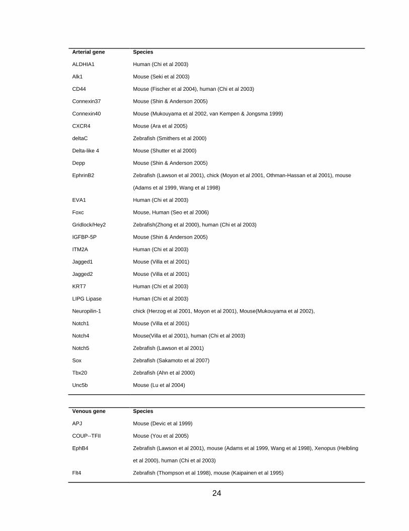
(Table I.1 and Figure I.2). For example, arterial ECs express high levels of
Notch1 and 4 (Villa et al 2001), Jagged1 and 2 (Villa et al 2001), Delta-like ligand
4 (Dll4) (Shutter et al 2000), Ephrin B2 (Wang et al 1998), Neuropilin 1 (Nrp1)
(Mukouyama et al 2002) and Hey2 (Chi et al 2003); whereas venous ECs are
characterized predominately by the expression of EphB4 (Wang et al 1998), Nrp-
2 (Herzog et al 2001, Yuan et al 2002) and COUP-TFII (You et al 2005).
24
Arterial gene Species
ALDHIA1 Human (Chi et al 2003)
Alk1 Mouse (Seki et al 2003)
CD44 Mouse (Fischer et al 2004), human (Chi et al 2003)
Connexin37 Mouse (Shin & Anderson 2005)
Connexin40 Mouse (Mukouyama et al 2002, van Kempen & Jongsma 1999)
CXCR4 Mouse (Ara et al 2005)
deltaC Zebrafish (Smithers et al 2000)
Delta-like 4 Mouse (Shutter et al 2000)
Depp Mouse (Shin & Anderson 2005)
EphrinB2 Zebrafish (Lawson et al 2001), chick (Moyon et al 2001, Othman-Hassan et al 2001), mouse
(Adams et al 1999, Wang et al 1998)
EVA1 Human (Chi et al 2003)
Foxc Mouse, Human (Seo et al 2006)
Gridlock/Hey2 Zebrafish(Zhong et al 2000), human (Chi et al 2003)
IGFBP-5P Mouse (Shin & Anderson 2005)
ITM2A Human (Chi et al 2003)
Jagged1 Mouse (Villa et al 2001)
Jagged2 Mouse (Villa et al 2001)
KRT7 Human (Chi et al 2003)
LIPG Lipase Human (Chi et al 2003)
Neuropilin-1 chick (Herzog et al 2001, Moyon et al 2001), Mouse(Mukouyama et al 2002),
Notch1 Mouse (Villa et al 2001)
Notch4 Mouse(Villa et al 2001), human (Chi et al 2003)
Notch5 Zebrafish (Lawson et al 2001)
Sox Zebrafish (Sakamoto et al 2007)
Tbx20 Zebrafish (Ahn et al 2000)
Unc5b Mouse (Lu et al 2004)
Venous gene Species
APJ Mouse (Devic et al 1999)
COUP--TFII Mouse (You et al 2005)
EphB4 Zebrafish (Lawson et al 2001), mouse (Adams et al 1999, Wang et al 1998), Xenopus (Helbling
et al 2000), human (Chi et al 2003)
Flt4 Zebrafish (Thompson et al 1998), mouse (Kaipainen et al 1995)

25
GDF1 Human (Chi et al 2003)
Lefty1/2 Human (Chi et al 2003)
Myosin 1B Human (Chi et al 2003)
Neuropilin-2 Chick (Herzog et al 2001), Mouse (Yuan et al 2002)
smootherned Human (Chi et al 2003)
Tie2 Chick (Moyon et al 2001)
Table I.1: Molecules expressed preferentially in arterial and venous ECs

26
Figure I.2 Model of AV specification. VEGF interacts with the Flk-1-Nrp1
complex to activate downstream Plc-γ-Erk and Notch signaling pathways, thus
inducing expression of arterial gene markers Hey2 and EphrinB2, while
inhibiting expression of venous gene EphB4. Foxc proteins also activate the
Notch pathway, leading to an arterial identity. Activated Notch can suppress
COUP-TFII expression, thereby repressing a venous fate. Conversely, COUP-
TFII can inhibit Nrp1 expression and thus attenuate VEGF signaling and
downstream Notch activation. Moreover, the PI3K-AKT cascade represses Erk
signaling. Therefore, a venous fate is promoted. Unconfirmed interactions are
indicated by dashed arrows. The molecules and signaling pathways that favor
an arterial fate are shown in black, while those that promote a venous fate are
in grey.

27
Regulation of AV Specification
Ehprin/Eph
The first genes that were found to be expressed differentially in arterial and
venous endothelium were Ephrin B2 and EphB4 (Adams et al 1999, Wang et al
1998), which are members of the Eph receptor tyrosine kinases (RTK) - Ephrin
family. The ligand Ephrins are divided into 2 subclasses: the A-subclass
(EphrinA1-A6), which anchored to the cell surface via a
glycosylphosphatidylinositol (GPI); and the B-subclass (EphrinB1-B3), which has
a transmembrane domain, followed by a short cytoplasmic region. Similarly, the
Eph receptors are divided into A and B subclass (EphA1-A8 and EphB1-B6,
respectively). The interactions of Ephrins and Ephs are prominent and are not
class restricted (Klein 2004, Kullander & Klein 2002). Because Ephrins and Ephs
are both membrane-bound, Ephrin-Eph signaling requires cell-cell contact. A
unique feature of Ephrins is that they are capable of receptor-like active
signaling. This results in bidirectional signal transduction, where conserved
tyrosine residues in the approximately 85aa long cytoplasmic domain of Ephrin
ligand are phosphorylated and recruit signaling effectors (Bruckner et al 1997,
Holland et al 1996).
The distinct expression pattern of arterial EphrinB2 and venous EphB4 was first
discovered in mice in the primary capillary plexus before initiation of circulation
(Adams et al 1999, Gerety et al 1999, Wang et al 1998). Since then, it has been
confirmed in chicks and zebrafish (Lawson et al 2001, Lawson et al 2002, Moyon

28
et al 2001, Othman-Hassan et al 2001, Zhong et al 2001). More importantly, the
interaction of EphinB2 with EphB4 is critical for proper vascular development. In
knockout mice lacking EphrinB2, the primary capillary plexus forms, but its
remodeling into a hierarchical, organized vasculature is arrested (Adams et al
1999, Wang et al 1998). Moreover, the dorsal aorta forms in these animals, while
the cardinal vein remains a loose, nonfunctional network of ECs (Adams et al
1999). These two vessels initially are formed by vasculogeneis. Similar
phenotypes are present in EphB4-deficient mice (Gerety et al 1999). Thus,
EphrinB2 and EphB4 are not acquired for determining EC fate during
vasculogenesis, but they are required to define and maintain the arterial and
venous interface. Mice lacking the EphrinB2 cytoplasmic domain exhibit defects
in vascular remodeling similar to those in mice that are completely deficient for
EphrinB2 or EphB4. This suggests that their bidirectional signaling is also
important for proper AV specification (Adams et al 2001).
Although EphrinB2 and EphB4 are markers of arteries and veins, studies in
zebrafish and mice have demonstrated some other critically important upstream
transcription factors that help determine arterial-venous EC fate, as discussed on
the following pages.
Notch
The Notch signaling pathway is an evolutionarily conserved pathway that is
involved in a variety of developmental processes. Notch family members and

29
Notch ligands are expressed throughout early vascular development and later
are restricted to arteries (see Table I.1). Mice with targeted deletion of Notch
molecules including Notch1 (Huppert et al 2000, Krebs et al 2000, Limbourg et al
2005), Notch4 (Carlson et al 2005, Krebs et al 2000, Uyttendaele et al 2001),
Jagged1 (Xue et al 1999), Dll4 (Duarte et al 2004, Gale et al 2004, Krebs et al
2004), Rbpj (Krebs et al 2004), Hey1 and Hey2 (Fischer et al 2004, Kokubo et al
2005) display severely impaired vascular remodeling in the yolk sac and in the
embryo proper, which indicates the essential roles of Notch molecules in
vascular development. In particular, the role of Notch molecules in arterial
specification has been extensively demonstrated in mice and zebrafish embryos,
and in human ECs (Gridley 2007, Roca & Adams 2007, Rossant & Hirashima
2003, Rossant & Howard 2002, Shawber & Kitajewski 2004, Siekmann & Lawson
2007).
Although Notch4 knockout mice do not exhibit any apparent deficiencies in
vessel formation, targeted deletion of Notch1 inhibits proper remodeling and
embryos die in utero. Moreover, Notch1/Notch4 double-knockout mice display
abnormal vascular development that is even more severe than that seen in
Notch1 knockout mice (Krebs et al 2000), suggesting that Notch1 and Notch4
functions partially overlap. Dll4, a ligand of Notch1 and Notch4, is haploid-
sufficient — loss of a single Dll4 allele results in reduced EphrinB2 expression
and increased EphB4 expression, along with a failure in arterial differentiation
(Duarte et al 2004). This defect is similar to that observed in Rbpj- and

30
Hey1/Hey2-knockout animals (Fischer et al 2004, Kokubo et al 2005, Krebs et al
2004). Most recently, Dll1 has been established as a requirement for maintaining
arterial identity during fetal development and a mediator of postnatal
arteriogenesis in mice with hindlimb ischemia (Limbourg et al 2007, Sorensen et
al 2009). Collectively, these findings indicate Notch activity is important for
promoting an arterial EC fate in mice.
Disruption of Notch signaling in zebrafish has many similar consequences as in
mice. Mindbomb (mib) in zebrafish encodes a RING ubiquitin ligase that
promotes ubiquitylation and internalization of Delta. The disruption of Notch
signaling in mib mutant leads to reduced expression of arterial markers EphrinB2
and Notch5 and increases expression of venous marker Flt4 in the dorsal aorta.
Similarly, overexpression of activated Notch5 in the posterior cardinal vein
causes a decrease in Flt4 expression. The expression of Notch’s downstream
target gridlock (grl), a zebrafish orthologue of mammalian Hey2, is restricted to
the dorsal aorta. Knockdown of grl represses Ephrin B2 expression and leads to
an expansion of adjacent veins. Conversely, overexpression of grl suppresses
venous growth with loss of Flt4 expression but does not affect dorsal aorta
formation (Zhong et al 2001, Zhong et al 2000). Although grl is needed for arterial
differentiation, its expression is not reduced in mib mutations or embryos injected
with Su(H) (dominant negative suppressor of Hairless), suggesting grl might not
be a direct target of Notch, making the role of grl in arterial specification in
zebrafish obscure.

31
VEGF
VEGF is one of the most potent and ubiquitous vascular growth factors that
affects many aspects of EC biology (Coultas et al 2005, Rossant & Hirashima
2003, Rossant & Howard 2002, Ruhrberg 2003). Consistent with VEGF’s
promotion of arterial EC differentiation in zebrafish, mice embryonic angioblasts
treated with either VEGF120 or VEGF164 can induce an arterial EC expression
pattern and undergo arterial specification (Mukouyama et al 2002). Similarly,
overexpression of VEGF164 in cardiomyocytes in transgenic mice results in an
increase of EphrinB2+ vessels (Visconti et al 2002). Interestingly, VEGF signaling
in the regulation of arterial differentiation in mice is in an isoform-sensitive
manner. Loss of VEGF164 impairs retinal arterial endothelial outgrowth, whereas
ablation of VEGF164 and VEGF188 perturbs renal arteriogenesis (Mattot et al
2002, Stalmans et al 2002).
Furthermore, recent work in zebrafish has shown Plc-γ/Erk under VEGF
signaling favors an arterial EC specification. A molecular cascade (VEGF Plc-
γ Pkc Raf Mek Erk) has been identified for promoting an arterial fate
of ECs (Lawson et al 2003). In contrast, constitutive activation of PI3K/AKT
induces venous EC fate, while repressing Erk activation (Hong et al 2006).
Inactivation of AKT with flavone GS4898 reverses PI3K inhibitory effect on Plc-
γ/Erk and thus promotes arterial EC specification (Hong et al 2006).

32
Sonic hedgehog (Shh)
The Hedgehog (Hh) family is composed of three ligands: sonic hedgehog, indian
hedgehog and desert hedgehog. They all signal through the same receptors
called PTCH1 and 2. The Hh signaling functions broadly in vascular development
including coronary vascular development (Lavine et al 2008, Lavine et al 2006)
and arterial specification (Lawson et al 2001). In zebrafish, loss of Shh activity
either in the null mutant embryo sonic-you (syu) or in the embryo that treated with
Shh signaling inhibitor cyclopamine results in loss of arterial identity and gain of
venous maker expression (Lawson et al 2002). Conversely, overexpression of
Shh leads to a switch from venous to arterial identity in posterior vein (Lawson et
al 2002). In addition, regulation of arterial specification by Shh is critically
associated with VEGF in the somite (Pola et al 2001). Microinjection of VEGF
mRNA in Shh-deficient embryos can rescue arterial differentiation. However,
injection of VEGF mRNA does not promote arterial differentiation in Notch-
deficient embryos. Instead, activation of the Notch pathway in the absence of
VEGF signaling can induce artery-specific gene expression (Lawson et al 2002).
Together, these findings indicate that VEGF acts downstream of Shh and
upstream of the Notch pathway to determine arterial EC fate.
Forkhead box c (Foxc)
The highly conserved Forkhead box (Fox) proteins are important in
cardiovascular development (Kume et al 2001). Recent studies demonstrated
that two Fox family members, Foxc1 and Foxc2, are essential for arterial

33
specification. Foxc1/Foxc2-null mice exhibit a fusion of the dorsal aorta with the
posterior cardinal vein. In addition, these mutants lack induction of the
expression of arterial markers including Notch1, Notch4, Dll4, Jagged1, Hey2
and EphrinB2; whereas, venous markers such as COUP-TFII and EphB4 are
expressed normally, suggesting that mutant ECs fail to acquire an arterial fate
(Seo et al 2006). Overexpression of Foxc genes in vitro consistently induces
expression of arterial markers. The promoters of Dll4 and Hey2 are directly
bound to and activated by Foxc proteins via a Foxc-binding site (Hayashi &
Kume 2008, Seo et al 2006). Moreover, activation of Dll4 and Hey2 occurs to a
greater extent when VEGF acts in combination with either Foxc1 or Foxc2.
Additionally, the transcriptional activity of Foxc proteins in Dll4 and Hey2
induction can be further modulated by VEGF-activated PI3K and ERK signaling
pathways, making the regulation of AV specification via Foxc proteins more
complicated. Collectively, Foxc transcriptional factors interact with VEGF and
Notch signaling to regulate arterial gene expression.
Sry-related HMG box (Sox)
Transcription factors of the Sox family, such as Sox7 and Sox18, are highly
expressed in developing vasculature and have recently been identified to be
involved in AV differentiation in zebrafish (Herpers et al 2008, Sakamoto et al
2007). Although disruption of either Sox protein does not cause any visible
vascular defects, simultaneous blockage of Sox7 and Sox18 leads to severe AV
malformation, characterized by vessel fusion and loss of circulation in the

34
posterior part of embryos (Herpers et al 2008). Further examination reveals that
arterial specification of ECs is severely interrupted in double-knockdown
embryos, which is often accompanied by failure of arteries to segregate from
veins. Therefore, Sox7 and Sox18 are dispensable for vascular development
individually, but synergistically they promote the specification of arterial ECs.
COUP- transcription factor 2 (COUP-TFII)
COUP-TFII, a member of the orphan nuclear receptor family, has been identified
as the first regulator that positively mediates venous EC identity (You et al 2005).
Targeted disruption of COUP-TFII results in embryonic lethality and displays a
phenotype opposite of that observed in the loss of Notch activity: a partial loss of
venous identity with reduced (but not completely abolished) EphB4 expression,
accompanied by ectopic expression of arterial gene markers such as Notch1 and
EphrinB2. Conditional lack of COUP-TFII in the endothelium causes veins to
acquire an arterial identity. In contrast, ectopic expression of COUP-TFII in the
endothelium results in AV malformation, which phenocopies the vascular
deficiency observed in Nrp1- and Notch1-knockout mice. Moreover,
overexpression of COUP-TFII represses the expression of Nrp1 and other
arterial gene markers. It is now known that COUP-TFII suppresses Nrp1
expression by binding to the Nrp1 promoter. Thus, COUP-TFII is a critical factor
that regulates the determination of venous EC fate.

35
Plasticity of AV Differentiation
Although it is now clear that the acquisition of arterial and venous identity is
determined genetically, there is no doubt that epigenetic factors such as oxygen
tension, blood flow and the vessel wall’s microenvironment also can influence
this process. Plasticity of ECs has been observed in the developing embryo by
performing quail-chick grafting experiments (Moyon et al 2001, Othman-Hassan
et al 2001). The quail arterial or venous vessel fragments, together with their
vessel wall from various stages of embryonic development, were grafted into
developing chick hosts. Quail ECs from arteries and veins were able to populate
both host arteries and veins with equal efficiency, and the arterial marker
expression changed with respect to the novel environment, regardless of its
origin. However, this EC plasticity diminished progressively after E11 (Moyon et
al 2001). Interestingly, removal of the vessel wall from the aorta, carotid artery
and vena cava in E14 quail can fully restore the capacity of ECs to colonize to
host arteries and veins, irrespective of their origin (Moyon et al 2001, Othman-
Hassan et al 2001). These results suggest that specification of arterial and
venous fate is, to some extent, reversible and the vessel wall may help stabilize
the arterial and venous choice of ECs. How the vessel wall precisely affects
endothelial AV determination is yet unknown.
EC plasticity with respect to AV differentiation also occurs during development
according to blood flow. It is observed that initiation of arterial and venous marker
expression in the primary capillary plexus occurs independently of flow (le Noble
36
et al 2004, Wang et al 1998). Shortly after the onset of circulation, the vitelline
artery forms from the arterial capillary plexus in the posterior pole of the embryo
by fusion of individual capillaries. But not all capillaries are involved in the
formation of the artery. Some of them become disconnected, exhibit
downregulation of arterial markers, and then serve to form the vitelline vein (le
Noble et al 2004). Furthermore, ligation of the vitelline artery on one side of the
yolk sac makes it become venularized over 24 hours, as judged by the direction
of blood flow and the expression of arterial makers. Similarly, flow manipulation
can switch veins to arteries (le Noble et al 2004). Therefore, hemodynamic forces
also can alter EC AV identity.
Oxygen tension differs in arteries and veins, and this can be another potential
instructive signal for AV differentiation. The influence of altered oxygen tension
on AV differentiation has been observed in the developing mouse retinal
vasculature (Claxton & Fruttiger 2005).
ECs in adults possess limited plasticity for AV differentiation, which has been
demonstrated in vein graft adaptation in humans and aged rats (Kudo et al
2007). EphB4 expression was lost and intima-media thickening was observed,
but arterial markers EphrinB2 and Dll4 were not strongly induced during vein
graft adaptation to the arterial environments (Kudo et al 2007).

37
Taken together, recent studies have demonstrated that AV specification is
genetically predetermined, and many molecules involved in the regulation of this
process have been identified. In addition, environmental factors such as oxygen
tension, hemodynamic forces and the microenvironment of vessel walls can
further influence vascular identity in terms of forming and maintaining arteries
and veins.
C. Endothelial Colony Forming Cells (ECFCs)
The concept that circulating bone-marrow-derived endothelial progenitor cells
(EPCs) could contribute to neovascularization via postnatal vasculogenesis was
first proposed in 1997 (Asahara et al 1997). Since then, EPCs have been studied
widely and regarded as a mechanism for maintenance of vascular homeostasis.
Defective regulation of EPCs leads to pathogenesis of various disorders.
However, to date, no cell surface molecules that uniquely identify EPCs have
been reported in human or other vertebrate species. This effort continues, as
researchers seek to isolate and define EPCs based on in vitro adhesion and
growth, and cell surface phenotype selection using fluorescent-labeled antibodies
and flow cytometry (see Figure I.3) (reviewed in (Hirschi et al 2008, Yoder &
Ingram 2009, Yoder et al 2007)).
In the ―cell adhesion and growth‖ approach, low-density mononuclear cells from
human peripheral blood are isolated and plated on fibronectin- and gelatin-
coated dishes. After several days in culture, nonadherent cells are removed. The

38
adherent cells display the ability to ingest acetylated low-density lipoprotein
(AcLDL) and bind to fluorescently labeled Ulex europaeus agglutinin I (UEA I)
plant lectin and thus are deemed as EPCs. These putative EPCs are released
from culture and undergo staining to confirm expression of endothelial markers
such as von Willebrand factor (vWF), VE-Cadherin and KDR by flow cytometry.
In additions, an inverse relationship between the circulating concentration of
these EPCs and an increased risk for developing coronary artery disease in
human subjects has been reported (Vasa et al 2001). However, others have
shown that putative EPCs isolated in this method may also express, to varying
degrees, CD45, CD11b, CD11c, CD14, CD68, eNOS, and E-selectin, and ingest
India ink like macrophages do (Rehman et al 2003, Rohde et al 2007, Rohde et
al 2006, Zhang et al 2006). Actually, the use of fibronectin- and gelatin-coated
tissue culture plates has been recognized as a method to isolate human blood
monocytes for differentiation into macrophages (Freundlich & Avdalovic 1983).
Moreover, monocytes/macrophages are well known to express ―endothelial-
specific‖ proteins, particularly when cultured in conditions that promote
endothelial growth. Therefore, the use of this method for isolating EPC is not
reliable.

39
Figure I.3 Common methods of “EPC” culture. Culture of CFU-Hill cells
includes a 5-day process where nonadherent peripheral blood MNCs plated on
FN-coated dishes give rise to colonies. ECFCs are derived from adherent
peripheral blood or cord blood MNCs cultured for 6 to 21 days on rat-tail collagen
I coated dishes, and colonies display cobblestone morphology. When adherent
peripheral blood MNCs cultured in FN-coated dishes, they typically do not display
colony formation. These cells are able to ingest AcLDL and bind to UEA I lectin.
The other method is to sort CD34+AC133+KDR+ cells directly from peripheral
blood MNCs instead of plating them.

40
A second method utilizes fluorescent-labeled antibodies and flow cytometry
analysis to enumerate putative EPC concentrations in human circulation. In the
first EPC paper, Asahara (Asahara et al 1997) reasoned that putative circulating
EPCs may express surface molecules shared by endothelial and hematopoietic
stem/progenitor cells because these two lineages share a similar mesoderm
origin during early embryogenesis. Thus, human circulating CD34+ cells were
isolated. They reportedly adhered to fibronectin-coated plates with greater
frequency than to type 1 collagen-coated plates and displayed a spindle-shaped
morphology. The adherent putative EPCs expressed a variety of cell surface
markers that normally were present in human umbilical vein endothelial cells
(HUVECs). Further studies demonstrated that these putative EPCs (CD34+ or
KDR+) home to areas of neovascularization when injected in vivo into
immunodeficient mice with induced hindlimb ischemia. These studies collectively
indicated that CD34+ or KDR+ circulating cells in human peripheral blood may be
EPCs and can contribute to postnatal angiogensis.
The number of endothelial cells circulating in the bloodstream in healthy subjects
is very low, but can increase under certain conditions (Blann et al 2005). Peichev
(Peichev et al 2000) attempted to define a panel of cell-surface antigens that may
distinguish EPCs from circulating endothelial cells (CECs). Peichev utilized
CD34, KDR, and AC133 as EPC markers. AC133, originally identified on
neuroepithelial cells, is a 5-transmembrane domain cell-surface glycoprotein that
is expressed in epithelial, hematopoietic, and various cancer stem cells (Mizrak

41
et al 2008). Peichev rationalized that the expression of CD34, KDR and AC133
should be present in EPCs and the expression of AC133 and CD34 would be
downregulated in CECs, which is similar to gene expression observed in
hematopoietic stem cells as they differentiate into more mature progenitors.
Reportedly, a CD34+KDR+AC133+ population can be identified in adult
peripheral blood, umbilical cord blood, and human fetal liver samples; but the
concentration is very low. Subsequently, multiple combinations of CD34, AC133
and/or KDR have been used extensively to identify circulating EPCs in human
subjects (reviewed by (Timmermans et al 2009)); and the concentration of
circulating EPCs has been correlated with many human diseases (Bertolini et al
2006, Jujo et al 2008, Pompilio et al 2009). However, recent studies have been
demonstrated that cells expressing CD34, AC133 and/or KDR are enriched for
hematopoietic colony-forming cells and do not form endothelial cells in vitro and
in invo (Case et al 2007, Timmermans et al 2007). Thus, the expression of CD34,
AC133 and KDR in human circulating cells fails to specifically identify a
circulating EPC.
The third approach utilizes in vitro colony-forming assays to identify putative
EPCs. Human peripheral blood mononuclear cells are placed on fibronectin-
coated dishes for 48 hours; then the nonadherent cells are replated to quantify
the emergence of the EPC colony forming units several days later. The putative
EPCs (that give rise to progeny forming the colony) have been referred to as
colony forming unit-Hill (CFU-Hill). The CFU-Hill assay has been used to

42
demonstrate a significant inverse correlation between the concentration of
circulating CFU-Hill and Framingham cardiovascular risk scores in human
subjects (Hill et al 2003). CFU-Hill has been recognized as being composed of
an aggregate of round cells overlying adherent spindle-shaped cells expressing
many proteins similar to the primary endothelial cells. However, these adherent
spindle-shaped cells also express several myeloid progenitor cell markers and
mature into macrophages that can readily ingest bacteria. In addition, these cells
neither proliferate extensively to give rise to secondary colonies in vitro nor form
blood vessels spontaneously when implanted in vivo in collagen gels (they do not
display postnatal vasculogenic activity) (Yoder et al 2007). Furthermore, recent
studies have demonstrated that CFU-Hill cells are mainly T cells and monocytes
admixed with B cells and natural killer (NK) cells. The combination of purified T
cells with monocytes is able to form CFU-Hill colonies (Rohde et al 2007, Rohde
et al 2006). Thus, the CFU-Hill assay identifies hematopoietic cells instead of
endothelial cells.
We and others have isolated ECFCs from human peripheral blood and cord
blood (Au et al 2008a, Ingram et al 2004, Melero-Martin et al 2007). Human
blood low-density mononuclear cells are placed on rat-tail collagen I coated
dishes, and the nonadherent cells are removed. Several days later, ECFC
colonies with the typical cobblestone morphology emerge in a medium that
benefits endothelial cell growth. ECFCs express cell surface proteins (KDR,
CD34, vWF, eNOS, VE-cadherin, and others) similar to those on primary ECs,

43
but ECFCs do not express hematopoietic cell markers such as CD45, AC133,
CD11b and CD14 (Ingram et al 2004, Yoder et al 2007). They can proliferate at a
clonal plating level and replate into secondary and tertiary ECFCs (Ingram et al
2004). They are able to incorporate AcLDL and form capillary-like structures in
vitro (Ingram et al 2004). Most importantly, in sharp contrast to CFU-Hill and any
other putative EPCs, ECFCs are capable of forming human blood vessels in vivo
in immunodeficient mice and incorporate with murine vasculature to become part
of murine systemic circulation (Yoder et al 2007). Thus, only ECFCs display all
the property of EPCs.
In summary, the types of cells being considered as EPCs vary widely from study
to study, causing controversies in this field. However, the strictest definition of an
EPC is a circulating cell that 1) displays the ability to produce endothelial progeny
that form endothelial tubes in vitro and 2) contribute to the functional endothelial
lining of injured vascular structure via angiogenesis and/or vasculogenesis in
vivo. Thus, to date, only human ECFCs fit the definition of true EPCs and display
all the activities described on these pages.

44
CHAPTER II
A Hierarchy of Endothelial Colony Forming Cell (ECFC) Activity is
Displayed by Bovine Corneal Endothelial Cells (BCECs)
Introduction
Corneal endothelial cells (ECs) are derived from neural crest precursors during
embryonic development. These unique cells form a distinctive monolayer of
hexagonally packed cells attached to their specialized basement membrane,
Descemet’s membrane, and form the most posterior portion of the cornea.
Corneal ECs fail to proliferate in vivo in response to injury, disease, or aging and
are arrested in the G1 phase of the cell cycle. However, corneal ECs possess
replicative potential that can be revealed upon in vitro endothelial cell culture
and/or application of a stress such as mechanical wounding or ethylene diamine
tetraacetic acid (EDTA) treatment to disrupt cell-cell interactions within the
endothelial monolayer on the cornea. A number of laboratories have also
reported successful growth of untransformed corneal ECs in vitro. Using in vitro
culture approaches, recent studies demonstrate that the proliferation of corneal
ECs varies with the age of the donor (cells derived from younger donors divide
more than those from older donors) and varies from the central cornea (low
proliferative potential) to peripheral cornea (high proliferative potential). To date,
it remains unclear if each corneal EC displays proliferative potential in vitro or if
the replicative ability resides in only a subset of the cells.

45
We have reported on development of methods to examine clonal proliferative
behavior of circulating and vascular endothelial cells and have identified a
hierarchy of endothelial colony forming cell (ECFC) activity ranging from high
proliferative potential ECFC (HPP-ECFC) to non-dividing mature ECs (Ingram et
al 2005b, Ingram et al 2004). Human umbilical cord blood is enriched in
circulating ECFCs and the distribution of ECFCs is skewed to a high percentage
of HPP-ECFCs compared to human adult peripheral blood samples (Ingram et al
2004). Both cord blood and adult peripheral blood ECFCs display phenotypical
and functional properties which are also present in ECs derived from human
blood vessels (Ingram et al 2005b). Furthermore circulating or resident ECFCs
form human blood vessels de novo when subcutaneously implanted into
immunodeficient mice and these vessels participate in carrying blood as a part of
the host murine systemic circulation (Au et al 2008a, Schechner et al 2000,
Yoder et al 2007). These current studies suggest that ECFCs represent
stem/progenitor cells for the endothelial lineage.
We questioned whether corneal ECs, although derived from neural crest rather
than mesoderm and differing in anatomic position, exposure to blood flow and
blood constituents, and displaying unique physiological functions, displayed
clonal ECFC potential. We report that BCECs display a complete hierarchy of
ECFCs that is similar to the distribution of ECFC activity in ECs isolated from
bovine aorta, coronary artery, and pulmonary artery. HPP-ECFCs in corneal
endothelium can be replated into at least secondary colonies and retain high

46
levels of telomerase activity similar to HPP-ECFCs derived from resident
endothelium in blood vessels. These data suggest that the fundamental
paradigm for endothelial cell lineage development and maintenance may be
similar in corneal ECs as with other vascular ECs, but the mechanisms for EC
repair and regeneration are regulated by dominant tissue specific requirements
and niches.

47
Materials and Methods
Isolation of bovine peripheral blood mononuclear cells (MNCs)
Blood (50-100ml) was collected from a local slaughterhouse and diluted one to
one with Dulbecco's Phosphate-Buffered Saline (DPBS) (1X) without Ca or Mg
(Invitrogen, Grand Island, NY)/1% bovine serum albumin (BSA)/ethylenediamine
tetraacetic acid (EDTA) solution and layered (2:1 ratio) onto Histopaque1119
(Sigma-Aldrich, St Louis, MO). Cells were centrifuged for 30 minutes at 1800 rpm
at room temperature (Beckman Coulter, Fullerton, CA). Low density mononuclear
cells (MNCs) were isolated as the floating monolayer and washed three times
with DPBS. Finally, MNCs were resuspended in DPBS with 2% fetal bovine
serum (FBS) (Hyclone, Logan, UT) for direct analysis by fluorescence activated
cell sorting (FACS).
Culture of bovine vessel derived endothelial cells (ECs)
Bovine aortic endothelial cells (BAECs), bovine coronary arterial endothelial cells
(BCAECs) and bovine pulmonary arterial endothelial cells (BPAECs) were
purchased from Lonza (Lonza, Walkersville, MD). These bovine vascular
endothelial cells were plated onto type I rat tail collagen (50 µg/ml) (BD
Biosciences, Bedford MA) pre-coated tissue culture flasks and cultured in
endothelial cell growth medium EGM-MV (Lonza) supplemented with 1.5%
antibiotic-antimycotic (Invitrogen) at 37°C, 5% CO2 in a humidified incubator.

48
Isolation and culture of bovine corneal ECs
The isolation of bovine corneal endothelial cells (BCECs) was performed as
previously described (Bonanno and Giasson, 1992). These corneal ECs were
plated in tissue culture flasks filled with DMEM supplemented with 5% FBS and
1.5% antibiotic-antimycotic. Corneal ECs were grown to a confluent monolayer
and the cells displayed a typical hexagonal appearance. These ECs were
released from the culture dish by using TryPLE (Gibco, Grand Island, NY) and
re-plated onto 75 cm2 tissue culture flasks for further passage in the
supplemented DMEM.
Immunophenotyping of endothelial cells
Early passage (2-4) bovine vascular ECs and corneal ECs as well as bovine
blood MNCs were stained with a mouse monoclonal antibody against bovine
CD45 conjugated to fluorescein isothiocyanate (FITC) (SEROTEC, UK), FITC
conjugated Lycopersicon Esculentum (Tomato) lectin (LEL) and Griffonia
simplicifolia I lectin (GSL I) (Vector Laboratories, Burlingame, CA) or mouse
isotype control antibody for 30 minutes at 4°C, washed three times, and analyzed
for cell surface expression using a FACS Calibur flow cytometer (Becton
Dickinson, San Diego, CA) as previously described (Huang et al 2007).
Ingestion of 488-conjugated acetylated-low density lipoprotein (488-AcLDL)
Early passage (2-4) bovine vessel wall derived ECs and corneal ECs were
incubated with 10 µg/mL of 488-AcLDL (Invitrogen) in the media for 8 hours at

49
37°C. Cells were washed three times, co-stained with 1.5µg/mL of the nuclear
stain, 4’, 6-diamidino-2-phenylindole dihydrochloride (DAPI) (Sigma), and
examined by inspection through an inverted fluorescence microscope (Zeiss,
Thornwood, NY) at 40 x magnification.
Tube formation assay
Capillary tube formation by the various ECs was performed as previously
described using Matrigel coated dishes (Ingram et al 2004). Early passage (2-4)
bovine vascular ECs and corneal ECs were seeded onto 96-well tissue culture
plates coated with 30µl Matrigel (BD Biosciences) at a cell density of 10,000-
30,000 cells per well. Cells were observed every two hours by an inverted
microscope for the formation of capillary-like structure.
RT-PCR
Total cellular RNA was extracted with TRIzol (Invitrogen) in a single-step method
as described by the manufacturer. RT reactions were performed using a
SuperScript First-Strand Synthesis system (Invitrogen). PCR was conducted
using Go Tap Flexi DNA Polymerase (Promega, Madison, WI) according to the
manufacturer’s instructions. The primer sequences were shown in Table II.1. The
PCR cycle was 94°C, 5 min; 94°C 30s, 53 or 57 or 59°C (depending on the
different primers) 30s, 72°C 45s, and 32 cycles with a final 72°C 7 min cycle.

50
PCR products were added to wells in a 2% agarose/ethidium bromide gel,
exposed to electophoresis current, and migrating bands were photographed
under UV light.

51
Gene Forward Reverse Tm Product size
S100B GGTGACAAGCACAAGCTGAA CAGTGGTAATCATGGCAACG 55 184
Enolase 2 GGACTTGGATGGGACTGAGA GCGTCCTTGCCATACTTGTC 57 327
N-Cadherin AGCAACTGCAATGGGAAAAG GATGGGAGGGATAACCCAGT 55 461
VE-Cadherin GAGTGTGGACCCCAAGAAGA GCTGGTACACGACAGAAGCA 57 359
eNOS TGAGCAGCTGCTGAGCCAGG CAGCTCGCTCTCTCGGAGGT 57 209
PECAM I ATGTGCTGCTTCACAACGTC TGAATTCCAGCGTCACAAAA 53 345
Vimentin CTTCGCCAACTACATCGACA GGATTCCACTTTACGCTCCA 55 340
Enolase 1 GCAGGAGAAGATCGACAAGC GTAAACCTCTGCTCCGATGC 57 310
ZO 1 GAATCCGATGTGGGTGATTC GCAGGTTTCTCTTGGAGCTG 57 310
Factor Viii ACTGCCTCATCCCACTTACG GGGGTCTAGAGCATTCACCA 57 327
GAPDH GGTGAAGGTCGGAGTGAACG GGGTCATTGATGGCGACGA 59 117
LDLR ACAACCCCGTGTACCAGAAG AGGGTCAGGGGAGAAAGTGT 57 195
CD36 AACCACTTTCATCAGACCCG ACGTGTCATCCTCAGTTCCA 57 475
Table II.1: Primers used for conventional RT-PCR

52
Single cell clonogenic assay
Early passage (2-4) bovine vascular ECs and corneal ECs, were selected by size
and cell complexity using a FacsVantage Sorter (Becton Dickinson) and
deposited as one cell per well into 96 well plates pre-coated with type I rat tail
collagen in 200µl of media as previously described (Huang et al 2007). Cells
were cultured at 37°C, 5% CO2 in a humidified incubator. Media was changed
every five days. After 14 days of culture, the cells were fixed with 4%
paraformaldehyde (Sigma, St. Louis, MO) in DPBS for 30 minutes at room
temperature, then washed twice and stained with 1.5 µg/ml DAPI, and examined
for determination of EC number. Those wells containing two or more cells were
identified as positive for proliferation under a fluorescent microscope at 10x
magnification. Culture wells containing fewer than 50 cells were counted by direct
visual inspection with a fluorescent microscope at 10x magnification. For those
wells with more than 50 cells, colonies were imaged and cell number quantified
using an Image J 1.36v program (Wayne Rasband, NIH).
Sphere forming assay
Early passage (1-2) bovine corneal ECs were released from adhering to the
culture dish using TryPLE and then suspended into single cells in the culture
medium. Cells were seeded at a density of 50cells/µl on plastic dishes as
hanging drops (10 µl each) to allow spheroid formation by cell aggregation. Cells
then were cultured at 37°C, 5% CO2 in a humidified incubator 3 days. Under this

53
condition, the suspended cells contributed to formation of a single spheroid per
drop of defined size (diameter > 100µm).
Telomerase assay
Telomerase activity was measured by the telomeric amplification protocol
(TRAP) as previously described (Kim et al 1994) using the TRAPeze telomerase
detection kit (Chemicon, Temecula, CA). Cells lysate from 1000 cells were used
in each assay. Hela cell extract served as a positive control and lysis buffer only
as negative control. The PCR products were exposed to an electrophoretic
current on a 12.5% non-denaturing polyacrylamide gel and visualized by SYBR
gold staining (Molecular Probe, Eugene, OR).
Statistical Analysis
Results are expressed as mean±SEM for the study variables. Data were
compared with ANOVA test and significant differences were set at the P < 0.05
level. All analyses were performed using GraphPad InStat software (GraphPad
Software Inc, La Jolla, CA)

54
Results
Characterization of BCECs compared to bovine vascular endothelial cells
BCECs have been successively isolated and expanded in vitro. We isolated and
cultured BCECs from 18 and 24 month old mixed breed steers and identified the
samples as BCEC 18 and BCEC 24, respectively. These primary cultured
BCECs displayed a typical hexagonal morphology (similar to the endothelial
morphology in the intact cornea by direct visualization) at confluence. In
comparison, bovine ECs from aorta, coronary artery, and pulmonary artery
exhibited more morphological heterogeneity with more spindle-shaped and large
oval cells (Figure II.1).
To determine whether ECs displayed any common hematopoietic antigens, we
examined BCECs as well as bovine vascular ECs for expression of the leukocyte
common antigen CD45 (known to be expressed on all nucleated blood cells).
None of the endothelial cells expressed CD45 while the peripheral blood low
density MNC (comprised of leukocytes) highly expressed CD45 (Figure II.2A).
BCECs and bovine vessel wall derived ECs were able to bind LEL while
peripheral blood MNCs did not display this activity (Figure II.2A). Additionally,
BCECs, vascular ECs, and MNCs were all able to bind GSL I (Figure II.2A).
Actually, BCECs and bovine vascular ECs had the same lectin binding pattern for
all 30 lectins tested (data not shown). Thus BCECs share some cell surface
molecule expression with bovine vessel wall derived ECs but there is no

55
evidence that any of the ECs express the most common hematopoietic antigen
CD45.
Ingestion of AcLDL is a feature displayed by many ECs and not surprisingly, all
bovine vessel wall derived ECs readily ingested the lipoprotein complex (Figure
II.2B). In contrast, BCECs were unable to ingest AcLDL (Figure II.2B). The LDL
receptor (LDLR) and scavenger receptors, such as CD36, mediate native and
modified LDL-ingestion. We further examined LDLR and CD36 expression in
bovine vascular and corneal endothelial cells after exposing the endothelial cells
to AcLDL. LDLR transcripts were detectable in both types of ECs while CD36
transcripts were only present in vascular ECs after 8h of exposure to AcLDL
(Figure II.2C). Capillary-like tube formation is another feature displayed
essentially by all vascular ECs when examined in vitro. Surprisingly, BCECs,
similar to all of the bovine vascular ECs, readily formed capillary-like structures
when plated on Matrigel-coated dishes (Figure II.2D). Therefore, while BCECs
always exist as a flattened monolayer lining the most posterior aspect of the
cornea in vivo, removal of the cells and replating in vitro permits some functional
properties of vascular ECs to emerge from the corneal ECs.

56
Figure II.1 The morphology of cultured bovine vessel wall derived ECs
and bovine corneal ECs. Bovine vascular ECs derived from aorta (BAECs),
coronary artery (BCAECs) and pulmonary artery (BPAECs) display spindle
shape morphology and BCECs present a hexagonal monolayer. (10x
magnification, scale bar represents 100µm)

57
A
B
C
D

58
Figure II.2 Phenotypic and functional characterization of bovine vessel
wall derived ECs and bovine corneal ECs. (A) Immunophenotyping of bovine
peripheral blood MNCs and the monolayers derived from BAECs, BCAECs,
BPAECs and BCECs by fluorescence cytometry. BAECs, BCAECs, BPAECs
and BCECs can bind to the lectins GSL I and LEL but do not express common
leukocyte antigen CD45. Negative controls are overlayed in green on each
histogram. (B) Incorporation of 488-AcLDL in bovine vessel wall derived ECs
and corneal ECs. BAECs, BCAECs and BPAECs are able to ingest 488-AcLDL
(green) but not BCECs. The cell nuclei are stained with DAPI (blue). (40x
magnification, scale bar represents 100µm) (C) LDLR and CD36 expression in
bovine vascular and corneal ECs after exposed to AcLDL using RT-PCR. LDLR
is expressed in all types of ECs while CD36 is only detectable in bovine
vascular ECs after ingesting AcLDL. (D) Formation of capillary-like structures
when bovine vascular ECs and BCECs are plated in Matrigel. (40x
magnification, scale bar represents 100µm) Three independent experiments
show similar results.

59
Gene expression of BCECs compared to bovine vascular endothelial cells
Although corneal ECs and vascular ECs possess differences in developmental
origin, anatomic location, and physiological function, they have showed some
similarities in phenotype and function in the above studies. To further compare
and contrast these sources of ECs, we interrogated all of the EC lines for the
expression of certain mRNA transcripts that are published as commonly
expressed in all vascular endothelial cells (Figure II.3) (Albelda et al 1991, Huang
et al 2007, Ohashi et al 2007). Remarkably, BCECs did not express vascular
endothelial-cadherin (VE-Cadherin), nitric oxide synthase 3 (endothelial nitric
oxide synthase; eNOS) or platelet/endothelial cell adhesion molecule 1
(PECAM1) which are expressed in bovine vascular ECs derived from aorta,
coronary artery, and pulmonary artery. While there has been no single specific
genetic marker for corneal ECs yet identified, we have scanned the published
literature and identified numerous genes, including S100B, enolase2, N-cadherin,
vimentin, ZO-1 and factor VIII, which are reportedly expressed preferentially in
corneal ECs. S100B mRNA was present in BCECs and BPAECs, but absent in
BAECs and BCAECs. Neuron specific enolase 2 mRNA was present in BCEC 18
and BCAECs and in three experiments, detectable in BCEC 24 but at the low
level of detection. N-Cadherin mRNA was present in all ECs except BAECs.
Furthermore, vimentin, enolase1, ZO1 and factor VIII mRNA were detectable in
all ECs. Thus, BCECs displayed a gene expression profile that differed
somewhat from bovine aortic, coronary artery, and pulmonary artery ECs,
however, no one single gene product was BCEC specific. Whether these

60
differences are related to the known influence of in vitro culture on modulating
gene expression and cell function in all the endothelial cells tested or truly
represent tissue specific differences in gene expression will require further
search for specific markers for these different endothelial populations that
permits prospective isolation from each tissue and gene expression interrogation.

61
Figure II.3 RT-PCR analysis of gene expression in bovine vessel wall
derived ECs and bovine corneal ECs. PECAM1, VE-Cadherin and eNOS are
specifically expressed in bovine vascular ECs but not in corneal ECs. S100B
transcripts are detectable in BCECs but not in vessel wall derived ECs.
Enolase2 are also can be detected in BCECs with variable expression level.
The other genes related to corneal ECs can also be detected in vascular ECs.
Three independent experiments show similar results.

62
Hierarchical organization of the proliferative potential in BCECs and bovine
vascular endothelial cells
We have recently developed a single cell clonogenic assay to quantitate the
proliferative capacity of individual ECs. Using this method, we have defined a
hierarchy of ECFCs present in both circulating and vessel wall derived ECs in
human subjects and other vertebrates. Here we tested whether bovine vessel-
derived ECs and corneal ECs possessed a similar hierarchy of cells with varying
levels of proliferative potential. It was apparent that vascular ECs and corneal
ECs displayed diverse proliferative ability at a clonal level. Some ECs did not
divide and some divided and gave rise to various sized colonies of endothelium
(Figure II.4). Significantly more single BAECs executed at least one cell division
during 14 days of culture compared to BCECs (BAECs vs. BCEC 18 vs. BCEC
24 was 65.67 ± 2.52% vs. 32.63 ± 1.22% vs. 41.40 ± 5.24%). However, there
was no significant difference in the frequency of dividing cells among BAECs,
BCAECs (48.00 ± 4.67%) and BPAECs (46.93 ± 7.46%) or among BCAECs,
BPAECs and BCECs (Figure II.4A). Surprisingly, 39.82 ± 2.71% of the individual
BCEC 18 and 43.25 ± 3.49% of the individual BCEC 24 cells that divided gave
rise to well circumscribed colonies containing more than 10,000 progeny in the 2
weeks’ assay. This frequency was significantly higher than that measured for
individually plated BAECs (33.67 ± 4.22%). Differences in the ECFC distribution
were observed in BCEC 18 cells versus BCEC 24 cells (at the same passage
number), suggesting that some variability may be observed between donors with
respect to this kind of distribution analysis.

63
We recently defined HPP-ECFCs as cells that can yield macroscopic colonies (at
least more than 2000 cells) and can also form secondary HPP-ECFC colonies
upon replating. To examine whether any BCECs or bovine vascular ECs possess
HPP-ECFC activity, the progeny of primary ECFC colonies containing more than
10,000 cells were trypsinized and replated into 96-well tissue culture plates at
one cell per well. After another 14 days of culture, the individually replated cells
generated all sizes of colonies including some colonies containing more than
10,000 cells (data not shown). Thus a complete hierarchy of ECFCs was
identified in BCECs and bovine vascular ECs, and these ECFC-derived progeny
were comprised of non-dividing mature ECs, endothelial clusters (2-50
cells/colony), low proliferative potential-ECFCs (LPP-ECFCs, 51-2000
cells/colony) and HPP-ECFCs (2001 or greater cells/colony).

64
A
B
Figure II.4 Quantitation of the clonogenic and proliferative potential of
single endothelial cells derived from bovine vascular endothelium and
bovine corneal endothelium. (A) The percentage of single BAECs, BCAECs,
BPAECs and BCECs dividing at least once after 14 days culture. There are
significantly fewer BCECs undergoing division comparing to bovine vessel wall
derived ECs. (B) The distribution of different size of colonies derived from
single ECs in an individual well after 14 days of culture. There is dramatically
higher percent of HPP-ECFCs than LPP-ECFCs and endothelial clusters in
BAECs and BCECs. *P < 0.05, **P < 0.01, ***P < 0.001 by parametric ANOVA.
(n=3)

65
High levels of telomerase activity in BCECs and bovine vascular
endothelial cells
We have previously reported that human cord blood HPP-ECFC possesses high
levels of telomerase activity. Remarkably, the progeny of HPP-ECFC isolated
from BCECs displayed telomerase activity similar to that of the progeny of HPP-
ECFC derived from BAECs, BCAECs and BPAECs (Figure II.5). Thus, HPP-
ECFCs in both BCECs and bovine vascular ECs express quantifiable levels of
telomerase activity suggesting a potential mechanism through which proliferative
potential is retained within the subset of endothelial cells that possess
proliferative potential.

66
Figure II.5 Telomerase activity of HPP-ECFCs derived from bovine vessel
wall derived ECs and BCECs. P indicates telomerase activity in Hela cells,
which acts as a positive control, N indicates a negative control and IC indicates
internal control. Three independent experiments show similar results.
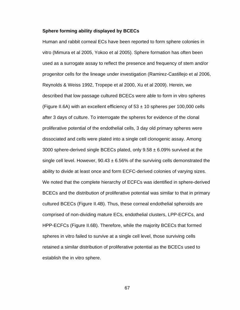
67
Sphere forming ability displayed by BCECs
Human and rabbit corneal ECs have been reported to form sphere colonies in
vitro (Mimura et al 2005, Yokoo et al 2005). Sphere formation has often been
used as a surrogate assay to reflect the presence and frequency of stem and/or
progenitor cells for the lineage under investigation (Ramirez-Castillejo et al 2006,
Reynolds & Weiss 1992, Tropepe et al 2000, Xu et al 2009). Herein, we
described that low passage cultured BCECs were able to form in vitro spheres
(Figure II.6A) with an excellent efficiency of 53 ± 10 spheres per 100,000 cells
after 3 days of culture. To interrogate the spheres for evidence of the clonal
proliferative potential of the endothelial cells, 3 day old primary spheres were
dissociated and cells were plated into a single cell clonogenic assay. Among
3000 sphere-derived single BCECs plated, only 9.58 ± 6.09% survived at the
single cell level. However, 90.43 ± 6.56% of the surviving cells demonstrated the
ability to divide at least once and form ECFC-derived colonies of varying sizes.
We noted that the complete hierarchy of ECFCs was identified in sphere-derived
BCECs and the distribution of proliferative potential was similar to that in primary
cultured BCECs (Figure II.4B). Thus, these corneal endothelial spheroids are
comprised of non-dividing mature ECs, endothelial clusters, LPP-ECFCs, and
HPP-ECFCs (Figure II.6B). Therefore, while the majority BCECs that formed
spheres in vitro failed to survive at a single cell level, those surviving cells
retained a similar distribution of proliferative potential as the BCECs used to
establish the in vitro sphere.

68
A
B
Figure II.6 The formation of sphere colony by bovine corneal endothelial
cells. (A) Representative photograph of sphere derived from plated BCECs.
(Scale bar represents 100µm) (B) The distribution of different size of colonies
derived from single BCECs dissociated from corneal endothelial sphere colonies
in an individual well after 14 days of culture. The complete hierarchy of ECFC is
present in BCECs residing in the spheres.

69
Discussion
It is well known that corneal ECs are apparently restrained from proliferating in
vivo, but they retain proliferative potential and can be expanded in vitro. The
present study is the first to demonstrate that not all corneal ECs display the same
ability to proliferate at a clonal level. In fact, we define a hierarchy of ECFCs in
bovine corneal ECs based on their proliferative potential using a single-cell
clonogenic assay. We report that the distribution of ECFCs derived from single
BCECs is quite similar to bovine vascular endothelium from aorta, coronary
artery, and pulmonary artery and is consistent with circulating and resident
vascular endothelial cells in adult human subjects.
The proliferative activity of corneal endothelium in vitro has been observed under
a variety of in vitro conditions. EDTA can release endothelial cells from contact
inhibition and promote proliferation within the endothelial monolayer (Joyce &
Zhu 2004, Senoo et al 2000). Cytokines such as epidermal growth factor (EGF)
and nerve growth factor (NGF) can also induce corneal ECs proliferation in vitro
(Joyce & Zhu 2004). Furthermore, the overexpression of oncogene proteins
(SV40 large T antigen or E6/E7) or the transcription factor E2F2 in corneal ECs
can induce proliferation (Joyce 2003, McAlister et al 2005). These observations
indicate that at least some corneal ECs inherently possess high proliferative
capacity. Most recently, Mimura and colleagues (Mimura & Joyce 2006) have
reported that peripheral corneal ECs display more proliferative capacity than
those more centrally located. Moreover, Mimura and Yamagami (Mimura et al

70
2005, Yamagami et al 2007) demonstrated that there are a greater number of
corneal EC precursor cells in the periphery than in the center by performing a
sphere-forming assay, and concluding that the higher frequency of sphere-
forming ability in the periphery must be related to the retention of precursor cells.
However, these important studies did not use a single cell clonogenic assay,
which can quantitatively and stringently interrogate the proliferative potential of
individual ECs, to test whether corneal ECs harbored different populations that
could be distinguished by their clonogenic potential.
Since we previously identified the entire hierarchy of ECFCs in circulating and
vessel-derived ECs in human, pig and rat, it’s not surprising that this hierarchy of
proliferation is also present in bovine vascular ECs. However, it is of interest that
corneal ECs can also be discriminated by similar proliferative and clonogenic
properties. In the present study, we have provided evidence that corneal
endothelial HPP-ECFC give rise to all subsequent stages of ECFC development.
Thus, corneal endothelium possesses the complete hierarchy of ECFCs, and
HPP-ECFCs display properties of a corneal endothelial progenitor cell as they
have the most proliferative capacity and can be cultured in vitro in the absence of
stroma cells.
The existence of stem cells for the corneal endothelium has been reported
recently. By using telomerase activity assay and BrdU (bromodeoxyridine)
incorporation assay, Whikehart et al (Whikehart et al 2005) argued that the

71
putative stem cells for corneal endothelium are located in a niche at the posterior
limbus between the peripheral endothelium and the trabecular meshwork (TM).
More recently, McGowan et al (McGowan et al 2007) provided further evidence
for the presence of such a population in this area by demonstrating the
expression of stem cell markers (nestin, Oct-3/4 and Wnt-1) in the unwounded
cornea. They observed that the differentiation markers (Pax-6, Sox-2) were
induced in this area and extended to peripheral corneal endothelium after
wounding, which suggested that these putative stem cells respond to corneal
damage and initiate endothelial repair. However, Mimura and Yokoo recently
demonstrated that they were able to isolate corneal endothelial precursor cells
from corneal endothelium with a sphere-forming assay and that the corneal
endothelium harbors precursor cells which exhibit high proliferative capacity,
form colonies in a sphere-forming assay, and have self-renewal ability to give
rise to the secondary colonies (Mimura et al 2005, Yokoo et al 2005). Additionally,
the sphere-forming ability of the peripheral corneal ECs was reported to be
greater than centrally located ECs (Mimura et al 2005, Yamagami et al 2007).
Moreover, the progeny of isolated spheres display hexagonal morphology and
transport activity.
Although a sphere-forming assay has been extensively utilized to quantitatively
measure stem cell frequency in many fields including neural stem cells and
cancer stem cell (Ramirez-Castillejo et al 2006, Reynolds & Weiss 1992,
Tropepe et al 2000, Xu et al 2009), recent studies have indicated that not all cells

72
capable of forming a sphere meet the criteria for a stem cell (Singec et al 2006).
Moreover, the in vitro conditions to form spheres often permits the highly motile
spheres to merged together, which argues against these spheroid structures
arising from a single stem cell (Singec et al 2006). Therefore, use of a sphere-
forming assay may not permit accurate determination of which individual cells
display proliferative potential. In our current study, we utilized a single cell
clonogenic assay to provide direct evidence that corneal EC precursors exist in
corneal endothelium. HPP-ECFCs display the property of corneal endothelial
progenitor cells as they have the most clonal proliferative capacity, can be
replated to form secondary HPP-ECFC, and give rise to all other ECFC colonies
and/or mature non-dividing endothelium. Whether the peripheral corneal
endothelium harbors more HPP-ECFCs than the central endothelium needs to be
further examined and is currently being investigated in our lab.
In this study, we did not directly isolate sphere colonies from bovine corneal
endothelium. Instead, we tried to generate corneal endothelial spheroids from
plated BCECs from low passage cultures (passage 1 or 2). We noticed that
without addition of fetal calf serum to the culture medium, isolated cells only
aggregated but did not form spheres. Furthermore, when plated in the floating
culture method as previously described (Mimura et al 2005, Yamagami et al 2007,
Yokoo et al 2005), BCEC frequently became free of the sphere and reattached to
the culture substrate. Thus, we had to use a hanging drop method of sphere
formation that was modified from previous publications (Del Duca et al 2004,

73
Timmins & Nielsen 2007). Under these culture conditions, BCECs were able to
form numerous spheres when 500 cells were deposited per drop of medium. This
number is much lower than reported for a human corneal endothelial cell sphere-
forming assay, where 257 ± 83 spheres were generated from 50,000 cells after
10 days of culture (Yokoo et al 2005). This might be due to obvious differences in:
1) culture conditions, 2) species, 3) use of freshly isolated corneal ECs versus
plated corneal ECs. It is of interest that BCECs residing in the spheres displayed
the complete hierarchy of ECFC (Figure II.6B) when examined clonally, and the
distribution of proliferative potential was similar to BCECs that were resident in
corneal endothelium prior to plating (Figure II.4B). Taken together, we propose
that a single cell clonogenic assay is an alternative method to stringently and
directly quantitate corneal endothelial progenitor cell residence.
Consistent with their great proliferative ability, HPP-ECFCs in corneal
endothelium display high levels of telomerase activity (Figure II.5). The telomere
length in corneal endothelium has been measured in vivo and in vitro. In human
subjects, the average telomere length has been reported as 11-14kb (Amano
2003, Egan et al 1998). It should be realized that there was no statistically
significant difference in the telomere length of the corneal ECs residing in the
central and peripheral areas of freshly examined corneal endothelium or when
corneal EC from younger and older age groups were compared (Amano 2003,
Egan et al 1998, Konomi & Joyce 2007). These observations have been
interpreted as evidence that the limited replicative ability of corneal ECs

74
displayed in vivo is not due to senescence caused by successive shortening of
telomeres. In contrast, it has been reported that in vitro passaging does shorten
the telomere length in cultured corneal ECs (Amano 2003, Whikehart et al 2005),
which leads to replicative senescence.
Corneal endothelium is formed by migration and proliferation of neural crest-
derived mesenchymal cells and plays a barrier-bump function to maintain corneal
clarity. Their developmental origin, anatomical localization and physical function
are completely different from vascular ECs. Thus these two types of endothelial
cells would be expected to exhibit different gene expression and functional
properties. We report that VE-Cadherin, PECAM1 and eNOS which are specific
markers associated with human vascular ECs are not expressed in human
corneal ECs at the transcriptional level (Figure II.3), although Scheef et al.
(Scheef et al 2007) reported that VE-Cadherin can be expressed in murine
corneal ECs. The disparity of VE-Cadherin expression might be due to the
difference between these two species. While incorporation of AcLDL is an
important property of vascular ECs, Elner and Chang reported that corneal EC
ingest native circulating LDL (Chang et al 1991, Elner et al 1991) but not
chemically modified lipoproteins such as AcLDL. Our data, consistent with these
previous studies, demonstrated that corneal ECs cannot incorporate AcLDL,
which might be result from the absence of expression of the scavenger receptors
CD36. Perhaps most surprising, bovine corneal ECs formed capillary-like
structures when plated in Matrigel which is a characteristic of vascular ECs. All

75
these observations indicate that although corneal ECs are not exposed to the
same environment as vascular ECs in vivo, they are flexible and are able to
acquire some vascular ECs properties if given appropriate stimuli in vitro. This
poses an intriguing possibility that, if vascular ECs can be manipulated to display
corneal ECs characteristics, they may serve as an alternative autologous cell
source for repair of damaged or senescent corneal endothelium.

76
CHAPTER III
Human Cord Blood Plasma Can Replace Fetal Bovine Serum (FBS) for in
vitro Expansion of Functional Human Endothelial Colony Forming Cells
(ECFCs)
Introduction
The progenitor cells for the endothelial lineage play critical roles in vascular
homeostasis and regeneration in adult subjects (Asahara et al 1997, Hirschi et al
2008, Khakoo & Finkel 2005, Kovacic et al 2008, Rafii & Lyden 2003). High
proliferative potential endothelial colony forming cells (HPP-ECFCs) have been
identified as endothelial progenitor cells (EPCs) with robust proliferative potential
in vitro and vessel-forming ability in vivo (Ingram et al 2004, Yoder et al 2007).
Additionally, recent studies reveal that the concentration of ECFCs in circulation
increases after vascular ischemia, which implies a possible contribution to
vascular repair (Guven et al 2006, Huang et al 2007). Thus, ECFCs become an
attractive target for new vascular regenerative therapies. However, the study of
ECFCs is hampered by multiple challenges in culturing the cells.
Current protocols for in vitro expansion of ECFCs mostly depend on the
presence of fetal bovine serum or fetal calf serum (FBS or FCS) in the culture
medium. Our preliminary data (Grimes BR, manuscript in preparation) has
indicated that a high concentration of FBS destabilizes chromosomes in ECFCs
and the frequency of the diploid karyotype decreases, while tetraploid or
aneuploid karyotypes increase in ECFCs cultured in increasing concentrations of

77
FBS (Corselli et al 2008). Moreover, we found that differential expression of
some arterial and venous endothelial cell (EC) gene markers in freshly isolated
arterial and venous EC becomes indistinct when these cells are plated in a
culture containing a high concentration of FBS (Figure III.1). This observation
suggests that FBS might modify gene expression in ECFCs, and thus may affect
cell properties and functions. In addition, FBS/FCS may contain potentially
harmful xenogenic compounds associated with risks of transmitting infectious
agents and inducing immune reactions when used in a transplantation setting
(Halme & Kessler 2006, Mannello & Tonti 2007). Based on research and clinical
considerations, a well-defined, serum-reduced or serum-depleted culture medium
for in vitro isolation and expansion of ECFCs is greatly needed.
Human blood derivatives have been considered as alternatives to FBS. Human
autologous or allogeneic serum/plasma, human umbilical cord serum, platelet
enriched plasma, platelet lysate and platelet-released growth factors have been
used as supplements in culture media to promote proliferation, migration and
differentiation of mesenchymal stromal stem cells (MSCs) (Bieback et al 2009,
Doucet et al 2005, Kilian et al 2004, Muller et al 2006, Vogel et al 2006).
Similarly, human platelet lysate (Reinisch et al 2009, Reinisch & Strunk 2009) or
platelet-derived growth factors (Kilian et al 2004) are able to enhance
proliferation and migration of ECs and retain their vessel-forming ability.

78
In this study, we describe a novel culture medium supplemented with six growth
factors and 1.5% human cord plasma, to efficiently recover ECFCs from human
cord blood mononuclear cells (MNCs). We call this medium serum reduced
medium (SRM). We demonstrate that ECFCs can be propagated in SRM,
retaining their endothelial phenotype and function. When cultured in SRM,
ECFCs exhibit the complete hierarchy of proliferative potential distribution and
maintain genomic stability.

79
Figure III.1 The expression of arterial and venous specific genes in human
umbilical artery and vein endothelial cells (HUAEC and HUVEC). The distinct
arterial and venous gene expression patterns are exhibited in freshly isolated
HUAECs and HUVECs, but these patterns become indistinct when cells are
cultured in complete endothelial growth medium-2 (cEGM-2) containing 10%
defined FBS.

80
Materials and Methods
Media and supplements
Human Endothelial Serum Free Medium (SFM; Invitrogen, Grand Island, NY)
was supplemented with 20 ng/ml human recombinant basic fibroblast growth
factor (hrbFGF) (Invitrogen), 10 ng/ml human recombinant epidermal growth
factor (hrEGF) (R&D, Minneapolis, MN), 10 ng/ml vascular endothelial growth
factor 165 (VEGF165) (R&D), 10 ng/ml VEGF121 (R&D), 10 ng/ml stem cell factor
(SCF) (R&D), 5 ng/ml stromal cell derived 1 alpha (SDF1 α) (R&D), 10 ng/ml
interleukin 6 (IL6) (R&D) and 1.5% human umbilical cord plasma (HCP), to
create our serum-reduced medium (SRM). As a control, human EGM-2 medium
(Lonza, Walkersville, MD) was supplemented with 10% FBS (Hyclone, Logan,
UT) and 1.5% penicillin/streptomyocin (Invitrogen), and called complete EGM-2
medium, or cEGM-2.
Preparation of pooled HCP
Human umbilical cord blood (UCB) samples (50-100 mL) were collected in
heparin-coated syringes (20 to 30 USP units of heparin/mL of blood) from healthy
newborns (38- 40 weeks gestation). The Institutional Review Board at Indiana
University School of Medicine reviewed and approved this study with exempt IRB
status. UCB was diluted 1:1 with Dulbecco’s Phosphate Buffered Saline (DPBS)
(Invitrogen) and overlaid onto Ficoll-Paque PLUS (GE Healthcare, Piscataway,
NJ) according to the manufacturer’s instructions. Cells were centrifuged for 30
minutes at room temperature at 1500 rpm. After centrifugation, the mononuclear

81
cells (MNCs) were collected for culturing endothelial cell colonies, and the
supernatant was collected for preparing human cord plasma (HCP).
Subsequently, the supernatant was aliquoted and frozen at -80°C. After thawing,
aliquots with the same volume from at least 20 samples were pooled and sterilely
filtered through a 0.2 µm filter. The pooled HCP was then added to SRM.
Isolation and culture of UCB-derived ECFCs
MNCs were isolated and washed with DPBS. For outgrowth of ECFC colonies,
MNCs either were resuspended in SRM or cEGM-2 medium. MNCs (3 x 10⁷
/well) were seeded onto 6-well tissue culture plates precoated with Type I rat-tail
collagen (BD Biosciences; Bedford, MA) and cultured as previously described
(Ingram et al 2004). Spindle-shaped ECFC colonies emerged sequentially from
the MNCs and the first day of ECFC colony emergence was recorded. The
frequency of ECFC colonies was determined by measuring the total number of
colonies in the primary culture on day 10 (as no ECFC ever emerged at a later
timepoint). Subsequently, the ECFC-derived ECs were released from the primary
culture dish by TrypLE™ Express (Gibco, Grand Island, NY) and replated onto
25 cm2 tissue culture flasks pre-coated with Type I rat-tail collagen for
subsequent passage.
Immunophenotyping of ECFC derived ECs
Early passaged (1-2) ECFC-derived ECs (5 x 10⁴) were incubated at 4°C for 30
minutes in 100 µl of medium with varying concentrations of the primary or isotype

82
control antibody as outlined below, washed three times, and analyzed by
fluorescence-activated cell sorting (FACS) (Becton Dickinson, San Diego, CA).
The primary antibodies we used included anti-human CD31 conjugated to
phycoerythrin (PE) (BD Biosciences Pharmingen; Bedford, MA), anti-human
CD34 conjugated to allophycocyanin (APC) (BD Biosciences Pharmigen), anti-
human CD144 conjugated to PE (BD Biosciences Pharmingen), anti-human
CD146 conjugated to PE (BD Biosciences Pharmingen), anti-human cKIT
conjugated to APC (eBioscience; San Diego, CA), anti-human VEGFR1
conjugated to PE (BD Biosciences Pharmingen), anti-human VEGFR2
conjugated to fluorescein isothiocyanate (FITC) (BD Biosciences Pharmingen),
anti-human VEGFR3 conjugated to APC (R&D), anti-human Nrp1 conjugated to
PE (Miltenyi Biotec; Auburn, CA), anti-human Nrp2 (R&D) conjugated to Alexa
Fluor 647 (Molecular Probes, Eugene, OR), anti-human CD14 conjugated to PE
(BD Biosciences Pharmingen), anti-human CD45 conjugated to FITC (BD
Biosciences Pharmingen), anti-human AC133 conjugated to APC (Miltenyi
Biotec) and anti-human CXCR4 conjugated to FITC (BD Biosciences
Pharmingen). For negative controls, we used directly conjugated mouse IgG
isotypes (BD Biosciences Pharmingen).
FISH analysis of interphase cells
ECFC derived ECs after 30 days of culture from initiation (representing
approximately 15 population doubling) were collected and resuspended in
hypotonic solution, followed by fixation in 3:1 w/v methanol acetic acid and

83
dropped onto microscope slides, as previously described (Grimes et al 2009).
Probes specific to the centromeres of chromosome 17 (CEP 17, Spectrum
Green) and chromosome X (CEP X, Spectrum Orange) were purchased from
Abbott Molecular Inc. (Des Plaines, IL). Following probe hybridization and
washes, cells were counterstained with 4’,6-diamidino-2-phenylindole (DAPI;
Vector Labs, Burlingame, CA). The signals obtained with the 17 and X probes
indicating at least 200 interphase nuclei per experiment were counted using a
Leica DM5000B fluorescent microscope (Leica Microsystems; Bannockburn, IL).
Images were captured with a Spot RTKE camera (Diagnostic Instruments;
Sterling Heights, MI) at 100x magnification.
Single cell clonogenic assays
Early-passage (1-2) ECFC-derived ECs were plated at one cell per well into 96
well plates pre-coated with Type I rat-tail collagen in 200 µl of cEGM-2 medium.
Cells were cultured at 37°C in a humidified incubator with 5% CO2. Media were
changed every five days. After 14 days of culturing, cells were fixed with 4%
paraformaldehyde (Sigma; St. Louis, MO) in phosphate-buffered saline for 30
minutes at room temperature, then washed twice, stained with 1.5 µg/ml DAPI,
and examined for the growth of ECs. Those wells containing two or more cells
were identified as positive for proliferation under a fluorescent microscope at 10x
magnification. Wells containing fewer than 50 cells were counted by visual
inspection with a fluorescent microscope at 40x magnification. For those wells
84
with more than 50 cells, colonies were imaged and cell number quantified using
an Image J1.36v program (Wayne Rasband, NIH).
In vivo matrix implantation assays
Early-passaged (3-5) ECFC-derived ECs (2 x 10⁶ cells/mL) were suspended in a
1.5 mg/mL collagen-fibronectin matrix as previously described (Yoder et al 2007).
Aliquotes (250µl) were pipetted into wells of 48 well plates, allowed to polymerize
at 37°C for 30 minutes, and covered with 500µl of culture medium for overnight
incubation at 37°C, in 5% CO2. After 18 hours of ex vivo culture, cellularized
matrices were implanted into the flanks of 6- to 8-week-old NOD/SCID mice as
previously described (Yoder et al 2007). After 14 days, mice were euthanized
and the grafts were harvested, fixed in formalin-free zinc fixative (BD
Biosciences), paraffin embedded, bisected, and sectioned (6 µm) for analysis by
histology and immunohistochemistry (n=6).
Histology and Immunohistochemistry
Sections were stained as previously described (Yoder et al 2007). Paraffin-
embedded tissue sections were deparaffinized and then either directly stained
with hematoxylin and eosin (H&E) or immersed in retrieval solution (Dako,
Carpenteria, CA) for 20 minutes at 90-99°C. Slides were incubated at room
temperature for 30 minutes with anti-human CD31 (clone JC70/A, Abcam),
followed by a 10-minute incubation with LASB2 link-biotin and streptavidin-HRP

85
(Dako), then developed with DAB (Vector, Burlingame, CA) solution for 5
minutes.
Statistical Analysis
Results are shown as the mean ± the standard error of the mean (SEM). Data
were analyzed with ANOVA; parametric test and significant differences were set
at P < 0.05. All analyses were performed using GraphPad InStat software
(GraphPad Software Inc. La Jolla, CA).

86
Results
Isolation and expansion of human UCB ECFCs
We and others have successfully isolated human ECFC-derived EC colonies
from low-density MNCs in umbilical cord blood by utilizing cEGM-2 medium (Au
et al 2008a, Ingram et al 2004, Melero-Martin et al 2007). To evaluate whether
SRM is able to promote ECFC outgrowth and proliferation, we compared these
two culture media directly. MNCs from the same donor were divided into portions
with half of them cultured in SRM and the rest in cEGM-2. The first ECFC
colonies were detected after 4.35 ± 0.25 days in SRM, compared to 6.10 ± 0.38
days in cEGM-2 (p < 0.001, Figure III.2A). No difference was observed in the
frequency of ECFCs recovered on day 10 under these two conditions (Figure
III.2B). Colonies in SRM displayed a cobblestone appearance with variations in
colony size, which indicated their heterogeneous proliferative abilities (Figure
III.2C) as previously reported (Ingram et al 2004).

87
A
B
C
Figure III.2 Isolation of human cord blood ECFC-derived EC colonies from
UCB MNCs by using SRM. (A) Time of initial ECFC derived EC colonies
emerged from MNCs after culture initiation in SRM and cEGM-2. Results
represent the mean number of days before initial EC appearance ± SEM (n =
23, *P < 0.001). (B) Number of ECFC-derived EC colonies outgrown per 107
MNCs after 10 days of culture initiation in SRM and cEGM-2. Results represent
the average number of EC colonies ± SEM (n = 23). (C) Representative
photomicrographs of individual human ECFC-derived EC colonies from UCB in
SRM. Scale bar represents 100 µm.

88
Phenotypic characterization of human UCB ECFCs
The ECFC colonies expanded and formed an endothelial monolayer in both
types of culture media conditions. Immunophenotyping of the endothelial
monolayer (Figure III.3) revealed that ECs cultured in SRM expressed
endothelial cell-surface antigens CD31, CD34, CD144, CD146, VEGFR1,
VEGFR2 and VEGFR3, which was similar with those cultured in cGEM-2.
However, the expression of cKIT (the receptor of SCF) and CXCR4 (the receptor
of SDFα1) was higher in ECs in SRM than in cEGM-2. This observation can be
the result of the addition of hSCF and hSDFα1 in the endothelial culture medium
(Broudy et al 1994, Volin et al 1998). Most importantly, the EC colonies cultured
in SRM did not express the hematopoietic cell surface antigens CD11b, CD14,
CD45 or AC133, which indicates that the HCP-supplemented culture
environment was devoid of hematopoietic cell contamination.

89
A
B
Figure III.3 Phenotypic analysis of human cord blood ECFC-derived ECs
cultured in SRM. Immunophenotyping of EC from the cultured monolayer
derived from human cord blood ECFC in SRM (A) and cEGM-2 (B) by
fluorescence cytometry. Similar to cells grown in cEGM-2, the ECs cultured in
SRM expressed CD31, CD34, CD144, CD146, Flt-1, Flk-1, Flt-4, and Nrp2 but
not CD45, CD14, CD11b or AC133. Moreover, the expression of cKit and
CXCR4 was detectable in ECs grown in SRM, but not in cEGM-2.

90
Clonogenic ability and genomic stability maintained in human UCB ECFCs
A complete hierarchy of ECFC in human peripheral and UCB derived ECs, based
on proliferative and clonogenic abilities, has been previously identified (Ingram et
al 2004). To determine whether such a proliferative hierarchy was also present in
the ECs cultured in SRM, a single-cell clonogenic assay was performed. After
single cells were plated in culture, some cells didn’t divide; while other cells
divided and formed colonies of different sizes comprised of varying cell numbers.
The frequency of single cells undergoing division was similar between samples
cultured in SRM and those in cEGM-2 (28.10 ± 21.04 vs 34.30 ± 20.89,
respectively). Moreover, the entire hierarchy of ECFCs, composed of high
proliferative (HPP)-, low proliferative (LPP)-ECFC, endothelial-cluster and non-
dividing mature ECs, was exhibited in ECs cultured in SRM (Figure III.4).
FISH with probes specific to the centromeres of chromosome 17 and
chromosome X was utilized to detect chromosomal stability in the ECFC
progeny. In preliminary experiments, we found that sequential passage of ECFC
in cEGM-2 resulted in a decrease of the frequency of the diploid (2n) karyotype
and an increase of tetraploid (4n) and aneuploid karyotypes, which indicated that
a high concentration of FBS may disturb the chromosome stability (Grimes BR,
manuscript in preparation). Here we showed that 1.5% HCP-supplemented SRM
better maintains genetic stability in ECFC progeny than cEGM-2 (Figure III.5).
After 30 days of culturing, 85.45% of all cells grown on SRM retained their diploid
karyotype, which was higher than cells grown in cEGM-2 (73.54%). Conversely,

91
the frequency of the tetraploid karyotype was noticeably higher in cEGM-2 (25%)
than in SRM (7.27%).

92
A
B
Figure III.4 Quantitation of the clonogenic and proliferative potential of
single ECs derived from human cord blood cultured in SRM. (A) The
distribution of colony sizes, where colonies were derived from single ECs
grown in individual wells after 14 days of culture. The complete hierarchy of
ECFCs was present in ECs cultured in SRM; that is similar to those grown in
cEGM-2. Inset chart is the percentage of single ECs dividing at least once after
growing 14 days in culture. No statistical difference in this frequency was
observed between cells grown in SRM and those in cEGM-2. (n = 5) (B)
Representive photomicrographs of EC colonies with varied sizes derived from
single ECs. Scale bar represents 100µm.

93
A
B
Figure III.5 Genomic stability in human cord blood ECFCs cultured in
SRM. (A) Fluorescence in situ hybridization (FISH) analysis of ECs using
centromere probes specific for the X chromosome (red) and chromosome 17
(cyan). Nuclei are stained with DAPI (blue). Normal (diploid) female cells
display two X chromosomes (red spots) and two chromosome 17s (cyan
spots). Tetraploid female cells display four X chromosomes (4 red spots) and
four chromosome 17s (4 cyan spots). (B) FISH analysis of > 200 ECs after 30
days of culture initiation revealed a higher diploid content in cells grown in SRM
than those in cEGM-2. Chromosomally aberrant cells (tetraploid) were
detectable at a frequency up to 25% in cells cultured in cEGM-2.

94
In vivo formation of chimera blood vessels
We (Yoder et al 2007) and others (Au et al 2008a, Au et al 2008b, Melero-Martin
et al 2008, Melero-Martin et al 2007) have demonstrated that human UCB-
derived ECFCs possess the potential to form de novo blood vessels when
suspended in a collagen-fibronectin matrix or Matrigel and implanted
subcutaneously into immunodeficient mice. To test the in vivo vessel-forming
ability of UCB ECFCs cultured in SRM, the same methods were employed. After
14 days of carrying implants containing ECFC cultured in SRM or cEGM2, the
mice were euthanized; the grafts were harvested, and analyzed for human or
murine blood vessel formation.
H&E staining revealed the formation of human microvessels perfused with
murine red blood cells in the graft, indicating human vessel anastomoses with the
surrounding murine vasculature (Figure III.6A). To further verify the human origin
of these vessels, an immunohistochemistry study with a specific anti-human
CD31 antibody was conducted and the results are shown on Figure III.6A. Thus,
ECFC progeny cultured in SRM can also form functional human-murine chimeric
vessels in a short-term xenograft model of blood vessel formation similar to
cEGM2 media cultured cells. Quantification of human microvessels that carry
murine erythrocytes (Figure III.6B) showed that there was no statistical difference
between the implanted cells cultured in these two culture media (SRM vs CEGM-
2 is 28.48 ± 14.86 vs. 14.73 ± 6.69 vessels/mm2). Furthermore, the size

95
distribution of these functional microvessels formed by ECFC cultured in SRM
was similar to those cultured in cEGM-2 (Figure III.6C).

96
A
B
C
Figure III.6 Human cord-blood-derived ECFC cultured in SRM
demonstrate the potential to form functional microvessels in
immunodeficient mice. (A) H&E staining indicates microvessel formation in
collagen-fibronectin gel after 14 days of implantation in NOD/SCID mice. Anti-
human CD31 staining further confirms the human origin of these vessels. Scale
bar represents 100 µm. (B) The number of vessels formed by human cord-
blood-derived ECFCs and perfused with murine red blood cells per mm2 in the
gel after 14 days of implantation. (n=6) (C) The size distribution of the
microvessels formed by human-cord-blood ECFCs. These data indicate that
there is no difference in the vessel-forming abilities of ECs cultured in SRM
versus those in cEGM-2.

97
Discussion
We have demonstrated a novel method that, for the first time, allows us to
efficiently isolate and expand human ECFCs in vitro with a low concentration of
HCP (1.5%) with no bovine serum additives. In SRM, human ECFC colony yields
remain quantitatively similar as those in cEGM-2. Cells display robust
proliferative ability in vitro and vessel forming capacity in vivo. Most importantly,
cells in SRM are noted proved to be more genomically stable than those cultured
in cEGM-2 as indicated by FISH analysis.
A recent study illustrated that human platelet lysate (HPL) can replace FBS for
large-scale propagation of human ECFCs (Reinisch et al 2009). This study
supported the idea that human blood products can maintain endothelial cell
growth in vitro similar to the support provided to MSC (Bieback et al 2009,
Schallmoser et al 2007, Schallmoser et al 2008). Human endothelial serum-free
medium (SFM), the basal medium in this culture system, is commercially
available and when supplemented with 20 ng/ml recombinant hbFGF and10
ng/ml recombinant hEGF is reported by the manufacturer to support the isolation
and long-term culture of human umbilical cord artery and vein endothelial cells
(HUAECs and HUVECs) and human dermal microvascular endothelial cells
(HMVECs) (Invitrogen manuals). However, that culture medium formulation did
not support human cord blood ECFC outgrowth in our pilot assay. Consequently,
we had to consider addition of other factors.

98
An increasing number of cytokines and chemokines have been demonstrated as
displaying proangiogenic properties. Among them, bFGF, EGF, VEGF, SCF,
SDF1 α and IL6 have been extensively studied for their effects on angiogenesis
(Ahmed & Bicknell 2009, Coultas et al 2005, Fan et al 2008, Gerritsen et al 2003,
Heidemann et al 2004, Murakami & Simons 2008, Piao et al 2009). Thus, we
added these cytokines to the basal media separately or in combination to
examine whether they could promote ECFC emergence from human low-density
MNCs. We found a mixture of growth factors with added 1.5% HCP to maximally
promoted ECFCs to emerge from MNCs and propagate (data not shown).
While 1.5% HCP substituted for 10% FBS in our studies, we were unable to
isolate and expand the ECFC in the absence of HCP. This indicates that some
yet-unidentified factors reside in the cord blood plasma that are critical for
endothelial cell survival and proliferation are still missing. Therefore, it is very
important to attempt to define such components, as that will ultimately allow the
establishment of a completely defined, serum-depleted culture system.
Additionally, the molecular mechanisms underlying the interaction of these
factors supporting ECs growth need to be further investigated, to help us fully
understand the biology of ECs.
Emerging research debates whether human serum is better for maintaining
genomic stability than FBS. Some studies suggest that autologous serum may
favor chromosomal stability as compared to FBS (Schallmoser et al 2008,

99
Shahdadfar et al 2005), while others indicate that chromosomal stability is
independent of the serum source (Dahl et al 2008, Meza-Zepeda et al 2008). Our
prior study (Grimes, BR manuscript in preparation) and current observation
support the opinion that genomic stability of ECFC may be related to serum
concentration instead of serum source. Our investigation illustrated that a high
concentration of FBS (10%) led to a higher frequency of abnormal tetraploid and
aneuploid forms in ECFCs during 30 days of tissue culture, compared to a low a
concentration of FBS (2%) or HCP (1.5%)(Grimes, BR manuscript in
preparation). One possible explanation for a serum effect on maintenance of
chromosomal stability, is that a high concentration of serum speeds cell division
and results in cytokinetic failure at binucleated intermediates, producing
tetraploidy; or it contributes to chromosome segregation errors, increasing
aneuploidy (John 1981, Lingle et al 2005, Shahdadfar et al 2005). Thus, serum-
replacement (like well-defined serum derived recombinant proteins and growth
factors) supplemented or serum-depleted culture media may need to be
established to safely culture cells for eventual human therapeutic interventions.
As mentioned earlier, human arterial and venous ECs cultured in cEGM-2
exhibited modified arterial and venous gene expression patterns compared to
freshly isolated ECs. We examined arterial and venous gene expression profiles
in cells cultured with SRM. Similarly, we found some genes were upregulated
and some were downregulated even after 7 days of culture from initiation (Figure
III.7). This indicates that ECs can express different gene patterns when plated in
100
vitro, which is not surprising. In vivo, vascular endothelial cells line the inner layer
of blood vessels; they are stabilized by mural cells (pericytes and smooth muscle
cells) and the extracellular matrix, and are exposed to various oxygen tension
and hemodynamic forces. However, in vitro, they were placed on collagen I
coated plates filled with cEGM-2/SRM and were cultured under static conditions,
which are totally different from an in vivo microenvironment. Therefore, a big
challenge in studying ECFCs is to develop a new strategy for expanding them,
while minimizing any disturbances in their phenotypical and functional properties.
Manipulating culture conditions to maintain undifferentiated status in embryonic
stem cells, hematopoietic stem cells, neuronal stem cells, as well as MSC, has
been extensively studied (Ludwig et al 2006, Mannello & Tonti 2007, Moon et al
2006, Reynolds & Rietze 2005). In sharp contrast, such studies on ECFCs are
scarcely found. This is partially due to the paucity of knowledge regarding how
endothelial stem/progenitor cells differentiate to various lineages: arterial ECs,
venous ECs and capillary ECs. However, such studies are necessary and
important, as they can lay the foundation for new therapies for vascular
disorders.
In summary, we report here the development of a novel formula of endothelial
cell culture medium that is free of FBS and is supplemented with 1.5% HCP for
expansion of human cord blood ECFCs in vitro. In creating this medium, we have
also demonstrated that ECFCs grown on this medium retain their phenotype and

101
function, as well as their proliferative and neoangiogenic properties. This work
contributes to the development of a complete serum-free culture system for
ECFCs that we and others (Reinisch & Strunk 2009) believe will be required for
future use as a human cell therapeutic.

102
A
B
Figure III.7 The expression of arterial and venous specific genes in
HUAECs and HUVECs is measured by quantitative PCR. (A) Compared to
freshly isolated HUAECs (D0), the arterial gene expression pattern was modified
in HUAECs plated in SRM for 7days (D7). (B) HUVECs cultured in SRM for 7
days adopt a gene expression profile which was different from freshly isolated
HUVECs.

103
CHAPTER IV
Dose-dependent Effects of Vascular Endothelial Growth Factor165 (VEGF165)
and Notch Ligand Delta like 1 (Dll1) on in vitro Differentiation of Human
Cord Blood Derived Endothelial Colony Forming Cells (ECFCs)
Introduction
The vascular system is systematically organized into arteries, veins and
capillaries. It has been believed that the endothelial cell (EC)-derived primary
capillary plexus differentiates into arteries and veins due to the influence of
hemodynamic forces. However, recent studies reveal that arterial and venous
specification is at least partially governed by genetic factors. In murine embryos,
arterial- and venous-specific molecular markers are detectable when the primary
capillary plexus is formed, before circulation is established. Numerous molecules
and signaling pathways have been described that participate in arteriovenous
(AV) differentiation. Arterial endothelial cells express high levels of Notch1,
Jagged1, Delta like ligand 4 (Dll4), EphrinB2 (EDNB2), Neuropilin 1 (Nrp1) and
Hey2 (Duarte et al 2004, Krebs et al 2000, Lawson et al 2002, Shutter et al 2000,
Villa et al 2001, Wang et al 1998, Zhong et al 2001). On the other hand, venous
endothelial cells are characterized by high level expression of EphB4, Neuropilin
2 (Nrp2), and COUP TFII (NR2F2) (Wang et al 1998, You et al 2005).
The Notch signaling pathway is an evolutionarily conserved pathway that is
involved in a variety of developmental processes. Notch family members and
Notch ligands are expressed throughout early vascular development and later

104
restricted in arteries. The expression of Notch1, Notch4, Jagged2, Dll1 and Dll4
are specifically expressed in arterial endothelium. The role of these molecules in
arterial specification has been demonstrated during murine embryogenesis
(Adams & Alitalo 2007, Alva & Iruela-Arispe 2004, Rossant & Howard 2002,
Shawber & Kitajewski 2004, Siekmann et al 2008). VEGF is one of the most
potent and ubiquitous vascular growth factors that affect many aspects of
endothelial cell biology. Recently, VEGF-A has been found to interact with the
activated Notch pathway to determine and maintain arterial endothelial cell fate.
We have successfully isolated circulating ECFCs from human umbilical cord
blood. Using single cell clonogenic assays and functional assays, a hierarchy of
ECFCs has been identified (Ingram et al 2004). Human cord blood ECFCs form
microvessels in immunodeficient mice after subcutaneous implantation in
collagen-fibronectin gels (Yoder et al 2007). Thus, human ECFCs derived from
cord blood display properties consistent with stem/progenitor cells for the
endothelial lineage. However, whether these cells can be directly specified to an
arterial or venous fate has so far not been addressed. We hypothesized that
human cord blood derived ECFCs are not committed to either an arterial or
venous fate and are able to be differentiated into arterial ECs when given
appropriate stimuli.

105
Materials and Methods
Media and supplements
Human Endothelial Serum Free Medium (SFM; Invitrogen, Grand Island, NY)
was supplemented with 20 ng/ml human recombinant basic fibroblast growth
factor (hrbFGF) (Invitrogen), 10 ng/ml human recombinant epidermal growth
factor (hrEGF) (R&D, Minneapolis, MN), 10 ng/ml vascular endothelial growth
factor 165 (VEGF165) (R&D), 10 ng/ml VEGF121 (R&D), 10 ng/ml stem cell factor
(SCF) (R&D), 5 ng/ml stromal cell derived 1alpha (SDF1α) (R&D), 10 ng/ml
interleukin 6 (IL6) (R&D) and 1.5% human umbilical cord plasma (HCP), to
create serum-reduced medium (SRM).
Isolation and culture of human umbilical cord blood (UCB) derived ECFCs
Human UCB samples (50-100 mL) were collected in heparin-coated syringes
from healthy newborns (38-40 weeks gestation). The Institutional Review Board
at Indiana University School of Medicine reviewed and approved this study with
exempt IRB status. UCB was diluted 1:1 with DPBS (DPBS; Invitrogen) and
overlaid onto Ficoll-Paque PLUS (GE Healthcare, Piscataway, NJ). Cells were
centrifuged for 30 minutes at room temperature (RT) at 1500 rpm. Mononuclear
cells (MNCs) were isolated and washed with DPBS. For outgrowth of ECFC
colonies, MNCs were resuspended in SRM. 3 x 10⁷ MNCs were seeded onto
each well of 6-well tissue culture plates precoated with Type I rat-tail collagen
(BD Biosciences; Bedford, MA) and cultured as described in Chapter III. ECFC
colonies appeared around 4 days of culture and were identified as spindle-

106
shaped in appearance. After approximately 10 days of culture, the ECFC-derived
ECs were released from the culture dish by TrypLE™ Express (Gibco, Grand
Island, NY) and replated onto 25 cm2 tissue culture flasks pre-coated with Type I
rat-tail collagen for subsequent passage. Characterization of human UCB ECFC
derived ECs was conducted using monoclonal antibodies and fluorescence-
activated cell sorter (FACS) analysis as described in Chapter III.
Isolation and culture of human umbilical artery endothelial cells (HUAECs)
and human umbilical vein endothelial cells (HUVECs)
Human umbilical cords were obtained from healthy newborns (38-40 weeks
gestation). The Institutional Review Board at the Indiana University School of
Medicine reviewed and approved this study with exempt IRB status. In each
cord, either an umbilical artery or umbilical vein was canalized, rinsed twice with
DPBS supplied with 4.8 mM sodium pyruvate (Invitrogen), and then infused with
100 µg/mL Liberase Blendzyme2 (Roche Applied Science, Indianapolis, IN ) and
4.8 mM sodium pyruvate supplemented DPBS. After 14 minutes of incubation at
37°C, detached ECs were eluted into a 50 mL Falcon tube and centrifuged at
1300 rpm for 10 minutes. The cell pellet was resuspended and cells were
washed with DPBS twice. The ECs were then stained with anti-human CD31 and
CD45 antibodies (BD Biosciences Pharmingen, San Diego, CA) and were sorted
in a BD FACSAria cell sorter (BD Biosciences) to isolate a CD31+CD45-
population of purified ECs using morphologic phenotypic and functional validation
as previously described (Ingram et al 2005b).

107
Immobilization of Delta1ext-IgG protein
Delta1ext-IgG protein (the extracellular domain of Dll1 fused to the Fc domain of
human IgG1) was provided by Dr. Irwin D. Bernstein (Fred Hutchinson Cancer
Research Center, Seattle, WA) (Delaney et al 2005, Varnum-Finney et al 2003).
Non-tissue culture-treated plates were coated with decreasing concentrations of
Delta1ext-IgG (20, 10, 5, 2.5, 1.25, 0.625 and 0.3125 µg/mL) or the same
concentration of human IgG (Sigma-Aldrich, St. Louis, MO), diluted in PBS
together with 5 µg/mL fibronectin fragment CH-296 (Takara Shuzo, Otsu, Japan).
The plates were incubated overnight at 4°C, washed with PBS 3 times, and
further incubated with 2% bovine serum albumin (BAS) dissolved in PBS at 37°C
for 1 hour. Thereafter, the plates were washed with PBS 3 times and ready for
plating cells.
In vitro arterial specification of human UCB derived ECFCs
Early-passage (2-4) human cord blood ECFCs were cultured in SRM with 50 or
100 µg/mL of rhVEGF165 , or plated in Delta1ext-IgG coated non-tissue culture
plates (as described above) with SRM for 14 days to induce an arterial EC
phenotype. Media were changed every 2 days.
RNA isolation and conventional/quantitative RT-PCR
Total cellular RNA was extracted with an RNeasy Micro extraction kit (Qiagen,
Valencia, CA) as described by the manufacturer. RT reactions were performed
using an Omniscript RT Kit (Qiagen). Conventional PCR was conducted by using

108
Go Tap Flexi DNA Polymerase (Promega, Madison, WI) according to the
manufacturer’s instructions. The primer sequences are shown in Table IV.1.
Gene Forward Reverse Tm Product
size (bp)
DLL4 TATTGGGCACCAACTCCTTC
ACATAGTGGCCGAAGTGGTC
55 349
NOTCH1 GGCCAGAACTGTGAGGAAAA
GCAGTAGAAGGAGGCCACAC
57 327
NOTCH4 TCTCCCTCTCCATTGACACC
TGGAAGCACTCGTTGACATC
55 323
HEY2 TGGGGAGCGAGAACAATTAC
GCACTCTCGGAATCCTATGC
55 329
VEGFR1 AGTTTAAAAGGCACCCAGCA
ACGAGCTCCCTTCCTTCAGT
55 362
VEGFR2 GAGGGACTTGGACTGGCTTT
GATTTGAAATGGACCCGAGA
55 302
VEGFR3 TGAACATCACGGAGGAGTCA
TCAGGCTTGTTGATGAATGG
55 337
NRP1 GAAAAATGCGAATGGCTGAT
AATCCGGGGGACTTTATCAC
53 335
NRP2 CAAACACTGTGGGAACATCG
TGTCCAGCCAATCGTACTTG
55 338
EFNB2 (C) TTATTTGCCCCAAAGTGGAC
CCTGGTTGATCCAGCAGAAC
55 347
EPHB4 GAGCTGTGTGGCAATCAAGA
ACTTTGCAGACGAGGTTGCT
55 345
COUPTFII AACACATCGAGTGCGTGGT
CAGGTACGAGTGGCAGTTGA
55 311
PECAM1 GCAAAATGGGAAGAACCTGA
ACAGTTGACCCTCACGATCC
55 316
VECAD TGGACAAGGACACTGGTGAA TCTTGCAGAGTGACCAGCAC
55 382
ACTB GCCAGCTCACCATGGATGAT GTCTCAAACATGATCTGGGTC 57 388
Table IV.1: Primers used for conventional RT-PCR

109
The PCR cycle profile was 94°C for 5 minutes; 94°C for 30 seconds, 53 or 57°C
(depending on the different primers) for 30 seconds, 72°C for 45 seconds, and 32
cycles with a final 72°C for 7-minute. PCR products were added to wells in a 2%
agarose/ethidium bromide gel and exposed to electophoresis current. Migrating
bands were photographed under UV light.
Quantitative PCR was performed using FastStart Universal SYBR Green Master
2x (Rox) (Roche). Amplification was carried out in an ABI 7500 (Applied
Biosystems, Foster City, CA), using its default program. The relative standard
curve of each gene amplification was first generated to determine the
amplification efficiency (Eff). ATP5B was used as a housekeeping gene
expression reference. To compare gene expression levels among HUAEC,
HUVEC and ECFCs, results were presented as the ratio of each gene to ATP5B
expression. For rhVEGF165 or Delta1ext-IgG induction, gene expression levels in
non-treated cells at Day 0 were considered as controls. Results were expressed
as a fold change (in logarithmic scale) in comparison to the control. The
quantitative analysis was performed according to Pfaffl’s method (Pfaffl 2001).
The primer sequences are shown in Table IV.2.
Etarget: amplification efficiency of target gene
Eref: amplification efficiency of reference gene
ΔCt target (control-treated)= Ct target,control -
Cttarget,treated
ΔCt ref (control-treated)= Ct ref,control - Ct ref,treated

110
Gene Forwards Reverse Eff
VEGFR1 GCTTCTGACCTGTGAAGCAA
CTCGTGTTCAAGGGAGTGGT
1.987
VEGFR2 GAACATTTGGGAAATCTCTTGC CGGAAGAACAATGTAGTCTTTGC 1.917
VEGFR3 AGACAAGAAAGCGGCTTCAG TTGGGAGTCAGGGTGTGC 1.860
NRP1 GTTGTGTCTTCAGGGCCATT
AATCCGGGGGACTTTATCAC
2.036
NRP2 TCTGCGCTACGAGATCTTCA GTGCAGTCCAAGTTGTGTGG 1.862
DLL4 GACCACTTCGGCCACTATGT TTGCTGCAGTAGCCATTCTG 1.967
NOTCH1 CTTCAATGACCCCTGGAAGA
GAAGTGGAAGGAGCTGTTGC
1.950
NOTCH4 CTGCTGCTGCTGCTATGTGT GTCAGGAAACTGGCACGTCT 1.833
HES1 TGCTTCACTGTCATTTCCAGA
GAAAGTCTGAGCCAGCTGAA
1.958
HEY2 CTTGTGCCAACTGCTTTTGA TCATGAAGTCCATGGCAAGA 1.912
EFNB2 TCTTTGGAGGGCCTGGAT CCAGCAGAACTTGCATCTTG 1.986
EPHB4 TTTGGCTCCTTCGAGCTG GGCCAAGATTTTCTTCTGGTG 1.880
COUPTFII CCAAGAGCAAGTGGAGAAGC TCCACATGGGCTACATCAGA 1.988
ATP5B CCACTACCAAGAAGGGATCTATCA GGGCAGGGTCAGTCAAGTC 1.960
Table IV.2: Primers used for quantitative RT-PCR

111
Western Blot
Protein extracts were prepared as described previously (Yang et al 2009),
electrophoresed using sodium dodecyl sulfate–polyacrylamide gel
electrophoresis (SDS-PAGE), transferred to nitrocellulose, and probed with anti-
human CoupTFII (R&D), anti-human Hes1 (Abcam, Cambridge, MA), anti-human
Hey2 (Abcam) and anti-human β-actin (Abcam). All signals were detected by
enhanced chemiluminescence.
Implantation of human cord blood derived ECFCs into NOD/SCID mice
Cellularized gel implants were cast as previously described (Yoder et al 2007).
Cultured ECFCs (2.4 x 10⁶ cells/mL) were suspended in a solution containing 1.5
mg/mL rat-tail collagen I (BD Biosciences), 100 µg/mL human fibronectin
(Chemicon), 1.5 mg/mL sodium bicarbonate (Sigma), 25 mM HEPES (Cambrex),
10% FBS, 30%SFM, pH-adjusted to 7.4. Then 250 µL of the cell suspension was
pipetted into one well of a 48-well tissue culture plate, allowed to polymerize at
37°C for 30 minutes, and covered with 500 µL SRM for overnight incubation at
37°C, in 5% CO2. Gels were implanted into the flanks of anesthetized 6- to 9-
week-old NOD/SCID mice. After 14 days, the mice were sacrificed; the grafts
were excised and analyzed by histology and immunohistochemistry (n=6) as
previously described (Yoder et al 2007).

112
Histology and Immunohistochemistry
Zinc-fixed, paraffin-embedded tissue sections (6 µm) were deparaffinized and
then either directly stained with hematoxylin and eosin (H&E) or immersed in a
retrieval solution (Dako, Carpenteria, CA) for 20 minutes at 95°C to 99°C. Vector
M.O.M. Immunodetection kit (Vector laboratories, Burlingame, CA) was then
utilized according to the manufacturer’s instructions. Sections were stained for
anti-human CD31 (clone JC70A, Abcam) and Cy3 conjugated anti-mouse
smooth muscle α actin (αSMA) (clone 1A4, Sigma). Purified class- and species-
matched immunoglobulins (BD Pharmingen, San Jose, CA) were used for
isotype controls. Sections were incubated with appropriate biotinylated
secondary antibodies (Vector Laboratories, Burlingame, CA) and fluorescein
streptavidin or streptavidin-horseradish peroxidase (HRP) (Vector laboratories),
followed by development with 3,3-diaminobenzidine (DAB) solution (Vector
laboratories). All the slides were mounted using 4’, 6-diamidino-2-phenylindole
dihydrochloride (DAPI) (Molecular Probe, Eugene, OR) as a nuclear marker and
then analyzed by visual inspection under 40x magnification. Images were
acquired using a SPOT RT color camera (Diagnostic Instruments, Sterling
Heights, MI) and analyzed using Metamorph 6.1 software (Universal Imaging
System Corp, Westerchester, PA).
Statistical Analysis
Results are expressed as mean ± the standard error of the mean (SEM) for the
study variables. Comparison of gene expression in the 3 types of ECs was

113
assessed by One-way ANOVA with a Tukey post test, performed using
GraphPad InStat version 3.00 (GraphPad Software, La Jolla, CA). The change of
gene expression after rhVEGF165 or Dll1 induction in ECFC were assessed by
Student’s paired t-test. The vessel number and size distribution were evaluated
by Students unpaired t test. A statistically significant difference was set at P <
0.05.

114
Results
Arterial and venous endothelial gene expression patterns are present in
human cord-blood ECFCs
Prior studies in chick, zebrafish and mice have suggested that many molecules
are involved in arterial-venous determination during embryonic development
(Table I.1). Moreover, global gene expression profiling revealed both arterial and
venous gene expression patterns in cultured human vascular endothelial cells
(Chi et al 2003). However, it is unknown to what extent these in vitro identified
arterial and venous EC gene expression profiles reflect in vivo expression
patterns. Thus, we directly evaluated gene expression in freshly isolated
HUAECs and HUVECs.
HUAECs and HUVECs were harvested from umbilical cords and successively
sorted into a purified CD31+CD45- EC population (Figure IV.1A). Subsequently,
cells were lysed and PCR was performed. As shown in Figure IV.1A, freshly
isolated HUAECs and HUVECs displayed distinct arterial/venous EC gene
expression patterns, which were consistent with published data (Chi et al 2003,
Swift & Weinstein 2009). Dll4 and Notch4 were more abundant in HUAECs, but
Notch1 predominated in HUVECs. For VEGF receptors, VEGFR2 (KDR) and
NRP1 were expressed mainly in HUAECs, while VEGFR3 (FLT4) and NRP2
mRNA were present in greater amounts in HUVECs. In particular, the important
arterial EC gene marker Hey2 was predominately expressed in HUAECs; while
the critical venous EC gene marker CoupTFII was enriched in HUVECs.

115
We next examined the expression of these genes in cultured human cord blood
ECFCs. Of interest, ECFCs were able to exhibit both arterial and venous EC
gene expression patterns, which were not weighted toward either an arterial or
venous identity. Additionally, HUAECs, HUVECs and ECFCs displayed several
typical EC surface markers such as PECAMI (CD31) and VE-Cadherin (VE-Cad),
which confirmed their endothelial identity.
We further quantified arterial and venous EC gene expression in HUAECs,
HUVECs and ECFCs by utilizing qRT-PCR (Figure IV.1C). The expression of
VEGFR2, Dll4, Notch4 and Hey2 was restricted in HUAECs, while NRP2 and
CoupTFII were expressed preferentially in HUVECs. The transcriptional level of
EprhinB2 was significantly higher in HUAECs than HUVECs; expression of
EphB4 was higher in HUVECs than HUAECs. Therefore, in this study, Dll4,
Notch4, Hey2 and EphrinB2 were assigned as arterial endothelial cell gene
markers, and EphB4 and CoupTFII as venous endothelial cell gene markers. It is
noteworthy that human cord blood ECFCs were able to express low levels of
both arterial and venous endothelial cell gene markers, and thus are not
committed in vitro to either an arterial or venous fate.

116
A
B

117
C

118
Figure IV.1 The expression of arterial and venous endothelial cell genes in
freshly isolated HUAEC, HUVEC and human cord blood derived ECFCs. (A)
Representative flow-cytometric analysis of freshly isolated HUAECs and
HUVECs stained with isotype controls and monoclonal antibodies against human
CD31 and CD45. The CD31+CD45- population was sorted as purified endothelial
cells. (B) Distinct arterial and venous gene expression patterns are exhibited in
freshly isolated HUAECs and HUVECs using conventional PCR. Human cord
blood derived ECFCs present both arterial and venous gene expression profiles.
Moreover, three types of cells all express endothelial-cell gene markers PECAM I
and VE-Cadherin. (C) Quantitative PCR further confirms the distinct gene
expression patterns in freshly isolated HUAECs and HUVECs. Human cord blood
derived ECFCs are able to express both arterial and venous genes, suggesting
the arterial or venous fate is not determined in this cell population. The mRNA
levels in all panels are expressed in ratio to a housekeeping gene ATP5B. n = 5.
*P < 0.05; **P < 0.01 and ***P < 0.001 by One-way ANOVA with a Tukey post
test.

119
Dose-dependent and time-sensitive effects of rhVEGF165 on induction of
an arterial endothelial gene expression pattern in human cord blood ECFCs
VEGF is an important upstream factor that regulates arterial differentiation during
embryogenesis and throughout life (Lawson et al 2002, Visconti et al 2002). We
next determined whether rhVEGF165 is capable of inducing an arterial endothelial
cell gene expression pattern in human cord blood ECFCs in vitro. Early passage
(2-4) of pooled human cord blood ECFCs (n = 5) were cultured in the presence of
rhVEGF165 over 14 days. Arterial and venous endothelial cell gene expression
patterns were examined throughout the progression of stimulation by quantitative
RT-PCR (data not shown). This revealed that the arterial endothelial cell gene
marker EphrinB2 was significantly upregulated after 14 days of culture with 50
ng/mL of rhVEGF165. In contrast, the venous endothelial cell gene markers Coup
TFII and EphB4 were not significantly affected nor were expression of the other
arterial endothelial cell gene markers Dll4, Notch4 and Hey2 (Figure IV.2). These
observations suggest that an arterial endothelial cell gene expression profile is
not completely induced under rhVEGF165 stimulation in vitro.

120

121
Figure IV.2: The alteration of arterial and venous endothelial cell gene
expression in human cord-blood-derived ECFCs’ response to 14 days of
rhVEGF165 stimulation. Q-RT-PCR analysis shows upregulation of expression
of some arterial endothelial cell gene markers (VEGFR2, Dll4, Notch4,). IN
particular, EphrinB2 mRNA significantly increases upon stimulation with 50
µg/mL of rhVEGF165, while the mRNA expression of the venous gene marker
CoupTFII decreases. Expression levels are presented as a fold change (in
logarithmic scale) compared to baseline levels, which are normalized using
ATP5B as a housekeeping gene. The expression at Day 0 in non-treated cells
acts as baseline. n = 5. *P < 0.05 by paired Student’s t-test.

122
Dose-dependent and time-sensitive effects of Dll1 on induction of an
arterial endothelial gene expression pattern in human cord blood ECFCs in
vitro
Developmental studies indicate that activation of Notch signaling is located
downstream of VEGF stimulation and further drives an arterial endothelial cell
gene expression pattern in endothelial cells. We decided to test whether an
arterial endothelial cell gene expression pattern could be induced in human cord-
blood ECFCs directly via activated Notch signaling. In this study, the engineered
Notch ligand, Delta1ext-IgG, consisting of the extracellular domain of Dll1 fused to
the Fc portion of human IgG, was used to activate Notch signaling. An enzyme-
linked immunosorbent assay was utilized to confirm that the concentration of
ligand coated on the tissue culture plate surface correlated with the amount of
ligand bound as previously described (Delaney et al 2005). The linear
relationship between the amount of immobilized ligand and activation of Notch
signaling was measured by the expression of Notch downstream target genes
Hes1 and Hey2 (Figure IV 3A and 3B). After 3 days of culturing, cells were
harvested and the expression of Hes1 and Hey2 was examined by quantitative
RT-PCR. Expression of Hes1 and Hey2 increased as the concentration of
Delta1ext-IgG increased. This indicated that human cord blood derived ECFCs
were capable of responding to Notch ligand Dll1 stimulation.
We next evaluated whether Delta1ext-IgG influences arterial/venous endothelial cell
gene expression patterns in human cord blood ECFCs. Early passaged (2-4)

123
pooled human cord blood ECFCs (n = 5) were cultured in wells coated with
Delta1ext-IgG at concentrations ranging from 0.312 to 20 µg/mL, or with the same
concentration of human IgG as the control ligand. ECFCs displayed a Delta1ext-
IgG dose-dependent increase in Hes1- and Hey2-mRNA expression over 14 days
of culture (data not shown). It is noteworthy that the arterial endothelial cell gene
markers Hey2 and EphrinB2 were significantly upregulated after 3 days of
Delta1ext-IgG induction (10 µg/mL). In contrast, the expression of the venous
endothelial cell gene markers CoupTFII and EphB4 were not significantly
affected (Figure VI.3C). Consistently, the alteration of gene expression at the
transcriptional level was confirmed at the translational level (Figure IV.3D). These
results demonstrated that Notch ligand Dll1 was able to enhance the arterial
endothelial cell gene marker expression in human cord blood ECFCs in vitro.

124
A
B
C
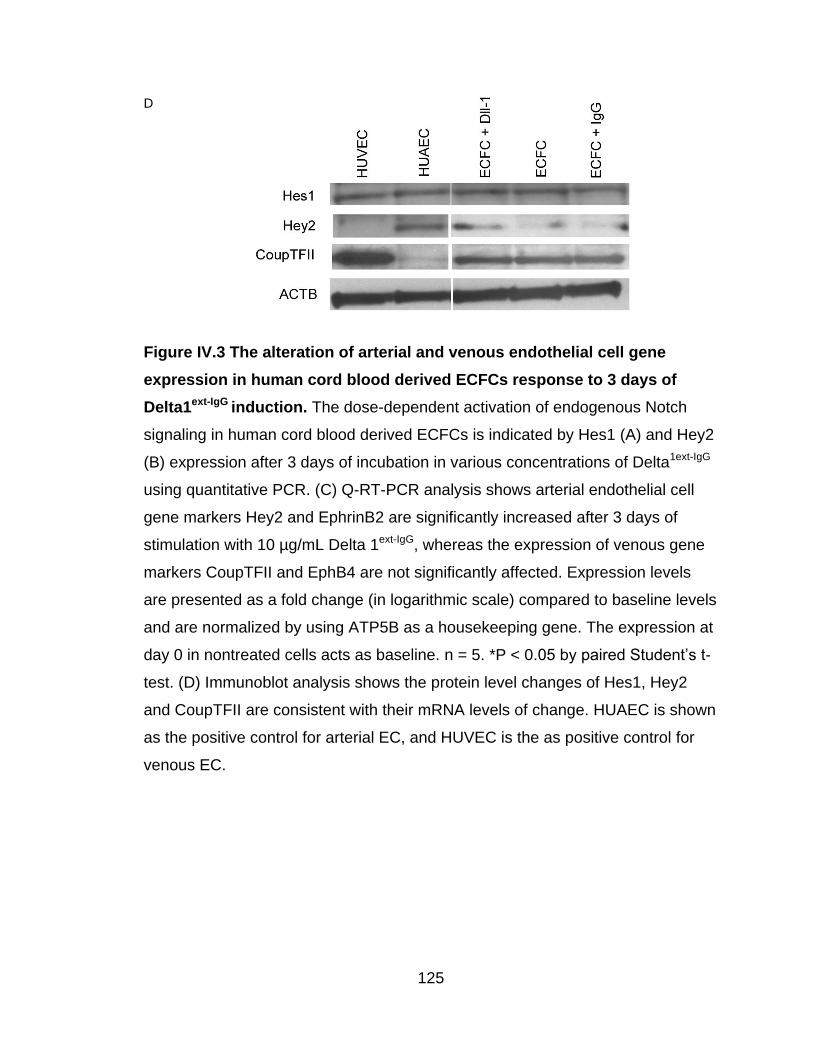
125
D
Figure IV.3 The alteration of arterial and venous endothelial cell gene
expression in human cord blood derived ECFCs response to 3 days of
Delta1ext-IgG induction. The dose-dependent activation of endogenous Notch
signaling in human cord blood derived ECFCs is indicated by Hes1 (A) and Hey2
(B) expression after 3 days of incubation in various concentrations of Delta1ext-IgG
using quantitative PCR. (C) Q-RT-PCR analysis shows arterial endothelial cell
gene markers Hey2 and EphrinB2 are significantly increased after 3 days of
stimulation with 10 µg/mL Delta 1ext-IgG, whereas the expression of venous gene
markers CoupTFII and EphB4 are not significantly affected. Expression levels
are presented as a fold change (in logarithmic scale) compared to baseline levels
and are normalized by using ATP5B as a housekeeping gene. The expression at
day 0 in nontreated cells acts as baseline. n = 5. *P < 0.05 by paired Student’s t-
test. (D) Immunoblot analysis shows the protein level changes of Hes1, Hey2
and CoupTFII are consistent with their mRNA levels of change. HUAEC is shown
as the positive control for arterial EC, and HUVEC is the as positive control for
venous EC.

126
Dll1-primed human cord blood ECFCs failed to enhance the formation of
arteriole-like microvessels in vivo
We further determined whether human cord blood ECFCs programmed to an
arterial endothelial cell-like fate will form arteriole or arteriole-like vessels after
they are implanted in vivo. ECFCs were cultured in a standard medium or treated
with 10 µg/mL of Delta1ext-IgG for 3 days; then they were suspended in a collagen-
fibronectin matrix and subcutaneously transplanted into immunodeficient mice.
After 14 days, the mice were euthanized; the grafts were harvested and analyzed
for blood vessel formation. H&E staining and immunohistochemistry analysis
using a specific anti-human CD31antibody revealed human cord blood ECFCs
were able to form microvessels perfused with murine red blood cells in the graft
(Figure IV.4A). Quantification of murine erythrocyte-containing human
microvessels (Figure IV. 4B) showed no statistical difference between control
and Delta1ext-IgG-treated cell implants (28.48 ± 14.86 vs. 36.37 ± 16.37
vessels/mm2, respectively). Furthermore, the size distribution of these functional
microvessels formed by treated and nontreated ECFCs were similar (Figure
IV.4B).
To further assess the formation of arterial-like microvessels, double-staining with
anti-human CD31 and anti-mouse α smooth-muscle actin was conducted, as
prior studies had demonstrated that human vessel recruitment of murine
perivascular support cells in vivo (Enis et al 2005, Melero-Martin et al 2008,
Melero-Martin et al 2007, Schechner et al 2000). Human ECFC-derived vessels

127
coated with murine αSMA-positive pericytes were indeed observed in the graft
(Figure IV.4C). However, there was no statistical increase of such vessels in
implants containing human cord blood ECFC primed with Delta1ext-IgG compared
to control nontreated cells and HUAECs (data not shown). Together, these data
indicate that in vitro Delta1ext-IgG-primed human cord blood ECFCs were unable to
enhance arterial-like vessel formation in vivo, although they can be induced to
express an arterial endothelial cell-like gene expression pattern in vitro.

128
A
B
C

129
D
Figure IV.4 in vitro Dll1 primed ECFCs fail to display significant skewing
toward arterial EC phenotype and function in vivo upon implantation. (A)
H&E staining indicates microvessel formation in collagen-fibronectin gel after 14
days of implantation. Anti-human CD31 staining further confirms the human
origin of these vessels. Results suggest that human cord blood derived ECFCs
cultured in the presence of Delta 1ext-IgG retain vessel-forming ability in vivo.
Moreover, the number of vessels formed by human cord blood derived ECFCs
and perfused with murine red blood cells per mm2 in the gel demonstrate no
observable difference between Delta 1ext-IgG treated and nontreated samples (B).
In additions, the size distribution of both populations of microvessels is similar
(C). (n = 6) (D) Vessels formed by human cord blood derived ECFCs are positive
for anti-human CD31 staining (green). Murine αSMA specifically reacts with
murine pericytes (red). Double-positive staining with human CD31 and murine
αSMA (*) indicates recruitment and investment of murine mural cells to human
ECFC-formed microvessels, thus representing an arterial EC property. However,
in vitro Dll1-primed ECFCs don’t skew to display arterial EC properties when
implanted in vivo; thus the number of double- positive staining vessels is not
significantly increased compared to cells under standard culture conditions.
Scale bar represents 100 µm.

130
Discussion
Studies in chick, zebrafish and mice have revealed molecular distinctions
between arterial and venous ECs, as well the signaling pathways underlying AV
specification during development (Aitsebaomo et al 2008, Rocha & Adams 2009,
Swift & Weinstein 2009). The most recent studies of murine embryonic stem cells
(ESCs) (Lanner et al 2007, Yurugi-Kobayashi et al 2006), human multipotent
adult progenitor cells (MAPCs) (Aranguren et al 2007) and human bone
mesenchymal stem cell (MSCs) (Zhang et al 2008) have illustrated the potential
of these stem cells to differentiate into cells displaying gene expression patterns
similar to arterial and/or venous ECs. However, no reports to date have shown
that human ECFCs are able to adopt an arterial or venous endothelial cell-like
fate. Thus, this study is the first to demonstrate that human cord blood derived
ECFCs heterogeneously express molecules that normally are restricted to
arterial and venous endothelium. In vitro, exposure to Notch ligand Dll1 is
sufficient to induce an arterial gene expression pattern in ECFCs. However, the
in vitro Dll1-primed ECFCs, when implanted, failed to display significant skewing
toward an arterial EC phenotype or function in vivo. Nonetheless, this study
contributes to the understanding of possible mechanisms involved in AV
differentiation in vitro and postnatal arteriogenesis in humans, which ultimately
may provide potential new therapeutic strategies for human cardiovascular
diseases.

131
Studies in zebrafish and mice have indicated that VEGF plays a critical role in
determining AV specification and that the concentration of VEGF influences this
determination. In murine ESCs, a high concentration of VEGF (50 ng/mL) drove
arterial endothelial cell gene expression (Dll4, Notch4, Nrp1 and EphrinB2), while
a low dosage of VEGF (2 ng/mL) encouraged venous endothelial cell gene
expression (CoupTFII) (Lanner et al 2007). Similarly, 100 ng/mL of rhVEGF165
could upregulate arterial endothelial cell gene marker expression in hMAPCs
(Dll4, Hey2, EphrinB1 and EphrinB2) and in hMSCs (Dll4, Notch4 and EphrinB2)
(Aranguren et al 2007, Zhang et al 2008). Our results consistently showed that
VEGF at 50 ng/mL was sufficient to enhance expression of the arterial
endothelial gene EphrinB2 only. Although the expression of Dll4 and Notch4 was
increased, the change was not statistically significant. Likewise, upon VEGF
stimulation, the expression of venous gene marker CoupTFII was downregulated
but not significantly. These findings suggest that a high concentration of VEGF is
able to induce the expression of at least one arterial endothelial cell gene in
cultured human circulating ECFCs.
Notch is another critical factor in determining and promoting vascular
development. Mice with deficiencies in Dll4, Notch1, RBP-Jk and Hey1/2 lose
expression of arterial endothelial cell gene markers (Duarte et al 2004, Fischer et
al 2004, Kokubo et al 2005, Krebs et al 2004, Uyttendaele et al 2001). Dll1
recently has been established as necessary for maintaining arterial identity
during fetal development and mediating postnatal arteriogenesis in mice with

132
hindlimb ischemia (Limbourg et al 2007, Sorensen et al 2009). Dll1 expression in
the vascular endothelium is detected in murine embryos at embryonic day 13.5
(E13.5) in arteries but not in capillaries and veins. Dll1-mediated Notch activity
via Notch1 is pivotal for controlling arterial identity during fetal development
(Sorensen et al 2009). Similarly, Dll1-dependent Notch signaling is required for
upregulation of EphrinB2 in growing arteries (Limbourg et al 2007). In this regard,
the signaling cascade (Dll-Notch/Hey2/EphrinB2) responsible for determining
arterial identity, which has been extensively studied in Dll4 (Aitsebaomo et al
2008, Rocha & Adams 2009, Swift & Weinstein 2009), is now also present in
Dll1. However, Dll1’s influence on arterial specification in human ECFCs has not
yet been addressed. In this study, we showed that human ECFCs were
responsive to Dll1 stimulation in a dosage-and time-dependent manner. After 3
days of Dll1 induction (10 µg/mL), the expression of critical arterial gene markers
(Hey2 and EphrinB2) were noticeably increased, which is consistent with prior
studies of HUVECs where Dll4 and Notch4 were constitutively expressed
(Shawber et al 2003, Williams et al 2006). On the other hand, venous endothelial
cell gene markers (CoupTFII and EphB4) were not influenced under Dll1
induction, which also has been observed when Notch4 or the Notch intracelluar
domain (NCID) was overexpressed in HUVECs (Iso et al 2006, Shawber et al
2003). Thus, these data illustrate that DII1 induction of arterial endothelial cell-
like gene expression is independent of suppression of the venous endothelial
cell-like phenotype in human circulating ECFCs cultured ex vivo.

133
Although an arterial endothelial cell-like gene expression pattern was able to be
induced in human circulating ECFCs in vitro upon Dll1 stimulation, these ECFCs
failed to display significant skewing toward an arterial EC phenotype and arterial
functions in vivo. A possible reason for this is that these cells were not
continuously exposed to Dll1 stimulation. It had been reported that only
immobilized Dll1 can activate Notch, and soluble Dll1 inhibits Notch (Varnum-
Finney et al 2000). Delta-1ext-IgG was not directly supplemented in the implanted
collagen-fibronectin gel in this present study. To overcome this diffusion
restriction, some carriers or scaffolds will need to be employed to immobilize Dll1
in the gel in future studies. In this case, both the cell carrier and cell density
should be determined to ensure that ligand-cell and cell-cell interfaces are
interacting directly to induce Notch activation.
It has been reported that Bcl2-transduced HUVECs suspended in a collagen-
fibronectin matrix and implanted under mouse abdominal skin were able to be
remodeled into arterioles, venules and capillaries after 60 days (Enis et al 2005,
Schechner et al 2000). Additionally, human ECs co-implanted with murine
mesenchymal precursor cells 10T1/2 or human bone-marrow-derived MSCs
were capable of giving rise to long-lasting, stable, engineered vessels and
demonstrating distinct arterial and venous functional characteristics (Au et al
2008a, Au et al 2008b). These findings suggest the potential of human ECs to be
differentiated into functional arterial ECs in vivo, which is consistent with our
short-term implantation outcomes. In the present study, anti-human CD31 and

134
anti-murine αSMA staining revealed that some human ECFC-formed vessels
were covered with murine periendothelial mesenchymal cells, which indicated
that human ECFCs adopt an arterial endothelial cell-like phenotype in vivo.
However, there was no dramatic increase in recruitment and investment of mural
cells to vessels derived from Dll1-primed ECFC compared to those formed by
nontreated ECFC or HUAEC (data not shown). Addition of mural cells to the
ECFC may be an additional strategy to provide Notch ligand exposure to the
implanted human ECFC and progeny.
In summary, we have demonstrated that human cord blood derived ECFCs are
not committed to either an arterial or venous fate during in vitro culture but can
be induced in vitro to display an arterial endothelial cell-like gene expression
pattern after exposure to Notch ligand Dll1. However, in vitro Dll1-treated ECFCs
fail to display significant skewing toward an arterial EC identity upon implantation,
suggesting that in vitro priming is not sufficient for in vivo specification in our
current experimental model. Our findings contribute to a greater understanding of
the mechanisms underlying A/V differentiation in human ECFCs and may also
provide novel insights into future therapeutic strategies for vascular regeneration
in human subjects with vascular disease

135
CHAPTER V
Acute Myocardial Infarction in Swine Rapidly and Selectively Releases
Highly Proliferative Endothelial Colony Forming Cells (ECFCs) into
Circulation
The following chapter represents a manuscript published in the journal Cell
Transplantation, 2007;16(9):887-97. PMID: 18293887
Contributing authors
Lan Huang, MS1, 2, 4, Dongming Hou, MD/PhD3,5, Meredith Thompson1, 2, Sarah
Baysden1, 2, W. Christopher Shelley, BA1, 2, David A. Ingram, MD1, 2, 4, 5, Keith L.
March, MD/PhD3,5, Mervin C. Yoder, MD1, 2, 4, 5
Department of Pediatrics1, Herman B Wells Center for Pediatric Research2,
Department of Medicine3, Department of Biochemistry and Molecular Biology4,
and Indiana Center for Vascular Biology and Medicine5, Indiana University
School of Medicine, Indianapolis, IN.

136
Abstract
Background: We have recently identified endothelial colony forming cells
(ECFCs) in human blood and blood vessels and ECFC are elevated in patients
with coronary artery disease. Since pigs are a favored model for studying
myocardial ischemia, we questioned whether ECFCs also exist in swine and
whether myocardial ischemia would alter the number of ECFC in circulation.
Methods and Results: ECFCs were present in circulating blood and aortic
endothelium of healthy pigs. In pigs with an acute myocardial infarction (AMI)
(n=9), the number of circulating ECFC was markedly increased compared to
sham control pigs (15 6 vs. 1 1 colonies/100cc blood, P < 0.05). Moreover,
the percentage of circulating high proliferative potential-ECFCs (HPP-ECFCs)
was significantly increased following AMI induction compared to sham control
(38.4 5.8% vs. 0.4 0.4%, P < 0.05) and to baseline (38.4 5.8% vs. 2.4
2.4%, P < 0.05) blood samples.
Conclusions: This is the first study to report that ECFCs are present in blood
and aorta in healthy pigs and that the number and distribution of circulating
ECFCs is altered following AMI. Since circulating ECFC are also altered in
human subjects with severe coronary artery disease, the pig model of AMI may
be an excellent preclinical model to test the role of ECFC in the pathophysiology
of AMI.

137
Introduction
In 1997, Asahara and colleagues described a cell population derived from human
circulating CD34+ cells that could differentiate ex vivo into cells with endothelial
cell-like characteristics (Asahara et al 1997). These cells were termed endothelial
progenitor cells (EPCs). Subsequent evidence suggested that these cells are
derived from bone marrow, circulate in peripheral blood, and play an important
role in both postnatal vasculogenesis and vascular homeostasis (Asahara &
Kawamoto 2004, Khakoo & Finkel 2005, Kubota et al 2003, Miyamoto et al 2004,
Rafii & Lyden 2003). However, the characterization of EPCs remains
controversial. Currently, human EPCs are defined as cells that express CD34,
CD133, and vascular endothelial growth factor receptor 2 (VEGFR2;
KDR)(Peichev et al 2000). Human EPCs and their progeny have also been
shown to express the endothelial markers von Willebrand factor (vWF), platelet
endothelial cell adhesion molecule-1 (CD31), and to ingest acetylated low-
density lipoprotein (LDL) and bind to Ulex europaeus agglutinin I (Ingram et al
2005a). Efforts to accurately define EPCs have been complicated by the
presence of circulating cells that have certain properties of endothelial cells, but
have a monocytic origin, termed circulating angiogenic cells (CACs) (Rehman et
al 2003), as well as mature circulating endothelial cells (CECs) (Blann et al 2005,
Dignat-George & Sampol 2000). These latter cells express the S-endothelial
antigen (CD146) but also express some of the same proteins as EPCs, such as
CD31, VEGFR2 and vWF.

138
In humans, another population of EPCs, termed late outgrowth endothelial cells
has been reported (Gulati et al 2003, Lin et al 2000). These are marrow-derived
cells that display robust proliferative potential ex vivo. Recently, we have
determined that the late outgrowth endothelial cells represent one subpopulation
of circulating endothelial colony forming cells (ECFCs) (Ingram et al 2004). A
complete hierarchy of ECFCs displaying varying levels of proliferative potential
has been isolated and characterized in human umbilical and adult peripheral
blood and as well as in a variety of blood vessels(Ingram et al 2005a, Ingram et
al 2005b, Ingram et al 2004). The cord blood ECFCs express high levels of
telomerase and contribute to new vessel formation when implanted in vivo
(Yoder et al 2007).
Several recent reports have demonstrated that increased circulating
concentrations of various populations of EPCs are correlated with reduced risk
for an adverse cardiovascular outcome (Chen et al 2004, Ding et al 2007, Eizawa
et al 2004, Güven et al 2006, Hill et al 2003, Loomans et al 2004, Mutunga et al
2001, Nadar et al 2005, Schmidt-Lucke et al 2005, Solovey et al 1997, Vasa et al
2001, Wang et al 2005, Werner et al 2005). However, patients with severe
coronary artery stenosis that are clinically selected for a revascularization
procedure also display the highest concentration of circulating late outgrowth
cells (Güven et al 2006), highlighting the linked questions whether the increase of
these cells represents a direct response to the occurrence of ischemia and
whether they play a risk-modulating role in the context of ischemia. Since pigs

139
are a favored species to examine the pathophysiology of coronary artery injury
and acute myocardial ischemia (AMI), we questioned whether circulating ECFCs
are present in the blood of healthy pigs and whether inducing an AMI would
specifically alter the circulating concentration of ECFCs.
We have determined that ECFCs are resident in the aortic endothelium of normal
pigs and circulate in pig peripheral blood, however, the frequency of these
circulating cells is extremely rare. The circulating ECFCs in healthy pigs display
proliferative potential that is limited. In pigs that have been stressed by an
induced AMI, however, ECFCs with high proliferative potential (HPP-ECFC)
promptly emerge in the circulation. Thus, swine possess circulating ECFCs as do
adult human subjects, and the onset of myocardial ischemia leading to infarction
is accompanied by an increase in concentration of the ECFCs, with preferential
mobilization of a highly proliferative subset.

140
Materials and Methods
Blood sample collection
Animal handing and care was performed under the recommendations of the
National Institutes of Health (NIH) Guide for the Care and Use of Laboratory
Animals. Yorkshire domestic pigs (25-35 Kg) of mixed gender were enrolled in
this study. Study animals were sedated with IM Ketamine (20 mg/kg), and
atropine (0.05 mg/kg), followed by IV sodium pentothal (25 mg/kg). After
intubation, anesthesia was maintained with Isoflurane (2.5%). Arterial access
was obtained via the common carotid artery using a cutdown technique, and an
8F vascular sheath was used to cannulate the artery. Left heart catheterization
was performed using a hockey-stick catheter (Cordis) for coronary cannulation.
A femoral artery sheath was placed to maintain continuous hemodynamic
monitoring during the procedure.
AMI was created by inflating an angioplasty balloon for 45 minutes in the
proximal LAD artery immediately distal to the first septal perforator. All study
animals received IV amiodarone to minimize ventricular arrhythmias; amiodarone
dosage was 75 mg IV bolus over 10 minutes given just prior to balloon inflation,
followed by 1 mg/min infusion continued for the duration between balloon
inflation and deflation. Nevertheless, sustained ventricular tachycardia and
ventricular fibrillation occurred during balloon inflation in every animal (2.9 1.6
episodes/procedure) and was terminated using biphasic DC cardioversion
(Medtronic). Peripheral blood was collected at baseline before AMI,

141
immediately post AMI and at sacrifice 7 days later (Figure V.1A). Sham control
pigs underwent all operative preparation, catheter instrumentation, and three DC
cardioversions but were not made ischemic (Figure V.1B).

142
A
B
Figure V.1 Overview of experimental design. The circles indicate blood
sampling times in the infarcted (A) and control sham pigs (B).
143
The extent of myocardial damage created by coronary occlusion was assessed
by tetrazolium trichloride (TTC) staining of harvested hearts (7 days following
AMI). TTC staining was performed using a standard protocol(Adegboyega et al
1997). The hearts were taken immediately after sacrifice and were sliced into 5
sections of 1.5-2.0 cm thickness along the apical-basal axis. These sections
were immersed into TTC solution for 10 minutes at 37C. Afterwards, MI areas
(pale areas not staining with TTC solution) were quantified by manual tracing
using NIH imaging software and were expressed as a percentage of the total left
ventricular area. The mean myocardial infarction was 26 ± 5% of the whole left
ventricular area in this series of 9 pigs. This model results in a decrease in left
ventricular ejection fraction approximating 46% (Hou et al 2005).
Isolation and culture of porcine blood derived endothelial cells
Blood (50-100ml) was diluted one to one with Hank’s/bovine serum albumin
(BSA)/ethylenediamine tetraacetic acid (EDTA) solution and layered (2:1 ratio)
onto Histopaque1119 (Sigma-Aldrich, St. Louis, MO). Cells were centrifuged for
30 minutes at 1800 rpm at room temperature (Beckman Coulter, Allgera Tm 6R
centrifuge). Mononuclear cells (MNCs) were isolated and washed three times
with endothelial cell basal medium-2 (EBM-2) (Cambrex, Walkersville, MD)
supplemented with 20% porcine plasma derived serum (PDS) (Animal
Technologies Inc. Tyler, TX), 2% antibiotic-Antimycotic (Invitrogen, Grand Island,
NY) (complete EGM-2 medium). MNCs were resuspended in 5 ml of complete
EGM-2 medium and cells were plated onto a six well plate pre-coated with mg/ml

144
type I rat tail collagen (BD Biosciences, Bedford, MA) and incubated at 37°C, 5%
CO2 in a humidified incubator. After 48 hours of culture, the non-adherent cells
were removed and complete EGM-2 medium was added to each well. Medium
was changed daily for the first seven days and then every other day until the first
passage.
Colonies of endothelial cells appeared in general following 7-14 days of culture
and were identified by their cobblestone appearance. Endothelial colonies were
enumerated by visual inspection with an inverted microscope (Olympus, Lake
Success, NY). These ECFC-derived endothelial cells were released from the
culture dish by 0.25% trypsin-EDTA (Gibco, Grand Island, NY) and replated onto
25 cm2 tissue culture flasks pre-coated with type I rat tail collagen for subsequent
passage.
Isolation and culture of porcine aortic endothelial cells
Endothelial cells were obtained from fresh aorta pieces by gently and thoroughly
scraping the vessel wall with a disposal scraper (Fisher Scientific, Pittsburgh,
PA). These cells were completely transferred from the edge of scraper into six
well plates filled with complete EGM-2 medium and pre-coated with type I rat tail
collagen. After 48 hours of culture, the non-adherent cells were removed and
complete EGM-2 medium was added to each well. Medium was changed every
other day until the first passage.

145
Endothelial colonies appeared on day 3 of culture and kept growing to form an
endothelial monolayer with typical cobblestone appearance. These aortic ECFC-
derived endothelial cells were released from the culture dish by 0.25% trypsin-
EDTA (Gibco, Grand Island, NY) and replated onto 25 cm2 tissue culture flasks
pre-coated with type I rat tail collagen for future passage.
Uptake of Dil-acetylated-low density lipoprotein (Dil-Ac-LDL)
Early passage (2-4) ECFC-derived endothelial cells were incubated with 10
µg/mL of Dil-Ac-LDL (Biomedical Technologies Inc., Stoughton, MA) in complete
EGM-2 medium for 4 hours at 37°C. Cells were washed three times, stained with
1.5µg/mL of 4’, 6-diamidino-2-phenylindole dihydrochloride (DAPI) (Sigma), and
examined by inspection through an inverted fluorescence microscope (Zeiss,
Thornwood, NY) at 32x magnification.
RT-PCR for eNOS
Total cellular RNA was extracted with TRIzol (Invitrogen, Grand Island, NY) in a
single-step method. RT reactions were performed using a SuperScript First-
Strand Synthesis system (Invitrogen, Grand Island, NY). PCR was performed
using the PuReTaq Ready-To-Go PCR Beads (GE Healthcare UK Limited, UK)
according to the manufacturer’s instructions. The primer sequences for pig eNOS
included: forward, 5’- AGCGGCTGCATGACATTGAGA-3’; reverse, 5’-
ATGTCCTCGTGATAGCGTTGCT-3’ with a 397 base pair expected band. The
primer sequences for pig GAPDH included: forward, 5’-

146
ACATCAAGAAGGTGGTGAAGCAGG-3’; reverse, 5’-
CACCCTGTTGCTGTAGCCAAAT-3’ with a 201 base pair expected band. The
PCR cycle was 94°C, 5 min; 94°C 30s, 57°C 30s, 72°C 1min, and 30 cycles with
a final 72°C 7 min cycle. PCR products were added to wells in a 2%
agarose/ethidium bromide gel, exposed to electophoresis current, and migrating
bands photographed under UV light.
Immunophenotyping of endothelial cells
Early passage (2-4) ECFC-derived endothelial cells were stained with
monoclonal antibodies including, mouse anti-rat/pig CD31 (RDI. Flanders, NJ),
mouse anti-pig CD45 (SEROTEC, UK), mouse anti-pig pan tissue (BD
Biosciences, Bedford, MA), mouse anti-pig SLA-DR (BD Biosciences, Bedford,
MA), or mouse isotype control antibody for 30 minutes at 4°C, washed three
times, and analyzed by fluorescence activated cell sorting (FACS) using a FACS
Calibur flow cytometer (Becton Dickinson, San Diego, CA) as previously
described (Ingram et al 2005a, Ingram et al 2005b, Ingram et al 2004, Yoder et al
2007).
Single cell assays
For early passage (2-4) ECFC-derived endothelial cells, the FACS Vantage
Sorter (Becton Dickinson) was used to place one cell per well into 96 well plates
pre-coated with type I rat tail collagen in 200µl of complete EGM-2 medium.
Cells were cultured at 37°C, 5% CO2 in a humidified incubator. Media was

147
changed every five days. After 14 days of culture, the cells were fixed with 4%
paraformaldehyde (Sigma, St. Louis, MO) in phosphate-buffered saline for 4
hours at room temperature, then washed twice, stained with 1.5 µg/ml DAPI, and
examined for the growth of endothelial cells. Those wells containing two or more
cells were identified as positive for proliferation under a fluorescent microscope
at 10x magnification. Wells containing fewer than 50 cells, were counted by
visual inspection with a fluorescent microscope at 10x magnification. For those
wells with more than 50 cells, colonies were imaged and cell number quantified
using an Image J 1.36v program (Wayne Rasband, NIH).
Statistical Analysis
Results are expressed as mean±SEM. Date were compared with nonparametric
(Wilcoxon or Mann-Whitney) test and significant differences were set at the P <
0.05 level. All analyses were performed using InStat 2.0 software.

148
Results
Characterization of porcine endothelial colony forming cells (ECFCs)
We have previously reported that human peripheral blood and vessels contain a
hierarchy of ECFCs (Ingram et al 2005a, Ingram et al 2005b, Ingram et al 2004).
However, the existence of this ECFC hierarchy in porcine blood and vascular
endothelium has not been demonstrated. To isolate ECFCs, we harvested swine
peripheral blood MNCs and aortic endothelial cells. Both ECFC-derived
endothelial cells (ECs) were able to form a monolayer with typical cobblestone
morphology of ECs (Figure V.2A,B). Generally there were 4 2 colonies growing
from 8.5 ± 4.0 x 10⁸ blood MNCs of healthy swine (n = 12). The appearance of
the first aortic EC colonies (3 0 days) was earlier than any colonies identified
from PB (11 1 days); from our human studies, those colonies emerging early
generally display higher proliferative potential (Ingram et al 2005a, Ingram et al
2005b, Ingram et al 2004). The PB and aortic ECFC-derived cells ingested Dil-
labeled acetylated low-density lipoprotein (AcLDL) (Figure V.3A). Moreover,
endothelial nitric oxide synthase (eNOS), an enzyme generally thought to be
endothelial-specific, was expressed at the mRNA level in both aortic and PB
ECFC- derived ECs using reverse transcription-polymerase chain reaction (RT-
PCR) (Figure V.3B). To evaluate whether these cells displayed cell surface
proteins with an EC phenotype, we assayed the ECFC-derived ECs for the
expression of EC and leukocyte surface markers. All the ECFC-derived cells
showed positive immunostaining for anti-platelet endothelial adhesion cell
adhesion molecule-1 (CD31) and anti-pan-tissue antibodies, but did not express

149
either the common leukocyte surface marker CD45 or the SLA-DR antigen
(Figure V.3C). Accordingly, the cultured cells derived from pig aorta and
peripheral blood ECFCs were confirmed to be endothelial cells by both
phenotypic and functional properties.

150
Control group (n=3) Experimental group (n=9)
Baseline Instrumented Baseline Post MI Sacrifice
MNCs (x108) 8.5±4.0 2.3±1.6 3.5±1.3 4.3±0.9 3.1±0.6
ECFC
Colony
5±2 1±1 5±2 15±6 6±2
Values are mean±SEM, Instrumented vs. Post MI, p < 0.05
Table V.1 The number of MNC and ECFC colony in 100cc porcine blood

151
Figure V.2 The formation of porcine aortic and PB ECFC-derived
endothelial colony. (A) Aortic ECFC-derived endothelial colony as it appeared
on day 3, day 5, day 7 and day 10 of culture. (B) PB ECFC-derived endothelial
colony from adherent MNCs emerged on day 8, day 10, day 12 and day 15 of
culture. (10x magnification, scale bar represents 100µm)

152
A
B
C
Figure V.3 Phenotypic and functional analysis of both aortic and PB ECFC-
derived ECs. (A) Aortic and PB ECFC-derived ECs incorporated Dil-Ac-LDL
(red) and also stained with DAPI (blue) (32x magnification, scale bar represents
100m). (B) Expression of eNOS in aortic and PB ECFC-derived ECs by RT-
PCR. (C) Immunophenotyping of MNCs and cell monolayers derived from aortic
and PB ECFC-derived ECs by fluorescence cytometry. Both aorta and PB
derived ECs expressed CD31, and Pan-tissue but not CD45 or SLA-DR
antigens.

153
Hierarchical organization of porcine ECFC proliferative potential
Circulating EPCs have been reported to serve as a robust biomarker for
predicting the severity and risk of adverse cardiovascular illnesses in human
subjects (Hill et al 2003, Werner et al 2005). Though EPCs are heterogeneously
defined, EPCs (including ECFCs) and CECs have been used as biomarkers. We
recently developed a single cell colony-forming assay to quantitate the
clonogenic potential of individual human EPCs (Ingram et al 2005a, Ingram et al
2005b, Ingram et al 2004). Using this assay we tested whether there was a
hierarchy of proliferative potential in ECFC present in porcine vascular
endothelium and peripheral blood. As shown in Figure V.4A, 70.4 13.9% of
single plated aortic ECFC-derived and 46.2 14.4% of PB ECFC-derived ECs
divided at least once during 14 days of culture. The hierarchy of ECFC is
comprised of colonies that may be further described by the total number of
progeny produced during a 14 day culture. Thus, single cells that gave rise to a
colony containing more than 2000 cells were called high proliferative potential-
endothelial colony forming cells (HPP-ECFC). Single cells that formed a colony
containing 51 to 2000 cells were called low proliferative potential-endothelial
colony forming cells (LPP-ECFC), whereas single cells that produced 2-50 cells
were defined as endothelial cluster forming cells, and mature non-dividing
endothelial cells retained no proliferative potential. In healthy animals at
baseline, both aorta and PB contained ECFC with the full complement of dividing
and non-dividing cells. The majority of these ECFC-derived colonies contained
less than 2000 cells, however approximately 10.3 3.5% of aortic ECFC and 1.0

154
1.0% of PB ECFC were classified as HPP-ECFC (Figure V.4B-C).
Photomicrographs of the size of the various endothelial colonies/clusters are
shown in Figure V.4D. To determine if the HPP-ECFC-derived progeny
possessed self-renewal ability (cells displaying the ability to give rise to colonies
of similar or greater proliferative potential), we replated aortic and PB HPP-ECFC
progeny in a single cell clonal assay. After replating, 39.6 7.0% of the clonal
progeny of single aortic HPP-ECFC could give rise to all subsequent stages of
endothelial progenitors in addition to forming secondary HPP-ECFC (Figure
V.4E-F). Similarly, 23.9 2.4% of clonal progeny of single PB HPP-ECFC were
able to form secondary colonies upon replating. (Figure V.4E, G). These colonies
could be further replated to form tertiary colonies (data not shown). Only HPP-
ECFC retained the ability to self-renew into secondary colonies with retained
HPP-ECFC activity, distinguishing this colony as functionally distinct from LPP-
ECFC or endothelial cluster forming cells. Thus, a hierarchy of endopoiesis was
demonstrated to exist in porcine endothelial cells isolated from aorta and from
circulating blood.

155
A
B
C
D
E
F
G

156
Figure V.4 Quantitation of the clonogenic and proliferative potential of
single endothelial cells derived from aortic and PB ECFCs. (A) The
percentage of single aortic and PB ECFC-derived ECs dividing at least once
after 14 days culture. (B-C) The distribution of different size of colonies derived
from single aortic (B) and PB (C) derived ECs in an individual well after 14
days of culture. (D) Representative DAPI- stained (blue) photomicrographs
(10x magnification, scale bar represents 100µm) of the different endothelial cell
clusters (< 50 cells) and colonies (> 50 cells) derived from single ECs. (E) The
percentage of single endothelial cells from primary aortic and blood HPP-
ECFCs that gave rise to secondary colonies after 14 days of culture. (F-G) The
distribution of different sizes of secondary colonies derived from single
endothelial cells, which in turn were derived from primary aortic (F) and blood
(G) HPP-ECFCs. Results represent the average of at least three independent
experiments from different animals. (n=5)

157
Alteration of the distribution of ECFCs in a porcine model of acute
myocardial ischemia (AMI)
A variety of reports indicate that the concentration of EPCs and CECs changes in
human subjects with acute cardiovascular injury and in those at risk for
developing an adverse cardiovascular event (Chen et al 2004, Ding et al 2007,
Eizawa et al 2004, Güven et al 2006, Hill et al 2003, Loomans et al 2004,
Mutunga et al 2001, Nadar et al 2005, Schmidt-Lucke et al 2005, Solovey et al
1997, Vasa et al 2001, Wang et al 2005, Werner et al 2005). To examine
whether the distribution of ECFCs in the circulating blood was affected during a
controlled ischemic cardiovascular stress, we isolated blood derived ECFCs from
pigs undergoing an experimental AMI. The AMI was created by balloon catheter
inflation in the left anterior descending coronary artery (LAD) for 45 minutes. We
harvested blood samples (n=9) before AMI (baseline), following AMI (post AMI)
and at sacrifice 7 days later (sacrificed). Additionally, sham control pigs were
anaesthetized, instrumented, and experienced 3 cardioversion procedures, but
the balloon was not inflated to induce myocardial injury. There was no statistical
difference in the blood MNC cell count between the 2 groups at baseline, post
infarction and sacrifice (Table V.1). However, the number of circulating ECFC
was noticeably higher in post AMI samples than in sham control pig blood (15 6
vs. 1 1 colonies/100cc blood, P < 0.05). Thus, AMI causes a significant
increase in circulating ECFC in affected pigs.

158
Next, we questioned whether the distribution of ECFC proliferative potential
would be altered following ischemia in these different test blood samples. There
was no statistical difference in the percentage of single cells dividing at least
once between the groups at baseline, post infarction and 7 days later (Figure
V.5A-B). Strikingly, the percentage of single cells giving rise to colonies of more
than 2000 cells (HPP-ECFCs) dramatically increased in the blood following AMI
compared to the sham control blood samples (38.4 5.8% vs. 0.4 0.4%, P <
0.05) (Figure V.5C) and also to baseline blood samples (38.4 5.8% vs. 2.4
2.4%, P < 0.05) (Figure V.5D). On the other hand, the percentage of single cells
that formed colonies containing 51-500 cells remarkably decreased following
infarction compared to sham control samples (29.2 2.2% vs. 76.2 4.5%, P <
0.05) (Figure V.5C). The percentage of endothelial clusters (endothelial colony
with 2-50 cells) present in the blood after AMI was also reduced compared to
baseline (Figure V.5D). Finally, the percentage of HPP-ECFC in the blood
samples drawn 7 days after AMI remained 2-fold greater than that measured at
baseline, but this trend did not reach significance. In contrast, the distribution of
ECFC in sham control animals was not disturbed from baseline following
instrumentation (Figure V.5E).

159
A
B
C
D
E
Figure V.5 Quantitation of the clonogenic and proliferative potential of
single endothelial cells derived from PB ECFCs in the swine undergoing
experimental AMI. (A-B) The percentage of single PB ECFC-derived ECs
dividing at least once after 14 days culture in the sham control group (n=3) (A)
and in the experimental group (n=5) (B) when isolated at the indicated
timepoints. (C) The comparison of the distribution of different sized colonies
from a single PB ECFC-derived ECs in an individual well after 14 days of
culture between Post MI and sham control, each isolated at 45 min following

160
coronary cannulation (D-E). The harvest time-dependence of the distribution of
different sized colonies from single PB ECFC-derived ECs are shown for
individual wells in the experimental group (D) and in the control group (E).
Observations were made after 14 days of culture. *P<0.05 by nonparametric
(Wilcoxon or Mann-Whitney) test.

161
Discussion
This is the first study to define a hierarchy of ECFCs circulating in the
bloodstream or present in blood vessels in swine. Using a single cell clonogenic
assay, ECFCs could be resolved based on their proliferative potential. Further,
we find that the number and distribution of circulating ECFCs is altered in
experimental pigs within 45 minutes following onset of AMI.
Previously we reported a hierarchy of ECFCs exists in human blood and blood
vessels (Ingram et al 2005a, Ingram et al 2005b, Ingram et al 2004). The ECFCs
were present in higher frequency in human umbilical cord blood than in adult
peripheral blood and the distribution of ECFCs was weighted toward more HPP-
ECFC in the cord blood samples. The human ECFCs displayed a variety of cell
surface proteins previously identified on human blood vessel endothelium and
formed blood vessels when implanted in collagen gels into immunodeficient mice
(Yoder et al 2007). Thus, human ECFCs display properties consistent with
stem/progenitor cells for the endothelial lineage. Using the same strategy, a
hierarchy of ECFCs was identified in porcine circulating blood and blood vessels
in the current study.
Growing evidence suggests that changes in the number of EPCs and CECs
correlate with the risk of developing certain human clinical disorders. A decrease
in number or impaired function of EPCs has been observed in patients with
diabetes, heart failure and coronary artery diseases (Chen et al 2004, Eizawa et

162
al 2004, Loomans et al 2004, Tepper et al 2002). Moreover, the number and
function of EPCs inversely correlates with the occurrence of cardiovascular
events and death from cardiovascular causes (Hill et al 2003, Schmidt-Lucke et
al 2005, Vasa et al 2001, Werner et al 2005). Conversely, elevated numbers of
CECs have been demonstrated in patients with a variety of clinical disorders
such as sickle cell crisis, septic shock, acute myocardial infarction, acute
ischemic stroke and coronary artery disease (Güven et al 2006, Mutunga et al
2001, Nadar et al 2005, Solovey et al 1997, Wang et al 2005). Consistent with
these findings, late outgrowth ECFCs are increased in the bloodstream of
patients with severe coronary artery disease (Güven et al 2006). Based on these
human clinical observations, we have hypothesized that the change in circulating
ECFC count and/or distribution could also be a surrogate biological marker for
vascular function and cumulative cardiovascular risk and be utilized to predict the
outcomes of vascular diseases.
In this study, the frequency of circulating ECFCs in the blood of normal swine
was quite low at baseline, but quickly increased with the induction of cardiac
injury. Additionally, our study, based on a single cell clonogenic assay, provides
evidence that an AMI could affect the proliferative distribution of ECFCs,
specifically stimulating more HPP-ECFC emergence in the circulating blood.
These results suggest that an AMI induces either an acute release of the ECFC
from the damaged area or release of a mobilizing molecule that recruits the HPP-
ECFC into the circulation. Of interest, Güven et al (Güven et al 2006) recently

163
reported that the concentration of late outgrowth endothelial cells is increased in
those patients with the most severe coronary artery disease, and those patients
selected for a revascularization procedure displayed a significantly higher
number of circulating late outgrowth endothelial colony forming cells than
patients who did not require a revascularization procedure; the mechanisms
underlying this finding are not yet known. Multiple growth factors and cytokines
are known to be released in the setting of AMI or exercise-induced ischemia, but
their significance for ECFC dynamics is not known. The establishment here of an
animal model with the property of robust and predictable ECFC mobilization
forms the background for future studies directed towards uncovering potential
molecular mechanisms for this mobilization. A comparison of factors circulating
at baseline and during ischemia, and selective manipulation of these factors
should provide insight into these mechanisms as well as potential future
therapeutic targets.
Endothelial cell proliferation in normal, mature vessels in most mammals is
reported to be extremely low. In some experimental animals, such as rats, guinea
pigs and dogs tritiated thymidine labeling studies demonstrated that 0.1-3.0% of
endothelial cells proliferate daily (Caplan & Schwartz 1973, Schwartz & Benditt
1973, Schwartz & Benditt 1976). In contrast to this slow turnover of endothelial
cells in normal vessels, in vitro plating of swine aortic endothelial cells was
associated with rapid proliferation, a finding consistent with numerous studies
using human or bovine vessel-derived endothelial cells (Bompais et al 2004,

164
Cajero-Juarez et al 2002, Ingram et al 2005b, Ryan et al 1978). Furthermore, a
complete hierarchy of ECFCs was present in the porcine vessel wall-derived
endothelial cells, similar to that observed in circulating blood and consistent with
that published for human blood and blood vessel-derived endothelial cells
(Ingram et al 2005a, Ingram et al 2005b, Ingram et al 2004). These data suggest
that some of the endothelial cells residing in the blood vessel intimal layer under
normal circumstances are quiescent but retain proliferative potential that is
displayed upon releasing the cells from contact inhibition and plating the cells in
vitro. Future studies will be required to determine whether the source of inducible
ECFC mobilization in porcine and human subjects is derived from the vascular
endothelial intima of the host vessels or from extravascular marrow niches.
In this study we compared the expression of a number of cell surface antigens in
porcine hematopoietic MNCs and aortic and blood ECFC-derived ECs. Both
types of endothelial cells showed similar expression pattern of these surface
proteins, which was different from that of blood MNCs (Figure V.3C).
Interestingly, these porcine ECFC-derived ECs expressed some surface antigens
that were reported to be present in hematopoietic cells, such as the pan-tissue
marker (granulocytes, monocytes and lymphocytes), whereas the common
leukocyte surface marker CD45 and SLA-DR were not detected. Available
evidence suggests that during embryonic development the hematopoietic stem
cells (HSCs) and EPCs are derived from a common precursor, the
hemangioblast, and then differentiate into different lineages and play distinct

165
functions (Baron 2003, Cogle & Scott 2004). This fact has led some investigators
to postulate that circulating blood cells isolated from human and murine bone
marrow transplant recipients or following development of leukemia provide
evidence of a hemangioblast precursor for the hematopoietic and endothelial
lineages in these adult subjects. Other than the pan-tissue marker, we saw no
other evidence of cell surface antigen co-expression of markers between the
hematopoietic and ECFC in this study.
In conclusion, we report that ECFCs are present in normal circulating swine
blood and comprise a proportion of the endothelial cells that represent the aortic
endothelial intima. Though the circulating ECFCs are rare, some of these cells
possess remarkable proliferative potential, also displayed by some of the ECFCs
that reside as aortic endothelial cells. The onset of acute myocardial ischemia
leads to prompt changes in circulating ECFC number and the selective
mobilization of highly proliferative subpopulations. Identification of populations of
circulating and resident vascular endothelial cells with such potent proliferative
potential may improve our understanding of the cellular mechanisms of
angiogenesis and permit new strategies to manipulate these cells to protect the
vascular endothelium from injury and disease.

166
CHAPTER VI
Summary and Perspectives
Circulating EPCs have been identified for more than a decade and have been
indicated to contribute to neovascularization via postnatal vasculogenesis.
However, the definition of an EPC remains controversial. Our lab has
successively isolated and identified ECFCs in circulating blood and blood vessels
in human and other species. Based on the strictest definition of an EPC as a cell
that 1) displays the ability to produce endothelial progeny that form endothelial
tubes in vitro and 2) contributes to the functional endothelial lining of injured
vascular structure via angiogenesis and/or vasculogenesis in vivo, to date, only
ECFCs fit the definition of true EPCs and display all the activities described on
these chapters. Furthermore, we have extended this definition and identified the
stem/progenitor cells for the corneal endothelial lineage.
Corneal endothelium is formed by migration and proliferation of neural crest-
derived mesenchymal cells and plays a barrier-bump function to maintain corneal
clarity. Unfortunately, corneal ECs fail to proliferate in vivo even in response to
corneal trauma or diseases which causes cell loss and eventually leads to the
loss of vision. However, they do display replicative capacity in vitro. In the
present study, using a single-cell clonogenic assay, we are able to first
demonstrate that not all corneal ECs display the same ability to proliferate at a
clonal level. We have defined a hierarchy of ECFCs in bovine corneal
endothelium based on their proliferative potential, which is quite similar to bovine

167
vascular ECs resident in aorta, coronary artery, and pulmonary artery and is
consistent with circulating and resident vascular ECs in adult human subjects.
Thus, this study provides a new conceptual framework for defining corneal EPC
potential and contributes to identify corneal endothelial precursor which is
currently controversial. Additionally, HPP-ECFCs in corneal endothelium with
their vast proliferative potential may represent a novel cell source for
reconstituting corneal endothelium and be of great benefit to corneal
transplantation.
Although human cord blood derived ECFCs are able to de novo form vessels,
molecular mechanisms underlying the roles of ECFCs in neoangiogensis remain
largely unknown. Several fundamental biological questions on EPCs’ mobilization,
homing, differentiation, lumenization and more are pending answered. One of
them that whether circulating ECFCs can be specified to an arterial or venous
fate has so far not been addressed. Thus, we hypothesized that human cord
blood derived ECFCs are not committed to either an arterial or venous fate and
are able to be differentiated into arterial ECs when given appropriate stimuli. In
the present study, we first demonstrate that human cord blood derived ECFCs
heterogeneously express molecules that normally are restricted to ECs resident
in arteries and veins. In vitro, exposure to Notch ligand Dll1 is sufficient to induce
an arterial gene expression pattern in ECFCs. However, the in vitro Dll1-primed
ECFCs, when implanted, failed to display significant weighting toward an arterial
EC phenotype or function in vivo. Therefore, the in vivo formation of human cord
168
blood ECFCs derived, long-lasting, functional vessels with an arterial identity
needs further investigated probably by manipulating implant microenvironments.
Nevertheless, this study contributes to the understanding of possible
mechanisms involved in AV differentiation in vitro and postnatal arteriogenesis in
humans and provides novel insights into the development of cellular therapeutic
strategies for patients with coronary and peripheral arterial diseases.
There is growing evidence suggesting that the concentration of circulating ECs is
close related to the adverse progression of cardiovascular disorders. Based on
human clinical observations, we hypothesized that the alteration in circulating
ECFC count and/or proliferative distribution could be a surrogate biological
marker for vascular function and cumulative cardiovascular risk and be utilized to
predict the outcomes of vascular diseases. Pigs are a favored species to
examine the pathophysiology of coronary artery injury and acute myocardial
ischemia (AMI). By using this animal model, we have demonstrated that the
number of circulating ECFCs in the bloodstream in healthy swine is quite rare.
The onset of AMI leads to a prompt increase in circulating ECFC number and a
selective mobilization of HPP-ECFCs. The exact role of the mobilized HPP-
ECFCs in homing and participation in repair of the ischemic tissue remains
unknown. Moreover, multiple growth factors and cytokines have been known to
be released in the setting of AMI. Future, a comparison of factors circulating
before, during and after ischemia, and selective manipulation of these factors
would provide insights into these uncovering potential molecular mechanisms for

169
the mobilization of HPP-ECFCs. The identification and characterization of HPP-
ECFC may improve our understanding of the cellular mechanisms of
neoangiogenesis and permit new strategies to manipulate these cells to protect
the vascular endothelium from injury and disease.
Based on their multiple therapeutic potentials, human ECFCs have become an
attractive target for novel vascular regenerative therapies. However, the study of
ECFCs is hampered by several challenges in culturing cells. Current protocols for
in vitro expansion of ECFCs mostly depend on the presence of FBS or FCS in
the culture medium which may disturb chromosome stability, modify gene
expression and thereafter affect cell properties and functions, and have risky
potentials of transmitting infectious agents and inducing immune reactions when
future used in a transplantation setting. According to these regards, we have
developed a novel formula of endothelial cell culture medium that is free of FBS
and is supplemented with 1.5% HCP for expansion of human cord blood ECFCs
in vitro. In creating this medium, we have also demonstrated that ECFCs grown
on this medium retain their phenotype and function, as well as their proliferative
and neoangiogenic properties. This work contributes to the development of a
complete serum-free culture system for ECFCs that we and others believe will be
required for future use in human cell therapeutics.

170
In summary, these studies contribute to an improved understanding of ECFCs
and their roles in postnatal vascularization and suggest several possible
therapeutic applications of ECFCs.

171
REFERENCES
Adams RH, Alitalo K. 2007. Molecular regulation of angiogenesis and lymphangiogenesis. Nat Rev Mol Cell Biol 8: 464-78
Adams RH, Diella F, Hennig S, Helmbacher F, Deutsch U, Klein R. 2001. The cytoplasmic domain of the ligand ephrinB2 is required for vascular morphogenesis but not cranial neural crest migration. Cell 104: 57-69
Adams RH, Wilkinson GA, Weiss C, Diella F, Gale NW, et al. 1999. Roles of ephrinB ligands and EphB receptors in cardiovascular development: demarcation of arterial/venous domains, vascular morphogenesis, and sprouting angiogenesis. Genes Dev 13: 295-306
Adegboyega PA, Adesokan A, Haque AK, Boor PJ. 1997. Sensitivity and specificity of triphenyl tetrazolium chloride in the gross diagnosis of acute myocardial infarcts. Arch Pathol Lab Med 121: 1063-8
Ahmed Z, Bicknell R. 2009. Angiogenic signalling pathways. Methods Mol Biol 467: 3-24
Ahn DG, Ruvinsky I, Oates AC, Silver LM, Ho RK. 2000. tbx20, a new vertebrate T-box gene expressed in the cranial motor neurons and developing cardiovascular structures in zebrafish. Mech Dev 95: 253-8
Aird WC. 2007a. Phenotypic heterogeneity of the endothelium: I. Structure, function, and mechanisms. Circ Res 100: 158-73
Aird WC. 2007b. Phenotypic heterogeneity of the endothelium: II. Representative vascular beds. Circ Res 100: 174-90
Aitsebaomo J, Portbury AL, Schisler JC, Patterson C. 2008. Brothers and sisters: molecular insights into arterial-venous heterogeneity. Circ Res 103: 929-39
Akerman ME, Pilch J, Peters D, Ruoslahti E. 2005. Angiostatic peptides use plasma fibronectin to home to angiogenic vasculature. Proc Natl Acad Sci U S A 102: 2040-5
Albelda SM, Muller WA, Buck CA, Newman PJ. 1991. Molecular and cellular properties of PECAM-1 (endoCAM/CD31): a novel vascular cell-cell adhesion molecule. J Cell Biol 114: 1059-68
Allende ML, Proia RL. 2002. Sphingosine-1-phosphate receptors and the development of the vascular system. Biochim Biophys Acta 1582: 222-7
Allende ML, Yamashita T, Proia RL. 2003. G-protein-coupled receptor S1P1 acts within endothelial cells to regulate vascular maturation. Blood 102: 3665-7
Alva JA, Iruela-Arispe ML. 2004. Notch signaling in vascular morphogenesis. Curr Opin Hematol 11: 278-83
Amano S. 2003. Transplantation of cultured human corneal endothelial cells. Cornea 22: S66-74
Ara T, Tokoyoda K, Okamoto R, Koni PA, Nagasawa T. 2005. The role of CXCL12 in the organ-specific process of artery formation. Blood 105: 3155-61
Aragones J, Schneider M, Van Geyte K, Fraisl P, Dresselaers T, et al. 2008. Deficiency or inhibition of oxygen sensor Phd1 induces hypoxia tolerance by reprogramming basal metabolism. Nat Genet 40: 170-80

172
Aranguren XL, Luttun A, Clavel C, Moreno C, Abizanda G, et al. 2007. In vitro and in vivo arterial differentiation of human multipotent adult progenitor cells. Blood 109: 2634-42
Argraves WS, Drake CJ. 2005. Genes critical to vasculogenesis as defined by systematic analysis of vascular defects in knockout mice. Anat Rec A Discov Mol Cell Evol Biol 286: 875-84
Arman E, Haffner-Krausz R, Chen Y, Heath JK, Lonai P. 1998. Targeted disruption of fibroblast growth factor (FGF) receptor 2 suggests a role for FGF signaling in pregastrulation mammalian development. Proc Natl Acad Sci U S A 95: 5082-7
Armulik A, Abramsson A, Betsholtz C. 2005. Endothelial/pericyte interactions. Circ Res 97: 512-23
Asahara T, Kawamoto A. 2004. Endothelial progenitor cells for postnatal vasculogenesis. Am J Physiol Cell Physiol 287: C572-9
Asahara T, Murohara T, Sullivan A, Silver M, van der Zee R, et al. 1997. Isolation of putative progenitor endothelial cells for angiogenesis. Science 275: 964-7
Au P, Daheron LM, Duda DG, Cohen KS, Tyrrell JA, et al. 2008a. Differential in vivo potential of endothelial progenitor cells from human umbilical cord blood and adult peripheral blood to form functional long-lasting vessels. Blood 111: 1302-5
Au P, Tam J, Fukumura D, Jain RK. 2008b. Bone marrow-derived mesenchymal stem cells facilitate engineering of long-lasting functional vasculature. Blood 111: 4551-8
Avraamides CJ, Garmy-Susini B, Varner JA. 2008. Integrins in angiogenesis and lymphangiogenesis. Nat Rev Cancer 8: 604-17
Bar RS, Boes M, Booth BA, Dake BL, Henley S, Hart MN. 1989. The effects of platelet-derived growth factor in cultured microvessel endothelial cells. Endocrinology 124: 1841-8
Baron MH. 2003. Embryonic origins of mammalian hematopoiesis. Exp Hematol 31: 1160-9
Battegay EJ, Rupp J, Iruela-Arispe L, Sage EH, Pech M. 1994. PDGF-BB modulates endothelial proliferation and angiogenesis in vitro via PDGF beta-receptors. J Cell Biol 125: 917-28
Baumwell S, Karumanchi SA. 2007. Pre-eclampsia: clinical manifestations and molecular mechanisms. Nephron Clin Pract 106: c72-81
Bertolini F, Shaked Y, Mancuso P, Kerbel RS. 2006. The multifaceted circulating endothelial cell in cancer: towards marker and target identification. Nat Rev Cancer 6: 835-45
Betsholtz C, Lindblom P, Gerhardt H. 2005. Role of pericytes in vascular morphogenesis. EXS: 115-25
Bieback K, Hecker A, Kocaomer A, Lannert H, Schallmoser K, et al. 2009. Human Alternatives to Fetal Bovine Serum for the Expansion of Mesenchymal Stromal Cells from Bone Marrow. Stem Cells

173
Bix G, Fu J, Gonzalez EM, Macro L, Barker A, et al. 2004. Endorepellin causes endothelial cell disassembly of actin cytoskeleton and focal adhesions through alpha2beta1 integrin. J Cell Biol 166: 97-109
Blann AD, Woywodt A, Bertolini F, Bull TM, Buyon JP, et al. 2005. Circulating endothelial cells. Biomarker of vascular disease. Thromb Haemost 93: 228-35
Bompais H, Chagraoui J, Canron X, Crisan M, Liu XH, et al. 2004. Human endothelial cells derived from circulating progenitors display specific functional properties compared with mature vessel wall endothelial cells. Blood 103: 2577-84
Breier G, Risau W. 1996. The role of vascular endothelial growth factor in blood vessel formation. Trends Cell Biol 6: 454-6
Brotherton TW, Chui DH, McFarland EC, Russell ES. 1979. Fetal erythropoiesis and hemoglobin ontogeny in tail-short (Ts/+) mutant mice. Blood 54: 673-83
Broudy VC, Kovach NL, Bennett LG, Lin N, Jacobsen FW, Kidd PG. 1994. Human umbilical vein endothelial cells display high-affinity c-kit receptors and produce a soluble form of the c-kit receptor. Blood 83: 2145-52
Bruckner K, Pasquale EB, Klein R. 1997. Tyrosine phosphorylation of transmembrane ligands for Eph receptors. Science 275: 1640-3
Burri PH, Hlushchuk R, Djonov V. 2004. Intussusceptive angiogenesis: its emergence, its characteristics, and its significance. Dev Dyn 231: 474-88
Cai H, Reed RR. 1999. Cloning and characterization of neuropilin-1-interacting protein: a PSD-95/Dlg/ZO-1 domain-containing protein that interacts with the cytoplasmic domain of neuropilin-1. J Neurosci 19: 6519-27
Cai W, Vosschulte R, Afsah-Hedjri A, Koltai S, Kocsis E, et al. 2000. Altered balance between extracellular proteolysis and antiproteolysis is associated with adaptive coronary arteriogenesis. J Mol Cell Cardiol 32: 997-1011
Cajero-Juarez M, Avila B, Ochoa A, Garrido-Guerrero E, Varela-Echavarria A, et al. 2002. Immortalization of bovine umbilical vein endothelial cells: a model for the study of vascular endothelium. Eur J Cell Biol 81: 1-8
Caplan BA, Schwartz CJ. 1973. Increased endothelial cell turnover in areas of in vivo Evans Blue uptake in the pig aorta. Atherosclerosis 17: 401-17
Carlson TR, Yan Y, Wu X, Lam MT, Tang GL, et al. 2005. Endothelial expression of constitutively active Notch4 elicits reversible arteriovenous malformations in adult mice. Proc Natl Acad Sci U S A 102: 9884-9
Carmeliet P. 2000. Mechanisms of angiogenesis and arteriogenesis. Nat Med 6: 389-95
Carmeliet P, Dor Y, Herbert JM, Fukumura D, Brusselmans K, et al. 1998. Role of HIF-1alpha in hypoxia-mediated apoptosis, cell proliferation and tumour angiogenesis. Nature 394: 485-90
Carmeliet P, Ferreira V, Breier G, Pollefeyt S, Kieckens L, et al. 1996. Abnormal blood vessel development and lethality in embryos lacking a single VEGF allele. Nature 380: 435-9

174
Case J, Mead LE, Bessler WK, Prater D, White HA, et al. 2007. Human CD34+AC133+VEGFR-2+ cells are not endothelial progenitor cells but distinct, primitive hematopoietic progenitors. Exp Hematol 35: 1109-18
Chang IL, Elner G, Yue YJ, Cornicelli A, Kawa JE, Elner VM. 1991. Expression of modified low-density lipoprotein receptors by trabecular meshwork cells. Curr Eye Res 10: 1101-12
Chappell JC, Taylor SM, Ferrara N, Bautch VL. 2009. Local guidance of emerging vessel sprouts requires soluble Flt-1. Dev Cell 17: 377-86
Chen JZ, Zhang FR, Tao QM, Wang XX, Zhu JH. 2004. Number and activity of endothelial progenitor cells from peripheral blood in patients with hypercholesterolaemia. Clin Sci (Lond) 107: 273-80
Chen YH, Wu HL, Li C, Huang YH, Chiang CW, et al. 2006. Anti-angiogenesis mediated by angiostatin K1-3, K1-4 and K1-4.5. Involvement of p53, FasL, AKT and mRNA deregulation. Thromb Haemost 95: 668-77
Cheresh DA, Stupack DG. 2008. Regulation of angiogenesis: apoptotic cues from the ECM. Oncogene 27: 6285-98
Chi JT, Chang HY, Haraldsen G, Jahnsen FL, Troyanskaya OG, et al. 2003. Endothelial cell diversity revealed by global expression profiling. Proc Natl Acad Sci U S A 100: 10623-8
Chittenden TW, Claes F, Lanahan AA, Autiero M, Palac RT, et al. 2006. Selective regulation of arterial branching morphogenesis by synectin. Dev Cell 10: 783-95
Claxton S, Fruttiger M. 2005. Oxygen modifies artery differentiation and network morphogenesis in the retinal vasculature. Dev Dyn 233: 822-8
Cogle CR, Scott EW. 2004. The hemangioblast: cradle to clinic. Exp Hematol 32: 885-90
Compernolle V, Brusselmans K, Acker T, Hoet P, Tjwa M, et al. 2002. Loss of HIF-2alpha and inhibition of VEGF impair fetal lung maturation, whereas treatment with VEGF prevents fatal respiratory distress in premature mice. Nat Med 8: 702-10
Coulet F, Nadaud S, Agrapart M, Soubrier F. 2003. Identification of hypoxia-response element in the human endothelial nitric-oxide synthase gene promoter. J Biol Chem 278: 46230-40
Coultas L, Chawengsaksophak K, Rossant J. 2005. Endothelial cells and VEGF in vascular development. Nature 438: 937-45
Dahl JA, Duggal S, Coulston N, Millar D, Melki J, et al. 2008. Genetic and epigenetic instability of human bone marrow mesenchymal stem cells expanded in autologous serum or fetal bovine serum. Int J Dev Biol 52: 1033-42
Davis GE, Senger DR. 2005. Endothelial extracellular matrix: biosynthesis, remodeling, and functions during vascular morphogenesis and neovessel stabilization. Circ Res 97: 1093-107
Deindl E, Buschmann I, Hoefer IE, Podzuweit T, Boengler K, et al. 2001. Role of ischemia and of hypoxia-inducible genes in arteriogenesis after femoral artery occlusion in the rabbit. Circ Res 89: 779-86

175
Dejana E, Tournier-Lasserve E, Weinstein BM. 2009. The control of vascular integrity by endothelial cell junctions: molecular basis and pathological implications. Dev Cell 16: 209-21
Del Duca D, Werbowetski T, Del Maestro RF. 2004. Spheroid preparation from hanging drops: characterization of a model of brain tumor invasion. J Neurooncol 67: 295-303
Delaney C, Varnum-Finney B, Aoyama K, Brashem-Stein C, Bernstein ID. 2005. Dose-dependent effects of the Notch ligand Delta1 on ex vivo differentiation and in vivo marrow repopulating ability of cord blood cells. Blood 106: 2693-9
deMello DE, Sawyer D, Galvin N, Reid LM. 1997. Early fetal development of lung vasculature. Am J Respir Cell Mol Biol 16: 568-81
Deng CX, Wynshaw-Boris A, Shen MM, Daugherty C, Ornitz DM, Leder P. 1994. Murine FGFR-1 is required for early postimplantation growth and axial organization. Genes Dev 8: 3045-57
Devic E, Rizzoti K, Bodin S, Knibiehler B, Audigier Y. 1999. Amino acid sequence and embryonic expression of msr/apj, the mouse homolog of Xenopus X-msr and human APJ. Mech Dev 84: 199-203
Dewey CF, Jr., Bussolari SR, Gimbrone MA, Jr., Davies PF. 1981. The dynamic response of vascular endothelial cells to fluid shear stress. J Biomech Eng 103: 177-85
Dignat-George F, Sampol J. 2000. Circulating endothelial cells in vascular disorders: new insights into an old concept. Eur J Haematol 65: 215-20
Ding DC, Shyu WC, Lin SZ, Li H. 2007. The role of endothelial progenitor cells in ischemic cerebral and heart diseases. Cell Transplant 16: 273-84
Dixelius J, Makinen T, Wirzenius M, Karkkainen MJ, Wernstedt C, et al. 2003. Ligand-induced vascular endothelial growth factor receptor-3 (VEGFR-3) heterodimerization with VEGFR-2 in primary lymphatic endothelial cells regulates tyrosine phosphorylation sites. J Biol Chem 278: 40973-9
Donnini S, Ziche M. 2002. Constitutive and inducible nitric oxide synthase: role in angiogenesis. Antioxid Redox Signal 4: 817-23
Doucet C, Ernou I, Zhang Y, Llense JR, Begot L, et al. 2005. Platelet lysates promote mesenchymal stem cell expansion: a safety substitute for animal serum in cell-based therapy applications. J Cell Physiol 205: 228-36
Downs KM, Gifford S, Blahnik M, Gardner RL. 1998. Vascularization in the murine allantois occurs by vasculogenesis without accompanying erythropoiesis. Development 125: 4507-20
Downs KM, Harmann C. 1997. Developmental potency of the murine allantois. Development 124: 2769-80
Drake CJ, Fleming PA. 2000. Vasculogenesis in the day 6.5 to 9.5 mouse embryo. Blood 95: 1671-9
Duan LJ, Zhang-Benoit Y, Fong GH. 2005. Endothelium-intrinsic requirement for Hif-2alpha during vascular development. Circulation 111: 2227-32
Duarte A, Hirashima M, Benedito R, Trindade A, Diniz P, et al. 2004. Dosage-sensitive requirement for mouse Dll4 in artery development. Genes Dev 18: 2474-8

176
Dumont DJ, Fong GH, Puri MC, Gradwohl G, Alitalo K, Breitman ML. 1995. Vascularization of the mouse embryo: a study of flk-1, tek, tie, and vascular endothelial growth factor expression during development. Dev Dyn 203: 80-92
Dumont DJ, Gradwohl G, Fong GH, Puri MC, Gertsenstein M, et al. 1994. Dominant-negative and targeted null mutations in the endothelial receptor tyrosine kinase, tek, reveal a critical role in vasculogenesis of the embryo. Genes Dev 8: 1897-909
Dumont DJ, Gradwohl GJ, Fong GH, Auerbach R, Breitman ML. 1993. The endothelial-specific receptor tyrosine kinase, tek, is a member of a new subfamily of receptors. Oncogene 8: 1293-301
Dyer MA, Farrington SM, Mohn D, Munday JR, Baron MH. 2001. Indian hedgehog activates hematopoiesis and vasculogenesis and can respecify prospective neurectodermal cell fate in the mouse embryo. Development 128: 1717-30
Egan CA, Savre-Train I, Shay JW, Wilson SE, Bourne WM. 1998. Analysis of telomere lengths in human corneal endothelial cells from donors of different ages. Invest Ophthalmol Vis Sci 39: 648-53
Eizawa T, Ikeda U, Murakami Y, Matsui K, Yoshioka T, et al. 2004. Decrease in circulating endothelial progenitor cells in patients with stable coronary artery disease. Heart 90: 685-6
Eklund L, Olsen BR. 2006. Tie receptors and their angiopoietin ligands are context-dependent regulators of vascular remodeling. Exp Cell Res 312: 630-41
Elner SG, Elner VM, Pavilack MA, Davis HR, Cornicelli JA, Yue BY. 1991. Human and monkey corneal endothelium expression of low-density lipoprotein receptors. Am J Ophthalmol 111: 84-91
Enis DR, Shepherd BR, Wang Y, Qasim A, Shanahan CM, et al. 2005. Induction, differentiation, and remodeling of blood vessels after transplantation of Bcl-2-transduced endothelial cells. Proc Natl Acad Sci U S A 102: 425-30
Eswarakumar VP, Lax I, Schlessinger J. 2005. Cellular signaling by fibroblast growth factor receptors. Cytokine Growth Factor Rev 16: 139-49
Fan Y, Ye J, Shen F, Zhu Y, Yeghiazarians Y, et al. 2008. Interleukin-6 stimulates circulating blood-derived endothelial progenitor cell angiogenesis in vitro. J Cereb Blood Flow Metab 28: 90-8
Ferguson JE, 3rd, Kelley RW, Patterson C. 2005. Mechanisms of endothelial differentiation in embryonic vasculogenesis. Arterioscler Thromb Vasc Biol 25: 2246-54
Ferkowicz MJ, Starr M, Xie X, Li W, Johnson SA, et al. 2003. CD41 expression defines the onset of primitive and definitive hematopoiesis in the murine embryo. Development 130: 4393-403
Ferkowicz MJ, Yoder MC. 2005. Blood island formation: longstanding observations and modern interpretations. Exp Hematol 33: 1041-7
Ferrara N, Carver-Moore K, Chen H, Dowd M, Lu L, et al. 1996. Heterozygous embryonic lethality induced by targeted inactivation of the VEGF gene. Nature 380: 439-42

177
Fiedler U, Reiss Y, Scharpfenecker M, Grunow V, Koidl S, et al. 2006. Angiopoietin-2 sensitizes endothelial cells to TNF-alpha and has a crucial role in the induction of inflammation. Nat Med 12: 235-9
Fischer A, Schumacher N, Maier M, Sendtner M, Gessler M. 2004. The Notch target genes Hey1 and Hey2 are required for embryonic vascular development. Genes Dev 18: 901-11
Flamme I, Frolich T, Risau W. 1997. Molecular mechanisms of vasculogenesis and embryonic angiogenesis. J Cell Physiol 173: 206-10
Fong GH, Rossant J, Gertsenstein M, Breitman ML. 1995. Role of the Flt-1 receptor tyrosine kinase in regulating the assembly of vascular endothelium. Nature 376: 66-70
Fong GH, Zhang L, Bryce DM, Peng J. 1999. Increased hemangioblast commitment, not vascular disorganization, is the primary defect in flt-1 knock-out mice. Development 126: 3015-25
Forsythe JA, Jiang BH, Iyer NV, Agani F, Leung SW, et al. 1996. Activation of vascular endothelial growth factor gene transcription by hypoxia-inducible factor 1. Mol Cell Biol 16: 4604-13
Fraisl P, Mazzone M, Schmidt T, Carmeliet P. 2009. Regulation of angiogenesis by oxygen and metabolism. Dev Cell 16: 167-79
Freundlich B, Avdalovic N. 1983. Use of gelatin/plasma coated flasks for isolating human peripheral blood monocytes. J Immunol Methods 62: 31-7
Gale NW, Dominguez MG, Noguera I, Pan L, Hughes V, et al. 2004. Haploinsufficiency of delta-like 4 ligand results in embryonic lethality due to major defects in arterial and vascular development. Proc Natl Acad Sci U S A 101: 15949-54
Gao Y, Li M, Chen W, Simons M. 2000. Synectin, syndecan-4 cytoplasmic domain binding PDZ protein, inhibits cell migration. J Cell Physiol 184: 373-9
Gerber HP, Condorelli F, Park J, Ferrara N. 1997. Differential transcriptional regulation of the two vascular endothelial growth factor receptor genes. Flt-1, but not Flk-1/KDR, is up-regulated by hypoxia. J Biol Chem 272: 23659-67
Gerety SS, Wang HU, Chen ZF, Anderson DJ. 1999. Symmetrical mutant phenotypes of the receptor EphB4 and its specific transmembrane ligand ephrin-B2 in cardiovascular development. Mol Cell 4: 403-14
Gerhardt H. 2008. VEGF and endothelial guidance in angiogenic sprouting. Organogenesis 4: 241-6
Gerhardt H, Golding M, Fruttiger M, Ruhrberg C, Lundkvist A, et al. 2003. VEGF guides angiogenic sprouting utilizing endothelial tip cell filopodia. J Cell Biol 161: 1163-77
Gerritsen ME, Tomlinson JE, Zlot C, Ziman M, Hwang S. 2003. Using gene expression profiling to identify the molecular basis of the synergistic actions of hepatocyte growth factor and vascular endothelial growth factor in human endothelial cells. Br J Pharmacol 140: 595-610
Gridley T. 2007. Notch signaling in vascular development and physiology. Development 134: 2709-18

178
Grimes BR, Steiner CM, Merfeld-Clauss S, Traktuev DO, Smith D, et al. 2009. Interphase FISH demonstrates that human adipose stromal cells maintain a high level of genomic stability in long-term culture. Stem Cells Dev 18: 717-24
Gulati R, Jevremovic D, Peterson TE, Chatterjee S, Shah V, et al. 2003. Diverse origin and function of cells with endothelial phenotype obtained from adult human blood. Circ Res 93: 1023-5
Guven H, Shepherd RM, Bach RG, Capoccia BJ, Link DC. 2006. The number of endothelial progenitor cell colonies in the blood is increased in patients with angiographically significant coronary artery disease. J Am Coll Cardiol 48: 1579-87
Güven H, Shepherd RM, Bach RG, Capoccia BJ, Link DC. 2006. the number of endothelial progenitor cell colonies in the blood is increased in patients with angiographically significant coronary artery disease. J Am Coll Cardiol 48: 1579-87
Haase VH, Glickman JN, Socolovsky M, Jaenisch R. 2001. Vascular tumors in livers with targeted inactivation of the von Hippel-Lindau tumor suppressor. Proc Natl Acad Sci U S A 98: 1583-8
Halme DG, Kessler DA. 2006. FDA regulation of stem-cell-based therapies. N Engl J Med 355: 1730-5
Hayashi H, Kume T. 2008. Foxc transcription factors directly regulate Dll4 and Hey2 expression by interacting with the VEGF-Notch signaling pathways in endothelial cells. PLoS One 3: e2401
He H, Venema VJ, Gu X, Venema RC, Marrero MB, Caldwell RB. 1999. Vascular endothelial growth factor signals endothelial cell production of nitric oxide and prostacyclin through flk-1/KDR activation of c-Src. J Biol Chem 274: 25130-5
Heidemann J, Ogawa H, Rafiee P, Lugering N, Maaser C, et al. 2004. Mucosal angiogenesis regulation by CXCR4 and its ligand CXCL12 expressed by human intestinal microvascular endothelial cells. Am J Physiol Gastrointest Liver Physiol 286: G1059-68
Heil M, Schaper W. 2004. Influence of mechanical, cellular, and molecular factors on collateral artery growth (arteriogenesis). Circ Res 95: 449-58
Helbling PM, Saulnier DM, Brandli AW. 2000. The receptor tyrosine kinase EphB4 and ephrin-B ligands restrict angiogenic growth of embryonic veins in Xenopus laevis. Development 127: 269-78
Heldin CH, Westermark B. 1999. Mechanism of action and in vivo role of platelet-derived growth factor. Physiol Rev 79: 1283-316
Hellstrom M, Kalen M, Lindahl P, Abramsson A, Betsholtz C. 1999. Role of PDGF-B and PDGFR-beta in recruitment of vascular smooth muscle cells and pericytes during embryonic blood vessel formation in the mouse. Development 126: 3047-55
Herpers R, van de Kamp E, Duckers HJ, Schulte-Merker S. 2008. Redundant roles for sox7 and sox18 in arteriovenous specification in zebrafish. Circ Res 102: 12-5

179
Herzog Y, Kalcheim C, Kahane N, Reshef R, Neufeld G. 2001. Differential expression of neuropilin-1 and neuropilin-2 in arteries and veins. Mech Dev 109: 115-9
Hill JM, Zalos G, Halcox JP, Schenke WH, Waclawiw MA, et al. 2003. Circulating endothelial progenitor cells, vascular function, and cardiovascular risk. N Engl J Med 348: 593-600
Hiratsuka S, Minowa O, Kuno J, Noda T, Shibuya M. 1998. Flt-1 lacking the tyrosine kinase domain is sufficient for normal development and angiogenesis in mice. Proc Natl Acad Sci U S A 95: 9349-54
Hirschi KK, Ingram DA, Yoder MC. 2008. Assessing identity, phenotype, and fate of endothelial progenitor cells. Arterioscler Thromb Vasc Biol 28: 1584-95
Holderfield MT, Hughes CC. 2008. Crosstalk between vascular endothelial growth factor, notch, and transforming growth factor-beta in vascular morphogenesis. Circ Res 102: 637-52
Holland SJ, Gale NW, Mbamalu G, Yancopoulos GD, Henkemeyer M, Pawson T. 1996. Bidirectional signalling through the EPH-family receptor Nuk and its transmembrane ligands. Nature 383: 722-5
Hong CC, Peterson QP, Hong JY, Peterson RT. 2006. Artery/vein specification is governed by opposing phosphatidylinositol-3 kinase and MAP kinase/ERK signaling. Curr Biol 16: 1366-72
Horowitz A, Simons M. 2008. Branching morphogenesis. Circ Res 103: 784-95 Hou D, Youssef EA, Brinton TJ, Zhang P, Rogers P, et al. 2005. Radiolabeled
cell distribution after intramyocardial, intracoronary, and interstitial retrograde coronary venous delivery: implications for current clinical trials. Circulation 112: I150-6
Huang L, Hou D, Thompson MA, Baysden SE, Shelley WC, et al. 2007. Acute myocardial infarction in swine rapidly and selectively releases highly proliferative endothelial colony forming cells (ECFCs) into circulation. Cell Transplant 16: 887-97
Huppert SS, Le A, Schroeter EH, Mumm JS, Saxena MT, et al. 2000. Embryonic lethality in mice homozygous for a processing-deficient allele of Notch1. Nature 405: 966-70
Ingram DA, Caplice NM, Yoder MC. 2005a. Unresolved questions, changing definitions, and novel paradigms for defining endothelial progenitor cells. Blood 106: 1525-31
Ingram DA, Mead LE, Moore DB, Woodard W, Fenoglio A, Yoder MC. 2005b. Vessel wall-derived endothelial cells rapidly proliferate because they contain a complete hierarchy of endothelial progenitor cells. Blood 105: 2783-6
Ingram DA, Mead LE, Tanaka H, Meade V, Fenoglio A, et al. 2004. Identification of a novel hierarchy of endothelial progenitor cells using human peripheral and umbilical cord blood. Blood 104: 2752-60
Iruela-Arispe ML, Davis GE. 2009. Cellular and molecular mechanisms of vascular lumen formation. Dev Cell 16: 222-31

180
Iso T, Maeno T, Oike Y, Yamazaki M, Doi H, et al. 2006. Dll4-selective Notch signaling induces ephrinB2 gene expression in endothelial cells. Biochem Biophys Res Commun 341: 708-14
Ito WD, Arras M, Scholz D, Winkler B, Htun P, Schaper W. 1997. Angiogenesis but not collateral growth is associated with ischemia after femoral artery occlusion. Am J Physiol 273: H1255-65
Itoh N. 2007. The Fgf families in humans, mice, and zebrafish: their evolutional processes and roles in development, metabolism, and disease. Biol Pharm Bull 30: 1819-25
Jain RK. 2003. Molecular regulation of vessel maturation. Nat Med 9: 685-93 Jimenez B, Volpert OV, Crawford SE, Febbraio M, Silverstein RL, Bouck N.
2000. Signals leading to apoptosis-dependent inhibition of neovascularization by thrombospondin-1. Nat Med 6: 41-8
John H, Radtke K, Standker L, Forssmann WG. 2005. Identification and characterization of novel endogenous proteolytic forms of the human angiogenesis inhibitors restin and endostatin. Biochim Biophys Acta 1747: 161-70
John PCL. 1981. The Cell cycle: Cambridge [Cambridgeshire]; New York : Cambridge University Press
Jones EA, le Noble F, Eichmann A. 2006. What determines blood vessel structure? Genetic prespecification vs. hemodynamics. Physiology (Bethesda) 21: 388-95
Joyce NC. 2003. Proliferative capacity of the corneal endothelium. Prog Retin Eye Res 22: 359-89
Joyce NC, Zhu CC. 2004. Human corneal endothelial cell proliferation: potential for use in regenerative medicine. Cornea 23: S8-S19
Jujo K, Ii M, Losordo DW. 2008. Endothelial progenitor cells in neovascularization of infarcted myocardium. J Mol Cell Cardiol 45: 530-44
Kabrun N, Buhring HJ, Choi K, Ullrich A, Risau W, Keller G. 1997. Flk-1 expression defines a population of early embryonic hematopoietic precursors. Development 124: 2039-48
Kaipainen A, Korhonen J, Mustonen T, van Hinsbergh VW, Fang GH, et al. 1995. Expression of the fms-like tyrosine kinase 4 gene becomes restricted to lymphatic endothelium during development. Proc Natl Acad Sci U S A 92: 3566-70
Kappas NC, Zeng G, Chappell JC, Kearney JB, Hazarika S, et al. 2008. The VEGF receptor Flt-1 spatially modulates Flk-1 signaling and blood vessel branching. J Cell Biol 181: 847-58
Kataoka H, Takakura N, Nishikawa S, Tsuchida K, Kodama H, et al. 1997. Expressions of PDGF receptor alpha, c-Kit and Flk1 genes clustering in mouse chromosome 5 define distinct subsets of nascent mesodermal cells. Dev Growth Differ 39: 729-40
Kawasaki T, Kitsukawa T, Bekku Y, Matsuda Y, Sanbo M, et al. 1999. A requirement for neuropilin-1 in embryonic vessel formation. Development 126: 4895-902

181
Kendall RL, Thomas KA. 1993. Inhibition of vascular endothelial cell growth factor activity by an endogenously encoded soluble receptor. Proc Natl Acad Sci U S A 90: 10705-9
Khakoo AY, Finkel T. 2005. Endothelial progenitor cells. Annu Rev Med 56: 79-101
Kilian O, Flesch I, Wenisch S, Taborski B, Jork A, et al. 2004. Effects of platelet growth factors on human mesenchymal stem cells and human endothelial cells in vitro. Eur J Med Res 9: 337-44
Kim NW, Piatyszek MA, Prowse KR, Harley CB, West MD, et al. 1994. Specific association of human telomerase activity with immortal cells and cancer. Science 266: 2011-5
Kinnaird T, Stabile E, Burnett MS, Shou M, Lee CW, et al. 2004. Local delivery of marrow-derived stromal cells augments collateral perfusion through paracrine mechanisms. Circulation 109: 1543-9
Kitsukawa T, Shimono A, Kawakami A, Kondoh H, Fujisawa H. 1995. Overexpression of a membrane protein, neuropilin, in chimeric mice causes anomalies in the cardiovascular system, nervous system and limbs. Development 121: 4309-18
Klein R. 2004. Eph/ephrin signaling in morphogenesis, neural development and plasticity. Curr Opin Cell Biol 16: 580-9
Koblizek TI, Weiss C, Yancopoulos GD, Deutsch U, Risau W. 1998. Angiopoietin-1 induces sprouting angiogenesis in vitro. Curr Biol 8: 529-32
Koga K, Osuga Y, Yoshino O, Hirota Y, Ruimeng X, et al. 2003. Elevated serum soluble vascular endothelial growth factor receptor 1 (sVEGFR-1) levels in women with preeclampsia. J Clin Endocrinol Metab 88: 2348-51
Koh W, Mahan RD, Davis GE. 2008. Cdc42- and Rac1-mediated endothelial lumen formation requires Pak2, Pak4 and Par3, and PKC-dependent signaling. J Cell Sci 121: 989-1001
Kokubo H, Miyagawa-Tomita S, Nakazawa M, Saga Y, Johnson RL. 2005. Mouse hesr1 and hesr2 genes are redundantly required to mediate Notch signaling in the developing cardiovascular system. Dev Biol 278: 301-9
Kono M, Mi Y, Liu Y, Sasaki T, Allende ML, et al. 2004. The sphingosine-1-phosphate receptors S1P1, S1P2, and S1P3 function coordinately during embryonic angiogenesis. J Biol Chem 279: 29367-73
Konomi K, Joyce NC. 2007. Age and topographical comparison of telomere lengths in human corneal endothelial cells. Mol Vis 13: 1251-8
Kovacic JC, Moore J, Herbert A, Ma D, Boehm M, Graham RM. 2008. Endothelial progenitor cells, angioblasts, and angiogenesis--old terms reconsidered from a current perspective. Trends Cardiovasc Med 18: 45-51
Krebs LT, Shutter JR, Tanigaki K, Honjo T, Stark KL, Gridley T. 2004. Haploinsufficient lethality and formation of arteriovenous malformations in Notch pathway mutants. Genes Dev 18: 2469-73
Krebs LT, Xue Y, Norton CR, Shutter JR, Maguire M, et al. 2000. Notch signaling is essential for vascular morphogenesis in mice. Genes Dev 14: 1343-52

182
Kubota Y, Kishi K, Satoh H, Tanaka T, Nakajima H, Nakajima T. 2003. Transplanted endothelial progenitor cells augment the survival areas of rat dorsal flaps. Cell Transplant 12: 647-57
Kudo FA, Muto A, Maloney SP, Pimiento JM, Bergaya S, et al. 2007. Venous identity is lost but arterial identity is not gained during vein graft adaptation. Arterioscler Thromb Vasc Biol 27: 1562-71
Kullander K, Klein R. 2002. Mechanisms and functions of Eph and ephrin signalling. Nat Rev Mol Cell Biol 3: 475-86
Kume T, Jiang H, Topczewska JM, Hogan BL. 2001. The murine winged helix transcription factors, Foxc1 and Foxc2, are both required for cardiovascular development and somitogenesis. Genes Dev 15: 2470-82
Kurz H, Burri PH, Djonov VG. 2003. Angiogenesis and vascular remodeling by intussusception: from form to function. News Physiol Sci 18: 65-70
Kurz H, Gartner T, Eggli PS, Christ B. 1996. First blood vessels in the avian neural tube are formed by a combination of dorsal angioblast immigration and ventral sprouting of endothelial cells. Dev Biol 173: 133-47
Lanner F, Sohl M, Farnebo F. 2007. Functional arterial and venous fate is determined by graded VEGF signaling and notch status during embryonic stem cell differentiation. Arterioscler Thromb Vasc Biol 27: 487-93
Larrivee B, Freitas C, Suchting S, Brunet I, Eichmann A. 2009. Guidance of vascular development: lessons from the nervous system. Circ Res 104: 428-41
Lavine KJ, Long F, Choi K, Smith C, Ornitz DM. 2008. Hedgehog signaling to distinct cell types differentially regulates coronary artery and vein development. Development 135: 3161-71
Lavine KJ, White AC, Park C, Smith CS, Choi K, et al. 2006. Fibroblast growth factor signals regulate a wave of Hedgehog activation that is essential for coronary vascular development. Genes Dev 20: 1651-66
Lawson ND, Mugford JW, Diamond BA, Weinstein BM. 2003. phospholipase C gamma-1 is required downstream of vascular endothelial growth factor during arterial development. Genes Dev 17: 1346-51
Lawson ND, Scheer N, Pham VN, Kim CH, Chitnis AB, et al. 2001. Notch signaling is required for arterial-venous differentiation during embryonic vascular development. Development 128: 3675-83
Lawson ND, Vogel AM, Weinstein BM. 2002. sonic hedgehog and vascular endothelial growth factor act upstream of the Notch pathway during arterial endothelial differentiation. Dev Cell 3: 127-36
Le Bras A, Lionneton F, Mattot V, Lelievre E, Caetano B, et al. 2007. HIF-2alpha specifically activates the VE-cadherin promoter independently of hypoxia and in synergy with Ets-1 through two essential ETS-binding sites. Oncogene 26: 7480-9
le Noble F, Moyon D, Pardanaud L, Yuan L, Djonov V, et al. 2004. Flow regulates arterial-venous differentiation in the chick embryo yolk sac. Development 131: 361-75
Lee S, Chen TT, Barber CL, Jordan MC, Murdock J, et al. 2007. Autocrine VEGF signaling is required for vascular homeostasis. Cell 130: 691-703

183
Licht AH, Muller-Holtkamp F, Flamme I, Breier G. 2006. Inhibition of hypoxia-inducible factor activity in endothelial cells disrupts embryonic cardiovascular development. Blood 107: 584-90
Limbourg A, Ploom M, Elligsen D, Sorensen I, Ziegelhoeffer T, et al. 2007. Notch ligand Delta-like 1 is essential for postnatal arteriogenesis. Circ Res 100: 363-71
Limbourg FP, Takeshita K, Radtke F, Bronson RT, Chin MT, Liao JK. 2005. Essential role of endothelial Notch1 in angiogenesis. Circulation 111: 1826-32
Lin Y, Weisdorf DJ, Solovey A, Hebbel RP. 2000. Origins of circulating endothelial cells and endothelial outgrowth from blood. J Clin Invest 105: 71-7
Lingle WL, Lukasiewicz K, Salisbury JL. 2005. Deregulation of the centrosome cycle and the origin of chromosomal instability in cancer. Adv Exp Med Biol 570: 393-421
Loomans CJ, de Koning EJ, Staal FJ, Rookmaaker MB, Verseyden C, et al. 2004. Endothelial progenitor cell dysfunction: a novel concept in the pathogenesis of vascular complications of type 1 diabetes. Diabetes 53: 195-9
Lu X, Le Noble F, Yuan L, Jiang Q, De Lafarge B, et al. 2004. The netrin receptor UNC5B mediates guidance events controlling morphogenesis of the vascular system. Nature 432: 179-86
Lucitti JL, Jones EA, Huang C, Chen J, Fraser SE, Dickinson ME. 2007. Vascular remodeling of the mouse yolk sac requires hemodynamic force. Development 134: 3317-26
Ludwig TE, Levenstein ME, Jones JM, Berggren WT, Mitchen ER, et al. 2006. Derivation of human embryonic stem cells in defined conditions. Nat Biotechnol 24: 185-7
Maisonpierre PC, Suri C, Jones PF, Bartunkova S, Wiegand SJ, et al. 1997. Angiopoietin-2, a natural antagonist for Tie2 that disrupts in vivo angiogenesis. Science 277: 55-60
Makanya AN, Hlushchuk R, Djonov VG. 2009. Intussusceptive angiogenesis and its role in vascular morphogenesis, patterning, and remodeling. Angiogenesis 12: 113-23
Maltepe E, Schmidt JV, Baunoch D, Bradfield CA, Simon MC. 1997. Abnormal angiogenesis and responses to glucose and oxygen deprivation in mice lacking the protein ARNT. Nature 386: 403-7
Mancuso MR, Davis R, Norberg SM, O'Brien S, Sennino B, et al. 2006. Rapid vascular regrowth in tumors after reversal of VEGF inhibition. J Clin Invest 116: 2610-21
Mannello F, Tonti GA. 2007. Concise review: no breakthroughs for human mesenchymal and embryonic stem cell culture: conditioned medium, feeder layer, or feeder-free; medium with fetal calf serum, human serum, or enriched plasma; serum-free, serum replacement nonconditioned medium, or ad hoc formula? All that glitters is not gold! Stem Cells 25: 1603-9

184
Mattot V, Moons L, Lupu F, Chernavvsky D, Gomez RA, et al. 2002. Loss of the VEGF(164) and VEGF(188) isoforms impairs postnatal glomerular angiogenesis and renal arteriogenesis in mice. J Am Soc Nephrol 13: 1548-60
Maynard SE, Min JY, Merchan J, Lim KH, Li J, et al. 2003. Excess placental soluble fms-like tyrosine kinase 1 (sFlt1) may contribute to endothelial dysfunction, hypertension, and proteinuria in preeclampsia. J Clin Invest 111: 649-58
McAlister JC, Joyce NC, Harris DL, Ali RR, Larkin DF. 2005. Induction of replication in human corneal endothelial cells by E2F2 transcription factor cDNA transfer. Invest Ophthalmol Vis Sci 46: 3597-603
McGowan SL, Edelhauser HF, Pfister RR, Whikehart DR. 2007. Stem cell markers in the human posterior limbus and corneal endothelium of unwounded and wounded corneas. Mol Vis 13: 1984-2000
McGrath KE, Palis J. 2005. Hematopoiesis in the yolk sac: more than meets the eye. Exp Hematol 33: 1021-8
Melero-Martin JM, De Obaldia ME, Kang SY, Khan ZA, Yuan L, et al. 2008. Engineering robust and functional vascular networks in vivo with human adult and cord blood-derived progenitor cells. Circ Res 103: 194-202
Melero-Martin JM, Khan ZA, Picard A, Wu X, Paruchuri S, Bischoff J. 2007. In vivo vasculogenic potential of human blood-derived endothelial progenitor cells. Blood 109: 4761-8
Meza-Zepeda LA, Noer A, Dahl JA, Micci F, Myklebost O, Collas P. 2008. High-resolution analysis of genetic stability of human adipose tissue stem cells cultured to senescence. J Cell Mol Med 12: 553-63
Miller DL, Ortega S, Bashayan O, Basch R, Basilico C. 2000. Compensation by fibroblast growth factor 1 (FGF1) does not account for the mild phenotypic defects observed in FGF2 null mice. Mol Cell Biol 20: 2260-8
Mimura T, Joyce NC. 2006. Replication competence and senescence in central and peripheral human corneal endothelium. Invest Ophthalmol Vis Sci 47: 1387-96
Mimura T, Yamagami S, Yokoo S, Araie M, Amano S. 2005. Comparison of rabbit corneal endothelial cell precursors in the central and peripheral cornea. Invest Ophthalmol Vis Sci 46: 3645-8
Miyamoto M, Yasutake M, Takano H, Takagi H, Takagi G, et al. 2004. Therapeutic angiogenesis by autologous bone marrow cell implantation for refractory chronic peripheral arterial disease using assessment of neovascularization by 99mTc-tetrofosmin (TF) perfusion scintigraphy. Cell Transplant 13: 429-37
Mizrak D, Brittan M, Alison MR. 2008. CD133: molecule of the moment. J Pathol 214: 3-9
Moon SY, Park YB, Kim DS, Oh SK, Kim DW. 2006. Generation, culture, and differentiation of human embryonic stem cells for therapeutic applications. Mol Ther 13: 5-14

185
Morita M, Ohneda O, Yamashita T, Takahashi S, Suzuki N, et al. 2003. HLF/HIF-2alpha is a key factor in retinopathy of prematurity in association with erythropoietin. EMBO J 22: 1134-46
Moyon D, Pardanaud L, Yuan L, Breant C, Eichmann A. 2001. Plasticity of endothelial cells during arterial-venous differentiation in the avian embryo. Development 128: 3359-70
Mukouyama YS, Shin D, Britsch S, Taniguchi M, Anderson DJ. 2002. Sensory nerves determine the pattern of arterial differentiation and blood vessel branching in the skin. Cell 109: 693-705
Muller I, Kordowich S, Holzwarth C, Spano C, Isensee G, et al. 2006. Animal serum-free culture conditions for isolation and expansion of multipotent mesenchymal stromal cells from human BM. Cytotherapy 8: 437-44
Murakami M, Simons M. 2008. Fibroblast growth factor regulation of neovascularization. Curr Opin Hematol 15: 215-20
Mutunga M, Fulton B, Bullock R, Batchelor A, Gascoigne A, et al. 2001. Circulating endothelial cells in patients with septic shock. Am J Respir Crit Care Med 163: 195-200
Nadar SK, Lip GY, Lee KW, Blann AD. 2005. Circulating endothelial cells in acute ischaemic stroke. Thromb Haemost 94: 707-12
Nishikawa S. 1997. Embryonic stem cells as a source of hematopoietic and vascular endothelial cells in vitro. J Allergy Clin Immunol 100: S102-4
Noguera-Troise I, Daly C, Papadopoulos NJ, Coetzee S, Boland P, et al. 2006. Blockade of Dll4 inhibits tumour growth by promoting non-productive angiogenesis. Nature 444: 1032-7
O'Reilly MS, Boehm T, Shing Y, Fukai N, Vasios G, et al. 1997. Endostatin: an endogenous inhibitor of angiogenesis and tumor growth. Cell 88: 277-85
Ohashi T, Sugaya Y, Sakamoto N, Sato M. 2007. Hydrostatic pressure influences morphology and expression of VE-cadherin of vascular endothelial cells. J Biomech 40: 2399-405
Ortega S, Ittmann M, Tsang SH, Ehrlich M, Basilico C. 1998. Neuronal defects and delayed wound healing in mice lacking fibroblast growth factor 2. Proc Natl Acad Sci U S A 95: 5672-7
Othman-Hassan K, Patel K, Papoutsi M, Rodriguez-Niedenfuhr M, Christ B, Wilting J. 2001. Arterial identity of endothelial cells is controlled by local cues. Dev Biol 237: 398-409
Paik JH, Skoura A, Chae SS, Cowan AE, Han DK, et al. 2004. Sphingosine 1-phosphate receptor regulation of N-cadherin mediates vascular stabilization. Genes Dev 18: 2392-403
Park JE, Chen HH, Winer J, Houck KA, Ferrara N. 1994. Placenta growth factor. Potentiation of vascular endothelial growth factor bioactivity, in vitro and in vivo, and high affinity binding to Flt-1 but not to Flk-1/KDR. J Biol Chem 269: 25646-54
Patan S. 2004. Vasculogenesis and angiogenesis. Cancer Treat Res 117: 3-32 Patan S, Haenni B, Burri PH. 1993. Evidence for intussusceptive capillary growth
in the chicken chorio-allantoic membrane (CAM). Anat Embryol (Berl) 187: 121-30

186
Peichev M, Naiyer AJ, Pereira D, Zhu Z, Lane WJ, et al. 2000. Expression of VEGFR-2 and AC133 by circulating human CD34(+) cells identifies a population of functional endothelial precursors. Blood 95: 952-8
Pfaffl MW. 2001. A new mathematical model for relative quantification in real-time RT-PCR. Nucleic Acids Res 29: e45
Phng LK, Gerhardt H. 2009. Angiogenesis: a team effort coordinated by notch. Dev Cell 16: 196-208
Piao CS, Gonzalez-Toledo ME, Xue YQ, Duan WM, Terao S, et al. 2009. The role of stem cell factor and granulocyte-colony stimulating factor in brain repair during chronic stroke. J Cereb Blood Flow Metab 29: 759-70
Pola R, Ling LE, Silver M, Corbley MJ, Kearney M, et al. 2001. The morphogen Sonic hedgehog is an indirect angiogenic agent upregulating two families of angiogenic growth factors. Nat Med 7: 706-11
Pompilio G, Capogrossi MC, Pesce M, Alamanni F, DiCampli C, et al. 2009. Endothelial progenitor cells and cardiovascular homeostasis: clinical implications. Int J Cardiol 131: 156-67
Poole TJ, Finkelstein EB, Cox CM. 2001. The role of FGF and VEGF in angioblast induction and migration during vascular development. Dev Dyn 220: 1-17
Presta M, Dell'Era P, Mitola S, Moroni E, Ronca R, Rusnati M. 2005. Fibroblast growth factor/fibroblast growth factor receptor system in angiogenesis. Cytokine Growth Factor Rev 16: 159-78
Puri MC, Rossant J, Alitalo K, Bernstein A, Partanen J. 1995. The receptor tyrosine kinase TIE is required for integrity and survival of vascular endothelial cells. EMBO J 14: 5884-91
Rafii S, Lyden D. 2003. Therapeutic stem and progenitor cell transplantation for organ vascularization and regeneration. Nat Med 9: 702-12
Ramirez-Castillejo C, Sanchez-Sanchez F, Andreu-Agullo C, Ferron SR, Aroca-Aguilar JD, et al. 2006. Pigment epithelium-derived factor is a niche signal for neural stem cell renewal. Nat Neurosci 9: 331-9
Ramsauer M, D'Amore PA. 2002. Getting Tie(2)d up in angiogenesis. J Clin Invest 110: 1615-7
Rankin EB, Higgins DF, Walisser JA, Johnson RS, Bradfield CA, Haase VH. 2005. Inactivation of the arylhydrocarbon receptor nuclear translocator (Arnt) suppresses von Hippel-Lindau disease-associated vascular tumors in mice. Mol Cell Biol 25: 3163-72
Rehman J, Li J, Orschell CM, March KL. 2003. Peripheral blood "endothelial progenitor cells" are derived from monocyte/macrophages and secrete angiogenic growth factors. Circulation 107: 1164-9
Reinisch A, Hofmann NA, Obenauf AC, Kashofer K, Rohde E, et al. 2009. Humanized large-scale expanded endothelial colony-forming cells function in vitro and in vivo. Blood 113: 6716-25
Reinisch A, Strunk D. 2009. Isolation and animal serum free expansion of human umbilical cord derived mesenchymal stromal cells (MSCs) and endothelial colony forming progenitor cells (ECFCs). J Vis Exp

187
Resink TJ, Philippova M, Joshi MB, Kyriakakis E, Erne P. 2009. Cadherins and cardiovascular disease. Swiss Med Wkly 139: 122-34
Reynolds BA, Rietze RL. 2005. Neural stem cells and neurospheres--re-evaluating the relationship. Nat Methods 2: 333-6
Reynolds BA, Weiss S. 1992. Generation of neurons and astrocytes from isolated cells of the adult mammalian central nervous system. Science 255: 1707-10
Ridgway J, Zhang G, Wu Y, Stawicki S, Liang WC, et al. 2006. Inhibition of Dll4 signalling inhibits tumour growth by deregulating angiogenesis. Nature 444: 1083-7
Risau W, Flamme I. 1995. Vasculogenesis. Annu Rev Cell Dev Biol 11: 73-91 Robert B, St John PL, Abrahamson DR. 1998. Direct visualization of renal
vascular morphogenesis in Flk1 heterozygous mutant mice. Am J Physiol 275: F164-72
Roca C, Adams RH. 2007. Regulation of vascular morphogenesis by Notch signaling. Genes Dev 21: 2511-24
Rocha SF, Adams RH. 2009. Molecular differentiation and specialization of vascular beds. Angiogenesis
Rohde E, Bartmann C, Schallmoser K, Reinisch A, Lanzer G, et al. 2007. Immune cells mimic the morphology of endothelial progenitor colonies in vitro. Stem Cells 25: 1746-52
Rohde E, Malischnik C, Thaler D, Maierhofer T, Linkesch W, et al. 2006. Blood monocytes mimic endothelial progenitor cells. Stem Cells 24: 357-67
Roman BL, Weinstein BM. 2000. Building the vertebrate vasculature: research is going swimmingly. Bioessays 22: 882-93
Rossant J, Hirashima M. 2003. Vascular development and patterning: making the right choices. Curr Opin Genet Dev 13: 408-12
Rossant J, Howard L. 2002. Signaling pathways in vascular development. Annu Rev Cell Dev Biol 18: 541-73
Ruhrberg C. 2003. Growing and shaping the vascular tree: multiple roles for VEGF. Bioessays 25: 1052-60
Ryan HE, Lo J, Johnson RS. 1998. HIF-1 alpha is required for solid tumor formation and embryonic vascularization. EMBO J 17: 3005-15
Ryan US, Clements E, Habliston D, Ryan JW. 1978. Isolation and culture of pulmonary artery endothelial cells. Tissue Cell 10: 535-54
Saharinen P, Kerkela K, Ekman N, Marron M, Brindle N, et al. 2005. Multiple angiopoietin recombinant proteins activate the Tie1 receptor tyrosine kinase and promote its interaction with Tie2. J Cell Biol 169: 239-43
Sakamoto Y, Hara K, Kanai-Azuma M, Matsui T, Miura Y, et al. 2007. Redundant roles of Sox17 and Sox18 in early cardiovascular development of mouse embryos. Biochem Biophys Res Commun 360: 539-44
Sato TN, Tozawa Y, Deutsch U, Wolburg-Buchholz K, Fujiwara Y, et al. 1995. Distinct roles of the receptor tyrosine kinases Tie-1 and Tie-2 in blood vessel formation. Nature 376: 70-4

188
Sato Y, Rifkin DB. 1989. Inhibition of endothelial cell movement by pericytes and smooth muscle cells: activation of a latent transforming growth factor-beta 1-like molecule by plasmin during co-culture. J Cell Biol 109: 309-15
Schallmoser K, Bartmann C, Rohde E, Reinisch A, Kashofer K, et al. 2007. Human platelet lysate can replace fetal bovine serum for clinical-scale expansion of functional mesenchymal stromal cells. Transfusion 47: 1436-46
Schallmoser K, Rohde E, Reinisch A, Bartmann C, Thaler D, et al. 2008. Rapid large-scale expansion of functional mesenchymal stem cells from unmanipulated bone marrow without animal serum. Tissue Eng Part C Methods 14: 185-96
Schaper W. 2009. Collateral circulation: past and present. Basic Res Cardiol 104: 5-21
Schaper W, Schaper J. 1997. Reperfusion Injury: An Opinionated View. J Thromb Thrombolysis 4: 113-6
Schechner JS, Nath AK, Zheng L, Kluger MS, Hughes CC, et al. 2000. In vivo formation of complex microvessels lined by human endothelial cells in an immunodeficient mouse. Proc Natl Acad Sci U S A 97: 9191-6
Scheef EA, Huang Q, Wang S, Sorenson CM, Sheibani N. 2007. Isolation and characterization of corneal endothelial cells from wild type and thrombospondin-1 deficient mice. Mol Vis 13: 1483-95
Schmidt-Lucke C, Rossig L, Fichtlscherer S, Vasa M, Britten M, et al. 2005. Reduced number of circulating endothelial progenitor cells predicts future cardiovascular events: proof of concept for the clinical importance of endogenous vascular repair. Circulation 111: 2981-7
Scholz D, Ito W, Fleming I, Deindl E, Sauer A, et al. 2000. Ultrastructure and molecular histology of rabbit hind-limb collateral artery growth (arteriogenesis). Virchows Arch 436: 257-70
Scholz D, Ziegelhoeffer T, Helisch A, Wagner S, Friedrich C, et al. 2002. Contribution of arteriogenesis and angiogenesis to postocclusive hindlimb perfusion in mice. J Mol Cell Cardiol 34: 775-87
Schwartz SM, Benditt EP. 1973. Cell replication in the aortic endothelium: a new method for study of the problem. Lab Invest 28: 699-707
Schwartz SM, Benditt EP. 1976. Clustering of replicating cells in aortic endothelium. Proc Natl Acad Sci U S A 73: 651-3
Scortegagna M, Morris MA, Oktay Y, Bennett M, Garcia JA. 2003. The HIF family member EPAS1/HIF-2alpha is required for normal hematopoiesis in mice. Blood 102: 1634-40
Seki T, Yun J, Oh SP. 2003. Arterial endothelium-specific activin receptor-like kinase 1 expression suggests its role in arterialization and vascular remodeling. Circ Res 93: 682-9
Semenza GL. 2003. Targeting HIF-1 for cancer therapy. Nat Rev Cancer 3: 721-32
Semenza GL, Wang GL. 1992. A nuclear factor induced by hypoxia via de novo protein synthesis binds to the human erythropoietin gene enhancer at a site required for transcriptional activation. Mol Cell Biol 12: 5447-54

189
Senoo T, Obara Y, Joyce NC. 2000. EDTA: a promoter of proliferation in human corneal endothelium. Invest Ophthalmol Vis Sci 41: 2930-5
Seo S, Fujita H, Nakano A, Kang M, Duarte A, Kume T. 2006. The forkhead transcription factors, Foxc1 and Foxc2, are required for arterial specification and lymphatic sprouting during vascular development. Dev Biol 294: 458-70
Shahdadfar A, Fronsdal K, Haug T, Reinholt FP, Brinchmann JE. 2005. In vitro expansion of human mesenchymal stem cells: choice of serum is a determinant of cell proliferation, differentiation, gene expression, and transcriptome stability. Stem Cells 23: 1357-66
Shalaby F, Rossant J, Yamaguchi TP, Gertsenstein M, Wu XF, et al. 1995. Failure of blood-island formation and vasculogenesis in Flk-1-deficient mice. Nature 376: 62-6
Shawber CJ, Das I, Francisco E, Kitajewski J. 2003. Notch signaling in primary endothelial cells. Ann N Y Acad Sci 995: 162-70
Shawber CJ, Funahashi Y, Francisco E, Vorontchikhina M, Kitamura Y, et al. 2007. Notch alters VEGF responsiveness in human and murine endothelial cells by direct regulation of VEGFR-3 expression. J Clin Invest 117: 3369-82
Shawber CJ, Kitajewski J. 2004. Notch function in the vasculature: insights from zebrafish, mouse and man. Bioessays 26: 225-34
Shibuya M, Yamaguchi S, Yamane A, Ikeda T, Tojo A, et al. 1990. Nucleotide sequence and expression of a novel human receptor-type tyrosine kinase gene (flt) closely related to the fms family. Oncogene 5: 519-24
Shin D, Anderson DJ. 2005. Isolation of arterial-specific genes by subtractive hybridization reveals molecular heterogeneity among arterial endothelial cells. Dev Dyn 233: 1589-604
Shutter JR, Scully S, Fan W, Richards WG, Kitajewski J, et al. 2000. Dll4, a novel Notch ligand expressed in arterial endothelium. Genes Dev 14: 1313-8
Siekmann AF, Covassin L, Lawson ND. 2008. Modulation of VEGF signalling output by the Notch pathway. Bioessays 30: 303-13
Siekmann AF, Lawson ND. 2007. Notch signalling limits angiogenic cell behaviour in developing zebrafish arteries. Nature 445: 781-4
Singec I, Knoth R, Meyer RP, Maciaczyk J, Volk B, et al. 2006. Defining the actual sensitivity and specificity of the neurosphere assay in stem cell biology. Nat Methods 3: 801-6
Sirard C, de la Pompa JL, Elia A, Itie A, Mirtsos C, et al. 1998. The tumor suppressor gene Smad4/Dpc4 is required for gastrulation and later for anterior development of the mouse embryo. Genes Dev 12: 107-19
Smith JC. 1989. Induction and early amphibian development. Curr Opin Cell Biol 1: 1061-70
Smithers L, Haddon C, Jiang YJ, Lewis J. 2000. Sequence and embryonic expression of deltaC in the zebrafish. Mech Dev 90: 119-23
Solovey A, Lin Y, Browne P, Choong S, Wayner E, Hebbel RP. 1997. Circulating activated endothelial cells in sickle cell anemia. N Engl J Med 337: 1584-90

190
Sorensen I, Adams RH, Gossler A. 2009. DLL1 mediated Notch activation regulates endothelial identity in mouse fetal arteries. Blood
Spiegel S, Milstien S. 2003. Sphingosine-1-phosphate: an enigmatic signalling lipid. Nat Rev Mol Cell Biol 4: 397-407
Stabile E, Burnett MS, Watkins C, Kinnaird T, Bachis A, et al. 2003. Impaired arteriogenic response to acute hindlimb ischemia in CD4-knockout mice. Circulation 108: 205-10
Stalmans I, Ng YS, Rohan R, Fruttiger M, Bouche A, et al. 2002. Arteriolar and venular patterning in retinas of mice selectively expressing VEGF isoforms. J Clin Invest 109: 327-36
Steiner R, Vogel H. 1973. On the kinetics of erythroid cell differentiation in fetal mice. I. Microspectrophotometric determination of the hemoglobin content in erythroid cells during gestation. J Cell Physiol 81: 323-38
Stratman AN, Malotte KM, Mahan RD, Davis MJ, Davis GE. 2009. Pericyte recruitment during vasculogenic tube assemblystimulates endothelial basement membrane matrix formation. Blood
Suri C, Jones PF, Patan S, Bartunkova S, Maisonpierre PC, et al. 1996. Requisite role of angiopoietin-1, a ligand for the TIE2 receptor, during embryonic angiogenesis. Cell 87: 1171-80
Swift MR, Weinstein BM. 2009. Arterial-venous specification during development. Circ Res 104: 576-88
Takahashi T, Ueno H, Shibuya M. 1999. VEGF activates protein kinase C-dependent, but Ras-independent Raf-MEK-MAP kinase pathway for DNA synthesis in primary endothelial cells. Oncogene 18: 2221-30
Takashima S, Kitakaze M, Asakura M, Asanuma H, Sanada S, et al. 2002. Targeting of both mouse neuropilin-1 and neuropilin-2 genes severely impairs developmental yolk sac and embryonic angiogenesis. Proc Natl Acad Sci U S A 99: 3657-62
Takeda K, Cowan A, Fong GH. 2007. Essential role for prolyl hydroxylase domain protein 2 in oxygen homeostasis of the adult vascular system. Circulation 116: 774-81
Takeda N, Maemura K, Imai Y, Harada T, Kawanami D, et al. 2004. Endothelial PAS domain protein 1 gene promotes angiogenesis through the transactivation of both vascular endothelial growth factor and its receptor, Flt-1. Circ Res 95: 146-53
Tam PP, Behringer RR. 1997. Mouse gastrulation: the formation of a mammalian body plan. Mech Dev 68: 3-25
Tepper OM, Galiano RD, Capla JM, Kalka C, Gagne PJ, et al. 2002. Human endothelial progenitor cells from type II diabetics exhibit impaired proliferation, adhesion, and incorporation into vascular structures. Circulation 106: 2781-6
Thomas M, Augustin HG. 2009. The role of the Angiopoietins in vascular morphogenesis. Angiogenesis 12: 125-37
Thompson MA, Ransom DG, Pratt SJ, MacLennan H, Kieran MW, et al. 1998. The cloche and spadetail genes differentially affect hematopoiesis and vasculogenesis. Dev Biol 197: 248-69
191
Thurston G, Suri C, Smith K, McClain J, Sato TN, et al. 1999. Leakage-resistant blood vessels in mice transgenically overexpressing angiopoietin-1. Science 286: 2511-4
Timmermans F, Plum J, Yoder MC, Ingram DA, Vandekerckhove B, Case J. 2009. Endothelial progenitor cells: identity defined? J Cell Mol Med 13: 87-102
Timmermans F, Van Hauwermeiren F, De Smedt M, Raedt R, Plasschaert F, et al. 2007. Endothelial outgrowth cells are not derived from CD133+ cells or CD45+ hematopoietic precursors. Arterioscler Thromb Vasc Biol 27: 1572-9
Timmins NE, Nielsen LK. 2007. Generation of multicellular tumor spheroids by the hanging-drop method. Methods Mol Med 140: 141-51
Torres-Vazquez J, Kamei M, Weinstein BM. 2003. Molecular distinction between arteries and veins. Cell Tissue Res 314: 43-59
Tropepe V, Coles BL, Chiasson BJ, Horsford DJ, Elia AJ, et al. 2000. Retinal stem cells in the adult mammalian eye. Science 287: 2032-6
Uyttendaele H, Ho J, Rossant J, Kitajewski J. 2001. Vascular patterning defects associated with expression of activated Notch4 in embryonic endothelium. Proc Natl Acad Sci U S A 98: 5643-8
van Kempen MJ, Jongsma HJ. 1999. Distribution of connexin37, connexin40 and connexin43 in the aorta and coronary artery of several mammals. Histochem Cell Biol 112: 479-86
van Oostrom MC, van Oostrom O, Quax PH, Verhaar MC, Hoefer IE. 2008. Insights into mechanisms behind arteriogenesis: what does the future hold? J Leukoc Biol 84: 1379-91
van Weel V, Toes RE, Seghers L, Deckers MM, de Vries MR, et al. 2007. Natural killer cells and CD4+ T-cells modulate collateral artery development. Arterioscler Thromb Vasc Biol 27: 2310-8
Varnum-Finney B, Brashem-Stein C, Bernstein ID. 2003. Combined effects of Notch signaling and cytokines induce a multiple log increase in precursors with lymphoid and myeloid reconstituting ability. Blood 101: 1784-9
Varnum-Finney B, Wu L, Yu M, Brashem-Stein C, Staats S, et al. 2000. Immobilization of Notch ligand, Delta-1, is required for induction of notch signaling. J Cell Sci 113 Pt 23: 4313-8
Vasa M, Fichtlscherer S, Aicher A, Adler K, Urbich C, et al. 2001. Number and migratory activity of circulating endothelial progenitor cells inversely correlate with risk factors for coronary artery disease. Circ Res 89: E1-7
Villa N, Walker L, Lindsell CE, Gasson J, Iruela-Arispe ML, Weinmaster G. 2001. Vascular expression of Notch pathway receptors and ligands is restricted to arterial vessels. Mech Dev 108: 161-4
Vincent KA, Shyu KG, Luo Y, Magner M, Tio RA, et al. 2000. Angiogenesis is induced in a rabbit model of hindlimb ischemia by naked DNA encoding an HIF-1alpha/VP16 hybrid transcription factor. Circulation 102: 2255-61
Visconti RP, Richardson CD, Sato TN. 2002. Orchestration of angiogenesis and arteriovenous contribution by angiopoietins and vascular endothelial growth factor (VEGF). Proc Natl Acad Sci U S A 99: 8219-24

192
Vogel JP, Szalay K, Geiger F, Kramer M, Richter W, Kasten P. 2006. Platelet-rich plasma improves expansion of human mesenchymal stem cells and retains differentiation capacity and in vivo bone formation in calcium phosphate ceramics. Platelets 17: 462-9
Volin MV, Joseph L, Shockley MS, Davies PF. 1998. Chemokine receptor CXCR4 expression in endothelium. Biochem Biophys Res Commun 242: 46-53
Wang C, Li H, Fu P, Zhang S, Xiu R. 2005. Serum C-reactive protein and circulating endothelial cells in patients with acute myocardial infarction. Clin Hemorheol Microcirc 32: 287-96
Wang HU, Chen ZF, Anderson DJ. 1998. Molecular distinction and angiogenic interaction between embryonic arteries and veins revealed by ephrin-B2 and its receptor Eph-B4. Cell 93: 741-53
Wang J, Wynshaw-Boris A. 2004. The canonical Wnt pathway in early mammalian embryogenesis and stem cell maintenance/differentiation. Curr Opin Genet Dev 14: 533-9
Weinstein M, Yang X, Li C, Xu X, Gotay J, Deng CX. 1998. Failure of egg cylinder elongation and mesoderm induction in mouse embryos lacking the tumor suppressor smad2. Proc Natl Acad Sci U S A 95: 9378-83
Wells JM, Melton DA. 1999. Vertebrate endoderm development. Annu Rev Cell Dev Biol 15: 393-410
Werner N, Kosiol S, Schiegl T, Ahlers P, Walenta K, et al. 2005. Circulating endothelial progenitor cells and cardiovascular outcomes. N Engl J Med 353: 999-1007
Whikehart DR, Parikh CH, Vaughn AV, Mishler K, Edelhauser HF. 2005. Evidence suggesting the existence of stem cells for the human corneal endothelium. Mol Vis 11: 816-24
Williams CK, Li JL, Murga M, Harris AL, Tosato G. 2006. Up-regulation of the Notch ligand Delta-like 4 inhibits VEGF-induced endothelial cell function. Blood 107: 931-9
Winnier G, Blessing M, Labosky PA, Hogan BL. 1995. Bone morphogenetic protein-4 is required for mesoderm formation and patterning in the mouse. Genes Dev 9: 2105-16
Xia P, Aiello LP, Ishii H, Jiang ZY, Park DJ, et al. 1996. Characterization of vascular endothelial growth factor's effect on the activation of protein kinase C, its isoforms, and endothelial cell growth. J Clin Invest 98: 2018-26
Xu Q, Yuan X, Tunici P, Liu G, Fan X, et al. 2009. Isolation of tumour stem-like cells from benign tumours. Br J Cancer 101: 303-11
Xue Y, Gao X, Lindsell CE, Norton CR, Chang B, et al. 1999. Embryonic lethality and vascular defects in mice lacking the Notch ligand Jagged1. Hum Mol Genet 8: 723-30
Yamagami S, Yokoo S, Mimura T, Takato T, Araie M, Amano S. 2007. Distribution of precursors in human corneal stromal cells and endothelial cells. Ophthalmology 114: 433-9

193
Yamaguchi TP, Harpal K, Henkemeyer M, Rossant J. 1994. fgfr-1 is required for embryonic growth and mesodermal patterning during mouse gastrulation. Genes Dev 8: 3032-44
Yancopoulos GD, Klagsbrun M, Folkman J. 1998. Vasculogenesis, angiogenesis, and growth factors: ephrins enter the fray at the border. Cell 93: 661-4
Yang X, Li C, Xu X, Deng C. 1998. The tumor suppressor SMAD4/DPC4 is essential for epiblast proliferation and mesoderm induction in mice. Proc Natl Acad Sci U S A 95: 3667-72
Yang Z, Kondo T, Voorhorst CS, Nabinger SC, Ndong L, et al. 2009. Increased c-Jun expression and reduced GATA2 expression promote aberrant monocytic differentiation induced by activating PTPN11 mutants. Mol Cell Biol 29: 4376-93
Yoder MC, Ingram DA. 2009. Endothelial progenitor cell: ongoing controversy for defining these cells and their role in neoangiogenesis in the murine system. Curr Opin Hematol 16: 269-73
Yoder MC, Mead LE, Prater D, Krier TR, Mroueh KN, et al. 2007. Redefining endothelial progenitor cells via clonal analysis and hematopoietic stem/progenitor cell principals. Blood 109: 1801-9
Yokoo S, Yamagami S, Yanagi Y, Uchida S, Mimura T, et al. 2005. Human corneal endothelial cell precursors isolated by sphere-forming assay. Invest Ophthalmol Vis Sci 46: 1626-31
You LR, Lin FJ, Lee CT, DeMayo FJ, Tsai MJ, Tsai SY. 2005. Suppression of Notch signalling by the COUP-TFII transcription factor regulates vein identity. Nature 435: 98-104
Yuan L, Moyon D, Pardanaud L, Breant C, Karkkainen MJ, et al. 2002. Abnormal lymphatic vessel development in neuropilin 2 mutant mice. Development 129: 4797-806
Yurugi-Kobayashi T, Itoh H, Schroeder T, Nakano A, Narazaki G, et al. 2006. Adrenomedullin/cyclic AMP pathway induces Notch activation and differentiation of arterial endothelial cells from vascular progenitors. Arterioscler Thromb Vasc Biol 26: 1977-84
Zerlin M, Julius MA, Kitajewski J. 2008. Wnt/Frizzled signaling in angiogenesis. Angiogenesis 11: 63-9
Zhang G, Zhou J, Fan Q, Zheng Z, Zhang F, et al. 2008. Arterial-venous endothelial cell fate is related to vascular endothelial growth factor and Notch status during human bone mesenchymal stem cell differentiation. FEBS Lett 582: 2957-64
Zhang SJ, Zhang H, Wei YJ, Su WJ, Liao ZK, et al. 2006. Adult endothelial progenitor cells from human peripheral blood maintain monocyte/macrophage function throughout in vitro culture. Cell Res 16: 577-84
Zhong TP, Childs S, Leu JP, Fishman MC. 2001. Gridlock signalling pathway fashions the first embryonic artery. Nature 414: 216-20
Zhong TP, Rosenberg M, Mohideen MA, Weinstein B, Fishman MC. 2000. gridlock, an HLH gene required for assembly of the aorta in zebrafish. Science 287: 1820-4

194
Zhou M, Sutliff RL, Paul RJ, Lorenz JN, Hoying JB, et al. 1998. Fibroblast growth factor 2 control of vascular tone. Nat Med 4: 201-7
Ziegelhoeffer T, Fernandez B, Kostin S, Heil M, Voswinckel R, et al. 2004. Bone marrow-derived cells do not incorporate into the adult growing vasculature. Circ Res 94: 230-8

CURRICULUM VITAE
Lan Huang
Education
2009 Ph.D. Indiana University, IN
2003 M.S. Xiamen University, China
2000 B.A. Xiamen University, China
Fellowships and Awards
2009 Travel award, the 7th Midwest Blood Group Meeting
2008 AHA Predoctoral Fellowship (0810062Z), “Are circulating
endothelial progenitor cells committed to an arterial or
venous fate?”
2007 Graduate fellowship in 'Translational Research', Indiana
University School of Medicine
2007 Travel award, Indiana University School of Medicine
2007 Travel award, the 6th Annual Gene Therapy Symposium for
Heart, Lung, and Blood Diseases
2000 Outstanding undergraduate research project, Xiamen
University, China
Professional Affiliation
North America Vascular Biology Organization
International Society of Experimental Hematology

Presentations
2009 Dose-dependent effects of VEGF165 and the Notch ligand Delta1
on ex vivo differentiation of human cord blood derived endothelial
colony forming cells (ECFCs). 7th Midwest Blood Group Meeting,
Cincinnati Ohio (Oral)
2008 Endothelial colony forming cells (ECFCs) isolated from the rhesus
monkey are phenotypically and functionally similar to human
circulating ECFCs. ISEH 37th Annual Scientific Meeting Featuring
the 6th International Neonatal Hematology and Immunology
Meeting, Boston, MA (Poster)
2007 A hierarchy of endothelial colony forming cell proliferative activity is
displayed by bovine corneal endothelial cells. 5th Midwest Blood
Group Meeting, Cincinnati Ohio (Poster)
2007 A hierarchy of endothelial colony forming cell proliferative activity is
displayed by bovine corneal endothelial cells. 48th Annual Midwest
Society for Pediatric Research Scientific Meeting, Indianapolis, IN
(Poster)
2007 Endothelial colony forming cells (ECFCs) isolated from the rhesus
monkey are phenotypically and functionally similar to human
circulating ECFCs. 6th Annual Gene Therapy Symposium for Heart,
Lung, and Blood Diseases, Sonoma, CA (Oral)
2006 Alteration of the distribution of endothelial colony forming cells
(ECFCs) in a swine model of acute myocardial ischemia (AMI).

Gordon Research Conference in Endothelial Cell Phenotypes in
Health & Disease, Biddeford, ME (Poster)
2006 Alteration of the distribution of endothelial colony forming cells
(ECFCs) in a swine model of acute myocardial ischemia (AMI).
47th Annual Midwest Society for Pediatric Research Scientific
Meeting, Indianapolis, IN (Oral)
Publications
1. Lung microvascular endothelium is enriched with progenitor cells that exhibit
rapid vasculogenic capacity. Alvarez DF, Huang L, King JA, Yoder MC,
Stevens T. Am J Physiol Lung Cell Mol Physiol. 2008 Mar;294(3):L419-30.
2. Regulatory role for nucleosome assembly protein-1 in the proliferative and
vasculogenic phenotype of pulmonary endothelium. Clark J, Alvarez DF,
Alexeyev MF, King JA, Huang L, Yoder MC, Stevens T. Am J Physiol Lung
Cell Mol Physiol. 2008 Mar;294(3):L431-9.
3. Circulating angiogenic precursors in idiopathic pulmonary arterial
hypertension. Asosingh K, Aldred MA, Vasanji A, Drazba J, Sharp J, Farver
C, Comhair SA, Xu W, Licina L, Huang L, Anand-Apte B, Yoder MC, Tuder
RM, Erzurum SC. Am J Pathol. 2008 Mar;172(3):615-27.
4. Acute Myocardial Infarction in swine rapidly and selectively releases highly
prolifereative Endothelial Colony Forming Cells (ECFCs) into circulation.
Huang L, Hou DM, Thompson MA, Baysden SE, Shelley WC, Ingram DA,
March KL, Yoder MC. Cell Transplantation. 2007;16(9):887-97.

5. VEGF and IHH rescue definitive hematopoiesis in Gata-4 and Gata-6
deficient murine embryoid bodies. Pierre M, Yoshimoto M, Huang L, Yoder
MC. Exp Hematol. 2009;37(9):1038-53.
6. A hierarchy of endothelial colony forming cell activity is displayed by bovine
corneal endothelial cells. Huang L, Harkenrider M, Baysden SE, Thompson
MA, Zeng PY , Bonanno JA, Ingram DA, Yoder MC (in preparation)
7. Dose-dependent effects of VEGF165 and the Notch ligand Delta1 on ex vivo
and in vivo differentiation of human cord blood derived endothelial colony
forming cells (ECFCs). Huang L, Critser PJ, Mallett CP, Yoder MC (in
preparation)