Embed Size (px)
Citation preview

Endovascular approach for isolated common iliacaneurysm and severe kyphoscoliosis
Tratamento endovascular de aneurisma isolado de artéria ilíacacomum e cifoescoliose grave
Alexandre Campos Moraes Amato, Germano Melissano, Xiaobing Liu,
Efrem Civilini, Roberto Chiesa*
Introduction
Isolated iliac artery aneurysms are rare.1 They are
found in only about 0.03%2 of the general population and
represent 2% of all abdominal aneurysms.3-5 Moreover, its
association with severe kyphoscoliosis, to our best know-
ledge, was not previously reported.
Although open surgical repair with prosthetic graft is
the gold standard treatment for iliac artery aneurysms,3,4,6
an increasing number of reports show that endovascular
repair is possible, with several advantages.4,7-11
The purpose to this study is to report a case of a patient
with an isolated left common iliac aneurysm with occlusi-
on of the right common iliac artery and severe kyphoscoli-
osis and gibbosity causing extreme vessel tortuosity. He
was successfully treated with a carefully planned endovas-
cular approach.
Case report
A 72-year-old man was admitted at our service with a
5.6 cm isolated left common iliac aneurysm with occlusion
of right common iliac artery discovered during ultrasound
screening.
The patient was a former heavy smoker who also had
hypertension. He had no previous history of aneurysms.
However, 2 years before, he had a trauma with lumbar ver-
tebrae fracture (L2 and L3) and secondary spinal canal ste-
nosis. His physical examination revealed severe kyphos-
coliosis, gibbosity in lumbar region, significant thoracic
asymmetry and obesity.
A preoperative CT scan was performed (Figure 1)
showing the isolated left common iliac aneurysm and an
important tortuosity of the abdominal aorta subsequent to
the tortuosity of the spine (video available online at
277
J Vasc Bras. 2009;8(3):277-280.Copyright © 2009 by Sociedade Brasileira de Angiologia e de Cirurgia Vascular
* Chair of Vascular Surgery, Vita-Salute University, Scientific Institute H. San Raffaele, Milan, Italy.
No conflicts of interest declared concerning the publication of this article.Manuscript received Nov 11 2008, accepted for publication May 05 2009.
CASE REPORT
AbstractWe report the case of a 72-year-old patient presenting with an iso-
lated common iliac aneurysm with occlusion of contralateral common
iliac artery and severe kyphoscoliosis. Because of high risk for open sur-
gery due to chronic obstructive pulmonary disease, this patient was
treated with an endovascular approach using an aortomonoiliac stent
graft, followed by a femoro-femoral crossover bypass. This report illus-
trates the usefulness of a minimally invasive approach, and feasibility
even for patients with difficult anatomy.
Keywords: Aneurysm; aortic and iliac surgery; endovascular
treatment, adult; therapeutic; iliac aneurysm; stents; tomography, treat-
ment outcome; vascular patency.
ResumoRelatamos o caso de um paciente de 72 anos com aneurisma
isolado de ilíaca, oclusão contralateral de artéria ilíaca comum e cifoes-
coliose grave. Devido ao alto risco para cirurgia convencional em razão
de doença pulmonar obstrutiva crônica, o paciente foi tratado com abor-
dagem endovascular, utilizando uma endoprótese aortomonoilíaca, se-
guida de uma derivação fêmoro-femoral cruzada. Este relato ilustra a
utilidade de uma abordagem minimamente invasiva e demonstra que,
mesmo para pacientes com anatomia difícil, é factível.
Palavras-chave: Aneurisma; cirurgia aorto-ilíaca; tratamento en-
dovascular, adulto; terapêutico; aneurisma de ilíaca; stents; tomografia,
desfecho de tratamento; patência vascular.

www.scielo.br/jvb). In the radiological examination, left
convex dorsal and right convex lumbar scoliosis were sta-
ted, denoting an 81-degree lumbar scoliosis in frontal pla-
ne (Figure 1A) and a 65-degree kyphotic curvature in the
sagittal plane (Figure 1B).
During surgical risk stratification, electrocardio-
graphy stated left bundle branch block, echocardiography
revealed moderate left ventricular hypertrophy and a rest
ejection fraction of 55%, suggesting a previous mild
asymptomatic myocardial infarction. He also had a severe
respiratory insufficiency due not only to chronic obstructi-
ve pulmonary disease, but also to restrictive disorder,
which turned him into a night bi-level positive airway
pressure dependent.
Due to the obvious risks of open surgery and despite
the anatomical difficulties, endovascular approach was
preferred over open surgery. The procedure was perfor-
med in the operating room and a portable digital C-arm
image intensifier was used. Under local anesthesia, left fe-
moral artery was surgically exposed. At this time, 5000 IU
of unfractionated heparin were administered intrave-
nously. A standard 8F sheath was inserted over guidewire.
Angiography showed a large left common iliac aneurysm.
Selective catheterization using a Simmons-2 catheter and
left hypogastric artery embolization with five coils
(0.035 inch in diameter and 5 cm in length; MReye stain-
less-steel coils; William Cook Europe) were performed. A
Lunderquist extra-stiff guidewire was inserted through the
catheter, over which a stent graft (24-12 mm in diameter
and 131 mm in length; Zenith® Aortomonoiliac Graft
ZCMD-24-12-131-SR-UNI-E-ENDO; William Cook Eu-
rope Aps) was infrarenally deployed, excluding the com-
mon iliac aneurysm and covering collateral circulation.
Completion angiography revealed correct placement of
the endograft, with complete exclusion of the aneurysm
and hypogastric artery without evidence of endoleaks and
good renal flow. Following the endovascular procedure,
right femoral artery was surgically exposed, and a femo-
ro-femoral crossover bypass procedure (InterGard® 6 mm
ringed, InterVascular) was performed.
The postoperative period was uneventful. The patient
was discharged home 3 days after the procedure. He is ali-
ve and asymptomatic at 1-year follow-up.
278 J Vasc Bras 2009, Vol. 8, N° 3 Endovascular approach for common iliac aneurysm and kyphoscoliosis – Amato ACM et al.
Figure 1 - Three-dimensional reconstruction of preoperative CT scan with OsiriX software12 showing the left common iliac aneurysm, oc-clusion of the right common iliac artery and vicarious collateral circulation. A) Anteroposterior view shows extreme lumbar scoliosis;B) left sagittal view shows a severe kyphotic curvature
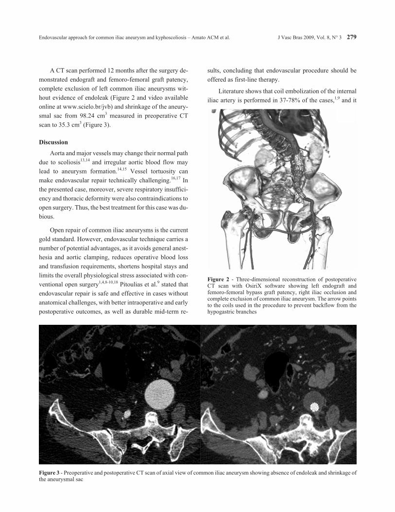
A CT scan performed 12 months after the surgery de-
monstrated endograft and femoro-femoral graft patency,
complete exclusion of left common iliac aneurysms wit-
hout evidence of endoleak (Figure 2 and video available
online at www.scielo.br/jvb) and shrinkage of the aneury-
smal sac from 98.24 cm3 measured in preoperative CT
scan to 35.3 cm3 (Figure 3).
Discussion
Aorta and major vessels may change their normal path
due to scoliosis13,14 and irregular aortic blood flow may
lead to aneurysm formation.14,15 Vessel tortuosity can
make endovascular repair technically challenging.16,17 In
the presented case, moreover, severe respiratory insuffici-
ency and thoracic deformity were also contraindications to
open surgery. Thus, the best treatment for this case was du-
bious.
Open repair of common iliac aneurysms is the current
gold standard. However, endovascular technique carries a
number of potential advantages, as it avoids general anest-
hesia and aortic clamping, reduces operative blood loss
and transfusion requirements, shortens hospital stays and
limits the overall physiological stress associated with con-
ventional open surgery1,4,8-10,18 Pitoulias et al.9 stated that
endovascular repair is safe and effective in cases without
anatomical challenges, with better intraoperative and early
postoperative outcomes, as well as durable mid-term re-
sults, concluding that endovascular procedure should be
offered as first-line therapy.
Literature shows that coil embolization of the internal
iliac artery is performed in 37-78% of the cases,1,9 and it
Endovascular approach for common iliac aneurysm and kyphoscoliosis – Amato ACM et al. J Vasc Bras 2009, Vol. 8, N° 3 279
Figure 2 - Three-dimensional reconstruction of postoperativeCT scan with OsiriX software showing left endograft andfemoro-femoral bypass graft patency, right iliac occlusion andcomplete exclusion of common iliac aneurysm. The arrow pointsto the coils used in the procedure to prevent backflow from thehypogastric branches
Figure 3 - Preoperative and postoperative CT scan of axial view of common iliac aneurysm showing absence of endoleak and shrinkage ofthe aneurysmal sac

was also performed in the case reported here to prevent
backflow into the aneurysm.
Post-processing preoperative CT scan with OsiriX
software12 allowed accurate measurement and planning of
the endovascular procedure. The aortomonoiliac endograft
used to expressly adapt to the patient’s particular anatomy,
with a short proximal large segment, designed to fit the
aorta, followed by a long narrow iliac segment, designed to
fit the iliac artery, allowed it to be deployed even in this
tortuous artery.
Complete exclusion of the iliac aneurysm resulted in
significant shrinkage of the aneurysmal sac after only 1
year, proving the efficacy of the method. Our encouraging
result demonstrates acceptable mid-term graft patency.
In conclusion, this report confirms the feasibility of
endovascular repair of isolated common iliac aneurysms in
complex vessel anatomy worsened by severe kyphoscolio-
sis. New generation devices are more adaptable to difficult
anatomy, broadening endovascular approach and allowing
us to make a personalized choice for each patient.
Supplementary online information: Video available
at www.scielo.br/jvb - Three-dimensional reconstruction
movie of preoperative and postoperative CT scan with
OsiriX software.
References
1. Boules TN, Selzer F, Stanziale SF, et al. Endovascular man-
agement of isolated iliac artery aneurysms. J Vasc Surg.
2006;44:29-37.
2. Brunkwall J, Hauksson H, Bengtsson H, Bergqvist D,
Takolander R, Bergentz SE. Solitary aneurysms of the iliac
arterial system: an estimate of their frequency of occurrence.
J Vasc Surg. 1989;10:381-4.
3. Lowry SF, Kraft RO. Isolated aneurysms of the iliac artery.
Arch Surg. 1978;113:1289-93.
4. Huang Y, Gloviczki P, Duncan AA, et al. Common iliac ar-
tery aneurysm: expansion rate and results of open surgical
and endovascular repair. J Vasc Surg. 2008;47:1203-10; dis-
cussion 1210-1.
5. Richardson JW, Greenfield LJ. Natural history and manage-
ment of iliac aneurysms. J Vasc Surg. 1988;8:165-71.
6. Minato N, Itoh T, Natsuaki M, Nakayama Y, Yamamoto H.
Isolated iliac artery aneurysm and its management.
Cardiovasc Surg. 1994;2:489-94.
7. Marin ML, Veith FJ, Lyon RT, Cynamon J, Sanchez LA.
Transfemoral endovascular repair of iliac artery aneurysms.
Am J Surg. 1995;170:179-82.
8. Caronno R, Piffaretti G, Tozzi M, et al. Endovascular treat-
ment of isolated iliac artery aneurysms. Ann Vasc Surg.
2006;20:496-501.
9. Pitoulias GA, Donas KP, Schulte S, Horsch S, Papadimitriou
DK. Isolated iliac artery aneurysms: endovascular versus
open elective repair. J Vasc Surg. 2007;46:648-54.
10. Wolf F, Loewe C, Cejna M, et al. Endovascular management
performed percutaneously of isolated iliac artery aneurysms.
Eur J Radiol. 2008;65:491-7.
11. Chaer RA, Barbato JE, Lin SC, Zenati M, Kent KC,
McKinsey JF. Isolated iliac artery aneurysms: a contempo-
rary comparison of endovascular and open repair. J Vasc
Surg. 2008;47:708-13.
12. Ratib O, Rosset A. Open-source software in medical imag-
ing: development of OsiriX. Int J Comput Assist Radiol
Surg. 2006;1:187-96.
13. Sucato DJ, Duchene C. The position of the aorta relative to
the spine: a comparison of patients with and without idio-
pathic scoliosis. J Bone Joint Surg Am. 2003;85-A:1461-9.
14. Richardson NG. A case of ruptured abdominal aortic aneu-
rysm in association with congenital kyphoscoliosis. Eur J
Vasc Surg. 1993;7:586-7.
15. Vollmar JF, Paes E, Pauschinger P, Henze E, Friesch A. Aor-
tic aneurysms as late sequelae of above-knee amputation.
Lancet. 1989;2:834-5.
16. Erzurum VZ, Sampram ES, Sarac TP, et al. Initial manage-
ment and outcome of aortic endograft limb occlusion. J Vasc
Surg. 2004;40:419-23.
17. Carroccio A, Faries PL, Morrissey NJ, et al. Predicting iliac
limb occlusions after bifurcated aortic stent grafting: Ana-
tomic and device-related causes. J Vasc Surg.
2002;36:679-84.
18. Laganà D, Carrafiello G, Recaldini C, et al. Endovascular
treatment of isolated iliac artery aneurysms: 2-year fol-
low-up. Radiol Med. 2007;112:826-36.
Correspondence:
Dr. Germano Melissano, MD
IRCCS H. San Raffaele, Department of Vascular Surgery
Via Olgettina, 60
20132 – Milan, Italy
Tel.: +39 02.2643.7146
Fax: +39 02.2643.7148
E-mail: [email protected]
280 J Vasc Bras 2009, Vol. 8, N° 3 Endovascular approach for common iliac aneurysm and kyphoscoliosis – Amato ACM et al.


![Agenesis of common iliac vein encroaching development of … · 2020-06-20 · Agenesis of common iliac vein 23 vein embolization and sampling of renal and adrenal veins [14]. Ruggeri](https://img.pdfslide.net/doc/110x75/5fa85145ea725f20c15155f3/agenesis-of-common-iliac-vein-encroaching-development-of-2020-06-20-agenesis-of.jpg)