Embed Size (px)
Citation preview

Março, 2017
UN
IVER
SID
AD
E D
E C
OIM
BR
A
Henrique Miguel Marques Bom Borges Alexandrino
ENERGY FOR LIVER REGENERATION:MITOCHONDRIAL BIOENERGETICS AND THE PATHOGENESIS OF
POSTHEPATECTOMY LIVER DYSFUNCTION
Doctoral Thesis in Health Sciences, branch of Medicine, supervised by Prof. Doutor Francisco José Franquera Castro e Sousa, MD, PhD and by Prof. Doutor Carlos Manuel Marques Palmeira, PhD and presented to the Faculty of Medicine of the University of Coimbra
Henriqu
e Migu
el Ma
rque
s Bo
m Bo
rges Alex
andrino
ENER
GY FOR
LIVE
R RE
GENE
RATIO
N:MITO
CHON
DRIAL
BIOEN
ERGE
TICS AN
D TH
E PA
THOG
ENESIS
OF P
OSTH
EPATEC
TOMY
LIVE
R DY
SFUN
CTION


Henrique Miguel Marques Bom Borges Alexandrino
Energy for Liver Regeneration:
Mitochondrial Bioenergetics and the Pathogenesis of
Posthepatectomy Liver Dysfunction
Energia para a Regeneração Hepática:
Bioenergética Mitocondrial e a Patogénese da Insuficiência Hepática
Pós-Hepatectomia
Março, 2017
Doctoral Thesis in Health Sciences, branch of Medicine, supervised by Prof. Doutor Francisco José Franquera Castro e Sousa, MD, PhD and by Prof. Doutor Carlos Manuel Marques Palmeira, PhD and presented to
the Faculty of Medicine of the University of Coimbra


À memória dos meus Avós
Rosa e Urbano


“We make our world significant by the courage of our questions
and the depth of our answers”
Carl Sagan, in “Cosmos” 1980


Table of Contents

Table of Contents ........................................................................................................................................ 7
Acknowledgements / Agradecimentos .................................................................................................... 13
Abstract / Resumo .................................................................................................................................... 19
List of abbreviations ................................................................................................................................. 27
Foreword ................................................................................................................................................... 33
Chapter I - Mitochondria and Liver Regeneration: Review of the Role of Energy Metabolism in the Pathophysiology of Posthepatectomy Liver Failure .............................................................................. 39
Introduction ............................................................................................................................................ 41
1. Mitochondria – The Powerhouse of Eukaryote cells ..................................................................... 42
2. Liver Regeneration – A highly energetic cellular process ............................................................. 44
3. Posthepatectomy Liver Failure – Evidence for disturbed bioenergetics in pathophysiology ........ 46
4. Hepatectomy in Chronic Liver Injury: Mitochondrial dysfunction as a key factor in the
Pathophysiology of Posthepatectomy Liver Failure .............................................................................. 53
4.1 Steatosis and steatohepatitis ................................................................................................. 53
4.2 Cirrhosis ............................................................................................................................... 58
4.3 Chemotherapy-induced liver injury ...................................................................................... 59
4.4 Chronic biliary obstruction ................................................................................................... 61
5. Novel markers of mitochondrial dysfunction – Earlier diagnosis of Posthepatectomy Liver
Failure .................................................................................................................................................... 63
6. Improving liver regeneration – Hepatocyte energetics as a potential target for prevention of
Postoperative Liver Failure .................................................................................................................... 68
6.1 Pharmacological therapy ...................................................................................................... 69
6.2 Improving hepatocyte oxygenation ...................................................................................... 71
6.3 Parenchymal modulation techniques .................................................................................... 74
6.4 Stem cell therapy .................................................................................................................. 75
Conclusions and future prospects ........................................................................................................... 76
Chapter II - Mitochondrial Bioenergetics, Hepatic Pedicle Clamping and Posthepatectomy Liver Dysfunction ............................................................................................................................................... 79
I. Introduction ................................................................................................................................... 81
II. Materials and Methods .................................................................................................................. 82
A. Experimental study ................................................................................................................... 82
1. Study animals ....................................................................................................................... 82
2. Surgical protocol................................................................................................................... 83
3. Mitochondrial isolation ......................................................................................................... 84
4. Measurement of mitochondrial membrane potential ............................................................ 84
5. Measurement of oxygen consumption .................................................................................. 85
6. Blood biochemistry............................................................................................................... 85
7. Histology .............................................................................................................................. 85
B. Clinical study ............................................................................................................................ 86

1. Study population and surgical procedures ............................................................................ 86
2. Collection of biopsies ........................................................................................................... 87
3. Mitochondrial isolation, measurement of mitochondrial membrane potential and oxygen
consumption .................................................................................................................................. 87
4. Measurement of Adenosine Triphosphate (ATP) content .................................................... 87
5. Postoperative liver function and clinical course ................................................................... 89
C. Statistical analysis ..................................................................................................................... 89
III. Results ....................................................................................................................................... 90
A. Experimental study ................................................................................................................... 90
1. Effect of Hepatectomy with Hepatic Pedicle Clamping on Mitochondrial Oxidative
Phosphorylation and Respiration ................................................................................................... 90
2. Effect of Hepatectomy with Hepatic Pedicle Clamping on markers of hepatocellular death90
3. Effect of Hepatectomy with Hepatic Pedicle Clamping on liver histology .......................... 90
B. Clinical study ............................................................................................................................ 92
1. Clinical outcome ................................................................................................................... 92
2. Baseline mitochondrial function ........................................................................................... 93
3. Hepatic Pedicle Clamping and mitochondrial function ........................................................ 93
4. Mitochondrial function and hepatocellular necrosis and function ........................................ 93
5. Mitochondrial function and postoperative morbidity ........................................................... 95
IV. Discussion ................................................................................................................................. 97
Colour Plates ........................................................................................................................................... 101
Chapter III - Chemotherapy Associated Liver Injury and Posthepatectomy Morbidity: Possible contribution of Mitochondrial Dysfunction to the Pathogenesis of Sinusoidal Obstruction Syndrome .................................................................................................................................................................. 113
I. Introduction ................................................................................................................................. 115
II. Material and Methods .................................................................................................................. 118
A. Clinical study .......................................................................................................................... 118
1. Study population ................................................................................................................. 119
2. Neoadjuvant chemotherapy ................................................................................................ 119
3. Operative data ..................................................................................................................... 120
4. Postoperative course ........................................................................................................... 120
5. Patient characteristics: chemotherapy vs. non-chemotherapy ............................................ 121
6. Pathological analysis .......................................................................................................... 122
B. Experimental study ................................................................................................................. 123
1. Study animals ..................................................................................................................... 123
2. Chemotherapy-induced liver injury model ......................................................................... 123
3. Surgical protocol................................................................................................................. 124
4. Mitochondrial bioenergetics ............................................................................................... 125
5. Blood biochemistry............................................................................................................. 127

6. Histology ............................................................................................................................ 127
7. Electron Microscopy ........................................................................................................... 127
C. Statistical Analysis .................................................................................................................. 128
III. Results ..................................................................................................................................... 128
A. Clinical Study ......................................................................................................................... 128
1. Postoperative morbidity and mortality ............................................................................... 128
2. Prevalence and patterns of histologic liver injury ............................................................... 129
3. Impact of neoadjuvant chemotherapy on liver injury ......................................................... 129
4. Impact of co-morbidities on liver injury ............................................................................. 129
5. Impact of neoadjuvant chemotherapy on morbidity and mortality ..................................... 130
6. Impact of liver injury on morbidity and mortality .............................................................. 130
7. Multivariate analysis ........................................................................................................... 133
B. Experimental study ................................................................................................................. 133
1. Induction of sinusoidal obstruction syndrome with chemotherapy .................................... 133
2. Effect of chemotherapy on mitochondrial energetics and structure .................................... 133
3. Effect of hepatectomy with hepatic pedicle clamping on mitochondrial energetics and hepatocellular function ................................................................................................................ 135
4. Correlation of mitochondrial dysfunction with postoperative hepatocellular function ...... 136
IV. Discussion ............................................................................................................................... 137
Chapter IV - Bioenergetic Adaptations of the Liver in the ALPPS Procedure: How Liver Regeneration Correlates with Mitochondrial Energy Status ............................................................. 145
I. Introduction ................................................................................................................................. 147
II. Patients and Methods .................................................................................................................. 149
A. Study population: ALPPS group ........................................................................................ 149
B. Operative procedures .......................................................................................................... 149
C. Control groups: Minor and Major hepatectomy ................................................................. 150
D. Volumetric and functional analysis .................................................................................... 152
E. Collection of biopsies ......................................................................................................... 152
F. Postoperative serum biochemistry ...................................................................................... 155
G. Postoperative liver function and clinical course ................................................................. 156
H. Statistical analysis............................................................................................................... 156
III. Results ..................................................................................................................................... 156
A. Clinical outcome and volumetric growth ............................................................................ 156
B. Liver bioenergetics compared: ALPPS T1, T2 and controls .............................................. 157
C. Energy metabolism and postoperative liver remnant function and volume growth............ 160
D. Gene expression and protein content in ALPPS ................................................................. 160
IV. Discussion ............................................................................................................................... 164
Concluding remarks ............................................................................................................................... 171

References ............................................................................................................................................... 181

Acknowledgements / Agradecimentos

Acknowledgements / Agradecimentos
15
A presente Tese não teria sido possível sem o constante estímulo e apoio de inúmeras pessoas,
que de uma forma ou de outra, contribuíram significativamente para a sua realização. Não podendo
enumerar todas, sob pena de injustamente excluir o concurso determinante de algumas, irei apenas
deixar patente o meu tributo às mais relevantes.
Em primeiro lugar, gostaria de agradecer ao Professor Doutor Francisco Castro e Sousa,
digníssimo Professor Catedrático da Faculdade de Medicina da Universidade de Coimbra, e meu Director,
Orientador Científico, Mestre, Mentor e Amigo. Demonstro aqui a minha admiração pela sua inigualável
capacidade de trabalho, sublime aptidão analítica, vocação científica invulgar, cultura de excelência, rigor
profissional, dedicação ao Serviço, ao Hospital e à Universidade, numa constante e contagiante busca de
aperfeiçoamento profissional. Destaco particularmente a forma amável e aberta como, em Junho de
2011, me recebeu no desde então nosso Serviço de Cirurgia A, após uma turbulenta restruturação dos
Serviços de Cirurgia dos Hospitais da Universidade de Coimbra. A magnanimidade com que me acolheu,
permitindo-me partilhar da sua dedicação e entusiasmo pela fascinante área da Cirurgia Hepatobiliar,
motivou todo o meu esforço e empenho, impelindo de forma marcante a minha carreira médica e
científica. Assim, por ter acreditado em mim nesse momento conturbado, testemunho aqui a minha mais
sincera gratidão. Por todas as incríveis oportunidades, valorosos ensinamentos, pertinentes críticas,
frutíferas discussões e amenas conversas, a ele dedico de forma muito particular este trabalho. Não
menos importante, estimo sobejamente a calorosa amizade com que me honra.
Logo de seguida tenho que reconhecer a pessoa do meu Co-Orientador, Professor Doutor
Carlos Marques Palmeira, digníssimo Professor Catedrático da Faculdade de Ciências e Tecnologia da
Universidade de Coimbra. Recordo aprazivelmente o nosso primeiro contacto e o entusiasmo
contagiante, personalidade franca e amável, exímia aptidão analítica, qualidades pedagógicas e sincera
curiosidade científica, que tanto me impressionaram. Graças ao genuíno sentimento de descoberta
científica que irradia da figura do Professor Doutor Carlos Marques Palmeira, não poderia de forma
alguma ter encontrado ambiente mais apropriado para o completo desenvolvimento deste trabalho. A ele
fica também aqui patente a minha enorme dívida de gratidão, também pelo constante e inestimável apoio,
e amizade.
Reconheço igualmente a figura do Professor Doutor Júlio Soares Leite, Professor Catedrático da
Faculdade de Medicina da Universidade de Coimbra, e Regente da Cadeira de Propedêutica. Sem dúvida
um exemplo como Clínico, Investigador e Docente, deixo aqui vincado o meu reconhecimento pela
distinta capacidade de trabalho, rigor e propensão para o Ensino, quer Pré- quer Pós-Graduado.
Não poderia imaginar esta Tese no seu início sem o imprescindível impulso do Professor Doutor
Guilherme Tralhão, Professor Agregado da Faculdade de Medicina da Universidade de Coimbra.
Reconheço aqui o enorme contributo que, pelas inúmeras oportunidades concedidas, deu à minha
carreira clínica e científica, do qual ficarei sempre devedor. Verdadeiro catalisador e impulsionador do
trabalho científico, dotado de uma capacidade de trabalho inigualável, personificando a figura do
Cirurgião Académico e revelando sempre um entusiasmo contagiante, a ele devo a possibilidade de ter
iniciado e desenvolvido este trabalho. Agradeço igualmente toda a amizade demonstrada nestes anos.
Destaco também a pessoa da Professora Doutora Anabela Rolo, Professora Agregada da
Faculdade de Ciências e Tecnologia da Universidade de Coimbra, pelas inúmeras e tão proveitosas
discussões críticas, constante apoio e marcante impulso à conclusão desta Tese.
Tenho do mesmo modo de reconhecer o papel daqueles que, muito antes do início dos trabalhos
conducentes a esta Tese, marcaram a minha formação Médica e Humana. Destaco em lugar cimeiro o

Acknowledgements / Agradecimentos
16
Dr. Fernando Martinho, antigo Director do saudoso Serviço de Cirurgia 2 dos Hospitais da Universidade
de Coimbra, meu ex-Director, Mestre, Mentor e Amigo. Assinalo aqui a sua indelével marca na minha
formação, como Homem e como Cirurgião. Pela dedicação à causa de tratar o ser humano doente de
forma abnegada e generosa. Pela exímia capacidade de juntar as capacidades humanas, clínicas e
técnicas, definindo aquilo que um Cirurgião deve ser. Foi para mim um privilégio, pessoal e profissional,
ter beneficiado do seu exemplo e da sua liderança durante a fase inicial da minha formação cirúrgica.
Sublinho igualmente o papel do Dr. Emanuel Furtado, Coordenador da Unidade de
Transplantação Hepática Pediátrica e de Adultos do Centro Hospitalar e Universitário de Coimbra.
Seguidor de uma cultura de excelência e dotado de uma inteligência crítica e capacidade técnica a todos
os níveis notáveis, foi e é para mim um sublime exemplo de rigor e de dedicação ao pormenor,
motivando-me numa permanente busca de aperfeiçoamento. Não menos importante, agradeço toda a
amizade com que me honra de há longa data.
A todos os meus colegas e colaboradores do Serviço de Cirurgia A e Bloco Operatório Central
do Centro Hospitalar e Universitário de Coimbra, pelo constante apoio e incentivo que tornaram possível
este trabalho. Um reconhecimento muito particular ao Dr. Carlos Mesquita, Dra. Maria Gorete Jorge, Dra.
Beatriz Costa, Enfª. Paula Moura, Enfª. Sandra Botelho, Enfª. Patrícia Ribeiro, Dra. Mónica Martins, Dr.
César Carvalho, Dr. Ricardo Martins e Dr. Miguel Fernandes. Igualmente destaco o papel relevante de
todos os Médicos Internos do Serviço de Cirurgia A, pelo inesgotável entusiasmo e vontade de aprender.
Devo sem dúvida um reconhecimento assaz especial ao Dr. Marco Serôdio e ao Dr. Luís Ferreira, não só
pelo incondicional e inesgotável apoio mas também por me privilegiarem com a sua amizade. Obrigado
por estarem sempre lá.
Como não poderia deixar de ser, deixo uma palavra muito especial de apreço a todos os meus
caros colegas do Mitolab, nomeadamente ao Prof. Doutor João Soeiro Teodoro, Prof. Doutor Filipe
Valente Duarte e Prof.ª Doutora Ana Teresa Varela. Por todo a inefável ajuda, constante entusiasmo,
esforço empenhado e, não menos importante, inesgotável paciência para comigo, fica a minha grata e
sentida homenagem. É com muito orgulho que sinto pertencer ao Mitolab. Obrigado por me acolherem.
Realço igualmente o papel de sobremaneira relevante que tiveram a Dra. Maria Augusta
Cipriano e o Dr. Rui Caetano Oliveira (Serviço de Anatomia Patológica – Centro Hospitalar e Universitário
de Coimbra), incansáveis na análise histológica dos diversos trabalhos aqui incluídos, pela análise crítica
dos resultados e por todo o apoio. De referir também o imprescindível contributo do Dr. Henrique Donato
e do Professor Doutor Filipe Caseiro Alves (Serviço de Imagem Médica - Centro Hospitalar e Universitário
de Coimbra), da Prof.ª Doutora Margarida Abrantes (Laboratório de Biofísica da Faculdade de Medicina
da Universidade de Coimbra), do Dr. Nuno Marques e Dr. José Feio (Serviços Farmacêuticos - Centro
Hospitalar e Universitário de Coimbra), do Dr. Rui Pratas e do Dr. Fernando Rodrigues (Serviço de
Patologia Clínica - Centro Hospitalar e Universitário de Coimbra) e do Dr. Paulo João Soares (Laboratório
S. José).
Não podia deixar de referir a enorme estima e dívida de gratidão que tenho para com os meus
Professores, em todos os níveis de ensino, que com o seu encorajamento e estímulo permanente,
sempre me impulsionaram a procurar, pesquisar e aperfeiçoar cada vez mais. Um reconhecimento
especial também aos meus Alunos, muito em particular ao João Martins, à Daniela Falcão e ao João
Cardoso, pela constante curiosidade e entusiasmo inesgotável.
Acknowledgements / Agradecimentos
17
O presente trabalho foi premiado em 2015 com a Bolsa da Associação Portuguesa de Estudo do
Fígado (APEF / MSD), pelo que agradeço à direcção da referida sociedade científica a confiança
depositada.
Aos meus Amigos que, nas horas boas e nos momentos menos bons, sempre encontraram
muito de si para me dar. Obrigado Vera, Gonçalo e, muito em particular, Luísa, pelo incondicional
encorajamento, generosidade e carinho.
À minha Família, em particular aos meus pais, Mário e Maria da Conceição, cujo amor
incondicional reconheço como dos maiores bens imateriais de que posso dispor. Ao meu irmão Nuno, por
tudo o que sempre fez por mim. À Xana, por todo o apoio. E muito em especial à minha querida filha
Madalena, pela Alegria, Amor e Serenidade com que preenche a minha vida.
Finalmente, expresso o meu mais sincero reconhecimento aos meus Doentes, Passados,
Presentes e Futuros. Serão sempre, em última instância, o motivo último de toda a minha actividade
Clínica e Científica e é a eles que esta Tese se destina.


Abstract / Resumo


Abstract / Resumo
21
Abstract
Clinical success of hepatectomy relies on the liver’s unique capacity to
regenerate, a highly energy-dependent process. When this capacity is surpassed
Posthepatectomy Liver Failure (PHLF) ensues, resulting in increased morbidity and
mortality.
Mitochondria are the powerhouses of the eukaryote cell and decision-makers of
cell death. Mitochondrial metabolism supplies the energy for liver regeneration, but the
clinical consequences of mitochondrial dysfunction in posthepatectomy morbidity and
liver function are presently unknown. Other unresolved issues are the effects of
chemotherapy-associated liver injury (CALI) on outcome, as well as the possible
contribution of bioenergetic dysfunction to the pathogenesis of CALI. Finally, two-
stage hepatectomies rely on an extremely rapid and significant inter-stages regenerative
response, but the energetic adaptations taking place in the future liver remnant (FLR)
are largely unknown. Mitochondrial oxidative phosphorylation and respiration are key
events in cellular energy metabolism and can be directly measured in liver biopsies.
In this Doctoral Thesis the following objectives were pursued: 1) Review the
role of energy metabolism in liver regeneration; 2) Determine whether changes in
mitochondrial function correlate with clinical outcome in Humans undergoing
hepatectomy; 3) Assess the impact of hepatic pedicle clamping (HPC) on intraoperative
mitochondrial function; 4) Evaluate the impact of chemotherapy-induced hepatotoxicity
on postoperative morbidity and the putative role of mitochondrial dysfunction in its
pathogenesis; 5) Investigate the bioenergetics adaptations underlying the enhanced
regenerative response taking place in two-stage hepatectomies.
For the first objective, a non-systematic review of relevant published material in
the English language was conducted. Reference lists were cross-checked for further
relevant publications. Mitochondrial oxidative phosphorylation was summarily
reviewed, as well as the role of mitochondria in both apoptotic and necrotic cell death.
The key role of mitochondrial metabolism in the cellular events leading to liver
regeneration was confirmed.

Abstract / Resumo
22
For the second and third objectives, both experimental and clinical works were
performed. First, a prospective study of patients undergoing hepatectomy for diverse
indications (N=30) was conducted. Liver biopsies were performed in two distinct
moments, at the beginning and at the end of surgery. Mitochondria were isolated and
membrane potential and respiration were measured. Mitochondrial lag phase, reflecting
the time required for adenosine diphosphate phosphorylation, presented high sensitivity
and specificity for prediction of PHLF; a finding previously unreported in the scientific
literature. Several key markers of postoperative liver function presented significant
correlations with intraoperative fluctuations in mitochondrial membrane potential and
respiration. An experimental study (N=35 male Wistar rats) was also outlined to explore
the effect of 70% hepatectomy with HPC on energy metabolism, liver function and
injury. In both studies, clinical and experimental, HPC was associated with depressed
mitochondrial function.
The fourth objective was addressed with two different methods. First, a clinical
and pathologic review of 140 patients undergoing hepatectomy for colorectal cancer
liver metastases was performed to look into the incidence, pathological spectrum and
clinical consequences of CALI. Sinusoidal obstruction syndrome (SOS) was present in
52% of patients and independently associated with overall and liver-specific morbidity.
Secondly, an experimental study (N=12 male Wistar rats), attempted to replicate a
previously described animal model of SOS. Hepatectomy with HPC was performed to
explore the possible link of mitochondrial dysfunction in the pathogenesis of
posthepatectomy liver dysfunction in CALI. Although the characteristic histologic
findings of SOS were not found, chemotherapy-treated animals presented evidence of
disturbed hepatocellular bioenergetics, namely longer lag phase and increased
mitochondrial size.
Finally, another original prospective clinical study was conducted on patients
undergoing the Associating Liver Partition and Portal Vein Ligation for Staged
Hepatectomy (ALPPS) procedure (N=5). In this study, mitochondrial membrane
potential and respiration were measured as well as gene expression profile. An inter-
stages increase in energetic capacity was demonstrated in the FLR, as well as a strong
and significant correlation of energy status with inter-stages volume growth. An
increased expression of several genes associated with liver regeneration (Augmenter of
Abstract / Resumo
23
Liver Regeneration; Small heterodimer partner; Signal Transducer and Activator of
Transcription 3), mitochondrial biogenesis (Peroxisome proliferator-activated receptor
gamma coactivator 1-alpha) and energy metabolism (Cytochrome oxidase subunit I;
Nicotinamide phosphoribosyltransferase) was also described. To our knowledge, this is
the first ever documentation of adaptations in energy metabolism in two-stage
hepatectomies in Humans.
In conclusion, not only is Liver Regeneration highly dependent on energy
metabolism, but also mitochondrial dysfunction was demonstrated to be involved in the
pathogenesis of PHLF. The clinical relevance of mitochondrial bioenergetics in clinical
liver resection deserves further exploration, with particular emphasis on the accurate
perioperative staging of liver energy status as well as energy-conditioning therapies
aiming at decreasing morbidity and mortality.

Abstract / Resumo
24
Resumo
O sucesso clínico da Cirurgia Hepática de exérese depende da capacidade
singular do fígado para regenerar, um processo altamente dependente de energia.
Quando esta capacidade é ultrapassada, desencadeia-se a Insuficiência Hepática Pós-
Hepatectomia (PHLF), com consequente morbi-mortalidade.
As mitocôndrias são as responsáveis pela produção de energia nas células
eucariotas e decisores-chave na morte celular. O metabolismo mitocondrial fornece
energia para a regeneração hepática, mas desconhecem-se as consequências clínicas da
disfunção mitocondrial sobre a morbilidade e função hepatocelular pós-hepatectomia.
Outra questão que ainda carece definição é o efeito da lesão hepática associada à
quimioterapia (CALI) nos resultados clínicos, bem como o possível papel do
metabolismo energético na sua patogénese. Finalmente, as hepatectomias iterativas
dependem de uma importante resposta regenerativa, mas as adaptações energéticas que
ocorrem no fígado remanescente (FLR) são desconhecidas. A fosforilação oxidativa e a
respiração mitocondriais são eventos-chave no metabolismo energético e podem ser
medidas directamente em biópsias hepáticas.
Nesta dissertação doutoral foram perseguidos os seguintes objectivos: 1)
Revisão do papel do metabolismo energético na regeneração hepática; 2) Determinar a
relação entre a função mitocondrial e os resultados clínicos após hepatectomia no
Humano; 3) Avaliar o impacto da clampagem do pedículo hepático (HPC) sobre a
função mitocondrial; 4) Aferir as consequências da hepatotoxicidade induzida pela
quimioterapia na morbilidade pós-hepatectomia, bem como o papel da disfunção
mitocondrial na sua patogénese; 5) Investigar as adaptações bioenergéticas subjacentes
à marcada resposta regenerativa que ocorre nas hepatectomias iterativas.
Para o primeiro objectivo foi realizada uma revisão não sistemática da literatura
em língua inglesa, com pesquisa de artigos presentes nas listas de referências. A
fosforilação oxidativa foi sumariamente revista, bem como o papel das mitocôndrias
nos processos de apoptose e necrose. Foi assim confirmado o papel do metabolismo
mitocondrial nos eventos celulares que culminam na regeneração hepática.

Abstract / Resumo
25
Para responder ao segundo e terceiro objectivos foram realizados um estudo
experimental e outro clínico. Em primeiro lugar, foram estudados prospectivamente
doentes submetidos a hepatectomia por indicações diversas (N=30), com realização de
biópsias hepáticas intra-operatórias, no início e no final da intervenção, e medição do
potencial de membrana e respiração mitocondriais. A “lag phase” mitocondrial, o
tempo necessário à completa fosforilação da adenosina difosfato, apresentou uma
elevada sensibilidade e especificidade na previsão de PHLF, um achado nunca antes
reportado. Vários marcadores de função hepatocelular pós-operatória apresentaram
correlações significativas com a flutuação peri-operatória do potencial de membrana e
respiração mitocondriais. Para além disso, um estudo experimental (N=35 ratos Wistar
machos) foi desenhado para explorar o efeito da hepatectomia de 70% com HPC no
metabolismo energético e na função e lesão hepática; em ambos os estudos foi
confirmado o efeito deletério da HPC sobre a função mitocondrial.
A resposta ao quarto objectivo decorreu de dois modos distintos. Em primeiro
lugar, foi realizada uma revisão clínica e patológica de 140 doentes submetidos a
hepatectomia por metástases de cancro colo-rectal, com o intuito de definir a incidência,
espectro patológico e consequências clínicas da CALI. O síndrome de obstrução
sinusoidal (SOS), presente em 52% dos doentes, associou-se de forma independente
com a morbilidade global e relacionada com o fígado. Em segundo lugar, procurou-se
replicar um modelo animal de SOS num estudo experimental (N=12 ratos Wistar
machos). Neste, realizaram-se hepatectomias com HPC de modo a explorar uma
possível conexão entre disfunção mitocondrial e disfunção hepatocelular na CALI.
Embora não tenham sido obtidas as lesões histológicas típicas do SOS, os animais
tratados com quimioterapia apresentaram significativa perturbação da bioenergética
hepatocelular, nomeadamente “lag phase” mais prolongada e aumento do tamanho
mitocondrial.
Finalmente, outro estudo clínico prospectivo original foi conduzido em doentes
submetidos a Associação de Laqueação Portal com Secção Parenquimatosa para
Hepatectomia a dois tempos (ALPPS) (N=5); neste estudo foi realizada a avaliação da
bioenergética mitocondrial e a análise da expressão génica. Verificou-se um aumento da
função mitocondrial entre estadios, bem como uma correlação forte e significativa entre
o status energético e o crescimento volumétrico do FLR. Foi ainda objectivado um
Abstract / Resumo
26
aumento da expressão de diversos genes associados à regeneração hepática (Augmenter
of Liver Regeneration; Small heterodimer partner; Signal Transducer and Activator of
Transcription 3), biogénese mitocondrial (Peroxisome proliferator-activated receptor
gamma coactivator 1-alpha) e metabolismo energético (Cytochrome oxidase subunit I;
Nicotinamide phosphoribosyltransferase). Tanto quanto nos é dado saber, trata-se da
primeira vez que, no Homem, se relatam adaptações do metabolismo energético em
hepatectomias iterativas.
Em conclusão, poder-se-á dizer que não só a regeneração hepática é um
processo altamente dependente de energia, mas, também, que a função mitocondrial
assume um papel relevante na fisiopatologia da PHLF. A possível relevância clínica da
bioenergética mitocondrial na Cirurgia Hepática merece exploração mais detalhada,
com particular ênfase na correcta aferição da capacidade energética peri-operatória, mas
também na possibilidade de melhorar os resultados, com o uso de terapêuticas de
condicionamento energético do FLR, reduzindo a morbi-mortalidade.

List of abbreviations


List of abbreviations
29
5-FU 5-fluorouracil
AALF Acetaminophen-induced acute liver failure
ADP Adenosine diphosphate
AKBR Arterial Ketone Body Ratio
ALPPS Associating Liver Partition and Portal Vein Ligation for Staged Hepatectomy
ALR Augmenter of Liver Regeneration
ALT Alanine aminotransferase
ANT Adenine Nucleotide Translocator
AST Aspartate aminotransferase
ATP Adenosine triphosphate
ATP/Pi Adenosine triphosphate to phosphate ratio
BDL Bile duct ligation
BLAST Basic Local Alignment Search Tool
BSA Bovine serum albumin
CACT Carnitine-acetylcarnitine translocase
CALI Chemotherapy-associated liver injury
CASH Chemotherapy-associated steatohepatitis
cDNA Complementary deoxyribonucleic acid
CECT Contrast-enhanced computed tomography
CK Creatine kinase
COX Cytochrome oxidase
COX1 Cytochrome oxidase subunit I
COX4 Cytochrome oxidase subunit IV
CRLM Colorectal cancer liver metastases
CT Computed tomography
DMSO Dimethyl sulfoxide
DNA Deoxyribonucleic acid
EDTA Ethylenediaminetetraacetic acid
List of abbreviations
30
EGF Epidermal growth factor
EGTA Ethylene glycol-bis(β-aminoethyl ether)-N,N,N',N'-tetraacetic acid
ETC Electron transport chain
FA Fatty acids
FADH2 or FAD Flavin adenine dinucleotide
FLR Future liver remnant
FOLFIRI Association of 5-fluoruracil with irinotecan
FOLFOX Association of 5-fluoruracil with oxaliplatin
FXR Farnesoid X receptor
GLDH Glutamate dehydrogenase
GLP-1 Glucagon-like peptide 1
GR Growth rate
GSH Glutathione
H2DCFDA 2′,7′-dichlorodihydrofluorescein diacetate
HABR Hepatic Artery Buffer Response
H&E Haematoxylin and Eosin
HBO Hyperbaric oxygen
HCC Hepatocellular carcinoma
HEPES 4-(2-hydroxyethyl)-1-piperazineethanesulfonic acid
HGF Hepatocyte Growth Factor
HPC Hepatic Pedicle Clamping
HR Hazard ratio
ICG Indocyanine Green
IL-6 Interleukin 6
INR International Normalized Ratio
IRI Ischemia-reperfusion injury
IQR Interquartile range
ISGLS International Study Group for Liver Surgery

List of abbreviations
31
KGR Kinetic growth rate
LDH Lactate dehydrogenase
LOS Length of stay
MaHp Major hepatectomy
MARS Molecular Adsorbent Recirculating System
MCT Monocrotaline
MELD Model for End Stage Liver Disease
miHp Minor hepatectomy
MPT Mitochondrial permeability transition
mtDNA Mitochondrial deoxyribonucleic acid
NADH or NAD+ Nicotinamide adenine dinucleotide
NAMPT Nicotinamide phosphoribosyltransferase
NCBI National Center for Biotechnology Information
NCT Neoadjuvant chemotherapy
NIM811 N-methyl-4-isoleucine cyclosporine
NRF-1 Nuclear respiratory factor 1
NRF-2 Nuclear factor erythroid 2-related factor 2
OR Odds ratio
PCR Polymerase chain reaction
PGC-1α Peroxisome proliferator-activated receptor gamma coactivator 1-alpha
PHLF Posthepatectomy Liver Failure
PVA Portal vein arterialization
PVE Portal vein embolization
PVL Portal vein ligation
PVP Portal vein pressure
RCR Respiratory Control Ratio
RNA Ribonucleic acid
ROS Reactive oxygen species
List of abbreviations
32
RR Relative risk
SAMe S-adenosyl-L-methionine
SD Standard deviation
SEC Sinusoidal endothelial cell
SEM Standard error of the mean
sFLR Standardized future liver remnant
SFSS Small-for-size syndrome
SH Steatohepatitis
SHP Small heterodimer partner
SirT1 Sirtuin 1
SOS Sinusoidal obstruction syndrome
STAT3 Signal Transducer and Activator of Transcription 3
TEM Transmission electron microscopy
TFAM Mitochondrial transcription factor A
TGF-α Transforming growth factor alpha
TNF-α Tumor Necrosis Factor α
TPP Tetraphenylphosphonium
TRAIL-R1 Tumor necrosis factor-related apoptosis-inducing ligand receptor 1
TRAIL-R2 Tumor necrosis factor-related apoptosis-inducing ligand receptor 2
UCP-2 Uncoupling protein 2

Foreword

Foreword
35
The current Doctoral Thesis revolves around the subject of Energy, which is
defined as “the capacity of a system to perform work” (Koolman, Roehm, 2005). The
Liver is the master regulator of complex multicellular animals and the inexhaustible
guardian of metabolic equilibrium. Either storing energy in the fed state, or releasing it
for other organs in the fast state, the Liver also synthetizes most of plasma proteins,
metabolizes and excretes endogenous and exogenous organic molecules in the bile,
while maintaining a discrete, yet vital, immune function. Furthermore, the Liver has the
extraordinary capacity to recuperate after insults, restoring its original size and function.
Such is the Liver’s work.
This potential for regeneration is the cornerstone of modern Hepatic Surgery,
whereby patients with primary and secondary malignant neoplasms are amenable to
cure after resection of a significant fraction of liver parenchyma, without resulting in
any permanent curtailment of function. Notwithstanding, Liver Regeneration is on
occasion inadvertently pushed beyond its limits. This results in one of the most ominous
complications of liver resection, Posthepatectomy Liver Failure, culminating in the
collapse of the metabolic building of the entire organism.
But what fuels Liver Regeneration? Where does the Liver obtain the energy to
replenish its cell population while ensuring the metabolic stability of the internal
milieu? More importantly, what energetic adaptations occur after liver resection and
how is the energy status related to the postoperative outcome? And how does hepatic
pedicle clamping influence liver energy status? What is the effect of chronic liver
disease on energy metabolism and on the response to surgical insult? Moreover, what
energy-based measures could be used for earlier detection, and even prevention, of the
dreaded Posthepatectomy Liver Failure? And finally, what is the energy source for the
short-interval two-stage hepatectomies, one of the most dramatic and risky challenges
that modern Hepatic Surgery places to Liver Regeneration? These were the research
questions underlying this project.
Soon a singular intracellular organelle assumed a pivotal role. Like the Liver,
Mitochondria are tireless providers of energy. By breaking down the carbon skeleton of
organic molecules in the presence of oxygen, Mitochondria supply heterotrophic
eukaryote cells with the fuel for all of Life’s functions. Additionally, they are key
regulators of cell death. If they malfunction the cell succumbs, relinquishing all activity.

Foreword
36
So, as the Liver is the metabolic coordinator of the Body, so are Mitochondria of the
Cell.
Under these postulates, research was conducted in a translational perspective.
Questions arose in the clinical setting, where rare, yet vexing cases of posthepatectomy
liver dysfunction ignited an intellectual unrest. The interrogations led to the laboratory,
where more profound insights were investigated, giving rise to novel problems and
innovative perspectives. And the quest for answers began. Theories were formulated
and tested. Results were compared and discussed. New information was collected and
summarized. And the end result of this work is: “Energy for Liver Regeneration:
Mitochondrial Bioenergetics and the Pathogenesis of Posthepatectomy Liver
Dysfunction”.
This Thesis is thus divided into four chapters. In the introductory chapter,
Chapter I - “Mitochondria and Liver Regeneration: Review of the Role of Energy
Metabolism in the Pathophysiology of Posthepatectomy Liver Failure”, the evidence for
the contribution of mitochondria in liver regeneration is scrutinized. Relevant scientific
literature, both in the fields of Basic and Applied sciences, was searched and critically
revised, with emphasis on the ramifications of mitochondrial function in the biological
processes occurring after liver resection. The clinical pertinence of the subject was
highlighted, mostly on three distinct tracks. First, as a growing proportion of patients
undergo resection of a chronically diseased liver parenchyma, the relevance of energy
status in the setting of diverse chronic hepatic disorders was explored. Secondly,
diagnostic markers of bioenergetics derangement for the earlier and more sensitive
detection of postoperative liver dysfunction were examined. And finally, the exhilarant
possibility of novel mitochondrial-based therapies for the improvement of
posthepatectomy outcome was reviewed.
In Chapter II - “Mitochondrial Bioenergetics, Hepatic Pedicle Clamping and
Posthepatectomy Liver Dysfunction”, original scientific data are displayed, in the search
for a nexus between energy status and clinical results of hepatectomy. Both clinical and
experimental works are detailed, and a definite link for mitochondrial homeostasis in
the liver’s recovery after ischemic and surgical insult is explored. And mitochondrial
oxidative phosphorylation is for the first time authenticated as a relevant vector in the
patient’s clinical outcome.
Foreword
37
Given the high incidence of colorectal cancer in developed nations, the
prevalence of liver metastases, as well as the definite benefits for their resection on
patient outcome, this Thesis was compelled to interrogate on this subject. In particular,
on a novel form of liver impairment, Chemotherapy-Associated Liver Injury. This was
the leitmotif for Chapter III - “Chemotherapy-Associated Liver Injury and
Posthepatectomy Morbidity: Possible contribution of Mitochondrial Dysfunction to the
Pathogenesis of Sinusoidal Obstruction Syndrome”. As evidence emerges for a
significant deterioration of liver reserve in this setting, a putative link with deranged
energy homeostasis was explored. An original retrospective clinical study on the
prevalence, pathologic spectrum and clinical consequences of chemotherapy-induced
parenchymal injury was conducted and presented. And an experimental model was
conceptualized to investigate a vinculum with mitochondrial dysfunction, with the
results detailed.
Although the indications for hepatectomy are expanding, the limits of resection
were at a standstill until recently, when a novel cutting-edge approach in the Liver
surgeons’ armamentarium emerged. Drawing on the liver’s notable capacity to
regenerate, and sometimes tipping this delicate equilibrium beyond its limits, the
Associating Liver Partition with Portal Vein Ligation for Staged Hepatectomy was
inadvertently discovered and developed. This remarkable technique undoubtedly relies
on the extremes of liver regenerative capacity, and possibly also on the bare limits of
energy homeostasis. This was again the focus of original clinical research unveiled in
the final chapter, Chapter IV – “Bioenergetic Adaptations of the Liver in the ALPPS
Procedure: How Liver Regeneration Correlates with Mitochondrial Energy Status”.
Here mitochondrial metabolism proved, once again, to be a ponderous factor in clinical
outcome.
A dissertation on the most meaningful findings of this Thesis, as well as a
discussion on the possible avenues to pursue in further research is laid out in the
Concluding Remarks section. And illustrating several salient aspects of Bioenergetics in
Liver Surgery, a pictorial essay of original photographs is presented in the Colour Plate
section.
With this Thesis, a Surgeon-Investigator’s effort is not complete, however.
Much more remains to be explored. And the enthralling encounter of two fascinating

Foreword
38
disciplines – Bioenergetics and Liver Surgery – will surely give rise to new questions;
and to the pursuit for new answers. Energy will surely be devoted to this work!
Coimbra, March 2017
Chapter I - Mitochondria and Liver Regeneration: Review of the Role of Energy Metabolism in the Pathophysiology of Posthepatectomy Liver Failure


Chapter I Mitochondria and Liver Regeneration: Review of the Role of Energy Metabolism in the Pathophysiology of Posthepatectomy Liver Failure
41
Introduction
Liver resection, founded on anatomical principles [1], is the only curative
therapy for most patients with hepatobiliary malignancies [2–6]. Resection of a
substantial amount of liver mass is only possible due to the liver’s remarkable capacity
to regenerate [7]. However, when this process is hampered Posthepatectomy Liver
Failure (PHLF) ensues. PHLF is a severe complication of liver resection, with
incidences ranging between 2.1 and 10.6% [8–10], resulting not only in perioperative
mortality of up to 54% [11], but also severe morbidity, long-term deterioration in liver
function and decreased long-term survival [12,13]. PHLF is characterized by jaundice,
ascites, encephalopathy and is usually complicated with renal dysfunction and sepsis
[9,14].
The mechanisms behind liver regeneration have been the focus of intense
research in the fields of Biology and Clinical Medicine [15–18]. Being a highly energy
dependent chain of events, liver regeneration is influenced by the energy status of the
main parenchymal cell – the hepatocyte. Mitochondria are the powerhouses of
eukaryote cells and key players in cell death. As such, they play a major role in the
organ’s response to major resection. Although this subject has been the focus of
investigation in the past, recent findings have led to a renewal of interest on disordered
bioenergetics in liver surgery [19–22]. And even more so, as the most upstream
mechanisms that trigger liver regeneration are slowly but surely unravelled, a definite
role for energy balance is gaining importance, putting the spotlight on mitochondria as
one of the master regulators of liver regeneration [23–26].
In this Chapter we will review the energetic mechanisms fuelling liver
regeneration, with emphasis on the role of mitochondria in energy homeostasis and cell
death. We will scrutinize the experimental and clinical evidence supporting a major role
for mitochondria in the liver’s response to resection, as well as the relevance of
mitochondrial derangement in the pathophysiology of PHLF. Furthermore, as a
significant proportion of patients undergoing hepatectomy have chronic liver diseases,
namely cirrhosis, biliary obstruction, steatosis or sinusoidal obstruction syndrome,
which are at increased risk of PHLF, we will recapitulate the disordered bioenergetics in
the pathophysiology of these conditions. We will also look into the possibility of

Chapter I
42
accurately predicting postoperative liver failure with non-invasive markers of
mitochondrial dysfunction, ultimately allowing an earlier diagnosis of PHLF and the
timely institution of therapy. Finally, we will elaborate on the evidence for a definitive
role of boosting energetic status of the liver parenchyma in improving the clinical
results of hepatectomy.
1. Mitochondria – The Powerhouse of Eukaryote cells
Mitochondria are singular organelles. They are descendent of α-proteobacteria
perfectly integrated with their host eukaryote cells, recapitulating one of the most
ancient symbiotic relationships of life on planet Earth and propelling eukaryotes’
evolution into complex multicellular organisms [27]. They provide over 90% of the
energy supply under aerobic conditions and are also key regulators of intracellular
calcium and programmed cell death [28]. Like their prokaryotic ancestors, mitochondria
are composed of an outer and an inner membrane (separated by the intermembrane
space) and a matrix containing a complex enzymatic machinery and a circular
deoxyribonucleic acid (DNA) molecule.
Since the liver is the hub of whole-body homeostasis, it is no surprise that there
is a constant need for energy in liver cells to perform the diverse anabolic and catabolic
reactions involving carbohydrate, lipid, protein, purine and xenobiotic metabolism. The
energy for this, in the form of adenosine tri-phosphate (ATP), is provided by
mitochondria, through the process of oxidative phosphorylation. In aerobic conditions
acetyl-coenzyme A (resulting from glycolysis and from β-oxidation of free fatty acids)
is completely oxidised to carbon dioxide in the tricarboxylic acid cycle, in which two
coenzymes are reduced: nicotinamide adenine dinucleotide (NAD+) and flavin adenine
dinucleotide (FAD) [29]. Both β-oxidation of free fatty acids and the tricarboxylic acid
cycle take place in the mitochondrial matrix. The reduced forms of NAD+ and FAD,
NADH and FADH2 respectively, transfer their electrons sequentially to the enzyme
complexes I to IV of the electron transport chain (ETC), in the mitochondrial inner
membrane, to the final acceptor, molecular oxygen, yielding water. During electron
transport, protons (H+) are pumped into the intermembrane space, creating an
electrochemical gradient across the mitochondrial inner membrane (proton-rich and

Mitochondria and Liver Regeneration: Energy Metabolism and Posthepatectomy Liver Failure
43
positively charged in the intermembrane space, versus proton-poor and electro-negative
in the matrix). The movement of protons back to the mitochondrial matrix is coupled to
ATP synthesis by complex V, F1F0 ATP-synthase. This enzyme uses the energy of
proton movement across the gradient for phosphorylation of adenosine di-phosphate
(ADP), generating ATP. The net yield of these reactions is the production of 32 mol of
ATP for every mol of glucose [30].
Oxidative phosphorylation is an extremely efficient process, because energy
transfer occurs in small steps, with several intermediary molecules and without much
dissipation of energy as heat. In spite of this, the transfer of electrons is sometimes
incomplete, leading to the production of reactive oxygen species. Reactive oxygen
species (ROS) is a collective term that broadly describes a variety of molecules and free
radicals (chemical species with one unpaired electron) derived from molecular oxygen:
superoxide, singlet oxygen, hydrogen peroxide, and hydroxyl radical. Production of
ROS is a physiologic event at basal levels and deleterious consequences are usually
prevented by several cellular antioxidant defence mechanisms, such as glutathione,
superoxide dismutase and catalase [31]. However, excessive production of ROS (for
instance in conditions of ischemia-reperfusion injury) can overwhelm these defences
and cause cell damage. Mitochondria are not only the producers but also the main
victims of increased ROS formation. Two mitochondrial elements are particularly
susceptible to oxidative stress: DNA and inner membrane lipids. Mitochondrial DNA
(MtDNA) is extremely sensitive to oxidative damage as it is located close to the inner
membrane (where most ROS are formed), lacks protective histones and has incomplete
repair mechanisms. Since it encodes 13 essential proteins for the ETC, mutations in
MtDNA can severely impair electron transport, propagating the vicious circle of
energetic dysfunction and oxidative damage. Lipid peroxidation by ROS, in particular
cardiolipin, can further compromise ETC function [32].
Besides the obvious contribution of mitochondrial dysfunction in cell death by
necrosis (ATP depletion causes failure of ATP-dependent ionic pumps and osmotic cell
swelling and death), mitochondria are also involved in apoptosis. In conditions of
mitochondrial calcium overload, especially during ischemia-reperfusion injury (IRI), a
non-selective conductive pore (the mitochondrial permeability transition – MPT) is
formed in the mitochondrial outer membrane, allowing the leakage of protons (with

Chapter I
44
subsequent reduced energy efficiency) and cytochrome c into the cytoplasm thus
activating the caspase-3 cell death pathway [33].
Mitochondrial quality is tightly regulated by a delicate balance between
formation of new mitochondria, in the process of mitochondrial biogenesis; and
degradation by autophagy. Several genes and transcription factors are responsible for
the tight regulation of mitochondrial biogenesis, namely the mitochondrial transcription
factor A (TFAM) and Peroxisome proliferator-activated receptor gamma coactivator 1-
alpha (PGC-1α), ensuring a coordinated collaboration between the mitochondrial 13-
gene DNA and the nuclear genome [34,35]. Mitochondrial autophagy or mitophagy, is
the process by which old or damaged mitochondria are engulfed in autophagosomes and
trafficked to lysosomes, thus maintaining a healthy and functioning mitochondrial pool
[36,37]. Remarkably, liver mitochondria have higher turnover rates than mitochondria
in other tissues [38].
2. Liver Regeneration – A highly energetic cellular process
Liver regeneration is a highly energy dependent process (Figure 1.1). After
hepatectomy the regenerative stimulus causes hepatocytes to undergo cell cycle
progression, DNA replication and protein synthesis, processes that require a large
amount of energy [15], mostly derived from β-oxidation of fatty acids [39]. Although
the increase in ATP synthesis precedes the peak in DNA synthesis [40,41], the net result
is a significant drop in liver ATP, more pronounced the more extended the resection of
parenchyma, as more energy is required for the diverse anabolic reactions of cell growth
and division. In fact, as early as 30 seconds after hepatectomy there is a 50% decrease in
liver ATP stores in rodents [24]. The decrease in ATP reaches a nadir at 48 hours but is
expected to fully recover by the fifth day. Likewise, there is a decrease in ATP to
phosphate (ATP/Pi) ratio, that significantly correlates with postoperative liver function
and markers of cellular proliferation [42]. Although the evidence for this is mostly
experimental, Mann et al, using 31
Phosphorus Magnetic Resonance Spectroscopy,
demonstrated that hepatectomy in human subjects was also associated with an early fall
in ATP/Pi ratio [43].
Mitochondria and Liver Regeneration: Energy Metabolism and Posthepatectomy Liver Failure
45
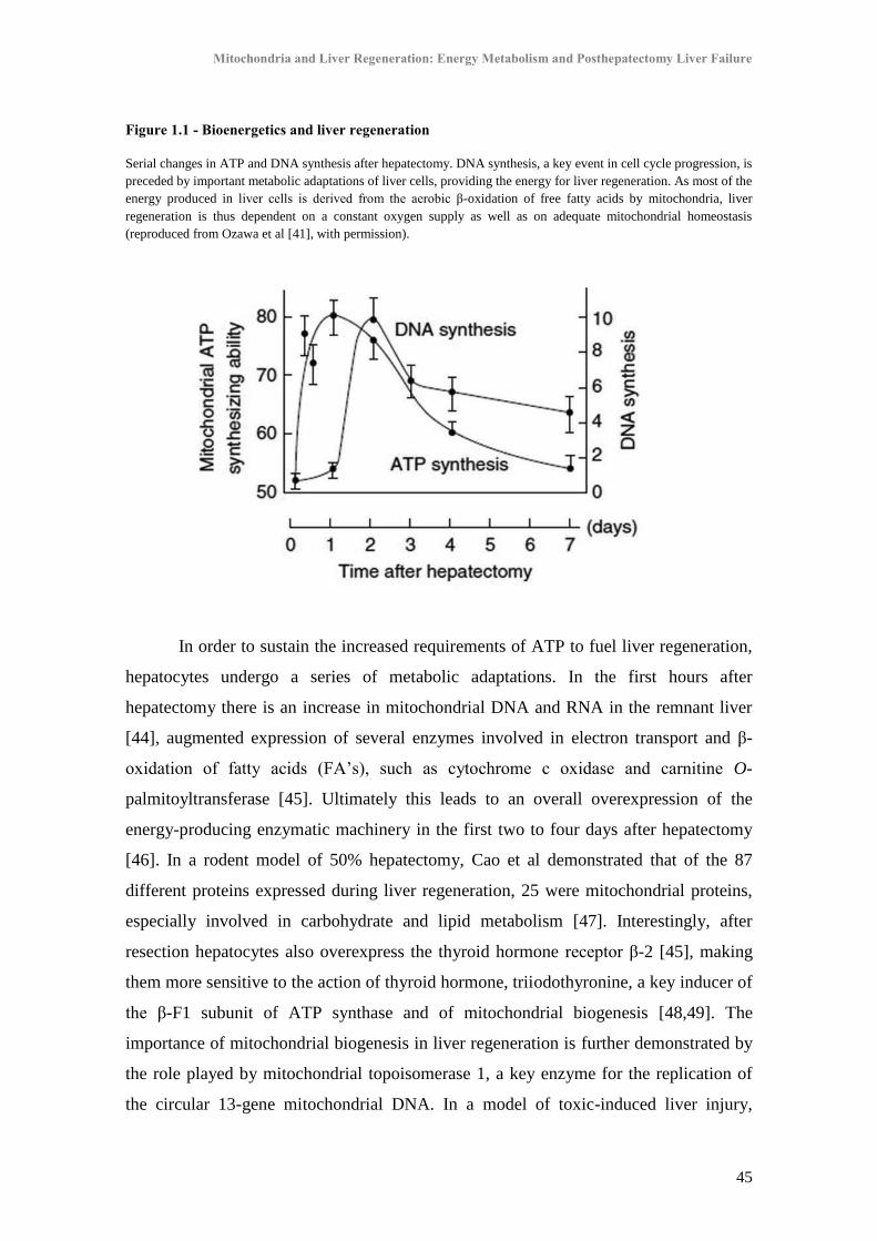
Figure 1.1 - Bioenergetics and liver regeneration
Serial changes in ATP and DNA synthesis after hepatectomy. DNA synthesis, a key event in cell cycle progression, is
preceded by important metabolic adaptations of liver cells, providing the energy for liver regeneration. As most of the
energy produced in liver cells is derived from the aerobic β-oxidation of free fatty acids by mitochondria, liver
regeneration is thus dependent on a constant oxygen supply as well as on adequate mitochondrial homeostasis
(reproduced from Ozawa et al [41], with permission).
In order to sustain the increased requirements of ATP to fuel liver regeneration,
hepatocytes undergo a series of metabolic adaptations. In the first hours after
hepatectomy there is an increase in mitochondrial DNA and RNA in the remnant liver
[44], augmented expression of several enzymes involved in electron transport and β-
oxidation of fatty acids (FA’s), such as cytochrome c oxidase and carnitine O-
palmitoyltransferase [45]. Ultimately this leads to an overall overexpression of the
energy-producing enzymatic machinery in the first two to four days after hepatectomy
[46]. In a rodent model of 50% hepatectomy, Cao et al demonstrated that of the 87
different proteins expressed during liver regeneration, 25 were mitochondrial proteins,
especially involved in carbohydrate and lipid metabolism [47]. Interestingly, after
resection hepatocytes also overexpress the thyroid hormone receptor β-2 [45], making
them more sensitive to the action of thyroid hormone, triiodothyronine, a key inducer of
the β-F1 subunit of ATP synthase and of mitochondrial biogenesis [48,49]. The
importance of mitochondrial biogenesis in liver regeneration is further demonstrated by
the role played by mitochondrial topoisomerase 1, a key enzyme for the replication of
the circular 13-gene mitochondrial DNA. In a model of toxic-induced liver injury,
Chapter I
46
knock-out mice for this enzyme presented with decreased mitochondrial DNA, lower
activities of ETC complexes I and IV and impaired hepatocyte replication [50].
These adaptations are paramount for the increased metabolic demand of liver
regeneration, as the recovery in energy status is preceded by an enhanced liver oxygen
consumption, which in turn is followed by the peak in DNA replication [51]. As a proof
of concept of the importance of hepatic energy status in liver regeneration, Satoh et al,
in an elegant experiment, used knock-in mice expressing creatine kinase (CK) in liver
cells. CK is normally expressed in skeletal and cardiac muscle, and in brain tissue, but
not in the liver. It constitutes an alternate source for adenosine triphosphate (ATP)
production by the transfer of a high-energy phosphate from creatine phosphate to ADP.
In this experiment, mice with liver expression of CK were fed either a high-creatine diet
or a control diet. After 70% hepatectomy, creatine-fed animals had higher hepatic ATP
synthesis and displayed increased bromodeoxyuridine incorporation and liver weight
gain, versus CK-positive controls with normal diet [52].
Since the availability of ATP is ultimately dependent upon the proper function
of mitochondria, it is expected that enhanced mitochondrial function would improve
liver regeneration. In fact, this is well illustrated by two experimental works that used
N-methyl-4-isoleucine cyclosporine (NIM811), a known inhibitor of mitochondrial
membrane permeability transition (MPT). Both in the setting of extended hepatectomy
[20] and small-for-size liver transplant [53], NIM811 inhibited the MPT, preserved liver
energy status and hepatocellular function and improved survival.
Having summarized the biological link of energy availability to cell proliferation
in liver regeneration, we will now discuss the evidence for a definite role of
bioenergetics dysfunction in the pathophysiology of PHLF.
3. Posthepatectomy Liver Failure – Evidence for disturbed bioenergetics in pathophysiology
Clinical success of hepatectomy depends on the liver’s unique ability to
regenerate. After major hepatectomy, the remnant liver must replace lost hepatocyte

Mitochondria and Liver Regeneration: Energy Metabolism and Posthepatectomy Liver Failure
47
mass, produce an acute-phase response and still carry the burden of maintaining
acceptable hepatocellular function for whole body homeostasis. After the surgical loss
of liver tissue, regeneration does not rely on the proliferation of a progenitor cell
population, but on the replication of normally quiescent hepatocytes, cells that already
have high metabolic demands [16]. The enormous amount of energy for these processes
is supplied by the oxidative phosphorylation of fatty acids by mitochondria under
aerobic conditions (Figure 1.2) [39,43]. However, if the energy demand exceeds the
supply, liver regeneration can be severely hampered resulting in PHLF and even death.
Figure 1.2 - Metabolic demands on mitochondria after hepatectomy
After hepatectomy mitochondria, through two oxygen-dependent enzymatic chains, the tricarboxylic acid cycle and
the electron transport chain, supply hepatocytes with increasing amounts of ATP needed to fuel biosynthesis of cell
components and progression through cell cycle. In the meantime, hepatocellular function is dependent upon a
constant ATP supply. In ideal conditions, hepatocyte function will not be severely hampered and should return to
normal as soon as possible. In the clinical setting good posthepatectomy outcome is confirmed by a return to normal
values of arterial lactate (reflecting gluconeogenesis), prothrombin time (reflecting adequate protein synthesis) and
serum bilirubin (reflecting conjugation and excretion of bilirubin, as well as other endo- and xenobiotics). All these
liver functions are endergonic, i.e. energy-dependent. Interestingly, mitochondrial biogenesis is, in itself, a needed
step for adequate hepatocyte replication.
Disturbed bioenergetics is an important factor in several chronic liver diseases
[54–56] and mitochondrial damage is involved in the pathophysiology of
acetaminophen-induced acute liver failure [57] and possibly in other etiologies [58].

Chapter I
48
Mitochondrial dysfunction is characterized by decreased cellular ATP stores,
compromised cellular processes and viability. Also, uncoupling of oxidative
phosphorylation leads to increased production of reactive oxygen species (ROS), which
can severely damage both mitochondrial DNA and inner membrane lipids, leading to
decrease mitochondrial biogenesis and further uncoupling, respectively. Furthermore,
mitochondrial membrane permeabilization releases cytochrome c in the cytoplasm and
activates the caspase-mediated pathway of apoptosis, further compromising liver
function (Figure 1.3). Finally, the delicate equilibrium of the cellular mitochondrial pool
is maintained by the balance of formation of new and degradation of damaged
mitochondria, by biogenesis and mitophagy, respectively, and if disturbed can further
compromise the energetic status of the cell.
Figure 1.3 - A look into the mechanisms of mitochondrial dysfunction in Posthepatectomy Liver Failure
Uncoupling of oxidative phosphorylation causes increased production of reactive oxygen species (ROS) and
decreased ATP production. ROS cause direct damage to mitochondrial DNA, further compromising mitochondrial
biogenesis, while peroxidation of mitochondrial membrane phospholipids decreases energy efficiency. Ultimately,
decreased ATP supply will hamper hepatocyte function and, if severe, lead to necrotic cell death through the loss of
osmotic gradient because of failure of ATP-dependent ionic pumps. Finally, mitochondrial membrane
permeabilization releases cytochrome c in the cytoplasm and activates the caspase-mediated pathway of apoptosis.
The net result is a decrease in functioning hepatocyte mass and a derangement in hepatocellular function, clinically
recognized as Posthepatectomy Liver Failure.

Mitochondria and Liver Regeneration: Energy Metabolism and Posthepatectomy Liver Failure
49
Clinical evidence for a role of bioenergetics derangement in posthepatectomy
liver dysfunction is mostly indirect. The Arterial Ketone Body Ratio (AKBR),
acetoacetate to 3-hydroxybutyrate ratio, is a marker of mitochondrial redox state. When
there is impairment of the mitochondrial respiratory chain, NADH cannot be recycled to
its reduced form (NAD+). In these conditions, higher quantities of 3-hydroxybutyrate
are formed, decreasing the AKBR. Ukikusa et al studied the AKBR in a rabbit model of
70% hepatectomy and concluded that the early drop in AKBR was associated with a
decrease in energy charge of the remnant liver [59]. These findings were corroborated in
the clinical setting, with several studies demonstrating that an AKBR under 0.4 after
hepatectomy was associated with decreased survival [60,61].
As the energy requirements for liver regeneration and function depend upon an
efficient oxidative phosphorylation, the hepatic venous oxygen saturation could reflect
the oxygen supply and demand of the liver parenchyma, and thus its energy charge [51].
The clinical evidence for this has been put forward by Kainuma et al [62]. The authors
demonstrated that sustained decreases in intraoperative hepatic venous haemoglobin
oxygen saturation were significantly correlated with peak postoperative
aminotransferases, risk of PHLF and death.
Correlation of mitochondrial derangement with clinical outcome has been
studied by Ozawa et al in a cohort of patients undergoing miscellaneous surgical
procedures. Using liver biopsies taken during surgery, the authors found that patients
with more pronounced changes in mitochondrial cytochrome activity were more likely
to present with previous liver dysfunction, and also more prone to postoperative
morbidity and mortality [63]. Later, the same author, with others, correlated the AKBR
and postoperative complications after hepatectomy, with redox activity in liver samples
[64]. However, more recently, another study failed to demonstrate a correlation between
hepatic pedicle clamping, peak aminotransferases and mitochondrial respiration in liver
resection [65].
We have recently demonstrated a direct relationship between mitochondrial
bioenergetics and the postoperative outcome of liver resection [19]. By measuring
mitochondrial membrane potential and oxygen consumption in two liver biopsies
performed during liver resection (one at the beginning of the resection and the other just

Chapter I
50
at the end) in a cohort of 30 patients, we have demonstrated that depressed oxidative
phosphorylation correlated with worse postoperative international normalized ratio
(INR) and bilirubin, reflecting decreased liver synthetic and excretory function,
respectively. Furthermore, depressed mitochondrial function was associated with
increased risk of PHLF and was an independent risk factor for liver-specific morbidity.
These data are further detailed in Chapter II – “Mitochondrial Bioenergetics, Hepatic
Pedicle Clamping and Posthepatectomy Liver Dysfunction”.
Bioenergetics dysfunction after hepatectomy is probably multifactorial. First,
transient deterioration of mitochondrial function has been proved to occur after
hepatectomy in an animal model [66]. On the other hand, hepatectomy is often
performed with Hepatic Pedicle Clamping (HPC) or Pringle manoeuvre [67,68], which
is known to decrease mitochondrial function experimentally [69,70] (Figure 1.4).
Clinical evidence for this has been indirect, with one electron microscopy study of
intraoperative biopsies demonstrating that hepatectomy with HPC can cause
mitochondrial swelling [71]. We sought to investigate on this matter and have found a
significant correlation between longer HPC time and worse mitochondrial
depolarization and lag phase [19]. Again the reader is referred to Chapter II for
presentation of original clinical and experimental data on this subject.
Moreover, other mechanisms could be at play, including the hemodynamic
changes after major hepatectomy. Since portal blood flow is dependent upon the
splanchnic bed, after extended hepatectomy the reduced liver mass is exposed to an
increased portal vein pressure (PVP). This causes increase in shear stress and induces
nitric oxide production, which is recognized as an important stimulus to liver
regeneration [72]. Paradoxically, excessive portal pressure can also be deleterious and
contribute to PHLF, both in non-cirrhotic and cirrhotic patients, especially when PVP
exceeds 20 mmHg [73,74]. The clinical presentation of disturbed synthetic function,
ascites and increased risk of sepsis recapitulates the “small-for-size” syndrome (SFSS)
of the transplant setting, occurring with the transplantation of a reduced size liver graft.
The pathologic changes found in SFSS are portal vein endothelial denudation,
centrilobular microvesicular steatosis and cholestasis, hepatocyte ballooning and
ischemia [75]. More recently, the emphasis has been placed not on the volume of the
Mitochondria and Liver Regeneration: Energy Metabolism and Posthepatectomy Liver Failure
51
liver remnant, but on the exaggerated portal hyperflow, leading to a proposed renaming
of the syndrome as “small for size and flow syndrome” [76].
However, shear stress is not the only mechanism thought to be involved. There
is a physiologic mechanisms regulating total liver flow, the Hepatic Arterial Buffer
Response (HABR), consisting of a reciprocal regulation of the hepatic artery flow by
the portal venous inflow. When portal blood flow decreases, arterial dilation of the
intrahepatic arterial bed compensates, maintaining constant hepatic blood flow.
Conversely, portal hyperperfusion leads to a reduction in hepatic arterial blood flow
[75,77,78]. The HABR is probably mediated by the washout of adenosine in the space
of Mall by the increased portal flow, causing arterial vasoconstriction [79]. Other
putative mediators of the HABR are nitric oxide, carbon monoxide and hydrogen
sulphide [80]. Increase in blood norepinephrine concentrations has also been proposed
as an underlying mechanism of hepatic artery vasoconstriction [81] but adrenergic
blockade does not seem to reverse this phenomenon [82].
Since portal blood is poor in oxygen, increase in portal blood flow and decrease
in arterial blood flow would lead to a state of parenchymal hypoxia, decreasing hepatic
oxygen extraction and causing mitochondrial dysfunction and bioenergetics failure of
the liver remnant. As such, we theorize that the deleterious effect of excessive portal
pressure after hepatectomy is, at least in part, mediated by a decrease in hepatic oxygen
extraction, which in turn leads to deficient ATP synthesis and bioenergetic failure
(Figure 1.4). Although a small animal model demonstrated that extended hepatectomy
was associated with decreased hepatic oxygenation and reduced mitochondrial oxidative
phosphorylation, no decrease in hepatic artery flow was observed [83]. Nonetheless,
this has not been the case in one larger animal model, where increase in portal vein flow
and corresponding decrease in arterial blood flow were proportional to the extent of
parenchymal resection [84].
Although appealing in theory, the decreased hepatic artery flow and subsequent
oxygen and energy deprivation of the liver remnant occurring in the small-for-size
setting is probably not the only factor involved in the pathophysiology of PHLF. Apart
from the deleterious effect of excessive shear stress, other mechanisms could be at play,
such as disorganized replication of hepatocytes and sinusoidal endothelial cells [85].

Chapter I
52
The haphazard regenerative process is not accompanied by an efficient vascular
network, potentially leading, early in the process, to an impaired oxygen supply to the
rapidly dividing and metabolically active liver cells.
Bioenergetics dysfunction could potentially be even more relevant in patients
with pre-existing mitochondrial impairment, such as chronic liver disease. In the
following section we will briefly review the experimental and clinical evidence for
impaired bioenergetics in the pathophysiology of PHLF in the setting of abnormal liver
parenchyma.
Figure 1.4 - Pathophysiology of bioenergetics dysfunction in clinical liver surgery
Energetic efficiency can be compromised after hepatectomy and several different mechanisms are at play. After
extended hepatectomy, a state of portal hyperperfusion of the liver remnant ensues. This is known as the “small-for-
size” or “small-for-flow” syndrome. Portal hyperperfusion causes a reduction in hepatic arterial blood flow and
oxygenation of the liver parenchyma, in a process mediated by the Hepatic Artery Buffer Response (HABR). Hepatic
pedicle clamping, if used and prolonged in time, also contributes to parenchymal hypoxia and causes ischemia-
reperfusion injury (IRI), which is known to cause mitochondrial dysfunction. Also, there is evidence for disturbed
bioenergetics in several liver diseases, namely cirrhosis, biliary obstruction and steatohepatitis, making hepatectomy
more risky in these settings. Finally, defective mitochondrial biogenesis and autophagy can decrease quality of liver
mitochondria, decreasing energy yield and increasing production of reactive oxygen species (see text for details).
Mitochondria and Liver Regeneration: Energy Metabolism and Posthepatectomy Liver Failure
53
4. Hepatectomy in Chronic Liver Injury: Mitochondrial dysfunction as a key factor in the Pathophysiology of Posthepatectomy Liver Failure
Mitochondrial dysfunction has been recently linked to the pathogenesis of many
acute and chronic liver diseases [54–56,86,87]. And while liver resection is ideally
performed in patients with intact liver function, some candidates for hepatectomy
present with chronic liver injury, which significantly increases the risk of postoperative
liver dysfunction. The obesity epidemic has led to an increased prevalence of non-
alcoholic fatty liver disease (NAFLD), making liver steatosis the main chronic liver
disease in developed countries [88]. Hepatocellular carcinoma (HCC) usually arises in
the setting of cirrhosis, and non-alcoholic steatohepatitis (NASH) is increasingly
recognized as an important risk factor [89]. Moreover, some patients with colorectal
cancer liver metastases, the main indication for hepatectomy in most countries, undergo
multiple cycles of hepatotoxic chemotherapy, increasing the risk of major morbidity
[90]. And finally, extended resections for hilar cholangiocarcinoma, are usually
performed in the presence of biliary obstruction [5].
In the aforementioned conditions there is some degree of hepatic mitochondrial
dysfunction, possibly aggravated in the postresection period. Thus it is no surprise that
hepatectomy is fraught with an increased incidence of postoperative liver failure,
mandating a correct preoperative stratification of surgical risk. In this section we will
elaborate on the evidence for disturbed mitochondrial bioenergetics in the
pathophysiology of PHLF in the setting of diseased liver parenchyma, hopefully aiding
in the reduction of morbidity and mortality of liver resection in patients with chronic
liver disease.
4.1 Steatosis and steatohepatitis
Steatosis and steatohepatitis are increasingly recognized as a significant public
health problems, associated with the obesity and diabetes epidemics [88]. There are two
distinct forms of non-alcoholic fatty liver disease (NAFLD): a more benign form,
simple steatosis; and a more progressive, severe form - non-alcoholic steatohepatitis
(NASH) often leading to cirrhosis, liver failure and hepatocellular carcinoma. In fact, in
Chapter I
54
developed countries NASH has supplanted other liver diseases as the main risk factor
for HCC [89].
Mitochondrial dysfunction is a hallmark of NAFLD and this link has been
extensively reviewed elsewhere [31,91–93]. The progression of simple fatty liver to
florid steatohepatitis, fibrosis and cirrhosis is often viewed as two-hit process. In the
first stage, caloric overload and insulin resistance cause intracellular accumulation of
triglycerides in hepatocytes. And in the second stage mitochondrial dysfunction ensues,
causing bioenergetics impairment, increased oxidative stress, inflammation and cellular
death.
Mitochondrial dysfunction in NAFLD is multifactorial and includes impairment
of oxidative phosphorylation and reduced ATP synthesis, loss of Adenine Nucleotide
Translocator (ANT) in the mitochondrial outer membrane, increase in Uncoupling
protein 2 (UCP-2), depletion of mitochondrial DNA and decreased expression of
important transcription coactivators, like peroxisome proliferator-activated receptor-γ
coactivator-1α (PGC-1α) [31]. More recently, epigenetic changes such as higher levels
of methylation of mitochondrial DNA in NASH patients have been found to correlate
with NAFLD activity score [94]. The progression of NAFLD to NASH is also
associated with an increase in oxidative and nitrosative stress and reduction of
antioxidant defences, like reduced mitochondrial glutathione (GSH) [31,95].
Other key pathophysiologic processes involved in the progression of fatty liver to
NASH are inflammation and cell death. Oxidative damage to the hepatocyte gives rise
to a pro-inflammatory state, further enhanced by systemic inflammation witnessed in
metabolic syndrome [96]. This leads to neutrophil chemotaxis and amplification of the
inflammation cascade. Tumor necrosis factor-α (TNF-α) is produced by Kupffer cells
and can stimulate constitutively expressed receptors in the hepatocyte membrane, like
Tumor necrosis factor-related apoptosis-inducing ligand receptor 1 and 2 (TRAIL-R1
and TRAIL-R2) [33]. Activation of these receptors leads to activation of caspase-8 and
initiation of cell death. Here too, mitochondrial intervention is of paramount importance
as hepatocytes require mitochondrial amplification of apoptotic response [97].
Studies of mitochondrial function in intraoperative liver biopsies have validated
the importance of mitochondrial dysfunction in steatosis, such as lower respiratory

Mitochondria and Liver Regeneration: Energy Metabolism and Posthepatectomy Liver Failure
55
control ratio and lower expression of NRF-1 and mitochondrial transcription factor A
(TFAM) [98], and lower baseline mitochondrial membrane potential and respiratory
control ratio [19]. And interestingly, experimental studies have demonstrated that
reversal of the metabolic derangements of NAFLD either by bariatric surgery [99] or by
pharmacological therapy [100,101], can lead to an improvement in hepatic
mitochondrial function.
Steatosis per se has been associated with impaired liver regeneration in
experimental models. In an animal model of choline/methionine deficient diet,
Veteläinen et al describe impaired liver regeneration after 70% hepatectomy, both in
mild and in severe steatosis [102,103]. However, these results were not replicated in
western-diet models of steatosis [104,105]. Nonetheless, since liver resection is often
performed with inflow occlusion, fatty livers are more susceptible to IRI due to
microcirculatory disturbances, Kupffer cell dysfunction, increased leukocyte
adhesiveness, ATP depletion and mitochondrial failure [106–108]. In fact, Cauchy et al
report that inflow clamping was an independent factor for major morbidity in patients
with metabolic syndrome [109].
Nevertheless, the clinical evidence for an increased risk of PHLF and
complications in steatotic livers is conflicting. There is even a suggestion for
improvement in long-term results, with one retrospective study demonstrating increased
survival in patients with steatosis after resection of colorectal cancer liver metastases
[110].
Multiple studies have been published on the role of simple steatosis on outcomes
of liver surgery, both in the setting of resection and in living-donor liver transplantation
[106]. In a meta-analysis of studies of liver resection for neoplasm and living-donor
procurement, Meijer et al concluded that patients with mild steatosis (< 30%) had
increased morbidity, while moderate and severe steatosis (> 30%) were associated with
increased morbidity and mortality [111]. However, most complications in patients with
mild steatosis are infective complications [112] and some studies do not demonstrate an
increase either in major morbidity or mortality [113–115].
Although controversial data emerges on the effect of milder degrees of steatosis
on postoperative complications, most studies agree that severe steatosis and, in

Chapter I
56
particular, steatohepatitis are associated with a high risk of postoperative morbidity
[116]. In a cohort of patients undergoing extended hepatectomy, Neal et al described
that NASH was an independent risk factor for major morbidity and PHLF [117].
However, simple steatosis was not associated with worse outcome, on univariate and
multivariate analysis. Interestingly, the only factor associated with increased risk of
mortality on multivariate analysis in this study was diabetes mellitus. Insulin-treated
diabetes was also found to significantly increase overall morbidity in another cohort of
patients [118]. A case-control study by Reddy et al [114] demonstrated that
steatohepatitis, but not simple steatosis, was associated with increased risk of overall
and hepatic-related morbidity. Interestingly, an original clinical study from our group
found that steatosis and steatohepatitis were associated with decreased morbidity [115]
(see also Chapter III – “Chemotherapy Associated Liver Injury and Posthepatectomy
Morbidity: Possible contribution of Mitochondrial Dysfunction to the Pathogenesis of
Sinusoidal Obstruction Syndrome”). A non-exhaustive review of relevant clinical
studies is detailed in Table 1.1.
Although the consequences of steatosis on clinical outcome after hepatectomy are
diverse, the role of mitochondrial dysfunction in fatty liver disease is unequivocal. This,
together with the increased susceptibility of fatty livers to IRI, could explain the
increased morbidity and mortality of liver resection in patients with the most severe
forms of NAFLD. Given the escalating epidemics of obesity, diabetes mellitus and
metabolic syndrome, together with the expanding indications for liver resection, the
spotlight is on understanding the mechanisms of bioenergetics dysfunction in steatosis,
thus enhancing the safety of liver resection.
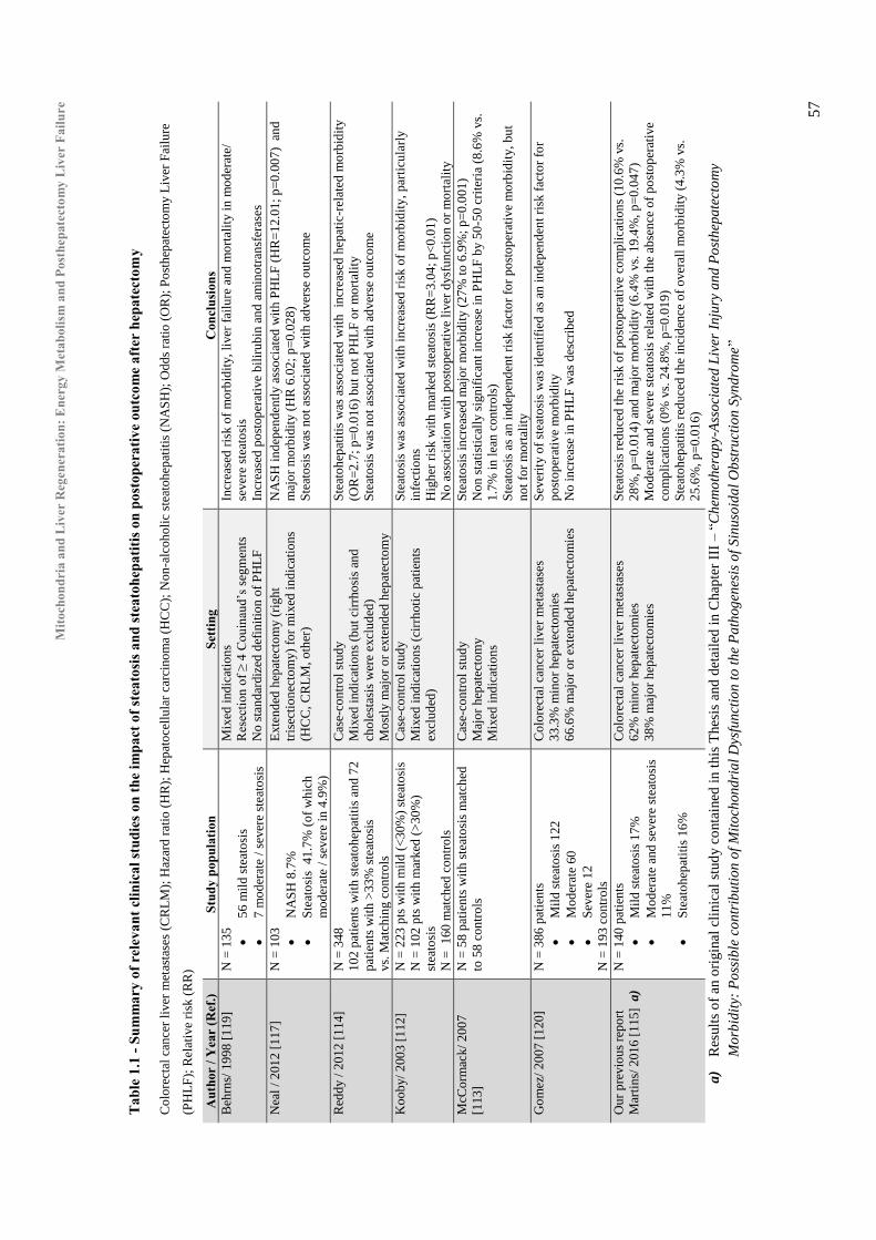
Mito
chon
dria
and
Liv
er R
egen
erat
ion:
Ene
rgy
Met
abol
ism
and
Pos
thep
atec
tom
y L
iver
Fai
lure
57
Tab
le 1
.1 -
Sum
mar
y of
rel
evan
t clin
ical
stud
ies o
n th
e im
pact
of s
teat
osis
and
stea
tohe
patit
is o
n po
stop
erat
ive
outc
ome
afte
r he
pate
ctom
y
Co
lore
ctal
can
cer
liver
met
asta
ses
(CR
LM
); H
azar
d r
atio
(H
R);
Hep
ato
cell
ula
r ca
rcin
om
a (H
CC
); N
on
-alc
oh
oli
c st
eato
hep
atit
is (
NA
SH
); O
dd
s ra
tio
(O
R);
Po
sth
epat
ecto
my L
iver
Fai
lure
(PH
LF
); R
elat
ive
risk
(R
R)
Aut
hor
/ Yea
r (R
ef.)
Stud
y po
pula
tion
Sett
ing
Con
clus
ions
B
ehrn
s/ 1
998
[1
19]
N =
135
5
6 m
ild
ste
ato
sis
7
mo
der
ate
/ se
ver
e st
eato
sis
Mix
ed i
nd
icat
ion
s
Res
ecti
on
of
≥ 4
Co
uin
aud
’s s
egm
ents
No
sta
nd
ardiz
ed d
efin
itio
n o
f P
HL
F
Incr
ease
d r
isk o
f m
orb
idit
y,
liver
fai
lure
an
d m
ort
alit
y i
n m
od
erat
e/
sever
e st
eato
sis
Incr
ease
d p
ost
op
erat
ive
bil
iru
bin
an
d a
min
otr
ansf
eras
es
Nea
l /
201
2 [
117]
N =
103
N
AS
H 8
.7%
S
teat
osi
s 4
1.7
% (
of
wh
ich
mo
der
ate
/ se
ver
e in
4.9
%)
Exte
nd
ed h
epat
ecto
my (
righ
t
tris
ecti
on
ecto
my)
for
mix
ed i
nd
icat
ion
s
(HC
C,
CR
LM
, o
ther
)
NA
SH
in
dep
end
entl
y a
sso
ciat
ed w
ith
PH
LF
(H
R=
12.0
1;
p=
0.0
07
) a
nd
maj
or
mo
rbid
ity (
HR
6.0
2;
p=
0.0
28
)
Ste
ato
sis
was
no
t as
soci
ated
wit
h a
dver
se o
utc
om
e
Red
dy /
20
12
[1
14]
N =
348
10
2 p
atie
nts
wit
h s
teat
oh
epat
itis
an
d 7
2
pat
ien
ts w
ith
>33
% s
teat
osi
s
vs.
Mat
chin
g c
on
tro
ls
Cas
e-co
ntr
ol
stud
y
Mix
ed i
nd
icat
ion
s (b
ut
cirr
ho
sis
and
cho
lest
asis
wer
e ex
clu
ded
)
Mo
stly
maj
or
or
exte
nd
ed h
epat
ecto
my
Ste
ato
hep
atit
is w
as a
sso
ciat
ed w
ith
in
crea
sed
hep
atic
-rel
ated
mo
rbid
ity
(OR
=2
.7;
p=
0.0
16
) bu
t n
ot
PH
LF
or
mo
rtal
ity
Ste
ato
sis
was
no
t as
soci
ated
wit
h a
dver
se o
utc
om
e
Ko
ob
y/
200
3 [
112]
N =
223
pts
wit
h m
ild
(<
30
%)
stea
tosi
s
N =
102
pts
wit
h m
arked
(>
30
%)
stea
tosi
s
N =
1
60
mat
ched
co
ntr
ols
Cas
e-co
ntr
ol
stud
y
Mix
ed i
nd
icat
ion
s (c
irrh
oti
c p
atie
nts
excl
ud
ed)
Ste
ato
sis
was
ass
oci
ated
wit
h i
ncr
ease
d r
isk o
f m
orb
idit
y,
par
ticu
larl
y
infe
ctio
ns
Hig
her
ris
k w
ith
mar
ked
ste
ato
sis
(RR
=3
.04
; p
<0
.01
)
No
ass
oci
atio
n w
ith
po
sto
per
ativ
e li
ver
dysf
un
ctio
n o
r m
ort
alit
y
McC
orm
ack/
20
07
[113]
N =
58
pat
ien
ts w
ith
ste
ato
sis
mat
ched
to 5
8 c
on
tro
ls
Cas
e-co
ntr
ol
stud
y
Maj
or
hep
atec
tom
y
Mix
ed i
nd
icat
ion
s
Ste
ato
sis
incr
ease
d m
ajo
r m
orb
idit
y (
27
% t
o 6
.9%
; p
=0.0
01
)
No
n s
tati
stic
ally
sig
nif
ican
t in
crea
se i
n P
HL
F b
y 5
0-5
0 c
rite
ria
(8.6
% v
s.
1.7
% i
n l
ean
con
tro
ls)
Ste
ato
sis
as a
n i
nd
epen
den
t ri
sk f
acto
r fo
r p
ost
op
erat
ive
mo
rbid
ity,
bu
t
no
t fo
r m
ort
alit
y
Go
mez
/ 2
007
[1
20]
N =
386
pat
ien
ts
M
ild
ste
ato
sis
122
M
od
erat
e 60
S
ever
e 1
2
N =
193
co
ntr
ols
Co
lore
ctal
can
cer
liver
met
asta
ses
33
.3%
min
or
hep
atec
tom
ies
66
.6%
maj
or
or
exte
nd
ed h
epat
ecto
mie
s
Sev
erit
y o
f st
eato
sis
was
id
enti
fied
as
an i
nd
epen
den
t ri
sk f
acto
r fo
r
po
stop
erat
ive
mo
rbid
ity
No
in
crea
se i
n P
HL
F w
as d
escr
ibed
Ou
r p
revio
us
rep
ort
Mar
tin
s/ 2
01
6 [
115]
a)
N =
140
pat
ien
ts
M
ild
ste
ato
sis
17
%
M
od
erat
e an
d s
ever
e st
eato
sis
11
%
S
teat
oh
epat
itis
16
%
Co
lore
ctal
can
cer
liver
met
asta
ses
62
% m
ino
r h
epat
ecto
mie
s
38
% m
ajo
r h
epat
ecto
mie
s
Ste
ato
sis
red
uce
d t
he
risk
of
po
sto
per
ativ
e co
mp
lica
tio
ns
(10
.6%
vs.
28
%, p
=0
.01
4)
and m
ajo
r m
orb
idit
y (
6.4
% v
s. 1
9.4
%,
p=
0.0
47
)
Mo
der
ate
and
sev
ere
stea
tosi
s re
late
d w
ith
th
e ab
sen
ce o
f p
ost
op
erat
ive
com
pli
cati
on
s (0
% v
s. 2
4.8
%, p
=0
.01
9)
Ste
ato
hep
atit
is r
edu
ced t
he
inci
den
ce o
f o
ver
all
mo
rbid
ity (
4.3
% v
s.
25
.6%
, p
=0.0
16
)
a)
Res
ult
s o
f an
ori
gin
al c
linic
al s
tud
y c
onta
ined
in
this
Thes
is a
nd
det
aile
d i
n C
hap
ter
III
– “
Ch
emo
ther
ap
y-A
sso
cia
ted
Liv
er I
nju
ry a
nd
Po
sth
epa
tect
om
y
Mo
rbid
ity:
Po
ssib
le c
on
trib
uti
on
of
Mit
och
on
dri
al
Dys
fun
ctio
n t
o t
he
Pa
thog
enes
is o
f S
inuso
ida
l O
bst
ruct
ion
Syn
dro
me
”
Chapter I
58
4.2 Cirrhosis
Cirrhosis is the end-stage of infectious, immune, toxic or metabolic insults to the
liver. It is characterized by decreased hepatocellular function and portal hypertension
[121]. Hepatocellular carcinoma is a frequent complication and resection is one of the
few curative treatments [4]. However, liver resection in cirrhosis is only possible in
patients with preserved liver function and even then at the cost of a high risk of PHLF
[122]. Moreover, cirrhotic livers are less tolerant to hepatic pedicle clamping, making
liver resection in these circumstances more demanding [123].
The decreased regenerative response of cirrhotic livers has been previously
noticed [124]. Among the contributing mechanisms, bioenergetics failure definitely
plays a key role. Although other factors, including the microvascular distortion of the
liver parenchyma act synergistically to decrease oxygen supply to liver cells [125], there
is significant clinical and experimental evidence of deranged mitochondrial function in
cirrhosis. Yang et al used a rodent model of chemically-induced cirrhosis to study the
mitochondrial respiratory function and antioxidant capacity after hepatectomy [126].
After inducing cirrhosis with intra-peritoneal thioacetamide in Wistar rats, the authors
performed 70% partial hepatectomy. After hepatectomy cirrhotic animals displayed
decreased state 3 respiration, decreased activities of NADH-cytochrome c reductase and
mitochondrial glutathione peroxidase, as well as decreased levels of mitochondrial
glutathione, when compared with non-cirrhotic controls. Nishikawa et al [55], using a
rat model of carbon tetrachloride and phenobarbital-induced cirrhosis, examined energy
metabolism in isolated hepatocytes and concluded that in compensated cirrhosis
maximal mitochondrial respiration is compromised and that ATP production was
maintained by an increase in the glycolytic pathway. As cirrhosis progresses to
decompensated form, failure of glycolytic pathway also occurs, leading to energetic
failure. The authors confirmed these findings in gene-expression profile of human liver
biopsy samples, as patients with compensated cirrhosis (Child A and B) had decreased
expression of cytochrome oxidase 1 and 2 genes, with increased expression of genes for
glycolytic enzymes. These enzymes were under-expressed in Child C cirrhosis,
underlying the energetic failure that occurs in this premorbid state of decompensated
liver disease. An interesting conclusion of this study is that mitochondrial function is
hampered in early stages of cirrhosis, with glycolysis taking over most of the energy

Mitochondria and Liver Regeneration: Energy Metabolism and Posthepatectomy Liver Failure
59
production. As the glycolytic pathway is much less energy-efficient than oxidative
phosphorylation, this could explain the decreased adaptation of compensated cirrhotic
livers to resection and other acute stressors, such as infection [127]; as failure to
increase ATP production would result in acute decompensation of liver function.
Deficient energetic recovery of the cirrhotic liver after hepatectomy has been
demonstrated by Mann et al. [128]. Using 31
Phosphorus Magnetic Resonance
Spectroscopy, the authors studied nine cirrhotic patients undergoing hepatectomy and
compared with nine other patients with normal liver parenchyma. Cirrhotic patients
experienced a sustained decrease in ATP to phosphate ratio and delayed regeneration
versus patients with normal liver parenchyma.
Given the evidence for disordered energetics in cirrhosis, interventions aimed at
improving energy status could be clinically relevant in prevention of hepatocellular
dysfunction after liver resection. This will be further explored in this Chapter in Section
6. Improving liver regeneration – Hepatocyte energetics as a potential target for
prevention of Postoperative Liver Failure.
4.3 Chemotherapy-induced liver injury
While hepatectomy is the gold standard of care for patients with colorectal
cancer liver metastases, preoperative chemotherapy is increasingly used, not only in a
conversion strategy but also in a true neoadjuvant intention [129,130]. The most
commonly used drugs are 5-fluorouracil (5-FU), irinotecan and oxaliplatin. The
cytotoxic agent 5-FU is a thymidylate synthase inhibitor and has been used for over 40
years in systemic therapy for advanced colorectal cancer. Irinotecan, or CPT-11, is a
prodrug. It suffers transformation by carboxylesterase into SN-38, a potent inhibitor of
topoisomerase-I. Oxaliplatin, a platinum derivative, forms DNA adducts and is directly
cytotoxic [131]. Molecular targeted therapies, such as monoclonal antibodies,
bevacizumab and cetuximab, have increased response and resectability rates [90].
Finally, in selected cases intra-arterial chemotherapy has demonstrated promising
results in conversion to resectability [132].
Chapter I
60
However, chemotherapy is associated with hepatocellular injury and increased
morbidity and even mortality after hepatectomy [116,133]. Although chemotherapy per
se produces a decline in hepatocellular function (as measured by indocyanine green
retention rate), independently of histologically-perceptible injury [134], chemotherapy-
associated liver injury (CALI) usually occurs in distinct histologic patterns: steatosis
and steatohepatitis (CASH); and sinusoidal obstruction syndrome (SOS).
Steatosis and steatohepatitis are usually associated with irinotecan-based
chemotherapy and one study demonstrated an increase in morbidity and even mortality
after liver resection [116]. Although the precise mechanism of hepatoxicity of irinotecan
is unknown, it is thought to involve mitochondrial dysfunction [135]. Vauthey et al
[116] reported a higher incidence of SH in patients treated with irinotecan-based
chemotherapy, in particular with Body Mass Index over 25 kg/m2. Patients with SH
were more likely to suffer from posthepatectomy liver failure and had higher
postoperative mortality. As the typical histologic injury of irinotecan-associated liver
injury is steatohepatitis, we refer to the previous section (4.1 – Steatosis and
steatohepatitis).
Sinusoidal obstruction syndrome (SOS) is another typical histologic pattern, and
is usually associated with oxaliplatin chemotherapy [136]. It is characterized by severe
sinusoidal dilation, sinusoidal endothelial cell necrosis and sloughing, causing
extravasation of blood into the space of Disse and decreased sinusoidal flow [137]. One
previous study from our group confirmed that SOS is an independent risk factor for
postoperative morbidity [115] and usually courses with increased portal pressure,
increased intraoperative bleeding, decreased tolerance to hepatic pedicle clamping and
lower regenerative capacity [138–141]. Narita et al described increased morbidity in
patients with histological evidence of chemotherapy injury during hepatectomy with
prolonged hepatic pedicle clamping [139]. In 53 patients with chemotherapy-associated
liver injury (mostly with oxilaplatin-related sinusoidal obstruction syndrome)
undergoing major hepatectomy, the authors found a statistically significant increase in
severe complications, namely sepsis and biloma, when hepatic pedicle clamping
exceeded 30 minutes. No significant association was found, however, with increased
incidence of postoperative liver failure, possibly due to the small study sample.

Mitochondria and Liver Regeneration: Energy Metabolism and Posthepatectomy Liver Failure
61
In yet another study, in a cohort of patients undergoing two-stage hepatectomy
for colorectal cancer liver metastases, Narita et al again reported that sinusoidal
obstruction syndrome was significantly associated with oxaliplatin-based
chemotherapy, hampered volumetric increase of the remnant liver after first-stage
hepatectomy, markedly depressed indocyanine green (ICG) clearance and caused a
trend towards increased incidence of postoperative liver dysfunction [142]. Also, a
blunted hypertrophic response was observed after portal vein embolization in patients
with previous exposure to platinum-based chemotherapy [143].
Apart from DNA-directed effects, there is evidence to support that oxaliplatin is
directly toxic to mitochondria, inhibiting oxidative phosphorylation in isolated liver
mitochondria [144,145] and inducing mitochondrial-dependent apoptosis in cell cultures
[146].
However, the lack of a valid animal model of oxaliplatin-induced toxicity has
been a major drawback. Up until recently the only existing model of posthepatectomy
liver dysfunction in toxic-induced SOS was the monocrotaline model [147]. The work
of Robinson et al has shed new light into the pathophysiology of SOS. The authors
validated an animal model of oxaliplatin-induced SOS in C57Bl/6 mice and found
increase in oxidative stress in the injured parenchyma [148].
Although there is still no definitive link between mitochondrial toxicity and the
development of SOS, we postulate that mitochondrial homeostasis could be severely
hampered in chemotherapy-associated liver injury, thus leading to an increased
susceptibility to postoperative liver dysfunction. Further investigation into this field is
of paramount importance and has thus been the subject of investigation in this Thesis. In
Chapter III original results from clinical and experimental works are presented.
4.4 Chronic biliary obstruction
Extended hepatectomy with en bloc bile duct resection is potentially curative for
hilar cholangiocarcinoma [5,149]. However, liver resection under biliary obstruction is
fraught with an extremely high incidence of postoperative liver dysfunction, with
significant mortality. In the experience of a high-volume centre, Yokoyama et al report
Chapter I
62
as much as 38% incidence of moderate and severe PHLF in patients undergoing major
or extended hepatectomy for biliary cancer, with severe PHLF associated with a 48%
mortality rate [150]. Thus, preoperative biliary decompression is recommended to
decrease the risk of PHLF and improve results in patients with high malignant biliary
obstruction [151] and most centres nowadays include it in the preoperative algorithm
for hilar cholangiocarcinoma, especially when there is a smaller future liver remnant
(FLR) [5,149,152–154]. However, there is uncertainty as to what is the defined upper
threshold of serum bilirubin mandating preoperative decompression; with cut-off values
ranging from 2 to 10 mg/dL in most series [5,149]. Chronic cholestasis induces liver
dysfunction through several mechanisms, including mitochondrial toxicity [155].
Conversely, biliary decompression results in an improvement in energy status of the
liver. This is supported by clinical and experimental evidence which we will now
summarily review.
Bile acids are directly toxic to mitochondria, causing a significant reduction of
membrane potential, state 3 respiration and Respiratory Control Ratio, as well as
increased susceptibility to Mitochondrial Permeability Transition (MPT) [156,157].
Mitochondrial biogenesis is also reduced by chronic biliary obstruction, as
demonstrated by Arduini et al. In a rat model of bile duct ligation (BDL) there was a
decrease in RCR and membrane potential, decreased expression of TFAM and depletion
of mitochondrial DNA [158]. Surprisingly, in another animal model of segmental
biliary obstruction, mitochondrial dysfunction was demonstrated not only in the
obstructed lobe but also in the non-obstructed one [159].
Partial reversal of physiologic derangements by biliary decompression was
demonstrated in an experimental model. Krähenbühl et al explored mitochondrial
homeostasis in a rat model of BDL followed by Roux-en-Y anastomosis four weeks
later [160]. The authors demonstrated that oxidative phosphorylation and β-oxidation of
fatty acids were inhibited during cholestasis. After relief of obstruction there was an
improvement in activities of complexes I and III of the mitochondrial electron transport
chain, whereas the activities of complexes II and IV remained impaired. Furthermore, β-
oxidation of fatty acids also remained depressed. Finally, in an extremely interesting
clinical study, Mann et al used 31
Phosphorus magnetic resonance spectroscopy to study
liver energy status in 10 patients with malignant biliary obstruction [161]. After

Mitochondria and Liver Regeneration: Energy Metabolism and Posthepatectomy Liver Failure
63
endoscopic or percutaneous biliary drainage, the ATP/Pi ratio, a measure of energy
status, significantly increased in one week time, reflecting the beneficial effect on the
entire liver parenchyma.
In conclusion, part of the biologic rationale for preoperative biliary
decompression in the management of jaundiced patients undergoing major hepatectomy
and bile duct resection can be related to improvement in mitochondrial function. If
proved, further therapies aimed at mitochondrial bioenergetics could lead to an
enhanced energetic status of the remnant liver, thereby decreasing the inherently high
risk of postoperative liver dysfunction. Ultimately, this could result in a significant
improvement in the management of perihilar cholangiocarcinoma.
5. Novel markers of mitochondrial dysfunction – Earlier diagnosis of Posthepatectomy Liver Failure
With the aim of standardizing the diagnosis and reporting of PHLF, a consensus
definition was established in 2010 [162]. It defined PHLF as a postoperatively acquired
deterioration in liver function, consisting of any increase of bilirubin and International
Normalized Ratio (INR) on or beyond the fifth postoperative day. Serum bilirubin and
INR reflect two major hepatocyte functions: excretory function (bilirubin uptake,
conjugation and excretion); and synthetic function (namely of plasma clotting factors II,
VII, IX and X) [163,164]. Usually, after uneventful hepatectomy these two parameters
are within normal range by the fifth postoperative day, reflecting appropriate liver
regeneration [11] (Figure 1.2). This consensus definition also defined clinical severity in
three grades, according to the intensity of medical support required and the coexistence
of other organ dysfunctions.
Another clinically used definition of PHLF using biochemical criteria is the “50-
50 criteria” defined by Balzan et al. In 803 consecutive patients that underwent liver
resection, the finding of bilirubin over 50 µmol/L and prothrombin time under 50% on
postoperative day 5 was associated, after multivariate analysis, with increased
postoperative mortality [165]. Different thresholds have been suggested in specific

Chapter I
64
subpopulations of surgical patients, such as peri-hilar cholangiocarcinoma [150] and
cirrhotic patients with hepatocellular carcinoma [166].
Although straightforward to use and solidly placed in the clinical routine, these
definitions are only valid on or beyond the fifth postoperative day, meaning that earlier
liver dysfunction could be underdiagnosed. Furthermore, their use can be compromised
in patients with previous biliary obstruction, who usually have high preoperative plasma
bilirubin levels, while plasma transfusion can significantly change postoperative INR
levels. Thus, the possibility of predicting liver dysfunction at an earlier time point is
attractive, since it would allow more intensive treatment.
Since mitochondrial derangement likely occurs before the development of full-
blown liver dysfunction [19], the resulting bioenergetics failure of the remnant liver can
hamper liver regeneration [43]. Thus, the discovery of non-invasive serum markers of
bioenergetics dysfunction is an attractive way for earlier detection of Posthepatectomy
Liver Failure. These biomarkers can either be indirect metabolic markers of disturbed
energetic status or can more directly reflect mitochondrial dysfunction.
Arterial lactate, an established prognostic marker in paracetamol-induced acute
liver failure [167] and critical illness [168], can reflect the energy status of the
hepatocyte. Lactate, produced by peripheral anaerobic glycolysis, is metabolized in the
liver through the Cori cycle, whereby it is recycled to pyruvate and glucose in energy-
consuming reactions partly occurring in the mitochondria [29]. In an animal model of
PHLF, consisting of extended resection and remnant liver ischemia, Detry et al found
significantly increased levels of lactate in the experimental group [169]. Watanabe et al
proved that elevated arterial lactate on admission to the Intensive Care Unit after
hepatectomy was independently associated with higher postoperative morbidity and
mortality [170]; and more recently the value of early postoperative arterial lactate as an
indicator of poor prognosis has been validated in a large prospective cohort [171].
Elevated arterial lactate not only reflects higher peripheral production (because of
systemic hypoperfusion) but mostly the liver’s inability to adequately perform
gluconeogenesis because of insufficient parenchyma or altered energy state. In fact, in a
cohort of 14 patients, Theodorakis et al have demonstrated that, in conditions of
ischemia-reperfusion, the liver becomes a net producer of lactate [172].
Mitochondria and Liver Regeneration: Energy Metabolism and Posthepatectomy Liver Failure
65
Serum phosphorus has also been scrutinized as a prognostic marker after
hepatectomy. As liver regeneration usually requires an enormous supply of inorganic
phosphate (for ATP synthesis, among other functions), a nadir in serum phosphate is not
only frequent but also desirable after liver resection [173]. In fact, in a cohort of 719
patients undergoing major hepatectomy, Squires et al [174] have demonstrated that
patients with profound hypophosphatemia (< 1.1 mg/dL) on postoperative day 2
experienced reduced incidence of PHLF and also major morbidity and 30-day mortality,
when compared to patients with normal phosphatemia. However in this study, the
majority of patients had parenteral phosphate supplementation. Furthermore, post
resection drop in serum phosphate is also probably related to increased urinary loss
[175], potentially compounding the use of phosphate as a reliable marker of acquired
liver dysfunction.
The Arterial Ketone Body Ratio (AKBR), acetoacetate to β-hydroxybutyrate
ratio, is a marker of mitochondrial redox state. In conditions of impairment of the
mitochondrial electron transport chain, nicotinamide adenine dinucleotide (NADH)
cannot be recycled to its oxidized form (NAD+), meaning that higher quantities of β-
hydroxybutyrate are formed, decreasing the AKBR. Nakamura et al demonstrated that
decreased hepatic perfusion during cardiopulmonary bypass in heart surgery was
associated with a lower AKBR; however without deleterious consequences [176]. In the
setting of liver resection Yamaguchi et al initially proposed that AKBR could predict
survival after hepatectomy [60]. In this study, an AKBR under 0.4 was associated with
increased arterial lactate and decreased survival. However, no association was sought
with other markers of postoperative liver function. In another study, Yan et al also
demonstrated that patients with AKBR under 0.4 had higher morbidity and mortality
[177]. Higashi et al have specifically investigated the value of arterial ketone bodies
measurement when compared with hepatic vein levels. They have found an excellent
correlation between both measurements, suggesting that arterial ketone body levels
reflect hepatic synthesis, rather than peripheral catabolism. In their cohort of 73
patients, the authors also found increased levels of β-hydroxybutyrate in patients with
impaired liver function (worse ICG retention rate, higher postoperative bilirubin) and
increased mortality [178]. Although promising, AKBR has not found its way into the
clinical routine of most hepatobiliary centres.
Chapter I
66
Direct assessment of intra- and postoperative liver metabolism is possible with
the use of microdialysis catheters, with serial measurements of intra-hepatic levels of
lactate, pyruvate, glucose and glycerol. In the setting of liver transplantation
microdialysis can assist in the early detection of severe ischemic injury [179,180]. At
least two studies have reported the use of this technique in liver surgery. Isaksson et al,
in 11 patients undergoing hepatectomy, reported increasing intra-hepatic levels of
lactate, glucose and glycerol and increased lactate / pyruvate ratio during hepatic
pedicle clamping [181]. And Windbladh et al explored the metabolic protection
afforded by ischemic preconditioning in a randomized control trial involving 32
patients, demonstrating lower peri- and postoperative levels of intra-hepatic lactate in
patients undergoing major hepatectomy [182]. Neither study, however, was designed to
validate microdialysis as an early predictor of PHLF. In concept microdialysis is an
extremely interesting approach, allowing bedside real-time monitoring of the lactate /
pyruvate ratio, which accurately reflects energy metabolism. However, microdialysis is
an invasive procedure as it requires the placement of an indwelling catheter on the liver
parenchyma during hepatectomy. Further studies are needed in order to validate this
technique for the early detection of PHLF.
Direct measurement of mitochondrial function in biopsies taken in the remnant
liver at the end of hepatectomy could help predict PHLF. In personal work of our group
we have previously demonstrated that mitochondrial lag phase (time to repolarize
membrane potential after addition of ADP) was significantly longer in patients that
went on to develop liver dysfunction, with a high specificity and sensitivity (area under
the curve: 0.933; p=0.008) (see also Chapter II) [19]. However, this requires further
confirmation in more studies. Furthermore, direct assessment of mitochondrial function
in liver tissue, although feasible through a needle biopsy [183], is an invasive test and
requires a dedicated bioenergetics laboratory for immediate sample processing. Ideally,
mitochondrial dysfunction could be detected with non-invasive serum markers, with
point of care tests that could find their way into routine clinical practice, from a “bench-
to-bedside” approach.
Several circulating serum biomarkers of mitochondrial dysfunction, including
ornithine carbamoyl transferase, alanine aminotransferase isoform 2 and cytochrome c,
have emerged in the setting of drug-induced liver injury [184]. In particular,

Mitochondria and Liver Regeneration: Energy Metabolism and Posthepatectomy Liver Failure
67
acetaminophen-induced liver injury (AALF), one of the most common causes of acute
liver failure in the developed world, has been the focus of attention. Mitochondrial
toxicity is a hallmark event in its pathogenesis and not all intoxicated patients develop
full-blown acute liver failure [57]. Thus, early and accurate identification of patients at
risk is extremely important, as it can help in proper allocation of resources and timely
transfer to higher levels of care, such as emergent referral for liver transplantation
[185,186]. Several markers of mitochondrial dysfunction have been sought, namely
mitochondrial DNA, circulating acylcarnitines and glutamate dehydrogenase (GLDH).
The latter seems particularly promising, since Schomaker et al reported a high
sensitivity and specificity of GLDH for diagnosis of toxic hepatocellular injury, with an
area under the curve of 0.98 [187].
McGill et al reported on the use of serum mitochondrial biomarkers in a cohort
of 69 patients with AALF [86]. They demonstrated that serum GLDH and serum levels
of mitochondrial DNA (specifically for NADH dehydrogenase and cytochrome c
oxidase subunit III) were higher in patients with AALF versus healthy controls.
Furthermore, they demonstrated that these markers were significantly higher in non-
survivors than in survivors. Using a Mitochondrial Biomarker Damage Index the
authors report increased sensitivity and specificity in prediction of outcome.
In another work by the same group [185], circulating acylcarnitines were
investigated in a cohort of patients with AALF. Acylcarnitines are carnitine-linked long
chain fatty acids (e.g. palmitoyl-, linoleoyl- and oleoyl-), that are transported into the
mitochondrial matrix for β-oxidation through facilitated diffusion by the carnitine-
acetylcarnitine translocase (CACT). When β-oxidation is impaired these fatty acids
derivatives accumulate in the cytosol and increased plasma levels are observed after cell
necrosis. However, although promising in the rodent model, this marker did not show
relevance in the clinical setting for AALF.
Albeit attractive, these mechanistic biomarkers were studied in the setting of
acetaminophen toxicity, which is highly dependent on mitochondrial dysfunction. As
such, extrapolation of these results for other mechanisms of liver injury, such as post-
resection and post-ischemia-reperfusion injury must await confirmation of further
studies.

Chapter I
68
In this Thesis we explored GLDH as a putative early diagnostic marker of
PHLF. Although the original studies herein included were underpowered to prove its
diagnostic accuracy, we nonetheless demonstrated important correlations of GLDH with
other markers of postoperative liver dysfunction, such as serum bilirubin and arterial
lactate (see Chapter III and Chapter IV – “Bioenergetic Adaptations of the Liver in
ALPPS: How Liver Regeneration Correlates with Mitochondrial Energy Status” for
more details). Hopefully, GLDH and other biomarkers of disturbed bioenergetics will
find their way into clinical practice and allow for an earlier diagnosis of PHLF.
6. Improving liver regeneration – Hepatocyte energetics as a potential target for prevention of Postoperative Liver Failure
PHLF is associated with high mortality and current therapies are mostly
supportive. The use of extracorporeal liver assist devices is an attractive option,
including the Molecular Adsorbent Recirculating System (MARS) [188] but results
have been largely disappointing and solid evidence is lacking in the particular
population of patients with PHLF [189]. The treatment of PHLF has been the focus of
extensive reviews and the reader is referred to them [8,10,190].
While in the previous sections we recapitulated the evidence for an important
role of mitochondrial function in liver recovery following resection, much more
appealing is the possibility of actually preventing PHLF by targeting mitochondrial
homeostasis. Plausibly, energetic conditioning of liver cells could aid in the
maintenance of adequate liver function and in the recovery of functioning liver mass
after major hepatectomy. Several possible pathways could be explored, either isolated
or in combination, include: improvement in mitochondrial pool by enhanced biogenesis
and more efficient mitophagy; more efficient oxidative phosphorylation through
increased parenchymal oxygenation; and decrease in mitochondrial permeability
transition (Figure 1.5).
Because of the considerable overlapping of these pathways, we will now dwell
on several therapeutic strategies, presenting them according to different methods: 1)

Mitochondria and Liver Regeneration: Energy Metabolism and Posthepatectomy Liver Failure
69
Pharmacological conditioning; 2) Strategies aiming at improving hepatocyte
oxygenation; 3) Parenchymal modulation techniques; 4) Stem cell therapies.
Figure 1.5 - Mitochondrial-centered perspective on posthepatectomy outcome
Adequate recovery of liver parenchyma after resection depends upon a proper cellular energy homeostasis.
Prevention of Posthepatectomy Liver Failure can be achieved by increasing hepatic oxygen uptake, such as
intraoperative hemodynamic modulation, prevention of postoperative portal hyperperfusion, hyperbaric oxygen
therapy or portal vein arterialization. Enhanced mitochondrial biogenesis is currently the focus of research in
pharmacological preconditioning strategies and is likely involved in the mechanism of action of portal vein
embolization and possibly two-stage hepatectomies. Improved mitochondrial autophagy and inhibition of the
mitochondrial permeability transition are novel and yet clinically unexplored pathways that could potentially improve
energy homeostasis in regenerating hepatocytes, enhancing ATP supply and decreasing production of ROS (see text
for further details).
6.1 Pharmacological therapy
Mitochondrial-based therapies have been the focus of recent research. S-
adenosyl-L-methionine (SAMe) is a methyl donor and a precursor of glutathione.
Interestingly, Bailey et al demonstrated that SAMe improved mitochondrial respiration
and oxidative phosphorylation, while decreasing mitochondrial ROS and damage to
mitochondrial DNA in chronic ethanol exposure [191]. In another work, Brown et al
report protection of SAMe against hepatic toxicity in an animal model of
acetaminophen-induced liver injury [192]. And in an animal model of 70% hepatic

Chapter I
70
ischemia-reperfusion, Jeon et al demonstrated that pre-treatment with SAMe reduced
peak AST, decreased lipid peroxidation, improved AKBR and maintained ATP levels
[193]. Clinical evidence for use of SAMe as a pharmacological conditioning agent in
liver resection has come from two randomized controlled trials [194,195]. Both studies
demonstrated a decrease in peak postoperative aminotransferases and bilirubin, in
particular with HPC over 15 minutes, in patients undergoing hepatectomy for
hepatocellular carcinoma in cirrhosis. However, no mechanistic study was performed
and we can only theorize that the protection afforded by SAMe is, at least in part,
directly related to improvement in mitochondrial respiration and in oxidative
phosphorylation. Further studies into the clinical applications of SAMe in liver surgery
are needed, in particular from a mitochondrial-centred perspective.
Berberine, an isoquinoline alkaloid found in several plants used in traditional
Chinese medicine, is another promising compound. Known for its antidiabetic
properties, it has demonstrated beneficial effects on mitochondrial oxidative
phosphorylation in fatty liver models, mostly through Sirtuin 3 mediated mechanism,
possibly involving enhanced mitochondrial biogenesis, among other actions [100].
Other possible mitochondrial-centred positive effects include ethanol-induced liver
toxicity and renal ischemia-reperfusion injury [196,197]. At least one clinical trial has
proved its safety and efficacy in NAFLD [198], suggesting that berberine could be a
valid alternative for energetic conditioning of liver cells before hepatectomy. Further
studies are needed, however.
One of the most exciting candidate molecules for energetic conditioning of liver
cells is the Augmenter of Liver Regeneration (ALR). Having been first isolated and
purified by Labrecque et al [199], ALR is also known as Hepatic Stimulator Substance
or Hepatopoietin, for its strong mitogenic effect, far surpassing other known hepatic
mitogens such as Epidermal growth factor (EGF) and Transforming growth factor alpha
(TGF-α). ALR is secreted in liver cells and acts on Kupffer cells and in autocrine and
paracrine fashion on hepatocytes. It is also found in the intracellular compartment,
namely in mitochondria, where it regulates oxidative phosphorylation and is essential
for mitochondrial biogenesis and ATP synthesis [200]. The importance of ALR in liver
regeneration seems to be dependent upon its effect on mitochondrial biogenesis, uphill
of other two co-factors, TFAM and PGC-1α [21]. In fact, in this Thesis we describe an
Mitochondria and Liver Regeneration: Energy Metabolism and Posthepatectomy Liver Failure
71
increased expression of ALR RNA in the remnant liver of patients during the second
stage of two-stage hepatectomies (see Chapter IV). In one experimental study,
transfection of isolated hepatocytes with antisense oligonucleotide against ALR brought
about energetic failure and cell death by apoptosis and necrosis [201], while transfection
of ALR via adenovirus in rodent models of hepatic ischemia reperfusion and toxic
injuries demonstrated improved oxidative phosphorylation, increased ATP production,
decreased mitochondrial permeability transition, reduced cell death and improved
survival [202,203]. Interestingly, delivery of ALR is not limited to gene transfection as
at least two studies demonstrated that intraperitoneal injection was also effective in
improving mitochondrial bioenergetics and biogenesis [204,205]. This ease of
administration, not requiring ALR integration into the host genome, could open the
possibility of expanding ALR therapy into the clinical arena for the treatment of acute
liver failure of several etiologies, including PHLF.
Another potential therapeutic target is inhibition of the MPT. At least two
experimental studies have demonstrated promising results with N-methyl-4-isoleucine
cyclosporine (NIM811) in the setting of posthepatectomy liver failure and in small-for-
size liver transplant [20,53]. The search for small molecule inhibitors of the MPT is
ongoing and future advances are expected in the clinical arena in the near future
[206,207].
Finally, induction of autophagy is yet another possible mechanism of hepatic
energetic conditioning and a plausible therapeutic avenue to pursue with pharmacologic
agents [37]. By the selective removal of damaged mitochondria, enhanced mitophagy is
thought to increase the energetic efficiency of cells and decrease oxidative stress, thus
improving liver regeneration and survival in a rodent model of 90% hepatectomy [208].
6.2 Improving hepatocyte oxygenation
Intraoperative increase of hepatic oxygen delivery and extraction could
potentially prevent postoperative hepatocellular dysfunction by increasing the energy
supply to the rapidly dividing and metabolically active liver parenchymal cells [62,209].
Since the use of inflow occlusion to minimize bleeding during liver resection causes

Chapter I
72
inevitable liver ischemia, diverse clamping techniques and strategies have been
developed to minimize the impact on the liver parenchyma. This subject has been
extensively detailed elsewhere [68,210–212]. In this review we will shed some light on
novel or less explored approaches to increase parenchymal oxygenation. Several
strategies aiming at this goal have been pursued, some of them with clinically relevant
results.
Intraoperative hyperdynamic circulatory pharmacological manipulation with β–
adrenergic agonists is an attractive option, as it increases hepatic arterial flow, portal
vein oxygen saturation and hepatic oxygen delivery, with subsequent increase in liver
lactate uptake [213]. In an extremely interesting prospective randomized trial with 30
compensated cirrhotic patients undergoing hepatectomy for HCC, Taurà et al
demonstrated that low dose β-adrenergic agent dobutamine decreased portal vein
resistance, increased hepatic blood flow and improved hepatic oxygen delivery,
consumption and extraction. In the dobutamine-treated group this resulted in increased
lactate clearance and decreased peak serum AST and peak postoperative bilirubin [214].
Although underpowered to prove a decrease in morbidity with the pharmacologic
intervention, this study is fascinating as it conclusively demonstrates that enhancement
of hepatic aerobic metabolism improves postoperative liver function.
Modulation of portal flow is another option, since extended hepatectomies result
in portal overflow and subsequent arterial vasoconstriction, mediated by the Hepatic
Arterial Buffer Response [79] (see Section 3. Posthepatectomy Liver Failure - Evidence
for disturbed bioenergetics in pathophysiology). This relative liver hypoxia during the
post-resection acute increase in metabolic demands could theoretically contribute to
early liver remnant bioenergetics failure. Thus, reversal of excessive portal flow has
been explored in order to prevent postoperative liver dysfunction. Several different
animal models have explored surgical measures to decrease splanchnic blood flow,
namely jejunal resection, splenectomy, splenic artery ligation and portal vein banding.
Kawano et al, performing jejunectomy and 70% hepatectomy in dogs, reported
improved hepatocellular function and decreased necrosis and apoptosis [215]. In a
rodent model of 90% hepatectomy, splenectomy has been demonstrated to decrease
portal vein flow, improve regeneration and decrease postoperative liver failure [216];
this effect is linked to an increase in hepatic arterial blood flow and hepatic oxygenation
Mitochondria and Liver Regeneration: Energy Metabolism and Posthepatectomy Liver Failure
73
and is likely mediated by the HABR [217,218]. Decreased mitochondrial swelling has
also been described after splenectomy as a portal flow modulation strategy after 80%
hepatectomy [219]. Splenic artery ligation has also been tested in experimental models,
with encouraging results [220]. Portal vein banding is another promising method and
one preclinical study in porcine model has reported improved postoperative liver
function, however without demonstrating increased parenchymal oxygenation or
improved hepatocellular metabolism [221]. These studies have been followed by
clinical attempts to modulate portal blood flow as a strategy to improve liver
regeneration, in the setting of partial liver transplantation [222,223] and extended
hepatectomy (E. Vibert, personal communication)[224,225].
Interestingly, improved arterial flow could in fact be modulated independently
of portal flow, as Kelly et al demonstrated in a porcine model of small-for-size liver
transplant. Using intra-arterial adenosine perfusion starting on postoperative day one
through a catheter placed in the gastroduodenal artery, the authors describe increasing
arterial blood flow without modulating portal flow, with adenosine treated animals
experiencing improved survival, decreased ascites, improved liver function and reduced
necrosis score, versus controls [226]. If further studies confirm these findings, the aim
of therapies could shift from reducing portal flow to increasing arterial flow, and hence
parenchymal oxygenation, in PHLF and SFSS.
Given that the rationale for the aforementioned strategies is the increase in liver
oxygen uptake, hyperbaric oxygen therapy (HBO) would seem a reasonable approach.
Although there is both preclinical and clinical evidence to support its use, it has been
largely unexplored. One experimental study has demonstrated improvement in liver
regeneration and ATP content after 90% hepatectomy with 2 atmospheres 80% O2
therapy [227]. Mitochondrial function was assessed in one animal model of 70%
hepatectomy and although there was a decrease in RCR in the HBO-treated group, there
was also an in improvement in liver regeneration rate, DNA content and proliferation
index [228]. There is undoubtedly a beneficial effect in liver bioenergetics, as HBO can
prevent deterioration of RCR, maintain mitochondrial membrane potential and sustain
ATP synthesis in isolated mitochondria in experimental conditions of cold ischemia
reperfusion injury [229]. The clinical evidence for the use of HBO in liver surgery has
come mostly from the pediatric transplantation setting where it has shown benefits in

Chapter I
74
the management of hepatic artery thrombosis [230,231]. Apart from case reports
demonstrating its anecdotal use in acute liver failure [232], including one case of PHLF
[233], at least two prospective clinical trials have reported encouraging results on its use
after hepatectomy. Ueno et al randomized 41 cirrhotic patients with hepatocellular
carcinoma after hepatectomy complicated with major intraoperative blood loss to either
HBO or standard management [234]. HBO-treated patients experienced lower
postoperative arterial lactate and bilirubin, higher hepatic vein oxygen saturation and a
trend towards improved hepatocellular function. However, in this study the definition of
PHLF was not standardized so no conclusions can be drawn on this respect. Suehiro et
al studied the effect of HBO on seven left liver living-donors and compared them to
seven standard-treated equally-matched controls [235]. HBO-treated donors presented
with better late postoperative liver function and increased liver remnant volume at four
weeks. As such, HBO has proved safe and feasible, mandating the call for a randomized
controlled trial on its use after major hepatectomy in patients at high risk of PHLF.
Inasmuch as increasing liver oxygen uptake could improve bioenergetics and
hence prevent liver dysfunction, another potentially useful technique would be portal
vein arterialization (PVA). In fact, PVA in animal models is known to increase portal
venous flow and oxygenation, liver ATP content and survival after extended
hepatectomy [236], with corresponding increase in liver regeneration rate and cell
proliferation [237,238]. Although an appealing therapy in concept, its clinical use has
mostly been reported for salvage therapy of a totally de-arterialized liver [239,240] and
cannot, at the present moment, be proposed as a first-line therapy for prevention of
PHLF.
6.3 Parenchymal modulation techniques
One of the most widely used strategies in prevention of PHLF is portal vein
embolization (PVE), or portal vein ligation (PVL). First described in the English
literature in 1986 [241], PVE is an Intervention Radiology technique that shunts portal
blood away from the tumour-bearing parenchyma into the FLR, causing atrophy of the
former and hypertrophy of the latter [242]. One randomized controlled trial
demonstrated that PVE decreased the rate of postoperative complications, improved

Mitochondria and Liver Regeneration: Energy Metabolism and Posthepatectomy Liver Failure
75
liver function and shortened intensive care unit and total hospital stay in patients with
chronic liver disease undergoing major hepatectomy [243]. There is experimental
evidence for modulation of mitochondrial function in the biologic rationale of PVE, as
there is an early increase in oxidative phosphorylation and respiration in the non-
embolized lobe in a rabbit model [244]. Katoh et al have demonstrated an early and
sustained increase in mitochondrial function in the non-embolized lobes in a rat model
[245], possibly mediated by a marked increase in mitochondrial DNA and mRNA that
occurs early after PVE [246]. More recently, the hypertrophic response to PVE was also
associated with the Nuclear factor erythroid 2-related factor 2 (Nrf-2) transcription
factor [247], a key inductor of mitochondrial biogenesis [34].
Two-stage hepatectomies, including the Associating Liver Partition and Portal
Ligation for Staged Hepatectomy (ALPPS) procedure [248,249], have emerged in the
armamentarium of liver surgeons. They use the same physiologic principle of PVE and
PVL, namely diversion of portal flow to the remnant liver. In the case of the ALPPS
procedure, parenchymal transection is also performed and is associated with a markedly
increased liver hypertrophy [250]. This greatly enhanced volume increase is dependent
upon an enhanced proliferative index, increase in hepatocyte cytoplasm and nuclear size
[251]. Although the parenchymal response is similar to that observed with PVE, the
hepatocyte morphology is more immature, with less organelles [252]. This inordinately
rapid regenerative response probably requires important metabolic adaptations in order
to sustain the increased energy requirements of rapidly dividing hepatocytes. However,
the mechanisms underlying these adaptations in cellular bioenergetics are largely
unknown and have also been the subject of interrogation in this Thesis. In Chapter IV
we present evidence for several inter-stages metabolic changes, including enhanced
bioenergetics and increased mitochondrial biogenesis in ALPPS, never previously
reported in the scientific literature.
6.4 Stem cell therapy
Cell therapy has emerged as an important approach for end-stage liver disease,
with both clinical and experimental studies reporting on the beneficial impact of
Chapter I
76
mesenchymal, embryonic and induced pluripotent stem cells on liver regeneration after
toxic and surgical insults [253].
In part, the effect of stem-cell therapy could be mediated by an improvement in
mitochondrial function, as there is an increase in mitochondrial number [254], decrease
in mitochondrial-mediated apoptosis [255] and improved mitochondrial respiration
[256]. In a very interesting study, Andrade et al have demonstrated that bone marrow
mononuclear cells transplanted through the jugular vein 14 day after BDL in Wistar rats
resulted in improved mitochondrial respiration (state 3 and RCR), decreased lipid
peroxidation and decreased fibrogenesis [257]. Tautenhahn et al [258] studied the effect
of mesenchymal stem cells on an animal model of PHLF. They found that the
improvement in liver function, decreased apoptosis, increased regeneration and overall
better metabolic profile occurred very early after therapy and were independent of stem
cells’ engraftment. As such, an interesting phenomenon could be taking place, as stem
cells could potentially transfer mitochondria to liver cells, thus improving their
metabolic capacity. The transfer of mitochondria is thought to occur through
intercellular nanotubes [259] and direct intrasplenic injection of isolated mitochondria
has also proved beneficial in experimental models of liver ischemia-reperfusion injury
[260]. Although stem cell therapy for PHLF has been mostly experimental, some
clinical trials have addressed the possibility of using CD133+ stem cells in the
prevention of PHLF, in particular as adjuvant to portal vein embolization [261,262]. As
further clinical studies are underway in cell therapy for liver disease, it would be
interesting to evaluate the effect on mitochondrial function, in particular in the setting of
acute liver failure and PHLF.
Conclusions and future prospects
Although the subject of bioenergetics in liver regeneration and clinical liver
surgery has been explored in the past, recent clinical and experimental evidence has
rekindled the interest in this field. With the increased prevalence of chronic liver disease
and the expanding indications for hepatectomy, novel strategies to improve the results
of liver surgery are paramount. Albeit a wondrous phenomenon, liver regeneration is

Mitochondria and Liver Regeneration: Energy Metabolism and Posthepatectomy Liver Failure
77
not inexhaustible and some patients still die from liver dysfunction in spite of optimal
pre-, intra- and postoperative management, even in the most experienced hepatobiliary
surgery centers. As liver physiology is evermore so taken to its limits, a profound
knowledge of the factors involved in liver regeneration is desperately needed, including
the energy source that fuels it. Mitochondria, the main energy suppliers of eukaryote
cells and key players in cell death, find themselves at the heart of this process, and in
the spotlight of biomedical research.
The clinical importance of bioenergetics in liver surgery is threefold. Firstly,
bioenergetics status of the liver parenchyma could become an important prognostic
marker of preoperative liver reserve, in the same way as volumetry or ICG retention rate
[263,264]. This would be of particular relevance in cases of diseased liver parenchyma,
whereby an accurate assessment of liver functional reserve is sometimes exceedingly
difficult. Indeed, the search for precise functional assessment of the FLR is still
ongoing, with an increasingly greater emphasis on dynamic or segmental function tests
(kinetic growth rate, differential assessment of right versus left liver function by
hepatobiliary scintigraphy or gadoxetic acid-enhanced magnetic resonance) rather than
static or whole-liver function tests [265–268]. As both dynamic and static liver function
tests ultimately depend on the assessment of endergonic, or ATP consuming reactions,
measurement of energy status of liver cells could in itself be considered a liver function
test [269]. In this regard, preoperative bioenergetics capacity would potentially translate
into clinical practice as a significant measure of parenchymal tolerance to surgical
insult.
Secondly, as the evidence for the key role of mitochondria in the
pathophysiology of posthepatectomy liver failure unfolds, so does the possibility of
using markers of mitochondrial dysfunction in the early detection of this dreaded
complication gain even more relevance. In fact, several serum biomarkers are currently
being explored in the setting of toxic-induced acute liver failure and could also prove
their worth in the early diagnosis of PHLF. Furthermore, novel approaches like
microdialysis could potentially aid in the early detection of the metabolic derangements
that precede full-blown liver failure in high-risk patients.
Chapter I
78
Finally, as the possibility of direct interventions on liver energy homeostasis
proves useful in improving clinical outcomes, a fascinating new field of mitochondrial-
directed therapies emerges into the clinical arena. Several noteworthy therapeutic
strategies developed to improve postoperative liver function have demonstrated to have
an underlying rationale based on enhanced cellular bioenergetics. This is the case for
preoperative biliary drainage, portal vein embolization and hyperbaric oxygen. Thus,
novel approaches such as pharmacological induction of mitochondrial biogenesis and
mitophagy, inhibition of the permeability transition, portal flow modulation or stem cell
therapy could ultimately result in an improvement of cellular energetics and
substantially decrease the morbidity and mortality of liver resection.
In conclusion, as Nagino et al brilliantly stated, “the tolerable extent of radical
resection depends on the capacity for regeneration of the remnant liver” [46]. And by
becoming able to improve energetic conditioning of the liver parenchyma we will safely
expand the limits of hepatic surgery; potentially benefiting patients nowadays deemed
inoperable due to a predicted insufficient liver remnant.

Part of this chapter has been published as: Mitochondrial bioenergetics and posthepatectomy liver dysfunction. Alexandrino H, Varela AT, Teodoro JS, Martins MA, Rolo AP, Tralhão JG, Palmeira CM,
Castro E Sousa F.
Eur J Clin Invest. 2016 Jul;46(7):627-35. doi: 10.1111/eci.12639.
Chapter II - Mitochondrial Bioenergetics, Hepatic Pedicle Clamping and Posthepatectomy Liver Dysfunction

Chapter II Mitochondrial Bioenergetics, Hepatic Pedicle Clamping and Posthepatectomy Liver Dysfunction
81
I. Introduction
Hepatectomy, the only curative treatment for many patients with primary and
secondary malignant neoplasms, ultimately relies on the liver’s capacity to replace lost
hepatocyte mass, while still producing an acute-phase response and maintaining
adequate hepatocellular function [43]. All these processes require an enormous energy
supply, which, if not met, can contribute to a derangement in liver function known as
Posthepatectomy Liver Failure (PHLF), a severe and sometimes lethal complication of
liver surgery [165].
Mitochondria are the powerhouses of eukaryote cells, hosting the most
important energy-producing reactions: β-oxidation of fatty acids, the tricarboxylic acid
cycle and the electron transport chain. The electron transport chain (ETC) is the key
enzymatic machinery for production of adenosine triphosphate (ATP). In the ETC
protons are moved from the mitochondrial matrix into the inter-membrane space,
creating an electro-chemical gradient. The energy for the creation of this chemiosmotic
gradient is provided by the sequential transport of electrons along the ETC complexes I
to IV from two intermediate molecules, flavin adenine dinucleotide (FADH2) and
nicotinamide adenine dinucleotide (NADH), to molecular oxygen. The return of protons
to the mitochondrial matrix is coupled to ATP synthesis by complex V (F1F0 ATP-
synthase). The uncoupling of oxidative phosphorylation causes a decrease in energy
production, and the resulting energetic failure causes cell death by necrosis. Apoptotic
cell death can also occur because of induction of mitochondrial membrane permeability
transition, releasing cytochrome c and activating the caspase-dependent pathway of
apoptosis. Mitochondrial membrane potential can be measured experimentally and is,
along with oxygen consumption and ATP content, an excellent indicator of cellular
energy homeostasis [270,271].
Mitochondrial dysfunction is an important factor in the pathophysiology of
many liver diseases [54–56,86,272]. Experimental evidence suggests a key role for
bioenergetics in post-resection liver regeneration [52,66] and this approach has recently
Chapter II
82
regained interest [20–22]. In order to reduce blood loss, hepatectomy is often performed
under inflow occlusion, or Hepatic Pedicle Clamping (HPC), causing ischemia-
reperfusion injury [67,68,273,274]. Isolated HPC without hepatectomy is known to
cause mitochondrial dysfunction experimentally [69]. However, the impact of
hepatectomy with HPC on mitochondrial function in animal models is largely unknown.
And furthermore, the precise role of mitochondrial dysfunction in clinical liver resection
is yet to be investigated. Hence, the research questions underlying this Chapter were:
1. What is the effect of hepatectomy with hepatic pedicle clamping on liver
mitochondrial function?
2. What is the clinical relevance of energetic status and mitochondrial function
on the postoperative outcome, liver function, and morbidity of patients
undergoing liver resection?
3. And as secondary endpoint, how are baseline mitochondrial oxidative
phosphorylation and respiration influenced by preoperative status, in
particular by age and co-morbidities?
In the attempt to answer these questions we designed an experimental study to
assess the effect of major hepatectomy with intermittent hepatic pedicle clamping on
mitochondrial oxidative phosphorylation, respiration and liver injury. A prospective,
observational clinical study was also conducted to investigate the intraoperative changes
in cellular bioenergetics in liver biopsies of patients undergoing hepatectomy; we then
correlated these findings with pre-, intra- and postoperative variables, especially
posthepatectomy liver function and morbidity.
II. Materials and Methods
A. Experimental study
1. Study animals
Animals, 12 weeks old male Wistar rats (Rattus norvegicus) weighing 267 – 323
g were purchased from Charles Rivers (Charles Rivers, France). Upon arrival animals
were allowed to acclimatize and were housed in 12-hours light-dark cycles, with
Mitochondrial Bioenergetics, Hepatic Pedicle Clamping and Posthepatectomy Liver Dysfunction
83
controlled temperature and humidity, with unlimited access to food and water. The
study protocol was approved by the Animal Ethics Committee of the Faculty of
Medicine of the University of Coimbra and all animals received care according to
institutional guidelines.
2. Surgical protocol
Surgical procedures were conducted under isoflurane anaesthesia, by the same
operator, between 7:00 and 8:00 am. The experimental model of hepatectomy consisted
of the resection of the median (ML) and left lateral lobes (LLL), comprising a 68% to
70% reduction, according to Martins et al [275]. A median laparotomy was performed
and the liver mobilized by division of the triangular and hepatogastric ligaments.
Ligation of the vasculo-biliary pedicles to the ML and LLL was performed with
polyglactin 4/0 (Vicryl, Ethicon, USA). For hepatic pedicle clamping, the liver hilum
was isolated just cranial to the duodenum and a microvascular bulldog clamp placed for
two periods of 15 minutes, with a 5-minutes interval of reperfusion. Adequate inflow
occlusion was confirmed by absence of visible pulsation in the hepatic artery distal to
the clamp and by the onset of venous congestion and edema in the splanchnic territory.
Animals (n=35) were divided in four groups:
Group 1 or control group (n= 7), underwent sham laparotomy, isolation of
hepatic pedicle and gentle liver manipulation, with the abdomen open during 35
minutes;
Group 2 (n= 14), underwent 70% hepatectomy; the hepatic pedicle was isolated
but not clamped and the abdomen was left open for 35 minutes;
Group 3 (n= 7), underwent intermittent HPC (during 15 minutes, followed by 5
minutes reperfusion and 15 minutes clamping again) and gentle liver
manipulation, but without hepatectomy;
Group 4 (n= 7), underwent 70% hepatectomy with intermittent HPC (during 15
minutes, followed by 5 minutes reperfusion and 15 minutes clamping again).
Chapter II
84
At the end of the procedure the abdomen was closed in two layers with
polyglactin 4/0 (Vicryl, Ethicon, USA) in all groups; all operated animals were allowed
to recover.
Animals were sacrificed at 12h, 24h, 48h and 72h after the surgical procedure,
by craniocervical dissociation. Blood was collected from the inferior vena cava, and
remnant liver (in groups 2 and 4) and whole liver (in groups 1 and 3), was weighed and
removed.
3. Mitochondrial isolation
Mitochondria were isolated in homogenization medium containing 250 mM
sucrose, 10 mM 4-(2-hydroxyethyl)-1-piperazineethanesulfonic acid (HEPES) (pH 7.4),
0.5 mM ethylene glycol-bis(β-aminoethyl ether)-N,N,N',N'-tetraacetic acid (EGTA),
and 0.1% fat-free bovine serum albumin (BSA), as previously described [69,276]. After
homogenization of the minced blood-free hepatic tissue, the homogenate was
centrifuged at 800 g for 10 min at 4º C. The supernatant was spun at 10,000 g for 10
min at 4º C to pellet mitochondria, which were re-suspended in a final washing medium.
EGTA and bovine serum albumin were omitted from the final washing medium,
adjusted at pH 7.4. Protein content was determined by the biuret method calibrated with
bovine serum albumin.
4. Measurement of mitochondrial membrane potential
Mitochondrial membrane potential was estimated using an ion-selective
electrode to measure the distribution of tetraphenylphosphonium (TPP+). The voltage
response of the TPP+ electrode to log (TPP
+) was linear with a slope of 59 ± 1, in
conformity with the Nernst equation. Reactions were carried out at 25º C, in a
temperature-controlled water-jacketed chamber with magnetic stirring. Mitochondria (1
mg) were suspended in 1 ml of standard respiratory medium (130 mM sucrose, 50 mM
KCl, 5 mM MgCl2, 5mM KH2PO4, 50 µM ethylenediaminetetraacetic acid EDTA, and

Mitochondrial Bioenergetics, Hepatic Pedicle Clamping and Posthepatectomy Liver Dysfunction
85
5 mM HEPES [pH 7.4] and 2 µM rotenone supplemented with 3 µM TPP+). A matrix
volume of 1.1 µl/mg protein was assumed.
The measured parameters were: membrane potential (mV); depolarization (mV);
lag phase (seconds); and repolarization (mV). Readings were recorded in duplicate.
5. Measurement of oxygen consumption
Oxygen consumption of isolated mitochondria was polarographically
determinated with a Clark oxygen electrode (Oxygraph, Hansatech Instruments Ltd,
UK) as described [69]. Mitochondria (1 mg) were suspended under constant stirring, at
25ºC, in 1.3 ml of standard respiratory medium (130 mM sucrose, 50 mM KCl, 5 mM
MgCl2, 5mM KH2PO4, 50 µM EDTA, and 5 mM HEPES (pH 7.4) and 2 µM rotenone).
State 3 respiration was induced by adding 200 nmol ADP. The oxygen consumption
was also measured in the presence of 1 µM carbonylcyanide-p-
trifluoromethoxyphenylhydrazon. State 3 and Respiratory Control Ratio (RCR) were
calculated according to Chance and Williams [277].
6. Blood biochemistry
Plasma samples were collected and enzymatic determinations of alanine
aminotransferase (ALT), aspartate aminotransferase (AST) and lactate dehydrogenase
(LDH) performed using the Architect c System (Abbott Laboratories, USA).
7. Histology
Tissue samples analysis was performed on formalin-fixed paraffin-embedded
tissue, using standard procedure: tissue samples were grossly inspected and sectioned,
fixated in 4% formaldehyde, embedded in paraffin and cut in 4 µm sections.
Examination by an experienced pathologist blinded to the experimental groups was
performed on Haematoxylin and Eosin (H&E, Polysciences, Sakura Autostainer –
Prisma 81D) stained slides observed in light microscope – Nikon Eclipse 50i, and

Chapter II
86
images obtained using a Nikon-Digital Sight DS-Fi1 camera. Sinusoidal dilation,
cytoplasmic vacuolation, hepatocellular ballooning, hepatocellular cytoaggregation,
nuclear pyknosis, steatosis, necrosis, polymorphonuclear inflammatory infiltrate,
mitotic activity and billirubinostasis were graded as absent, mild, moderate and severe;
in some cases the parameters were graded together as absent/mild versus
moderate/severe.
B. Clinical study
Consecutive patients undergoing hepatectomy between January and September
2014 were included. Exclusion criteria were: refusal to participate, age under 18 years,
laparoscopic hepatectomy, significant liver fibrosis (Metavir F3 or F4) and need for
associated vascular resection. Institutional ethics committee approved the study and
informed consent was obtained from each patient.
1. Study population and surgical procedures
Study population consisted of 30 patients, 16 male and 14 female, with a median
age of 62 years and a median Model for End Stage Liver Disease (MELD) score of 8
(range 6–12). Indications were: colorectal cancer liver metastases in 16 cases (53.3%);
non-colorectal cancer liver metastases in 7 (23.3%); cholangiocarcinoma in 3 (10%);
and other benign indications in 4 (13.3%).
Hepatectomies were performed through bilateral subcostal incisions with routine
used of intraoperative ultrasonography. Parenchymal transection was performed with
ultrasonic dissection with CUSA™ Ultrasonic Surgical Aspirator (Integra, Plainsboro,
N. USA) or Kelly-clamp crush technique, as previously described [115,273]. Twenty
two (73%) patients underwent minor hepatectomies (up to two Couinaud segments) and
eight (27%) major hepatectomies (three or more Couinaud segments). Hepatic pedicle
clamping (HPC) was used (only when deemed necessary) in 21 (70%) patients, with a
mean time of 39 ±25 minutes (range 10–99); an intermittent strategy of 15 minutes
clamping with 5 minutes reperfusion was used. Sixteen (53%) patients underwent 20 or

Mitochondrial Bioenergetics, Hepatic Pedicle Clamping and Posthepatectomy Liver Dysfunction
87
more minutes of HPC. Liver parenchyma was normal in all but four patients with mild
steatosis and in three with mild sinusoidal dilation. Clinical and operative data are
detailed in Table 2.1.
2. Collection of biopsies
Study protocol consisted of taking 2 x 2 cm wedge biopsies in the non-resected
liver at two time points: biopsy A, collected at the beginning of the procedure; and
biopsy B, collected at the end of the procedure, immediately after the last period of
clamping (if HPC was performed). Samples were divided. One fragment was placed in
4° C preservation solution (sucrose 250 mM, EGTA 0,5 mM, HEPES 10 mM, pH 7,4,
BSA 1%) and immediately transported to the laboratory for mitochondrial fraction
isolation and function tests: membrane potential and respiration. The other part of the
biopsy was frozen at –80º C for assessment of ATP content.
3. Mitochondrial isolation, measurement of mitochondrial membrane potential and oxygen consumption
Mitochondrial isolation, membrane potential measurement and oxygen
consumption were performed as described previously for the experimental study.
Variables obtained were: initial membrane potential (mV); depolarization (mV); lag
phase (seconds); and repolarization (mV). Readings were recorded in triplicate. Values
for baseline sample (biopsy A) and end of surgery sample (biopsy B) were obtained and
the difference was calculated as the value in sample B minus the value in sample A.
Initial and final values of RCR were obtained and the difference calculated, as above.
4. Measurement of Adenosine Triphosphate (ATP) content
Liver ATP was extracted using an alkaline extraction procedure, as described by
[278]. Tissue ATP levels were measured with the Luciferase / Luciferine assay (Sigma
Chemical Company, St. Louis, MO) with a PerkinElmer VICTOR 3 plate-reader
fluorometer (PerkinElmer, Waltham, MA), according with the manufacturer´s

Chapter II
88
instructions. Initial and final values of ATP were obtained and the difference calculated,
as above.
Table 2.1 - Clinical and operative data of the study population submitted to liver resection (N= 30 patients undergoing hepatectomy for diverse indications)
N % Male / Female 16 / 14
Age (median, range) 62 years
(42 – 80)
Body Mass Index (mean ± standard deviation, range) 27.0 ± 4.9 Kg.m-2
(20.0 – 38.0)
Diabetes mellitus
Yes 8 26.7
No 22 73.3
American Society of Anesthesiology classification
I 8 26.7
II 16 53.3
III 6 20
Preoperative chemotherapy
Yes 16 53.3
No 14 46.7
Indications
Colorectal cancer liver metastases 16 53.3
Non-colorectal cancer liver metastases 7 23.3
Perihilar cholangiocarcinoma 3 10
Hepatocellular adenoma 1 3.3
Hydatid cyst 3 10
Surgical procedures
Major hepatectomy 8 27%
Right hepatectomy 4 13.3
Extended right hepatectomy 1 3.3
Left hepatectomy 2 6.7
Central hepatectomy 1 3.3
Minor hepatectomy 22 73%
Bissegmentectomy 4 13.3
Segmentectomy 5 16.7
Subsegmentectomy 7 23.3
Multiple, atypical resections 6 20
Intraoperative transfusion
Yes 6 20
No 24 80

Mitochondrial Bioenergetics, Hepatic Pedicle Clamping and Posthepatectomy Liver Dysfunction
89
5. Postoperative liver function and clinical course
Arterial lactate was measured every six hours in the first 24 hours. Standard
biochemical determinations of International Normalized Ratio (INR), total bilirubin,
alanine aminotransferase (ALT) and aspartate aminotransferase (AST) were performed
on postoperative days 0, 1, 3, 5 and 7. Arterial lactate clearance was calculated
according to Wu et al [279].
Postoperative morbidity was defined and graded according to Dindo et al up to
the 90th
postoperative day [280]. Posthepatectomy liver failure (PHLF), was defined and
graded by International Study Group of Liver Surgery (ISGLS) consensus definition, as
an increased value of both bilirubin and INR on or after the 5th
postoperative day [162].
Bile leakage and biloma were defined by the ISGLS consensus [281]. Liver-specific
morbidity was defined as any complication directly related to liver resection, namely
PHLF, ascites, bleeding, bile leakage, biloma or intra-abdominal abscess.
C. Statistical analysis
All continuous variables were presented as mean ± standard error of the mean
unless otherwise specified. Normality of distribution of continuous variables was
assessed with Kolmogorov-Smirnov and Shapiro-Wilk tests, when indicated.
Categorical variables were compared with the Chi-square test. Continuous variables
with normal distribution were compared with Student t test and with non-normal
distribution with the Mann-Whitney U test; correlations were assessed with Pearson
correlation. Receiver operating characteristic (ROC) curve analysis was used to assess
the predictive value of mitochondrial function for morbidity. Binary logistic regression
was conducted to examine the individual effect of each parameter on postoperative
morbidity. Statistical analysis was performed using SPSS™ (version 21.0, SPSS inc.,
Chicago, IL.). Significance was considered when p < 0.05.
Chapter II
90
III. Results
A. Experimental study
1. Effect of Hepatectomy with Hepatic Pedicle Clamping on Mitochondrial Oxidative Phosphorylation and Respiration
Animals in group 4 presented significantly increased lag phase at 12h
posthepatectomy (122.4 ±19.6 seconds), indicating a longer time to repolarize the
electrochemical gradient across the mitochondrial inner membrane, when compared
with animals in other groups (92.2 ±4.1 seconds) (p=0.03) (Figure 2.1.A). However, no
significant differences were observed in other bioenergetics parameters, including
membrane potential, depolarization, repolarization or RCR.
2. Effect of Hepatectomy with Hepatic Pedicle Clamping on markers of hepatocellular death
Both groups 2 and 4 presented a statistically significant increase in serum ALT
(277 ±88.2 and 249 ±63.8 UI/L, respectively), when compared with groups 1 and 3 (93
±34.5 and 86 ±38.6 UI/L, respectively) (p=0.03) (Figure 2.1.B). No statistically
significant differences were observed in serum levels of AST or LDH.
3. Effect of Hepatectomy with Hepatic Pedicle Clamping on liver histology
As expected there was a significant increase in mitotic count at 24h and 48h in
animals undergoing hepatectomy (groups 2 and 4) (median 1; IQR 1-4) versus animals
not undergoing hepatectomy (groups 1 and 3) (median 0; IQR 0-0) (p=0.012). However,
there was no statistically significant difference in remnant liver weight in goups 2 and 4.
Macro and microvesicular steatosis at 48 hours was more prevalent in animals in group
4 than in the other groups (p=0.018) (Figure 2.2). No statistically significant difference
in necrosis, ballooning, pyknosis, inflammatory cell infiltrate or sinusoidal dilation was
observed between groups.
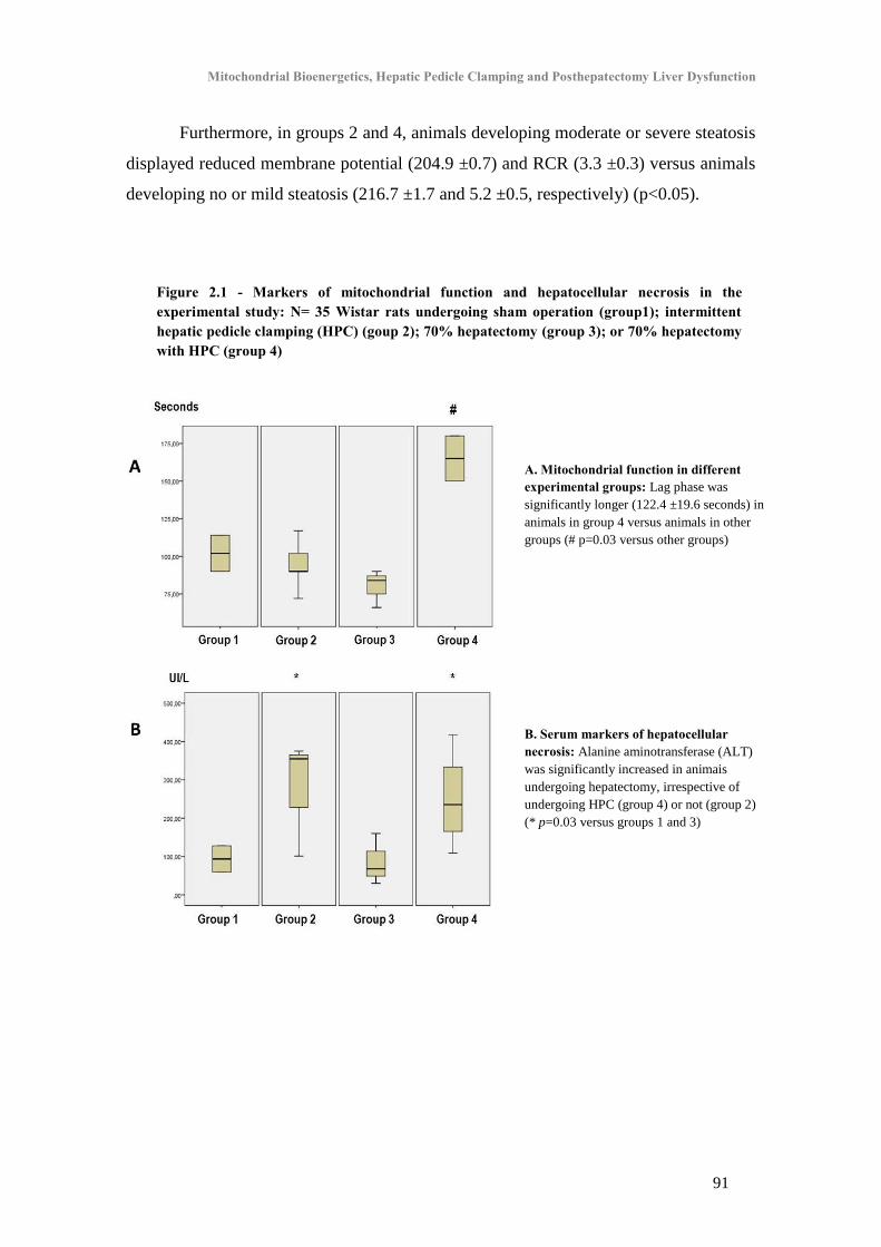
Mitochondrial Bioenergetics, Hepatic Pedicle Clamping and Posthepatectomy Liver Dysfunction
91
Furthermore, in groups 2 and 4, animals developing moderate or severe steatosis
displayed reduced membrane potential (204.9 ±0.7) and RCR (3.3 ±0.3) versus animals
developing no or mild steatosis (216.7 ±1.7 and 5.2 ±0.5, respectively) (p<0.05).
A. Mitochondrial function in different experimental groups: Lag phase was
significantly longer (122.4 ±19.6 seconds) in
animals in group 4 versus animals in other
groups (# p=0.03 versus other groups)
B. Serum markers of hepatocellular necrosis: Alanine aminotransferase (ALT)
was significantly increased in animais
undergoing hepatectomy, irrespective of
undergoing HPC (group 4) or not (group 2)
(* p=0.03 versus groups 1 and 3)
Figure 2.1 - Markers of mitochondrial function and hepatocellular necrosis in the experimental study: N= 35 Wistar rats undergoing sham operation (group1); intermittent hepatic pedicle clamping (HPC) (goup 2); 70% hepatectomy (group 3); or 70% hepatectomy with HPC (group 4)
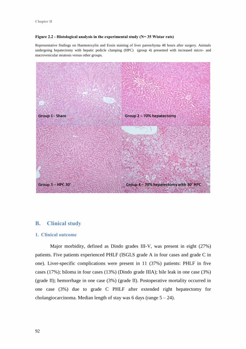
Chapter II
92
Figure 2.2 - Histological analysis in the experimental study (N= 35 Wistar rats)
Representative findings on Haemotoxylin and Eosin staining of liver parenchyma 48 hours after surgery. Animals
undergoing hepatectomy with hepatic pedicle clamping (HPC) (group 4) presented with increased micro- and
macrovesicular steatosis versus other groups.
B. Clinical study
1. Clinical outcome
Major morbidity, defined as Dindo grades III-V, was present in eight (27%)
patients. Five patients experienced PHLF (ISGLS grade A in four cases and grade C in
one). Liver-specific complications were present in 11 (37%) patients: PHLF in five
cases (17%); biloma in four cases (13%) (Dindo grade IIIA); bile leak in one case (3%)
(grade II); hemorrhage in one case (3%) (grade II). Postoperative mortality occurred in
one case (3%) due to grade C PHLF after extended right hepatectomy for
cholangiocarcinoma. Median length of stay was 6 days (range 5 – 24).
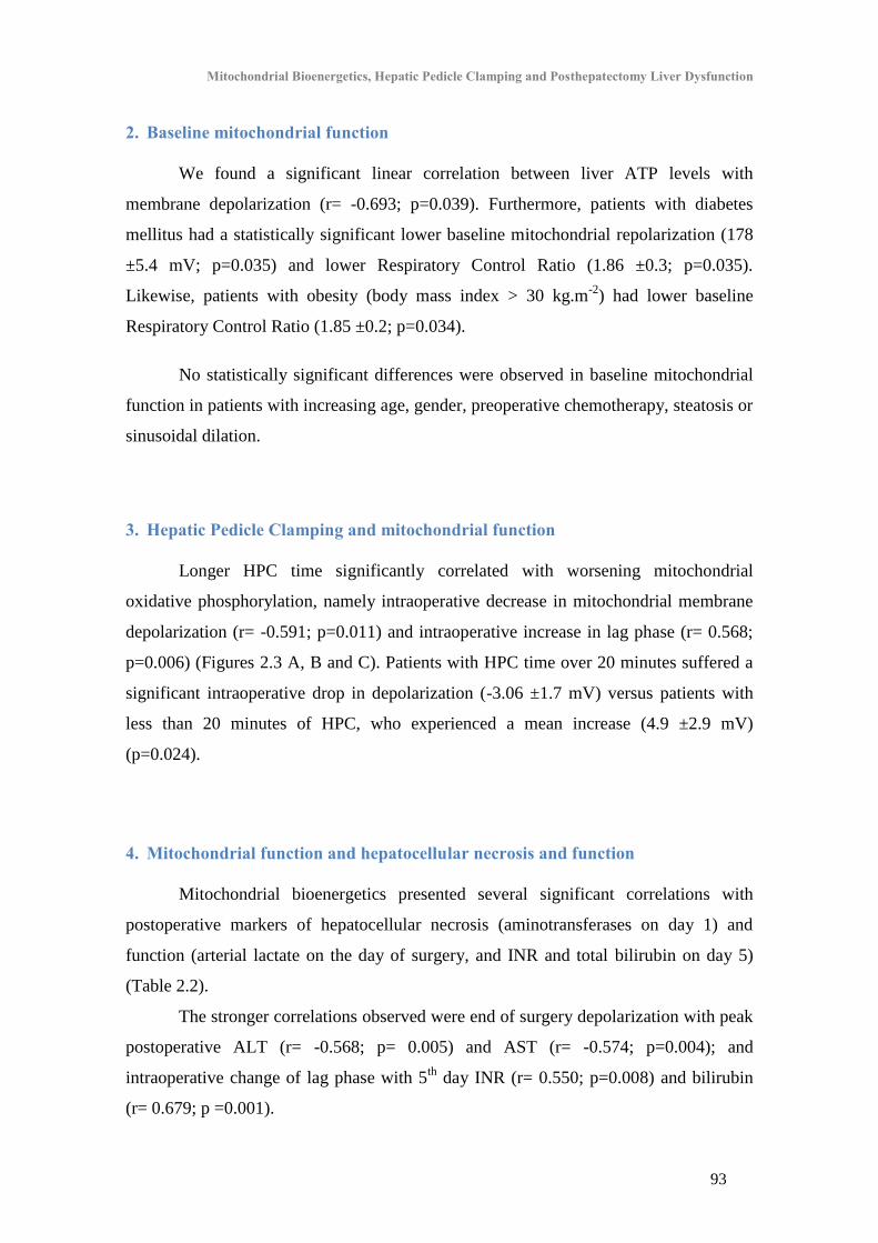
Mitochondrial Bioenergetics, Hepatic Pedicle Clamping and Posthepatectomy Liver Dysfunction
93
2. Baseline mitochondrial function
We found a significant linear correlation between liver ATP levels with
membrane depolarization (r= -0.693; p=0.039). Furthermore, patients with diabetes
mellitus had a statistically significant lower baseline mitochondrial repolarization (178
±5.4 mV; p=0.035) and lower Respiratory Control Ratio (1.86 ±0.3; p=0.035).
Likewise, patients with obesity (body mass index > 30 kg.m-2
) had lower baseline
Respiratory Control Ratio (1.85 ±0.2; p=0.034).
No statistically significant differences were observed in baseline mitochondrial
function in patients with increasing age, gender, preoperative chemotherapy, steatosis or
sinusoidal dilation.
3. Hepatic Pedicle Clamping and mitochondrial function
Longer HPC time significantly correlated with worsening mitochondrial
oxidative phosphorylation, namely intraoperative decrease in mitochondrial membrane
depolarization (r= -0.591; p=0.011) and intraoperative increase in lag phase (r= 0.568;
p=0.006) (Figures 2.3 A, B and C). Patients with HPC time over 20 minutes suffered a
significant intraoperative drop in depolarization (-3.06 ±1.7 mV) versus patients with
less than 20 minutes of HPC, who experienced a mean increase (4.9 ±2.9 mV)
(p=0.024).
4. Mitochondrial function and hepatocellular necrosis and function
Mitochondrial bioenergetics presented several significant correlations with
postoperative markers of hepatocellular necrosis (aminotransferases on day 1) and
function (arterial lactate on the day of surgery, and INR and total bilirubin on day 5)
(Table 2.2).
The stronger correlations observed were end of surgery depolarization with peak
postoperative ALT (r= -0.568; p= 0.005) and AST (r= -0.574; p=0.004); and
intraoperative change of lag phase with 5th
day INR (r= 0.550; p=0.008) and bilirubin
(r= 0.679; p =0.001).
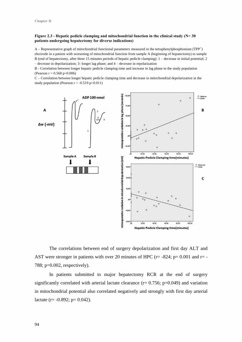
Chapter II
94
Figure 2.3 - Hepatic pedicle clamping and mitochondrial function in the clinical study (N= 30 patients undergoing hepatectomy for diverse indications)
A – Representative graph of mitochondrial functional parameters measured in the tetraphenylphosphonium (TPP+)
electrode in a patient with worsening of mitochondrial function from sample A (beginning of hepatectomy) to sample
B (end of hepatectomy, after three 15 minutes periods of hepatic pedicle clamping): 1 – decrease in initial potential; 2
– decrease in depolarization; 3- longer lag phase; and 4 – decrease in repolarization
B - Correlation between longer hepatic pedicle clamping time and increase in lag phase in the study population
(Pearson r = 0.568 p=0.006) C – Correlation between longer hepatic pedicle clamping time and decrease in mitochondrial depolarization in the
study population (Pearson r = -0.519 p=0.011)
The correlations between end of surgery depolarization and first day ALT and
AST were stronger in patients with over 20 minutes of HPC (r= -824; p= 0.001 and r= -
788; p=0.002, respectively).
In patients submitted to major hepatectomy RCR at the end of surgery
significantly correlated with arterial lactate clearance (r= 0.756; p=0.049) and variation
in mitochondrial potential also correlated negatively and strongly with first day arterial
lactate (r= -0.892; p= 0.042).

Mitochondrial Bioenergetics, Hepatic Pedicle Clamping and Posthepatectomy Liver Dysfunction
95
Table 2.2 - Mitochondrial function and postoperative liver function in the clinical study (N= 30 patients undergoing hepatectomy for diverse indications)
Correlations between mitochondrial bioenergetics parameters and markers of hepatocellular function and necrosis
(arterial lactate 6 hours after end of surgery, peak postoperative aminotransferases on postoperative day 1,
International Normalized Ratio on postoperative day 5 and total bilirubin on day 5) in the study population (* p <
0.05; ** p < 0.005)
Arterial lactate 6
h
Day 1
ALT
Day 1
AST
Day 5
Bilirubin
Day 5
INR
End of surgery
membrane potential
r = 0.068 r = - 0.326 r = - 0.225 r = - 0.015 r = - 0.219
End of surgery lag
phase
r = 0.452* r = 0.452* r = 0.415* r = 0.444* r = 0.627**
End of surgery
depolarization
r = - 0.282 r = - 0.568* r = - 0.574** r = - 0.179 r = - 0.224
End of surgery
repolarization
r = - 0.095 r = - 0.096 r = 0.003 r = 0.055 r = - 0.368
Change in
membrane potential
r = - 0.357 r = - 0.453* r = - 0.445* r = - 0.416 r = - 0.343
Change in lag phase
r = 0.508* r = 0.484* r = 0.460* r = 0.550* r = 0.679**
Change in
depolarization
r = - 0.426* r = - 0.571** r = - 0.553* r = - 0.279 r = - 0.257
Change in
repolarization
r = - 0.287 r = - 0.112 r = - 0.096 r = - 0.332 r = - 0.494*
5. Mitochondrial function and postoperative morbidity
Patients developing liver-specific morbidity and PHLF presented with worse end
of surgery mitochondrial bioenergetics (Table 2.3).
Multivariate analysis demonstrated that intraoperative deterioration in
mitochondrial membrane potential (HR=13.7; p=0.043) and preoperative chemotherapy
(HR=13.7; p=0.043) were independent risk factors for liver-specific morbidity (Table
2.4).

Chapter II
96
Table 2.3 - M
itochondrial bioenergetics and postoperative morbidity in the clinical study (N
= 30 patients undergoing hepatectomy for diverse indications)
Intrao
perativ
e measu
remen
ts of m
itoch
on
drial b
ioen
ergetics (sam
ple A
, samp
le B an
d in
traop
erative ch
ange) in
the stu
dy p
op
ulatio
n an
d in
patien
ts dev
elop
ing p
osto
perativ
e com
plicatio
ns
(liver-sp
ecific mo
rbid
ity an
d p
osth
epatecto
my liv
er failure). M
ann
-Wh
itney
two
-tailed: a) p
= 0
.03
7; b) p
= 0
.022; c) p
= 0
.03
2; d) p
= 0
.00
4; e) p =
0.0
18; f) p
= 0
.01
1; g
) p =
0.0
37
Study population (N
= 30) L
iver-specific morbidity (n = 11)
Posthepatectomy L
iver Failure (n = 5)
Parameter
Biopsy A
B
iopsy B
Variation
Biopsy A
B
iopsy B
Variation
Biopsy A
B
iopsy B
Variation
Mem
bran
e po
tential (m
V)
19
3.5
±2
.5
191.9 ±2.7 a) - 0.9 ±3.1
b) 1
96.4
±3
.2
18
5.1
±4
.7
- 7.7
±5
.5
20
0.1
±3
.7
178.8 ±6.9 a) - 17.1 ±7.8 b)
Lag p
hase (sec)
41
.9 ±
3.1
54.7 ±5.9 c), d)
+ 13.0 ±6.8 e), f) 3
9.3
±3.1
73.3 ±12.4 c)
+ 33.9 ±13.6 e) 4
0.5
±4.8
95.3 ±15.1 d)
+54.8 ±19.4 f)
Dep
olarizatio
n (m
V)
21
.6 ±
1.1
2
2.3
±1.0
+ 0.7 ±1.8 g)
24
.3 ±
1.9
2
0.4
±1.1
- 3.9 ±2.3 g)
24
.1 ±
2.4
1
9.2
±1.6
- 4
.9 ±
3.4
Rep
olarizatio
n (m
V)
18
7.1
±2
.6
18
5 ±
2.5
- 1
.3 ±
3.2
1
91.7
±1
.9
18
2.4
±3
.8
- 9.2
±4
.9
19
2.6
±3
.1
17
7.8
±3
.8
- 15.3
±5
.4
Resp
iratory
Co
ntro
l Ratio
2
.4 ±
0.2
2
.7 ±
0.2
+
0.3
±0
.2
2.7
±0
.2
2.7
±0
.4
+ 0
.2 ±
0.3
2
.8 ±
0.4
2
.8 ±
0.7
+
0.1
±0
.6
Table 2.4 - U
nivariate and multivariate analysis of factors associated w
ith liver-specific morbidity in the clinical study (N
= 30 patients undergoing hepatectomy
for diverse indications)
U
nivariate analysis M
ultivariate analysis
O
R
CI 95%
p
OR
C
I 95%
p
Preoperative chemotherapy
3.6
7
0.7
– 1
8.3
0
.10
7
13.7 1.1 – 173.7
0.043
Major hepatectom
y 7.1
1.1 – 46.7 0.043
_
_
n.s.
HPC
> 20 minutes
7.7 1.28 – 46.4
0.021 _
_
n
.s.
Decrease in m
itochondrial mem
brane potential 6
.3
0.9
– 4
2.7
0
.06
13.7 1.1 – 173.7
0.043

Mitochondrial Bioenergetics, Hepatic Pedicle Clamping and Posthepatectomy Liver Dysfunction
97
Elevated lag phase at the end of surgery, above 53.9 seconds, was found to be
highly predictive of PHLF (area under the curve: 0.933; p=0.008; sensitivity 100%;
specificity 83.3%). However, there was no statistically significant association of
mitochondrial dysfunction with major morbidity or mortality.
IV. Discussion
Posthepatectomy liver failure is a severe complication of liver surgery,
associated with increased mortality, risk of sepsis and decreased long term survival
[13,14,282]. Liver regeneration is a highly energy dependent process and bioenergetic
status of the main parenchymal cell of the liver, the hepatocyte, is of paramount
importance in this process. However, the exact role of bioenergetics in clinical
posthepatectomy liver dysfunction is largely unknown.
Experimental evidence confirms the key role of mitochondrial homeostasis in
liver regeneration. Improved energy status and decreased mitochondrial permeability
transition can improve liver regeneration, maintain hepatocellular function and decrease
mortality in animal models of major hepatectomy [20,52]. However, clinical evidence
for bioenergetics dysfunction in liver surgery is scarce. One study failed to show
significant correlations of mitochondrial respiration with clinical parameters, possibly
because only one intraoperative biopsy was made [65]. Furthermore, the effect of
hepatic pedicle clamping on mitochondrial function in the setting of liver resection has
not been fully clarified, both clinically and experimentally. This led to the design of two
distinct and original studies, one experimental and one clinical, to elucidate these issues.
In the clinical study we sought to investigate the change in liver mitochondrial
function by performing two intraoperative biopsies in the non-resected parenchyma, one
at the beginning of the procedure and one at the end. Rather than only measuring the
absolute value, we also measured the change in mitochondrial membrane potential,
oxygen consumption and ATP content between the two samples, thus reflecting the
intraoperative change in the liver energy state. We then sought to correlate these data
with preoperative liver function, HPC time, markers of hepatocellular necrosis and
function, and postoperative morbidity.
Chapter II
98
Unsurprisingly, there was a significant correlation between mitochondrial
membrane depolarization and ATP levels. There is a direct and biological rationale for
this finding, since ATP synthesis depends on proper function of the electron transport
chain, as well as adequate adenosine diphosphate transport into the mitochondrial
matrix and an efficient ATP-synthase.
We were not able to demonstrate any correlation of mitochondrial metabolism
with preoperative liver function. This is easily explained, not only because cirrhotic
patients were excluded from the study, but also because liver parenchyma was normal
all but in seven patients, who presented mild histologic changes; this was confirmed by
the low median MELD score of the study population. Regarding the effect of other
clinical variables on mitochondrial function, we also did not confirm any change with
advanced age or preoperative chemotherapy. Nonetheless, our results confirmed that
patients with diabetes and obesity had worse mitochondrial function and this finding is
in keeping with other studies [56,272] (and reviewed in Chapter I - “Bioenergetics and
Liver Regeneration: Review of the Role of Energy Status in the Pathophysiology of
Posthepatectomy Liver Failure”).
Regarding the association of HPC with mitochondrial dysfunction, answers were
sought in both the clinical and the experimental study.
The experimental study was designed to explore the effect of a well-tolerated
HPC strategy, namely 30 minutes intermittent clamping strategy. Although this duration
of HPC, if used continuously, is associated with increased hepatocellular injury in
animal models of 70% hepatectomy [220,283], it is well tolerated when used
intermittently [284]. Furthermore, intermittent clamping is the most widely used
strategy in the clinical setting [68,274,285]. We confirmed little evidence for increase in
hepatocyte death, as was demonstrated by similar ALT levels in both hepatectomy
groups, irrespective of HPC. Group 4 also did not present an enhanced necrosis score on
histology when compared with animals undergoing hepatectomy without HPC.
Nonetheless, animals undergoing hepatectomy with HPC presented longer lag phase at
12 hours posthepatectomy, reflecting an impaired phosphorylation of ADP. Group 4
also demonstrated an increased steatosis score at 48 hours, versus the other
experimental groups. Another interesting finding was that both groups of animals

Mitochondrial Bioenergetics, Hepatic Pedicle Clamping and Posthepatectomy Liver Dysfunction
99
undergoing hepatectomy presented an association of intracellular accumulation of lipids
with decreased mitochondrial respiration and membrane potential. In fact, steatosis is
known to occur during early liver regeneration and improvement in energy status can
potentially decrease intra-hepatocyte lipid accumulation [22,286]. These findings could
be easily explained, as hepatocytes become preferential users of free fatty acids released
from adipose tissue after surgical aggression. So, a disturbance in the mitochondrial
electron transport chain and subsequent decreased oxygen consumption would lead to
an increase in the reduced form of nicotinamide adenine dinucleotide (NADH) and
increased NADH/NAD+ ratio, thus halting the β-oxidation of fatty acids, causing their
accumulation in the hepatocytes’ cytoplasm.
As such, we demonstrated that mitochondrial dysfunction, in particular longer
lag phase, is a subtle yet measurable biologic change of the liver parenchyma after
hepatectomy with inflow occlusion in the experimental model. We postulate that longer
HPC time is associated with increased mitochondrial dysfunction, and this was
confirmed in the clinical study. In the cohort of patients we observed a moderately
strong and statistically significant correlation of duration of HPC with two relevant
bioenergetics variables. Furthermore, mitochondrial membrane depolarization was
significantly worsened in patients with over 20 minutes of HPC, whereas it was
improved in patients with under 20 minutes, possibly reflecting a mechanism of
ischemic preconditioning [108]. This confirms the previous notion that a strategy of
intermittent HPC in an on-need basis is usually considered safest [273,274].
Nevertheless, HPC time over 20 minutes has previously been associated with an
increased risk of liver failure and mortality [287].
In keeping with the second interrogation of this study, our clinical results clearly
demonstrate a significant relationship between mitochondrial bioenergetics with
postoperative markers of hepatocellular necrosis. There was a moderate correlation
between deterioration of mitochondrial depolarization and peak postoperative
aminotransferases. This correlation was even stronger in patients undergoing HPC over
20 minutes, possibly reflecting increased hepatocyte necrosis caused by bioenergetic
failure. Although peak postoperative aminotransferases do not necessarily reflect
outcome [288], they are a useful surrogate of remnant liver ischemia-reperfusion injury.
Chapter II
100
An additional finding was the important correlation of mitochondrial
dysfunction with two key postoperative markers of hepatocellular function, namely fifth
day INR and bilirubin [165,289]. Furthermore, patients undergoing major hepatectomy
also experienced a very strong correlation between mitochondrial dysfunction and
arterial lactate on postoperative day 1, a sensitive marker of liver function [171,279];
and between decreased oxygen consumption and depressed lactate clearance, possibly
reflecting lower hepatic lactate metabolism rather than increased peripheral production
[172].
One of the main results of our study was a strong association of mitochondrial
dysfunction with PHLF. Patients with PHLF presented lower end of surgery membrane
potential and increased lag phase, likely reflecting the development of bioenergetics
failure. In fact, end of surgery lag phase was a strong predictor of PHLF, with high
specificity and sensitivity. And finally, intraoperative decrease in mitochondrial
membrane potential was an independent risk factor for liver-specific morbidity.
This clinical study is, to the best of our knowledge, the first to demonstrate a
clear relationship between mitochondrial function and clinical outcome after
hepatectomy. The importance of these findings is twofold. First, markers of
mitochondrial dysfunction can be used for earlier diagnosis of PHLF. And finally,
research into the mechanisms of bioenergetics dysfunction in liver surgery could open
the way to novel pharmacological therapies. If translated into clinical practice,
improvement in hepatocyte bioenergetics could decrease susceptibility to ischemia-
reperfusion injury, accelerate liver regeneration and ultimately decrease morbidity and
mortality of liver resection. These concepts were explored in detail in Chapter I -
“Bioenergetics and Liver Regeneration: Review of the Role of Energy Status in the
Pathophysiology of Posthepatectomy Liver Failure”.

Colour Plates

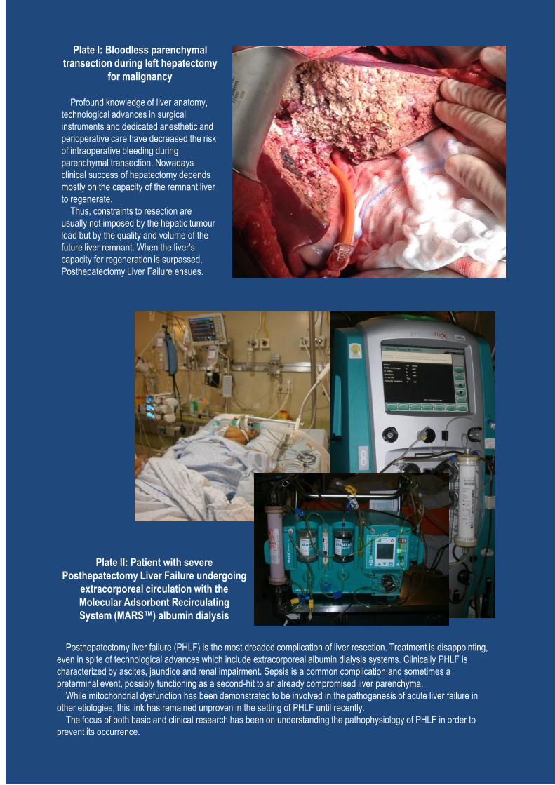
Plate II: Patient with severe
Posthepatectomy Liver Failure undergoing
extracorporeal circulation with the
Molecular Adsorbent Recirculating
System (MARS™) albumin dialysis
Posthepatectomy liver failure (PHLF) is the most dreaded complication of liver resection. Treatment is disappointing,
even in spite of technological advances which include extracorporeal albumin dialysis systems. Clinically PHLF is
characterized by ascites, jaundice and renal impairment. Sepsis is a common complication and sometimes a
preterminal event, possibly functioning as a second-hit to an already compromised liver parenchyma.
While mitochondrial dysfunction has been demonstrated to be involved in the pathogenesis of acute liver failure in
other etiologies, this link has remained unproven in the setting of PHLF until recently.
The focus of both basic and clinical research has been on understanding the pathophysiology of PHLF in order to
prevent its occurrence.
Plate I: Bloodless parenchymal
transection during left hepatectomy
for malignancy
Profound knowledge of liver anatomy,
technological advances in surgical
instruments and dedicated anesthetic and
perioperative care have decreased the risk
of intraoperative bleeding during
parenchymal transection. Nowadays
clinical success of hepatectomy depends
mostly on the capacity of the remnant liver
to regenerate.
Thus, constraints to resection are
usually not imposed by the hepatic tumour
load but by the quality and volume of the
future liver remnant. When the liver’s
capacity for regeneration is surpassed,
Posthepatectomy Liver Failure ensues.


Plate III: Normal liver anatomy of the
Wistar rat
The lobular structure and remarkable
regenerative capacity of the rat liver provide an
excellent model for the study of liver
regeneration. The most used experimental
procedure is the 70% hepatectomy model,
which involves resection of the left lateral and
median lobes. Liver regeneration starts almost
immediately after resection and is usually
complete by the seventh postoperative day.
Previous experimental works prove that liver
energy status is a relevant factor in this
biological process.
Left Lateral Lobe
MedianLobe
Right Superior Lobe
Right Inferior Lobe
Omental Lobe
Bile duct
Portal vein
Hepatic artery
Plate IV: Anatomy of the liver hilum of the Wistar rat and the microscopic appearance of the portal
triad – The vasculo-biliary tree, from the hilum to the acinus
The liver parenchyma is supported by portal and arterial inflow, biliary drainage and venous outflow. From the
liver pedicle (left) to the most fundamental functional unit – the acinus – portal, arterial and biliary branches
sequentially bifurcate in sectorial, segmental and subsegmental branches, supplying the entire liver mass. At the
microscopic level this is exemplified by the portal triad (right, Hematoxylin-Eosin 100x), the lowest division of the
elements of the hepatic pedicle, where the vasculo-biliary structures are enveloped in a connective tissue sheath
that is contiguous with the Glisson’s capsule.
The portal vein supplies the liver sinusoids with blood from the splanchnic organs, ensuring first contact of the
liver with absorbed nutrients and xenobiotics, high concentration of entero-hormones and recirculating bile salts. The
hepatic artery, on the other hand is responsible for the continuous delivery of oxygen, indispensable for the
extremely high metabolic demands of the liver in its tireless regulation of glucidic, proteic and lipidic metabolism.


Intraoperative appearance of Sinusoidal Obstruction Syndrome (SOS), or “Blue Liver”, in a patient undergoing hepatectomy
for colorectal cancer liver metastases after oxaliplatin-based chemotherapy. SOS is associated with increased risk of
intraoperative bleeding and complications after hepatectomy.
In order to reduce bleeding in clinical liver resection or liver trauma, the hepatic pedicle can be temporarily clamped, a
manoeuvre first described by Dr. John Hogarth Pringle, of Glasgow, in 1908. Hepatic Pedicle Clamping is an invaluable adjunct
as it reduces intraoperative bleeding but it should be used judiciously in patients with chronic liver disease, including SOS, due
to an increased susceptibility to ischemia-reperfusion injury and mitochondrial dysfunction.
Plate V: Hepatic Pedicle
Clamping in a patient
with “Blue Liver”
Plate VI: Histologic patters of Chemotherapy-
associated liver injury (CALI)
Preoperative chemotherapy is commonly used in the
management of patients with colorectal cancer liver
metastases, in order to downsize lesions and improve
survival. However, hepatotoxicity is a concern and
parenchymal injury can compromise liver regeneration
and increase morbidity and mortality of liver resection.
Three distinct patterns are recognized on liver
histology: simple steatosis, steatohepatitis and
sinusoidal obstruction syndrome. Disturbed cellular
bioenergetics could prove to be a contributing factor in
the pathophysiology of these conditions.
Steatosis(Hematoxylin-Eosin 100x)
Chemotherapy-associated Steatohepatitis(Hematoxylin-Eosin 200x)
Sinusoidal Obstruction Syndrome(Masson trichrome 40x)


Plate VII: Percutaneous Right Portal Vein
Embolization before Right Hepatectomy
To decrease the risk of Posthepatectomy Liver
Failure after major or extended liver resection,
Portal Vein Embolization (PVE) is occasionally
required. This technique was first described in 1986
and is usually performed under fluoroscopic
guidance, through direct liver puncture.
By shunting portal blood flow to the non-tumor
bearing parenchyma, PVE causes atrophy and
apoptosis in the embolized hemiliver; and cell
proliferation and hypertrophy in the non-embolized
future liver remnant. After some weeks major
hepatectomy can be performed with decreased
morbidity.
Experimental evidence suggests that important
adaptations in mitochondrial function take place in
the non-embolized parenchyma.
Liver remnant consisting of segments 2 and 3 after right extended hepatectomy with bile duct
resection for Bismuth type IV hilar cholangiocarcinoma
Extended resections, i.e. resections of five or more Couinaud segments, are one of the most risky procedures in
modern Hepatobiliary Surgery, on occasion resulting in Posthepatectomy Liver Failure.
Hilar cholangiocarcinoma is a particularly challenging condition, because patients usually present extremely
jaundiced and biliary obstruction can severely hamper the liver remnant’s capacity for regeneration. Both experimental
and clinical evidence suggest that liver mitochondrial function is deteriorated in chronic biliary obstruction and can
significantly recover after biliary drainage.
While meticulous surgical technique is mandatory, preoperative optimization of the future liver remnant is also a
critical factor in the success of liver resection. This patient underwent the following consecutive preoperative
percutaneous interventions: external biliary drainage; right and segment 4 portal vein embolization; and middle hepatic
vein occlusion. With these procedures the future liver remnant volume increased from 222 cm3 to 360 cm3 making
radical resection possible. She was discharged home on postoperative day 9 without any signs of liver dysfunction.
Plate VIII:


Plate X: Mitochondria – the engine behind the liver’s
amazing regenerative capacity
The liver’s extremely high metabolic demands can only be
maintained through the unremitting work of mitochondria. Most
liver functions are endergonic, or energy-dependent, requiring
substantial amounts of adenosine triphosphate, supplied
predominantly by mitochondrial oxidative phosphorylation under
aerobic conditions.
After hepatectomy the remaining hepatocytes must replace
lost liver mass while still maintaining liver function. This can
only happen with an increased energy supply by mitochondria.
Potentially, liver functional reserve before hepatectomy could
be quantified through the measurement of metabolic capacity,
instead of using single-function tests, like indocyanine green
retention rate or hepatobiliary scintigraphy.
Furthermore, mitochondrial-based therapies could plausibly
increase the liver’s regenerative capacity and decrease the
incidence of Posthepatectomy Liver Failure, surpassing the
current limits of liver resection and providing hope for cure to
patients nowadays deemed inoperable.
Plate IX: Volumetric analysis of the future
liver remnant with the Osirix™ software
before and after stage 1 of the ALPSS
procedure
Volumetric analysis by cross sectional imaging
is the gold standard for the correct assessment of
the response to Portal Vein Embolization or two-
stage procedures, like the Associating Liver
Partition and Portal Ligation for Staged
Hepatectomy (ALPPS).
Recent clinical evidence has shown that rather
than the absolute increase in volume, the main
factor in predicting regenerative capacity might
be the kinetic pattern of liver growth.
In the case illustrated the future liver remnant
sustained a volume increase of 43% in less than
two weeks and the patient underwent a non-
eventful recovery after the second stage of
ALPPS.
Being a highly-energy dependent chain of
events, liver regeneration is influenced by
mitochondrial status. In fact, sufficient evidence
exists for a definite link of mitochondrial oxidative
phosphorylation in the enhanced inter-stages
liver volume growth in ALPPS.

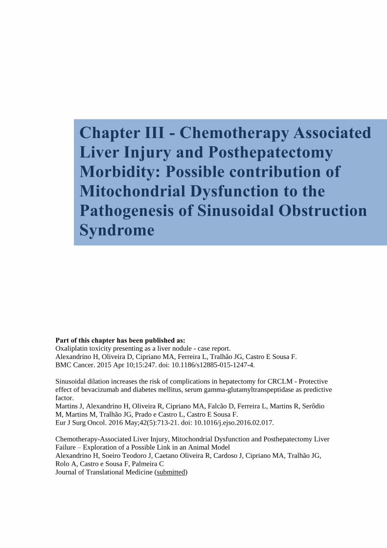
Part of this chapter has been published as: Oxaliplatin toxicity presenting as a liver nodule - case report.
Alexandrino H, Oliveira D, Cipriano MA, Ferreira L, Tralhão JG, Castro E Sousa F.
BMC Cancer. 2015 Apr 10;15:247. doi: 10.1186/s12885-015-1247-4.
Sinusoidal dilation increases the risk of complications in hepatectomy for CRCLM - Protective
effect of bevacizumab and diabetes mellitus, serum gamma-glutamyltranspeptidase as predictive
factor.
Martins J, Alexandrino H, Oliveira R, Cipriano MA, Falcão D, Ferreira L, Martins R, Serôdio
M, Martins M, Tralhão JG, Prado e Castro L, Castro E Sousa F.
Eur J Surg Oncol. 2016 May;42(5):713-21. doi: 10.1016/j.ejso.2016.02.017.
Chemotherapy-Associated Liver Injury, Mitochondrial Dysfunction and Posthepatectomy Liver
Failure – Exploration of a Possible Link in an Animal Model
Alexandrino H, Soeiro Teodoro J, Caetano Oliveira R, Cardoso J, Cipriano MA, Tralhão JG,
Rolo A, Castro e Sousa F, Palmeira C
Journal of Translational Medicine (submitted)
Chapter III - Chemotherapy Associated Liver Injury and Posthepatectomy Morbidity: Possible contribution of Mitochondrial Dysfunction to the Pathogenesis of Sinusoidal Obstruction Syndrome

Chapter III Chemotherapy-Associated Liver Injury and Posthepatectomy Morbidity: Possible contribution of Mitochondrial Dysfunction to the Pathogenesis of Sinusoidal Obstruction Syndrome
115
I. Introduction
Yearly, over 447.000 new cases of colorectal cancer are diagnosed in Europe
and around 50% of them will develop liver metastases [290]. Liver resection is
potentially curative in patients with colorectal cancer liver metastases (CRLM)
[90,291], but up to 85% of patients are considered unresectable due to tumor load [2]. In
these cases hepatectomy can be performed after aggressive chemotherapy in a
conversion strategy, downsizing the lesions and allowing for safe resection [2,90].
Moreover, neoadjuvant chemotherapy (NCT) in upfront operable patients can improve
overall and disease-free survival after hepatectomy [130,292]. Modern NCT consists of
5-fluorouracil (5-FU) in association with irinotecan (FOLFIRI) or oxaliplatin
(FOLFOX). Molecular-targeted agents, such as cetuximab and bevacizumab, have
improved response rates and resectability [293].
However, NCT is known to cause chemotherapy-associated liver injury (CALI)
and may increase morbidity and mortality after liver resection [116,133,294]. Irinotecan
has been reported to cause steatohepatitis (SH) [116], while oxaliplatin-based
chemotherapy has been associated with sinusoidal obstruction syndrome
[136,138,295,296]. Sinusoidal obstruction syndrome (SOS) consists of severe
sinusoidal dilation and congestion, sinusoidal endothelial cell necrosis and sloughing,
erythrocyte extravasation in the sinusoids, peliosis and decreased sinusoidal flow
[136,137]. SOS is associated with depressed preoperative liver function, increased
intraoperative bleeding, decreased tolerance to hepatic pedicle clamping (HPC), worse
posthepatectomy liver function and lower regenerative capacity after two-stage
hepatectomy or portal vein embolization [138–143,297].
Focal peliosis in SOS can also cause another unexpected side effect, which is the
development of liver nodules on cross-sectional imaging that can be mistaken for liver
metastases. We have previously reported the case of a 59 year old male patient with
T3N1bM0 sigmoid colon adenocarcinoma [298]. The patient underwent sigmoidectomy
and adjuvant chemotherapy with capecitabine and oxaliplatin. One year after surgery, a
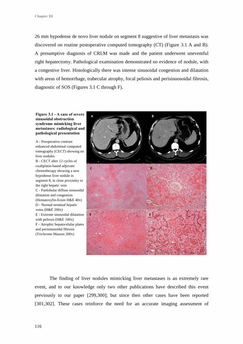
Chapter III
116
26 mm hypodense de novo liver nodule on segment 8 suggestive of liver metastasis was
discovered on routine postoperative computed tomography (CT) (Figure 3.1 A and B).
A presumptive diagnosis of CRLM was made and the patient underwent uneventful
right hepatectomy. Pathological examination demonstrated no evidence of nodule, with
a congestive liver. Histologically there was intense sinusoidal congestion and dilatation
with areas of hemorrhage, trabecular atrophy, focal peliosis and perisinuosoidal fibrosis,
diagnostic of SOS (Figures 3.1 C through F).
The finding of liver nodules mimicking liver metastases is an extremely rare
event, and to our knowledge only two other publications have described this event
previously to our paper [299,300]; but since then other cases have been reported
[301,302]. These cases reinforce the need for an accurate imaging assessment of
Figure 3.1 - A case of severe sinusoidal obstruction syndrome mimicking liver metastases: radiological and pathological presentation
A - Preoperative contrast
enhanced abdominal computed
tomography (CECT) showing no
liver nodules
B - CECT after 12 cycles of
oxaliplatin-based adjuvant
chemotherapy showing a new
hypodense liver nodule in
segment 8, in close proximity to
the right hepatic vein
C - Panlobular diffuse sinusoidal
dilatation and congestion
(Hematoxylin-Eosin H&E 40x)
D - Normal terminal hepatic
veins (H&E 200x)
E - Extreme sinusoidal dilatation
with peliosis (H&E 100x)
F - Atrophic hepatocelular plates
and perisinusoidal fibrosis
(Trichrome Masson 200x)
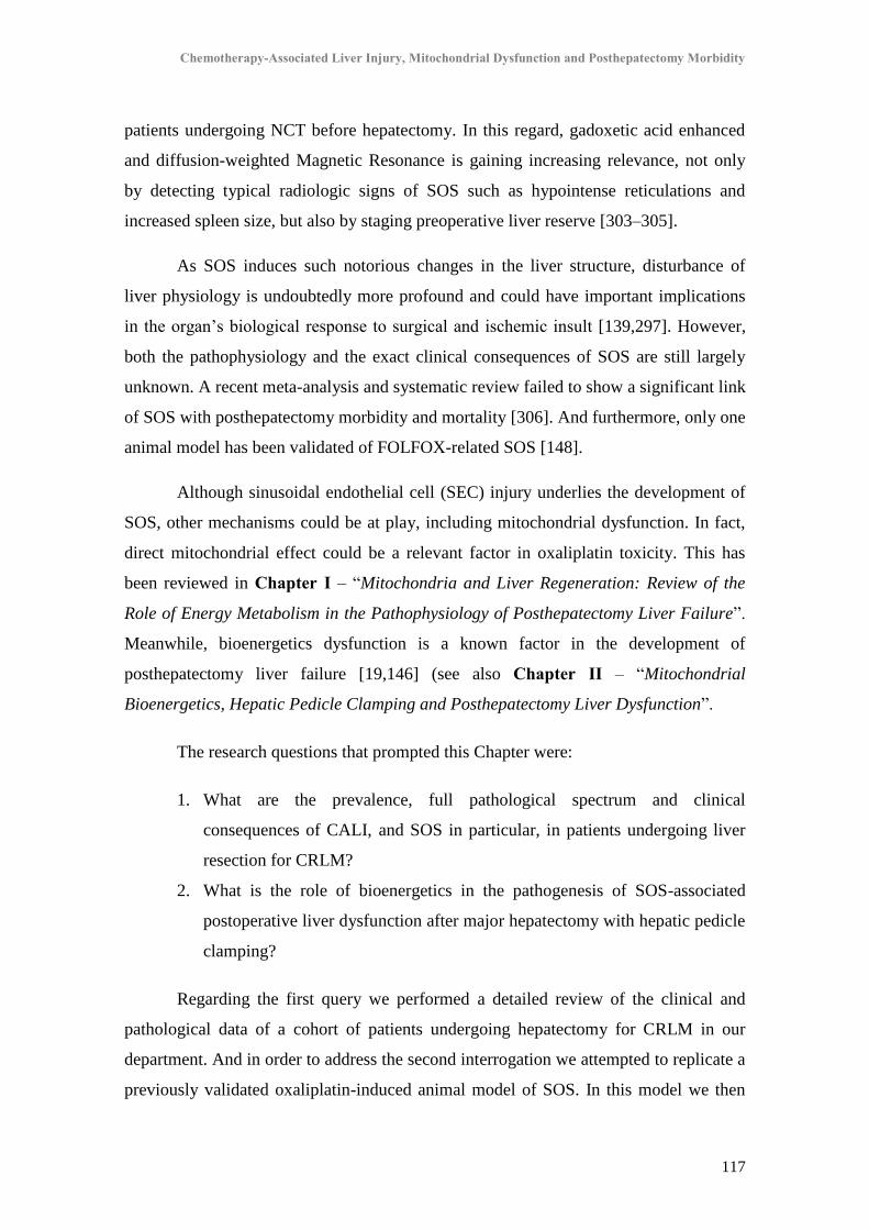
Chemotherapy-Associated Liver Injury, Mitochondrial Dysfunction and Posthepatectomy Morbidity
117
patients undergoing NCT before hepatectomy. In this regard, gadoxetic acid enhanced
and diffusion-weighted Magnetic Resonance is gaining increasing relevance, not only
by detecting typical radiologic signs of SOS such as hypointense reticulations and
increased spleen size, but also by staging preoperative liver reserve [303–305].
As SOS induces such notorious changes in the liver structure, disturbance of
liver physiology is undoubtedly more profound and could have important implications
in the organ’s biological response to surgical and ischemic insult [139,297]. However,
both the pathophysiology and the exact clinical consequences of SOS are still largely
unknown. A recent meta-analysis and systematic review failed to show a significant link
of SOS with posthepatectomy morbidity and mortality [306]. And furthermore, only one
animal model has been validated of FOLFOX-related SOS [148].
Although sinusoidal endothelial cell (SEC) injury underlies the development of
SOS, other mechanisms could be at play, including mitochondrial dysfunction. In fact,
direct mitochondrial effect could be a relevant factor in oxaliplatin toxicity. This has
been reviewed in Chapter I – “Mitochondria and Liver Regeneration: Review of the
Role of Energy Metabolism in the Pathophysiology of Posthepatectomy Liver Failure”.
Meanwhile, bioenergetics dysfunction is a known factor in the development of
posthepatectomy liver failure [19,146] (see also Chapter II – “Mitochondrial
Bioenergetics, Hepatic Pedicle Clamping and Posthepatectomy Liver Dysfunction”.
The research questions that prompted this Chapter were:
1. What are the prevalence, full pathological spectrum and clinical
consequences of CALI, and SOS in particular, in patients undergoing liver
resection for CRLM?
2. What is the role of bioenergetics in the pathogenesis of SOS-associated
postoperative liver dysfunction after major hepatectomy with hepatic pedicle
clamping?
Regarding the first query we performed a detailed review of the clinical and
pathological data of a cohort of patients undergoing hepatectomy for CRLM in our
department. And in order to address the second interrogation we attempted to replicate a
previously validated oxaliplatin-induced animal model of SOS. In this model we then

Chapter III
118
pursued to explore the potential contribution of mitochondrial dysfunction in the
pathophysiology of chemotherapy-induced hepatotoxicity, in particular in the setting of
major hepatectomy with hepatic pedicle clamping.
II. Material and Methods
A. Clinical study
This study consisted of clinical and pathological review of patients undergoing
liver resection for CRLM in Serviço de Cirurgia A/III of Hospitais da Univerdade de
Coimbra, Centro Hospitalar e Universitário de Coimbra (Head of Department: Prof.
Doutor Francisco Castro e Sousa, MD, PhD) between January 2010 and July 2013.
Exclusion criteria were poor histological material and/or insufficient clinical
information (Figure 3.2). The study was approved by the institution’s ethical committee
and complied with the principles of the Declaration of Helsinki.
Figure 3.2 - Study population and exclusion criteria in the clinical study

Chemotherapy-Associated Liver Injury, Mitochondrial Dysfunction and Posthepatectomy Morbidity
119
1. Study population
The study included a total of 140 patients, 100 men and 40 women, with a mean
age of 64±10 years (range 33-82); two thirds were older than 70 years. Twenty-seven
(19%) patients suffered from diabetes mellitus. Primary tumor was located in the colon
in 90 (64%) patients, in the rectum in 45 (32%) and in both sites in five (4%) patients.
According to TNM classification, 91 (67%) patients were staged as node-positive (N+).
Patients presented with a median of 2 liver metastases (range 1-16), with a mean
diameter of 4.1±3.2cm (range 0.5-22) for the largest lesion. Sixty (43%) patients had a
single nodule and 80 (57%) presented with multiple metastases. The location of the
lesions was: right liver in 50 (36%) patients, left liver in 37 (26%) and bilobar in 53
(38%).
Clinical presentation of liver metastases was synchronous with the primary
tumor in 74 (53%) patients and metachronous in 66 (47%). Among patients with
synchronous disease, 12 (18.2%) underwent synchronous resection and in 54 (71.8%)
the resection was metachronous: 44 underwent resection of the primary tumor before
hepatectomy; while in 10 a “liver first” strategy was used; five of these were never
operated on their primary lesion due to systemic tumor progression or major morbidity
after hepatectomy. Furthermore, 26 (18.6%) cases were re-hepatectomies and 14 of
those (10%) were two-stage hepatectomies, with an inter-stages median interval of 6
weeks (range 3-27).
2. Neoadjuvant chemotherapy
NCT was administered in 70 patients (50%); the remaining 70 patients (50%)
formed the non-neoadjuvant chemotherapy (Non-NCT) group. Sixty-three (90%)
patients received one line of chemotherapy and seven (10%) two. Chemotherapy
regimens included 5-FU monotherapy in two (3%) patients, FOLFIRI in 48 (72%)
patients and FOLFOX in 21 (31%). Twenty-nine (43%) patients were also treated with
bevacizumab and 19 (28%) with cetuximab. A mean of 10.7±5.5 cycles (range 2-24)
were administered. Thirty-eight (56%) patients received long-duration chemotherapy
(more than nine cycles) while 32 (44%) received short-duration treatment (one to eight

Chapter III
120
cycles). The interval between the end of NCT and hepatectomy was six weeks for
patients treated with bevacizumab and four weeks for all other agents.
3. Operative data
Hepatectomies were mostly performed through bilateral subcostal incisions,
with routine use of intraoperative ultrasonography. Parenchymal transection was
performed with ultrasonic dissection with CUSA™ Ultrasonic Surgical Aspirator
(Integra, Plainsboro, N. USA) or Kelly-clamp crush technique. Hepatic inflow
occlusion was used only when deemed necessary and in an intermittent strategy (15
minutes clamping with 5 minutes reperfusion) as previously reported [273]. Surgical
procedures are summarized in Table 3.1. Minor resections were performed in 87 (62%)
patients and major hepatectomy (resection of three or more Couinaud segments) in 53
(38%). Three patients (2%) underwent laparoscopic hepatectomy. Nineteen patients
underwent additional portal vein embolization or ligation. Two (1%) were also treated
with radiofrequency ablation. A mean of 346.5±855 mL of red blood cells were
administered (range 0-5850). Hepatic pedicle clamping was performed in 98 patients,
for a mean time of 28.2±23.2 minutes (range 0-104).
4. Postoperative course
Aminotransferase levels (AST and ALT), bilirubin and INR were collected on
postoperative days one, three, five and seven. The median hospital length of stay (LOS)
was 7 days (range 3-71). Postoperative morbidity was defined and graded up to the 90th
postoperative day, according to Dindo et al [280]. Patients grading IIIA or higher were
further classified as major morbidity. Posthepatectomy liver failure (PHLF) was defined
by the “50-50 criteria”, according to Balzan et al [165] and graded according to Rahbari
et al [162]. Bile leakage and postoperative hemorrhage were defined and graded
according to published consensus definitions [162,281]. Liver-specific morbidity was
defined as any complication directly related to liver resection, namely PHLF, bile
leakage, biliary stenosis, ascites, bleeding or intra-abdominal abscess.

Chemotherapy-Associated Liver Injury, Mitochondrial Dysfunction and Posthepatectomy Morbidity
121
Table 3.1 - Description of hepatectomies performed in the clinical study (N= 140 patients undergoing hepatectomy for colorectal cancer liver metastases)
N %
Right Hepatectomy
Classic 21 15
Extended 6 4.3
Classic + Left Atypical Resections 5 3.6
Extended + Left Atypical Resections 1 0.7
Left Hepatectomy
Classic 4 2.9
Extended 3 2.1
Classic + Right Atypical Resections 1 0.7
Extended + Right Atypical Resections 2 1.4
Other anatomical resections
Segmentectomy 19 13.6
Bisegmentectomy 14 10
Trisegmentectomy 3 2.1
Atypical resections
< 4 24 17.1
≥ 4 9 6.4
Other anatomical resections + Atypical resections 28 20
5. Patient characteristics: chemotherapy vs. non-chemotherapy
Comparing the NCT Group with Non-NCT Group, there were no significant
differences regarding age, sex and comorbidities. However, both the number (4.04 vs.
1.79, p<0.001) and diameter (4.63cm vs. 3.53cm, p=0.045) of hepatic lesions were
higher in patients undergoing NCT. Also the proportion of cases with synchronous
CRLM diagnosis (77.1% vs. 17.1%, p<0.001), major hepatectomies (48.6% vs. 27.1%,
p=0.014) and two-stage hepatectomies (20% vs. 0%, p<0.001) was significantly larger
in patients undergoing NCT when compared to those not treated with chemotherapy.
Chapter III
122
6. Pathological analysis
Archive pathological material from each patient was reviewed. A sample of non-
tumoral liver parenchyma distant from the neoplasm (≥2cm) was examined and staining
with hematoxylin and eosin (H&E), Masson’s trichrome and reticulin was conducted.
The histopathologic review was performed by two experienced hepatobiliary
pathologists, blinded to patients’ clinical data or outcome.
SOS-related lesions (Figure 3.3) were classified as follows: sinusoidal dilation,
according to Rubbia-Brandt et al [136]: absent, mild (centrilobular involvement limited
to less than one-third of lobular area), moderate (centrilobular involvement up to two-
thirds of lobular area) and severe (complete lobular involvement); perisinusoidal
hemorrhage, peliosis and necrosis: absent or present. Accordingly, we graded SOS as
mild (in the presence of mild sinusoidal dilation without necrosis or peliosis) and
moderate/severe (in the presence of mild sinusoidal dilation combined with necrosis, in
the presence of moderate or severe sinusoidal dilation, or in the presence of peliosis).
Figure 3.3 - Representative samples of histological findings in the non-tumour bearing parenchyma in the clinical study (N=140 patients undergoing hepatectomy for colorectal cancer liver metastases)
(A) Ballooning degeneration, H&E, 400x; (B) Steatohepatitis, H&E, 200x; (C) Severe sinusoidal dilation with
perisinusoidal hemorrhage, H&E, 100x; (D) Sinusoidal dilation, Masson’s trichrome, 40x.

Chemotherapy-Associated Liver Injury, Mitochondrial Dysfunction and Posthepatectomy Morbidity
123
SH-related lesions (Figure 3.3) were classified according to Kleiner et al [307]
as follows: steatosis (0-3): 0 for absent (less than 5% of hepatocytes involved), 1 for
mild (5 to 33%), 2 for moderate (33% to 66%) and 3 for severe (more than 66%);
lobular inflammation (0-3): 0 for absent, 1 for mild (less than two foci per 200x field), 2
for moderate (two to four foci per 200x field) and 3 for severe (more than 4 foci per
200x field); ballooning degeneration (0-2): 0 for absent, 1 for mild (few cells) and 2 for
moderate and severe (many cells). A score from 0 to 8 was calculated for every patient
and used to classify SH as it follows: absent (less than 3 points), borderline SH (3 or 4
points) and definite SH (more than 4 points) [307].
B. Experimental study
1. Study animals
Animals, 12 weeks old male Wistar rats (Rattus norvegicus) weighing 314 – 361
g, were purchased from Charles Rivers (Charles Rivers, France). Upon arrival animals
were allowed to acclimatize and were housed in 12-hours light-dark cycles, with
controlled temperature and humidity, with unlimited access to food and water. The
study protocol was approved by the Animal Ethics Committee of the Faculty of
Medicine of the University of Coimbra and all animals received care according to
institutional guidelines.
2. Chemotherapy-induced liver injury model
For the induction of SOS we replicated the FOLFOX model described by
Robinson et al [148], with a slight reduction in dosage based on our preliminary
exploratory work (unpublished data) and literature review [308]. Briefly, intraperitoneal
injection of oxalipatin (4 mg/kg) or vehicle (5% dextrose) was followed two hours later
by intraperitoneal 5-fluorouracil (25 mg/kg) or vehicle (0.9% NaCl). Doses were given
every week, during four weeks. In the end of the week 5, surgical procedures were
performed. Four animals in the chemotherapy group succumbed to purulent peritonitis
and were excluded from the final analysis.

Chapter III
124
All drugs were obtained in the Pharmaceutics Department of Centro Hospitalar e
Universitário de Coimbra.
3. Surgical protocol
The surgical procedure was similar to that previously described in the Methods
section of Chapter II - “Mitochondrial Bioenergetics, Hepatic Pedicle Clamping and
Posthepatectomy Liver Dysfunction”. In summary, under isoflurane anesthesia and after
median laparotomy, a 70% hepatectomy with 30 minutes intermittent clamping (two
periods of 15 minutes with 5-minute reperfusion) was performed, or sham procedure.
Animals (n=12) were divided in four groups:
Group 1 (n= 3) were injected with vehicle only and underwent sham
laparotomy, isolation of hepatic pedicle and gentle liver manipulation, with the
abdomen open during 35 minutes;
Group 2 (n= 3) were injected with vehicle only, underwent 70% hepatectomy
with 30 minutes intermittent HPC;
Group 3 (n= 3) underwent FOLFOX injection and sham laparotomy, with
isolation of hepatic pedicle and gentle liver manipulation, with the abdomen
open during 35 minutes;
Group 4 (n= 3) underwent FOLFOX injection and 70% hepatectomy with 30
minutes intermittent HPC.
The abdomen was closed in all groups and the animals allowed to recover.
Animals were sacrificed at 12h after the surgical procedure, by craniocervical
dissociation. Blood was collected from the inferior vena cava, and remnant liver (in
groups 2 and 4) and whole liver (in groups 1 and 3), was weighed and removed.

Chemotherapy-Associated Liver Injury, Mitochondrial Dysfunction and Posthepatectomy Morbidity
125
4. Mitochondrial bioenergetics
a) Mitochondrial isolation
Mitochondria were isolated in homogenization medium containing 250 mM
sucrose, 10 mM 4-(2-hydroxyethyl)-1-piperazineethanesulfonic acid (HEPES) (pH 7.4),
0.5 mM ethylene glycol-bis(β-aminoethyl ether)-N,N,N',N'-tetraacetic acid (EGTA),
and 0.1% fat-free bovine serum albumin, as previously described [69,276]. After
homogenization of the minced blood-free hepatic tissue, the homogenate was
centrifuged at 800 g for 10 min at 4º C. The supernatant was spun at 10,000 g for 10
min at 4º C to pellet mitochondria, which were re-suspended in a final washing medium.
EGTA and bovine serum albumin were omitted from the final washing medium,
adjusted at pH 7.4. Protein content was determined by the biuret method calibrated with
bovine serum albumin.
b) Measurement of mitochondrial membrane potential
Mitochondrial membrane potential was estimated using an ion-selective
electrode to measure the distribution of tetraphenylphosphonium (TPP+). The voltage
response of the TPP+ electrode to log (TPP
+) was linear with a slope of 59 ± 1, in
conformity with the Nernst equation. Reactions were carried out at 25º C, in a
temperature-controlled water-jacketed chamber with magnetic stirring. Mitochondria (1
mg) were suspended in 1 ml of standard respiratory medium (130 mM sucrose, 50 mM
KCl, 5 mM MgCl2, 5mM KH2PO4, 50 µM ethylenediaminetetraacetic acid [EDTA], and
5 mM HEPES [pH 7.4] and 2 µM rotenone supplemented with 3 µM TPP+). A matrix
volume of 1.1 µl/mg protein was assumed.
The measured parameters were: membrane potential (mV); depolarization (mV);
lag phase (seconds); and repolarization (mV). Readings were recorded in duplicate.
c) Measurement of oxygen consumption
Oxygen consumption of isolated mitochondria was polarographically
determinated with a Clark oxygen electrode (Oxygraph, Hansatech Instruments Ltd,
Chapter III
126
UK) as described [69]. Mitochondria (1 mg) were suspended under constant stirring, at
25ºC, in 1.3 ml of standard respiratory medium (130 mM sucrose, 50 mM KCl, 5 mM
MgCl2, 5mM KH2PO4, 50 µM EDTA, and 5 mM HEPES [pH 7.4] and 2 µM rotenone).
State 3 respiration was induced by adding 200 nmol ADP. The oxygen consumption
was also measured in the presence of 1 µM carbonylcyanide-p-
trifluoromethoxyphenylhydrazon. State 3 and Respiratory Control Ratio (RCR) were
calculated according to Chance and Williams [277].
d) Measurement of production of reactive oxygen species
Production of reactive oxygen species (ROS) was fluorometrically quantified
using a PerkinElmer VICTOR3 plate-reader, at an excitation wavelength of 485 nm and
an emission wavelength of 538 nm, respectively corresponding to the excitation and
emission wavelengths of 2′,7′-dichlorodihydrofluorescein diacetate (H2DCFDA) [309].
Isolated mitochondria (1 mg/mL) were suspended in standard respiratory medium and
loaded with succinate 5 mM and H2DCFDA 50 μM (prepared in dimethyl sulfoxide
DMSO) for 15 min at 25 °C. 200 μL of the mitochondrial suspension was loaded into a
96-well plate and the fluorescence was monitored for 30 min to calculate the rate of
ROS formation. The results were expressed as arbitrary relative fluorescence units.
e) Measurement of the mitochondrial permeability transition
Mitochondrial swelling was estimated by changes in light scattering, as
monitored spectrophotometrically at 540 nm [310]. Reactions were carried out at 25 °C
and measurement started with the addition of mitochondria (1 mg) to 2 mL of swelling
medium (200 mM sucrose, 10 mM Tris–MOPS, 1 mM KH2PO4 and 10 μM EGTA pH
7.4) supplemented with 5 mM succinate and 2 μM rotenone. After a brief period for the
recording of basal absorbance, different amounts of Ca2+ were added and the resulting
alterations in light scattering were registered.

Chemotherapy-Associated Liver Injury, Mitochondrial Dysfunction and Posthepatectomy Morbidity
127
5. Blood biochemistry
Blood samples were processed in the laboratory for alanine aminotransferase
(ALT), aspartate aminotransferase (AST) total bilirubin, gamma-glutamyltranspeptidase
(GGT), International Normalized Ratio (INR), Tumour Necrosis Factor alpha (TNF-α)
and glutamate dehydrogenase (GLDH). All analysis were performed with the Architect
c System (Abbott Laboratories, USA).
6. Histology
Tissue samples analysis was performed on formalin-fixed paraffin-embedded
tissue, using standard procedure: tissue samples were grossly inspected and sectioned,
fixated in 4% formaldehyde, embedded in paraffin and cut in 4µm sections.
Examination was performed on Haematoxylin and Eosin (H&E, Polysciences, Sakura
Autostainer – Prisma 81D) stained slides observed in light microscope – Nikon Eclipse
50i, and images obtained using a Nikon-Digital Sight DS-Fi1 camera.
Sinusoidal obstruction syndrome (SOS) related lesions were classified according
to Rubbia-Brandt et al: sinusoidal dilation – absent, mild (centrilobular involvement
limited to less than one third of lobular area), moderate (centrilobular involvement up to
two-thirds of lobular area) and severe (complete lobular involvement); perisinusoidal
hemorrhage, peliosis and necrosis – absent or present.
7. Electron Microscopy
Liver biopsies were immediately fixated with glutaraldehyde (1.5% in
cacodylate buffer 0.067 mol/L) and sliced in 1 mm3 blocks for transmission electron
microscopy (TEM). Afterwards the samples were washed with cacodylate buffer 0.2
mol/L and re-fixated in 1% osmium tetroxide in phosphate buffer for one hour. After
being washed again in phosphate buffer, samples were ethanol-dehydrated and
embedded in Epon. Ultra-thin sections (60 nm) were stained with uranyl and lead
citrate. Samples were assessed by an experienced hepatobiliary and ultrastructural
pathologist, blinded to the experimental groups. The following parameters were

Chapter III
128
investigated and graded as absent or present, according to Vreuls et al [311]: widening
of the space of Disse; erythrocytes or blebs in the space of Disse; change in format of
SEC’s; decrease in SEC’s organelles; intact cytoplasm of SEC’s; and enlarged
mitochondria in hepatocytes.
C. Statistical Analysis
Normality of distribution of continuous variables was assessed with
Kolmogorov-Smirnov and Shapiro-Wilk tests, when indicated. Unless otherwise
indicated, continuous variables with normal distribution were presented as mean ±
standard deviation (SD); and variables with non-normal distribution as median and
interquartile range (IQR). Mean values were compared using Student’s t tests for
variables with normal distribution and with Mann-Whitney U test for variables with
non-normal distribution. Categorical variables were described by absolute and relative
frequencies, and the distributions were compared using Chi squared tests. A two-sided p
value of ≤0.05 was considered to indicate statistical significance. A binary logistic
regression was conducted to examine the individual effect of each parameter on
postoperative morbidity and mortality. SPSS™ (version 21.0, SPSS inc., Chicago, IL.)
was used for statistical calculations.
III. Results
A. Clinical Study
1. Postoperative morbidity and mortality
Postoperative complications were present in 31 (22.1%) patients. Non-liver-
specific morbidity was reported in 7 (5%) patients. Liver-specific complications, were
present in 25 (17.9%) patients: biloma or intra-abdominal abscess in seven patients,
biliary fistula in five, biliary stenosis in two, liver hemorrhage in two, and PHLF in nine
(6.4%) patients, of which two were grade A (1.4%), two were grade B (1.4%) and five

Chemotherapy-Associated Liver Injury, Mitochondrial Dysfunction and Posthepatectomy Morbidity
129
grade C (3.6%). Twenty-one (15%) patients presented major morbidity. Six (4.3%)
postoperative deaths were reported, five of which due to PHLF grade C.
2. Prevalence and patterns of histologic liver injury
In the study population, steatosis was present in 47 patients (34%), of which 15
were moderate or severe steatosis (11%) and 23 steatohepatitis (16%). Sinusoidal
dilation was present in approximately half of the study population, 73 patients (52%).
Moderate or severe SOS was prevalent in 18 patients (13%), while peliosis was present
in six (4%).
3. Impact of neoadjuvant chemotherapy on liver injury
Patients who underwent NCT showed a higher incidence of peliosis (25.7% vs.
11.6%, OR 1.094, CI 95% 1.018-1.175, p=0.049) and moderate and severe SOS (21.4%
vs. 4.3%, OR 6.091, CI 95% 1.677-22.124, p=0.004), when compared with the non-
NCT group. The chemotherapy agent (oxaliplatin or irinotecan) did not significantly
correlate with any particular histological pattern. However, bevacizumab offered
significant protection against severe and moderate SOS. Only three (10.3%) patients
treated with bevacizumab presented with moderate and severe SOS, against 12 (31.6%)
not treated with this agent (OR 0.25, CI 95% 0.063-0.991, p=0.045). Longer NCT
(more than 9 cycles) was not associated with more liver injury.
4. Impact of co-morbidities on liver injury
Diabetic patients had significantly higher incidence of both steatosis (59.3% vs.
27.4%, OR 3.848, CI 95% 1.609-9.200, p=0.003) and moderate and severe steatosis
(25.9% vs. 7.1%, OR 4.594, CI 95% 1.497-14.099, p=0.01). However, there was a
reduced risk for sinusoidal dilation (33.3% vs. 56.6%, OR 0.383, CI 95% 0.158-0.925,
p=0.034).

Chapter III
130
5. Impact of neoadjuvant chemotherapy on morbidity and mortality
A statistically significant association between the administration of NCT and
postoperative complications or mortality was not found. Mean LOS was also similar
between the two groups (NCT group 10.9±11.8 vs. non-NCT group 10.6±10.2,
p=0.855). The number of NCT cycles did not correlate either with postoperative
morbidity or mortality, or with a higher LOS (<9 cycles 12.0±13.3 vs. ≥9 cycles
10.5±10.9, p=0.605).
6. Impact of liver injury on morbidity and mortality
Steatohepatitis-related lesions
The impact of SH-related lesions on morbidity and mortality is shown in Table
3.2. Steatosis reduced the risk of postoperative complications (10.6% vs. 28%, p=0.014)
and major morbidity (6.4% vs. 19.4%, p=0.047). Moderate and severe steatosis related
with the absence of postoperative complications (0% vs. 24.8%, p=0.019). The presence
of SH reduced the incidence of overall morbidity (4.3% vs. 25.6%, p=0.016).
SOS-lesions
The impact of SOS-related lesions on morbidity and mortality is shown in Table
3.2. The presence of sinusoidal dilation increased the risk of overall morbidity (31.5%
vs. 12%, p=0.008), liver-specific complications (26% vs. 9%, p=0.014) and major
morbidity (22% vs. 7.5%, p=0.019). Peliosis was a risk factor for PHLF (50% vs. 4.5%,
p=0.003), major morbidity (50% vs. 12.9%, p=0.044) and mortality (50% vs. 2.2%,
p=0.001). Patients with moderate and severe SOS had a higher incidence of
postoperative complications (44.4% vs. 18.9%, p=0.029), PHLF (27.8% vs. 3.3%,
p=0.002), liver-specific morbidity (38.9% vs. 14.8%, p=0.021), major complications
(33.3% vs. 12.3%, p=0.031) and mortality (16.7% vs. 2.5%, p=0.028).

Chemotherapy-Associated Liver Injury, Mitochondrial Dysfunction and Posthepatectomy Morbidity
131
Table 3.2 - Impact of liver injury on morbidity and mortality (univariate analysis) in the clinical study (N= 140 patients undergoing hepatectomy for colorectal cancer liver metastases) PHLF – Posthepatectomy Liver Failure; CASH – Chemotherapy-Associated Steatohepatitis; SOS –Sinusoidal
Obstruction Syndrome
Morbidity Liver-specific
Morbidity PHLF
Major
Morbidity Mortality
Steatosis (n=47) 5 (10.6%) 4 (8.5%) 1 (2.1%) 3 (6.4%) 1 (2.1%)
No Steatosis (n=93) 26 (28%) 21 (22.6%) 8 (8.6%) 18 (19.4%) 5 (5.4%)
OR 0.307 0.319 0.231 0.284 0.383
CI 95% 0.109-0.861 0.103-0.991 0.028-1.904 0.079-1.019 0.43-3.373
p 0.014 0.06 0.272 0.047 0.664
Moderate and Severe
Steatosis (n=15) 0 (0%) 0 (0%) 0 (0%) 0 (0%) 0 (0%)
Mild or No Steatosis
(n=125) 31 (24.8%) 25 (20%) 9 (7.2%) 21 (16.8%) 6 (4.8%)
OR 0.862 0.87 0.885 0.874 0.888
CI 95% 0.800-0.930 0.810-0.933 0.833-0.942 0.816-0.936 0.836-0.943
p 0.019 0.073 0.597 0.127 1
CASH (n=23) 1 (4.3%) 1 (4.3%) 0 (0%) 1 (4.3%) 0 (0%)
No CASH (n=117) 30 (25.6%) 24 (20.5%) 9 (7.7%) 20 (17%) 6 (5.1%)
OR 0.132 0.176 0.824 0.22 0.828
CI 95% 0.017-1.020 0.023-1.373 0.762-0.892 0.028-1.731 0.767-0.895
p 0.016 0.077 0.355 0.198 0.589
Sinusoidal Dilation
(n=73) 23 (31.5%) 19 (26%) 7 (9.6%) 16 (22%) 4 (5.5%)
No Sinusoidal Dilation
(n=67) 8 (12%) 6 (9%) 2 (3%) 5 (7.5%) 4 (6%)
OR 3.393 3.577 3.447 3.481 1.884
CI 95% 1.395-8.247 1.332-9.610 0.690-17.216 1.198-10.114 0.334-10.636
p 0.008 0.014 0.169 0.019 0.682
Peliosis (n=6) 3 (50%) 3 (50%) 3 (50%) 3 (50%) 3 (50%)
No Peliosis (n=134) 28 (20.9%) 22 (16.4%) 6 (4.5%) 18 (12.9%) 3 (2.2%)
OR 3.786 5.091 21.333 6.444 43.667
CI 95% 0.724-19.783 0.964-26.890 3.535-128.745 1.206-34.425 6.105-312.316
p 0.122 0.07 0.003 0.044 0.001
Moderate and Severe
SOS (n=18) 8 (44.4%) 7 (38.9%) 5 (27.8%) 6 (33.3%) 3 (16.7%)
Mild or No SOS (n=122) 23 (18.9%) 18 (14.8%) 4 (3.3%) 15 (12.3%) 3 (2.5%)
OR 3.443 3.677 11.346 3.567 7.933
CI 95% 1.224-9.689 1.259-10.736 2.704-47.608 1.165-10.921 1.467-42.909
p 0.029 0.021 0.002 0.031 0.028

Chapter III
132
However, patients with moderate and severe SOS underwent significantly more
major hepatectomies (72.2% vs. 32.8%, OR 5.33, CI 95% 1.777-15.988, p=0.003) and
longer periods of HPC (40.24±25.03 vs. 26.33±22.45, p=0.021). The number of nodules
was also higher in the patients with moderate and severe SOS (4.17±3.808 vs.
2.72±2.764, p=0.051). This prompted the performance of a multivariate analysis.
Table 3.3 - Multivariate analysis of risk factors for postoperative morbidity in the clinical study (N= 140 patients undergoing hepatectomy for colorectal cancer liver metastases) PHLF – Posthepatectomy Liver Failure; SOS – Sinusoidal Obstruction Syndrome; CASH – Chemotherapy-
Associated Steatohepatitis
Morbidity
Liver-specific Morbidity
PHLF Major
Morbidity Mortality
Sinusoidal Dilation
p OR
CI 95%
0.020 3.884
1.233-12.232
0.016 4.921
1.346-17.995
0.811
0.751
0.072-7.838
0.056
3.690
0.968-14.072
0.497
0.383
0.024-6.084
Fibrosis p
OR CI 95%
0.317
1.808
0.567-5.736
0.254
2.046
0.599-6.991
0.606
1.613
0.262-9.946
0.423
1.731
0.453-6.620
0.199
4.626
0.447-47.850
Moderate and Severe SOS
p OR
CI 95%
0.848
1.145
0.286-4.592
0.753
0.791
0.184-3.409
0.141
4.859
0.593-39.817
0.975
0.975
0.200-4.767
0.586
2.191
0.130-37.005
Steatosis p
OR CI 95%
0.393
0.521
0.117-2.325
0.282
0.377
0.064-2.229
0.915
1.145
0.095-13.753
0.447
0.499
0.083-2.997
0.965
0.942
0.065-13.589
CASH p
OR CI 95%
0.307
0.272
0.022-3.301
0.604
0.494
0.034-7.122
0.998
-
-
0.651
0.539
0.037-7.875
0.998
-
-
Hepatic Pedicle Clamping
p OR
CI 95%
0.967
0.976
0.315-3.023
0.894
0.920
0.270-3.130
0.626
0.636
0.103-3.923
0.513
1.604
0.389-6.624
0.903
1.162
0.105-12.874
Major Hepatectomy
p OR
CI 95%
0.268
1.747
0.652-4.685
0.093
2.511
0.859-7.345
0.184
3.333
0.565-19.663
0.086
2.786
0.863-8.992
0.208
4.541
0.431-47.818

Chemotherapy-Associated Liver Injury, Mitochondrial Dysfunction and Posthepatectomy Morbidity
133
7. Multivariate analysis
On multivariate analysis (Table 3.3) sinusoidal dilation was found to be an
independent risk factor for postoperative morbidity, increasing almost four times the
risk of overall complications (p=0.02, OR 3.884, CI 95% 1.233-12.232) and five times
the risk of liver-specific complications (p=0.016, OR 4.921, CI 95% 1.346-17.995).
However, no effect was found on the incidence of PHLF, major morbidity or mortality
B. Experimental study
1. Induction of sinusoidal obstruction syndrome with chemotherapy
No significant effect of FOLFOX was observed on H&E, including sinusoidal
dilation, hepatocellular necrosis or atrophy in chemotherapy-treated versus vehicle-
treated animals. There was also no widening or increased content in the space of Disse
and no injury to endothelial cells on TEM.
2. Effect of chemotherapy on mitochondrial energetics and structure
Animals in groups 3 and 4 (chemotherapy-treated) presented longer time to re-
energize the mitochondrial membrane potential after complete phosphorylation of ADP
into ATP (lag phase 13.4 ±1.9 seconds) versus animals in groups 1 and 2 (lag phase
10.2 ±1.1 seconds) (p=0.012) (Figure 3.4).
Increased-size mitochondria in hepatocytes were described more frequently on
TEM in chemotherapy-treated animals (5 out of 6) versus control animals (1 out of 6)
(p=0.021) (Figure 3.5). No statistically significant change was observed in the other
bioenergetics variables, including radical oxygen species and mitochondrial
permeability transition.

Chapter III
134
Figure 3.4 - Effect of chemotherapy on mitochondrial function in the experimental study (N= 12 Wistar rats undergoing oxaliplatin and 5-fluorouracil or vehicle injection for four weeks)
Animals treated with chemotherapy (groups 3 and 4) displayed a longer lag phase, i.e. the time to re-energize the
mitochondrial membrane potential after complete phosphorylation of ADP into ATP (13.4 ±1.9 seconds) versus
vehicle-treated animals (groups 1 and 2) (lag phase 10.2 ±1.1 seconds) (p=0.012)
Figure 3.5 - Transmission electron microscopy in the experimental study (N= 12 Wistar rats undergoing oxaliplatin and 5-fluoruracil or vehicle injection for four weeks)
A. Hepatocytes with normal-sized mitochondrial in the vehicle-treated animals (n=6) (white arrow)
B. Hepatocytes with swelled mitochondria in the chemotherapy-treated animals (n=6) (white arrow)

Chemotherapy-Associated Liver Injury, Mitochondrial Dysfunction and Posthepatectomy Morbidity
135
3. Effect of hepatectomy with hepatic pedicle clamping on mitochondrial energetics and hepatocellular function
Posthepatectomy liver mitochondrial function was slightly hampered. Animals
in groups 2 and 4 demonstrated lower mitochondrial membrane potential (209.3 ±1.6
mV) versus animals in groups 1 and 3 (211.7 ±1.5 mV) (p=0.045). Also glutamate
dehydrogenase, a marker of mitochondrial dysfunction, was increased in groups 2 and 4
taken together (134 miliunits/mL IQR 71 – 356) versus sham-operated animals (24.4
miliunits/mL IQR 5 – 58) (p=0.033) (Figure 3.6).
No statistically significant change was observed in the other bioenergetics
variables, including radical oxygen species and mitochondrial permeability transition.
Figure 3.6 - Noninvasive marker of mitochondrial dysfunction after hepatectomy with hepatic pedicle clamping in the experimental study (N= 12 Wistar rats undergoing oxaliplatin and 5-fluorouracil or vehicle injection for four weeks)
Glutamate dehydrogenase was increased in animals undergoing hepatectomy with hepatic pedicle clamping (groups 2
and 4) taken together (134 miliunits/mL IQR 71 – 356) versus sham-operated animals (groups 1 and 3) (24.4
miliunits/mL IQR 5 – 58) (* p=0.033)
Hepatectomy with HPC was associated with increased markers of hepatocellular
necrosis (ALT 1595 UI/L IQR 997 – 3652; AST 1810 IQR 1045 – 4190) versus sham-
operated animals (ALT 119 IQR 80 – 302; AST 275 IQR 249 – 944) (p=0.006 and

Chapter III
136
p=0.018, respectively). Also hepatocellular function was decreased in groups 2 and 4,
with higher peak bilirubin (1.1 mg/dL IQR 0.4 – 1.7) than sham operated animals (0.14
mg/dL IQR 0.1 – 0.17) (p=0.006) (Figure 3.7). No statistically significant difference in
these markers was observed in chemotherapy-treated versus vehicle-treated animals,
however. Furthermore, there was no statistically significant change in the levels of
TNF-α in either group.
4. Correlation of mitochondrial dysfunction with postoperative hepatocellular function
In all the study animals mitochondrial membrane potential inversely correlated
with ALT (r= -0.756; p=0.011) and AST (r= -0.770; p=0.009). Furthermore, GLDH
strongly correlated with bilirubin levels (r=0.801; p=0.005).
Figure 3.7 - Markers of hepatocellular function in the experimental study (N= 12 Wistar rats undergoing oxaliplatin and 5-fluoruracil or vehicle injection for four weeks)
Peak serum bilirubin was higher (1.1 mg/dL IQR 0.4 – 1.7) in animals undergoing 70% hepatectomy with hpatic
pedicle clamping (groups 2 and 4) versus sham operated animals (groups 1 and 3) (0.14 mg/dL IQR 0.1 – 0.17)
(*p=0.006)
Chemotherapy-Associated Liver Injury, Mitochondrial Dysfunction and Posthepatectomy Morbidity
137
IV. Discussion
In this Chapter two distinct investigation questions were formulated. The first
question concerned the association of CALI with posthepatectomy liver dysfunction,
while the second question dwelled on the involvement of mitochondrial dysfunction in
its pathogenesis.
The impact of neoadjuvant chemotherapy on the liver parenchyma and
associated posthepatectomy morbidity and mortality has been the subject of intense
research. Previous studies have already established an association between NCT and
liver injury, such as SOS and SH. [116,136,138,295,296] However, the impact of both
the chemotherapy itself and the drug-induced liver lesions on postoperative morbidity
remains controversial. These interrogations prompted a review of clinical and
pathological data of patients undergoing hepatectomy for CRLM.
In the clinical study we failed to confirm the deleterious effect of high number of
cycles of NCT on postoperative morbidity. Other studies have proved a direct
relationship between NCT and morbidity [133], while others have reported findings
similar to our results [116,138]. The only prospective randomized trial of NCT reported
a higher incidence of postoperative complications in patients that underwent NCT
[130].
We then investigated the patterns of liver injury and its consequences. Vauthey
et al reported a higher incidence of SH in patients treated with irinotecan-based
chemotherapy [116]. Reissfelder et al further confirmed this, but the type of NCT was
not differentiated [312]. Steatosis was found in 34% and steatohepatitis in 16% of the
study population but they were unrelated to chemotherapy in our study. Furthermore,
unlike what is described by Vauthey et al [116], SH did not increase postoperative
mortality. In fact, although the multivariate analysis failed to confirm it, both steatosis
and SH were associated with decreased postoperative morbidity. Interestingly, Viganò
et al had previously correlated steatosis with a better long-term prognosis [313].
Sinusoidal dilation was the most prevalent pattern of injury, present in half of
the patients, while moderate and severe SOS was present in 13% of patients. Univariate

Chapter III
138
analysis revealed that patients undergoing NCT had a higher incidence of sinusoidal
dilation, peliosis and moderate and severe SOS. Rubbia-Brandt et al were the first to
report an association between oxaliplatin-based NCT and sinusoidal dilation [295] and
other studies have confirmed this fining [136,138,296]. However, we did not find a
significant association of any NCT agent or combination with SOS. Nonetheless,
bevacizumab-treated patients had a lower incidence of moderate and severe SOS
(p=0.045). This protective effect of bevacizumab was first reported by Rubbia-Brandt et
al [136] and was later confirmed in other studies [294,296]. Surprisingly, in our study,
diabetic patients had lower incidence of sinusoidal dilation (p=0.034), even though the
multivariate analysis failed to confirm this finding. To our knowledge, only one study
has reported this finding before [314].
SOS, formerly known as veno-occlusive disease or Stuart-Bras syndrome [137],
is particularly relevant in liver surgery as it can increase perioperative bleeding [138],
postoperative morbidity [138,141] and decrease tolerance to HPC [139]. It also lowers
regenerative capacity, increases risk of postoperative liver failure [142] and can be
associated with decreased overall survival [315]. However, there is still no consensual
classification for SOS, as several lesions, such as sinusoidal dilation, perisinusoidal
hemorrhage, fibrosis and nodular regenerative hyperplasia, are included in this broad
spectrum.
In our series SOS significantly increased postoperative morbidity. Nevertheless
potential bias may have influenced this result, as patients with moderate and severe SOS
had higher tumor load, underwent major hepatectomies more often and had longer
periods of HPC. However, the multivariate analysis confirmed sinusoidal dilation as an
independent major risk factor for overall morbidity (OR 3.884; p=0.02) and liver-
specific complications (OR 4.921; p=0.016). As such, the results of the present clinical
study are in keeping with the previous reports of increased postoperative morbidity
associated with SOS. However, a definitive association of SOS with PHLF, major
morbidity or mortality was not demonstrated on multivariate analysis. This is also in
agreement with a recently published meta-analysis and systematic review on the subject
[306].

Chemotherapy-Associated Liver Injury, Mitochondrial Dysfunction and Posthepatectomy Morbidity
139
Having addressed the first investigation question, a thorough reflection on the
pathophysiology of chemotherapy-induced SOS, as well as its effects on liver
regeneration, are in order before addressing the second query. Sinusoidal endothelial
cell injury plays a pivotal role in the pathogenesis of SOS, possibly due to glutathione
depletion [137], emphasizing the potential role of oxidative stress [316,317]. Other
putative mechanisms involved in the pathogenesis of SOS include disturbances in
angiogenesis, coagulation, and cell adhesion. These are suggested by the protective role
of aspirin and bevacizumab [136,296,318–320], which was also confirmed in our
clinical study. Portal hypertension is also a common finding in SOS, as is reflected by
an increase in spleen size and decreased platelet count [297,303].
Current knowledge on the pathophysiology of SOS largely relies on the work of
DeLeve et al. The authors developed the cardinal experimental model of SOS after
exposure to the pyrrolizidine alkaloid monocrotaline (MCT), replicating the histologic
and ultrastructural findings of the disease, including SEC injury and detachment, as well
as destruction of the sinusoidal wall [321]. Injury progression leads to formation of gaps
in the sinusoidal membrane, leakage of blood cells into the space of Disse,
hepatocellular necrosis and deposition of extracellular matrix. Similar findings on TEM
were also observed by Vreus et al in human samples [311].
In attempts to investigate the clinical consequences of SOS in liver surgery,
several studies have confirmed that MCT-induced SOS decreases liver regeneration,
hampers liver function and increases susceptibility to ischemia-reperfusion injury in
animal models of major hepatectomy with or without hepatic pedicle clamping
[147,322].
However, experimental evidence for a direct deleterious effect of chemotherapy
on liver regeneration is conflicting. Rickenbacher et al investigated the effect of
exposure to different chemotherapy regimens on liver regeneration after 75%
hepatectomy in C57BL/6 mice [323]. After administration on five consecutive days of
several chemotherapy combinations (including FOLFIRI and FOLFOX), animals
underwent 75% hepatectomy and regeneration was assessed with PCNA and Ki-67. No
deleterious effect of chemotherapy was noted and the authors concluded that
chemotherapy had no toxic effect on liver regeneration after major hepatectomy.

Chapter III
140
Another study reported different findings after single dose injection of oxaliplatin,
oxaliplatin plus 5-FU and bevacizumab, followed three days later by 70% hepatectomy.
The authors report a decrease in bromodeoxyuridine positive nuclei and liver regrowth
ratio [324]. These results must be interpreted with caution, however. Of note, no
histological injury was present, possibly due to the short-term exposure to the drugs.
Also, in these studies hepatectomy was performed without hepatic pedicle clamping, a
known co-factor in hepatocellular injury.
Furthermore, all the aforementioned hepatotoxicity models rely on acute
exposure and acute toxic effects, unlike what is witnessed in the clinical setting with
chemotherapy-associated liver injury, in which longer exposure is the rule. A major
breakthrough in this field was provided by Robinson et al, with the publication of a
murine model of oxaliplatin-induced SOS [148]. The authors injected C57Bl/6 mice for
five weeks with oxaliplatin and 5-FU and describe mild sinusoidal dilation, SEC
disruption and glutathione depletion. As such, we decided to use this model, with slight
modifications based on our preliminary works, especially in dose adjustment for
decreased mortality (unpublished data), and on literature review [308].
Although most chronic liver diseases are associated with some degree of
mitochondrial dysfunction [54], this remains unproven in CALI and SOS. However, this
link is suggested by indirect data. As reviewed in Chapter I - “Mitochondria and Liver
Regeneration: Review of the Role of Energy Metabolism in the Pathophysiology of
Posthepatectomy Liver Failure”, oxaliplatin is directly toxic to mitochondria and can
hamper cellular respiration. A link with mitochondrial toxicity is also suggested by
another study of Robinson et al [325]. The authors describe an association of increased
expression of copper transporter ATP7B in tissue resistance to oxaliplatin. This is easily
explained as copper metabolism parallels that of platinum compounds [326].
Intriguingly, defects of ATP7B have been linked to decreased copper excretion and to a
Wilson’s disease-like phenotype in animal models, characterized by mitochondrial
dysfunction and acute liver dysfunction [327]. Finally, Kim et al noticed an increase in
glucose uptake on 18F-fluorodeoxyglucose positron emission tomography in the liver
parenchyma in patients with SOS after platinum-based chemotherapy [328]. This could
suggest an increase in the glycolytic pathway for the production of energy, to which
healthy liver cells only resort in the setting of failure of mitochondrial oxidative

Chemotherapy-Associated Liver Injury, Mitochondrial Dysfunction and Posthepatectomy Morbidity
141
phosphorylation [329]. One of the putative mechanisms of oxaliplatin-induced
mitochondrial injury is damage to mitochondrial DNA (MtDNA). Oxaliplatin forms
DNA adducts and MtDNA lacks protective histones and is more susceptible to damage
than the nuclear genome [31]. Since MtDNA contains 13 genes that code for important
protein subunits of the electron transport chain, hampered cellular bioenergetics could
be an early event in oxaliplatin-toxicity.
Another possible pathway for bioenergetics derangement in SOS is through
disturbances in microcirculatory flow caused by the SEC injury and detachment. This
could lead to decreased hepatic oxygen delivery and subsequent energetic dysfunction
of liver cells [330]. In fact, an increased collagen deposition in the space of Disse is a
hallmark of SOS, causing sinusoidal capillarization, a finding noted by Narita et al to be
associated with decreased indocyanine green excretion [331]. And Jafari et al describe a
decrease in parenchymal oxygenation in rats with MCT-induced SOS [322].
Whether by direct mitochondrial toxicity or by decreased parenchymal
oxygenation, bioenergetics derangement in SOS is a definite possibility. This postulate
led us to seek the answer for the second research question in this Chapter by performing
an experimental study using the Robinson et al FOLFOX-induced model. In this study,
we were able to demonstrate a modest yet significant effect of oxaliplatin on liver
energy metabolism. Mitochondrial lag phase, i.e. the time needed to repolarize the
mitochondrial membrane potential after complete ADP phosphorylation, was
significantly longer in chemotherapy-treated animals. This could result from a selective
impairment of the Adenine Nucleotide Translocator (ANT), the transmembrane
complex that exchanges ADP for ATP. Interaction with the ANT carrier has already
been described in isolated mitochondrial models of xenobiotic toxicity and DILI
[332,333]. ANT is also a component of the mitochondrial permeability transition (MPT)
pore, a non-selective channel in the mitochondrial outer membrane. The MPT causes
mitochondrial swelling and allows the leakage of protons, with subsequent delayed
phosphorylation and reduced energy efficiency. Cytochrome c is also released into the
cytoplasm, activating the caspase-3 cell death pathway [33]. In fact we observed an
increase in mitochondria size on TEM in chemotherapy-treated animals, suggesting
mitochondrial swelling and the development of MPT.
Chapter III
142
However, chemotherapy-treated animals did not present any relevant curtailment
of liver function, nor increased markers of hepatocellular necrosis. Also of note was the
absence of an increased production of ROS, which is in apparent contradiction with the
previous reports of oxidative stress in the pathogenesis of oxaliplatin toxicity [316].
Furthermore, sinusoidal dilation and SEC injury, the histologic hallmarks of
SOS, were not replicated in the chemotherapy-treated group. And neither TEM was able
to display more subtle ultrastructural changes in SEC’s. The absence of significant
parenchymal damage on histology suggests that microcirculatory derangement is a less
important contributory factor and could be an argument for direct mitochondrial toxicity
of oxaliplatin, but this remains unproven. Furthermore, a detailed analysis of the
Robinson et al models reveals that the authors only reported mild sinusoidal dilation in
their FOLFOX-model, unlike the MCT model where the complete pathologic spectrum
of SOS is found. And very recently, Lentschener et al have reported that they too were
unable to replicate Robinson’s FOLFOX-induced SOS model [334]. Further
investigation in this arena is obviously needed.
Other significant findings emerge from the experimental study. In our model
hepatectomy with HPC was associated with worse mitochondrial membrane potential.
This is congruent with our previous results, both in the clinical and experimental setting,
whereby HPC was associated with a derangement of mitochondrial oxidative
phosphorylation (see Chapter II - “Mitochondrial Bioenergetics, Hepatic Pedicle
Clamping and Posthepatectomy Liver Dysfunction”). Also in keeping with our previous
clinical study [19], there was a strong and significant correlation between mitochondrial
dysfunction and posthepatectomy levels of ALT and AST. Although aminotransferases
are not a surrogate marker of outcome, they are a sensitive marker of hepatocellular
ischemic injury and necrosis [288]. And interestingly, there was a very strong and
highly significant correlation between glutamate dehydrogenase, a non-invasive marker
of mitochondrial dysfunction, and bilirubin, one of the most clinically useful markers of
posthepatectomy liver function. We postulate that GLDH could become a sensitive and
specific marker of posthepatectomy liver outcome, much as it is in the setting of drug-
induced liver injury [187] (see also Chapter I – “Mitochondria and Liver
Regeneration: Review of the Role of Energy Metabolism in the Pathophysiology of
Posthepatectomy Liver Failure”).
Chemotherapy-Associated Liver Injury, Mitochondrial Dysfunction and Posthepatectomy Morbidity
143
In conclusion, although preoperative chemotherapy is a powerful tool for the
multidisciplinary management of CRLM, it can also severely injure the liver
parenchyma. In keeping with other reports, steatosis was not associated with increased
morbidity and sinusoidal injury was the prevalent histologic pattern. In fact, severe
sinusoidal injury was associated with an increased incidence of PHLF and SOS was an
independent risk factor for liver-specific morbidity. Whether because of sinusoidal
endothelial cell injury and subsequent disturbance of microcirculatory flow, or by direct
mitochondrial toxicity of oxaliplatin, bioenergetics failure possibly plays a key role in
the hepatocellular dysfunction and morbidity associated with hepatectomy in SOS.
Meanwhile, in the clinical setting preoperative identification of patients with
established SOS is of paramount importance, since surgical strategy could be modified
accordingly. In fact, a higher threshold for future liver remnant volume in CALI has
been put forward by Narita et al [140] and is an argument for the use of parenchymal
modulating therapies, such as portal vein embolization. And ultimately, advances in
pharmacogenomics could allow targeted selection of chemotherapeutic agents,
maximizing its effect while minimizing toxicity [335].
Although there is a suggestion for the definite involvement of mitochondrial
dysfunction in the pathogenesis of CALI-induced posthepatectomy morbidity, many
questions remain unanswered, especially regarding the exact molecular mechanisms of
oxaliplatin-induced injury. Furthermore, the intriguing observation of decreased
prevalence of SOS in diabetic patient cannot be easily explained and merits further
investigation.
Given the diverse and complex putative pathways involved, the pathophysiology
of SOS remains elusive. While hepatocyte and SEC co-cultures could provide the ideal
experimental model to investigate the role of energy metabolism in CALI [336,337],
further research will hopefully shed light on this complex liver disorder, as well as on
its consequences on the liver’s response to surgical aggression.

Part of this chapter has been submitted as: Bioenergetic Adaptations of the Liver in the ALPPS Procedure – How Liver Regeneration
Correlates with Mitochondrial Energy Status
Alexandrino H, Rolo A, Teodoro JS, Donato
H, Martins
R, Serôdio
M, Martins
M , Tralhão JG,
Caseiro Alves F, Palmeira C, Castro e Sousa
HPB – Journal of the Hepato-Pancreato-Biliary Association (submitted)
Chapter IV - Bioenergetic Adaptations of the Liver in the ALPPS Procedure: How Liver Regeneration Correlates with Mitochondrial Energy Status


Chapter IV Bioenergetic Adaptations of the Liver in the ALPPS Procedure: How Liver Regeneration Correlates with Mitochondrial Energy Status
147
I. Introduction
Liver resection is only limited by the capacity of the liver to recuperate lost
mass, which is dependent upon the size and quality of the future liver remnant (FLR)
[7]. The Associating Liver Partition and Portal Vein Ligation for Staged Hepatectomy,
or ALPPS, is a recently developed two-stage procedure that allows the performance of
extended resections while significantly increasing the volume of the FLR between
stages in an otherwise unresectable patient [249]. However, it is also associated with
increased morbidity and up to 12% mortality rate; mostly due to Posthepatectomy Liver
Failure (PHLF) [338,339]. In order to decrease complication rates variations on the
technique have been developed, with decreased parenchymal transection in the first
stage and longer inter-stages interval [340,341]. The ALPPS procedure induces a strong
and swift growth of the FLR [249] but static volumetric criteria per se are probably not
enough to guarantee adequate function [340,342]. The Kinetic Growth Rate (KGR), the
percentage volumetric increase of the FLR per unit of time, has emerged as a sensitive
marker of adequate liver function after portal vein embolization (PVE) or ALPPS
[267,343].
The remarkably rapid regenerative response of ALPPS is characterized by a
greatly enhanced proliferative index [251]. Since liver regeneration is a highly energy-
dependent process [43], the ALPPS procedure probably relies on important metabolic
adaptation of hepatocytes in order to sustain the immense energy requirements.
Mitochondria are the powerhouse of eukaryote cells, hosting the electron
transport chain (ETC), the final common pathway for production of adenosine
triphosphate (ATP). In the presence of oxygen, the ETC pumps protons from the
mitochondrial matrix into the inter-membrane space, creating an electro-chemical
gradient. The return of protons to the mitochondrial matrix is coupled to ATP synthesis
by the F1F0 ATP-synthase. Moreover, mitochondria are key players in the cell death
[33]. Mitochondrial dysfunction can cause necrotic cell death due to energetic failure; or
Chapter IV
148
apoptotic cell death due to membrane permeabilization, release of cytochrome c into the
cytoplasm and activation of Bax-mediated programmed cell death.
Mitochondrial function is an important factor in the liver’s response to resection,
providing the energy required for cell growth, division and maintenance of
hepatocellular function [43,52,40]. Furthermore, intraoperative mitochondrial
bioenergetics has been shown to correlate with posthepatectomy liver function [19]. The
mitochondrial enzymatic machinery, including ETC enzymes such as cytochrome
oxidase (COX) depends on the coordinated transcription of the nuclear and
mitochondrial genome, which is regulated by several transcription factors, such as
peroxisome proliferator-activated receptor-γ coactivator (PGC-1α) [34]. Mitochondrial
membrane potential and respiration can be measured in biopsy samples and are
excellent indicators of cellular energy homeostasis [270,271].
However, the metabolic and energetic adaptations of the FLR during two-stage
hepatectomy, and particularly the ALPPS procedure, have not been investigated up to
this day. Expanded knowledge on this field could pave the way for the discovery of
novel markers of liver metabolic capacity, accurately staging tolerance to extended
resection. And energetic conditioning of the liver parenchyma by pharmacologic
modulation of mitochondrial function [20] could potentially decrease morbidity and
mortality associated with the ALPPS procedure.
The main objectives pursued in this Chapter are:
1. To assess bioenergetics parameters in ALPPS patients and compare findings in
stage 1 with matched controls undergoing minor atypical hepatectomies; and
findings in stage 2 with matched patients undergoing major hepatectomy;
2. To correlate clinical and volumetric data in ALPPS patients with liver metabolic
status;
3. To investigate the biological mechanisms associated with the regenerative
response in the ALPPS procedure, in particular protein content and expression
of genes associated with liver regeneration, energy metabolism and apoptosis.

Bioenergetic Adaptations in the ALPPS Procedure: Liver Regeneration and Mitochondrial Energy Status
149
II. Patients and Methods
Consecutive patients undergoing the ALPPS procedure and matched patients
undergoing minor (miHp) and major hepatectomy (MaHp) in our department between
September 2015 and September 2016 were included. Institutional ethics committee
approved the study and informed consent was obtained from each patient.
Decision for resection was discussed in a multidisciplinary setting and patients
were eligible for ALPPS if they had: good performance status; multiple bilobar
metastases not amenable to one-stage parenchymal-sparing resection; adequate response
to neoadjuvant chemotherapy; no extra-hepatic disease with the exception of resectable
lung metastases; and a predicted FLR volume under 30% of the total standardized liver
volume; or under 37.5% in patients with more than six cycles of chemotherapy, as
reported by Narita et al [140].
A. Study population: ALPPS group
Study population (ALPPS group) consisted of five patients, three male and two
female, with a median age of 59 years (interquartile range IQR 55–68). All patients had
colorectal cancer liver metastases (CRLM), with a median number of eight nodules
(IQR 6.5–12.5) and a median size of 44 mm (IQR 28.5–62.5) for the largest lesion.
Preoperative chemotherapy was performed in all cases, with a median number of 14
cycles (IQR 6–15). Clinical variables of the ALPPS group are detailed in Table 4.1.
B. Operative procedures
Our department’s operative technique for liver resection has been previously
described [115,273] and we chose a partial ALPPS technique, according to a
modification proposed by Linecker et al [341]. First-stage ALPPS (T1) consisted of
atypical, parenchymal-sparing resections of liver metastases in the FLR, contralateral
portal vein ligation (PVL) and limited (up to 50%) parenchymal transection along the
Chapter IV
150
future line of division. Four cases underwent left FLR clearance and right PVL; and one
patient underwent right posterior section FLR clearance and left PVL. A median
number of three nodules (IQR 2-4) were resected in T1.
After a median time of 21 days (IQR 14–28), ALPPS patients underwent
extended hepatectomy (stage 2, or T2): right extended to segment 4 in four cases; and
left extended to segments 5 and 8 in one case.
Hepatic pedicle clamping (HPC), used only when deemed necessary (in an
intermittent strategy of 15 minutes clamping with 5 minutes reperfusion), was
performed for a shorter median time of 16 minutes (IQR 0–31) in T1 versus T2 (41
minutes IQR 19–45.5) (p=0.038). No transfusion was required in T1, while a median
volume of packed red blood cells of 560 mL (IQR 0-1000) was transfused in T2.
C. Control groups: Minor and Major hepatectomy
Patients with CRLM scheduled to go one-stage parenchymal-conservative
surgery served as matched controls for ALLPS T1 in the minor hepatectomy group
(miHp group). And patients scheduled to undergo one-stage major hepatectomy (MaHp
group) served as matched controls for ALPPS T2.
There were no significant differences in age, sex, biometric data or exposure to
preoperative chemotherapy between the ALPPS and both control groups. The size of the
largest nodule was larger in the ALPPS group but this was not statistically significant.
As expected, ALLPS patients had a higher number of metastases, 8 (IQR 6.5–12.5),
versus 3 (IQR 2–10) in the miHp group and 4 (IQR 2.5–5) in the MaHp group
(p=0.009).
Operative technique was similar in ALPPS and controls, with the obvious
exception of not performing PVL or parenchymal transection in miHp. There was also
an expected difference in the volume of the FLR in the ALPPS group versus the MaHp,
statistically significant before T1 (p=0.047) but barely significant before T2 (p=0.075).

Bioenergetic Adaptations in the ALPPS Procedure: Liver Regeneration and Mitochondrial Energy Status
151
Table 4.1 - Study population (n=5 patients undergoing ALPPS) and controls (n=10 patients undergoing minor and major hepatectomies) Clinical and operative characteristics of cases (patients undergoing Associating Liver Partition and Portal Vein
Ligation for Staged Hepatectomy - ALPPS) (n=5) and both control populations: patients undergoing minor (miHp
n=5) and major hepatectomy (MaHp n=5). Continuous variables are presented as median and interquartile range.
ALPPS
n= 5 Minor hepatectomy
n= 5 Major hepatectomy
n= 5 p
Age (years) 59 (55 – 68) 59 (54 – 64) 58 (53 – 66) 0.671
Sex M/F 3 / 2 4/1 3 / 2 0.490
Diabetes mellitus Y/N 1 / 4 1/4 2 / 3 0.490
Body Mass Index (kg.m-2) 24 (20.5 – 26.7) 24.2 (21.2 – 26) 27.6 (22.6 – 33.3) 0.251
Preoperative chemotherapy 5 (100%) 5 (100%) 5 (100%) 0.565
Irinotecan-based 4 3 3
Oxaliplatinum-based 1 2 1
Capecitabine 0 0 1
Number of cycles 14 (6 – 15) 9 (6 – 17) 9 (5.5 – 12) 0.292
Non-tumoral parenchyma 0.565
Normal 4 4 3
Mild sinusoidal dilation 1 0 2
Mild steatosis 0 1 0
Number of nodules 8 (6.5 – 12.5) 3 (2 – 10) 4 (2.5 – 5) 0.009
Size of largest nodule (mm) 44 (28.5 – 62.5) 30 (21 – 45) 30 (21 – 44) 0.295
Number of nodules resected
T1 3 (2 – 4) 3 (2 – 10) - 0.690
T2 5 (4 – 9) - 4 (2.5 – 5) 0.310
Hepatic pedicle clamping time (minutes)
T1 16 (0 – 31) 25 (17 – 62) - 0.463
T2 41 (19 – 45.5) - 44 (15.5 – 61.5) 0.673
Blood transfusion (mL)
T1 0 0 - 1
T2 560 (0 – 1000) - 480 (0 – 1100) 0.9
Future liver remnant volume (cm3)
721 (516 – 912)
Before ALPPS T1 330 (250 – 500) - 0.047
Before ALPPS T2 450 (350 – 610) - 0.075
Standardized Future Liver Remnant (%)
49.8 (37.7 – 56.1)
Before ALPPS T1 26 (20 – 37) - 0.05
Before ALPPS T2 36 (28 – 45) - 0.076
Chapter IV
152
Other variables, including number of resected lesions, duration of hepatic
pedicle clamping (also used in an intermittent strategy) and intraoperative transfusion
were all similar between ALPPS T1 and miHp group and ALPPS T2 and MaHp.
Clinical, pathologic and operative variables are detailed in Table 4.1.
D. Volumetric and functional analysis
Volumetric analysis was performed using an imaging processing software
(Osirix, Pixmeo, Geneva, Switzerland) on contrast-enhanced computed tomography
(CECT) before and at a median time of 8 days (IQR 7-12) after T1. Standardized future
liver remnant volume (sFLR) and Kinetic Growth Rate (KGR) were calculated
according previous studies [267,343,344]. Growth rate (GR) was defined as the volume
increase (in cm3) divided by the number of days between T1 and the volumetric
assessment with CT. The body surface area was calculated according to Mosteller [345].
Indocyanine green (ICG) clearance was assessed before T2, with a median
retention rate of 5.2% (IQR 2.4-7.8) at 15 minutes.
Volumetric analysis for the MaHp group consisted of measurement of the
volume of the FLR on the basis of the most recent preoperative CECT and calculation
of the standardized future liver remnant volume (sFLR) with the same method as above.
Because of the one-stage procedure no KGR was calculated in the MaHp group.
E. Collection of biopsies
Biopsies were performed in the FLR at two time-points: biopsy A, collected at
the beginning of the procedure; and biopsy B, collected at the end of the procedure,
immediately after the last period of clamping. One pair of biopsies was performed for
each ALPPS patient during T1 and another pair during T2. Both miHp and MaHp
patients had one pair of intraoperative biopsies. Thus, a total of 40 liver biopsies were
collected from 15 patients.
Bioenergetic Adaptations in the ALPPS Procedure: Liver Regeneration and Mitochondrial Energy Status
153
Samples were immediately placed in 4° C preservation solution (sucrose 250
mM, ethylene glycol-bis(β-aminoethyl ether)-N,N,N',N'-tetraacetic acid [EGTA] 0,5
mM, 4-(2-hydroxyethyl)-1-piperazineethanesulfonic acid [HEPES] 10 mM, pH 7.4,
bovine serum albumin [BSA 1%]) and immediately transported to the laboratory for
mitochondrial fraction isolation and function tests. In ALPPS patients another sample
was frozen at –80º C for assessment of gene expression and protein content.
1. Mitochondrial isolation, measurement of mitochondrial membrane potential
and oxygen consumption
Mitochondria were isolated and membrane potential was estimated using an ion-
selective electrode to measure the distribution of tetraphenylphosphonium (TPP+)
according to our previous work [19]. The measured parameters were: membrane
potential (mV); depolarization (mV); lag phase (seconds); and repolarization (mV)
(Figure 4.1.A). Values for baseline sample (biopsy A) and end of surgery sample
(biopsy B) were obtained and the difference was calculated as the value in sample B
minus the value in sample A.
Oxygen consumption of isolated mitochondria was polarographically
determinated with a Clark oxygen electrode (Oxygraph, Hansatech Instruments Ltd,
UK), also according to our previous work [19]. State 3 and Respiratory Control Ratio
(RCR) were calculated according to Chance and Williams [277]. Initial and final values
of RCR were obtained and the difference calculated, as above.
2. Gene expression
Ribonucleic acid (RNA) was isolated from flash-frozen liver samples from
ALPPS patients with Aurum™ Total RNA Mini Kit (Bio-Rad, Hercules, CA. USA),
according to the manufacturers’ instructions. Total RNA yield was quantified with a
Nanodrop instrument (Thermo Scientific, Waltham, MA. USA) and complementary
DNA was produced using 1 µg of RNA with a iScript cDNA Synthesis Kit (Bio-Rad).
Gene expression was evaluated by real-time Polymerase Chain Reaction (PCR) with
Chapter IV
154
Sybr Green Supermix (Bio-Rad) in a C-1000 Touch CFX 96 Real-Time System
equipment (Bio-Rad). Primers for the following genes were used: Signal Transducer
and Activator of Transcription 3 (STAT3); Augmenter of Liver Regeneration (ALR);
Cyclin D1; Hepatocyte Growth Factor (HGF); Peroxisome proliferator-activated
receptor gamma coactivator 1-alpha (PGC-1α); Mitochondrial transcription factor A
(TFAM); cytochrome oxidase subunit I (COX1); cytochrome oxidase subunit IV
(COX4); small heterodimer partner (SHP); Sirtuin 1 (SirT1); and nicotinamide
phosphoribosyltransferase (Nampt). The relative expression of each gene was
normalized to the expression of I.
All primers were designed using the Primer3™ (Thermo Fisher) on-line
software tool and validated using the National Center for Biotechnology Information
(NCBI) Basic Local Alignment Search Tool (BLAST). Primers were ordered from
Thermo Fisher. Primer sequences are listed in Table 4.2. Data were presented as a ratio
of content in biopsy B to basal expression in time-point A at each stage.
3. Measurement of protein content
Tissue homogenates were lysed in ice-cold RIPA lysis buffer (Sigma-Aldrich,
MO. USA) supplemented with a cocktail of protease inhibitors. 50 µg of protein were
loaded and electrophoresed on SDS-polyacrylamide gel and transferred to a
polyvinylidene difluoride membrane. Membranes were blocked with 5% non-fat milk
and incubated with anti-COX I (1:1000), anti-PGC-1α (1:100), anti-TFAM (1:2000),
anti-NAMPT (1:1000), anti-SIRT1 (1:1000), anti-phospho-Sirtuin 3 (SIRT3) (1:500) or
anti-BAX (1:1000), overnight at 4ºC. Immunodetection was performed with
WesternDot 625 goat anti-rabbit or goat anti-mouse western blot kits. Membranes were
imaged using a Versa Doc 3000 and the densitometry analysis was performed with the
Image J software.
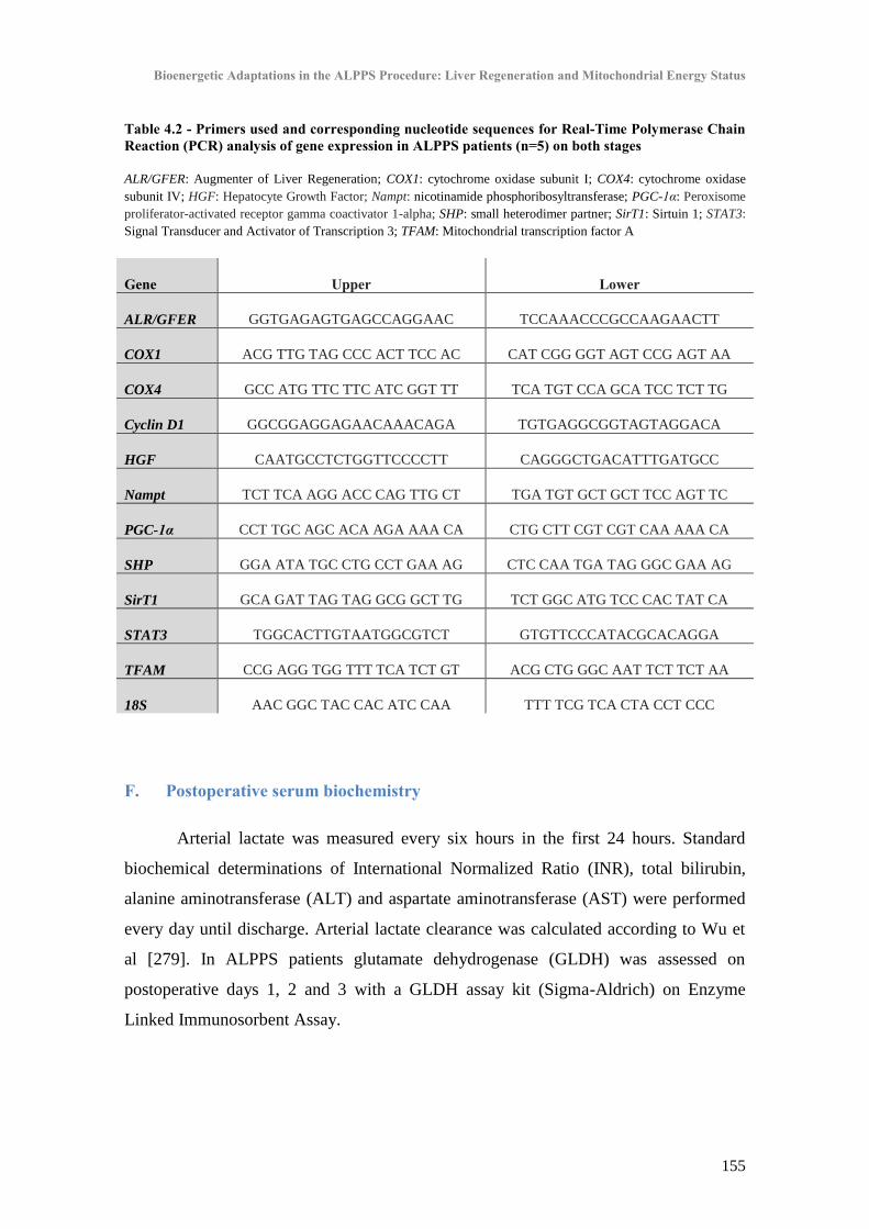
Bioenergetic Adaptations in the ALPPS Procedure: Liver Regeneration and Mitochondrial Energy Status
155
Table 4.2 - Primers used and corresponding nucleotide sequences for Real-Time Polymerase Chain Reaction (PCR) analysis of gene expression in ALPPS patients (n=5) on both stages
ALR/GFER: Augmenter of Liver Regeneration; COX1: cytochrome oxidase subunit I; COX4: cytochrome oxidase
subunit IV; HGF: Hepatocyte Growth Factor; Nampt: nicotinamide phosphoribosyltransferase; PGC-1α: Peroxisome
proliferator-activated receptor gamma coactivator 1-alpha; SHP: small heterodimer partner; SirT1: Sirtuin 1; STAT3:
Signal Transducer and Activator of Transcription 3; TFAM: Mitochondrial transcription factor A
Gene Upper Lower
ALR/GFER GGTGAGAGTGAGCCAGGAAC TCCAAACCCGCCAAGAACTT
COX1 ACG TTG TAG CCC ACT TCC AC CAT CGG GGT AGT CCG AGT AA
COX4 GCC ATG TTC TTC ATC GGT TT TCA TGT CCA GCA TCC TCT TG
Cyclin D1 GGCGGAGGAGAACAAACAGA TGTGAGGCGGTAGTAGGACA
HGF CAATGCCTCTGGTTCCCCTT CAGGGCTGACATTTGATGCC
Nampt TCT TCA AGG ACC CAG TTG CT TGA TGT GCT GCT TCC AGT TC
PGC-1α CCT TGC AGC ACA AGA AAA CA CTG CTT CGT CGT CAA AAA CA
SHP GGA ATA TGC CTG CCT GAA AG CTC CAA TGA TAG GGC GAA AG
SirT1 GCA GAT TAG TAG GCG GCT TG TCT GGC ATG TCC CAC TAT CA
STAT3 TGGCACTTGTAATGGCGTCT GTGTTCCCATACGCACAGGA
TFAM CCG AGG TGG TTT TCA TCT GT ACG CTG GGC AAT TCT TCT AA
18S AAC GGC TAC CAC ATC CAA TTT TCG TCA CTA CCT CCC
F. Postoperative serum biochemistry
Arterial lactate was measured every six hours in the first 24 hours. Standard
biochemical determinations of International Normalized Ratio (INR), total bilirubin,
alanine aminotransferase (ALT) and aspartate aminotransferase (AST) were performed
every day until discharge. Arterial lactate clearance was calculated according to Wu et
al [279]. In ALPPS patients glutamate dehydrogenase (GLDH) was assessed on
postoperative days 1, 2 and 3 with a GLDH assay kit (Sigma-Aldrich) on Enzyme
Linked Immunosorbent Assay.
Chapter IV
156
G. Postoperative liver function and clinical course
Patients were extubated in the operating room and admitted to the surgical
intensive care unit. Postoperative morbidity was defined and graded according to Dindo
et al up to the 90th
postoperative day [280]. Posthepatectomy liver failure (PHLF), was
defined according to the “50-50” criteria [165] and graded by International Study Group
of Liver Surgery (ISGLS) consensus definition [162]. Bile leakage was defined by the
ISGLS definition [281].
H. Statistical analysis
Normality of distribution was confirmed with the Shapiro-Wilk test. All
continuous variables were presented as mean ± standard deviation (SD) (if normally
distributed) or median and interquartile range (IQR). Continuous variables with normal
distribution were compared with two-tailed Student t test. Variables with non-normal
distribution with the two-tailed Mann-Whitney U test. Categorical variables were
compared with Chi-square test. Linear correlation between continuous variables was
assessed with Pearson coefficient. Statistical analysis was performed using SPSS™
(version 21.0, SPSS inc., Chicago, IL. USA). Significance was considered when p ≤
0.05 and a statistical trend with p ≤ 0.1.
III. Results
A. Clinical outcome and volumetric growth
There was no morbidity in the ALPPS group after T1. After T2, one patient
(20%) developed grade A bile leakage (Dindo grade II) and one patient (20%)
developed pleural effusion (Dindo grade II). One patients in the miHp group developed
grade B biloma (20%) (Dindo grade IIIA). No major morbidity was observed in the
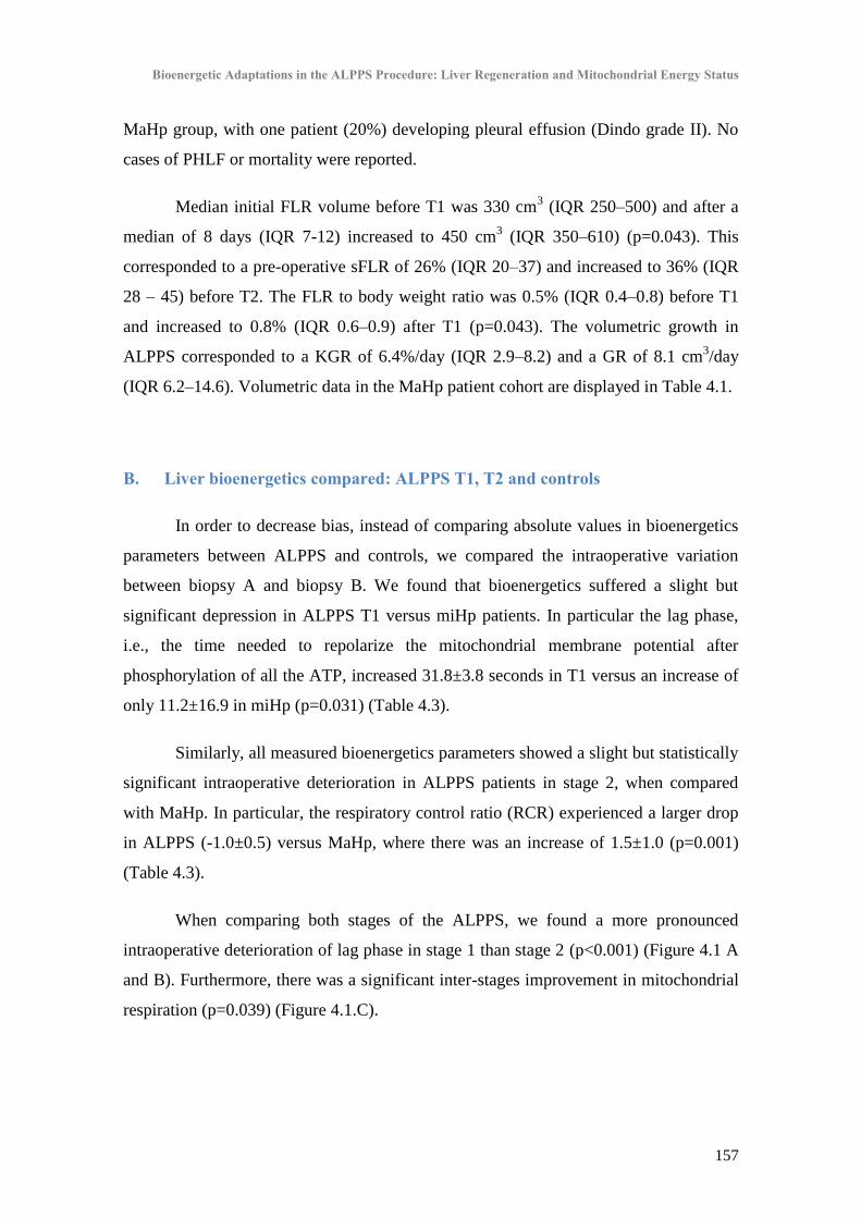
Bioenergetic Adaptations in the ALPPS Procedure: Liver Regeneration and Mitochondrial Energy Status
157
MaHp group, with one patient (20%) developing pleural effusion (Dindo grade II). No
cases of PHLF or mortality were reported.
Median initial FLR volume before T1 was 330 cm3 (IQR 250–500) and after a
median of 8 days (IQR 7-12) increased to 450 cm3 (IQR 350–610) (p=0.043). This
corresponded to a pre-operative sFLR of 26% (IQR 20–37) and increased to 36% (IQR
28 – 45) before T2. The FLR to body weight ratio was 0.5% (IQR 0.4–0.8) before T1
and increased to 0.8% (IQR 0.6–0.9) after T1 (p=0.043). The volumetric growth in
ALPPS corresponded to a KGR of 6.4%/day (IQR 2.9–8.2) and a GR of 8.1 cm3/day
(IQR 6.2–14.6). Volumetric data in the MaHp patient cohort are displayed in Table 4.1.
B. Liver bioenergetics compared: ALPPS T1, T2 and controls
In order to decrease bias, instead of comparing absolute values in bioenergetics
parameters between ALPPS and controls, we compared the intraoperative variation
between biopsy A and biopsy B. We found that bioenergetics suffered a slight but
significant depression in ALPPS T1 versus miHp patients. In particular the lag phase,
i.e., the time needed to repolarize the mitochondrial membrane potential after
phosphorylation of all the ATP, increased 31.8±3.8 seconds in T1 versus an increase of
only 11.2±16.9 in miHp (p=0.031) (Table 4.3).
Similarly, all measured bioenergetics parameters showed a slight but statistically
significant intraoperative deterioration in ALPPS patients in stage 2, when compared
with MaHp. In particular, the respiratory control ratio (RCR) experienced a larger drop
in ALPPS (-1.0±0.5) versus MaHp, where there was an increase of 1.5±1.0 (p=0.001)
(Table 4.3).
When comparing both stages of the ALPPS, we found a more pronounced
intraoperative deterioration of lag phase in stage 1 than stage 2 (p<0.001) (Figure 4.1 A
and B). Furthermore, there was a significant inter-stages improvement in mitochondrial
respiration (p=0.039) (Figure 4.1.C).

Chapter IV
158
Table 4.3 - E
nergy metabolism
in the study population (n= 5 patients undergoing AL
PPS) and controls (n= 10 patients undergoing minor and m
ajor hepatectomies)
Intrao
perativ
e variatio
n in
bio
energ
etics param
eters in A
ssociatin
g L
iver P
artition
and P
ortal V
ein L
igatio
n fo
r Stag
ed H
epatecto
my (A
LP
PS
n=
5) stag
e 1 (T
1) an
d m
ino
r hep
atectom
y co
ntro
ls
(n=
5); an
d in
AL
PP
S stag
e 2 (T
2) an
d m
ajor h
epatecto
my co
ntro
ls (n=
5). V
alues are p
resented
as mean
± stan
dard
dev
iation
.
Intraoperative change in: A
LPPS T
1 M
inor hepatectomy
p A
LPPS T
2 M
ajor hepatectomy
p
Mem
brane potential (mV
) - 2
6.4
± 4
.5
- 8.9
± 1
9.1
0
.10
9
- 30.6 ± 42.1 8.1 ± 9.9
0.025
Lag phase (seconds)
31.8 ± 3.8 11.2 ± 16.9
0.031 13.5 ± 2.3
- 8.7 ± 18.3 0.025
Depolarization (m
V)
- 13
.2 ±
14
.3
- 6.2
± 8
.6
0.3
72
- 10.1 ± 6.8
0.67 ± 3.4 0.027
Repolarization (m
V)
- 22.3 ± 6.6 - 6.4 ± 15.5
0.05 - 10.3 ± 0.5
6.4 ± 7.6 0.024
Respiratory control ratio
- 0.2
± 0
.2
0.0
2 ±
1.2
1
- 1.5 ± 0.5
1.5 ± 1.0 0.001
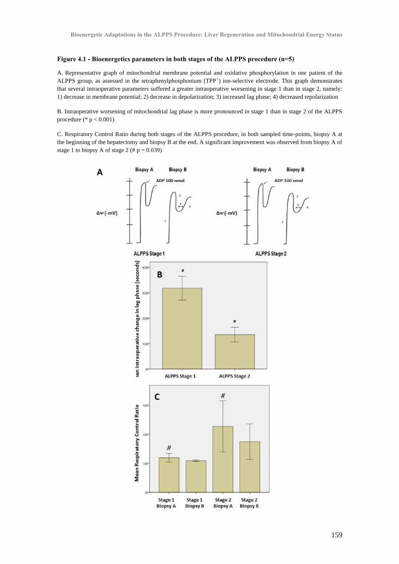
Bioenergetic Adaptations in the ALPPS Procedure: Liver Regeneration and Mitochondrial Energy Status
159
Figure 4.1 - Bioenergetics parameters in both stages of the ALPPS procedure (n=5)
A. Representative graph of mitochondrial membrane potential and oxidative phosphorylation in one patient of the
ALPPS group, as assessed in the tetraphenylphosphonium (TPP+) ion-selective electrode. This graph demonstrates
that several intraoperative parameters suffered a greater intraoperative worsening in stage 1 than in stage 2, namely:
1) decrease in membrane potential; 2) decrease in depolarization; 3) increased lag phase; 4) decreased repolarization
B. Intraoperative worsening of mitochondrial lag phase is more pronounced in stage 1 than in stage 2 of the ALPPS
procedure (* p < 0.001)
C. Respiratory Control Ratio during both stages of the ALPPS procedure, in both sampled time-points, biopsy A at
the beginning of the hepatectomy and biopsy B at the end. A significant improvement was observed from biopsy A of
stage 1 to biopsy A of stage 2 (# p = 0.039)
Chapter IV
160
C. Energy metabolism and postoperative liver remnant function and volume
growth
Mitochondrial bioenergetics presented several strong and significant correlations
with several markers of postoperative liver function in ALPPS patients. Post-T2 early
lactate clearance strongly correlated with RCR (r=0.994; p=0.006) and membrane
potential (r=0.969; p=0.031). Intraoperative change in mitochondrial membrane
potential during T2 inversely correlated with postoperative day 1 ALT (r=-0.932;
p=0.021) and AST (r=-0.969; p=0.006). Post-T2 fifth day INR correlated closely with
membrane potential (r=-0.982; p=0.003) and repolarization (r=-0.957; p=0.011). On
postoperative day 1 GLDH showed a very strong and significant correlation with
arterial lactate (r=0.905; p=0.035).
Decreased time to repolarize the mitochondrial membrane potential in T1
significantly correlated with improved KGR (r=-0.945; p=0.015). And intraoperative
increase in repolarization in T1 also demonstrated a very close linear correlation with
improved FLR growth (r=0.989; p=0.001) (Figure 4.2). Furthermore, improved
volumetrics also correlated with improved energetic status in T2, with the strongest
correlation demonstrated of KGR with improved State 3 respiration (r=0.931; p=0.021)
and GR with depolarization (r=0.919; p=0.027).
D. Gene expression and protein content in ALPPS
Liver samples in both stages of ALPPS were collected at the two time-points,
beginning and end of surgery for analysis of gene expression and protein content.
RNA content of genes associated with liver regeneration (STAT3, ALR)
(p<0.05), mitochondrial biogenesis (PGC-1α) (p=0.004) and energy metabolism
(COX1) (p=0.003) significantly increased intraoperatively during ALPPS T2, but not
during T1 (Figure 4.3). The expression of nicotinamide phosphoribosyltransferase
(Nampt) (p=0.003) was also significantly increased during both stages of ALPPS
(Figure 4.3) while the expression of SHP presented an intraoperative increase in T1
(p=0.034).
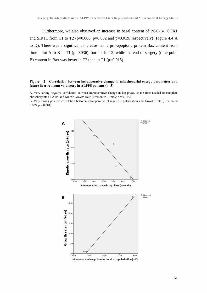
Bioenergetic Adaptations in the ALPPS Procedure: Liver Regeneration and Mitochondrial Energy Status
161
Furthermore, we also observed an increase in basal content of PGC-1α, COX1
and SIRT1 from T1 to T2 (p=0.006, p=0.002 and p=0.019, respectively) (Figure 4.4 A
to D). There was a significant increase in the pro-apoptotic protein Bax content from
time-point A to B in T1 (p=0.036), but not in T2; while the end of surgery (time-point
B) content in Bax was lower in T2 than in T1 (p=0.015).
Figure 4.2 - Correlation between intraoperative change in mitochondrial energy parameters and future liver remnant volumetry in ALPPS patients (n=5)
A. Very strong negative correlation between intraoperative change in lag phase, ie the time needed to complete
phosphorylate all ATP, and Kinetic Growth Rate (Pearson r= - 0.945; p = 0.015)
B. Very strong positive correlation between intraoperative change in repolarization and Growth Rate (Pearson r=
0.989; p = 0.001)

Chapter IV
162
Figure 4.3 - Gene expression in biopsies obtained in the liver remnant in both stages of the ALPPS procedure (n=5) Biopsies were collected at both time-points (time-point A at the beginning of the procedure, and time-point B at the end). Data are presented as a ratio of time-point B to basal expression in time-point A at each stage
A. Increase in expression of Signal Transducer and Activator of Transcription 3 (STAT3) at the end of stage 2 (* p =
0.036 versus sample A)
B. Increase in expression of Augmenter of Liver Regeneration (ALR) at the end of stage 2 (** p < 0.05 versus other
samples)
C. Expression of short heterodimer partner (SHP) signficantly increased during stage 1 of ALPPS (*** p = 0.034)
D. Increase in expression of Peroxisome proliferator-activated receptor gamma coactivator 1-alpha (PGC-1α) at the
end of stage 2 (**** p = 0.004 versus T1 and T2 sample A)
E. Increase in expression of cytochrome oxidase subunit 1 (COX1) at the end of stage 2 (# p = 0.003 versus other
samples)
F. Increase in expression of nicotinamide phosphoribosyltransferase (Nampt) at the end of stage 1 (## p = 0.01 versus
sample A) and at the end of stage 2 (### p = 0.003 versus sample A)

Bioenergetic Adaptations in the ALPPS Procedure: Liver Regeneration and Mitochondrial Energy Status
163
Figure 4.4 - Analysis of protein content (Western Blot) obtained in the liver remnant in both stages of the ALPPS procedure (n=5)
A. Representative Western blot analysis of protein content of several enzymes and transcription factors involved in
energy metabolism, mitochondrial biogenesis and apoptosis
B. Slight but significant increase in cytochrome oxidase subunit I content in the liver remnant between stage 1 and
stage 2 of ALPPS (* p = 0.006)
C. Significant increase in peroxisome proliferator-activated receptor gamma coactivator 1-alpha (PGC-1α) content in
the liver remnant between stage 1 and stage 2 of ALPPS (** p = 0.002)
D. Significant increase in Sirtuin 1 (SIRT1) content in the liver remnant between stage 1 and stage 2 of ALPPS (***
p = 0.019)

Chapter IV
164
IV. Discussion
Liver regeneration, albeit a wondrous phenomenon, is not inexhaustible and
some patients do not tolerate radical extended resections. Of the diverse techniques
available in modern Hepatobiliary surgery, the ALPPS procedure probably relies on the
extremes of the liver’s regenerative capacity, allowing resection in otherwise inoperable
patients [339]. This is clearly demonstrated by the successful completion of mono-
segment ALPPS [346]. However, ALPPS is fraught with high morbidity and mortality,
mostly due to PHLF [338].
Mitochondrial oxidative phosphorylation supplies the fuel for liver regeneration
and liver energy status is known to correlate with posthepatectomy liver function and
clinical outcome [19,41,43,52] (see also Chapter I - “Bioenergetics and Liver
Regeneration: Review of the Role of Energy Status in the Pathophysiology of
Posthepatectomy Liver Failure”). Experimental work has demonstrated that there is an
increase in mitochondrial DNA and RNA in the remnant liver in the first hours after
hepatectomy, as well as enhanced expression of several enzymes involved in energy
metabolism, such as the ETC enzyme cytochrome oxidase (COX) [44,45]. This results
in an overall increase of the liver’s energy-producing capacity in the first two to four
days after hepatectomy [46].
Mitochondrial quality and quantity in cells are influenced by the equilibrium of
formation of new organelles (biogenesis) and its destruction (mitophagy) [34]. Several
transcription factors are involved, including peroxisome proliferator-activated receptor-
γ coactivator (PGC-1α), for the orchestrated transcription of both the nuclear and the
13-gene mitochondrial genome to produce new mitochondria. Experimental evidence
has suggested that portal vein ligation (PVL) can improve mitochondrial function in the
non-ligated lobes in a rat model, possibly mediated by an increase in mitochondrial
biogenesis [245,246]. However, the adaptations occurring in human liver energy
metabolism in two stage hepatectomies, including ALPPS are unknown.
We intended to explore the role of mitochondrial bioenergetics and biogenesis in
the liver’s regenerative response in ALPPS. By studying a cohort of five consecutive
Bioenergetic Adaptations in the ALPPS Procedure: Liver Regeneration and Mitochondrial Energy Status
165
patients undergoing the ALPPS procedure, we performed intraoperative liver biopsies in
both stages, at two time-points, beginning (A) and end (B) of the procedure. With this
we examined intraoperative changes in mitochondrial membrane potential and
respiration in the remnant liver. The transmembrane potential developed by
mitochondria upon addition of succinate is essential for energy production and is an
extremely sensitive measure of metabolic capacity. And the Respiratory Control Ratio
(RCR) reflects the capacity for substrate oxidation and ATP synthesis.
First we examined energy metabolism in both stages of ALPPS and compared
them with matched controls. The control populations consisted of a cohort of patients
undergoing multiple, minor parenchymal-sparing resections for stage 1; and a major
hepatectomy for stage 2. Although the controls were adequately matched, unavoidable
differences could present a bias in the interpretation of the results, as ALPPS is a more
complex procedure: one-stage conservative resection does not include PVL or
parenchymal division; and major hepatectomies presented an obviously larger FLR
volume and were performed as a single stage procedure. So, in order to further reduce
the individual variability in energetics parameters, in this comparison we only assessed
the intraoperative change from biopsy A to biopsy B.
We found that mitochondrial function was hampered in both stages of ALPPS
when compared to matched controls. Even after taking into account the aforementioned
biases, this finding alerts to the possibility of mitochondrial dysfunction being a
contributing factor in the higher risk of PHLF and mortality associated with ALPPS;
and reinforces the caution surrounding its use [347].
However, we also found an extremely interesting improvement in mitochondrial
respiration and function from T1 to T2, in spite of a slightly longer duration of HPC in
the latter. Furthermore, we also describe a larger increase in the mitochondrial-
dependent apoptotic protein Bax in T1 versus T2. These findings suggest that important
metabolic adaptations occur in the FLR, promoting improved bioenergetics
performance and decreased apoptosis. To our knowledge this has never been
documented before.
Another interesting result was the correlation of mitochondrial energetics with
posthepatectomy liver function, which seems to confirm previous work from our group
Chapter IV
166
[19]. Although we experienced no cases of PHLF in this series, we found that
mitochondrial membrane potential in T2 closely correlated in a negative way with two
key markers of posthepatectomy liver function, peak arterial lactate and postoperative
day 5 INR [11,171]. Furthermore GLDH, a mitochondrial matrix urea cycle enzyme and
a very sensitive marker of liver necrosis and mitochondrial dysfunction [86,187], was
tightly correlated with arterial lactate. And postoperative aminotransferases were also
correlated with mitochondrial membrane potential. These results suggest that patients
with better intraoperative energy metabolism presented improved postoperative liver
function and decreased hepatocellular necrosis.
Also extremely interesting was the finding of a very close association of
improved cellular energetics with enhanced volume growth of the FLR. In fact, we
demonstrated that improved mitochondrial potential in T1 correlated with improved
volume gain. Although the small size of the series should lead to caution in the
interpretation of these results, this has, to our knowledge, also never been reported
previously in the scientific literature. And were the main reason to further expand our
study on this subject with detailed analysis of the expression of genes associated with
liver regeneration, mitochondrial biogenesis and energy metabolism.
The regenerative response of ALPPS and other two-stage hepatectomies is
linked to portal vein ligation (PVL). Like portal vein embolization (PVE), PVL shunts
portal blood away from the tumour-bearing parenchyma and into the FLR, causing
atrophy and apoptosis in the embolized hemiliver; and hypertrophy and increased
proliferation in the non-embolized hemiliver [348]. This is probably due to the exposure
of the FLR to higher concentrations of hepatotrophic factors from the gut. Possible
implicated molecules are bile acids and enterohormones, such as glucagon-like peptide
1 (GLP-1) [349,350]. Our finding of increased expression of SHP in stage 1 is
suggestive of the role of bile acids, as SHP is a pleiotropic transcription factor just
downstream of the Farnesoid X Receptor (FXR), which is the main bile acid-regulated
nuclear receptor involved in liver regeneration [351].
However, ALPPS differs from other two-stage hepatectomies because PVL is
supplemented by the in situ splitting (total or partial) of the liver, separating the FLR
from the tumour containing contralateral lobe. This parenchymal division causes a
Bioenergetic Adaptations in the ALPPS Procedure: Liver Regeneration and Mitochondrial Energy Status
167
surgical disconnection of left to right cross-portal shunts and undoubtedly contributes to
the enhanced regenerative response [352]. But the augmented liver growth is also
possibly linked with the increased systemic inflammation caused by the parenchymal
partition. Although increased expression of Interleukin 6 (IL-6) was not found in a
rabbit model of ALPPS [353], extra-hepatic injury with PVL is associated with similar
volume gain in a mouse model of ALPPS and this is possibly mediated by the IL-6-
STAT3 pathway [250]. Cytokines are known primers of liver regeneration and the
STAT3 pathway can significantly improve mitochondrial function [16,354]. Moreover,
mitochondrial respiration is improved in animal models of moderate but not severe
abdominal sepsis [355]. Our results are in support of these observations, as we describe
a significantly higher liver remnant expression of STAT3 at the end of stage 2.
Another interesting finding was the increased expression of Augmenter of Liver
Regeneration (ALR), a potent hepatotrophic factor with important regulatory function in
cellular respiration. ALR, also known as Hepatic Stimulator Substance or
Hepatopoietin, is one of the strongest hepatic cell mitogens and it also modulates
oxidative phosphorylation, mitochondrial biogenesis and ATP synthesis [200]. In fact,
ALR’s importance in energy metabolism seems to be dependent upon its effect on
mitochondrial biogenesis, upstream of other two co-factors, TFAM and PGC-1α [21].
Also testifying the role of energy metabolism in liver regeneration was the fact
that Nicotinamide phosphoribosyltransferase (Nampt), a key enzyme in the synthesis of
nicotinamide adenine dinucleotide (NAD, an intermediate in the electron transfer
between the citric acid cycle and the ETC) was highly expressed in the end biopsy of
both stages of ALPPS. Nampt has recently been demonstrated to be involved in the
energetic adaptations in experimental liver resection [22].
Finally, we think we have confirmed the importance of mitochondrial biogenesis
and energy metabolism in Human liver regeneration. This is supported by the enhanced
gene expression and increased protein content in T2 of peroxisome proliferator-
activated receptor gamma coactivator 1-alpha (PGC-1α), a pivotal regulator of
mitochondrial biogenesis; and of cytochrome oxidase subunit I (COX1), a component
of complex IV of the ETC which is coded by the 13-gene mitochondrial genome. There

Chapter IV
168
was also an increase in SIRT1 content, a metabolic sensor that acts directly on PGC-1α
[36].
In conclusion, this work clearly demonstrates, for the first time in the scientific
literature, the importance of adaptations in energy metabolism in the regenerative
response of the liver in two stage hepatectomies. Of striking relevance are the inter-
stages improvement in energy status and the correlation with volumetric growth. A
definite role for mitochondrial biogenesis in this process is confirmed. In fact, the first
stage of ALPPS seems to not only induce liver regeneration but also prompts energetic
conditioning of the FLR, inasmuch as exercise does for striated muscle [36]. In the
clinical setting liver regeneration is very likely tightly paralleled by an efficient
mitochondrial response, in order to provide the ATP to fuel the extremely high
metabolic requirements of swift cell replication.
The clinical implications of this study are twofold. First, bioenergetic status
could be used as a liver functional reserve test, by staging parenchymal capacity to
undergo extended resection. In fact, assessment of fitness to undergo stage 2 of ALPPS
is increasingly performed with functional tests such as hepatobiliary scintigraphy, rather
than volumetric-only tests [342]. The metabolism, conjugation and biliary excretion of
iminodiacetic acid (and other xenobiotics such as indocyanine green), like most liver
functions, is an endergonic, i.e. energy-requiring or ATP consuming reaction.
Considering that liver regeneration is just another, although seldom required, liver
function, we postulate that the assessment of energy status of liver cells could indicate
liver pre-resection reserve [269]. This could assist in the surgical decision-making
process: second stage of ALPPS could be performed earlier in patients with improved
cellular bioenergetics; and later, or not at all, in patients with underlying mitochondrial
dysfunction. However, as there were no cases of PHLF in our series we cannot
speculate on the accuracy of this method and further studies are needed to clarify this
issue.
Secondly, these findings open the way to the possibility of using mitochondrial-
targeted pharmacological therapies to improve cellular bioenergetics in clinical liver
surgery, enhancing liver regeneration and decreasing the incidence of PHLF. Several
drugs are currently under evaluation in the preclinical setting [20,204,206,207] and

Bioenergetic Adaptations in the ALPPS Procedure: Liver Regeneration and Mitochondrial Energy Status
169
could potentially translate into the clinical arena, reducing the morbidity and mortality
of liver surgery and hopefully expanding the current limits of hepatectomy.


Concluding remarks


Concluding remarks
173
Liver resection offers improved survival to patients with both primary and
secondary liver malignancies. While doing so, modern Hepatobiliary Surgery constantly
challenges the liver’s unique ability to regenerate. In fact, technical constraints to
resection are usually only imposed by the quality and volume of the predicted future
liver remnant. With adequate preoperative planning and preparation, thorough selection
of patients, meticulous surgical technique, dedicated intraoperative management and
close postoperative care, the rule is that the majority of patients undergo resection of a
significant fraction of parenchyma with an unimpeded recovery of mass and function of
the liver remnant. Notwithstanding, on occasion the liver’s capacity to regenerate is
pushed beyond its limits, resulting in a syndrome of decreased excretory and synthetic
functions known as Posthepatectomy Liver Failure (PHLF). Although often self-limited,
the consequences of PHLF can at times be more portentous, with impaired renal
function, increased risk of sepsis and death. Furthermore, evidence has emerged for a
deleterious effect on long-term outcome in patients surviving PHLF.
Multiple experimental and some notable clinical evidence has proved that liver
regeneration is a highly energy-dependent sequence of events. As was thoroughly
reviewed in this Doctoral Thesis, the energy source for liver regeneration is
mitochondrial oxidative phosphorylation, the highly efficient oxygen-dependent
metabolic chain that fuels most processes in higher heterotrophic organisms. However,
the link of mitochondrial function and postoperative outcome had not been clearly
documented up to this day. In particular, the parallel of mitochondrial energy status and
posthepatectomy liver dysfunction and morbidity remained unproven.
In this Thesis we sought to explore this relationship, and succeed in doing so. In
one of the original clinical studies herein displayed, intraoperative decrease of
mitochondrial membrane potential was confirmed for the first time as an independent
factor in the development of postoperative liver-specific morbidity, with
posthepatectomy liver function closely correlating with several bioenergetics
parameters; for example, a longer lag phase, reflecting a prolonged time to
phosphorylate adenosine diphosphate, was both highly sensitive and specific of the
development of PHLF.
Concluding remarks
174
Furthermore, Hepatic Pedicle Clamping (HPC), an almost universal manoeuvre
in the technical arsenal of liver surgeons, was demonstrated to have a distinct effect on
mitochondrial function. This is an extremely interesting finding as it has long been
known that longer duration of HPC, although causing ischemia-reperfusion injury is not
linearly associated with worse postoperative outcome. Thus, mitochondrial
susceptibility to HPC could in fact be a missing link in this equation and it was possible,
in our work, to demonstrated this relationship; both in clinical and experimental studies.
Chemotherapy offers a major contribution to the management of patients with
colorectal cancer liver metastases (CRLM), the most common indication for liver
resection in developed nations. By reducing the size of liver nodules, resection can be
offered to a significant proportion of patients initially deemed inoperable. However, one
of the unwanted consequences of chemotherapy is the development of liver injury in
three distinct patterns: steatosis, steatohepatitis and sinusoidal obstruction syndrome
(SOS). Although mitochondrial dysfunction is involved in the pathophysiology of many
liver diseases, its role in the pathogenesis of chemotherapy-associated liver injury
(CALI) was still unknown. Furthermore, the exact clinical consequences of CALI are
still under intense scrutiny, as a recent meta-analysis failed to prove the current notion
of worse postoperative outcome in SOS.
This led to the pursuit of two studies. First, a clinical and pathological review of
patients undergoing liver resection for CRLM was performed, confirming SOS as an
independent factor in overall and liver-specific morbidity. However, the association of
more severe forms of SOS with PHLF or mortality was not confirmed on multivariate
analysis. Interestingly, steatosis and steatohepatitis did not hamper postoperative liver
function or clinical outcome, challenging a deeply embedded dogma in the surgical
community. The second study attempted to explore the possible involvement of
mitochondrial dysfunction in the pathogenesis of SOS. The experimental study designed
to answer these questions failed to replicate the characteristic histologic injury of a
previously published model. Nonetheless, mitochondrial dysfunction was for the first
time demonstrated to occur in an experimental model of chemotherapy-induced
hepatotoxicity.

Concluding remarks
175
Although indications are constantly increasing, the technical limits of liver
resection are presently being reached. But an incidentally discovered development
recently expanded these boundaries, up to the point where, unthinkable a few years ago,
the future liver remnant would consist of a single Couinaud segment. The Associating
Liver Partition and Portal Vein Ligation for Staged Hepatectomy (ALLPS) procedure,
an exciting but nonetheless risky procedure, probably dwells on the extremes of the
liver’s regenerative capacity. However, the precise adaptations in remnant liver
bioenergetics occurring along with the profound inter-stages changes of ALPPS had
never been documented. Again, this Thesis interrogated on this issue and several
conclusions were drawn. First, mitochondrial function is worse in ALPPS than in other
hepatectomies, a finding that is in accordance with the increased postoperative
morbidity and mortality associated with this technique; and that reinforces the caution
surrounding its use [347]. Moreover, the extremely interesting correlation of liver
remnant mitochondrial membrane potential in the first stage with the inter-stages
volume growth rate had never before described in the scientific literature. And finally,
the inter-stages improvement in mitochondrial function suggested that important
adaptations were taking place in the future liver remnant. This was confirmed by the
paralleled expression profile of several key genes in liver regeneration, mitochondrial
biogenesis and energy metabolism, an exciting first ever description in the scientific
literature.
In conclusion, the research questions drawn at the start of this project were
answered. However, several unresolved issues remain to be addressed in future research
and will now be summarily catalogued.
In the first place, the exact mechanism of energetic dysfunction in major
hepatectomies deserves further scrutiny. An attractive theory is the portal vein
hyperflow and resultant increased shear stress. Although it does not explain the
deterioration in liver function that, albeit rare can occur in minor hepatectomies, this
theory elegantly fits the current knowledge on pathogenesis of PHLF in major and
extended hepatectomies. However, shear stress alone might not justify the pathogenesis
of the small-for-size syndrome. In fact, decreased arterial flow mediated by the Hepatic
Artery Buffer Response could be the most relevant factor. The resulting parenchymal
hypoxia would compromise mitochondrial energy production and hence clinical
Concluding remarks
176
outcome. In order to verify this theory, a large animal experimental model of extended
hepatectomy would be needed in order to correlate liver macrovascular changes
(including arterial and portal flow, pressure and shear stress), parenchymal oxygenation
and cellular energy metabolism. Furthermore, the effects of intraoperative modulation
of portal vein and hepatic artery flow on energy metabolism, liver function and outcome
would be more readily assessed.
Another key point needing clarification is the mechanistic effect of oxaliplatin
on mitochondrial function. In order to examine this question, a complete dissection of
the molecular events taking place would probably require at least one study on
hepatocyte culture. However, since SOS is also a sinusoidal endothelial cell disease, a
three-dimensional co-culture comprising distinct liver cell populations would be a more
appropriate research model. Hopefully, this would provide a significant contribution to
the yet elusive pathophysiology of chemotherapy-induced SOS.
And as the evidence for mitochondrial dysfunction in the pathogenesis of PHLF
builds up, it is unavoidable to consider future clinical applications. In this regard, three
distinct pathways have their start at the “bench” and could lead to the “bedside”.
First, perioperative and early postoperative non-invasive assessment of
mitochondrial function could assist in the timely recognition of PHLF. While
mitochondrial function testing in liver biopsy samples is undoubtedly the gold standard,
it is invasive and cumbersome. Thus, other promising modalities will surely find their
way, notably serum glutamate dehydrogenase and microdialysis. Further research on
these methods is warranted and could ensure accurate and early detection of
posthepatectomy liver dysfunction.
Most Hepatobiliary surgeons would agree that estimation of hepatic preoperative
reserve is one of the most important steps in the prehepatectomy decision-making
process. And this leads to the second translation point of the findings herein included.
Composite liver function tests alongside with remnant liver volume measurement have
for long been the gold standard [269]. Although useful, these tests do not account for
regional asymmetries in liver function. But nowadays other methods are allowing
assessment of segmental liver function and are proving to be invaluable. Since most
liver functions require energy, an estimation of liver energy capacity could prove to be
Concluding remarks
177
an excellent liver reserve test. However, the best way to achieve this goal is not clear at
the present moment. As was demonstrated in this Thesis, direct assessment of liver
mitochondrial function is possible in liver remnant biopsy samples collected during the
first stage of a two-stage hepatectomy, such as the ALPPS procedure. However, one-
stage resections would require a preoperative liver percutaneous liver biopsy to assess
mitochondrial function, which although feasible is an invasive and risky procedure
[183]. Ideally, appraisal of liver energy status should be performed with a non-invasive
method. With the ability to measure the ATP/Pi ratio, 31
Phosphorus Magnetic
Resonance Spectroscopy could aid in this quest, with the added advantage of assessing
segmental liver function with gadoxetic acid enhanced study, and also measuring future
liver remnant volume, as an all-in-one exam [128,268,356].
The third translation point is the extremely appealing potential for improving the
liver’s regenerative capacity with mitochondrial-based therapies. Although innovative
techniques such as ALPPS or simultaneous portal and hepatic vein embolization are
pushing the current limits of technical resectability beyond expectations [357,358],
these limits will be ultimately met nonetheless. Considering energy status as one of the
limiting factors in liver regeneration, energetic conditioning could potentially help
transcend the boundaries of liver resection. Several candidate therapies were reviewed
earlier and a few demonstrate enormous potential, such as: enhancing mitochondrial
biogenesis with new drugs such as berberine; decreasing permeability transition with
novel cyclophilin inhibitors; improving parenchymal oxygenation with hyperbaric
oxygen and/or portal flow modulation; and upgrading mitochondrial pool with
improved mitophagy. Some the aforementioned approaches could eventually find their
way into the clinical arena and hopefully decrease morbidity and mortality of liver
resection.
This Doctoral Thesis hopes to have provided a modest yet significant
contribution to the understanding of Posthepatectomy Liver Failure. Mitochondria, the
living relic of an early symbiotic relationship in Earth’s life, relentlessly provide liver
cells with the energy for regeneration. Their malfunction can severely impair clinical
results after hepatectomy, either through depression of cell function, mitochondrial-
induced programmed cell death or outright energetic failure and necrosis. And while
playing a distinct yet undefined role in the complex changes that the liver suffers after

Concluding remarks
178
chemotherapy, mitochondrial function is confirmed as a key vector in the liver’s
regenerative response in two-stage hepatectomies.
Meanwhile, we feel that it is incumbent upon Surgeons in Academic Centres to
foster basic and clinical research on this subject. Not only will this allow a deeper
understanding of the pathophysiology of PHLF, but hopefully aid in its prevention and
early detection. And with the development of energetic-conditioning strategies in the
near future, we will conceivably provide hope for patients nowadays deemed unfit for
surgery due to a predicted insufficient liver remnant. One dares to dream that those days
are nearer than imagined.
Finally, while the mechanisms underlying liver regeneration have been the focus
of intensive research in clinical and basic sciences, the most upstream primer of liver
regeneration has yet to be identified. Metabolic stimuli, namely a change in whole
organism energy status, could in fact be the igniter of hepatocyte proliferation [24,26].
Since liver resection is not a usual environmental stressor, the liver must have evolved
the capacity for regeneration because of the constant exposure to toxic, metabolic and
infectious aggressors carried by the portal blood flow. Given the central role of the liver
in glucose and lipid metabolism, it is conceivable that liver regeneration has thus
evolved as an adaptive response to two divergent phenomena: first, liver injury can
significantly decrease liver mass; and second, fluctuations in whole-body energy supply
and demand require enormous plasticity of the liver parenchyma. A hepatostat, i.e. a
sensor of hepatocyte mass to whole body metabolic needs fits nicely with the theory of
liver mass regulation by metabolic rather than hemodynamic stimuli [23,359–361].
Could it be that the hepatostat is none other than an elegantly placed intracellular
energy sensing system? Could the stimulus for liver regeneration start with an altered
cellular energy state, and thus the hepatostat would in reality be an “energostat”? An
interesting parallel emerges: as much as the Liver must recuperate lost mass to maintain
whole-body energy status, so Mitochondria should increase energy supply to fuel
hepatocyte replication and function. Perhaps Mitochondria’s role in Liver Regeneration
is not simply as the provider of energy. Perhaps by sensing the hepatocyte’s metabolic
overload, they also play a role in the key events initiating Liver Regeneration. Surely

Concluding remarks
179
this question would fuel the scientific curiosity to mandate further and more exciting
research. Energy will hopefully be applied in this endeavour.
Coimbra, March 2017


References

References
183
1. Bismuth H. Surgical anatomy and anatomical surgery of the liver. World J Surg.
1982 Jan;6(1):3–9.
2. Adam R, Delvart V, Pascal G, Valeanu A, Castaing D, Azoulay D, et al. Rescue
surgery for unresectable colorectal liver metastases downstaged by
chemotherapy: a model to predict long-term survival. Ann Surg.
2004;240(4):644-657-658.
3. Narita M, Oussoultzoglou E, Jaeck D, Fuchschuber P, Rosso E, Pessaux P, et al.
Two-stage hepatectomy for multiple bilobar colorectal liver metastases. Br J
Surg. 2011 Oct;98(10):1463–75.
4. Torzilli G, Belghiti J, Capussotti L, Vauthey J, Choti MA, Santibanes E De, et al.
A Snapshot of the Effective Indications and Results of Surgery for Hepatocellular
Carcinoma in Tertiary Referral Centers: Is It Adherent to the EASL / AASLD
Recommendations? Ann Surg. 2013;257(5):929–37.
5. Nagino M, Ebata T, Yokoyama Y, Igami T, Sugawara G, Takahashi Y, et al.
Evolution of Surgical Treatment for Perihilar Cholangiocarcinoma. Ann Surg.
2013;258(1):129–40.
6. Adam R, Chiche L, Aloia T, Elias D, Salmon R, Rivoire M, et al. Hepatic
resection for noncolorectal nonendocrine liver metastases: analysis of 1,452
patients and development of a prognostic model. Ann Surg. 2006
Oct;244(4):524–35.
7. Clavien P-A, Petrowsky H, DeOliveira ML, Graf R. Strategies for safer liver
surgery and partial liver transplantation. N Engl J Med. 2007 Apr
12;356(15):1545–59.
8. Golse N, Bucur PO, Adam R, Castaing D, Sa Cunha A, Vibert E. New paradigms
in post-hepatectomy liver failure. J Gastrointest Surg. 2013 Mar;17(3):593–605.
9. Hammond JS, Guha IN, Beckingham IJ, Lobo DN. Prediction, prevention and
management of postresection liver. Br J Surg. 2011;98(9):1188–200.
10. Qadan M, Garden OJ, Corvera CU, Visser BC. Management of Postoperative
Hepatic Failure. J Am Coll Surg. 2016 Feb;222(2):195–208.
11. Reissfelder C, Rahbari NN, Koch M, Kofler B, Sutedja N, Elbers H, et al.
Postoperative course and clinical significance of biochemical blood tests
following hepatic resection. Br J Surg. 2011 Jun;98(6):836–44.
12. Nakamura N, Hatano E, Iguchi K, Seo S, Taura K, Uemoto S. Posthepatectomy
Liver Failure Affects Long-Term Function After Resection for Hepatocellular
Carcinoma. World J Surg. 2015 Nov 20;40(4):929–36.
13. Vibert E, Pittau G, Gelli M. Actual incidence and long-term consequences of
posthepatectomy liver failure after hepatectomy for colorectal liver metastases.
Surgery. 2014;155(1):94–105.
14. Capussotti L, Vigano L, Giuliante F, Ferrero A, Giovannini I, Nuzzo G. Liver
dysfunction and sepsis determine operative mortality after liver resection. Br J
References
184
Surg. 2009;96(1):88–94.
15. Minuk GY. Hepatic regeneration: If it ain’t broke, don’t fix it. Can J
Gastroenterol. 2003;17(7):418–24.
16. Fausto N, Campbell JS, Riehle KJ. Liver regeneration. Hepatology. 2006
Feb;43(2 Suppl 1):S45-53.
17. Michalopoulos GK. Liver regeneration: alternative epithelial pathways. Int J
Biochem Cell Biol. Elsevier Ltd; 2011 Feb;43(2):173–9.
18. Tralhão JG, Abrantes AM, Hoti E, Oliveiros B, Cardoso D, Faitot F, et al.
Hepatectomy and liver regeneration: from experimental research to clinical
application. ANZ J Surg. 2013 May 8;84(9):665–71.
19. Alexandrino H, Varela AT, Teodoro JS, Martins MA, Rolo AP, Tralhão JG, et al.
Mitochondrial bioenergetics and posthepatectomy liver dysfunction. Eur J Clin
Invest. 2016 Jul;46(7):627–35.
20. Rehman H1, Sun J, Shi Y, Ramshesh VK, Liu Q, Currin RT, Lemasters JJ ZZ.
NIM811 prevents mitochondrial dysfunction, attenuates liver injury, and
stimulates liver regeneration after massive hepatectomy. Transplantation.
2012;91(4):406–12.
21. Han L-H, Dong L-Y, Yu H, Sun G-Y, Wu Y, Gao J, et al. Deceleration of liver
regeneration by knockdown of augmenter of liver regeneration gene is associated
with impairment of mitochondrial DNA synthesis in mice. Am J Physiol
Gastrointest Liver Physiol. 2015 May 14;309(2):112–22.
22. Mukherjee S, Chellappa K, Moffitt A, Ndungu J, Dellinger RW, Davis JG, et al.
Nicotinamide adenine dinucleotide biosynthesis promotes liver regeneration.
Hepatology. 2016 Nov 3;65(2):616–63.
23. Huang J, Rudnick DA. Elucidating the Metabolic Regulation of Liver
Regeneration. Am J Pathol. American Society for Investigative Pathology;
2014;184(2):309–21.
24. Crumm S, Cofan M, Juskeviciute E, Hoek JB. Adenine nucleotide changes in the
remnant liver: An early signal for regeneration after partial hepatectomy.
Hepatology. 2008 Sep;48(3):898–908.
25. Guerra MT, Fonseca EA, Melo FM, Andrade VA, Aguiar CJ, Andrade LM, et al.
Mitochondrial calcium regulates rat liver regeneration through the modulation of
apoptosis. Hepatology. 2011;54(1):296–306.
26. Ngala Kenda JF, de Hemptinne B, Lambotte L. Role of metabolic overload in the
initiation of DNA synthesis following partial hepatectomy in the rat. Eur Surg
Res Eur Chir Forschung Rech Chir Eur. 1984;16(5):294–302.
27. Yin F, Cadenas E. Mitochondria: the cellular hub of the dynamic coordinated
network. Antioxid Redox Signal. 2015 Apr 20;22(12):961–4.
28. Lemasters JJ, Theruvath TP, Zhong Z, Nieminen A-L. Mitochondrial calcium
References
185
and the permeability transition in cell death. Biochim Biophys Acta. Elsevier
B.V.; 2009 Nov;1787(11):1395–401.
29. Koolman J, Roehm K-H. Koolman, Color Atlas of Biochemistry. Koolman J,
Roehm K-H, editors. Color Atlas of Biochemistry. Thieme; 2005. 215 p.
30. Stillway W. Bioenergetics and Oxidative Metabolism. In: Dominiczak M,
Baynes J, editors. Medical Biochemistry. Mosby, Inc.; 1999. p. 83–94.
31. Rolo AP, Teodoro JS, Palmeira CM. Role of oxidative stress in the pathogenesis
of nonalcoholic steatohepatitis. Free Radic Biol Med. 2012 Jan 1;52(1):59–69.
32. Paradies G, Paradies V, Ruggiero FM, Petrosillo G. Oxidative stress, cardiolipin
and mitochondrial dysfunction in nonalcoholic fatty liver disease. World J
Gastroenterol. 2014 Oct 21;20(39):14205–18.
33. Luedde T, Kaplowitz N, Schwabe RF. Cell death and cell death responses in liver
disease: mechanisms and clinical relevance. Gastroenterology. 2014
Oct;147(4):765–783.e4.
34. Stotland A, Gottlieb RA. Mitochondrial quality control: Easy come, easy go.
Biochim Biophys Acta. 2015 Jan 14;1853(10 Pt B):2802–11.
35. Wenz T. Regulation of mitochondrial biogenesis and PGC-1α under cellular
stress. Mitochondrion. 2013 Mar;13(2):134–42.
36. Duarte F V, Amorim JA, Palmeira CM, Rolo AP. Regulation of Mitochondrial
Function and its Impact in Metabolic Stress. Curr Med Chem. 2015;22(20):2468–
79.
37. Madrigal-Matute J, Cuervo AM. Regulation of Liver Metabolism by Autophagy.
Gastroenterology. 2016 Feb;150(2):328–39.
38. Lee S, Kim J-S. Mitophagy: therapeutic potentials for liver disease and beyond.
Toxicol Res. 2014 Dec 31;30(4):243–50.
39. Nakatani T, Yasuda K, Ozawa K, Kawashima S, Tobe T. Effects of (+)-
octanoylcarnitine on deoxyribonucleic acid synthesis in regenerating rabbit liver.
Clin Sci (Lond). 1982 Mar;62(3):295–7.
40. Yamaoka Y, Ohsawa T, Takasan H, Ozawa K. Energy requirement in
regenerative and atrophic processes of the liver in man and other mammals. Surg
Gynecol Obstet. 1974 Aug;139(2):234–40.
41. Ozawa K, Kamiyama Y, Kimura K, Ukikusa M, Kono Y, Yamato T, et al. The
effects of heterologous liver cross-hemodialysis on adenylate energy charge of
the remnant liver after major hepatic resection. Artif Organs. 1982
Nov;6(4):447–52.
42. Corbin IR, Buist R, Volotovskyy V, Peeling J, Zhang M, Minuk GY.
Regenerative activity and liver function following partial hepatectomy in the rat
using (31)P-MR spectroscopy. Hepatology. 2002 Aug;36(2):345–53.
References
186
43. Mann D V, Lam WWM, Hjelm NM, So NMC, Yeung DKW, Metreweli C, et al.
Metabolic control patterns in acute phase and regenerating human liver
determined in vivo by 31-phosphorus magnetic resonance spectroscopy. Ann
Surg. 2002 Mar;235(3):408–16.
44. Koyama H, Kurokawa T, Nonami T, Nakao A, Sugiyama S. Increases in the
Mitochondrial DNA Replication and Transcription in the Remnant Liver of Rats.
1998;861(243):858–61.
45. Sun Y, Deng X, Li W, Yan Y, Wei H, Jiang Y, et al. Liver proteome analysis of
adaptive response in rat immediately after partial hepatectomy. Proteomics. 2007
Dec;7(23):4398–407.
46. Nagino M, Tanaka M, Nishikimi M, Nimura Y, Kubota H, Kanai M. Stimulated
Rat Liver Mitochondrial Biogenesis after Partial Hepatectomy. Cancer Res.
1989;49(17):4913–8.
47. Cao H, Yu J, Xu W, Jia X, Yang J, Pan Q, et al. Proteomic analysis of
regenerating mouse liver following 50% partial hepatectomy. Proteome Sci. 2009
Jan;7(48):doi: 10.1186/1477-5956-7-48.
48. Guerrieri F, Nicoletti C, Adorisio E, Caraccio G, Leonetti P, Zanotti F, et al.
Correlation between decreased expression of mitochondrial F0F1-ATP synthase
and low regenerating capability of the liver after partial hepatectomy in
hypothyroid rats. J Bioenerg Biomembr. 2000 May;32(2):183–91.
49. Moro L, Marra E, Capuano F, Greco M. Thyroid hormone treatment of
hypothyroid rats restores the regenerative capacity and the mitochondrial
membrane permeability properties of the liver after partial hepatectomy.
Endocrinology. 2004 Nov;145(11):5121–8.
50. Khiati S, Baechler SA, Factor VM, Zhang H, Huang S-YN, Dalla Rosa I, et al.
Lack of mitochondrial topoisomerase I (TOP1mt) impairs liver regeneration.
Proc Natl Acad Sci U S A. 2015 Sep 8;112(36):11282–7.
51. Yoshioka S, Miyazaki M, Shimizu H, Ito H, Nakagawa K, Ambiru S, et al.
Hepatic venous hemoglobin oxygen saturation predicts regenerative status of
remnant liver after partial hepatectomy in rats. Hepatology. 1998
May;27(5):1349–53.
52. Satoh S, Tanaka A, Hatano E, Inomoto T, Iwata S, Kitai T, et al. Energy
Metabolism and Regeneration in Transgenic Mouse Liver Expressing Creatine
Kinase After Major Hepatectomy. Gastroenterology. 1996;110(4):1166–74.
53. Zhong Z, Theruvath TP, Currin RT, Waldmeier PC, Lemasters JJ. NIM811, a
mitochondrial permeability transition inhibitor, prevents mitochondrial
depolarization in small-for-size rat liver grafts. Am J Transplant. 2007
May;7(5):1103–11.
54. Auger C, Alhasawi A, Contavadoo M, Appanna VD. Dysfunctional
mitochondrial bioenergetics and the pathogenesis of hepatic disorders. Front cell
Dev Biol. 2015 Jan;3:40.

References
187
55. Nishikawa T, Bellance N, Damm A, Bing H, Zhu Z, Handa K, et al. A switch in
the source of ATP production and a loss in capacity to perform glycolysis are
hallmarks of hepatocyte failure in advance liver disease. J Hepatol. 2014
Jun;60(6):1203–11.
56. Cortez-Pinto H, Chatham J, Chacko VP, Arnold C, Rashid A, Diehl AM.
Alterations in liver ATP homeostasis in human nonalcoholic steatohepatitis: a
pilot study. JAMA. 1999 Nov 3;282(17):1659–64.
57. McGill MR, Sharpe MR, Williams CD, Taha M, Curry SC, Jaeschke H. The
mechanism underlying acetaminophen-induced hepatotoxicity in humans and
mice involves mitochondrial damage and nuclear DNA fragmentation. J Clin
Invest. 2012 Apr;122(4):1574–83.
58. Lane M, Boczonadi V, Bachtari S, Gomez-Duran A, Langer T, Griffiths A, et al.
Mitochondrial dysfunction in liver failure requiring transplantation. J Inherit
Metab Dis. 2016;39(3):427–36.
59. Ukikusa M, Ozawa K, Shimahara Y, Asano M, Nakatani T, Tobe T. Changes in
blood ketone body ratio: their significance after major hepatic resection. Arch
Surg. 1981 Jun;116(6):781–5.
60. Yamaguchi T, Shimahara Y, Takada Y, Ino K, Mori K, Kobayashi N, et al.
Evaluation of ketogenesis in seriously reduced hepatic mitochondrial redox state.
An analysis of survivors and non-survivors in critically ill hepatectomized
patients. Scand J Gastroenterol. 1992 Jun;27(6):472–8.
61. Yamamoto Y, Ozawa K, Okamoto R, Kiuchi T, Maki A, Lin H, et al. Prognostic
implications of postoperative suppression of arterial ketone body ratio: time
factor involved in the suppression of hepatic mitochondrial oxidation-reduction
state. Surgery. 1990 Mar;107(3):289–94.
62. Kainuma M, Nakashima K, Sakuma I, Kawase M, Komatsu T, Shimada Y, et al.
Hepatic venous hemoglobin oxygen saturation predicts liver dysfunction after
hepatectomy. Anesthesiology. 1992;76(3):379–86.
63. Ozawa K, Kitamura O, Yamaoka Y, Mizukami T, Kamano T. Quantitative
analysis of respiratory enzymes of mitochondria isolated from liver tissue of
patients. J Lab Clin Med. 1973 Mar;81(3):379–92.
64. Ueda J, Mori K, Sakai Y, Tanaka A, Katayama T, Maki A, et al. Noninvasive
evaluation of cytochrome c oxidase activity of the liver. Its prognostic value for
hepatic resection. Arch Surg. 1994;129(3):303–8.
65. Castro O, David E, Ii M, Kumar A, Iii S, Eliza M, et al. Biochemical liver
function after partial hepatic resection with or without partial hepatic vascular
exclusion. Acta Cir Bras. 2011;26(Suppl 2):120–4.
66. Guerrieri F, Vendemiale G, Grattagliano I, Cocco T, Pellecchia G, Altomare E.
Mitochondrial oxidative alterations following partial hepatectomy. Free Radic
Biol Med. 1999 Jan;26(1–2):34–41.
References
188
67. Pringle JH. Notes on the Arrest of Hepatic Hemorrhage due to Trauma. Ann
Surg. 1908;4(4):541–9.
68. Chouillard EK, Gumbs A a, Cherqui D. Vascular clamping in liver surgery:
physiology, indications and techniques. Ann Surg Innov Res. 2010 Jan;4:2.
69. Varela AT, Simões AM, Teodoro JS, Duarte F V, Gomes AP, Palmeira CM, et
al. Indirubin-3’-oxime prevents hepatic I/R damage by inhibiting GSK-3beta and
mitochondrial permeability transition. Mitochondrion. Mitochondria Research
Society; 2010 Aug;10(5):456–63.
70. Bahde R, Spiegel HU. Hepatic ischaemia-reperfusion injury from bench to
bedside. Br J Surg. 2010;97(10):1461–75.
71. Wilasrusmee C, Siritheptawee S, Kanchanapanjapon S, Sopon P, Vanichanon C,
Limpthong W, et al. Ultrastructural changes in cirrhotic and noncirrhotic patients
due to hepatectomy. J Hepatobiliary Pancreat Surg. 2004 Jan;11(4):266–71.
72. Schoen JM, Wang HH, Minuk GY, Lautt WW. Shear stress-induced nitric oxide
release triggers the liver regeneration cascade. Nitric Oxide. 2001 Jan;5(5):453–
64.
73. Allard M-A, Adam R, Bucur P-O, Termos S, Cunha AS, Bismuth H, et al.
Posthepatectomy portal vein pressure predicts liver failure and mortality after
major liver resection on noncirrhotic liver. Ann Surg. 2013 Nov;258(5):822-9-30.
74. Chen X, Zhai J, Cai X, Zhang Y, Wei L, Shi L, et al. Severity of portal
hypertension and prediction of postoperative liver failure after liver resection in
patients with Child-Pugh grade A cirrhosis. Br J Surg. 2012 Dec;99(12):1701–10.
75. Demetris AJ, Kelly DM, Eghtesad B, Fontes P, Wallis Marsh J, Tom K, et al.
Pathophysiologic observations and histopathologic recognition of the portal
hyperperfusion or small-for-size syndrome. Am J Surg Pathol. 2006
Aug;30(8):986–93.
76. Golriz M, Majlesara A, El Sakka S, Ashrafi M, Arwin J, Fard N, et al. Small for
Size and Flow (SFSF) syndrome: An alternative description for posthepatectomy
liver failure. Clin Res Hepatol Gastroenterol. 2015;40(3):267–75.
77. Lautt WW, Legare DJ, Ezzat WR. Quantitation of the hepatic arterial buffer
response to graded changes in portal blood flow. Gastroenterology. 1990
Apr;98(4):1024–8.
78. Akamatsu N, Sugawara Y, Satou S, Mitsui T, Ninomiya R, Komagome M, et al.
Hemodynamic changes in the hepatic circulation after the modulation of the
splenic circulation in an in vivo human experimental model. Liver Transpl. 2014
Jan;20(1):116–21.
79. Lautt WW. Regulatory processes interacting to maintain hepatic blood flow
constancy: Vascular compliance, hepatic arterial buffer response, hepatorenal
reflex, liver regeneration, escape from vasoconstriction. Hepatol Res. 2007
Nov;37(11):891–903.
References
189
80. Eipel C, Abshagen K, Vollmar B. Regulation of hepatic blood flow: the hepatic
arterial buffer response revisited. World J Gastroenterol. 2010 Dec
28;16(48):6046–57.
81. Kostopanagiotou G, Pandazi A, Arkadopoulos N, Theodoraki K, Mystakidou K,
Costopanagiotou C, et al. Norepinephrine in Small-For-Size Liver Grafts: An
Experimental Study in Pigs. J Surg Res. 2007;141(2):257–61.
82. Zhu X, Fung JJ, Nakagawa S, Wang LF, Irefin S, Cocieru A, et al. Elevated
catecholamines and hepatic artery vasospasm in porcine small-for-size liver graft.
J Surg Res. 2012;174(1):157–65.
83. Dold S, Richter S, Kollmar O, Von Heesen M, Scheuer C, Laschke MW, et al.
Portal hyperperfusion after extended hepatectomy does not induce a hepatic
arterial buffer response (HABR) but impairs mitochondrial redox state and
hepatocellular oxygenation. PLoS One. 2015;10(11):e0141877.
84. Xiang L, Huang L, Wang X, Zhao Y, Liu Y, Tan J. How much portal vein flow is
too much for liver remnant in a stable porcine model? Transplant Proc.
2016;48(1):234–41.
85. Ninomiya M, Shirabe K, Terashi T, Ijichi H, Yonemura Y, Harada N, et al.
Deceleration of regenerative response improves the outcome of rat with massive
hepatectomy. Am J Transplant. 2010;10(7):1580–7.
86. McGill MR, Staggs VS, Sharpe MR, Lee WM, Jaeschke H. Serum mitochondrial
biomarkers and damage-associated molecular patterns are higher in
acetaminophen overdose patients with poor outcome. Hepatology. 2014
Oct;60(4):1336–45.
87. Helbling D, Buchaklian A, Wang J, Wong L-J, Dimmock D. Reduced
mitochondrial DNA content and heterozygous nuclear gene mutations in patients
with acute liver failure. J Pediatr Gastroenterol Nutr. 2013 Oct;57(4):438–43.
88. Ratziu V, Bellentani S, Cortez-Pinto H, Day C, Marchesini G. A position
statement on NAFLD/NASH based on the EASL 2009 special conference. J
Hepatol. 2010 Aug;53(2):372–84.
89. Mittal S, El-Serag HB, Sada YH, Kanwal F, Duan Z, Temple S, et al.
Hepatocellular Carcinoma in the Absence of Cirrhosis in US Veterans is
Associated with Non-Alcoholic Fatty Liver Disease. Clin Gastroenterol Hepatol.
2015 Jul 18;14(124–31).
90. Adam R, De Gramont A, Figueras J, Guthrie A, Kokudo N, Kunstlinger F, et al.
The oncosurgery approach to managing liver metastases from colorectal cancer: a
multidisciplinary international consensus. Oncologist. 2012 Jan;17(10):1225–39.
91. Gusdon AM, Song K-X, Qu S. Nonalcoholic Fatty liver disease: pathogenesis
and therapeutics from a mitochondria-centric perspective. Oxid Med Cell
Longev. 2014 Jan;2014:637027.
92. Pessayre D, Fromenty B. NASH: a mitochondrial disease. J Hepatol. 2005
References
190
Jun;42(6):928–40.
93. Grattagliano I, de Bari O, Bernardo TC, Oliveira PJ, Wang DQ-H, Portincasa P.
Role of mitochondria in nonalcoholic fatty liver disease--from origin to
propagation. Clin Biochem. The Canadian Society of Clinical Chemists; 2012
Jun;45(9):610–8.
94. Pirola CJ, Gianotti TF, Burgueño AL, Rey-Funes M, Loidl CF, Mallardi P, et al.
Epigenetic modification of liver mitochondrial DNA is associated with
histological severity of nonalcoholic fatty liver disease. Gut. 2013
Sep;62(9):1356–63.
95. Reiniers MJ, van Golen RF, van Gulik TM, Heger M. Reactive oxygen and
nitrogen species in steatotic hepatocytes: a molecular perspective on the
pathophysiology of ischemia-reperfusion injury in the fatty liver. Antioxid Redox
Signal. 2014 Sep 1;21(7):1119–42.
96. Kaur J. A Comprehensive Review on Metabolic Syndrome. Cardiol Res Pract.
2014 Jan;2014:1–21.
97. Guicciardi ME, Malhi H, Mott JL, Gores GJ. Apoptosis and necrosis in the liver.
Compr Physiol. 2013 Apr;3(2):977–1010.
98. Koliaki C, Szendroedi J, Kaul K, Jelenik T, Nowotny P, Jankowiak F, et al.
Adaptation of hepatic mitochondrial function in humans with non-alcoholic fatty
liver is lost in steatohepatitis. Cell Metab. 2015 May 5;21(5):739–46.
99. Verbeek J, Lannoo M, Pirinen E, Ryu D, Spincemaille P, Vander Elst I, et al.
Roux-en-y gastric bypass attenuates hepatic mitochondrial dysfunction in mice
with non-alcoholic steatohepatitis. Gut. 2015 Apr;64(4):673–83.
100. Teodoro JS, Duarte FV, Gomes AP, Varela AT, Peixoto FM, Rolo AP, et al.
Berberine reverts hepatic mitochondrial dysfunction in high-fat fed rats: a
possible role for SirT3 activation. Mitochondrion. Elsevier B.V.; 2013
Nov;13(6):637–46.
101. Perry RJ, Zhang D, Zhang X-M, Boyer JL, Shulman GI. Controlled-release
mitochondrial protonophore reverses diabetes and steatohepatitis in rats. Science.
2015 Mar 13;347(6227):1253–6.
102. Veteläinen R, Bennink RJ, van Vliet AK, van Gulik TM. Mild steatosis impairs
functional recovery after liver resection in an experimental model. Br J Surg.
2007 Aug;94(8):1002–8.
103. Veteläinen R, van Vliet AK, van Gulik TM. Severe steatosis increases
hepatocellular injury and impairs liver regeneration in a rat model of partial
hepatectomy. Ann Surg. 2007 Jan;245(1):44–50.
104. Sydor S, Gu Y, Schlattjan M, Bechmann LP, Rauen U, Best J, et al. Steatosis
does not impair liver regeneration after partial hepatectomy. Lab Investig. Nature
Publishing Group; 2012;93(1):20–30.
105. Garnol T, Kučera O, Staňková P, Lotková H, Červinková Z. Does Simple

References
191
Steatosis Affect Liver Regeneration after Partial Hepatectomy in Rats? Acta
medica (Hradec Králove) / Univ Carolina, Fac Medica Hradec Králove.
2016;59(2):35–42.
106. Veteläinen R, van Vliet A, Gouma DJ, van Gulik TM. Steatosis as a risk factor in
liver surgery. Ann Surg. 2007 Jan;245(1):20–30.
107. Varela AT, Rolo AP, Palmeira CM. Fatty liver and ischemia/reperfusion: are
there drugs able to mitigate injury? Curr Med Chem. 2011 Jan;18(32):4987–
5002.
108. Rolo AP, Teodoro JS, Peralta C, Rosello-Catafau J, Palmeira CM. Prevention of
I/R injury in fatty livers by ischemic preconditioning is associated with increased
mitochondrial tolerance: the key role of ATPsynthase and mitochondrial
permeability transition. Transpl Int. 2009 Nov;22(11):1081–90.
109. Cauchy F, Zalinski S, Dokmak S, Fuks D, Farges O, Castera L, et al. Surgical
treatment of hepatocellular carcinoma associated with the metabolic syndrome.
Br J Surg. 2013 Jan;100(1):113–21.
110. Parkin E, O’Reilly DA, Adam R, Kaiser GM, Laurent C, Elias D, et al. The
effect of hepatic steatosis on survival following resection of colorectal liver
metastases in patients without preoperative chemotherapy. HPB (Oxford). 2013
Jun;15(6):463–72.
111. de Meijer VE, Kalish BT, Puder M, Ijzermans JNM. Systematic review and
meta-analysis of steatosis as a risk factor in major hepatic resection. Br J Surg.
2010 Sep;97(9):1331–9.
112. Kooby DA, Fong Y, Suriawinata A, Gonen M, Allen PJ, Klimstra DS, et al.
Impact of steatosis on perioperative outcome following hepatic resection. J
Gastrointest Surg. 2003 Dec;7(8):1034–44.
113. McCormack L, Petrowsky H, Jochum W, Furrer K, Clavien P-A. Hepatic
steatosis is a risk factor for postoperative complications after major hepatectomy:
a matched case-control study. Ann Surg. 2007 Jun;245(6):923–30.
114. Reddy SK, Marsh JW, Varley PR, Mock BK, Chopra KB, Geller DA, et al.
Underlying steatohepatitis, but not simple hepatic steatosis, increases morbidity
after liver resection: A case-control study. Hepatology. 2012 Dec 14;56(6):2221–
30.
115. Martins J, Alexandrino H, Oliveira R, Cipriano MA, Falcão D, Ferreira L, et al.
Sinusoidal dilation increases the risk of complications in hepatectomy for
CRCLM – Protective effect of bevacizumab and diabetes mellitus, serum
gamma-glutamyltranspeptidase as predictive factor. Eur J Surg Oncol. 2016 Feb
24;42(5):713–21.
116. Vauthey J-N, Pawlik TM, Ribero D, Wu T-T, Zorzi D, Hoff PM, et al.
Chemotherapy regimen predicts steatohepatitis and an increase in 90-day
mortality after surgery for hepatic colorectal metastases. J Clin Oncol. 2006 May
1;24(13):2065–72.
References
192
117. Neal CP, Mann CD, Pointen E, McGregor A, Garcea G, Metcalfe MS, et al.
Influence of hepatic parenchymal histology on outcome following right hepatic
trisectionectomy. J Gastrointest Surg. 2012 Nov;16(11):2064–73.
118. Wiggans MG, Lordan JT, Shahtahmassebi G, Aroori S, Bowles MJ, Stell DA.
The Interaction between Diabetes, Body Mass Index, Hepatic Steatosis, and Risk
of Liver Resection: Insulin Dependent Diabetes Is the Greatest Risk for Major
Complications. HPB Surg. 2014 Jan;2014:1–10.
119. Behrns KE, Tsiotos GG, DeSouza NF, Krishna MK, Ludwig J, Nagorney DM.
Hepatic steatosis as a potential risk factor for major hepatic resection. J
Gastrointest Surg. 1998;2(3):292–8.
120. Gomez D, Malik HZ, Bonney GK, Wong V, Toogood GJ, Lodge JP a, et al.
Steatosis predicts postoperative morbidity following hepatic resection for
colorectal metastasis. Br J Surg. 2007 Nov;94(11):1395–402.
121. Schuppan D, Afdhal NH. Liver cirrhosis. Lancet. 2008 Mar 8;371(9615):838–51.
122. Cescon M, Cucchetti A, Grazi GL, Ferrero A, Viganò L, Ercolani G, et al.
Indication of the extent of hepatectomy for hepatocellular carcinoma on cirrhosis
by a simple algorithm based on preoperative variables. Arch Surg. 2009
Jan;144(1):57–63; discussion 63.
123. Sugiyama Y, Ishizaki Y, Imamura H, Sugo H, Yoshimoto J, Kawasaki S. Effects
of intermittent Pringle ’ s manoeuvre on cirrhotic compared with normal liver. Br
J Surg. 2010;97(7):1062–9.
124. Hashimoto M, Watanabe G. Functional restoration of cirrhotic liver after partial
hepatectomy in the rat. Hepatogastroenterology. 2005 Jan;52(63):897–902.
125. Froomes PR, Morgan DJ, Smallwood RA, Angus PW. Comparative effects of
oxygen supplementation on theophylline and acetaminophen clearance in human
cirrhosis. Gastroenterology. 1999 Apr;116(4):915–20.
126. Yang S, Tan TMC, Wee A, Leow CK. Mitochondrial respiratory function and
antioxidant capacity in normal and cirrhotic livers following partial hepatectomy.
Cell Mol Life Sci. 2004;61(2):220–9.
127. Arvaniti V, D’Amico G, Fede G, Manousou P, Tsochatzis E, Pleguezuelo M, et
al. Infections in patients with cirrhosis increase mortality four-fold and should be
used in determining prognosis. Gastroenterology. 2010 Oct;139(4):1246–56,
1256-5.
128. Mann D V, Lam WW, Hjelm NM, So NM, Yeung DK, Metreweli C, et al.
Human liver regeneration: hepatic energy economy is less efficient when the
organ is diseased. Hepatology. 2001 Sep;34(3):557–65.
129. Folprecht G, Grothey a, Alberts S, Raab H-R, Köhne C-H. Neoadjuvant
treatment of unresectable colorectal liver metastases: correlation between tumour
response and resection rates. Ann Oncol. 2005 Aug;16(8):1311–9.
130. Nordlinger B, Sorbye H, Glimelius B, Poston GJ, Schlag PM, Rougier P, et al.

References
193
Perioperative chemotherapy with FOLFOX4 and surgery versus surgery alone for
resectable liver metastases from colorectal cancer (EORTC Intergroup trial
40983): a randomised controlled trial. Lancet. 2008 Mar;371(9617):1007–16.
131. Lentz F, Tran A, Rey E, Pons G, Tréluyer J-M. Pharmacogenomics of
fluorouracil, irinotecan, and oxaliplatin in hepatic metastases of colorectal
cancer: clinical implications. Am J Pharmacogenomics. 2005 Jan;5(1):21–33.
132. Allard MA, Sebagh M, Baillie G, Lemoine A, Dartigues P, Faitot F, et al.
Comparison of complete pathologic response and hepatic injuries between
hepatic arterial infusion and systemic administration of oxaliplatin in patients
with colorectal liver metastases. Ann Surg Oncol. 2015 Jun;22(6):1925–32.
133. Karoui M, Penna C, Amin-Hashem M, Mitry E, Benoist S, Franc B, et al.
Influence of preoperative chemotherapy on the risk of major hepatectomy for
colorectal liver metastases. Ann Surg. 2006 Jan;243(1):1–7.
134. Takamoto T, Hashimoto T, Sano K, Maruyama Y, Inoue K, Ogata S, et al.
Recovery of liver function after the cessation of preoperative chemotherapy for
colorectal liver metastasis. Ann Surg Oncol. 2010 Oct;17(10):2747–55.
135. Labbe G, Pessayre D, Fromenty B. Drug-induced liver injury through
mitochondrial dysfunction: mechanisms and detection during preclinical safety
studies. Fundam Clin Pharmacol. 2008 Aug;22(4):335–53.
136. Rubbia-Brandt L, Lauwers GY, Wang H, Majno PE, Tanabe K, Zhu AX, et al.
Sinusoidal obstruction syndrome and nodular regenerative hyperplasia are
frequent oxaliplatin-associated liver lesions and partially prevented by
bevacizumab in patients with hepatic colorectal metastasis. Histopathology. 2010
Mar;56(4):430–9.
137. Fan CQ, Crawford JM. Sinusoidal Obstruction Syndrome (Hepatic Veno-
Occlusive Disease). J Clin Exp Hepatol. Elsevier Ltd; 2014;4(4):332–46.
138. Aloia T, Sebagh M, Plasse M, Karam V, Lévi F, Giacchetti S, et al. Liver
histology and surgical outcomes after preoperative chemotherapy with
fluorouracil plus oxaliplatin in colorectal cancer liver metastases. J Clin Oncol.
2006 Nov 1;24(31):4983–90.
139. Narita M, Oussoultzoglou E, Fuchshuber P, Chenard M-P, Rosso E, Yamamoto
K, et al. Prolonged portal triad clamping increases postoperative sepsis after
major hepatectomy in patients with sinusoidal obstruction syndrome and/or
steatohepatitis. World J Surg. 2012 Aug;36(8):1848–57.
140. Narita M, Oussoultzoglou E, Fuchshuber P, Pessaux P, Chenard M-P, Rosso E, et
al. What is a safe future liver remnant size in patients undergoing major
hepatectomy for colorectal liver metastases and treated by intensive preoperative
chemotherapy? Ann Surg Oncol. 2012 Aug;19(8):2526–38.
141. Nakano H, Oussoultzoglou E, Rosso E, Casnedi S, Chenard-Neu M-P, Dufour P,
et al. Sinusoidal injury increases morbidity after major hepatectomy in patients
with colorectal liver metastases receiving preoperative chemotherapy. Ann Surg.

References
194
2008 Jan;247(1):118–24.
142. Narita M, Oussoultzoglou E, Chenard MP, Rosso E, Casnedi S, Pessaux P, et al.
Sinusoidal obstruction syndrome compromises liver regeneration in patients
undergoing two-stage hepatectomy with portal vein embolization. Surg Today.
2011;41(1):7–17.
143. de Baere T, Teriitehau C, Deschamps F, Catherine L, Rao P, Hakime A, et al.
Predictive Factors for Hypertrophy of the Future Remnant Liver After Selective
Portal Vein Embolization. Ann Surg Oncol. 2010 Mar 17;17(8):2081–9.
144. Tabassum H, Waseem M, Parvez S, Qureshi MI. Oxaliplatin-induced Oxidative
Stress Provokes Toxicity in Isolated Rat Liver Mitochondria. Arch Med Res.
2015 Nov 9;6(8):597–603.
145. Rosen M, Figliomeni M, Simpkins H. The interaction of platinum antitumour
drugs with mouse liver mitochondria. Int J Exp Pathol. 1992 Feb;73(1):61–74.
146. Gourdier I, Crabbe L, Andreau K, Pau B, Kroemer G. Oxaliplatin-induced
mitochondrial apoptotic response of colon carcinoma cells does not require
nuclear DNA. Oncogene. 2004;(23):7449–57.
147. Schiffer E, Frossard J-L, Rubbia-Brandt L, Mentha G, Pastor CM. Hepatic
regeneration is decreased in a rat model of sinusoidal obstruction syndrome. J
Surg Oncol. 2009 Jun 1;99(7):439–46.
148. Robinson SM, Mann J, Vasilaki a, Mathers J, Burt a D, Oakley F, et al.
Pathogenesis of FOLFOX induced sinusoidal obstruction syndrome in a murine
chemotherapy model. J Hepatol. European Association for the Study of the Liver;
2013 Aug;59(2):318–26.
149. Neuhaus P, Jonas S, Bechstein WO. Extended Resections for Hilar
Cholangiocarcinoma. Ann Surg. 1999;230(6):808–19.
150. Yokoyama Y, Ebata T, Igami T, Sugawara G. Predictive power of prothrombin
time and serum total bilirubin for postoperative mortality after major
hepatectomy with extrahepatic bile duct resection. Surgery. Mosby, Inc.;
2011;155(3):504–11.
151. Iacono C, Ruzzenente A, Campagnaro T, Bortolasi L, Valdegamberi A,
Guglielmi A. Role of preoperative biliary drainage in jaundiced patients who are
candidates for pancreatoduodenectomy or hepatic resection: highlights and
drawbacks. Ann Surg. 2013 Mar;257(2):191–204.
152. Baton O, Azoulay D, Adam DVR, Castaing D. Major hepatectomy for hilar
cholangiocarcinoma type 3 and 4: prognostic factors and longterm outcomes. J
Am Coll Surg. 2007 Feb;204(2):250–60.
153. Igami T, Nishio H, Ebata T, Yokoyama Y, Sugawara G, Nimura Y, et al.
Surgical treatment of hilar cholangiocarcinoma in the “new era”: the Nagoya
University experience. J Hepatobiliary Pancreat Sci. 2010 Jul;17(4):449–54.
154. Wiggers JK, Groot Koerkamp B, Cieslak KP, Doussot A, van Klaveren D, Allen

References
195
PJ, et al. Postoperative Mortality after Liver Resection for Perihilar
Cholangiocarcinoma: Development of a Risk Score and Importance of Biliary
Drainage of the Future Liver Remnant. J Am Coll Surg. 2016 Aug;223(2):321–
331.e1.
155. Palmeira CM, Rolo AP. Mitochondrially-mediated toxicity of bile acids.
Toxicology. 2004;203(1–3):1–15.
156. Rolo AP, Oliveira PJ, Moreno JM, Palmeira CM. Bile Acids Affect Liver
Mitochondrial Bioenergetics : Possible Relevance for Cholestasis Therapy.
2000;57(1):177–85.
157. Schulz S, Schmitt S, Wimmer R, Aichler M, Eisenhofer S, Lichtmannegger J, et
al. Progressive stages of mitochondrial destruction caused by cell toxic bile salts.
BBA - Biomembr. Elsevier B.V.; 2013;81(9):2121–33.
158. Arduini A, Serviddio G, Escobar J, Tormos AM, Bellanti F, Viña J, et al.
Mitochondrial biogenesis fails in secondary biliary cirrhosis in rats leading to
mitochondrial DNA depletion and deletions. Am J Physiol Gastrointest Liver
Physiol. 2011 Jul;301(1):G119-27.
159. Kanai M, Tanaka M, Nimura Y, Nagino M, Katoh T, Ozawa T. Mitochondrial
dysfunction in the non-obstructed lobe of rat liver after selective biliary
obstruction. Hepatogastroenterology. 1992 Oct;39(5):385–91.
160. Krähenbühl L, Schäfer M, Krähenbühl S. Reversibility of hepatic mitochondrial
damage in rats with long-term cholestasis. J Hepatol. 1998 Jun;28(6):1000–7.
161. Mann D V, Lam WWM, Magnus Hjelm N, So NMC, Yeung DKW, Metreweli C,
et al. Biliary drainage for obstructive jaundice enhances hepatic energy status in
humans: a 31-phosphorus magnetic resonance spectroscopy study. Gut. 2002
Jan;50(1):118–22.
162. Rahbari NN, Garden OJ, Padbury R, Brooke-Smith M, Crawford M, Adam R, et
al. Posthepatectomy liver failure: a definition and grading by the International
Study Group of Liver Surgery (ISGLS). Surgery. 2011 May;149(5):713–24.
163. Siu J, Mccall J, Connor S. Systematic review of pathophysiological changes
following hepatic resection. HPB (Oxford). 2014;16(January 2000):407–21.
164. Hoekstra LT, de Graaf W, Nibourg GAA, Heger M, Bennink RJ, Stieger B, et al.
Physiological and Biochemical Basis of Clinical Liver Function Tests. Ann Surg.
2013 Jan;257(1):27–36.
165. Balzan S, Belghiti J, Farges O, Ogata S, Sauvanet A, Delefosse D, et al. The “50-
50 criteria” on postoperative day 5: an accurate predictor of liver failure and
death after hepatectomy. Ann Surg. 2005 Dec;242(6):824–8, NaN-9.
166. Kim SH, Kang DR, Lee JG, Kim DY, Ahn SH, Han K-H, et al. Early predictor of
mortality due to irreversible posthepatectomy liver failure in patients with
hepatocellular carcinoma. World J Surg. 2013 May;37(5):1028–33.
167. Bernal W. Lactate is important in determining prognosis in acute liver failure. J

References
196
Hepatol. European Association for the Study of the Liver; 2010 Jul;53(1):209–
10.
168. Rady MY. Bench-to-bedside review: Resuscitation in the emergency department.
Crit Care. 2005 Apr;9(2):170–6.
169. Detry O, Gaspar Y, Cheramy-Bien J-P, Drion P, Meurisse M, Defraigne J-O. A
modified surgical model of fulminant hepatic failure in the rat. J Surg Res.
Elsevier Ltd; 2013 May 1;181(1):85–90.
170. Watanabe I, Mayumi T, Arishima T, Takahashi H, Takezawa J, Medicine E, et al.
Hyperlactemia can predict the Prognosis of Liver Resection. Shock.
2007;28(1):35–8.
171. Vibert E, Boleslawski E, Cosse C, Adam R, Castaing D, Cherqui D, et al.
Arterial Lactate Concentration at the End of an Elective Hepatectomy Is an Early
Predictor of the Postoperative Course and a Potential Surrogate of Intraoperative
Events. Ann Surg. 2015 Nov;262(5):787–93.
172. Theodoraki K, Arkadopoulos N, Fragulidis G, Voros D, Karapanos K, Markatou
M, et al. Transhepatic lactate gradient in relation to liver ischemia/reperfusion
injury during major hepatectomies. Liver Transpl. 2006 Dec;12(12):1825–31.
173. Hallet J, Karanicolas PJ, Zih FSW, Cheng E, Wong J, Hanna S, et al.
Hypophosphatemia and recovery of post-hepatectomy liver insufficiency.
Hepatobiliary Surg Nutr. 2016 Jun;5(3):217–24.
174. Squires MH, Dann GC, Lad NL, Fisher SB, Martin BM, Kooby D a, et al.
Hypophosphataemia after major hepatectomy and the risk of post-operative
hepatic insufficiency and mortality: an analysis of 719 patients. HPB (Oxford).
2014 Oct;16(10):884–91.
175. Salem RR, Tray K. Hepatic resection-related hypophosphatemia is of renal origin
as manifested by isolated hyperphosphaturia. Ann Surg. 2005 Feb;241(2):343–8.
176. Nakamura K, Onitsuka T, Yano M, Yano Y. Study in three different types of
cardiopulmonary bypass on arterial ketone body ratio: its prognostic implication
and participation of body temperature. Interact Cardiovasc Thorac Surg. 2003
Mar;2(1):25–9.
177. Yan L-N, Chen X-L, Li Z-H, Li B, Lu S-C, Wen T-F, et al. Perioperative
management of primary liver cancer. World J Gastroenterol. 2007 Apr
7;13(13):1970–4.
178. Higashi Shushi, Tabata Naoto, Kondo Kazu-hiro, Kai Masahiro, Miyamoto Koji,
Maeda Yorio ST. A Predominant Increase of Arterial Beta-Hydroxybutyrate
Concentration during Partial Hepatectomies in Patients with Impaired
Indocyanine Green Clearance Test. J Surg Res. 1999;81(2):243–8.
179. Perera MTPR, Richards DA, Silva MA, Ahmed N, Neil DA, Murphy N, et al.
Comparison of energy metabolism in liver grafts from donors after circulatory
death and donors after brain death during cold storage and reperfusion. Br J Surg.
References
197
2014;101(7):775–83.
180. Haugaa H, Thorgersen EB, Pharo A, Boberg KM, Foss A, Line PD, et al. Early
bedside detection of ischemia and rejection in liver transplants by microdialysis.
Liver Transplant. 2012;18(7):839–49.
181. Isaksson B, D’souza MA, Jersenius U, Ungerstedt J, Lundell L, Permert J, et al.
Continuous assessment of intrahepatic metabolism by microdialysis during and
after portal triad clamping. J Surg Res. 2011 Aug;169(2):214–9.
182. Winbladh A, Björnsson B, Trulsson L, Offenbartl K, Gullstrand P, Sandström P.
Ischemic preconditioning prior to intermittent Pringle maneuver in liver
resections. J Hepatobiliary Pancreat Sci. 2012 Mar;19(2):159–70.
183. Chu MJJ, Phillips ARJ, Hosking AWG, Macdonald JR, Bartlett ASJR, Hickey
AJR. Hepatic Mitochondrial Function Analysis Using Needle Liver Biopsy
Samples. PLoS One. 2013;8(10):243–8.
184. Shi Q, Yang X, Mattes WB, Mendrick DL, Harrill AH, Beger RD. Circulating
mitochondrial biomarkers for drug-induced liver injury. Biomark Med. 2015
Jan;9(11):1215–23.
185. McGill MR, Li F, Sharpe MR, Williams CD, Curry SC, Ma X, et al. Circulating
acylcarnitines as biomarkers of mitochondrial dysfunction after acetaminophen
overdose in mice and humans. Arch Toxicol. 2014 Mar;88(2):391–401.
186. Bernal W, Auzinger G, Dhawan A, Wendon J. Acute liver failure. Lancet.
Elsevier Ltd; 2010 Jul 17;376(9736):190–201.
187. Schomaker S, Warner R, Bock J, Johnson K, Potter D, Winkle J Van, et al.
Assessment of Emerging Biomarkers of Liver Injury in Human Subjects. Toxicol
Sci. 2013;132(2):276–83.
188. Kellersmann R, Gassel H-J, Bühler C, Thiede A, Timmermann W. Application of
Molecular Adsorbent Recirculating System in patients with severe liver failure
after hepatic resection or transplantation: initial single-centre experiences. Liver.
2002;22(Suppl 2):56–8.
189. Barshes NR, Gay a N, Williams B, Patel AJ, Awad SS. Support for the acutely
failing liver: a comprehensive review of historic and contemporary strategies. J
Am Coll Surg. 2005 Sep;201(3):458–76.
190. Narita M, Oussoultzoglou E, Bachellier P, Jaeck D, Uemoto S. Post-hepatectomy
liver failure in patients with colorectal liver metastases. Surg Today. 2015 Oct
29;45(10):1218–26.
191. Bailey SM, Robinson G, Pinner A, Chamlee L, Ulasova E, Pompilius M, et al. S-
adenosylmethionine prevents chronic alcohol-induced mitochondrial dysfunction
in the rat liver. Am J Physiol Gastrointest Liver Physiol. 2006 Nov;291(5):G857-
67.
192. Brown JM, Kuhlman C, Terneus M V, Labenski MT, Lamyaithong AB, Ball JG,
et al. S-adenosyl-l-methionine protection of acetaminophen mediated oxidative
References
198
stress and identification of hepatic 4-hydroxynonenal protein adducts by mass
spectrometry. Toxicol Appl Pharmacol. 2014 Dec 1;281(2):174–84.
193. Jeon BR, Lee SM. S-adenosylmethionine protects post-ischemic mitochondrial
injury in rat liver. J Hepatol. 2001 Mar;34(3):395–401.
194. Su Z-R, Cui Z-L, Ma J-L, Li J-S, Ge Y, Yu J-H, et al. Beneficial effects of S-
adenosyl-L-methionine on post-hepatectomy residual liver function: a
prospective, randomized, controlled clinical trial. Hepatogastroenterology.
Jan;60(125):1136–41.
195. Liu G, Wang W, Jia W, Xu G, Ma J, Ge Y, et al. Protective effect of S-
adenosylmethionine on hepatic ischemia-reperfusion injury during hepatectomy
in HCC patients with chronic HBV infection. World J Surg Oncol. 2014
Jan;12:27.
196. Zhang P, Ma D, Wang Y, Zhang M, Qiang X, Liao M, et al. Berberine protects
liver from ethanol-induced oxidative stress and steatosis in mice. Food Chem
Toxicol. Elsevier Ltd; 2014;74:225–32.
197. Yu W, Sheng M, Xu R, Yu J, Cui K, Tong J, et al. Berberine protects human
renal proximal tubular cells from hypoxia/reoxygenation injury via inhibiting
endoplasmic reticulum and mitochondrial stress pathways. J Transl Med. Journal
of Translational Medicine; 2013 Jan;11(1):24.
198. Yan H-M, Xia M-F, Wang Y, Chang X-X, Yao X-Z, Rao S-X, et al. Efficacy of
Berberine in Patients with Non-Alcoholic Fatty Liver Disease. PLoS One.
2015;10(8):e0134172.
199. LaBrecque DR, Steele G, Fogerty S, Wilson M, Barton J. Purification and
physical-chemical characterization of hepatic stimulator substance. Hepatology.
7(1):100–6.
200. Gandhi CR. Augmenter of liver regeneration. Fibrogenesis Tissue Repair.
2012;5(1):10.
201. Thirunavukkarasu C, Wang LF, Harvey SAK, Watkins SC, Chaillet JR, Prelich J,
et al. Augmenter of liver regeneration: an important intracellular survival factor
for hepatocytes. J Hepatol. 2008 Apr;48(4):578–88.
202. Jiang S, Li W, An W. Adenoviral Gene Transfer of Hepatic Stimulator Substance
Confers Resistance Against Hepatic Ischemia – Reperfusion Injury by Improving
Mitochondrial Function. 2013;456(April):443–56.
203. Li S, Tang Z, Yu H, Li W, Jiang Y, Wang Y, et al. Administration of Naked
Plasmid Encoding Hepatic Stimulator Substance by Hydrodynamic Tail Vein
Injection Protects Mice from Hepatic Failure by Suppressing the Mitochondrial
Permeability Transition. J Pharmacol Exp Ther. 2011;338(3):750–7.
204. Polimeno L, Capuano F, Marangi LC, Margiotta M, Lisowsky T, Ierardi E, et al.
The augmenter of liver regeneration induces mitochondrial gene expression in rat
liver and enhances oxidative phosphorylation capacity of liver mitochondria. Dig

References
199
Liver Dis. 32(6):510–7.
205. Tang C, Lin H, Wu Q, Zhang Y, Bie P, Yang J. Recombinant human augmenter
of liver regeneration protects hepatocyte mitochondrial DNA in rats with
obstructive jaundice. J Surg Res. Elsevier Inc; 2015 Jun 1;196(1):90–101.
206. Naoumov N V. Cyclophilin inhibition as potential therapy for liver diseases. J
Hepatol. 2014 Nov;61(5):1166–74.
207. Šileikytė J, Forte M. Shutting down the pore: The search for small molecule
inhibitors of the mitochondrial permeability transition. Biochim Biophys Acta.
2016 Aug;1857(8):1197–202.
208. Lin C-W, Chen Y-S, Lin C-C, Chen Y-J, Lo G-H, Lee P-H, et al. Amiodarone as
an autophagy promoter reduces liver injury and enhances liver regeneration and
survival in mice after partial hepatectomy. Sci Rep. 2015;5:15807.
209. Meguro M, Mizuguchi T, Kawamoto M, Nakamura Y, Ota S, Kukita K, et al.
Continuous monitoring of central venous oxygen saturation predicts
postoperative liver dysfunction after liver resection. Surgery. 2013
Aug;154(2):351–62.
210. Richardson AJ, Laurence JM, Lam VWT. Portal triad clamping versus other
methods of vascular control in liver resection: a systematic review and meta-
analysis. 2012;14(6):355–64.
211. Hoekstra LT, van Trigt JD, Reiniers MJ, Busch OR, Gouma DJ, van Gulik TM.
Vascular occlusion or not during liver resection: the continuing story. Dig Surg.
2012 Jan;29(1):35–42.
212. Lesurtel M, Lehmann K, Rougemont O De, Clavien P. Clamping techniques and
protecting strategies in liver surgery. HPB (Oxford). 2009;11(4):290–5.
213. Nonami T, Asahi K, Harada A, Nakao A, Takagi H. Effect of hyperdynamic
circulatory support on hepatic hemodynamics, oxygen supply and demand after
massive hepatectomy. Surgery. 1991 Mar;109(3 Pt 1):277–83.
214. Taurà P, Fuster J, Mercadal J, Martinez-Palli G, Fondevila C, Blasi A, et al. The
use of beta-adrenergic drugs improves hepatic oxygen metabolism in cirrhotic
patients undergoing liver resection. J Hepatol. European Association for the
Study of the Liver; 2010 Mar;52(3):340–7.
215. Kawano Y, Akimaru K, Takubo K, Matsumoto K, Yoshida H, Mamada Y, et al.
Jejunectomy can reduce excessively elevated portal pressure after major
hepatectomy in beagle dogs. J Surg Res. 2006 Jan;130(1):24–33.
216. Glanemann M, Eipel C, Nussler AK, Vollmar B, Neuhaus P. Hyperperfusion
syndrome in small-for-size livers. Eur Surg Res. Jan;37(6):335–41.
217. Eipel C, Abshagen K, Ritter J, Cantré D, Menger MD, Vollmar B. Splenectomy
improves survival by increasing arterial blood supply in a rat model of reduced-
size liver. Transpl Int. 2010 Oct;23(10):998–1007.
References
200
218. Zhuang ZG, Xu JR, Qian LJ, Xia Q, Chi JC. Computed tomography perfusion
study of Hemodynamic Changes and portal hyperperfusion in a rabbit model of
small-for-size liver. Hepatobiliary Pancreat Dis Int. 2012;11(1):74–80.
219. Di Domenico S, Santori G, Traverso N, Balbis E, Furfaro A, Grillo F, et al. Early
effects of portal flow modulation after extended liver resection in rat. Dig Liver
Dis. 2011 Oct;43(10):814–22.
220. Ito K, Ozasa H, Noda Y, Koike Y, Arii S, Horikawa S. Splenic artery ligation
improves remnant liver function in partially hepatectomized rats with
ischemia/reperfusion injury. Liver Int. 2007 Apr;27(3):400–7.
221. Bucur PO, Bekheit M, Audebert C, Othman A, Hammad S, Sebagh M, et al.
Modulating Portal Hemodynamics With Vascular Ring Allows Efficient
Regeneration After Partial Hepatectomy in a Porcine Model. Ann Surg. 2017 Feb
1;(doi: 10.1097/SLA.0000000000002146. [Epub ahead of print]).
222. Taniguchi M, Shimamura T, Suzuki T, Yamashita K, Oura T, Watanabe M, et al.
Transient portacaval shunt for a small-for-size graft in living donor liver
transplantation. Liver Transpl. 2007 Jun;13(6):932–4.
223. Troisi R, de Hemptinne B. Clinical relevance of adapting portal vein flow in
living donor liver transplantation in adult patients. Liver Transpl. 2003
Sep;9(9):S36-41.
224. Famularo S, Nefotyou K, Fotiadis N, Khan N, Foxton M, Khan AZ. Small-for-
Size Liver Syndrome: a Case Series with a Proposal for Management Based on
Portal Flow Modulation. J Gastrointest Cancer. 2015 Jun;46(2):185–9.
225. Sato Y, Kobayashi T, Nakatsuka H, Yamamoto S, Oya H, Watanabe T, et al.
Splenic arterial ligation prevents liver injury after a major hepatectomy by a
reduction of surplus portal hypertension in hepatocellular carcinoma patients with
cirrhosis. Hepatogastroenterology. 2001;48(39):831–5.
226. Kelly DM, Zhu X, Shiba H, Irefin S, Trenti L, Cocieru A, et al. Adenosine
restores the hepatic artery buffer response and improves survival in a porcine
model of small-for-size syndrome. Liver Transpl. 2009 Nov;15(11):1448–57.
227. Nagamine K, Kubota T, Togo S, Nagashima Y, Mori M, Shimada H. Beneficial
effect of hyperbaric oxygen therapy on liver regeneration after 90% hepatectomy
in rats. Eur Surg Res Eur Chir Forschung Rech Chir Eur. Jan;36(6):350–6.
228. Tolentino EC, Castro e Silva O, Zucoloto S, Souza MEJ, Gomes MCJ,
Sankarankutty AK, et al. Effect of hyperbaric oxygen on liver regeneration in a
rat model. Transplant Proc. Jan;38(6):1947–52.
229. Sgarbi G, Giannone F, Casalena GA, Baracca A, Baldassare M, Longobardi P, et
al. Hyperoxia fully protects mitochondria of explanted livers. J Bioenerg
Biomembr. 2011 Dec;43(6):673–82.
230. Mazariegos G V, O’Toole K, Mieles LA, Dvorchik I, Meza MP, Briassoulis G, et
al. Hyperbaric oxygen therapy for hepatic artery thrombosis after liver

References
201
transplantation in children. Liver Transpl Surg. 1999 Sep;5(5):429–36.
231. Grover I, Conley L, Alzate G, Lavine J, Van Hoesen K, Khanna A. Hyperbaric
oxygen therapy for hepatic artery thrombosis following liver transplantation:
current concepts. Pediatr Transplant. 2006 Mar;10(2):234–9.
232. Ponikvar R, Buturović J, Cizman M, Mekjavić I, Kandus A, Premru V, et al.
Hyperbaric oxygenation, plasma exchange, and hemodialysis for treatment of
acute liver failure in a 3-year-old child. Artif Organs. 1998 Nov;22(11):952–7.
233. Asanuma Y, Sato T, Yasui O, Kurokawa T, Koyama K. Treatment for
postoperative liver failure after major hepatectomy under hepatic total vascular
exclusion. J Artif Organs. 2003 Jan;6(2):152–6.
234. Ueno S, Sakoda M, Kurahara H, Iino S, Minami K, Ando K, et al. Safety and
efficacy of early postoperative hyperbaric oxygen therapy with restriction of
transfusions in patients with HCC who have undergone partial hepatectomy.
Langenbeck’s Arch Surg / Dtsch Gesellschaft fur Chir. 2011 Jan;396(1):99–106.
235. Suehiro T, Shimura T, Okamura K, Okada T, Okada K, Hashimoto S, et al. The
effect of hyperbaric oxygen treatment on postoperative morbidity of left lobe
donor in living donor adult liver transplantation. Hepatogastroenterology.
Jan;55(84):1014–9.
236. Shimizu Y, Miyazaki M, Shimizu H, Ito H, Nakagawa K, Ambiru S, et al.
Beneficial effects of arterialization of the portal vein on extended hepatectomy.
Br J Surg. 2000 Jun;87(6):784–9.
237. Li J, Cai C, Guo H, Guan X, Yang L, Li Y, et al. Portal vein arterialization
promotes liver regeneration after extended partial hepatectomy in a rat model. J
Biomed Res. 2015 Jan;29(1):69–75.
238. Chen Y-L, Chen W-B, Wan Y-Y, Li W-G, Huang Z-Q, Wu X-T, et al. Effects of
partial portal vein arterialization on liver regeneration after hepatectomy in
minipigs with obstructive jaundice. Chin Med J (Engl). 2012 Jul;125(13):2302–5.
239. Bhangui P, Salloum C, Lim C, Andreani P, Ariche A, Adam R, et al. Portal vein
arterialization: a salvage procedure for a totally de-arterialized liver. The Paul
Brousse Hospital experience. HPB (Oxford). 2014 Aug;16(8):723–38.
240. Qiu J, Chen S, Pankaj P, Wu H. Portal vein arterialization as a bridge procedure
against acute liver failure after extended hepatectomy for hilar
cholangiocarcinoma. Surg Innov. 2014 Aug;21(4):372–5.
241. Kinoshita H, Sakai K, Hirohashi K, Igawa S, Yamasaki O, Kubo S. Preoperative
portal vein embolization for hepatocellular carcinoma. World J Surg. 1986
Oct;10(5):803–8.
242. de Baere T, Denys A, Madoff DC. Preoperative portal vein embolization:
indications and technical considerations. Tech Vasc Interv Radiol. 2007
Mar;10(1):67–78.
243. Farges O, Belghiti J, Kianmanesh R, Regimbeau JM, Santoro R, Vilgrain V, et

References
202
al. Portal vein embolization before right hepatectomy: prospective clinical trial.
Ann Surg. 2003 Feb;237(2):208–17.
244. Ozawa K, Takasan H, Kitamura O, Mizukami T, Kamano T. Effect of ligation of
portal vein on liver mitochondrial metabolism. J Biochem. 1971 Nov;70(5):755–
64.
245. Katoh T, Tanaka M, Nimura Y, Kanai M, Nagino M, Ozawa T. Enhancement of
rat liver mitochondrial function by portal branch ligation secures subsequent
extended hepatectomy. Biochem Int. 1991 May;24(1):107–16.
246. Shimizu Y, Suzuki H, Nimura Y, Onoue S, Nagino M, Tanaka M, et al. Elevated
mitochondrial gene expression during rat liver regeneration after portal vein
ligation. Hepatology. 1995 Oct;22(4 Pt 1):1222–9.
247. Shirasaki K, Taguchi K, Unno M, Motohashi H, Yamamoto M. NF-E2-related
factor 2 promotes compensatory liver hypertrophy after portal vein branch
ligation in mice. Hepatology. 2014 Jun 25;59(6):2371–82.
248. Adam R, Laurent A, Azoulay D, Castaing D, Bismuth H. Two-stage
hepatectomy: A planned strategy to treat irresectable liver tumors. Ann Surg.
2000 Dec;232(6):777–85.
249. Schnitzbauer AA, Lang SA, Goessmann H, Nadalin S, Baumgart J, Farkas SA, et
al. Right portal vein ligation combined with in situ splitting induces rapid left
lateral liver lobe hypertrophy enabling 2-staged extended right hepatic resection
in small-for-size settings. Ann Surg. 2012 Mar;255(3):405–14.
250. Schlegel A, Lesurtel M, Melloul E, Limani P, Tschuor C, Graf R, et al. ALPPS:
from human to mice highlighting accelerated and novel mechanisms of liver
regeneration. Ann Surg. 2014;260(5):839-46-7.
251. de Santibañes M, Dietrich A, Alvarez FA, Ardiles V, Loresi M, D’adamo M, et
al. Biological Substrate of the Rapid Volumetric Changes Observed in the
Human Liver During the Associating Liver Partition and Portal Vein Ligation for
Staged Hepatectomy Approach. J Gastrointest Surg. 2016 Mar;20(3):546–53.
252. Matsuo K, Murakami T, Kawaguchi D, Hiroshima Y, Koda K, Yamazaki K, et
al. Histologic features after surgery associating liver partition and portal vein
ligation for staged hepatectomy versus those after hepatectomy with portal vein
embolization. Surgery. 2016 Jan 8;159(5):1289–98.
253. Forbes SJ, Gupta S, Dhawan A. Cell therapy for liver disease: From liver
transplantation to cell factory. J Hepatol. 2015;62(S1):S157–69.
254. Kuai XL, Cong XQ, Du ZW, Bian YH, Xiao SD. Treatment of surgically
induced acute liver failure by transplantation of HNF4-overexpressing embryonic
stem cells. Chin J Dig Dis. 2006;7(2):109–16.
255. Cai Y, Zou Z, Liu L, Chen S, Chen Y, Lin Z, et al. Bone marrow-derived
mesenchymal stem cells inhibits hepatocyte apoptosis after acute liver injury. Int
J Clin Exp Pathol. 2015;8(1):107–16.

References
203
256. El’chaninov A V, Volodina MA, Arutyunyan I V, Makarov A V, Tarasova N V,
Kananykhina EY, et al. Effect of multipotent stromal cells on the function of cell
mitochondria in regenerating liver. Bull Exp Biol Med. 2015 Feb;158(4):566–72.
257. de Andrade DC, de Carvalho SN, Pinheiro D, Thole AA, Moura AS, de Carvalho
L, et al. Bone marrow mononuclear cell transplantation improves mitochondrial
bioenergetics in the liver of cholestatic rats. Exp Cell Res. Elsevier; 2015 May
12;336(1):15–22.
258. Tautenhahn HM, Bruckner S, Baumann S, Winkler S, Otto W, von Bergen M, et
al. Attenuation of Postoperative Acute Liver Failure by Mesenchymal Stem Cell
Treatment Due to Metabolic Implications. Ann Surg. 2015;263(3):546–56.
259. Vallabhaneni KC, Haller H, Dumler I. Vascular smooth muscle cells initiate
proliferation of mesenchymal stem cells by mitochondrial transfer via tunneling
nanotubes. Stem Cells Dev. 2012;21(17):3104–13.
260. Lin H-C, Liu S-Y, Lai H-S, Lai I-R. Isolated mitochondria infusion mitigates
ischemia-reperfusion injury of the liver in rats. Shock. 2013;39(3):304–10.
261. Esch JSA, Schmelzle M, Fürst G, Robson SC, Krieg A, Duhme C, et al. Infusion
of CD133+ Bone Marrow–Derived Stem Cells After Selective Portal Vein
Embolization Enhances Functional Hepatic Reserves After Extended Right
Hepatectomy. Ann Surg. 2012;255(1):79–85.
262. Treska V, Liska V, Fichtl J, Lysak D, Mirka H, Bruha J, et al. Portal vein
embolisation with application of haematopoietic stem cells in patients with
primarily or non-resectable colorectal liver metastases. Anticancer Res.
2014;34(12):7279–85.
263. Leung U, Simpson AL, Araujo RLC, Gönen M, McAuliffe C, Miga MI, et al.
Remnant Growth Rate after Portal Vein Embolization Is a Good Early Predictor
of Post-Hepatectomy Liver Failure. J Am Coll Surg. 2014 Oct;219(4):620–30.
264. Tralhao JG, Hoti E, Oliveiros B, Botelho MF, Sousa FC. Study of perioperative
liver function by dynamic monitoring of ICG-clearance. Hepatogastroenterology.
2012 Jun;59(116):1179–83.
265. Mullin EJ, Metcalfe MS, Maddern GJ. How much liver resection is too much?
Am J Surg. 2005 Jul;190(1):87–97.
266. de Santibañes E, Alvarez F a, Ardiles V. How to avoid postoperative liver
failure: a novel method. World J Surg. 2012 Jan;36(1):125–8.
267. Shindoh J, Truty MJ, Aloia T a, Curley S a, Zimmitti G, Huang SY, et al. Kinetic
growth rate after portal vein embolization predicts posthepatectomy outcomes:
toward zero liver-related mortality in patients with colorectal liver metastases and
small future liver remnant. J Am Coll Surg. American College of Surgeons; 2013
Feb;216(2):201–9.
268. Wibmer A, Prusa AM, Nolz R, Gruenberger T, Schindl M, Ba-Ssalamah A. Liver
failure after major liver resection: risk assessment by using preoperative
References
204
Gadoxetic acid-enhanced 3-T MR imaging. Radiology. 2013 Dec;269(3):777–86.
269. Mann D V. Assessment of Liver Function. In: Lau WY, editor. Hilar
Cholangiocarcinoma. Springer; 2013. p. 75–89.
270. Palmeira CM, Rolo AP. Mitochondrial membrane potential (ΔΨ) fluctuations
associated with the metabolic states of mitochondria. Methods Mol Biol. 2012
Jan;810:89–101.
271. Hall A, Larsen AK, Parhamifar L, Meyle KD, Wu L-P, Moghimi SM. High
resolution respirometry analysis of polyethylenimine-mediated mitochondrial
energy crisis and cellular stress: Mitochondrial proton leak and inhibition of the
electron transport system. Biochim Biophys Acta. 2013 Oct;1827(10):1213–25.
272. Grattagliano I, Caraceni P, Calamita G, Ferri D, Gargano I, Palasciano G, et al.
Severe liver steatosis correlates with nitrosative and oxidative stress in rats. Eur J
Clin Invest. 2008 Jul;38(7):523–30.
273. Tralhão JG, Hoti E, Oliveiros B, Abrantes AM, Botelho MF, Castro-Sousa F.
Intermittent pringle maneuver and hepatic function: Perioperative monitoring by
noninvasive ICG-clearance. World J Surg. 2009;33(12):2627–34.
274. Scatton O, Zalinski S, Jegou D, Compagnon P, Lesurtel M, Belghiti J, et al.
Randomized clinical trial of ischaemic preconditioning in major liver resection
with intermittent Pringle manoeuvre. Br J Surg. 2011;98:1236–43.
275. Martins PN a, Theruvath TP, Neuhaus P. Rodent models of partial
hepatectomies. Liver Int. 2008 Jan;28(1):3–11.
276. Palmeira CM, Moreno AJ, Madeira VM. Interactions of herbicides 2,4-D and
dinoseb with liver mitochondrial bioenergetics. Toxicol Appl Pharmacol. 1994
Jul;127(1):50–7.
277. Chance B, Williams GR. Respiratory enzymes in oxidative phosphorylation. VI.
The effects of adenosine diphosphate on azide-treated mitochondria. J Biol
Chem. 1956 Jul;221(1):477–89.
278. Stocchi V, Cucchiarini L, Magnani M, Chiarantini L, Palma P, Crescentini G.
Simultaneous extraction and reverse-phase high-performance liquid
chromatographic determination of adenine and pyridine nucleotides in human red
blood cells. Anal Biochem. 1985 Apr;146(1):118–24.
279. Wu J-F, Wu R-Y, Chen J, Ou-Yang B, Chen M-Y, Guan X-D. Early lactate
clearance as a reliable predictor of initial poor graft function after orthotopic liver
transplantation. Hepatobiliary Pancreat Dis Int. 2011 Dec;10(6):587–92.
280. Dindo D, Demartines N, Clavien P. Classification of surgical complications: a
new proposal with evaluation in a cohort of 6336 patients and results of a survey.
Ann Surg. 2004 Aug;240(2):205–13.
281. Koch M, Garden OJ, Padbury R, Rahbari NN, Adam R, Capussotti L, et al. Bile
leakage after hepatobiliary and pancreatic surgery: A definition and grading of
severity by the International Study Group of Liver Surgery. Surgery.

References
205
2011;149(5):680–8.
282. Mann CD, Palser T, Briggs CD, Cameron I, Rees M, Buckles J, et al. A review of
factors predicting perioperative death and early outcome in
hepatopancreaticobiliary cancer surgery. Hepatobiliary Pancreat Dis Int.
2010;12(6):380–8.
283. Ypsilantis P, Lambropoulou M, Tentes I, Anagnostopoulos K, Tsigalou C,
Papadopoulos N, et al. Impaired liver regeneration following partial hepatectomy
using the Pringle maneuver: Protective effect of mesna. J Gastroenterol Hepatol.
2009 Apr;24(4):623–32.
284. Seyama Y, Imamura H, Inagaki Y, Matsuyama Y, Tang W, Makuuchi M, et al.
Intermittent clamping is superior to ischemic preconditioning and its effect is
more marked with shorter clamping cycles in the rat liver. J Gastroenterol. 2013
Jan;48(1):115–24.
285. van der Bilt JDW, Livestro DP, Borren a, van Hillegersberg R, Borel Rinkes
IHM. European survey on the application of vascular clamping in liver surgery.
Dig Surg. 2007 Jan;24(6):423–35.
286. Rudnick DA, Davidson NO. Functional Relationships between Lipid Metabolism
and Liver Regeneration. Int J Hepatol. 2012;10(15).
287. Benzoni E, Lorenzin D, Baccarani U, Adani GL, Favero A, Cojutti A, et al.
Resective surgery for liver tumor: a multivariate analysis of causes and risk
factors linked to postoperative complications. Hepatobiliary Pancreat Dis Int.
2006 Nov;5(4):526–33.
288. Boleslawski E, Vibert E, Pruvot F-R, Le Treut Y-P, Scatton O, Laurent C, et al.
Relevance of postoperative peak transaminase after elective hepatectomy. Ann
Surg. 2014 Nov;260(5):815-20-1.
289. Lafaro K, Buettner S, Maqsood H, Wagner D, Bagante F, Spolverato G, et al.
Defining Post Hepatectomy Liver Insufficiency: Where do We stand? J
Gastrointest Surg. 2015 Nov;19(11):2079–92.
290. Ferlay J, Steliarova-Foucher E, Lortet-Tieulent J, Rosso S, Coebergh JWW,
Comber H, et al. Cancer incidence and mortality patterns in Europe: estimates for
40 countries in 2012. Eur J Cancer. 2013 Apr;49(6):1374–403.
291. Fong Y, Fortner J, Sun RL, Brennan MF, Blumgart LH. Clinical Score for
Predicting Recurrence After Hepatic Resection for Metastatic Colorectal Cancer
Analysis of 1001 Consecutive Cases. 1999;230(3):309–21.
292. Kopetz S, Chang GJ, Overman MJ, Eng C, Sargent DJ, Larson DW, et al.
Improved survival in metastatic colorectal cancer is associated with adoption of
hepatic resection and improved chemotherapy. J Clin Oncol. 2009;27(22):3677–
83.
293. Blazer DG, Kishi Y, Maru DM, Kopetz S, Chun YS, Overman MJ, et al.
Pathologic response to preoperative chemotherapy: a new outcome end point
References
206
after resection of hepatic colorectal metastases. J Clin Oncol. 2008
Nov;26(33):5344–51.
294. Kishi Y, Zorzi D, Contreras CM, Maru DM, Kopetz S, Ribero D, et al. Extended
preoperative chemotherapy does not improve pathologic response and increases
postoperative liver insufficiency after hepatic resection for colorectal liver
metastases. Ann Surg Oncol. 2010;17(11):2870–6.
295. Rubbia-Brandt L, Audard V, Sartoretti P, Roth AD, Brezault C, Le Charpentier
M, et al. Severe hepatic sinusoidal obstruction associated with oxaliplatin-based
chemotherapy in patients with metastatic colorectal cancer. Ann Oncol. 2004
Mar;15(3):460–6.
296. Hubert C, Sempoux C, Humblet Y, Van Den Eynde M, Zech F, Leclercq I, et al.
Sinusoidal obstruction syndrome (SOS) related to chemotherapy for colorectal
liver metastases: Factors predictive of severe SOS lesions and protective effect of
bevacizumab. HPB. 2013;15:858–64.
297. Soubrane O, Brouquet A, Zalinski S, Terris B, Brézault C, Mallet V, et al.
Predicting High Grade Lesions of Sinusoidal Obstruction Syndrome Related to
Oxaliplatin-Based Chemotherapy for Colorectal Liver Metastases. Ann Surg.
2010 Mar;251(3):454–60.
298. Alexandrino H, Oliveira D, Cipriano MA, Ferreira L, Tralhão JG, Castro E Sousa
F. Oxaliplatin toxicity presenting as a liver nodule - case report. BMC Cancer.
2015 Jan;15:247.
299. Uchino K, Fujisawa M, Watanabe T, Endo Y, Nobuhisa T, Matsumoto Y, et al.
Oxaliplatin-induced liver injury mimicking metastatic tumor on images: a case
report. Jpn J Clin Oncol. 2013 Oct;43(10):1034–8.
300. Xiong W-J, Hu L-J, Jian Y-C, He Y, Zhou W, Guo X-L, et al. Focal peliosis
hepatis in a colon cancer patient resembling metastatic liver tumor. World J
Gastroenterol. 2012 Nov 7;18(41):5999–6002.
301. Choi J-H, Won Y-W, Kim HS, Oh Y-H, Lim S, Kim H-J. Oxaliplatin-induced
sinusoidal obstruction syndrome mimicking metastatic colon cancer in the liver.
Oncol Lett. 2016 Apr;11(4):2861–4.
302. Kawai T, Yamazaki S, Iwama A, Higaki T, Sugitani M, Takayama T. Focal
Sinusoidal Obstruction Syndrome Caused by Oxaliplatin-Induced Chemotherapy:
A Case Report. Hepat Mon. 2016 Aug 21;16(9):e37572.
303. Overman MJ, Maru DM, Charnsangavej C, Loyer EM, Wang H, Pathak P, et al.
Oxaliplatin-Mediated Increase in Spleen Size As a Biomarker for the
Development of Hepatic Sinusoidal Injury. J Clin Oncol. 2010;28(15):2549–55.
304. Shin N-Y, Kim M-J, Lim JS, Park M-S, Chung Y-E, Choi J-Y, et al. Accuracy of
gadoxetic acid-enhanced magnetic resonance imaging for the diagnosis of
sinusoidal obstruction syndrome in patients with chemotherapy-treated colorectal
liver metastases. Eur Radiol. 2012 Apr;22(4):864–71.

References
207
305. Wibmer A, Prusa AM, Nolz R, Gruenberger T, Schindl M, Ba-Ssalamah A. Liver
failure after major liver resection: risk assessment by using preoperative
Gadoxetic acid-enhanced 3-T MR imaging. Radiology. 2013 Dec;269(3):777–86.
306. van Mierlo KMC, Zhao J, Kleijnen J, Rensen SS, Schaap FG, Dejong CHC, et al.
The influence of chemotherapy-associated sinusoidal dilatation on short-term
outcome after partial hepatectomy for colorectal liver metastases: A systematic
review with meta-analysis. Surg Oncol. 2016 Sep;25(3):298–307.
307. Kleiner DE, Brunt EM, Van Natta M, Behling C, Contos MJ, Cummings OW, et
al. Design and validation of a histological scoring system for nonalcoholic fatty
liver disease. Hepatology. 2005 Jun;41(6):1313–21.
308. Boeykens N, Ponsaerts P, Ysebaert D, Greef K De. Biochemical Parameters for
Longitudinal Monitoring of Liver Function in Rat Models of Partial Hepatectomy
Following Liver Injury. PLoS One. 2013;8(6):2–9.
309. Zhou S, Palmeira CM, Wallace KB. Doxorubicin-induced persistent oxidative
stress to cardiac myocytes. Toxicol Lett. 2001 May 19;121(3):151–7.
310. Palmeira CM, Wallace KB. Benzoquinone inhibits the voltage-dependent
induction of the mitochondrial permeability transition caused by redox-cycling
naphthoquinones. Toxicol Appl Pharmacol. 1997 Apr;143(2):338–47.
311. Vreuls CPH, Driessen A, Olde Damink SWM, Koek GH, Duimel H, van den
Broek MAJ, et al. Sinusoidal obstruction syndrome (SOS): A light and electron
microscopy study in human liver. Micron. 2016 May;84:17–22.
312. Reissfelder C, Brand K, Sobiegalla J, Rahbari NN, Bork U, Schirmacher P, et al.
Chemotherapy-associated liver injury and its influence on outcome after
resection of colorectal liver metastases. Surgery. 2014 Feb;155(2):245–54.
313. Viganò L, Capussotti L, De Rosa G, De Saussure WO, Mentha G, Rubbia-Brandt
L. Liver resection for colorectal metastases after chemotherapy: impact of
chemotherapy-related liver injuries, pathological tumor response, and
micrometastases on long-term survival. Ann Surg. 2013;258(5):731-40-2.
314. Pilgrim CHC, Satgunaseelan L, Pham A, Murray W, Link E, Smith M, et al.
Correlations between histopathological diagnosis of chemotherapy-induced
hepatic injury, clinical features, and perioperative morbidity. 2012;15(5):333–40.
315. Tamandl D, Klinger M, Eipeldauer S, Herberger B, Kaczirek K, Gruenberger B,
et al. Sinusoidal Obstruction Syndrome Impairs Long-Term Outcome of
Colorectal Liver Metastases Treated with Resection after Neoadjuvant
Chemotherapy. Ann Surg Oncol. 2011;18(2):421–30.
316. Vreuls CPH, Olde Damink SWM, Koek GH, Winstanley A, Wisse E, Cloots
RHE, et al. Glutathione S-transferase M1-null genotype as risk factor for SOS in
oxaliplatin-treated patients with metastatic colorectal cancer. Br J Cancer. Nature
Publishing Group; 2013;108(January):1–5.
317. Huezo-Diaz Curtis P, Uppugunduri CRS, Muthukumaran J, Rezgui MA, Peters

References
208
C, Bader P, et al. Association of CTH variant with sinusoidal obstruction
syndrome in children receiving intravenous busulfan and cyclophosphamide
before hematopoietic stem cell transplantation. Pharmacogenomics J. 2016 Oct
25;(doi: 10.1038/tpj.2016.65. [Epub ahead of print]).
318. Brouquet A, Benoist S, Julie C, Penna C, Beauchet A, Rougier P, et al. Risk
factors for chemotherapy-associated liver injuries: A multivariate analysis of a
group of 146 patients with colorectal metastases. Surgery. 2009;145(4):362–71.
319. Rubbia-Brandt L, Tauzin S, Brezault C, Delucinge-Vivier C, Descombes P,
Dousset B, et al. Gene expression profiling provides insights into pathways of
oxaliplatin-related sinusoidal obstruction syndrome in humans. Mol Cancer Ther.
2011 Apr;10(4):687–96.
320. Agostini J, Benoist S, Seman M, Julié C, Imbeaud S, Letourneur F, et al.
Identification of molecular pathways involved in oxaliplatin-associated
sinusoidal dilatation. J Hepatol. European Association for the Study of the Liver;
2012;56(4):869–76.
321. DeLeve LD, McCuskey RS, Wang X, Hu L, McCuskey MK, Epstein RB, et al.
Characterization of a reproducible rat model of hepatic veno-occlusive disease.
Hepatology. 1999 Jun;29(6):1779–91.
322. Jafari A, Wehner S, Kalff JC, Manekeller S. Sinusoidal obstruction syndrome in
the animal model: influence on liver surgery. Langenbeck’s Arch Surg. 2016 Sep
1;[Epub ahead of print].
323. Rickenbacher A, DeOliveira ML, Tian Y, Jang JH, Riener M-O, Graf R, et al.
Arguments against toxic effects of chemotherapy on liver injury and regeneration
in an experimental model of partial hepatectomy. Liver Int. 2011 Mar;31(3):313–
21.
324. Hubert C, Dahrenmoller C, Marique L, Jabbour N, Gianello P, Leclercq I.
Hepatic regeneration in a rat model is impaired by chemotherapy agents used in
metastatic colorectal cancer. Eur J Surg Oncol. 2015 Aug 21;41(11):1471–8.
325. Robinson SM, Mann J, Manas DM, Mann DA, White SA. An experimental study
to identify the potential role of pharmacogenomics in determining the occurrence
of oxaliplatin-induced liver injury. HPB. 2013;15(8):581–7.
326. Katano K, Kondo A, Safaei R, Holzer A, Samimi G, Mishima M, et al.
Acquisition of resistance to cisplatin is accompanied by changes in the cellular
pharmacology of copper. Cancer Res. 2002 Nov 15;62(22):6559–65.
327. Lichtmannegger J, Leitzinger C, Wimmer R, Schmitt S, Schulz S, Kabiri Y, et al.
Methanobactin reverses acute liver failure in a rat model of Wilson disease. J
Clin Invest. 2016 Jun 20;126(7):2721–35.
328. Kim H, Baek S-E, Moon J, Roh YH, Lee N, Cho A. Increased hepatic FDG
uptake on PET/CT in hepatic sinusoidal obstructive syndrome. Oncotarget. 2015
Jul 18;7(42):69024–69031.
References
209
329. Liemburg-Apers DC, Schirris TJJ, Russel FGM, Willems PHGM, Koopman
WJH. Mitoenergetic Dysfunction Triggers a Rapid Compensatory Increase in
Steady-State Glucose Flux. Biophys J. 2015 Oct;109(7):1372–86.
330. DeLeve L. Hepatic Microvasculature in Liver Injury. Semin Liver Dis. 2007
Nov;27(4):390–400.
331. Narita M, Oussoultzoglou E, Chenard M-P, Fuchshuber P, Rather M, Rosso E, et
al. Liver Injury Due to Chemotherapy-induced Sinusoidal Obstruction Syndrome
Is Associated with Sinusoidal Capillarization. Ann Surg Oncol. 2012 Jul
9;19(7):2230–7.
332. Duarte FV, Gomes AP, Teodoro JS, Varela AT, Moreno AJM, Rolo AP, et al.
Dibenzofuran-induced mitochondrial dysfunction: Interaction with ANT carrier.
Toxicol Vitr. 2013 Dec;27(8):2160–8.
333. Li Y, Couch L, Higuchi M, Fang J-L, Guo L. Mitochondrial Dysfunction Induced
by Sertraline, an Antidepressant Agent. Toxicol Sci. 2012 Jun 1;127(2):582–91.
334. Lentschener C, Nicco C, Terris B, Samama C-M, Coriat R. Comment on “The
potential contribution of tumour-related factors to the development of FOLFOX-
induced sinusoidal obstruction syndrome.” Br J Cancer. 2016 Oct 11;115(8):e7.
335. Chua W, Kho PS, Moore MM, Charles KA, Clarke SJ. Clinical, laboratory and
molecular factors predicting chemotherapy efficacy and toxicity in colorectal
cancer. Crit Rev Oncol / Hematol. Elsevier Ireland Ltd; 2011;79(3):224–50.
336. Bale SS, Golberg I, Jindal R, McCarty WJ, Luitje M, Hegde M, et al. Long-term
coculture strategies for primary hepatocytes and liver sinusoidal endothelial cells.
Tissue Eng Part C Methods. 2015 Apr;21(4):413–22.
337. Bale SS, Geerts S, Jindal R, Yarmush ML. Isolation and co-culture of rat
parenchymal and non-parenchymal liver cells to evaluate cellular interactions and
response. Sci Rep. 2016 May 4;6:25329.
338. Schadde E, Ardiles V, Robles-Campos R, Malago M, Machado M, Hernandez-
Alejandro R, et al. Early survival and safety of ALPPS: first report of the
International ALPPS Registry. Ann Surg. 2014 Nov;260(5):829-36-8.
339. Eshmuminov D, Raptis DA, Linecker M, Wirsching A, Lesurtel M, Clavien P-A.
Meta-analysis of associating liver partition with portal vein ligation and portal
vein occlusion for two-stage hepatectomy. Br J Surg. 2016 Sep;103(13):1768–82.
340. de Santibañes E, Alvarez FA, Ardiles V, Pekolj J, de Santibañes M. Inverting the
ALPPS paradigm by minimizing first stage impact: the Mini-ALPPS technique.
Langenbeck’s Arch Surg / Dtsch Gesellschaft fur Chir. 2016 Jun;401(4):557–63.
341. Linecker M, Kambakamba P, Reiner CS, Linh Nguyen-Kim TD, Stavrou GA,
Jenner RM, et al. How much liver needs to be transected in ALPPS? A
translational study investigating the concept of less invasiveness. Surgery. 2016
Nov 1;161(2):453–64.
342. Serenari M, Collaud C, Alvarez FA, de Santibañes M, Giunta D, Pekolj J, et al.
References
210
Interstage Assessment of Remnant Liver Function in ALPPS Using Hepatobiliary
Scintigraphy: Prediction of Posthepatectomy Liver Failure and Introduction of
the HIBA Index. Ann Surg. 2017 Jan 24;Jan 24(doi:
10.1097/SLA.0000000000002150. [Epub ahead of print]).
343. Kambakamba P, Stocker D, Reiner CS, Nguyen-Kim TD, Linecker M,
Eshmuminov D, et al. Liver kinetic growth rate predicts postoperative liver
failure after ALPPS. HPB (Oxford). 2016 Aug 11;18(10):800–5.
344. Vauthey J-N, Abdalla EK, Doherty DA, Gertsch P, Fenstermacher MJ, Loyer
EM, et al. Body surface area and body weight predict total liver volume in
Western adults. Liver Transpl. 2002 Mar;8(3):233–40.
345. Mosteller RD. Simplified calculation of body-surface area. N Engl J Med. 1987
Oct 22;317(17):1098.
346. Schadde E, Malagó M, Hernandez-Alejandro R, Li J, Abdalla E, Ardiles V, et al.
Monosegment ALPPS hepatectomy: Extending resectability by rapid
hypertrophy. Surgery. 2015 Apr;157(4):676–89.
347. Belghiti J, Dokmak S, Schadde E. ALPPS: Innovation for innovation’s sake.
Surgery. 2016 May;159(5):1287–8.
348. Komori K, Nagino M, Nimura Y. Hepatocyte morphology and kinetics after
portal vein embolization. Br J Surg. 2006 Jul;93(6):745–51.
349. van de Laarschot LFM, Jansen PLM, Schaap FG, Olde Damink SWM. The role
of bile salts in liver regeneration. Hepatol Int. 2016 Sep 5;10(5):733–40.
350. Valdecantos MP, Pardo V, Ruiz L, Castro-Sánchez L, Lanzón B, Fernández-
Millán E, et al. A novel glucagon-like peptide 1/glucagon receptor dual agonist
improves steatohepatitis and liver regeneration in mice. Hepatology. 2016 Nov
23;doi: 10.1002/hep.28962. [Epub ahead of print].
351. Ding L, Yang Y, Qu Y, Yang T, Wang K, Liu W, et al. Bile acid promotes liver
regeneration via farnesoid X receptor signaling pathways in rats. Mol Med Rep.
2015 Jan 29;11(6):4431–7.
352. Zeile M, Bakal A, Volkmer JE, Stavrou GA, Dautel P, Hoeltje J, et al.
Identification of cofactors influencing hypertrophy of the future liver remnant
after portal vein embolization-the effect of collaterals on embolized liver volume.
Br J Radiol. 2016 Dec;89(1068):20160306.
353. Olthof PB, Schadde E, van Lienden KP, Heger M, de Bruin K, Verheij J, et al.
Hepatic parenchymal transection increases liver volume but not function after
portal vein embolization in rabbits. Surgery. 2017 Feb 4;(doi:
10.1016/j.surg.2016.12.014. [Epub ahead of print]).
354. Jin X, Zhang Z, Beer-Stolz D, Zimmers TA, Koniaris LG. Interleukin-6 inhibits
oxidative injury and necrosis after extreme liver resection. Hepatology. 2007
Sep;46(3):802–12.
355. Herminghaus A, Barthel F, Heinen A, Beck C, Vollmer C, Bauer I, et al. Severity
References
211
of polymicrobial sepsis modulates mitochondrial function in rat liver.
Mitochondrion. 2015 Sep;24:122–8.
356. Yoon JH, Choi J-I, Jeong YY, Schenk A, Chen L, Laue H, et al. Pre-treatment
estimation of future remnant liver function using gadoxetic acid MRI in patients
with HCC. J Hepatol. 2016 Dec;65(6):1155–62.
357. de Santibañes E, Clavien P-A. Playing Play-Doh to Prevent Postoperative Liver
Failure. Ann Surg. 2012 Mar;255(3):415–7.
358. Guiu B, Chevallier P, Denys A, Delhom E, Pierredon-Foulongne M-A, Rouanet
P, et al. Simultaneous trans-hepatic portal and hepatic vein embolization before
major hepatectomy: the liver venous deprivation technique. Eur Radiol. 2016
Dec 18;26(12):4259–67.
359. Mortensen KE, Conley LN, Nygaard I, Sorenesen P, Mortensen E, Bendixen C,
et al. Increased sinusoidal flow is not the primary stimulus to liver regeneration.
Comp Hepatol. 2010 Jan 20;9(1):2.
360. Hohmann N, Weiwei W, Dahmen U, Dirsch O, Deutsch A, Voss-Böhme A. How
Does a Single Cell Know When the Liver Has Reached Its Correct Size? Avila
MA, editor. PLoS One. 2014 Apr 1;9(4):e93207.
361. Michalopoulos GK. Hepatostat: Liver regeneration and normal liver tissue
maintenance. Hepatology. 2016 Dec 20;(doi: 10.1002/hep.28988. [Epub ahead of
print]).